Embed Size (px)
Citation preview

1
Design of potent and selective hybrid inhibitors of the
mitotic kinase Nek2: SAR, structural biology and
cellular activity†
Paolo Innocenti,¥ Kwai-Ming J. Cheung,¥ Savade Solanki,¥ Corine Mas-Droux,§ Fiona Rowan,¥,§
Sharon Yeoh,¶ Kathy Boxall,¥ Maura Westlake,¥ Lisa Pickard,¥ Tara Hardy,¶ Joanne E. Baxter,¶ G.
Wynne Aherne,¥ Richard Bayliss,§,¶ Andrew M. Fry,¶ and Swen Hoelder*,¥
The Institute of Cancer Research, Division of Cancer Therapeutics, Cancer Research UK Cancer
Therapeutics Unit, 15 Cotswold Road, Sutton, Surrey SM2 5NG, United Kingdom, The Institute of
Cancer Research, Division of Structural Biology, Chester Beatty Laboratories, 237 Fulham Road,
London SW3 6JB, United Kingdom, Department of Biochemistry, University of Leicester, Henry
Wellcome Building, Lancaster Road, Leicester, LE1 9HN, United Kingdom.
RECEIVED DATE (to be automatically inserted after your manuscript is accepted if required
according to the journal that you are submitting your paper to)
†Atomic coordinates and structure factors for the crystal structure of ligand bound Nek2 can be accessed
using the following PDB codes: rac-17j (4A4X), rac-21 (4AFE).
*To whom correspondence should be addressed. Phone, +44 20 87224353; e-mail,
¥The Institute of Cancer Research, Division of Cancer Therapeutics.
§The Institute of Cancer Research, Division of Structural Biology.

2
¶Department of Biochemistry, University of Leicester.
Abbreviations: Abl, Abelson murine leukemia viral oncogene; ATP, adenosine triphosphate; AurA,
Aurora A; CDK2, cyclin-dependent kinase 2; C-Nap1, centrosomal Nek2-associated protein 1; DCE,
1,2-dichloroethane; DCM, dichloromethane; DME, 1,2-dimethoxyetane; DMF, N,N-
dimethylformamide; DFG, Asp-Phe-Gly; Hec1, highly expressed in cancer 1; DMSO, dimethyl
sulfoxide; HMGA2, high mobility group AT-hook 2; Lck, lymphocyte-specific protein tyrosine kinase;
LE, ligand efficiency; Mad, mitotic arrest deficient-like; MPS-1, human monopolar spindle 1; Nek,
NIMA (never in mitosis gene a)-related kinase; Nlp, ninein-like protein; PAMPA, parallel artificial
membrane assay; Plk, polo-like kinase; PMB, para-methoxybenzyl; SAC, spindle assembly checkpoint;
SAR, structure-activity relationship; SD, standard deviation; TFA, trifluoroacetic acid; THF,
tetrahydrofuran.
Abstract
We report herein a series of Nek2 inhibitors based on an aminopyridine scaffold. These compounds
have been designed by combining key elements of two previously discovered chemical series. Structure
based design led to aminopyridine (R)-21, a potent and selective inhibitor able to modulate Nek2
activity in cells.
Introduction
(Never in mitosis gene a)-related kinase-2 (Nek2) is a serine/threonine kinase involved in key mitotic
processes. It localizes to the centrosomes, and takes part in the regulation of spindle pole organization
and separation by phosphorylation of centrosomal Nek2-associated protein 1 (C-Nap1), rootletin, and
ninein-like protein (Nlp).1 Nek2 has also been reported to act as a regulator of the spindle assembly
checkpoint (SAC) through interaction or phosphorylation of highly expressed in cancer 1 (Hec1),
mitotic arrest deficient-like 1 (Mad1), and mitotic arrest deficient-like 2 (Mad2).2 In recent publications,

3
the role of Nek2 was probed by RNAi knockdown experiments and through inhibition by a covalent
inhibitor. These studies suggest that Nek2 is not essential for centrosome separation but plays a
supportive part.3 Moreover, definitive evidence for a role in the SAC is still lacking. Despite recent and
somewhat conflicting literature reports on its requirement for mitotic progression, Nek2 is an interesting
cancer target. High levels of Nek2 expression have been found in many tumors, and RNAi knockdown
experiments have shown antiproliferative effects in HeLa, MDA-MB-231, HuCCT1 and MCF7 cells in
vitro as well as in vivo.4
The paucity of potent, selective and cell-active inhibitors has hampered in-depth investigation of the
role of Nek2. Recently, propynamide 1 has been shown to be a nanomolar range inhibitor acting through
an irreversible mechanism (Figure 1).3a We previously described a pyrazine series of Nek2 inhibitors
(exemplified by 2, Figure 1) that attained an IC50 value of 0.23 M, displayed high selectivity, but failed
to achieve cellular activity due to poor permeability.5 In addition, we published results of a series of
benzimidazole compounds that showed comparable activity towards Nek2 and good permeability (see
structure (R)-3, Figure 1).6
Figure 1. Nek2 inhibitors described in the literature.
The structure-activity relationship (SAR) of this series was found to be non-linear: high Nek2
inhibition was achieved when the basic piperidine and the fully substituted benzyl ether were present in
the same molecule. Removal of either group resulted in a significant drop in potency. Despite these
interesting features, these compounds also did not show any cellular activity, most likely due to lack of

4
sufficient potency. Herein we report the exploration of hybrids of the two series that ultimately led to
potent compounds with the ability to modulate the phosphorylation status of a Nek2 substrate in cells.
Chemistry
The pyrazine compounds were prepared starting from commercially available methyl 4-bromo-2-
hydroxybenzoic acid 5, which was transformed into methyl ester 6 by treatment with sulfuric acid in
methanol (Scheme 1). Introduction of the benzyl ether functionality to afford compound rac-7 was
carried out under Mitsunobu conditions7 in the presence of ()-1-(2-(trifluoromethyl)phenyl)ethanol,
whereas transformation into the corresponding boronic acid ester rac-8 was accomplished by means of a
standard palladium catalyzed reaction.8 Coupling with known 3-bromo-5-chloropyrazin-2-amine9
afforded chloroderivative 9, which was further elaborated through Suzuki reactions to produce esters 10
(Scheme 1).10 The boronic acids or esters required for the couplings described above were either
commercially available or prepared according to standard procedures.11 Finally, esters 10 were heated
with ammonia in methanol to give primary amides 11 (Scheme 1).
Scheme 1. Synthesis of aminopyrazinesa
5
a Reagents and conditions: a) H2SO4, MeOH, 80 C; b) ()-1-(2-(trifluoromethyl)phenyl)ethanol, di-tert-butyl azodicarboxylate, PPh3, DCM, 0 C to rt; c) bis(pinacolato)diboron, NaOAc, Pd(dppf)Cl2DCM, DMF, W, 100 C; d) 3-bromo-5-chloropyrazin-2-amine, NaHCO3, Pd(dppf)Cl2DCM, DMF/water, W, 100 C; e) RB(OR)2 or RB(OH)2, Na2CO3, Pd(PPh3)4, DMF/water, W, 125-135 C; f) NH3, MeOH, 75-90 C.
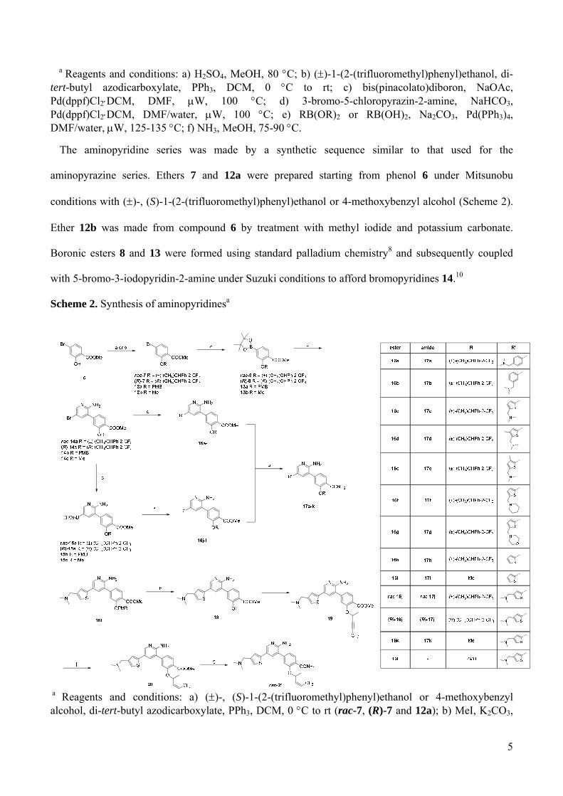
The aminopyridine series was made by a synthetic sequence similar to that used for the
aminopyrazine series. Ethers 7 and 12a were prepared starting from phenol 6 under Mitsunobu
conditions with ()-, (S)-1-(2-(trifluoromethyl)phenyl)ethanol or 4-methoxybenzyl alcohol (Scheme 2).
Ether 12b was made from compound 6 by treatment with methyl iodide and potassium carbonate.
Boronic esters 8 and 13 were formed using standard palladium chemistry8 and subsequently coupled
with 5-bromo-3-iodopyridin-2-amine under Suzuki conditions to afford bromopyridines 14.10
Scheme 2. Synthesis of aminopyridinesa
a Reagents and conditions: a) ()-, (S)-1-(2-(trifluoromethyl)phenyl)ethanol or 4-methoxybenzyl alcohol, di-tert-butyl azodicarboxylate, PPh3, DCM, 0 C to rt (rac-7, (R)-7 and 12a); b) MeI, K2CO3,

6
DMF, rt (12b); c) bis(pinacolato)diboron, NaOAc, Pd(dppf)Cl2DCM, DMF, W, 100 C; d) 5-bromo-3-iodopyridin-2-amine, NaHCO3, Pd(dppf)Cl2DCM, DMF/water, W, 100 C; e) RB(OR)2 or RB(OH)2, Na2CO3, Pd(PPh3)4, DMF/water or DME/water, W, 110-135 C (16a-i); f) 1-(5-bromothiophen-3-yl)-N,N-dimethylmethanamine, Na2CO3, Pd(PPh3)4, DME/water, W, 110 C (16j-l); g) NH3, MeOH, 70-80 C; h) TFA, DCM, 0 C; i) ()-5,5,5-trifluoropent-3-yn-2-ol, di-tert-butyl azodicarboxylate, PPh3, DCM, 0 C to rt; j) H2, Lindlar’s catalyst, MeOH, rt.
Aminopyridines 16a-i were formed by Suzuki coupling of the appropriate boronic esters or acids with
bromoderivatives 14 (Scheme 2).10,12 Derivatives 16j-l were prepared starting from 1-(5-bromothiophen-
3-yl)-N,N-dimethylmethanamine13 and suitably prepared boronic acids 15.10 Ammonolysis of esters 16
furnished the target primary amides 17. Compound 21 was prepared via a slightly different route:
aminopyridine 16l was deprotected with trifluoroacetic acid to afford phenol 18, which was derivatized
with ()-5,5,5-trifluoropent-3-yn-2-ol14 under Mitsunobu conditions to give ether 19. Target amide rac-
21 was obtained after hydrogenation with Lindlar catalyst to afford ester 20 followed by ammonolysis
(Scheme 2).
Results and discussion
Hybrid compounds design
As mentioned above, our previous studies of the benzimidazole series led to permeable and selective
inhibitors which suffered from insufficient potency and high lipophilicity (compound (R)-3, Figure 1).6
Our results indicated that the poor ligand efficiency (LE) and modest potency of these compounds were
at least in part due to inefficient binding of the phenyl substituted benzimidazole core 4 (IC50 value of
62 M, LE = 0.32, Figure 1). We speculated that a more potent compound could be obtained if the
substituents of benzimidazole 3 were to be grafted on a core that binds more efficiently to the hinge
region of Nek2. A replacement candidate for the benzimidazole moiety was the aminopyrazine-type
scaffold, which in our studies consistently showed better LE compared to the benzimidazoles. Indeed,
superposition of co-crystal structures of both series indicated that hypothetical hybrids of these series
can address all the pharmacophoric features of benzimidazole (R)-3 (Figure 2A). This prompted us to
prepare a series of compounds exploring this design principle and comprising the following key

7
features: the presence of a suitably placed amino-substituted aromatic group, an aminopyrazine-type
binding scaffold and a phenyl carboxyamide substituted with a benzyl ether (Figure 1).
SAR of benzyl ether-substituted hybrid compounds
The initial series of compounds were built by fusing the hinge-binding fragment of aminopyrazine 2
with the benzyloxy-substituted benzamide moiety of benzimidazole (R)-3 (Figure 1). In order to place a
suitable basic residue mimicking the piperidine ring of compound (R)-3, a para-N,N-
dimethylaminomethylphenyl group was used instead of the trimethoxyphenyl substituent. The resulting
hybrid 11a showed a Nek2 IC50 value of 0.79 M (Table 1) and proved to be approximately equipotent
to the parent compounds (0.23 M and 0.36 M, respectively, for pyrazine 2 and benzimidazole (R)-3,
Figure 1). Moving the amino group attachment point to the meta position resulted in aminopyrazine 11b
which demonstrated reduced Nek2 inhibition (IC50 value of 2.47 M, Table 1). During the course of
previous studies on the aminopyrazine series, it was found that switching to an aminopyridine hinge-
binding scaffold caused a marked increase in Nek2 inhibition.15,12 For this reason, aminopyridines 17a
and 17b were prepared, featuring a para- and a meta-substituted phenyl ring moiety, respectively.
Gratifyingly, both compounds showed improved activity against Nek2 with IC50 values of 0.12 and 0.21
M, respectively (Table 1). Overall, these hybrid compounds showed moderate activity and LE as well
as high predicted lipophilicity (Table 1).
Table 1. Phenyl substituted hybridsa
compound Nek2 IC50 (M)
LE Plk1 IC50 (M)
CLogP
11a
0.79 0.31 0.21 3.90 5.18

8
11b
2.47 0.66 0.20 12.1 5.18
17a
0.12 0.01 0.24 1.56 0.18 5.47
17b
0.21 0.03 0.24 3.29 1.22 5.47
a Results are mean (± SD) for n ≥ 3, or mean values of two independent determinations with individual determinations in parentheses or samples run n = 1; compounds are racemic unless otherwise stated.
In order to improve the potency of this series against Nek2, the aminomethyl-substituted phenyl ring
was replaced by an isosteric thiophene. Whereas in a previous series this modification had a generally
positive effect on Nek2 activity,5 these results showed that the outcome strongly depends on hinge-
binding motif (Table 2). Thus, introduction of an α’- or β’-aminomethyl substituted α-thiophene on the
aminopyrazine scaffold resulted in a drop in Nek2 inhibition, with compounds 11c and 11d showing
IC50 values of 2.68 and 2.63 M, respectively (Table 2; compare with aminopyrazines 11a and 11b,
Table 1). Conversely, when the same change was applied on the aminopyridine core, a significant
improvement in activity towards Nek2 was observed: the IC50 values for compounds 17c and rac-17j,
featuring an α’- or β’-aminomethyl substituent, are 0.047 and 0.059 M respectively (Table 2). Further

9
decoration of the thiophene group of aminopyridine 17c with methyl substituents either at the β’ or β
position resulted in virtually equipotent compounds: the IC50 values for aminopyridines 17d and 17e are
0.047 and 0.058 M, respectively (Table 3).
Table 2. Thiophene substituted hybridsa
compound Nek2 IC50
(M) LE
Plk1 IC50
(M) CLogP
11c
2.68 1.68 0.20 - 5.21
11d 2.63 1.42 0.20 - 5.13
17c
0.047 0.015 0.27 0.50 0.12 5.50
rac-17j 0.059 0.016 0.26 1.32 0.34 5.42

10
a Results are mean (± SD) for n ≥ 3, or mean values of two independent determinations with individual determinations in parentheses or samples run n = 1; compounds are racemic unless otherwise stated.
Table 3. SAR around the thiophene corea
compound Nek2 IC50 (M) LE CLogP
17d
0.047 0.005 0.26 5.99
17e
0.058 0.020 0.25 5.99
17f
0.093 0.017 0.24 6.42
17g
0.318 0.060 0.22 5.19
a Results are mean (± SD) for n ≥ 3, or mean values of two independent determinations with individual determinations in parentheses or samples run n = 1; compounds are racemic unless otherwise stated.
N NH2
CONH2
O
CF3
S
N

11
Next, we prepared two additional compounds bearing alterations of the basic residue: compound 17f
features a cyclic, more rigid amino group, while compound 17g features a less basic morpholine ring.
The former did show only a slight decrease in Nek2 inhibition (IC50 for 17f is 0.093 M, Table 3)
suggesting that the shape of the aminomethyl substituent is not crucial for activity. Conversely, the
reduced basicity of aminopyridine 17g had a more pronounced effect, with the IC50 value increasing
more than 7-fold to 0.318 M (compare compound 17c and 17g, Tables 2 and 3). These observations
are in line with previous findings on the role of amino substituents in benzimidazole-based Nek2
inhibitors, and stress the importance of a suitably placed basic residue with the ability to engage in
charge-charge type interactions.6
Since this series was initially inspired by Plk1 inhibitors16 and still maintained the original substituted
benzyl ether moiety, we investigated the Plk1 inhibition for selected compounds.
Whilst all showed a preference for Nek2, the window was relatively small: Plk1 IC50 values were 3.90,
1.56 and 1.32 M, respectively, for amides 11a, 17a and rac-17j, which translated to 10- to 25-fold
reduced activity when compared to Nek2 (Tables 1 and 2). Since we were interested in developing tool
compounds and Plk1 has been suggested to play a role upstream of Nek2,3b an improved selectivity
window was desirable. In addition, we were still concerned about the high lipophilicity of the described
compounds.
Co-crystal structure of aminopyrydine rac-17j bound to Nek2
In order to address these issues, we solved the co-crystal structure of rac-17j bound to Nek2. As
expected from our design hypothesis leading to this hybrid series, the binding mode shares many
features with that of rac-3, including a DFG-out conformation of the kinase.6 Here, we will focus on key
elements. The aminopyridine forms two hydrogen bonds with the hinge region at Glu87 and Cys89
(Figure 2B and 2C), compared to the single hydrogen bond formed between the imidazole ring of rac-3
and Cys89. The phenyl ring of the substituted benzamide sandwiches between the gatekeeper Met86,
and Phe148, whilst the amide group engages in two hydrogen bonds with the backbone NH and the side

12
chain carbonyl group of Asp159 of the DFG motif. As observed for rac-3, the substituted benzyl group
is well ordered with the trifluoromethyl group binding to a hydrophobic pocket formed by part of the
glycine rich loop, and one of the fluorine atoms is engaged in an orthogonal interaction with the
carbonyl group of Ile14 (3.6 Å). The position of the methyl group clearly reveals the R configuration at
the benzylic stereocentre of rac-17j even though the racemic mixture was used for the soaking process.
Figure 2. The structures of aminopyridines rac-17j and rac-21 bound to Nek2 as determined by X-ray
crystallography. (A) The rational basis of the hybrid design is illustrated by the superposition of the
structure of Nek2 (purple) bound to rac-3 (carbons colored yellow), extracted from PDB code 2XNP, on
the structure of Nek2 (not shown) bound to an aminopyrazine inhibitor (carbons colored green), PDB
code 2XKF. (B) View of the ATP binding site of Nek2 (carbons colored teal) occupied by
aminopyridine rac-17j (carbons colored orange). Wire-mesh represents the 2mFo-DFc electron density
map in the vicinity of the compound, contoured at 1.0 . Black dashed lines represent hydrogen bonds
formed between the protein and the compounds, magenta dashed lines represent the orthogonal
interaction between Ile14 and the trifluoromethyl group. (C) The structure viewed in a second
orientation, showing the hydrophobic contacts between the compound and protein (double-headed
arrows) and the hydrogen bonds between the amide group of the compound and Asp159 of the Nek2

13
DFG motif (black dashed lines). (D) Superposition of structures of compound rac-17j bound to Nek2
(teal) and compound rac-3 (PDB code 2XNP, carbons colored yellow) bound to Nek2 (not shown),
viewed from above. (E) The structure of rac-21 (carbons colored magenta) bound to Nek2 (light gray).
An ordered water molecule in the active site is shown as a red sphere. Wire-mesh represents the 2mFo-
DFc electron density map in the vicinity of the compound and the water molecule, contoured at 1.0 .
(F) Superposition of the structures of Nek2 (light gray) bound to rac-21 with the structure of Nek2 (not
shown) bound to rac-17j.
This confirmed that the R isomer is the most potent enantiomer for Nek2 binding as observed for the
benzimidazole series.6 The thiophene ring engages in hydrophobic contacts with Ile14 and Gly92. The
dimethylamino group is clearly resolved and superimposes well with the basic center of the piperidine
ring of rac-3 (Figure 2D). We have shown that the correct positioning of the piperidine ring of rac-3 is
crucial for high selectivity of this compound against Plk1 most likely by causing a clash with the
Arg136 residue in Plk1.6 We hypothesize that the N,N-dimethylamino group of rac-17j plays a similar
role, leading to the 25-fold selectivity with respect to Plk1 inhibition. The observation that rac-17j
shows a somewhat smaller selectivity window might be explained by the smaller size of the
dimethylamino group compared to the piperidine group leading to reduced steric interaction with
Arg136 in Plk1.
The binding mode revealed by the crystal structure thus confirmed our design hypothesis. However,
we were keen to further improve the selectivity against Plk1 and to reduce the lipophilicity of our
compounds. Our attention turned to the role of the benzyl ether moiety. Close examination of the X-ray
co-crystal structures of rac-17j and rac-3 revealed that this aromatic region is solvent exposed and has
few contacts with Nek2 (Figure 2B and 2D). Its main role appears to be as a scaffold for the
trifluoromethyl group that binds to the small hydrophobic groove formed partially by the Gly rich
region. As we have previously shown, the benzylic methyl group is important for activity since it
stabilizes the bioactive conformation in which the fluorinated moiety points towards the groove through
14
minimization of the 1,3-allylic strain.6 We hypothesized that the phenyl group is not needed for potent
Nek2 inhibition and can be replaced by a smaller moiety that leads to comparable positioning of the
trifluoromethyl group. Replacement of the aromatic residue would have the benefit of significantly
reducing the lipophilicity and molecular weight. In addition, we speculated that this change might be
significantly less tolerated for Plk1 and thus lead to an improved selectivity window.
Synthesis and profiling of truncated inhibitors
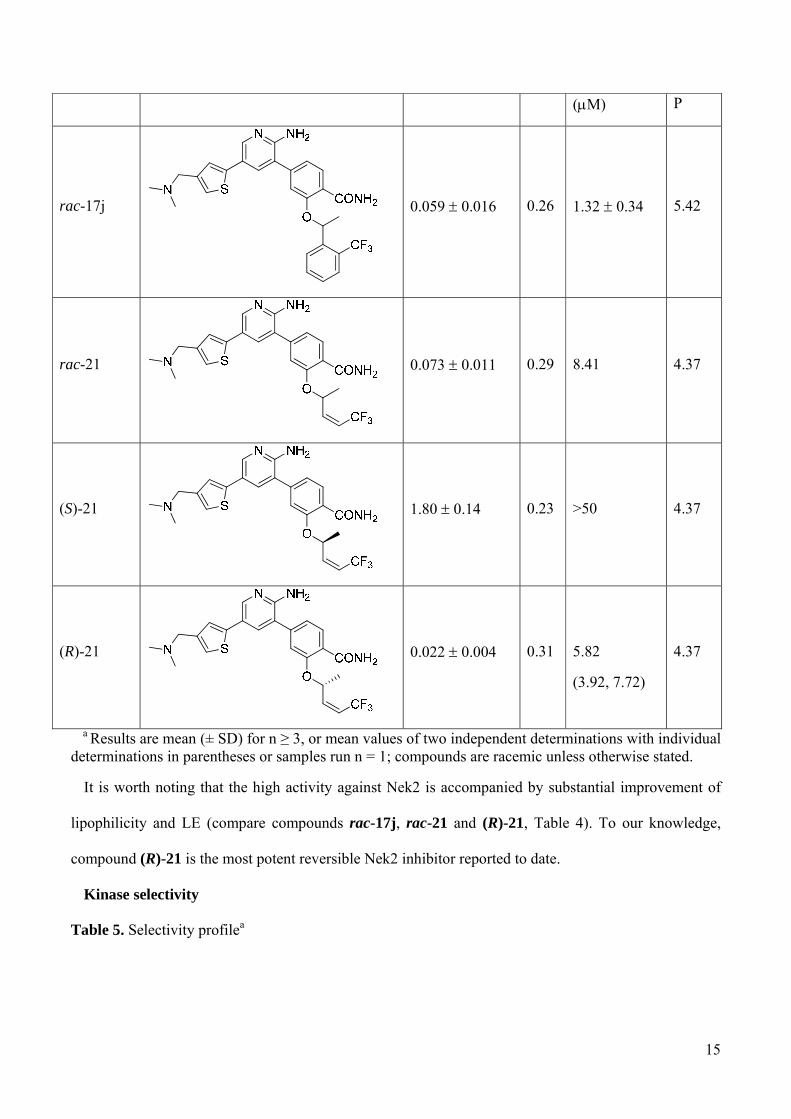
To explore this design rationale we prepared a compound in which the benzyl ether moiety was
replaced by an allylic ether bearing the desired Z configuration. Pleasingly, aminopyridine rac-21 was
roughly equipotent compared to the parent compound: the Nek2 IC50 value was found to be 0.073 M,
the LE value was 0.29 with a CLogP reduced to 4.37 (Table 4). These figures compared well with those
associated with benzyl ether rac-17j and demonstrate that replacing the aromatic ring with a smaller
allyl ether is tolerated and afforded a compound with better LE and lower CLogP (aminopyridine rac-
21, Table 4). The crystal structure of of rac-21 bound to Nek2 confirmed that the binding mode was as
expected (Figure 2E and 2F). The reduced bulk of the allylic ether compared to the benzyl ether allowed
the binding of an ordered water molecule within the active site, although this apparently had little
impact on the potency of inhibition. Our previous studies on benzimidazole-based Nek2 inhibitors
showed that the R configured enantiomer is significantly more potent than the S enantiomer.6 We
therefore separated the two enantiomers of aminopyridine rac-21 through chiral HPLC.17 The faster
eluting enantiomer showed a Nek2 IC50 value of 1.80 M, more than seventy times weaker than the
slower eluting enantiomer, which had an IC50 value of 0.022 M. Since we have previously shown that
the R configuration is preferred for Nek2 inhibition, we assigned the R configuration to the slower
eluting enantiomer, whereas the faster eluting compound was associated to the S configuration (Table
4).
Table 4. Alkene seriesa
compound Nek2 IC50 (M) LE Plk1 IC50 CLog
15
(M) P
rac-17j
0.059 0.016 0.26 1.32 0.34 5.42
rac-21
0.073 0.011 0.29 8.41 4.37
(S)-21
1.80 0.14 0.23 >50 4.37
(R)-21
0.022 0.004 0.31
5.82
(3.92, 7.72)
4.37
a Results are mean (± SD) for n ≥ 3, or mean values of two independent determinations with individual determinations in parentheses or samples run n = 1; compounds are racemic unless otherwise stated.
It is worth noting that the high activity against Nek2 is accompanied by substantial improvement of
lipophilicity and LE (compare compounds rac-17j, rac-21 and (R)-21, Table 4). To our knowledge,
compound (R)-21 is the most potent reversible Nek2 inhibitor reported to date.
Kinase selectivity
Table 5. Selectivity profilea

16
a Results are mean (± SD) for n ≥ 3, or mean values of two independent determinations with individual determinations in parentheses or samples run n = 1; compounds are racemic unless otherwise stated.
An assessment of the selectivity profile for key compounds was then carried out on four kinases
involved in the mitotic machinery: Plk1, MPS1, AurA and CDK2 (Table 5). For comparison purposes,
aminopyridine 17j was prepared as a single enantiomer in the R configuration (compound (R)-17j,
Table 5). In general, all the compounds showed a good level of selectivity. However, as anticipated, the
compound Nek2 IC50
(M) Plk1 IC50
(M)
MPS1 IC50
(M)
AurA IC50
(M)
CDK2 IC50
(M) CLogP
17a 0.12 0.01 1.56 0.18
3.06 ± 0.40
7.93
(8.44, 7.41)
2.74
(2.88, 2.60)
5.47
(R)-17j 0.037
0.82
(0.82, 0.83)
4.81 ± 0.31
8.81 ± 0.58
26.6
(26.7, 26.4)
5.42
(S)-21 1.80 0.14 >50 7.12 ± 0.13
2.33 ± 0.23
0.53
(0.52, 0.53)
4.37
(R)-21
0.022 0.004
5.82
(3.92, 7.72)
8.47 ± 0.23
4.94 ± 0.73
4.57
(4.77, 4.36)
4.37

17
truncated compound (R)-21 showed significantly improved selectivity against Plk1 (compare
aminopyridines 17a and (R)-17j and (R)-21, Table 5). Selectivity data on MPS1 inhibition showed a
similar trend, with the alkene compounds showing higher IC50 values (Table 5). Finally, all the
compounds demonstrated low activity towards Aurora A and CDK2 with the exception of the weak
Nek2 inhibitor (S)-21, which inhibited CDK2 with an IC50 value of 0.53 M (Table 5). Further profiling
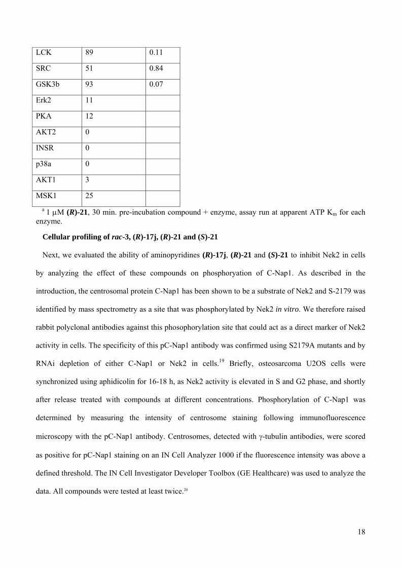
of compound (R)-21 was performed against a panel of 24 kinases using a ProfilerPro 1 kit. Again, at
least a 100-fold window of selectivity was observed in most cases even though a few cell cycle-
unrelated kinases showed a more pronounced inhibition (Table 6). Despite this, we concluded that
aminopyridine (R)-21 fulfils our requirements in terms of both potency and selectivity (IC50 value of
0.022 M, Gini coefficient18 = 0.514 (1 M), Table 5 and 6).
Table 6. Selectivity data for (R)-21 against a panel of kinases.a
kinase % of inhibition @ 1 M
IC50 (M)
MAPKAPK2 6
AurA 19
PKCz 12
RSK1 29
PRAK 7
Erk1 2
PKD2 77 0.33
CH1d 67 0.43
CHK1 11
ABL 46 1.11
FYN 57 0.75
LYN 43 1.36
CHK2 32 2.00
MET 46 1.17
18
LCK 89 0.11
SRC 51 0.84
GSK3b 93 0.07
Erk2 11
PKA 12
AKT2 0
INSR 0
p38a 0
AKT1 3
MSK1 25
a 1 M (R)-21, 30 min. pre-incubation compound + enzyme, assay run at apparent ATP Km for each enzyme.
Cellular profiling of rac-3, (R)-17j, (R)-21 and (S)-21
Next, we evaluated the ability of aminopyridines (R)-17j, (R)-21 and (S)-21 to inhibit Nek2 in cells
by analyzing the effect of these compounds on phosphoryation of C-Nap1. As described in the
introduction, the centrosomal protein C-Nap1 has been shown to be a substrate of Nek2 and S-2179 was
identified by mass spectrometry as a site that was phosphorylated by Nek2 in vitro. We therefore raised
rabbit polyclonal antibodies against this phosophorylation site that could act as a direct marker of Nek2
activity in cells. The specificity of this pC-Nap1 antibody was confirmed using S2179A mutants and by
RNAi depletion of either C-Nap1 or Nek2 in cells.19 Briefly, osteosarcoma U2OS cells were
synchronized using aphidicolin for 16-18 h, as Nek2 activity is elevated in S and G2 phase, and shortly
after release treated with compounds at different concentrations. Phosphorylation of C-Nap1 was
determined by measuring the intensity of centrosome staining following immunofluorescence
microscopy with the pC-Nap1 antibody. Centrosomes, detected with -tubulin antibodies, were scored
as positive for pC-Nap1 staining on an IN Cell Analyzer 1000 if the fluorescence intensity was above a
defined threshold. The IN Cell Investigator Developer Toolbox (GE Healthcare) was used to analyze the
data. All compounds were tested at least twice.20

19
Figure 3. C-Nap1 phosphorylation inhibition activity for rac-3, (R)-17j, (S)-21 and (R)-21. (A) C-
Nap1 phosphorylation experiments (results are expressed as the percent of cells with pC-Nap1 spot(s) as
a percent of DMSO (1%) control). Error bars represent the standard deviation of six replicates obtained
in two separate experiments. (B) IC50 values for C-Nap1 phosphorylation inhibition calculated for
compounds achieving more than 40 % inhibition.
Representative results are shown in Figure 3A. Pleasingly, aminopyridine (R)-21 indeed showed
concentration-dependent down-regulation of C-Nap1 phosphoryation with an IC50 value of 0.8 M
(Figure 3B). The approximately 40-fold drop of the IC50 values compared to the biochemical assays can
be attributed to the higher ATP concentrations in cells and is not uncommon for ATP competitive kinase
inhibitors. The corresponding benzyl ether compound (R)-17j also showed an effect in this assay albeit
at a slightly higher concentration leading to an IC50 of 2.7 M (Figure 3A and 3B). The somewhat
reduced activity might be due to the more lipophilic character of (R)-17j, causing binding to other
components and so reducing the effective concentration. It is worth noting that benzimidazole rac-3 did
not show any significant modulation of C-Nap1 phosphorylation in this assay even at high
concentrations (Figure 3A). Finally we tested aminopyridine (S)-21, a much weaker biochemical
inhibitor of Nek2, in this assay. Indeed, this compound showed significantly less suppression of C-Nap1
phosphorylation and a flatter concentration dependency (Figure 3). We attributed the weak effect of (S)-
21 in this assay to residual Nek2 activity, however other factors, such as inhibition of upstream kinases,
0.16 0.32 0.63 1.25 5 10 0.19 0.38 0.75 1.5 4.5 9 0.19 0.38 0.75 1.5 4.5 9 0.19 0.38 0.75 1.5 4.5 90
20
40
60
80
100
120
(R)-17j (S)-21 (R)-21rac-3
Compound (M)
PE
RC
EN
T O
F C
ON
TR
OL
N NH2
CONH2
O
CF3
SN
A B
compound C-Nap1 IC50 (M)
rac-3
-
(R)-17j
2.7
(S)-21
-
(R)-21
0.8

20
might also play a role. The flat concentration dependency could also be explained by the hypothesis that
the weak effect of aminopyridine (S)-21 in this assay is due to modulation of different targets, very
likely with different IC50 values. Overall, these experiments showed that (R)-17j, and particularly (R)-
21, inhibit the phosphorylation of C-Nap1, a cellular substrate of Nek2, in a concentration dependent
manner. Next, we tested the effect of benzimidazole rac-3 and of aminopyridines (R)-17j, (S)-21, and
(R)-21 on the growth of a panel of cancer cell lines. We determined the concentration required to inhibit
the growth by 50% (GI50) after 96 h. However, we did not observe any clear trends between
biochemical and C-Nap1 assay on one hand and antiproliferative effect on the other hand (Table 7).
These results suggest, at least in the case of the weak Nek2 inhibitor (S)-21, that inhibition of other
targets is responsible for the toxicity against the cancer cell lines after long term exposure. Similar off-
target inhibition might also contribute to the antiproliferative effects of aminopyridines (R)-17j and (R)-
21, albeit to a lesser degree.
The data collected in the C-Nap1 phosphorylation assay combined with the profiling against other
prominent cell cycle kinases suggest that aminopyridine (R)-21 can serve as a valuable tool for
investigating the role of Nek2 in the cell cycle and its function in mitotic organization. It will be
particularly useful in short term experiments using synchronized cell populations aiming to identify
additional substrates and functions of Nek2.
Lastly, racemic aminopyridines rac-17j and rac-21 showed medium to high permeability at
physiological pH in a parallel artificial membrane assay (PAMPA, 15.2 10-6 and 27.6 10-6 cm/s
respectively) as well as high metabolic degradation.21
Table 8. GI50 values for compounds rac-3, (R)-17j, (S)-21 and (R)-21a
compound
U2OS GI50 (M)
MDA-MB-231 GI50 (M)
HeLa GI50 (M)
MCF7 GI50 (M)
rac-3 2.96 (3.73, 2.17) 4.95 (4.81, 5.08) 1.20 0.23 2.03 (1.68, 2.38)

21
(R)-17j
0.48 (0.51, 0.45) 0.95 (0.87, 1.02) 0.14 0.06 0.22 (0.21, 0.23)
(S)-21
2.17 (2.17, 2.16) 7.25 (6.40, 8.09) 0.44 0.13 5.42 (4.72, 6.12)
(R)-21
1.82 (2.22, 1.41) 4.16 (3.94, 4.38) 0.59 0.06 2.17 (2.03, 2.31)
a Results are mean (± SD) for n ≥ 3, or mean values of two independent determinations with individual determinations in parentheses or samples run n = 1; compounds are racemic unless otherwise stated.
Confirmation of non linear SAR
Finally, we were interested in investigating whether the non-additive SAR observed in the
benzimidazole series is also translated in the aminopyridine series. As described above, we previously
found that in order to attain high level of Nek2 inhibition, the two key pharmacophoric elements
(namely the amino group and the substituted benzylic ether, see Figure 1) had to be present at the same
time.6 Thus, three truncated analogues of aminopyridine rac-17j were prepared, lacking the amino
group, the benzylic ether or lacking both groups at the same time. The results show that loss of either of
these moieties lead to a sharp decrease in activity to a level observed for the core compound 17i (Table
8). This confirms that neither of the two substituents significantly improves Nek2 activity on its own
(compare core compound 17i with aminopyridines 17h or 17k, IC50 values of 10.5, 9.0 and 2.26 M,
respectively, Table 8).
Table 8. Non-additive SARa
compound Nek2 IC50
(M) LE CLogP

22
rac-17j 0.059 0.016
0.26 5.42
17h
9.0 1.6 0.20 5.35
17k 2.26 0.36 0.29 2.52
17i
10.5 0.28 0.30 2.45
a Results are mean (± SD) for n ≥ 3, or mean values of two independent determinations with individual determinations in parentheses or samples run n = 1; compounds are racemic unless otherwise stated.
Instead, a large jump in activity (ca. 200-fold) takes place when both substituents are present in rac-
17j, as previously observed in the benzimidazole series. These results confirm that the non-linear SAR
observed for benzimidazoles translates into the aminopyridine series.
Conclusions
Starting from two different sets of Nek2 ligands, we designed a series of hybrid inhibitors. Using
structure-based design we optimized Nek2 potency, kinase selectivity, lipophilicity and ligand
efficiency to the desired range, ultimately culminating in aminopyridine (R)-21. To our knowledge (R)-
21 is the most potent reversible inhibitor of Nek2 known to date. In our cellular assay, compound (R)-21
N NH2
CONH2
O
SN

23
showed a concentration dependent inhibition of phosphorylation of C-Nap1, a known substrate of Nek2.
Gratifyingly, aminopyridine (R)-21 also showed sufficient selectivity against the most relevant cell
cycle kinases and will therefore make for a useful tool compound to probe the role of Nek2 in cell cycle
control. The results of these experiments will be reported in due course.
Experimental section
General Chemistry Information. Starting materials, reagents and solvents for reactions were reagent
grade and used as purchased. Chromatography solvents were HPLC grade and were used without
further purification. Thin layer chromatography (TLC) analysis was performed using Merck silica gel
60 F-254 thin layer plates. Flash column chromatography was carried out using columns pre-packed
with 40-63 m silica. NMR spectra were recorded on a Bruker Avance 500 MHz spectrometer and
samples were referenced to the appropriate internal nondeuterated solvent peak. The data is given as
follows: chemical shift (δ) in ppm, multiplicity (where applicable), coupling constants (J) in Hz (where
applicable) and integration (where applicable). LCMS analyses were performed on a Micromass
LCT/Waters Alliance 2795 separations module HPLC system with a Merck Chromolith SpeedROD RP-
18e 50 x 4.6mm column at a temperature of 22 °C. The following solvent system, at a flow rate of 2
mL/min, was used: solvent A: methanol; solvent B: 0.1% formic acid in water. Gradient elution was as
follows: 1:9 (A:B) to 9:1 (A:B) over 2.25 min., 9:1 (A:B) for 0.75 min. then reversion back to 1:9 (A:B)
over 0.3 min., 1:9 (A:B) for 0.2 min. Detection was carried out with a Waters 2487 Dual Absorbance
Detector (detecting at 254nm) and ionisation was electrospray (ESI). Some LCMS and all HRMS
analyses were performed on a Agilent 1200 series HPLC system with a Merck Chromolith SpeedROD
RP-18e 50x4.6mm column at a temperature of 22 °C. The following solvent system, at a flow rate of 2
mL/min, was used: solvent A: methanol; solvent B: 0.1% formic acid in water. Gradient elution was as
follows: 1:9 (A:B) to 9:1 (A:B) over 2.5 min., 9:1 (A:B) for 1 min. then reversion back to 1:9 (A:B)
over 0.3 min., 1:9 (A:B) for 0.2 min. This was connected to a Agilent 6200 Time of Flight (ToF) mass
spectrometer (simultaneous ESI and APCI or ESI only) with detection at 254nm. The following
reference masses were used for HRMS analysis: Caffeine [M + H]+ = 195.087652, reserpine [M + H]+

24
= 609.280657 and (1H,1H,3H-tetrafluoropentoxy)phosphazene [M + H]+ = 922.009798. The purity of
final compounds was determined by HPLC as described above and is ≥ 95% unless specified otherwise.
Methyl 4-bromo-2-hydroxybenzoate 6 A solution of 4-bromo-2-hydroxybenzoic acid 5 (5.00 g,
23.04 mmol) in MeOH (20 mL) was treated with sulfuric acid (1.7 mL). The mixture was refluxed for
24 h, poured onto ice-water and extracted with DCM. The combined organics were washed with satd
aqueous NaHCO3, dried (Na2SO4) and concentrated to give ester 6 (3.97 g, 75%). 1H NMR (500 MHz,
CDCl3) δ 10.82 (s, 1H), 7.70 (d, J = 8.5 Hz, 1H), 7.20 (d, J = 1.9 Hz, 1H), 7.04 (dd, J = 8.5, 1.9 Hz,
1H), 3.97 (s, 3H).
Methyl 4-bromo-2-((4-methoxybenzyl)oxy)benzoate 12a A solution of phenol 6 (1.70 g, 7.36
mmol), 4-methoxybenzyl alcohol (1.29 g, 9.30 mmol) and triphenylphosphine (2.81 g, 10.71 mmol) in
DCM (35 mL) was cooled at 0 °C and treated with di-tert-butyl azodicarboxylate (2.45 g, 10.65 mmol).
The reaction was allowed to reach rt and stirred overnight. The mixture was diluted with DCM and
quenched with water. The organic layer was separated and extracted with DCM. The combined organics
were dried (Na2SO4), concentrated and purified by Biotage column chromatography (07%
EtOAc/cycloexane) to give ether 12a (2.04 g, 79%). LCMS (ESI) m/z 373 (M+Na). 1H NMR (500
MHz, CDCl3) δ 7.70 (d, J = 8.3 Hz, 1H), 7.43 – 7.40 (m, 2H), 7.20 (d, J = 1.8 Hz, 1H), 7.15 (dd, J = 8.3,
1.8 Hz, 1H), 6.96 – 6.93 (m, 2H), 5.11 (s, 2H), 3.89 (s, 3H), 3.83 (s, 3H).
Methyl 4-(2-amino-5-bromopyridin-3-yl)-2-((4-methoxybenzyl)oxy)benzoate 14b A solution of
bromide 12a (1.10 g, 3.13 mmol), bis(pinacolato)diboron (1.20 g, 4.72 mmol), potassium acetate (925
mg, 9.44 mmol) and 1,1′-bis(diphenylphosphino)ferrocene]dichloropalladium(II)DCM (130 mg, 0.16
mmol) in DMF (15 mL) was stirred at 100 °C under microwave irradiation for 1 h 30 min. The reaction
was quenched with brine and extracted with AcOEt. The combined organics were washed with brine,
dried (Na2SO4), and concentrated to afford the crude boronic ester 13a.
A solution of crude boronic ester 13a (ca. 3.13 mmol), sodium bicarbonate (480 mg, 5.71 mmol), 1,1′-
bis(diphenylphosphino)ferrocene]dichloropalladium(II)DCM (125 mg, 0.15 mmol) and 5-bromo-3-

25
iodopyridin-2-amine (850 mg, 2.84 mmol) in DMF/water (8/1, 15 mL) was stirred at 100 °C under
microwave irradiation for 1 h 30 min. The reaction was quenched with brine and extracted with EtOAc.
The combined organics were washed with brine, dried (Na2SO4), concentrated and purified by Biotage
column chromatography (030% EtOAc/cyclohexane) to give bromopyridine 14b (1.02 g, 81%).
HRMS (ESI) m/z calcd for C21H20BrN2O4 (M+H) 443.0601, found 443.0617. 1H NMR (500 MHz,
CDCl3) δ 8.13 (d, J = 2.4 Hz, 1H), 7.90 (d, J = 7.8 Hz, 1H), 7.45 (d, J = 2.4 Hz, 1H), 7.43 – 7.40 (m,
2H), 7.07 – 7.04 (m, 2H), 6.95 – 6.92 (m, 2H), 5.17 (s, 2H), 4.57 (br. s, 2H), 3.94 (s, 3H), 3.83 (s, 3H).
Methyl 4-(2-amino-5-(4-((dimethylamino)methyl)thiophen-2-yl)pyridin-3-yl)-2-((4-
methoxybenzyl)oxy)benzoate 16l A solution of bromide 14b (1.01 g, 2.28 mmol),
bis(pinacolato)diboron (870 mg, 3.43 mmol), potassium acetate (680 mg, 6.94 mmol) and 1,1′-
bis(diphenylphosphino)ferrocene]dichloropalladium(II)DCM (200 mg, 0.25 mmol) in DMF (11 mL)
was stirred at 100 °C under microwave irradiation for 1 h 30 min. The reaction was quenched with brine
and extracted with AcOEt. The combined organics were washed with brine, dried (Na2SO4), and
concentrated to afford the crude boronic acid 15b.
A solution of crude boronic acid 15b (ca. 2.28 mmol), 1-(5-bromothiophen-3-yl)-N,N-
dimethylmethanamine13 (1.0 g, 4.54 mmol), tetrakis(triphenylphosphine)palladium(0) (250 mg, 0.22
mmol) and sodium carbonate (485 mg, 4.58 mmol) in DME/water (8/1, 15 mL) was heated to 110 °C
under microwave irradiation for 1h. The reaction was quenched with brine and extracted with EtOAc.
The combined organics were washed with brine, dried (Na2SO4), concentrated and purified by Biotage
column chromatography (025% MeOH/DCM) to give amine 16l (569 mg, 50% over two steps).
LCMS (ESI) m/z 504 (M+H). 1H NMR (500 MHz, CDCl3) δ 8.36 (s, 1H), 7.92 (d, J = 7.9 Hz, 1H), 7.56
(d, J = 2.3 Hz, 1H), 7.44 – 7.41 (m, 2H), 7.24 (s, 1H), 7.14 – 7.10 (m, 3H), 6.97 – 6.91 (m, 2H), 5.19 (s,
2H), 4.59 (br. s, 2H), 3.94 (s, 3H), 3.82 (s, 3H), 3.54 (s, 2H), 2.36 (s, 6H).
Methyl 4-(2-amino-5-(4-((dimethylamino)methyl)thiophen-2-yl)pyridin-3-yl)-2-hydroxybenzoate
18 A solution of phenol ether 16l (560 mg, 1.11 mmol) in DCM (7 mL) was treated with trifluoroacetic

26
acid (800 µL, 10.81 mmol) at 0 °C. After 1 h 30 min. the reaction was brought to pH ca. 56 with 1M
NaOH and 1M HCl, the aqueous layer separated and extracted with DCM. The combined organic layers
were concentrated and purified by Biotage column chromatography (015% MeOH/DCM) to give
phenol 18 (394 mg, 92%). HRMS (ESI) m/z calcd for C20H22N3O3S (M+H) 384.1376, found 384.1391.
1H NMR (500 MHz, MeOD) δ 8.28 (d, J = 2.4 Hz, 1H), 7.99 (d, J = 8.2 Hz, 1H), 7.68 (d, J = 2.4 Hz,
1H), 7.58 (d, J = 1.4 Hz, 1H), 7.39 (d, J = 1.4 Hz, 1H), 7.10 (d, J = 1.7 Hz, 1H), 7.07 (dd, J = 8.2, 1.7
Hz, 1H), 4.30 (s, 2H), 4.00 (s, 3H), 2.88 (s, 6H).
()-(Z)-Methyl 4-(2-amino-5-(4-((dimethylamino)methyl)thiophen-2-yl)pyridin-3-yl)-2-((5,5,5-
trifluoropent-3-en-2-yl)oxy)benzoate 20 A solution of phenol 18 (64 mg, 0.17 mmol), ()-5,5,5-
trifluoropent-3-yn-2-ol14 20% w/w solution in EtOAc, 0.31 mmol) and triphenylphosphine (70 mg, 0.27
mmol) in DCM (1 mL) was cooled at 0 °C and treated with di-tert-butyl azodicarboxylate (60 mg, 0.26
mmol). The reaction was allowed to reach rt and stirred overnight. Additional batches of reagents were
added as required until the reaction reached completion. The mixture was diluted with DCM and
quenched with water. The organic layer was separated and extracted with DCM. The combined organics
were dried (Na2SO4), concentrated and purified by Biotage column chromatography (015%
MeOH/DCM) to give ether 19.
A solution of ether 19 (ca. 0.050 mmol) in MeOH (2 ml) was treated with palladium on calcium
carbonate (poisoned with lead (Lindlar’s catalyst), 5% w/w, 5 mg, 2.4 µmol) and stirred in an
atmosphere of hydrogen overnight. Additional batches of palladium on calcium carbonate (poisoned
with lead, 5% w/w, 7 mg, 3.3 µmol) were added and the mixture stirred in an atmosphere of hydrogen
until completion. The mixture was filtrated over Celite washing with MeOH, the solvent removed under
reduced pressure and the residue purified by semi-preparative reverse phase HPLC (Phenomenex
Gemini C18 column; 15 min. gradient 25-50% MeOH/water, 0.1% formic acid; 5 mL/min.) to give
alkene 20 (12 mg, 14% over two steps). HRMS (ESI) m/z calcd for C25H27F3N3O3S (M+H) 506.1720,
found 506.1701. 1H NMR (500 MHz, MeOD) δ 8.29 (d, J = 2.4 Hz, 1H), 7.88 (d, J = 8.0 Hz, 1H), 7.65

27
(d, J = 2.4 Hz, 1H), 7.56 (s, 1H), 7.37 (d, J = 1.5 Hz, 1H), 7.22 (dd, J = 8.0, 1.5 Hz, 1H), 7.17 (s, 1H),
6.27 (dd, J = 12.1, 8.7 Hz, 1H), 5.94 – 5.85 (m, 1H), 5.49 – 5.45 (m, 1H), 4.24 (s, 2H), 3.92 (s, 3H),
2.83 (s, 6H), 1.55 (d, J = 6.4 Hz, 3H).
()-(Z)-4-(2-Amino-5-(4-((dimethylamino)methyl)thiophen-2-yl)pyridin-3-yl)-2-((5,5,5-
trifluoropent-3-en-2-yl)oxy)benzamide rac-21 Ester 20 (16 mg, 0.032 mmol) was treated with
ammonia in methanol (7M, 4 mL) and heated to 75 °C in a closed-cap vial for 4 days. The mixture was
concentrated, and the residue purified by Biotage column chromatography (015% MeOH/DCM) to
give amide rac-21 (9 mg, 56%). HRMS (ESI) m/z calcd for C24H26F3N4O2S (M+H) 491.1723, found
491.1749. 1H NMR (500 MHz, (CD3)2SO) δ 8.27 (d, J = 2.4 Hz, 1H), 7.88 (d, J = 8.0 Hz, 1H), 7.65 (br.
s, 1H), 7.57 (br. s, 1H), 7.53 (d, J = 2.4 Hz, 1H), 7.26 (d, J = 1.5 Hz, 1H), 7.22 (dd, J = 8.0, 1.5 Hz, 1H),
7.18 (s, 1H), 7.09 (d, J = 1.5 Hz, 1H), 6.41 (dd, J = 12.1, 8.5 Hz, 1H), 6.09 – 6.04 (m, 1H), 5.98 (s, 2H),
5.58 – 5.54 (m, 1H), 3.36 (s, 2H), 2.15 (s, 6H), 1.53 (d, J = 6.3 Hz, 3H).
Aminopyridine rac-21 was separated into enantiomers by chiral HPLC (LUX cellulose II; 90%
acetonitrile/2-propanol; 1 mL/min):
Peak 1: retention time 12.3 min: (S)-(Z)-4-(2-Amino-5-(4-((dimethylamino)methyl)thiophen-2-
yl)pyridin-3-yl)-2-((5,5,5-trifluoropent-3-en-2-yl)oxy)benzamide (S)-21 HRMS (ESI) m/z calcd for
C24H26F3N4O2S (M+H) 491.1723, found 491.1722. 1H NMR (500 MHz, (CD3)2SO) δ 8.27 (d, J = 2.4
Hz, 1H), 7.88 (d, J = 8.0 Hz, 1H), 7.65 (br. s, 1H), 7.57 (br. s, 1H), 7.53 (d, J = 2.4 Hz, 1H), 7.26 (d, J =
1.5 Hz, 1H), 7.22 (dd, J = 8.0, 1.5 Hz, 1H), 7.18 (s, 1H), 7.09 (d, J = 1.5 Hz, 1H), 6.41 (dd, J = 12.1, 8.5
Hz, 1H), 6.09 – 6.04 (m, 1H), 5.98 (s, 2H), 5.58 – 5.54 (m, 1H), 3.36 (s, 2H), 2.15 (s, 6H), 1.53 (d, J =
6.3 Hz, 3H).
Peak 2: retention time 15.2 min: (R)-(Z)-4-(2-Amino-5-(4-((dimethylamino)methyl)thiophen-2-
yl)pyridin-3-yl)-2-((5,5,5-trifluoropent-3-en-2-yl)oxy)benzamide (R)-21 HRMS (ESI) m/z calcd for
C24H26F3N4O2S (M+H) 491.1723, found 491.1719. 1H NMR (500 MHz, (CD3)2SO) δ 8.27 (d, J = 2.4
Hz, 1H), 7.88 (d, J = 8.0 Hz, 1H), 7.65 (br. s, 1H), 7.57 (br. s, 1H), 7.53 (d, J = 2.4 Hz, 1H), 7.26 (d, J =

28
1.5 Hz, 1H), 7.22 (dd, J = 8.0, 1.5 Hz, 1H), 7.18 (s, 1H), 7.09 (d, J = 1.5 Hz, 1H), 6.41 (dd, J = 12.1, 8.5
Hz, 1H), 6.09 – 6.04 (m, 1H), 5.98 (s, 2H), 5.58 – 5.54 (m, 1H), 3.36 (s, 2H), 2.15 (s, 6H), 1.53 (d, J =
6.3 Hz, 3H).
Biochemical assays. Nek2 and Plk1 biochemical assays were performed as reported previously.
MPS1, AurA and CDK2 counterscreen assays were carried out using similar procedures.5,6
Cellular assays. CellTiter-Blue Assay for Growth Inhibition
U2OS human osteosarcoma cells (American Type Culture Collection, Manassas, Virginia, United
States) were grown in McCoy’s 5A medium supplemented with 1.5 mM L-glutamine, 25 mM HEPES,
2% penicillin/streptomycin (Invitrogen, Paisley, United Kingdom), and 10% fetal bovine serum
(Biosera, Ringmer, East Sussex, United Kingdom). Cells were maintained in a humidified atmosphere
of 5% CO2 at 37°C. The medium was aspirated and the cells were washed with PBS (Invitrogen,
Paisley, United Kingdom), trypsinized (Internal supply, 0.25% versene trypsin with EDTA),
neutralized, and counted. Cells were seeded into 384-well clear tissue culture treated microtiter plates
(Corning B.V. Life Sciences, Amsterdam, The Netherlands) at 200 cells per well in a 45 µL volume of
the respective media. Columns 1 and 24 had no cells added and were plated with 45 µL of media alone.
Cells were incubated at 37°C / 5% CO2. At 24 hours after plating, test compounds, Etoposide as
positive control (Sigma-Aldrich, Gillingham, Dorset, United Kingdom), or DMSO at 1% v/v final
concentration (Fisher Scientific, Loughborough, Leicestershire, United Kingdom) were dispensed using
an Echo liquid handling system (Labcyte, Dublin, Ireland). After 92 hours, 5 µL of CellTiter-Blue
Reagent (Promega, Southampton, United Kingdom) was added to the cells using a Multidrop dispenser
(Thermo Electron, Basingstoke, Hants, United Kingdom) and incubated for 4 hours in a humidified
atmosphere of 5% CO2 at 37°C. After the incubation, the plates were placed at room temperature for 40
minutes before fluorescence was recorded (560Ex/590Em) on an EnVision 2103 plate reader
(PerkinElmer Life Sciences). Data were plotted as percentage of DMSO control against compound
concentration using GraphPad Prism 5 Software. The 50% growth inhibition (GI50) was calculated as

29
the compound concentration required to reduce the cell number by 50% compared with the DMSO
control.
MDA-MB-231 cells (American Type Culture Collection, Manassas, Virginia, United States) were
grown in RPMI medium (Invitrogen, Paisley, United Kingdom) supplemented with 2%
penicillin/streptomycin and 10% fetal bovine serum.
HeLa cells (American Type Culture Collection, Manassas, Virginia, United States) were grown in
Dulbecco's Modified Eagle Medium (Invitrogen, Paisley, United Kingdom) supplemented with 2%
penicillin/streptomycin and 10% fetal bovine serum.
MCF7 cells (American Type Culture Collection, Manassas, Virginia, United States) were grown in
Dulbecco's Modified Eagle Medium supplemented with 2% penicillin/streptomycin and 10% fetal
bovine serum. They were plated at 800 cells per well.
Phosphorylated C-Nap1 in Cell Assay
U2OS human osteosarcoma cells (American Type Culture Collection, Manassas, Virginia, United
States) were grown in McCoy’s 5A medium supplemented with 1.5 mM L-glutamine, 25 mM HEPES,
2% penicillin/streptomycin (Invitrogen, Paisley, United Kingdom), and 10% fetal bovine serum
(Biosera, Ringmer, East Sussex, United Kingdom). Cells were maintained in a humidified atmosphere
of 5% CO2 at 37°C. The medium was aspirated and the cells were washed with PBS (Invitrogen,
Paisley, United Kingdom), trypsinized (Internal supply, 0.25% versene trypsin with EDTA),
neutralized, and counted. Cells were seeded into 96-well black clear bottom tissue culture treated
microtiter plates (PerkinElmer Life Sciences, Waltham, Massachusetts, USA) at 10,000 cells per well in
a 180 µL volume of media. Cells were incubated for 24 hours at 37°C / 5% CO2 after which
Aphidicolin (Sigma-Aldrich, Gillingham, Dorset, United Kingdom) was added to all wells for cell
synchronization at a final concentration of 2 µg/mL for 16-18 hours. The cells were then released into
180 µL of fresh media. Simultaneously, the cells were treated in triplicate wells with test compound in
DMSO at 1% v/v final concentration (Fisher Scientific, Loughborough, Leicestershire, United
Kingdom) and incubated for 3 hours. 1% DMSO final concentration was used as a negative control.

30
The cells were then fixed for 15 minutes at 2-8°C with 50 µL cold 100% methanol. The plates were
washed once with 100 µL PBS before blocking non-specific binding with 50 µL 5% w/v Bovine Serum
Albumin (Sigma-Aldrich, Gillingham, Dorset, United Kingdom) in PBS for 1 hour at room temperature
with gentle agitation. A rabbit polyclonal antibody raised to phosphorylated C-Nap1 (generated by
Peptide Specialty Laboratories, GmbH, Heidelberg) was added at a dilution of 1:750 , and a monoclonal
antibody raised to the centrosomal marker γ-tubulin (Sigma-Aldrich, Gillingham, Dorset, United
Kingdom) was added at a dilution of 1:500 for 1 hour at room temperature with gentle agitation.
Following a further wash with PBS, Alexa fluor 488 goat anti-rabbit IgG and Alexa fluor 568 goat anti-
mouse IgG (Invitrogen, Paisley, United Kingdom) were added at a final concentration of 4 µg/mL in 5%
BSA/PBS for 1 hour at room temperature under gentle agitation. After another wash with PBS, the
nuclear stain DAPI (Invitrogen, Paisley, United Kingdom) was added at a final concentration of 2.5
µg/mL in PBS for 10 minutes at room temperature with gentle agitation. The plate was washed in PBS
once again and refrigerated until ready to image. The assay plates were read on the IN Cell Analyzer
1000 using the Workstation 1000 acquisition software (GE Healthcare, Amersham, United Kingdom).
The instrument was equipped with a 20X dry Nikon objective, a D360/40X DAPI excitation filter, an
HQ480/40X FITC excitation filter, an HQ565/30X excitation filter, an HQ460/40M bandwidth emission
filter, an HQ535nm/50M bandwidth emission filter, and an HQ620nm/60M bandwidth emission filter.
The exposure times were consistently 200 ms in the DAPI channel, 400 ms in the FITC channel, and
1000 ms in the red channel. Fifteen fields of view were imaged in all wells. IN Cell Investigator
Developer Toolbox (GE Healthcare) was used to analyze the assay data. The algorithm was written to
identify the DAPI-stained nuclei, and then segment the cells setting a small collar around the nuclei as
the centrosomal inclusion area. The γ-tubulin-identified centrosomes and phosphorylated C-Nap1 spots
were chosen based on size and pixel intensity and were only counted if they were located within the
centrosomal inclusion area. A “One to One link” was written into the protocol so that only
phosphorylated C-Nap1 spots colocalized with the centrosomal γ-tubulin were counted. Data were

31
plotted as percentage of DMSO control against compound concentration using GraphPad Prism 5
Software.
Aknowledgement. We acknowledge NHS funding to the NIHR Biomedical Research Centre and
funding from Cancer Research UK programme grant number C309/A8274. AMF acknowledges support
from Cancer Research UK, The Wellcome Trust and the Association for International Cancer Research.
RB acknowledges support from Cancer Research UK (grant C24461/A10285 and infrastructure support
for Structural Biology at the ICR), a Royal Society Research Fellowship and the Career Development
Programme of the ICR. We are thankful to Prof. Julian Blagg, Prof. Roger Griffin, Prof. Herbie Newell,
Dr. Ian Collins and Prof. Paul Workman for helpful discussions. We also thank Dr. Amin Mirza, Mr.
Meirion Richards and Dr. Maggie Liu for their help with HPLC, NMR and mass spectrometry. Ms
Jessica Schmitt is acknowledged for biochemical assays. We are indebted to the staff of ESRF beamline
ID29 as well as our many colleagues of the Section of Structural Biology (ICR in London) for their
support during data collection.
Supporting Information Available: Experimental procedures, analytical data for final compounds
11, 17 and intermediates, a summary of crystallographic analysis of aminopyridine rac-17j and rac-21,
HPLC traces for compounds 21, C-Nap1 antibody characterization and pictures of assay plates. This
material is available free of charge via the Internet at http://pubs.acs.org.
References
1 O’Regan, L.; Blot, J.; Fry, A. M. Mitotic regulation by NIMA-related kinases. Cell Div. 2007, 2, 25–
36.
2 a) Lou, Y.; Yao, J.; Zereshki, A.; Dou, Z.; Ahmed, K.; Wang, H.; Hu, J.; Wang, Y.; Yao, X. NEK2A
interacts with MAD1 and possibly functions as a novel integrator of the spindle checkpoint signaling. J.
Biol. Chem. 2004, 279, 20049–20057; b) Chen, Y.; Riley, D. J.; Zheng, L.; Chen, P. L.; Lee, W. H.

32
Phosphorylation of the mitotic regulator protein Hec1 by Nek2 kinase is essential for faithful
chromosome segregation. J. Biol. Chem. 2002, 277, 49408–49416.
3 a) Henise, J. C.; Taunton, J. Irreversible Nek2 kinase inhibitors with cellular activity. J. Med. Chem.
2011, 54, 4133–4146; b) Mardin, B. R.; Lange, C. L.; Baxter, J. E.; Hardy, T.; Scholz, S. R.; Fry, A. M.;
Schiebel, E. Components of the Hippo pathway cooperate with Nek2 kinase to regulate centrosome
disjunction. Nat. Cell Biol. 2010, 12, 1166–1176.
4 a) Fletcher, L.; Cerniglia, G. J.; Nigg, E. A.; Yend, T. J.; Muschel, R. J. Inhibition of centrosome
separation after DNA damage: a role for Nek2. Radiat. Res. 2004, 162, 128–135; b) Kokuryo, T.;
Senga, T.; Yokoyama, Y.; Nagino, M.; Nimura, Y.; Hamaguchi, M. Nek2 as an effective target for
inhibition of tumorigenic growth and peritoneal dissemination of cholangiocarcinoma Cancer Res.
2007, 67, 9637–9642; c) Tsunoda, N.; Kokuryo, T.; Oda, K.; Senga, T.; Yokoyama, Y.; Nagino, M.;
Nimura, Y.; Hamaguchi, M. Nek2 as a novel molecular target for the treatment of breast carcinoma.
Cancer Sci. 2009, 100, 111–116.
5 Whelligan, D. K.; Solanki, S.; Taylor, D.; Thomson, D. W.; Cheung, K. M. J.; Boxall, K.; Mas-
Droux, C.; Barillari, C.; Burns, S.; Grummitt, C. G.; Collins, I.; van Montfort, R. L. M.; Aherne, G. W.;
Bayliss, R.; Hoelder, S. Aminopyrazine inhibitors binding to an unusual inactive conformation of the
mitotic kinase Nek2: SAR and structural characterization. J. Med. Chem. 2010, 53, 7682–7698.
6 Solanki, S.; Innocenti, P.; Mas-Droux, C; Boxall, K.; Barillari, C.; van Montfort, R. L. M.; Aherne,
G. W.; Bayliss, R.; Hoelder, S. Benzimidazole inhibitors induce a DFG-out conformation of never in
mitosis gene A-related kinase 2 (Nek2) without binding to the back pocket and reveal a nonlinear
structure−activity relationship. J. Med. Chem. 2011, 54, 1626–1639.
7 Mitsunobu, O. The use of diethyl azodicarboxylate and triphenylphosphine in synthesis and
transformation of natural-products. Synthesis 1981, 1–28.

33
8 Ishiyama, T.; Murata, M.; Miyaura, N. Palladium(0)-catalyzed cross-coupling reaction of
alkoxydiboron with haloarenes: a direct procedure for arylboronic esters. J. Org. Chem. 1995, 60, 7508–
7510.
9 a) Kundo, M.; Khairatkar-Joshi, N.; Nadkarni, S. M.; Pansare, R. M.; Karnik, P. V. Bicyclic
heteroaryl derivatives as cannabinoid receptor modulators. (Glenmark pharmaceuticals SA). Patent WO
2007/096764, 2007; b) Binch, H.; Robinson, D.; Miller, A.; Fraysse, D. Pyrrolopyrazines and
pyrazolopyrazines useful as inhibitors of protein kinases. (Vertex pharma). Patent WO 2006/058074,
2006.
10 Miyaura, N.; Suzuki, A. Palladium-catalyzed cross-coupling reactions of organoboron compounds.
Chem. Rev. 1995, 95, 2457–2483.
11 See Supporting Information for detailed procedures.
12 Hilton, S.; Naud, S.; Caldwell, J. C.; Boxall, K; Burns, S.; Anderson, V. E.; Antoni, L; Allen, C. A.;
Pearl, L. H.; Oliver, A. W.; Aherne, G. W.; Garrett, M. D.; Collins, I. Identification and characterisation
of 2-aminopyridine inhibitors of checkpoint kinase 2. Bioorg. Med. Chem. 2010, 18, 707–718.
13 1-(5-Bromothiophen-3-yl)-N,N-dimethylmethanamine was prepared by reductive amination of the
known corresponding aldehyde: Deng, J.; Kerns, J. K.; Jin, Q.; Lin, G.; Lin, X.; Lindenmuth, M.;
Neipp, C. E.; Nie, H.; Thomas, S. M.; Widdowson, K. L. Chemical compounds. (Smithkline Beecham
Corporation). Patent WO 2007/005534, 2007. See Supporting Information for experimental details.
14 ()-5,5,5-trifluoropent-3-yn-2-ol was prepared according to a modified literature method: Yamazaki,
T.; Mizutani, K.; Kitazume, T. Modified preparation of trifluoromethylated propargylic alcohols and its
application to chiral 2,6-dideoxy-6,6,6-trifluorosugars. J. Org. Chem. 1995, 60, 6046–6056. See
Supporting Information for experimental details.

34
15 Whelligan, D. K.; Solanki, S.; Taylor, D.; Thomson, D. W.; Cheung, K. M. J.; Hoelder, S.
unpublished results.
16 a) Rapley, J.; Baxter, J. E.; Blot, J.; Wattam, S. L.; Casenghi, M.; Meraldi, P.; Nigg, E. A.; Fry, A.
M. Coordinate regulation of the mother centriole component Nlp by Nek2 and Plk1 protein kinases.
Mol. Cell. Biol. 2005, 25, 1309–1324; b) for a study on Plk1 inhibitors, see: Emmitte, K. A.; Adjebang,
G. M.; Andrews, C. W.; Alberti, J. G.; Bambal, R.; Chamberlain, S. D.; Davis-Ward, R. G.; Dickson, H.
D.; Hassler, D. F.; Hornberger, K. R.; Jackson, J. R.; Kuntz, K. W.; Lansing, T. J.; Mook, R. A., Jr.;
Nailor, K. E.; Pobanz, M. A.; Smith, S. C.; Sung, C. M.; Cheung, M. Design of potent thiophene
inhibitors of polo-like kinase 1 with improved solubility and reduced protein binding. Bioorg. Med.
Chem. Lett. 2009, 19, 1694–1697.
17 See Experimental Section and Supporting Information for details.
18 Graczyk, P. P. Gini coefficient: a new way to express selectivity of kinase inhibitors against a family
of kinases. J. Med. Chem. 2007, 50, 5773–5779.
19 See Figure S2 in Supporting Information for more details.
20 See Figure S3 in the Supporting Information for samples of the assay readout.
21 87% and 95% respectively of aminopyridines rac-17j and rac-21 were metabolized in MLM after
30 min. Data for the benzimidazole series have been previously published (see ref. 6).
Table of Contents Graphic


![Class 3 Ab + L AbL Review d/ dt [ AbL ] = k on [ Ab ][L] – k off [ AbL ]](https://img.pdfslide.us/doc/110x75/56815b21550346895dc8dddd/class-3-ab-l-abl-review-d-dt-abl-k-on-ab-l-k-off-abl-.jpg)