Embed Size (px)
Citation preview

ElastographyLecture 13

Elastography
• Mechanical property imaging of tissue
• Imaging modality – Ultrasound, MRI, OCT, etc
• Non-invasive, convenient, precise, (low-cost)
Disease pathophysiologyDiagnosisTreatment

Elastic property of Tissue
Elastic modulus:
• stress/strain
• resistance to axial deformation
• stiffness

Diagnosis with Elasticity
• In old Egypt, 5 000 years ago, physicians examined the different parts of the body to evaluate elasticity, they knew that a hard mass in an organ is pathologic.
• In Greek ancient age, for Hippocratic medicine, palpating was an essential time of physical examination.
• In 21st century, imaging take preeminent place in medicine and Elastography could be considered as an « imaging palpation »

Diagnosis with Elasticity
• Disease changes tissue elasticity
• Palpation: Used for centuries – low resolution, not depth resolved, highly subjective
• Elasticity can vary by up to four orders of magnitude
Ex) Compression test on 142 breast tissue samples38 fat31 glandular tissue18 fibrous tissue23 intraductal carcinoma32 infiltrating ductal carcinoma

US elastography - breast cancer diagnosis
Fibroadenoma
Benign Benign Malignant Benign
Results of study
296 solid lesions from 232 patients
• Sonography – 72.6% accuracy
• Elastography – 88.2% accuracy
Features of US elastography
• Deep penetration
• Poor resolution
• Commercially available
Invasive ductal carcinoma
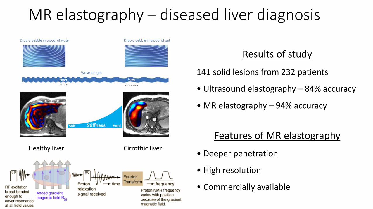
MR elastography – diseased liver diagnosis
Results of study
141 solid lesions from 232 patients
• Ultrasound elastography – 84% accuracy
• MR elastography – 94% accuracy
Features of MR elastography
• Deeper penetration
• High resolution
• Commercially available
Healthy liver Cirrothic liver

Cornea - RK Wang group
OCT elastography – emerging applications
Breast tumour, lymph nodes, skeletal muscle
OCE
needle probe
Breast tumour
5 mm
Kennedy et al., Sampson group, J
Biomed Opt, Dec 2013; Opt Lett May 2014; Biomed OptExpress June 2014
Human lymph node
Human breast cancer
Rat muscle
Human breast cancer
Micro-OCE
Features of OCT elastography
• Higher resolution (1 ~ 50 um)
• Higher sensitive (sub nm disp.)
• Higher acquisition speed ( >1kHz)
• Very low penetration (0.5 ~ 3 mm)
• Clinical applications NOT available

Histology Enface OCT Elastogram (strain)

Medical Imaging Techniques
Pen
etra
tio
n D
epth
Resolution
Confocal Microscopy
OCT
CT/MRI
US
10 μm
1 mm
1 cm
10 cm
1 μm
100 μm
1 m
1 μm 10 μm 100 μm 1 mm 1 cm
Atomic Force Microscopy
Lateral resolution: ~20umAxial resolution: ~ 8um

Scale of measurement
Cancer cells are 10 times softer than healthy cellsOn the macroscopic scale–
Cancerous tissue >10 times stiffer than healthy tissue
Elasticity depends on tissue material and structure at the scale being probed
Example: cancer cells versus cancerous tissue
AFM of cancercell
Force sensor with piconewton resolutionTip on cantilever senses sample surface
Lateral resolution: 1 nmAxial resolution: 1 Å
On the microscopicscale –
So, how do we measure mechanical properties in elastography?

• We need to make someassumptions:
Assumption 1:Mechanically homogeneous over a resolution element
Imaging tissue deformation
Describe behaviour using continuum mechanics
• Take a complex block of tissue and break it up into
homogeneous, small volumes
How is a load realized in each small volume?

Stress and Strain
xx
u
yy
x
vy
zz
wz
x
y
z
σxx
σyy
τyx
τzx
σzz
τyz
τxz
τxy
τzy
• Stress causes a shape change
y
z
εxx
εyy
γyx
γzx
εzz
γyzγ
xz
γxy
γzy
• When any load (force) is applied,a stressresults on each surface of eachvolume
σ: normal force τ: shear force ε: normal strain γ: shear strain

Assumption 2:Isotropic (direction independent)properties
Relating stress and strain
ij Cijklij
Reduces to two elasticconstants• shear modulus, G• bulk modulus, K
•Relate stress and strain through an elastic constant• 9 stress x 9 strain = 81 elastic constants to describe
behaviour!
x
y
3D stress tensor – 9 stress
components
σxx
σyy
τyx
τzx
σzz
τyzτxz
τxy
τ zy
x
y
3D strain tensor – 9 straincomponents
εxx
εyy
γyx
γzx
εzz
γyzγ xz
γxy
γ zy
z z

Elastic moduli
Shear Modulus, G – shear stress andstrain Describes tendency to change inshape
x
y
Young’s Modulus, E – special case:longitudinal stress and strain, mostcommonly used to quantifystiffness
z
Poisson’s ratio, υ – relates change in shape to change involume
xx
zz
E 3G
Assumption 3:Tissue is incompressible (Volume is conserved)
Bulk Modulus, K – compressibility Describes tendency to change involume
x
y
z
K E
3(12)
Relate E and G throughgeometry:
E 2G(1)
Relate E and K throughgeometry:
= 0.5
for tissue
G
E

So, which moduli for Tissue?
Howdo we get to a modulus in elastography?
Assumption 4: Displacement can be related to modulus
Imaging system AlgorithmTissuemotion Displacement ElastogramModulus
Shear modulus (G) or Young’s modulus (E = 3G) has largest dynamic range in tissue

Deformation types (inducing strain)Ex
cita
tio
n
CompressionDirect strain
measurement
Indirect strain measurement
Shear WaveMechanical
excitation
Remote excitation
Phased array
Single transducerPressure
WaveTOF
For homogeneous isotropic
linear elastic materials
𝐸 =𝜎
𝜀
𝐺 = 𝜌𝑣𝑠2
𝐾 = 𝜌𝑣𝑐2
Newton-Laplace equation
𝐸 = 2𝐺 1 + 𝜈 = 3𝐾(1 − 2𝜈)
(Poisson’s ratio ~ 0.499)
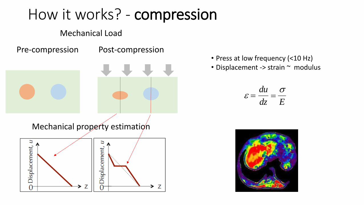
Mechanical Load
Pre-compression Post-compression
Mechanical property estimation
How it works? - compression
• Press at low frequency (<10 Hz)• Displacement -> strain ~ modulus
du
dz E

How it works - transient (SW)
csE
3
Shear wave speed to modulus:
Ultrasound image and shear wavespeed image of invasive ductal carcinoma in human breast (M. Tanter et al., Ultrasound Med. Biol., 2008)
•Acoustic radiation force impulse
•Generates shear waves
•Displacement -> shear wave speed ~ modulus

How it works - supersonic SW
• Higher SNR
• Wider spatial extent of shear wave
• Lower frame rates
• Displacement -> shear wave speed ~modulusMach 3 supersonic regime in an elastic phantom

Type E(kPa) G(kPa) K(GPa) ct (m/s) cs (m/s)
Fat -1 -0.3 2-2.5 -0.5 1490-1540
Liver 1-24 0.3-8 2-2.5 0.5-2.8 1490-1540
Muscle 3-30 1-10 2-2.5 1-3.2 1490-1540
Prostate 6-45 2-15 2-2.5 1.4-3.9 1490-1540
Myocardium 20-150 6.7-50 2-2.5 2.6-7.1 1490-1540
Fibrotic Liver 30-300 10-100 2-2.5 3.2-10 1490-1540

Is tissue purely elastic?
Hysteresis
•Young’s modulus defines elastic (linear, instantaneous) material behaviour
•But more commonly tissue is viscoelastic
•Viscosity is resistance to flow
•Viscoelastic behaviour is non-linear and time-dependent
Creep – time-dependent strain In elastography,
− small
− wait for stress
relaxation before
acquiring, OR
− use acquisition
speed >> tissue flow
Relaxation – time-dependent stress

•Assumed in most elastography techniques to simply quantify stiffness
•But disease alters tissue viscoelasticity as well as stiffness
• Possibility for viscoelastic contrast in elastography – changes in time-dependent properties
• Study of viscous or viscoelastic properties is called rheology
Assumption 5: Tissue is linear elastic
Viscoelasticity in elastography
Creep response of breast tissue in compression elastography Shear modulus Viscosity
Shearwave speed SW attenuation

Viscoelastic measurement
Shearwave speed equation derived from the Voigt modelIf viscosity,μ2 is zero, ??
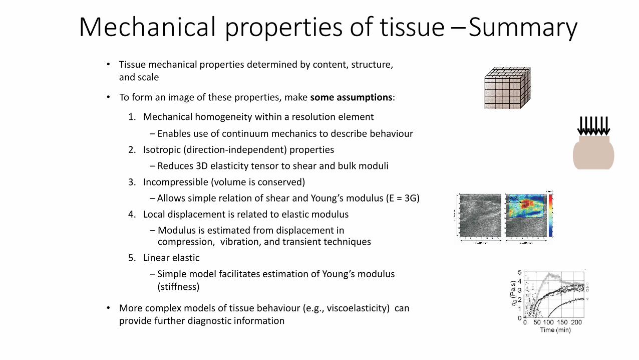
Mechanical properties of tissue –Summary• Tissue mechanical properties determined by content, structure,
and scale
• To form an image of these properties, make some assumptions:
1. Mechanical homogeneity within a resolution element
– Enables use of continuum mechanics to describe behaviour
2. Isotropic (direction-independent) properties
– Reduces 3D elasticity tensor to shear and bulk moduli
3. Incompressible (volume is conserved)
– Allows simple relation of shear and Young’s modulus (E = 3G)
4. Local displacement is related to elastic modulus
– Modulus is estimated from displacement in compression, vibration, and transient techniques
5. Linear elastic
– Simple model facilitates estimation of Young’s modulus (stiffness)
• More complex models of tissue behaviour (e.g., viscoelasticity) can provide further diagnostic information

Feasibility of a hybrid elastographic-microfluidic device to rapidly process and assess pancreatic cancer biopsies for pathologists
• Ronnie Das, Thu-Mai Nguyen, Saniel D. Lim, Matt O'Donnell, Ruikang K. Wang and Eric J. Seibel
• IEEE EMBS Special Topic Conference on Healthcare Innovations & Point-of-Care Technologies, Oct 8-10, 2014, Seattle WA
• Objectives:
1) To measure the elasticity of pancreatic tissue specimens using optical coherence tomography shear wave elastography (OCT-SWE)
2) To determine feasibility of OCT-SWE to identify distinct structures in the specimens


Methods
• Flesh and formalin-fixed porcine pancreas samples in rectangular and square microchannels made of glass and PDMS, respectively.
• 1% agarose hydrogel phantom with 4% agarose inclusion

Results
Remarks• No significant difference by channels
• Glass substrate/ enclosed glass / PDMS channels
• Measured shear wave speed distribution• Flesh vs Fixed: 3.5 m/s vs 14.5 m/s
• Estimated shear modulus• Flesh vs Fixed: 18 kPa vs 227 kPa

Results
Remarks• Differenciated 4% agarose from 1% agarose
hydrogel phantom
• Shear wave : 11.18±1.48 m/s vs 6.62±2.65 m/s
free space (upper), microchannel (lower)

Fixed pancreatic tissue, placed on a glass plate

Comparisons of Loading Schemes
Loading Method
Measured Parameter
Axial Resolution*
Lateral Resolution*
Assumptions ** Quantitative? Non-contact?
Strain Contrast Elasticity
Quasi-static Compression
Local Strain 50 ~ 200 μm 10 ~ 30 μmUniform
Stress Field
Required Local Stress Distribution
No No
Dynamic Compression
No No
Dynamic Compression
/w stress sensor
Local StrainLocal Stress
n/a 10 μmYes No
Shear Wave Variation
Elasticity
Shear Wave by Piezo
Phase Velocity, Cs ~ 10 μm ~ 10 μm N/AUniformDensity
Yes No
Shear Wave by ARF
Yes Yes

Mechanical Load
Pre-compression Post-compression
Imaging
Ultrasound MRI OCT
Mechanical property estimation
Stiffness1
Stiffness2
Stiffness3
From displacement to modulus


Excitation Methods
• Mechanical excitation• Compression loading (quasi-static / dynamic)
• Vibration by piezoelectric actuator (dynamic)
• Acoustic radiation force (ultrasound)• Internal
• Internal shear wave
• Internal endogenous force• Respiratory, heart