Embed Size (px)
Citation preview

Design and Implementation of Cell-Based AssaysTo Model Human DiseaseJeremy O. Jones and Marc I. Diamond*Departments of Neurology and Cellular and Molecular Pharmacology, University of California, San Francisco,California 94143-2280
ABSTRACT Cell-based assays, if appropriately designed, can be used to rap-idly identify molecular mechanisms of human disease and develop novel thera-peutics. In the last 20 years, many genes that cause or contribute to diverse dis-orders, including cancer and neurodegenerative disease, have been identified.With such genes in hand, scientists have created numerous model systems to dis-sect the molecular mechanisms of basic cellular and developmental biology.Meanwhile, techniques for high-throughput screening that use large chemical li-braries have been developed, as have cDNA and RNA interference libraries thatcover the entire human genome. By combining cell-based assays with chemicaland genetic screens, we now have vastly improved our ability to dissect molecu-lar mechanisms of disease and to identify therapeutic targets and therapeutic leadcompounds. However, cell-based screening systems have yet to yield many funda-mental insights into disease pathogenesis, and the development of therapeuticleads is frustratingly slow. This may be due to a failure of such assays to accu-rately reflect key aspects of pathogenesis. This Review attempts to guide the de-sign of productive cellular models of human disease that may be used in high-throughput chemical and genetic screens. We emphasize two points: (i) modelsystems should use quantifiable molecular indicators of a pathogenic process,and (ii) small chemical libraries that include molecules with known biological ac-tivity and/or acceptable safety profiles are very useful.
A ll experimental research on human disease de-pends on accurately replicating some aspect ofpathogenesis in a controlled system, and no
perfect models exist. Pharmaceutical companies areadept at using in vitro assays of protein function to iden-tify novel inhibitors. If a specific therapeutic target isknown, a biochemical assay can be readily developed,and this remains a desirable way to design and optimizenew drugs. For example, the identification of Bcr-Abl asa therapeutic target for chronic myelogenous leukemiahas allowed the discovery of effective therapies basedon in vitro functional assays (1). However, many caus-ative genes encode proteins of unknown function orwithout obvious catalytic sites amenable to targetingwith in vitro assays. In addition, many proteins can onlybe evaluated in the context of an intact cell (e.g., trans-membrane proteins or proteins that function in a multi-component complex), and thus in such cases cell-basedassays are required for productive high-throughputscreening (HTS).
In creating a disease model, a researcher must bal-ance the ease of working with the system against the rel-evance to the disease process. Typically, “relevance” isdetermined empirically when predictions made by asimple system (e.g., a cellular assay) are tested in amore complex system (e.g., a transgenic mouse model).The balance between tractability and relevance be-comes even more challenging when attempting to cre-ate a cellular model that is amenable to HTS. We arguethat using a discrete and quantifiable molecular eventthat closely correlates with pathogenesis as the readoutfor cell-based HTS increases the likelihood of discover-ing compounds with activity in more stringent in vivomodels. We contrast this strategy with that of using acellular phenotype as an end point (e.g., cell death inneurodegeneration models or cell proliferation in can-
*Corresponding author,[email protected].
Received for review August 17, 2007and accepted October 16, 2007.
Published online November 16, 2007
10.1021/cb700177u CCC: $37.00
© 2007 American Chemical Society
REVIEW
ACS CHEMICAL BIOLOGY • VOL.2 NO.11 www.acschemicalbiology.org718

cer models), which has a high potential for false posi-tive and negative results.
For example, the identification of genes that causeneurodegenerative disease, such as superoxide dis-mutase (amyotrophic lateral sclerosis), amyloid precur-sor protein (Alzheimer’s disease), polyglutamine pro-teins (Huntington’s disease and others), and �-syn-uclein (Parkinson’s disease), has led to a wealth of cel-lular and animal models ranging from yeast to mice,many of which have been adapted for HTS (2–12). It ispossible to model neurodegeneration by culturing neu-ronal cells ex vivo, in which overexpression of toxic pro-teins causes cell death. This model appears to provide asimple, convenient readout, but the use of a pheno-typic end point such as cell death is fraught with pit-falls. Neuronal cells certainly die in the course of neuro-degeneration. However, tissue culture cells typically diewithin days of expression of a toxic protein, comparedwith degeneration, which generally takes years withinthe brain of an affected individual. Furthermore, ampleevidence indicates that significant neuronal dysfunctionoccurs prior to detectable cell death (13–15). Myriadpathways influence cell death, and thus a compoundcan prevent cell death in this assay in many ways thatdo not relate to a disease-specific mechanism, such asthe up-regulation of drug exporters or the down-regulation of apoptotic pathways. Thus, it is uncertainwhether cell death models in tissue culture accuratelyrepresent crucial events in neurodegeneration. Not sur-prisingly, the ability of phenotypic cell models of neuro-degeneration to predict success in vivo has been verylow. The same holds true for cell proliferation models ofcancer. As an alternative, we describe several assaysthat exploit molecular, rather than phenotypic, readouts.
Molecular Indicators in Disease Models. The valueof a cell-based assay is greatly enhanced if the endpoint is specific to the disease mechanism. Most basiccellular phenotypes that can be readily measured (e.g.,proliferation, death, and process extension) are underthe control of multiple regulatory pathways. When suchreadouts are used in screening assays to identify geneticor chemical modifiers, there is consequently a greatchance that a nonspecific effect will account for the re-sult. Conversely, when a highly specific molecular event(e.g., protein folding) is monitored within a cell, theodds are much lower that nonspecific processes couldaccount for the result. Here, we provide examples of mo-
lecular events monitored in cell-based assays that havebeen or could be adapted to HTS.
Protein Aggregation in Neurodegeneration. Misfold-ing and aggregation of pathogenic peptides within thecell is a central feature of many neurodegenerative dis-eases. Polyglutamine diseases are a family of at leastnine dominantly inherited neurodegenerative disor-ders (16). Each derives from an expanded glutaminetract that destabilizes a target protein. This leads to mis-folding, aggregation, and toxicity in central nervous sys-tem neurons. Various relatively inaccurate methodshave been used to quantify intracellular aggregation: di-rect counting of large cellular inclusions by microscopy;filter trap assays, in which cell lysates are passedthrough a membrane with small pore size to detectlarge aggregates; and Western blots to detect high-mobility complexes. Such approaches either are notamenable to HTS or are unable to detect subtle changesin aggregation.
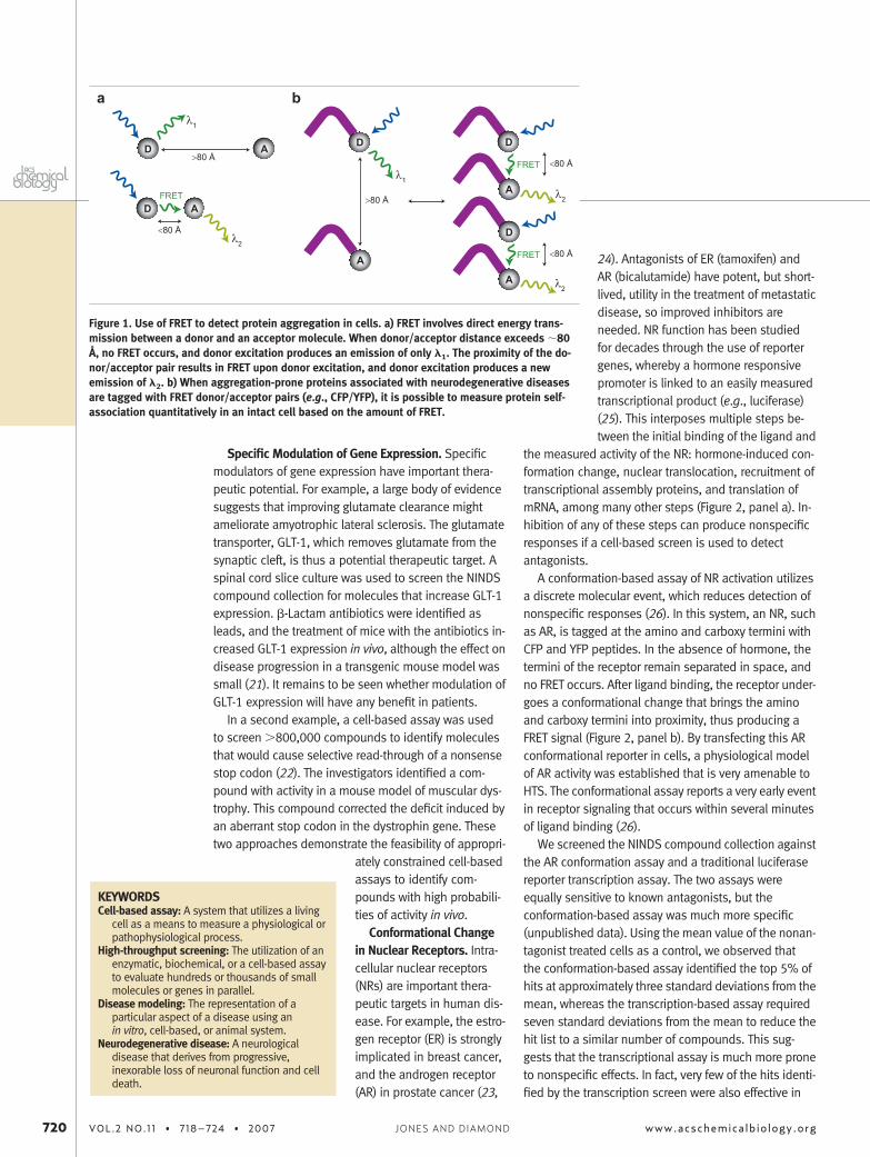
Fluorescence resonance energy transfer (FRET) is apowerful method for detecting protein interactions andcan be exploited in cell-based assays for HTS (Figure 1,panel a). FRET involves the direct transfer of energyfrom a donor to an acceptor molecule, which is de-tected by spectroscopy. The green fluorescent proteinderivatives cyan (CFP) and yellow (YFP) fluorescent pro-teins are useful FRET donor/acceptor pairs in cell-basedassays. The fusion of aggregation-prone proteins to CFPand YFP allows quantitative detection of FRET based onprotein interactions (Figure 1, panel b). Cells expressingthese fusion proteins are cultured in a microtiter for-mat, and the FRET signal is quantitatively measured byusing a micrometer-based fluorescence plate reader(17). The utility of this system was demonstrated byscreening the National Institute of Neurological Disor-ders and Stroke (NINDS) compound collection (www.msdiscovery.com) (18, 19), which is composed of U.S.Food and Drug Administration (FDA)-approved drugsand natural products. Eleven compounds were iden-tified that inhibited intracellular polyglutamine proteinaggregation (20). Six of these compounds diminishedneurodegeneration in a Drosophila model of neurode-generation created by overexpression of the pathogenicpeptide that causes Huntington’s disease (20), provid-ing a striking degree of corroboration in vivo. The quan-titative nature of the FRET-based assay also facilitatedgenetic studies to determine molecular mechanismsthat regulate protein aggregation (17).
REVIEW
www.acschemicalbiology.org VOL.2 NO.11 • 718–724 • 2007 719
Specific Modulation of Gene Expression. Specificmodulators of gene expression have important thera-peutic potential. For example, a large body of evidencesuggests that improving glutamate clearance mightameliorate amyotrophic lateral sclerosis. The glutamatetransporter, GLT-1, which removes glutamate from thesynaptic cleft, is thus a potential therapeutic target. Aspinal cord slice culture was used to screen the NINDScompound collection for molecules that increase GLT-1expression. �-Lactam antibiotics were identified asleads, and the treatment of mice with the antibiotics in-creased GLT-1 expression in vivo, although the effect ondisease progression in a transgenic mouse model wassmall (21). It remains to be seen whether modulation ofGLT-1 expression will have any benefit in patients.
In a second example, a cell-based assay was usedto screen �800,000 compounds to identify moleculesthat would cause selective read-through of a nonsensestop codon (22). The investigators identified a com-pound with activity in a mouse model of muscular dys-trophy. This compound corrected the deficit induced byan aberrant stop codon in the dystrophin gene. Thesetwo approaches demonstrate the feasibility of appropri-
ately constrained cell-basedassays to identify com-pounds with high probabili-ties of activity in vivo.
Conformational Changein Nuclear Receptors. Intra-cellular nuclear receptors(NRs) are important thera-peutic targets in human dis-ease. For example, the estro-gen receptor (ER) is stronglyimplicated in breast cancer,and the androgen receptor(AR) in prostate cancer (23,
24). Antagonists of ER (tamoxifen) andAR (bicalutamide) have potent, but short-lived, utility in the treatment of metastaticdisease, so improved inhibitors areneeded. NR function has been studiedfor decades through the use of reportergenes, whereby a hormone responsivepromoter is linked to an easily measuredtranscriptional product (e.g., luciferase)(25). This interposes multiple steps be-tween the initial binding of the ligand and
the measured activity of the NR: hormone-induced con-formation change, nuclear translocation, recruitment oftranscriptional assembly proteins, and translation ofmRNA, among many other steps (Figure 2, panel a). In-hibition of any of these steps can produce nonspecificresponses if a cell-based screen is used to detectantagonists.
A conformation-based assay of NR activation utilizesa discrete molecular event, which reduces detection ofnonspecific responses (26). In this system, an NR, suchas AR, is tagged at the amino and carboxy termini withCFP and YFP peptides. In the absence of hormone, thetermini of the receptor remain separated in space, andno FRET occurs. After ligand binding, the receptor under-goes a conformational change that brings the aminoand carboxy termini into proximity, thus producing aFRET signal (Figure 2, panel b). By transfecting this ARconformational reporter in cells, a physiological modelof AR activity was established that is very amenable toHTS. The conformational assay reports a very early eventin receptor signaling that occurs within several minutesof ligand binding (26).
We screened the NINDS compound collection againstthe AR conformation assay and a traditional luciferasereporter transcription assay. The two assays wereequally sensitive to known antagonists, but theconformation-based assay was much more specific(unpublished data). Using the mean value of the nonan-tagonist treated cells as a control, we observed thatthe conformation-based assay identified the top 5% ofhits at approximately three standard deviations from themean, whereas the transcription-based assay requiredseven standard deviations from the mean to reduce thehit list to a similar number of compounds. This sug-gests that the transcriptional assay is much more proneto nonspecific effects. In fact, very few of the hits identi-fied by the transcription screen were also effective in
KEYWORDSCell-based assay: A system that utilizes a living
cell as a means to measure a physiological orpathophysiological process.
High-throughput screening: The utilization of anenzymatic, biochemical, or a cell-based assayto evaluate hundreds or thousands of smallmolecules or genes in parallel.
Disease modeling: The representation of aparticular aspect of a disease using anin vitro, cell-based, or animal system.
Neurodegenerative disease: A neurologicaldisease that derives from progressive,inexorable loss of neuronal function and celldeath.
D A>80 Å
λ1
D A
λ2
a b
λ1
>80 Å
D
A
D
A
<80 Å
λ2
FRET
D
A
<80 Å
λ2
FRET
<80 Å
FRET
Figure 1. Use of FRET to detect protein aggregation in cells. a) FRET involves direct energy trans-mission between a donor and an acceptor molecule. When donor/acceptor distance exceeds �80Å, no FRET occurs, and donor excitation produces an emission of only �1. The proximity of the do-nor/acceptor pair results in FRET upon donor excitation, and donor excitation produces a newemission of �2. b) When aggregation-prone proteins associated with neurodegenerative diseasesare tagged with FRET donor/acceptor pairs (e.g., CFP/YFP), it is possible to measure protein self-association quantitatively in an intact cell based on the amount of FRET.
720 VOL.2 NO.11 • 718–724 • 2007 www.acschemicalbiology.orgJONES AND DIAMOND

secondary assays of antiandrogen activity, and thosethat were effective were also identified by the conforma-tion screen (unpublished data). Many NRs, includingER, could be adapted to a conformational assay. Thismethod has been used to characterize compounds thatinduce subtle conformational changes of intracellularER-� and -� (27), illustrating how specific molecularreadouts can also be used to better characterize regula-tors of protein function in secondary analyses.
Nuclear Localization. Several assays that model othersteps along the NR activation pathway are also ame-nable to HTS. Upon ligand binding, AR concentrates inthe nucleus, where it forms dimers prior to activatinggene expression (26). A highly quantitative cell-basedscreen was developed to detect antagonists of ARnuclear translocation by using a genetically encoded
bioluminescent indicator (28). AR was fused to the
amino-terminal half of Renilla luciferase, while the
carboxy-terminus of Renilla luciferase contained a po-tent nuclear localization signal, making it constitutivelynuclear. With nuclear translocation of AR, the reconstitu-tion of these split fragments produces quantifiable bi-oluminescence in the presence of the appropriate sub-strate (Figure 2, panel c). This assay was used to screenfor compounds that inhibit nuclear translocation of AR,a discrete step in the AR activation pathway. The assayyielded important leads and was even adapted to anin vivo assay in mice (28). This work exemplifies how an-other important molecular event (nuclear localization)can be adapted to a quantitative cell-based model forHTS.
Cofactor Interactions. To activate gene expression,many NRs recruit cofactors that utilize LXXLL motifs tobind to NR ligand binding domains (LBDs) (29). Nosingle NR�cofactor interaction is known to be abso-lutely critical in the progression of disease; however, it
Figure 2. Use of FRET to monitor NR function. a) NRs undergo multiple steps of processing after ligand activation, which can produce nonspecifichits during a screen. b) The amino and carboxy termini of an NR are tagged with a FRET donor (D) and acceptor (A). Conformational change inducedby hormone binding reduces the intramolecular distance and increases the FRET signal. c) The amino terminus of an NR is tagged with one-half of aluciferase enzyme. The second half is tagged with a nuclear localization sequence and is constitutively nuclear. Nuclear translocation of the NR al-lows reconstitution of the luciferase activity. d) The LBD of an NR is tagged with a FRET donor, and a coactivator protein (CoA) is tagged with a FRETacceptor. Hormone binding induces intermolecular FRET. Alternatively, a single fusion protein has a FRET donor fused to the LBD, fused in turn to acoactivator peptide motif, and then fused to a FRET acceptor. Hormone binding induces intramolecular FRET.
REVIEW
www.acschemicalbiology.org VOL.2 NO.11 • 718–724 • 2007 721
is possible that inhibitors of these interactions couldbe useful therapeutics. Two reports describe the use ofFRET reporters to identify novel inhibitors of NR�
coactivator interactions. In the first, a FRET acceptor(YFP) is fused to the LBD of either the peroxisome prolif-erator activated receptor-� or ER-�. A FRET donor isfused to NR coactivators CREB-binding protein or ste-roid receptor coactivator-1 (SRC-1) (Figure 2, panel d)(30). Known agonists of the two NRs increased NR asso-ciation with the coactivators, while antagonists inhib-ited the ligand-induced cofactor association. In the sec-ond, YFP was fused to the ER-� LBD, attached by aflexible linker sequence to the LXXLL motif of SRC-1fused to CFP (Figure 2, panel d) (31). This intracellular re-porter also demonstrated the ligand-dependent asso-ciation of ER with a coactivator and was used to deter-mine the activity of various compounds as agonists andantagonists of the LXXLL-LBD association. The FRET as-says of NR�coactivator association replicated the find-ings of other assays, such as immunoprecipitation andyeast two hybrid. However, unlike these other tech-niques, the FRET assay may be more likely to identifyleads with in vivo activity because it uses an intracellu-lar, quantifiable molecular event and it is readily adapt-able to HTS.
HTS: Start with Small Libraries. The choice of a li-brary is critical to the success of a screening project. Acomprehensive discussion of library design is beyondthe scope of this Review (for an excellent review of idealcompound composition, see ref (32)), but it is impor-tant to consider the size and cost of the library, how todetermine the mechanisms of action of hits, and how totranslate them into viable drugs. Although some hits
from large synthetic chemi-cal libraries have been trans-lated into important thera-pies, discovering new leadcompounds via HTS by us-ing these libraries has notbeen widely successful, andmany companies are nowdeveloping smaller, focusedlibraries in hopes of greatersuccess rates (33, 34).
Because the cost of alarge library is high, and thesuccess rate is low, we sug-gest that academic laborato-
ries begin by screening small collections of biologicallyactive molecules in cell-based assays. Ideally, com-pounds should be cell-permeant, nontoxic, and non-mutagenic and have good bioavailability. These charac-teristics will make them more effective in live cells andimprove their likelihood of activity in vivo. We also rec-ommend that the collection include natural productsand drugs already known to be safe and/or effective inhumans.
Screening an established chemical space offers manyadvantages. These compounds are generally known tobe biologically active, which increases their chance ofmodifying the molecular event of interest. Also, a com-pound that influences a discrete molecular event withina cell need not directly bind the protein target. Proteinbehavior (e.g., aggregation) is often subject to the influ-ence of cell signaling pathways (17, 35), which can cre-ate opportunities for therapeutic modulation as well asdetermination of important pathogenic mechanisms.
Putative mechanisms of action of many natural prod-ucts and known drugs have often been previously de-scribed. This can provide an instant wealth of informa-tion about a confirmed hit and greatly aids furtherpharmacological and genetic evaluation of molecularmechanisms. A lead compound can thus become a“biological probe” in the identification of novel regula-tory pathways and new therapeutic targets. In addition,libraries of FDA-approved drugs and natural productsare composed of compounds safe for human use, andthis expedites their translation from discovery to theclinic (36). Natural products often have very good phar-macokinetic profiles, and nearly half of currently ap-proved drugs mimic them or have been inspired by them(37). For these reasons, we suggest that using a librarythat includes natural products and compounds ofknown function, with in vivo tolerability, greatly en-hances the chance of finding a useful lead.
Screening known compounds presents potential dis-advantages, including the perceived lack of novelty andthe difficulties of creating novel intellectual property(IP). However, assay development, chemical and ge-netic screening, and subsequent characterization ofpathways that regulate key pathological mechanismsare vital aspects of translational research. The creationof novel IP from leads is essential for successful transla-tional research and is certainly more challenging whenone is working with previously described compounds. Itis always vital to determine a structure–activity relation-
KEYWORDSNuclear hormone receptors: Proteins that sense
the presence of specific hormones andtransduce their activity by direct binding toparticular promoter elements and regulationof gene expression. Before they bindhormone, these receptors may reside in thecytoplasm or nucleus, but they are localizedto the nucleus after ligand activation.
Library choice: The selection of collections ofsmall molecules for use in high-throughputscreens.
Molecular readouts: The use of specific proteinconformational change, protein–proteininteractions, or changes in subcellularlocalization as the basis of a cellular assay toidentify chemical or genetic modifiers.
722 VOL.2 NO.11 • 718–724 • 2007 www.acschemicalbiology.orgJONES AND DIAMOND

ship (SAR) for any promising lead. This can help vali-date its putative mechanism and may also reveal a moreefficacious compound with a novel structure and thatis more amenable for IP protection. The ZINC databasecan help identify commercially available compounds forSARs (38). Finally, it remains possible to protect an off-patent compound for a new clinical indication, espe-cially if novel formulations are created and new mecha-nisms of action are defined. Many small companieshave now been started based on rescreening knowncompounds to develop drugs for novel indications. Thismay ultimately constitute a highly productive use of the“therapeutic chemical space”. Ultimately, the advan-tages of rescreening previously characterized com-pounds should not be discounted.
Conclusion. Mammalian cell based HTS has not yetlived up to its promise of drug and target discovery.
We argue that cell-based disease models that rely ona discrete molecular event will improve the chances offinding valuable leads. For academic research,cellular assays based on validated disease mecha-nisms increase the odds of identifying factors or sig-naling pathways that modulate a key event. Such as-says may be exploited for complementary DNAor RNA interference library screens and for subse-quent molecular studies of pathogenesis. Thesescreens are not limited to mechanisms relevant onlyto neurodegeneration or cancer but can be extendedto any molecular event that defines disease progres-sion. The design of specific intracellular molecular endpoints, and the careful choice of libraries, will facili-tate informative investigations that make the best useof precious resources for an academic laboratory orsmall enterprise.
REFERENCES1. La Rosee, P., O’Dwyer, M. E., and Druker, B. J. (2002) Insights from
pre-clinical studies for new combination treatment regimens withthe Bcr-Abl kinase inhibitor imatinib mesylate (Gleevec/Glivec) inchronic myelogenous leukemia: a translational perspective, Leu-kemia 16, 1213–1219.
2. Bandyopadhyay, S., Ni, J., Ruggiero, A., Walshe, K., Rogers, M. S.,Chattopadhyay, N., Glicksman, M. A., and Rogers, J. T. (2006) A high-throughput drug screen targeted to the 5=untranslated region of Al-zheimer amyloid precursor protein mRNA, J. Biomol. Screening 11,469–480.
3. Ahn, J. S., Musacchio, A., Mapelli, M., Ni, J., Scinto, L., Stein, R., Ko-sik, K. S., and Yeh, L. A. (2004) Development of an assay to screenfor inhibitors of tau phosphorylation by cdk5, J. Biomol. Screen-ing 9, 122–131.
4. Kosik, K. S., Ahn, J., Stein, R., and Yeh, L. A. (2002) Discovery of com-pounds that will prevent tau pathology, J. Mol. Neurosci. 19, 261–266.
5. Yager, D., Watson, M., Healy, B., Eckman, E. A., and Eckman, C. B.(2002) Natural product extracts that reduce accumulation of the Al-zheimer’s amyloid beta peptide: selective reduction in A beta42,J. Mol. Neurosci. 19, 129–133.
6. Haugabook, S. J., Le, T., Yager, D., Zenk, B., Healy, B. M., Eckman,E. A., Prada, C., Younkin, L., Murphy, P., Pinnix, I., Onstead, L., Sam-bamurti, K., Golde, T. E., Dickson, D., Younkin, S. G., and Eckman,C. B. (2001) Reduction of Abeta accumulation in the Tg2576 ani-mal model of Alzheimer’s disease after oral administration of thephosphatidyl-inositol kinase inhibitor wortmannin, FASEB J. 15, 16–18.
7. Utsuki, T., Yu, Q. S., Davidson, D., Chen, D., Holloway, H. W.,Brossi, A., Sambamurti, K., Lahiri, D. K., Greig, N. H., and Gior-dano, T. (2006) Identification of novel small molecule inhibitors ofamyloid precursor protein synthesis as a route to lower Alzhei-mer’s disease amyloid-beta peptide, J. Pharmacol. Exp. Ther. 318,855–862.
8. Morse, L. J., Payton, S. M., Cuny, G. D., and Rogers, J. T. (2004) FDA-preapproved drugs targeted to the translational regulation and pro-cessing of the amyloid precursor protein, J. Mol. Neurosci. 24,129–136.
9. Coufal, M., Maxwell, M. M., Russel, D. E., Amore, A. M., Altmann,S. M., Hollingsworth, Z. R., Young, A. B., Housman, D. E., and Kazant-sev, A. G. (2007) Discovery of a novel small-molecule targeting se-lective clearance of mutant huntingtin fragments, J. Biomol. Screen-ing 12, 351–360.
10. Hu, M., Schurdak, M. E., Puttfarcken, P. S., El Kouhen, R., Go-palakrishnan, M., and Li, J. (2007) High content screen microscopyanalysis of Abeta(1–42)-induced neurite outgrowth reduction inrat primary cortical neurons: neuroprotective effects of alpha7 neu-ronal nicotinic acetylcholine receptor ligands, Brain Res. 1151,227–35.
11. Wang, W., Duan, W., Igarashi, S., Morita, H., Nakamura, M., andRoss, C. A. (2005) Compounds blocking mutant huntingtin toxicityidentified using a Huntington’s disease neuronal cell model, Neuro-biol. Dis. 20, 500–508.
12. Zhang, X., Smith, D. L., Meriin, A. B., Engemann, S., Russel, D. E.,Roark, M., WA, S. L., Maxwell, M. M., Marsh, J. L., Thompson, L. M.,Wanker, E. E., Young, A. B., Housman, D. E., Bates, G. P., Sher-man, M. Y., and Kazantsev, A. G. (2005) A potent small molecule in-hibits polyglutamine aggregation in Huntington’s disease neuronsand suppresses neurodegeneration in vivo, Proc. Natl. Acad. Sci.U.S.A. 102, 892–897.
13. Stack, E. C., Kubilus, J. K., Smith, K., Cormier, K., Del Signore, S. J.,Guelin, E., Ryu, H., Hersch, S. M., and Ferrante, R. J. (2005) Chronol-ogy of behavioral symptoms and neuropathological sequela inR6/2 Huntington’s disease transgenic mice, J. Comp. Neurol. 490,354–370.
14. Li, J. Y., Popovic, N., and Brundin, P. (2005) The use of the R6 trans-genic mouse models of Huntington’s disease in attempts to de-velop novel therapeutic strategies, NeuroRx 2, 447–464.
15. Gregory, C. A., Serra-Mestres, J., and Hodges, J. R. (1999) Early diag-nosis of the frontal variant of frontotemporal dementia: how sensi-tive are standard neuroimaging and neuropsychologic tests? Neu-ropsychiatry Neuropsychol. Behav. Neurol. 12, 128–135.
16. Zoghbi, H. Y., and Orr, H. T. (2000) Glutamine repeats and neurogen-eration, Annu. Rev. Neurosci. 23, 217–247.
17. Pollitt, S. K., Pallos, J., Shao, J., Desai, U. A., Ma, A. A., Thompson,L. M., Marsh, J. L., and Diamond, M. I. (2003) A rapid cellular FRET as-say of polyglutamine aggregation identifies a novel inhibitor, Neu-ron 40, 685–694.
REVIEW
www.acschemicalbiology.org VOL.2 NO.11 • 718–724 • 2007 723

18. Finkelstein, R., Miller, T., and Baughman, R. (2002) The challengeof translational research—a perspective from the NINDS, Nat. Neuro-sci. 5 Suppl., 1029–1030.
19. Abbott, A. (2002) Neurologists strike gold in drug screen effort, Na-ture 417, 109.
20. Desai, U. A., Pallos, J., Ma, A. A., Stockwell, B. R., Thompson, L. M.,Marsh, J. L., and Diamond, M. I. (2006) Biologically active mole-cules that reduce polyglutamine aggregation and toxicity, Hum. Mol.Genet. 15, 2114–2124.
21. Rothstein, J. D., Patel, S., Regan, M. R., Haenggeli, C., Huang, Y. H.,Bergles, D. E., Jin, L., Dykes Hoberg, M., Vidensky, S., Chung, D. S.,Toan, S. V., Bruijn, L. I., Su, Z. Z., Gupta, P., and Fisher, P. B. (2005)Beta-lactam antibiotics offer neuroprotection by increasing gluta-mate transporter expression, Nature 433, 73–77.
22. Welch, E. M., Barton, E. R., Zhuo, J., Tomizawa, Y., Friesen, W. J., Tri-fillis, P., Paushkin, S., Patel, M., Trotta, C. R., Hwang, S., Wilde, R. G.,Karp, G., Takasugi, J., Chen, G., Jones, S., Ren, H., Moon, Y. C., Cor-son, D., Turpoff, A. A., Campbell, J. A., Conn, M. M., Khan, A., Alm-stead, N. G., Hedrick, J., Mollin, A., Risher, N., Weetall, M., Yeh, S.,Branstrom, A. A., Colacino, J. M., Babiak, J., Ju, W. D., Hirawat, S.,Northcutt, V. J., Miller, L. L., Spatrick, P., He, F., Kawana, M., Feng,H., Jacobson, A., Peltz, S. W., and Sweeney, H. L. (2007) PTC124 tar-gets genetic disorders caused by nonsense mutations, Nature 447,87–91.
23. Culig, Z., Klocker, H., Bartsch, G., Steiner, H., and Hobisch, A. (2003)Androgen receptors in prostate cancer, J. Urol. 170, 1363–1369.
24. Shao, W., and Brown, M. (2004) Advances in estrogen receptor biol-ogy: prospects for improvements in targeted breast cancer therapy,Breast Cancer Res. 6, 39–52.
25. Korner, W., Vinggaard, A. M., Terouanne, B., Ma, R., Wieloch, C.,Schlumpf, M., Sultan, C., and Soto, A. M. (2004) Interlaboratorycomparison of four in vitro assays for assessing androgenic and an-tiandrogenic activity of environmental chemicals, Environ. HealthPerspect. 112, 695–702.
26. Schaufele, F., Carbonell, X., Guerbadot, M., Borngraeber, S., Chap-man, M. S., Ma, A. A., Miner, J. N., and Diamond, M. I. (2005) Thestructural basis of androgen receptor activation: intramolecular andintermolecular amino-carboxy interactions, Proc. Natl. Acad. Sci.U.S.A. 102, 9802–9807.
27. Cvoro, A., Paruthiyil, S., Jones, J. O., Tzagarakis-Foster, C., Clegg, N. J.,Tatomer, D., Medina, R. T., Tagliaferri, M., Schaufele, F., Scanlan,T. S., Diamond, M. I., Cohen, I., and Leitman, D. C. (2007) Selectiveactivation of estrogen receptor-beta transcriptional pathways byan herbal extract, Endocrinology 148, 538–547.
28. Kim, S. B., Ozawa, T., Watanabe, S., and Umezawa, Y. (2004) High-throughput sensing and noninvasive imaging of protein nucleartransport by using reconstitution of split Renilla luciferase, Proc.Natl. Acad. Sci. U.S.A. 101, 11542–11547.
29. Savkur, R. S., and Burris, T. P. (2004) The coactivator LXXLL nuclearreceptor recognition motif, J. Pept. Res. 63, 207–212.
30. Zhou, G., Cummings, R., Li, Y., Mitra, S., Wilkinson, H. A., Elbrecht,A., Hermes, J. D., Schaeffer, J. M., Smith, R. G., and Moller, D. E.(1998) Nuclear receptors have distinct affinities for coactivators:characterization by fluorescence resonance energy transfer, Mol. En-docrinol. 12, 1594–1604.
31. Awais, M., Sato, M., Sasaki, K., and Umezawa, Y. (2004) A geneti-cally encoded fluorescent indicator capable of discriminating estro-gen agonists from antagonists in living cells, Anal. Chem. 76,2181–2186.
32. Irwin, J. J. (2006) How good is your screening library? Curr. Opin.Chem. Biol. 10, 352–356.
33. Golebiowski, A., Klopfenstein, S. R., and Portlock, D. E. (2003) Leadcompounds discovered from libraries: part 2, Curr. Opin. Chem. Biol.7, 308–325.
34. Golebiowski, A., Klopfenstein, S. R., and Portlock, D. E. (2001) Leadcompounds discovered from libraries, Curr. Opin. Chem. Biol. 5,273–284.
35. Diamond, M. I., Robinson, M. R., and Yamamoto, K. R. (2000) Regu-lation of expanded polyglutamine protein aggregation and nuclearlocalization by the glucocorticoid receptor, Proc. Natl. Acad. Sci.U.S.A. 97, 657–661.
36. Clardy, J., and Walsh, C. (2004) Lessons from natural molecules, Na-ture 432, 829–837.
37. Butler, M. S. (2005) Natural products to drugs: natural product de-rived compounds in clinical trials, Nat. Prod. Rep. 22, 162–195.
38. Irwin, J. J., and Shoichet, B. K. (2005) ZINC—a free database of com-mercially available compounds for virtual screening, J. Chem. Inf.Model. 45, 177–182.
724 VOL.2 NO.11 • 718–724 • 2007 www.acschemicalbiology.orgJONES AND DIAMOND