Embed Size (px)
Citation preview
Design and Fabrication of PEG and PVA Based
Hydrogels for Potential Use as Artificial Vitreous
Substitutes
A DISSERTATION SUBMITTED BY
REESHA K.V.
IN PARTIAL FULFILLMENT OF THE REQUIREMENTS
FOR THE AWARD OF
MASTER OF PHILOSOPHY
SREE CHITRA TIRUNAL INSTITUTE FOR MEDICAL
SCIENCE AND TECHNOLOGY
THIRUVANANTHAPURAM
INDIA
JULY 2016
DECLARATION
I, Reesha K. V., hereby certify that I had personally carried out the entire work on
depicted in the thesis entitled, “Design and fabrication of PEG and PVA based hydrogels
for potential use as an artificial vitreous substitute” for the degree of Master of Philosophy
in Polymer Division, Biomedical Technology Wing. Other sources of support and
information have been acknowledged in the text. I also declare that the content of this
dissertation has not previously submitted for the award of any degree or diploma up to this
date.
Thiruvananthapuram Reesha K. V.
Roll No: 160001, 2015/ MPhil/01
SREE CHITHRA TIRUNAL INSTITUTE FOR MEDICAL SCIENCES &
TECHNOLOGY
Thiruvananthapuram – 695011, INDIA
(An Institute of National Importance under Govt. of India)
Phone - (91) 0471-2520248 Fax - (91) 0471-2341814
CERTIFICATE
This is to certify that Ms. Reesha K. V. submitted her thesis entitled “Design and
Fabrication of PEG and PVA Based Hydrogels for Potential Use as Artificial Vitreous
Substitutes” is bonafide work done by her in Polymer Division, Biomedical Technology
Wing under my supervision in partial fulfillment for the Degree of Master in Philosophy.
No part of this embodied matter was submitted for any award of degree or diploma prior to
this date.
Place: Thiruvananthapuram
Date:
Dr. M Jayabalan
Scientist-G & Head
Polymer Division
BMT Wing, SCTIMST
The Dissertation entitled
“Design and Fabrication of PEG and PVA Based Hydrogels for Potential
Use as Artificial Vitreous Substitutes”
Submitted by
Reesha K. V.
For the degree of
Masters of Philosophy in Biomedical Technology
SREE CHITRA TIRUNAL INSTITUTE FOR MEDICAL SCIENCES &
TECHNOLOGY, THIRUVANANTHAPURAM – 695011, INDIA
is evaluated and approved
by
………………………. ………………………..
Dr. M Jayabalan Ph.D., D.Sc., Examiner’s
Scientist G & Head Name and Designation
Polymer Division
BMT wing, SCTIMST
(Research Supervisor)

Acknowledgements
I wish to express my heartfelt gratitude and respect to my supervisor Dr. M. Jayabalan,
Scientist – G & Head, Polymer Division, BMT Wing, SCTIMST for giving me an incredible opportunity
to be part of the lab along with his constant encouragement, guidance ,crictical evaluations, discussions
and fatherly suggestions throughout my M.Phil. programme.
I am also highly indebted to my coguide, Dr. Shivaram Selvam, INSPIRE Faculty for his
guidance and support.
My gratitude to Dr. Sunita Prem Victor, Scientist – D Adhoc, Polymer Division, for her
support. I place my gracious thanks to my lab mates Ms. Remya (SRF), Mr. Adarsh (Project Assistant),
Mr. Vineeth (SRF), Mr. Shamon (Project Engineer) ,Ms.Gayathri (JRF) for their constant support and
encouragement.
I convey my indebtedness and gratitude towards, the Previous M.Phil. co-ordinators
Dr.M.Jayabalan, for his support and valuable suggestions during my initial phase of M.Phil. programme
also I acknowledge present M.Phil. co-ordinator Dr.Manoj Komath ,Dr.Maya Nandakumar for their
valuable guidance and support .
Heartfelt thanks to all my friends and well wishers who supported me throughout the course.
Especially, Riju, Abin, Jimna, Archana, Prabha, Deepa, Lekha, Sneha, Sreelakshmi and all my
classmates. Last but not least I thank all the faculty members, technical staff, admin staff and the entire
Chitra family for all the learning, fun and joy. I also take the opportunity to thank all the academic staff
especially The Head, BMT Wing, Deputy Registrar and The Director, SCTIMST for facilities provided.
I am also very much indebted to my family. Words cannot express my indebtedness and deepest
sense of gratitude towards my dearest Achan, Amma and brother for their unconditional love,
encouragement and emotional support that keep doing well. Lastly, I thank God almighty for enabling
me to continue my studies and constantly guiding me through the life.
Thiruvananthapuram Reesha K.V.
Table of Contents
Page
No.
Chapter I
INTRODUCTION 1
1. Polymers for medical applications 1
1.1 Synthetic polymeric biomaterials 2
1.2. Degradable polymers for medical applications 4
1.2.1. Hydrolytically degradable polymers 5
Poly glycolide
Polylactide
Polyethers
1.3. Vitreous substitute 7
1.4. Structure and function of vitreous body 7
1.4.1. Anatomical Properties 7
1.4.2. Chemical Composition 8
1.4.3. Physical properties 9
1.5. Function of native vitreous 10
1.6. Artificial vitreous substitutes 11
1.6.1.Gas based substitutes 11
1.6.2.Expansible Gas-based Substitutes 11
1.6.3. Per fluorocarbon liquid 12
1.6.4.Semi fluorinated alkanes 13
1.6.5.Silicone oil 13
1.6.6. Silicone oil/SFA combinations 14
1.7. Ideal vitreous substitute 15
1.8.Polymeric hydrogels and vitreous characteristics 15
1.8.1 Natural polymeric based vitreous substitutes 15
1.8.2. Synthetic polymeric vitreous substitute 16
Chapter II
2. OBJECTIVES AND SCOPE 19
Chapter III
3. EXPERIMANTAL 21
3.1Materials 21
3.2 Synthesis of Poly (ethylene glycol fumarate) (OPF) 21
3.3 Synthesis of allyl polyvinyl alcohol (APVA) 23
3.4 Preparation of pregel formulation 24
3.4.1 OPF pregel 24
3.4.2 APVA pregel 24
3.5 Preparation of hydrogels 24
3.5.1 OPF based hydrogels 24
3.5.2 APVA based hydrogels 25
3.6 Physiocochemical Characterization of prepolymer 25
3.6.1 Nuclear magnetic resonance (NMR) 25
3.6.2. Fourier transform Infrared spectroscopy (FTIR) 26
3.6.3 Gel permeation chromatography (GPC) 26
3.6.4 Determination of Viscosity of pregel formulations 27
3.7. Characterizations of hydrogels 27
3.7.1. Swelling Studies 27
3.7.2 Crosslink Density 28
3.7.3 Mechanical Property 29
3.7.4 Degradation 29
3.7.5 .Surface Morphology 30
3.7.6 Differential Scanning Calorimetry (DSC) 31
3.7.7 Thermogravimetric Analysis (TGA) 31
3.7.8. Contact angle sessile drop method 32
3.8 Optical properties 32
3.8.1 Refractive Index 32
3.8.2 Transparency 33
3.9. Biological Characterization of hydrogels 33
3.9.1 Cytotoxicity assay 33
3.9.2. MTT Assay of polymer resin 33
3.9.3. Direct Contact Assay 34
3.9.4. Live /dead cell assay of direct contact 34
3.9.5. MTT Assay of Hydrogels 35
Chapter IV
4. RESULTS AND DISCUSSION 36
4.1. Synthesis of pregel formulations 36
4.1.1. Oligo polyethylene glycol fumarate oligomer (OPF) 36
4.1.2. Allyl - poly vinyl alcohol 37
4.2. Characterization of pregel formulations 38
4.2.1. Spectral analysis of oligo polyethylene glycol 39
4.2.2. Spectral analysis of Allyl polyvinyl alcohol 40
4.2.3 FTIR analyses of oligo polyethylene glycol fumarate (OPF) 41
4.2.4. FTIR analysis of Allyl poly vinyl alcohol 42
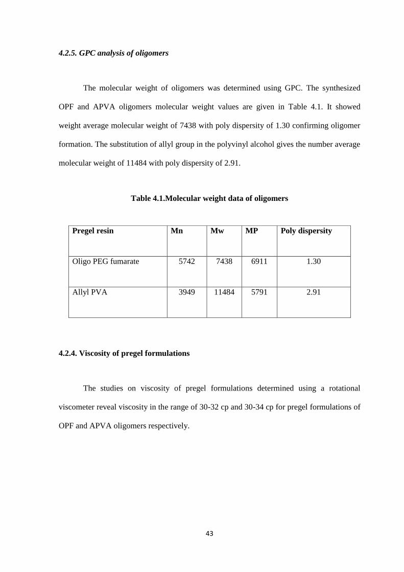
4.2.5. GPC analysis of OPF and APVA 42
4.2.6. Viscosity of pregel formulations 43
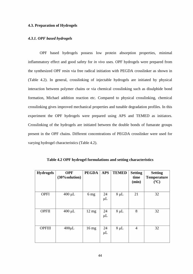
4.3. Preparation of Hydrogels 43
4.3.1. OPF based hydrogels 44
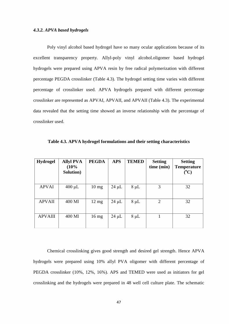
4.3.2. APVA based hydrogels 47
4.4 Hydrogel physiochemical properties 49
4.4.1. Swelling studies of OPF hydrogels 49
4.4.2. Swelling studies of APVA hydrogels 50
4.4.3 Crosslinking density and molecular weight between crosslinks 51
4.4.4. Mechanical Property 52
4.4.5. Degradation Studies 53
4.4.6. Surface Morphology 56
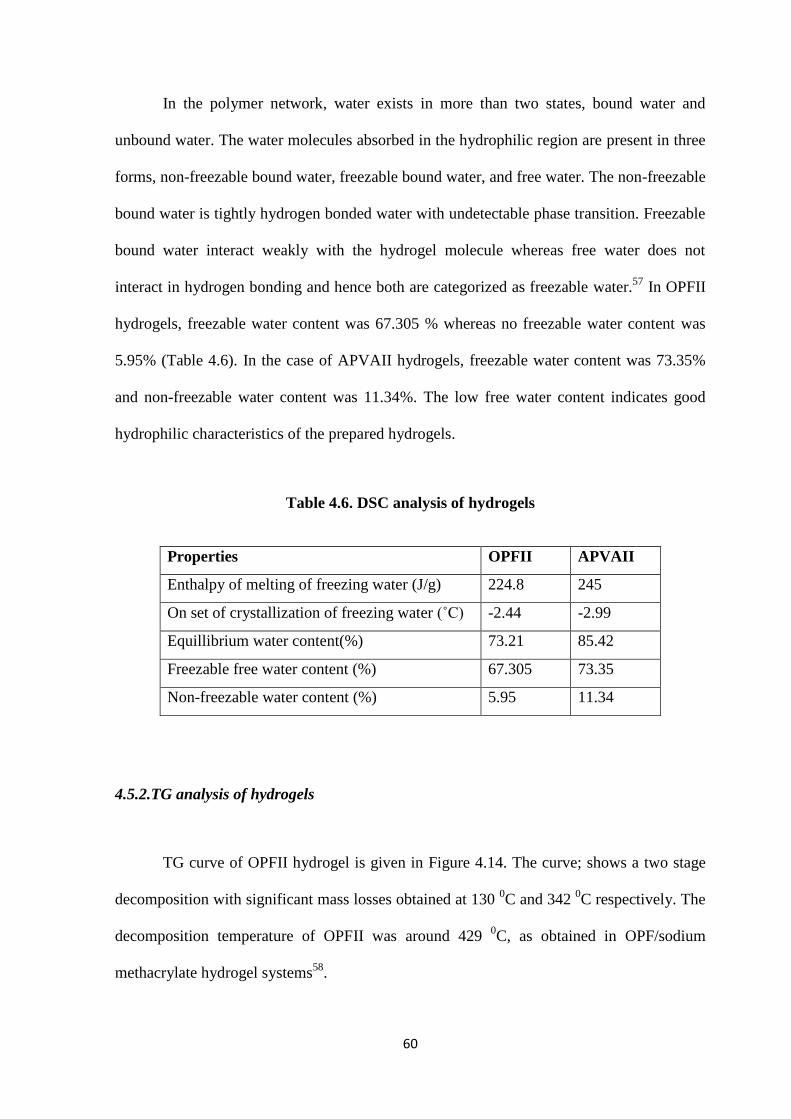
4.5 .Thermal properties of hydrogel 59
4.5.1.DSC analysis of hydrogels 59
4.5.2.TG analysis of hydrogels 60
4.6. Contact Angle Measurements 62
4.7. Optical properties 63
4.7.1. Refractive Index 63
4.7.2. Transmittance 64
4.8 In vitro Cytotoxicity 65
4.8.1 MTT Assay of resin 65
4.8.2. Direct Contact Assay 67
4.8.3. Live/Dead cell Assay 68
4.8.4. MTT Assay of prepared hydrogels 70
Chapter V
5. SUMMARY AND CONCLUSION 72
References 75
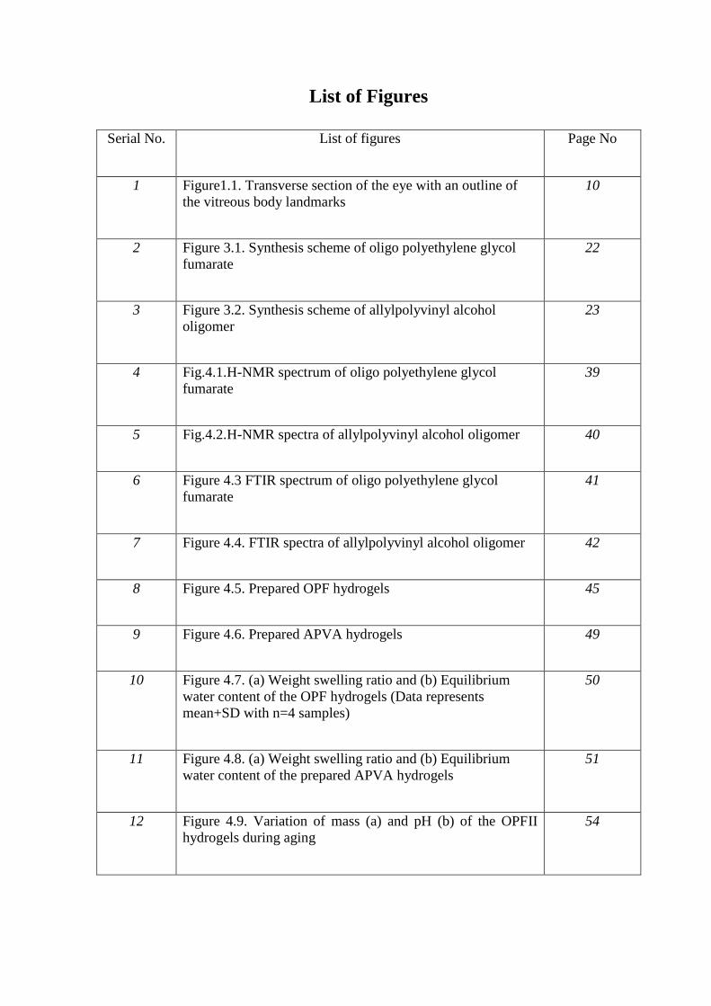
List of Figures
Serial No. List of figures Page No
1 Figure1.1. Transverse section of the eye with an outline of
the vitreous body landmarks
10
2 Figure 3.1. Synthesis scheme of oligo polyethylene glycol
fumarate
22
3 Figure 3.2. Synthesis scheme of allylpolyvinyl alcohol
oligomer
23
4 Fig.4.1.H-NMR spectrum of oligo polyethylene glycol
fumarate
39
5 Fig.4.2.H-NMR spectra of allylpolyvinyl alcohol oligomer 40
6 Figure 4.3 FTIR spectrum of oligo polyethylene glycol
fumarate
41
7 Figure 4.4. FTIR spectra of allylpolyvinyl alcohol oligomer 42
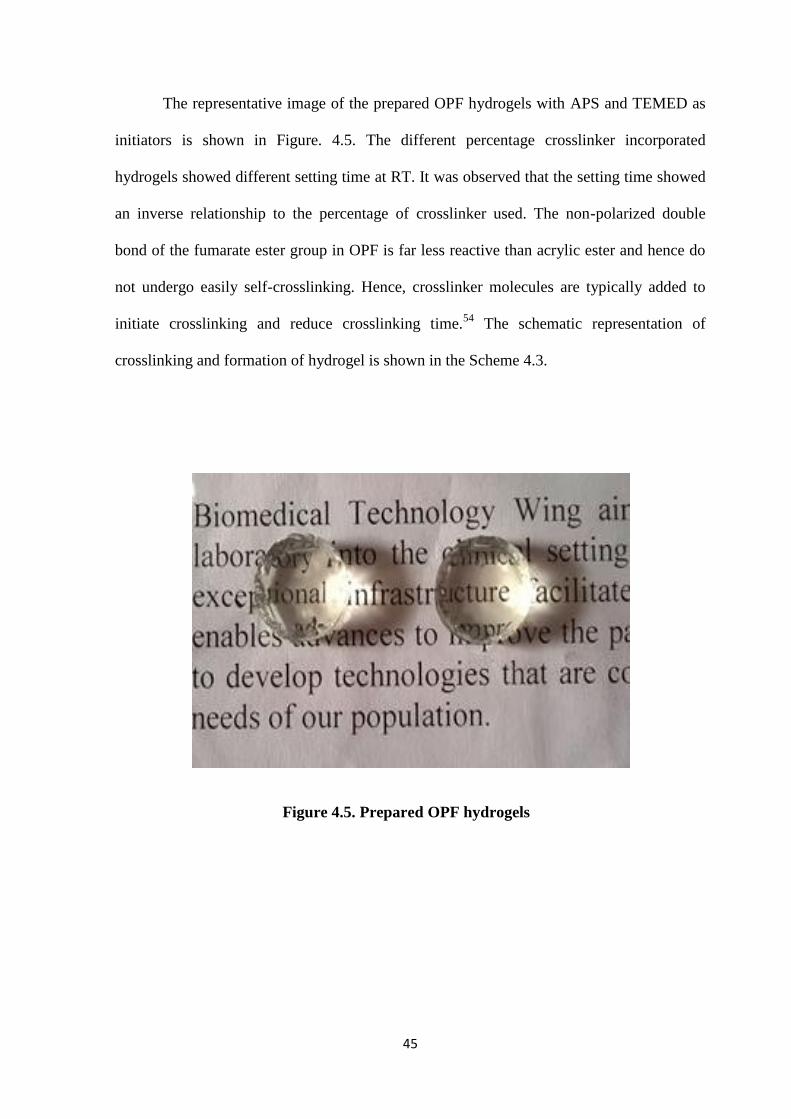
8 Figure 4.5. Prepared OPF hydrogels 45
9 Figure 4.6. Prepared APVA hydrogels 49
10 Figure 4.7. (a) Weight swelling ratio and (b) Equilibrium
water content of the OPF hydrogels (Data represents
mean+SD with n=4 samples)
50
11 Figure 4.8. (a) Weight swelling ratio and (b) Equilibrium
water content of the prepared APVA hydrogels
51
12 Figure 4.9. Variation of mass (a) and pH (b) of the OPFII
hydrogels during aging
54
13 Figure 4.10. Variation of mass (a) and pH (b) of the APVAII
hydrogels during aging
55
14 Figure 4.11.Surface morphology of OPFII hydrogels. virgin
hydrogel (a), hydrogel after 3rd
week of aging (b) and
hydrogel after 6th
week of aging (c)
57
15 Figure 4.12.Surface morphology of APVAII hydrogels.
virgin hydrogel (a), hydrogel after 3rd
week of aging (b) and
hydrogel after 6th
week of aging (c)
58
16 Figure 4.13. DSC curve of hydrogels. (a) OPFII and (b)
APVAII.
59
18 Figure 4.14. TG curve of OPFII hydrogel. 61
19 Figure 4.15. TG curve of APVAII hydrogel. 62
20 Figure 4.16. Contact angle measurements. OPFII hydrogel
(a) and APVAII hydrogel (b).
63
21 Figure 4.17.Transparency of hydrogels. OPFII (a) and
APVAII hydrogel (b).
64-65
22 Figure 4.18.MTT assay of leachable from the OPF pregel
solution
66
23 Figure 4.19. MTT assay of leachable from the APVA pregel
solution
66
24 Figure 4.20. Cytocompatibility- Direct contact assay of
hydrogels. OPFII hydrogel (a), APVAII hydrogel (b) and
L929 control cells (c)
67-68
25 Figure 4.21. Cell viability of cells on direct contact with
hydrogels. OPFII hydrogel (a) , APVA hydrogels (b) L929
Control cells (c) (Ethidium bromide/Acridine orange
staining)
69-70
26 Figure.4.22. MTT assay of leachables from the OPFII
hydrogel
71
27 Figure.4.23. MTT assay of leachables from the APVAII
hydrogel
71
List of Tables
Serial no List of tables Page No
1 Table.1 Main properties and applications of synthetic polymeric
biomaterials
3
2 Table.1.1.Biodegradable polymers and their degradation
profiles
5
3 Table.4.1. Molecular weight data of oligomers 43
4 Table 4.2 OPF hydrogel formulations and their setting
characteristics
44
5 Table 4.3. APVA hydrogel formulations and their setting
characteristics
47
6 Table.4.4. Crosslinking density and Molecular weight between
crosslinks
52
7 Table 4.5. Mechanical properties of OPF and APVA hydrogels 53
8 Table 4.6. DSC analysis of hydrogels 54
9 Table 4.7. Refractive index of OPFII and APVAII hydrogels 64
List of Schemes
Serial No List of scheme Page no.
1 Scheme 4.1. Schematic representation of synthesis of oligo
polyethylene glycol fumarate
37
2 Scheme 4.2 Schematic representation of synthesis of allyl-
poly vinyl alcohol.
38
3 Scheme 4.3 Crosslinking of OPF oligomer in the formation
of hydrogel
46
4 Scheme 4.4 Crosslinking of APVA hydrogels schematic
representation
48
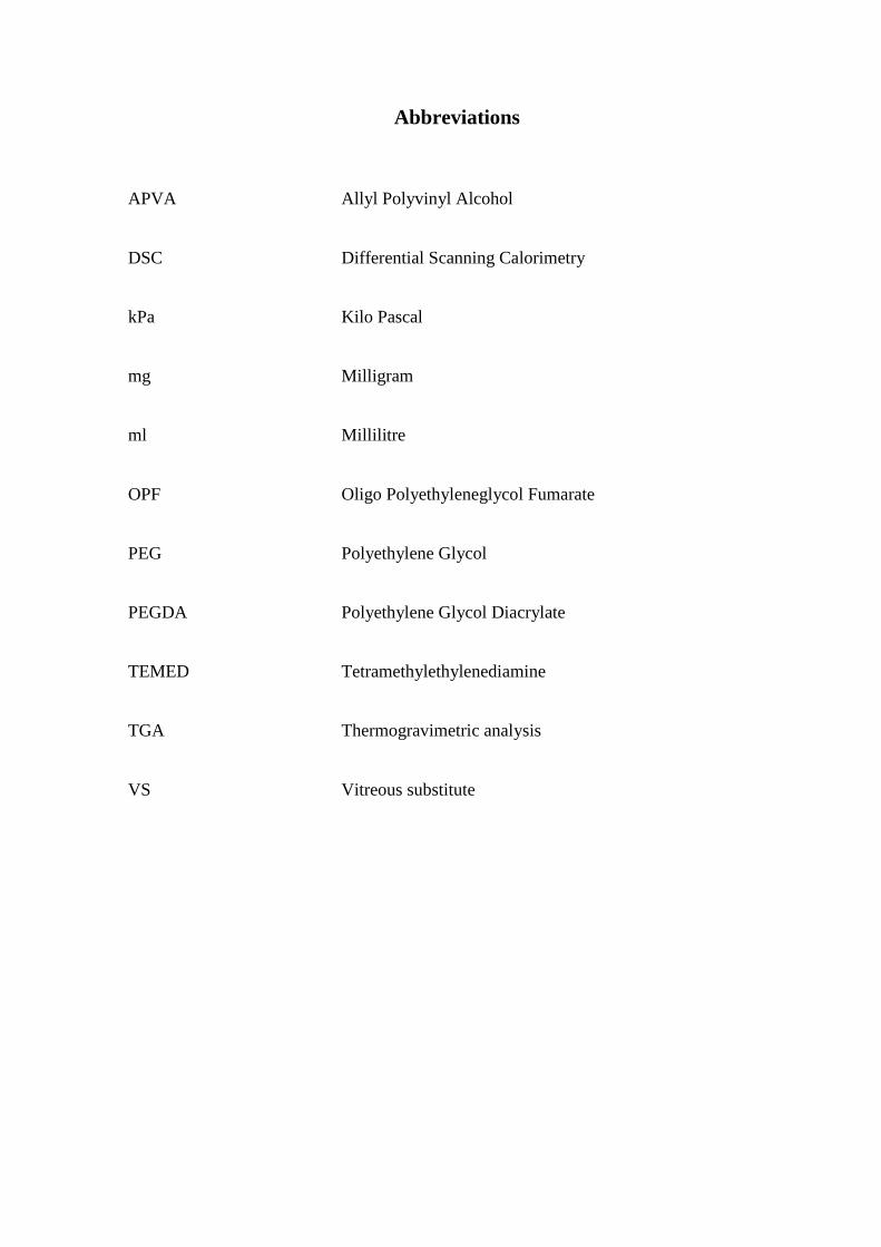
Abbreviations
APVA Allyl Polyvinyl Alcohol
DSC Differential Scanning Calorimetry
kPa Kilo Pascal
mg Milligram
ml Millilitre
OPF Oligo Polyethyleneglycol Fumarate
PEG Polyethylene Glycol
PEGDA Polyethylene Glycol Diacrylate
TEMED Tetramethylethylenediamine
TGA Thermogravimetric analysis
VS Vitreous substitute
1
Chapter I
INTRODUCTION
1. Polymers for medical applications
For the last few decades, polymers have found extensive applications in the medical
field, such as scaffolds for tissue engineering, biosensors, actuators, bio separation devices,
reactive coatings, medical imaging, and medical devices. The production of polymers and
their preparation in different structures and compositions gives appropriate physiochemical,
interfacial and biomimetic properties for specific applications. Polymeric biomaterials have
ease of manufacturability and can be molded into films, gels, membranes, sheets, capsules,
spheres, particles, 3-D scaffolds. They have good processability, reasonable cost and
possess desired mechanical and physical properties1.
Polymers are large organic macromolecules consisting of repeating monomeric
units. Polymers are formed by monomers which are covalently bonded together by chains of
atoms. The entangled chains of the polymer are formed by the weak secondary bonds of
attraction between the covalent bonds such as hydrogen and Vander Waals bonds. Polymers
show weak thermal and electrical properties due to their covalent interatomic bonding
2
within the molecules. The molecular weight, chemical side groups, chain structure and
composition of backbone are responsible for the mechanical and thermal behavior of
polymers.2
According to polymerization techniques, polymers can be classified into bulk
polymerization, condensation polymerization, and emulsion and suspension polymerization.
In bulk polymerization, monomers are reacted with each other using appropriate catalyst
and initiators fed in to the reactor without solvent. At the end of the reaction, a solid mass of
synthesized polymer being formed. In condensation polymerization different monomer units
are condensed to form a polymer with byproducts. The reactions are slightly exothermic and
have low viscosity thereby enhancing ready mixing. In solution polymerization,
polymerization takes place in a solvent in which both monomers and formed polymers are
soluble. In suspension polymerization, the monomer is a dispersed phase in an aqueous
medium and the polymer produced from the reactions are formed as a solid dispersed phase.
Emulsion polymerization is same as suspension polymerization but differences are that the
initiator is dispersed in the aqueous phase compared to suspension and polymer formed is
obtained as very small particles.3
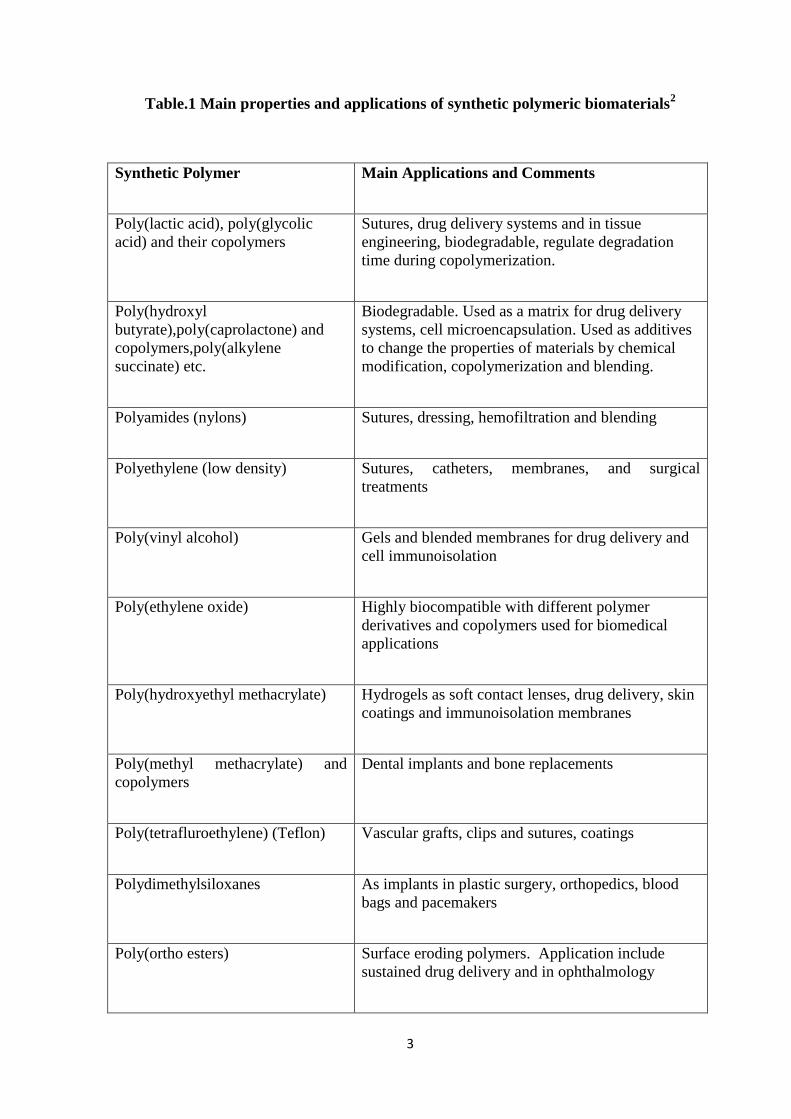
1.1 Synthetic polymeric biomaterials
Synthetic polymers have found various applications as biocompatible biomaterials
because of their well-known synthesis and moldable properties. Several synthetic materials
are already used as medical products such as poly glycolic acid as absorbable sutures and
polyurethane in artificial heart. Several synthetic polymeric materials and their applications
in the biomedical field are given below. (Table.1)
3
Table.1 Main properties and applications of synthetic polymeric biomaterials2
Synthetic Polymer Main Applications and Comments
Poly(lactic acid), poly(glycolic
acid) and their copolymers
Sutures, drug delivery systems and in tissue
engineering, biodegradable, regulate degradation
time during copolymerization.
Poly(hydroxyl
butyrate),poly(caprolactone) and
copolymers,poly(alkylene
succinate) etc.
Biodegradable. Used as a matrix for drug delivery
systems, cell microencapsulation. Used as additives
to change the properties of materials by chemical
modification, copolymerization and blending.
Polyamides (nylons) Sutures, dressing, hemofiltration and blending
Polyethylene (low density) Sutures, catheters, membranes, and surgical
treatments
Poly(vinyl alcohol) Gels and blended membranes for drug delivery and
cell immunoisolation
Poly(ethylene oxide) Highly biocompatible with different polymer
derivatives and copolymers used for biomedical
applications
Poly(hydroxyethyl methacrylate) Hydrogels as soft contact lenses, drug delivery, skin
coatings and immunoisolation membranes
Poly(methyl methacrylate) and
copolymers
Dental implants and bone replacements
Poly(tetrafluroethylene) (Teflon) Vascular grafts, clips and sutures, coatings
Polydimethylsiloxanes As implants in plastic surgery, orthopedics, blood
bags and pacemakers
Poly(ortho esters) Surface eroding polymers. Application include
sustained drug delivery and in ophthalmology
4
Polymers undergo degradation into smaller fragmented chain units followed by
mineralization and absorption. The biodegradation process is primarily chemical
degradation through hydrolysis. Physical forces such as wetting/drying, cooling, heating
helps in the deterioration of the polymer chains which is exacerbated by biochemical agents
that come into contact with the polymer surface fulfilling the degradation process.
In medical device applications, polymer degradation often limits the performance of
the implanted material. Depending on the application, different products with different
degradation rates are required. For example, polymers used as tissue engineering scaffolds
should have controllable degradation rate for enhancing tissue regeneration. On the other
hand, polymers for bone implants should have good strength and less degradability.
Polymers used for drug delivery applications should have tunable degradability ranging
between days to months.
1.2. Degradable polymers for medical applications
A lot of efforts are focused on developing biodegradable materials based on natural
and synthetic polymers.4 Biodegradable natural polymers cause less toxicity and does not
evoke significant foreign-body reactions in host tissues compared to synthetic materials. In
biomedical applications, notable characteristics are required including, controllable
degradability, less inflammatory reactions in host tissues, desired mechanical properties,
good sterilizability and process ability.5
Biodegradable polymers are composed of hydrolytically and enzymatically
degradable polymers. Hydrolytically degradable polymers break down due to their
hydrolytically unstable chemical bonds and undergo degradation. Hydrolytically degradable
5
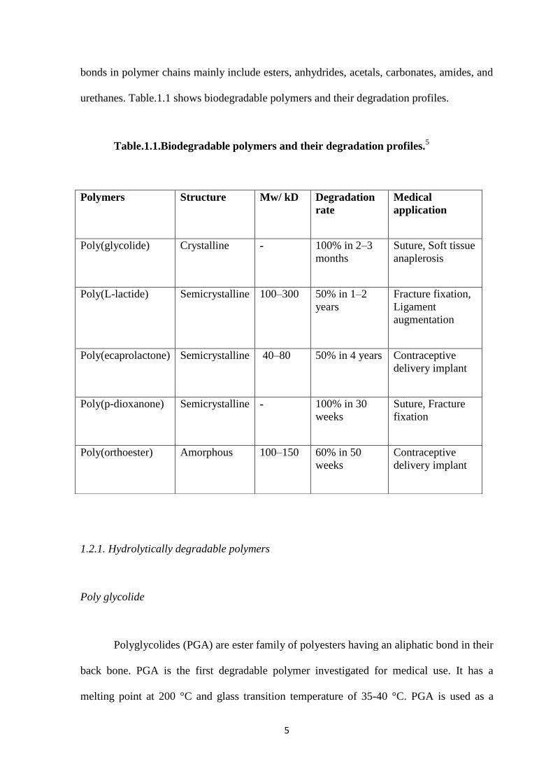
bonds in polymer chains mainly include esters, anhydrides, acetals, carbonates, amides, and
urethanes. Table.1.1 shows biodegradable polymers and their degradation profiles.
Table.1.1.Biodegradable polymers and their degradation profiles.5
1.2.1. Hydrolytically degradable polymers
Poly glycolide
Polyglycolides (PGA) are ester family of polyesters having an aliphatic bond in their
back bone. PGA is the first degradable polymer investigated for medical use. It has a
melting point at 200 °C and glass transition temperature of 35-40 °C. PGA is used as a
Polymers Structure Mw/ kD Degradation
rate
Medical
application
Poly(glycolide) Crystalline - 100% in 2–3
months
Suture, Soft tissue
anaplerosis
Poly(L-lactide) Semicrystalline 100–300 50% in 1–2
years
Fracture fixation,
Ligament
augmentation
Poly(ecaprolactone) Semicrystalline 40–80 50% in 4 years Contraceptive
delivery implant
Poly(p-dioxanone) Semicrystalline - 100% in 30
weeks
Suture, Fracture
fixation
Poly(orthoester) Amorphous 100–150 60% in 50
weeks
Contraceptive
delivery implant
6
degradable suture, DEXON®, from 1970s. It is mainly used in short term tissue engineering
applications.
Polylactide
Polylactide is a naturally occurring polymer from lactic acid monomers. Poly
lactides include poly(L-lactic acid) (PLLA), poly(D-lactic acid) (PDLA), and poly(D,L-
lactic acid) (PDLLA), a racemic mixture of PLLA and PDLA, have been extensively used
for various applications. PLLA has melting point of 175 °C and glass transition of 60 -
65°C. It is used as bone fixator and as tissue engineering scaffolds for bone applications.
Polyethers
Most of the biomedical research with synthetic polyethers focus on the use of
poly(propylene glycol) (PPG) and poly(ethylene glycol) (PEG). Polyethers have good
stability and they do not readily undergo hydrolytic degradation. Polyether with lower
molecular weight is used for biomedical applications because of its lower accumulation of
degradation product compared to higher molecular weight polyethers. PEG based hydrogels
have been used as in situ-forming injectable hydrogels. It has also been tested in
formulations for intravitreal drug delivery, repair of scleral incisions and sealing retinal
detachments. For example, 5 wt% PEG in phosphate-buffered saline have been
administrated as artificial vitreous substitutes (VS). The injected VS had good mechanical
and optical properties similar to the natural vitreous and was well tolerated by the retina
with minimal histological electrophysiological changes during the rabbit implanted studies.6
7
1.3. Vitreous substitute
Retinal detachment is a significant cause of vision loss. Recently, efforts have been
undertaken for the development of innovative approaches in vitreo retinal treatments.
Vitreous humor is a gelatinous structure that fills the space between the lens and the retina7
and is composed of 98 - 99% water by weight. The human vitreous is composed of
collagen fibers and hyaluronic acid and hence is a natural polymeric hydrogel.8 The vitreous
is the largest part of the eye by volume and plays an important role in the support and
growth of its structures. The retinal pigment epithelium (RPE) is dependent on the vitreous
hyaluronic acid composition9. The vitreous is a highly transparent substance that transmits
90% visible light and gives support to lens capsule.10,11
Vitreous humor helps in the
circulation of metabolites throughout the eye and decreases lens exposure to oxygen.12
1.4. Structure and function of vitreous body
1.4.1. Anatomical Properties
Vitreous humor consists of several parts with different densities and biochemical
compositions so it is not homogeneous. The vitreous cortex is closest to the retina and is
also the denser part of the vitreous with variable thickness.13
Vitreous is overlying the
vitreous base which is denser than the cortex, also called as ora serrata. Vitreoretinal
interface is a membrane complex seen between border of vitreous and retina. It is comprised
of internal limiting membrane of the retina (ILM) and a layer of dense collagen fibers
adjacent to the ILM14
.
8
1.4.2. Chemical Composition
Vitreous body contains 98% weight is water, but is not in free form as some of it is
bound to proteins and glycosaminoglycans.15
Proteins: The content of proteins in the vitreous is small when compared to the size of the
vitreous body. The vitreous synthesizes transferrin and iron-binding proteins which gives a
protective effect towards small vitreal hemorrhages.16 The most abundant insoluble collagen
concentration in vitreous is 300μg/ml and is found in the form of type II (60-70%), type IX
(25%), type V/XI (10-25%) and type IV (<10%) collagen.17
Glycosaminoglycan: It is an important constituent of the vitreous, and is present as
hyaluronic acid (HA), chondroitin sulfate (CS) and heparin sulfate. Hyaluronic acid
determines the viscosity of the vitreous body and serves as a template for assembly of other
extracellular matrix.18
Chondriotin sulfate in vitreous is in the form of two proteoglycans,
versican and type IX collagen. A small percentage of heparin sulfate is present in the
vitreous and is presumed to maintain adequate spacing between the collagen fibrils.
Metabolites: The major metabolites, glucose and lactic acid, are found in vitreous and is
needed for support of enzymatic activity. The concentration of glucose is found to be half
that in plasma for most species.
Ascorbic acid: Ascorbic acid found in vitreous body is higher than in plasma and is
associated with the process of liquefaction during aging. Recent studies have showed that it
plays an important role as a potent antioxidant and prevents early cataract formation.12
9
Amino acids: Amino acids concentration in the vitreous is similar to those in plasma.
Fatty acids: The total lipid concentration in the vitreous is stable with age and unsaturated
fatty acids constitute around 50-55%19
.
Prostaglandins: Prostaglandins are a group of physiologically active lipid compounds
composed of unsaturated carboxylic acids. The human vitreous contains 100 g/ml of
prostaglandins.
Cells: Three types of cells are mainly identified in vitreous body. They are hyalocytes,
fibrocytes and macrophages. Hyalocytes are located on the vitreous surface adjoining the
retinal ILM. Fibrocytes help in the secretion of collagen and macrophages are also involved
in phagocytosis.20
1.4.3. Physical properties
Organization: The long chain collagen fibrils and hyaluronic acid molecules makes the
vitreous gelatinous and provides it with viscoelastic properties.21
Gradients: The vitreous contains several compositional gradients including collagen,
protein, bound water, HA, glucose, lactate and oxygen. A small pressure gradient also helps
in the movement of ions. The standing potential of eye is fortified by the gradients and
constant movement of ions. Gradients in anterior and posterior of the eye part is much more
viscous and denser22
compared to the middle segment of the eye.
10
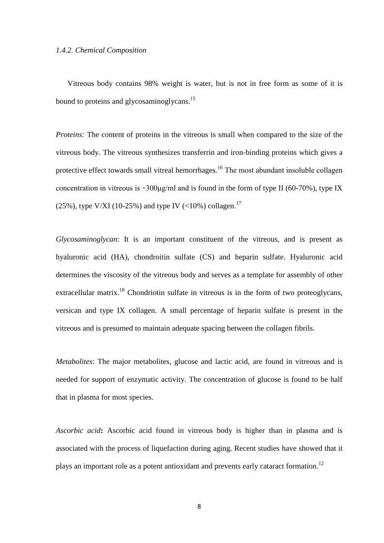
Figure1.1. Transverse section of the eye with an outline of the vitreous body landmarks.23
1.5. Function of native vitreous
The vitreous plays an essential role in the growth of RPE. Hyaluronic acid in
vitreous plays a role in sustaining internal pressure within the eye.24
The high HA content in
the vitreous protects the eye from low frequency mechanical stress and vibration. HA
behaves as viscoelastic body rather than a viscous liquid thereby acting as shock absorber of
the eye. The highly transparent ocular medium that transmit 90% visible and near infrared
light also acts as a support for the lens capsule.10
The vitreous acts as a barrier to
biochemical substances and cells thereby inhibiting proliferation, inflammation and
neovascularization. Vitreous helps in the metabolism of the surrounding tissue by
transporting essential molecules and minimizes the risk of cataract formation by protecting
the lens from oxidative damage.12
As age increases, the vitreous undergoes liquefaction which affects normal vision.
11
Vitreous liquefaction causes retinal detachment, a condition where the neurosensory retinal
segments separate from posterior part of the eye, which leads to dangerous eye
complications such as macular hole, vitreous hemorrhage, vision loss, etc.25
To treat this
condition, vitrectomy, a surgical procedure which involves the removal of the vitreous in
the central cavity of the eye and replacing with an artificial substitute, is performed.
Currently used vitreous substitutes have the ability to reproduce some of the important
characteristics of native vitreous.
1.6. Artificial vitreous substitutes
There are several types of VS currently available. These include gases (air, expansile
gases), liquids (perflurocarbon liquids) and polymers.
1.6.1.Gas based substitutes
Air-based substitutes: In 1911 Ohm first used air as VS for retinal detachments.
Cheaply and readily available air can easily absorb by eye and replaced by aqueous humor.
Air based materials have short residence time and hence have limited use as a vitreous
substitute.
1.6.2.Expansible Gas-based Substitutes
Sulfur hexafluoride (SF6) and perfluoropropane are heavier than air and are
commonly used as VS. Both are colorless, odorless, non toxic and are used in non-expansile
concentrations to fill the vitreous cavity.
12
1.6.3. Per fluorocarbon liquid
The more ideal vitreous substitute is a liquid or gelatinous solid that can respond to
head movement and maintain tamponade effect without being absorbed or degraded. Water
and balanced salt solutions were the first liquids used as VS.26
In 1990s, modern liquid
substitutes such as perfluorocarbon liquids (PFCLs) were investigated. PFCLs are synthetic,
fluorinated, carbon containing compounds with clear, colorless, and odorless characteristics.
They possess an extensive capacity for transporting and releasing both O2 and CO2 and is
twice denser than water.27
They have good oxygen carrying capability and was originally
designed as blood substitutes.28
The most commonly used PFCLs are perfluorodecalin
(PFD), perfluorohexyloctane (F6H8), perfluoroperhydrophenanthrene, and
octafluoropropane.
Use: PFCLs could potentially be used as long-term tamponade of inferior retinal
detachments by virtue of being heavier than water. In complex retinal detachments PFCLs
are used intraoperatively to temporarily flatten the retina and are used as replacement for
silicone oil or another long-term substitute. However, they are currently limited by long-
term toxicity.
Advantages: PFCL’s high specific gravity makes them useful for the intraoperative
repair of complex retinal tears. The intraoperative removal of PFCLs gives peculiar
complications in anterior and posterior segments.28
The high oxygen solubility of PFCL’s
provides good neuroprotective effect on ischemic retina29
.
13
Disadvantages: PFCL’s are intraopratively limited because of their long-term
toxicity. Disorganization of retinal cell growth pattern, neuritis, and other toxic effects are
showed by PFD and perfluoroperhydrophenanthrene in in vitro experiments.30
1.6.4.Semi fluorinated alkanes
Semifluorinated alkanes are also known as partially fluorinated alkanes. It is the first
tamponade agent used for intraoperative setting and is heavier than water.31
It have low
specific gravity, 1.35 g/mL, compared to PFCLs with a refractive index of 1.3.
Use: In earlier days, SFAs were used as a solvent for silicone oil and was
temporarily used as endotamponade for special cases of retinal detachment when silicone
oil did not offer desired results.
Disadvantage: It is associated with cataract formation, emulsification followed by
the presence of soft epiretinal membranes.
1.6.5.Silicone oil
In 1960s silicone has been used as a VS. Its structure is similar to that of silicone
rubber but shorter polymer chains and absence of crosslinking makes it exist as a liquid
form. It has hydrophobic characteristics with a specific gravity of 0.97 g/mL and refractive
index of 1.4.
14
Use: It is the currently accepted vitreous substitute for long term application. It is
available in many viscosity variants which is measured in centistokes. Clinically 1000 to
5000 centistoke silicone oils (SO) are used now. SO are typically removed after 3-6 months.
Advantages: SO is very good VS because of its high surface tension, low toxicity,
ease of removal and transparency. It possesses 70% anatomical integrity due to its
immiscible characteristics with water which gives it high surface tension and low specific
gravity. Difficulties in post operative positioning with SO in children and adult is very less
so it is a versatile tamponade agent for VS.
Disadvantages: SO can undergo traction because it can pass through retinal breaks
due to low surface tension property. SO need optimal adjustments due to its refractive index
value close to 1.4. Post-operatively, SO shows oil emulsification, incidence of corneal
abnormalities at 24 months, which did not differ significantly from sulfur hexafluoride gas,
and has been reported to cause problems with both native and implanted lenses
1.6.6. Silicone oil/SFA combinations
One of the clinically approved tamponade agent for the vitreous is silicone oil/SFA
combination. The combination of both mixture gives the advantages of both liquids such as
high specific gravity of SFAs and high viscosity of silicone oil making it a good VS with
low emulsification. The solubility of this mixture called heavy silicone oils can produce a
homogeneous clear solution and varies with the ratio of liquids. The percentage of SO is
kept as lower to avoid toxicity.
15
1.7. Ideal vitreous substitute
The currently used clinically approved vitreous tamponade have many drawbacks.
According to chirila et al, the ideal VS should have transparency and clarity, biological &
chemical inertness, refractive index and density equal to that of the natural vitreous,
sufficient rigidity to act as tamponade agent, ability to allow transfer of metabolites, non-
absorbable and non-biodegradable characteristics and should have storable, sterilizable,
viscoelastic properties.32
1.8.Polymeric hydrogels and vitreous characteristics
Polymeric hydrogels are wet and soft materials that are composed of three
dimensional hydrophilic network structure containing large amount of water (50−90%) 33
.
Hydrogels are convenient materials for biomedical applications as they are soft materials
with hydrophilic and porous nature whose stiffness can be tailored depending on the
application.34
Injectable hydrogels are excellent biomaterials for tissue engineering applications as
they possess good biocompatibility, easily controllable physical properties, minimally
invasive delivery and undergo solution to gelation (sol-gel) transition in situ through
physical or chemical stimuli.35
1.8.1 Natural polymeric based vitreous substitutes
Collagen based semisynthetic polymer VS were used during the early days of
vitrectomy. Kurt et al reported that tropocollagen isolated from calf skin offered clear,
16
stable hydrogels with properties close to native vitreous when implanted in rabbit and
monkey model.36
Pruett et al evaluated proctase-treated collagen VS but the gel lacked
rigidity.37
An alkaline–solubilized collagen with hyaluronic acid VS in rabbit model
revealed no adverse effect on ocular tissues with increased collagen half-life was reported
by Nakagava et al.38
Recently Lai et al developed a chemically modified gelatin hydrogels
by crosslinking it with glutaraldehyde (GTA) and ethyl dimethyl amino propyl
carbodiimide (EDC). In vivo studies in rabbit model exhibited good biocompatibility in
EDC cross-linked gelatin gels.39
Commercially available sodium hyaluronidate showed
excellent biocompatibility and tolerance in ocular tissues but due to its short residence time
in vitreous cavity and poor tamponade effect it was unsuitable to serve as an ideal VS.40
Surie et al formed a gellan gum gel at RT and their structure was maintained in body
temperature. In vitro studies on mouse fibroblasts showed excellent biocompatibility but
poor rheological properties than that of natural vitreous. However, mechanical properties
was improved by adding CaCl2 to the gellan gum/HA mixture.41
1.8.2. Synthetic polymeric vitreous substitute
There are several synthetic polymeric materials that show promise as good VS.
Current research is increasingly focusing on synthesizing highly biocompatible polymers
that might be able to mimic the functional properties of the natural vitreous, rather than its
physical characteristics. Poly(2-hydroxyethyl methacrylate) (PHEMA) is a solid hydrogel
that was well-tolerated by ocular tissues and did not elicit retinal damages without
undergoing bioabsorption or biodegradation. However, PHEMA implantation required
difficult surgical procedures and therefore, was no longer investigated.42
Polyvinyl alcohol
(PVA) also shows great promise as an artificial VS. Maruoka et al developed an autoclaved
PVA hydrogels and tested in a monkey’s eye. These hydrogels showed good optical
17
properties and long-term biocompatibility.10
Recently, Cavalieri et al studied poly(vinyl
alcohol methacrylate) (PVA-MA) hydrogels. The polymer contained a photo initiator that
could form a 3-D gel network by irradiating with light at 365 nm. The crosslinking density
can be varied by adjusting photo initiator concentration and the radiation exposure time to
form in situ gelification. In vitro test showed that hydrogel degradation occurred in the
presence of low degree of crosslinking indicating that suitable hydrogels would be those
synthesized at high polymer concentrations and with high degree of cross-linking. However,
such gels were found to be significantly stiffer than natural vitreous. Nevertheless, PVA-
MA hydrogels offers promise for long-term VS application.43
Hydrogels offer great potential for the development of an artificial VS, however,
their drawbacks such as short residence time and uncontrolled biodegradation in vivo limits
their application. Several researchers are focused on clinically utilizable hydrogel
preparation for vitreous substitutes. Feng et al developed a novel approach of using foldable
capsular vitreous body (FCVB) injected with PVA hydrogel as VS. These crosslinked
hydrogels were formed using gamma irradiation and they exhibited good rheological and
physical properties with no potential toxicity in vitro. After 180 days retention, 3% PVA
hydrogels inside FCVB remained transparent, had good elasticity, minimal biodegradation,
good biocompatibility and retina support.44
Stimuli-responsive hydrogels have achieved notable consideration due to their
manifold applications. These hydrogels shrink or expand or switch between sol to gel state
in response to the environmental changes like pH, temperature, or light. Smart hydrogel can
be prepared, stored and injected in solution state and undergo in situ gelation via external
stimuli. For example, vitreous Pluronic F127 45
ocular and WTG-12746
have been attempted
for use as artificial VS. However, F127 was found to cause severe retinal toxicity and
18
WTG-12 exhibited a short degradation time and tendency to drift under retinal tears before
complete gelation.47
Work is ongoing to prepare smart hydrogels that not only mimic the
optical and rheological properties of the natural vitreous but also bring in antifouling
properties and ease of application. Lirong et al developed a hydrogel using inexpensive
commercially available components: polyethylene glycol acrylate and dithiothreitol. The
prepared PEG hydrogels have a compressive modulus of up to 104 Pa and comprised of
87.5% water making their mechanical performance comparable to cross-linked PEG gels.
Furthermore, the dynamic nature of the boronate ester linkages imparted the hydrogel with
self-healing properties and the gels can be healed within 30 min without external stimulus48
19
Chapter II
OBJECTIVES AND SCOPE
The removal of the natural vitreous from the eye during vitrectomy and the loss of
vision associated with retinal detachment after vitrectomy are major complications in the
area of ophthalmology. The development of an artificial vitreous is greatly needed to fill the
role as a tamponade agent to prevent retinal detachment in the posterior part of the eye.
Many materials have been developed and used for last few years; but none possess the ideal
characteristics of the natural vitreous due to their short term lifetime and biocompatibility
issues.
The objectives of this study were to design transparent, injectable pregel
formulations that can crosslink via free radical polymerization using a PEGDA as
crosslinker and form crosslinked hydrogels in situ. Toward this end, we have synthesized
two injectable pregel polymer solutions based on polyethylene glycol and polyvinyl alcohol
as main polymer chain units. We have introduced fumarate groups in PEG backbone and
allyl functional groups in PVA chain to yield a crosslinked hydrogel. The physiocochemical
properties, optical characteristics, degradation profiles and biological studies of the prepared
PEG and PVA hydrogels were evaluated and detailed below.
20
Main objective of the study include
Synthesis and characterization of oligo polyethylene glycol fumarate (OPF) and
allyl functionalized polyvinyl alchol (PVA) oligomers
Characterization of prepared hydrogels by studying the swelling, degradation,
mechanical and optical properties
Evaluation of cytocompatibilty of hydrogels
21
Chapter III
EXPERIMENTAL
3.1. Materials
PEG (Poly ethylene glycol, MW: 600) and fumaryl chloride were purchased from
Merck (India). Potassium carbonate, dichloromethane, polyvinyl alcohol (Mw = 18,000),
allyl bromide, sodium hydroxide, ammonium persulphate, tetramethylethylenediamine
(TEMED), and polyethylene glycol diacrylate (PEGDA, MW: 700) were acquired from
Sigma (Bangalore, India). High glucose Dulbecco’s Modified Eagle’s Medium (DMEM),
fetal bovine serum (FBS) and 100X antibiotic-antimycotic solution were obtained from
Invitrogen (India). MTT (3-(4,5-dimethylthiazol-2-yl)-2,5- diphenyltetrazolium bromide)
assay kit procured from Invitrogen.
3.2. Synthesis of poly (ethylene glycol fumarate) oligomer (OPF)
The oligo polyethylene glycol fumarate (OPF) was synthesized using the
experimental setup as shown below (Figure 1). Dried PEG, fumaryl chloride, potassium
carbonate (K2CO3) were measured out in a 1:1:1.5 molar ratio. PEG was dissolved in a

22
methylene chloride (1:2 v/v) and placed in a two neck flask along with powdered K2CO3.
The mixture was stirred with an magnetic stirrer to form a slurry to which fumaryl chloride
dissolved in methylene chloride (1:1 v/v) was added dropwise. The reaction mixture was
maintained at room temperature (RT) for 50°C for 12 h under reflux using a condenser
under nitrogen atmosphere. After polymerization for 12 h the reaction was cooled down and
transferred to centrifuge tubes and centrifuged for 15 min at 3000 rpm until potassium
chloride (KCl) and unreacted K2CO3 were pelleted out. The supernatant was then added
drop wise to petroleum ether to extract the oligomer and rotary evaporated to yield a light
yellow viscous product.49
Figure 3.1. Synthesis scheme of oligo polyethylene glycol fumarate
23
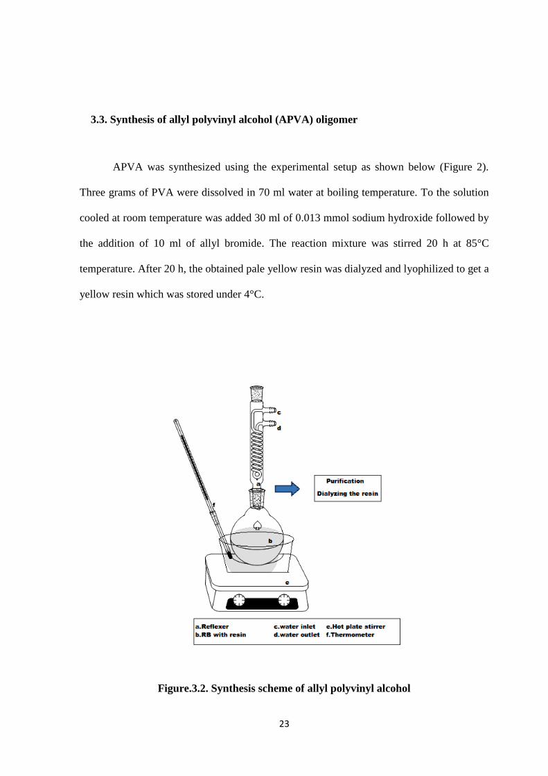
3.3. Synthesis of allyl polyvinyl alcohol (APVA) oligomer
APVA was synthesized using the experimental setup as shown below (Figure 2).
Three grams of PVA were dissolved in 70 ml water at boiling temperature. To the solution
cooled at room temperature was added 30 ml of 0.013 mmol sodium hydroxide followed by
the addition of 10 ml of allyl bromide. The reaction mixture was stirred 20 h at 85°C
temperature. After 20 h, the obtained pale yellow resin was dialyzed and lyophilized to get a
yellow resin which was stored under 4°C.
Figure.3.2. Synthesis scheme of allyl polyvinyl alcohol
24
3.4. Preparation of pregel formulation
3.4.1 OPF pregel
Briefly, 0.3 g OPF resin was dissolved in 1 ml of distilled water. The mixture was
then gently neutralized with sodium hydroxide solution (2 M) to bring pH to 7.4. The pregel
solution was then mixed thoroughly and was used as the working solution.
3.4.2 APVA pregel
Briefly, 0.1 g APVA resin was dissolved in 1 ml of hot distilled water. The pregel
solution was mixed thoroughly and was used as the working solution.
3.5. Preparation of hydrogels
3.5.1 OPF based hydrogels
For the preparation of OPF hydrogel, 400 μl of this pregel solution was added to a
well of a 48-well tissue culture plate and added 12 mg of PEGDA crosslinker and then
polymer crosslinking was initiated by adding the redox initiators, APS (24 μL, 2 M) and
TEMED (8 μL, 6 M) was mixed thoroughly. After 15 min, the disc-shaped hydrogels (12
mm diameter and 3.5 mm height) were scooped out using a clean spatula and were
subsequently used for various characterization studies. Three different hydrogels with 6 mg,
12 mg and 16 mg concentration of PEGDA crosslinker were prepared for initial
optimization of gel setting and characterization.
25
3.5.2 APVA based hydrogels
For the preparation of APVA hydrogel, 400 μl of this pregel solution was added to a
well of a 48-well tissue culture plate and added 12 mg of PEGDA crosslinker then polymer
crosslinking was initiated by adding the redox initiators, APS (24 μL, 2 M) and TEMED (8
μL, 6 M) was mixed thoroughly. After 2 min, the disc-shaped hydrogels (12 mm diameter
and 3.5 mm height) were scooped out using a clean spatula and were subsequently used for
various characterization studies. Three different hydrogels with 10 mg, 12 mg and 16 mg
concentration of PEGDA crosslinker were prepared for initial optimization of gel setting
and characterization.
3.6 Physiocochemical characterization of prepolymer
3.6.1 Nuclear magnetic resonance (NMR)
Nuclear magnetic resonance (NMR) spectroscopy is a strong analytical technique
used for the determination of molecular structure and purity of the material. The principle
behind NMR is that the nuclei of atoms have spin and are electrically charged. When an
external magnetic field is applied, an energy transfer from base energy to a higher energy
level takes place at a wavelength that corresponds to radio frequencies and energy emitted
when the spin returns to its base level is of the same frequency. The signal that matches this
transfer is measured in many ways and processed in order to yield an NMR spectrum for the
nucleus concerned. The NMR analysis of samples were done using Bruker 500 MHZ
spectrometer (Bruker Biospin, Billerica, MA) with DMSO as solvent
26
3.6.2. Fourier transform Infrared spectroscopy (FTIR)
FTIR spectroscopy is a sensitive technique to determine the functional group
analysis of unknown compounds. In FTIR, when infrared radiation is passed through a
sample, some of the incident infrared radiation is absorbed by the sample and some are
transmitted resulting in a spectrum that gives the molecular absorption and transmission of
the sample. This spectrum gives the molecular spectroscopic finger print of the sample.
The synthesized polymer’s functional groups were determined using a JASCO
FT/IR-4200 spectrometer that included JASCO’S proprietary spectra Manager TM II cross
platform software. The FTIR spectrum was recorded by sandwiching samples between two
potassium bromide pellets and scanned in the range from 4000 –400 cm-1
at a resolution of
4 cm-1
.
3.6.3 Gel permeation chromatography (GPC)
GPC is a chromatographic technique for analyzing molecular weight of polymers.
Chromatography systems rely on pumps to pass a pressurized liquid solvent containing a
sample mixture through a column filled with a solid adsorbent material. Each component in
the sample interacts slightly differently with the adsorbent material thereby causing a shift
in flow rates for the different components which lead to the separation of the components as
they flow out the column. Particles are eluted based on the pore size of the adsorbent
material and volume of sample injected into the column. In general, low molecular weight
polymers have longer retention times compared to high molecular weight polymers.
Molecular weight of the sample was determined using the Waters GPC system 600 series
pump with waters Styragel column (HR5E/4E/2/0.5). Five milligram samples were
27
dissolved in tetrahydrofuran for OPF and THF was used as mobile phase with flow rate of 1
ml/min. For APVA, samples were dissolved in distilled water and sodium nitrate was used
as mobile phase with flow rate of 1 ml/min. Waters ultrahydrogel columns 2000/1000/500
with series were used. Dextran and PEG were used for relative calculation.
3.6.4. Determination of Viscosity of pregel formulations
The viscosity of pregel formulations of oligomer was determined using a rotational
viscometer (RVA-Starch Master 2, Newport Scientific). The polymer was dissolved in
water at 37°C and viscosity was measured at 200 rpm as per machine guidelines. Hydrogel
pregel formulations of 25 ml was directly put in to the constant stirring using a rotational
viscometer (RVA StarchMaster 2, Newport Scientific) and measure the viscosity at 37°C,
and 200 rpm for 15 min.
3.7. Characterizations of hydrogels
3.7.1. Swelling Studies
Swelling studies were performed to evaluate the water absorbing capacity of
hydrogels. Hydrogels were freeze-dried using a lyophilizer. The freeze-dried weight of
sample was measured and the hydrogels were incubated in either (PBS, pH~7.4) or water
for 24 h at 37°C. After 24 h incubation, the sample wet weight was measured and the
weight swelling ratio and equllibrium water content of each sample was calculated
according to the following equation.
Weight swelling ratio = Ww /Wd
28
where, Wd is the weight of the freeze-dried hydrogel and Ww is the weight of the wet
hydrogel incubated in PBS or water.
3.7.2 Crosslink Density
The crosslink density and number average molecular weight between crosslinks of
hydrogels were determined. The prepared hydrogels were freeze-dried and their weight was
measured. The diameter and thickness of the dried sample was measured using digital
vernier calipers. The pre-weighed dried gels were incubated in water for 24 h in 37°C. After
24 h incubation the sample wet weight was measured. Modified Florry-Rehner’s equation
was used for the calculation of the above parameters.50
where, is the Huggin’s polymer-solvent interaction coefficient, assumed to be 0.34 and Vr
is the volume fraction of the polymer in water-swollen hydrogel, which can be calculated
from the swelling coefficient, using the relation,
29
where, Ws is the weight of the solvent in the swollen polymer, Wp is the weight of the
swollen hydrogel, Dp is the density of hydrogel, Ds is the density of water and dr is the
density of the polymer. The number average molecular weight between crosslinks (MC)
was calculated as the reciprocal of crosslink density (mol cm 3).
3.7.3 Mechanical Property
Hydrogels were prepared in circular plastic mold with 1 cm height and 1 cm
diameter with different concentration of crosslinker. The samples were lyophilized and
incubated in water for 24 h at 37°C. After swelling, the height and diameter of the sample
was again measured using digital vernier calipers. Compression test was carried out in an
Instron instrument (model 2510-104) with a 500 N load cell and crosshead speed of 5
mm/min-1
at RT with samples compressed to 60% of their thickness. Compressive stress,
load at break and Young’s modulus were calculated using Instron’s proprietary Bluehill 3
software.
3.7.4 Degradation
The degradation studies of lyophilized samples were carried out in PBS (pH~7.4) by
aging at 37 °C for 7, 14, 21, 28 and 35 days of incubation. Disc shaped hydrogels with 6
mm diameter and 3 mm height were prepared. Gels were incubated in PBS and placed in a
orbital shaker under constant agitation of 100 rpm with temperature set to 37 °C. At each
time point, the change in pH of the medium was recorded using a pH meter (Cyber scan pH
510, Eutech instruments); the hydrogels were removed, lyophilized and weighed. The
weight loss of hydrogels was calculated using the equation below.
30
Where, Mo is the initial mass of the hydrogel and Mt is the mass of the hydrogel at various
time points.
3.7.5 .Surface Morphology
Scanning Electron Microscopy (SEM) is a versatile methodology for analyzing the
surface morphology of the material. It helps observe the minute details related to the
material’s surface characteristics and corresponding pore sizes. SEM provides high
resolution images by using electron beams instead of light waves. SEM uses vacuum
atmosphere in a column that contain electron guns which emits beams of high energy
electrons. The electron beams travels downwards, passess through a series of lenses and
focuses on the sample. The kinetic energy carried by accelerated electrons interact with the
sample which in turn produces various signals and secondary electrons that are detected by
detectors.
Surface morphology and pore size distribution of the hydrogel were recorded using
an environmental scanning electron microscope (ESEM). The analysis was performed on
lyophilized samples at different time points (days 0, 21 and 42) and visualized under low
vacuum conditions (FEI, Quanta 200, USA).
31
3.7.6 Differential Scanning Calorimetry (DSC)
Differential scanning calorimetry is a thermoanalytical technique in which the
difference in the amount of heat required to increase the temperature of a sample and
reference are measured as a function of temperature. DSC gives the glass transition
temperature, melting point, phase transformation information of the samples. The transition
of water content in the hydrogels was measured using Differential scanning calorimeter
(DSC Q20 V24.4 Build 116, TA Instruments). The lyophilized hydrogel samples of OPF
and APVA were swelled in distilled water for 24 h. From that 5 mg samples were cooled to
-50 °C and heated to 100°C at a heating rate of 5 °C/min in a nitrogen atmosphere. An
empty aluminum pan was used as reference. The ratio of endothermic heat flow of hydrogel
to the endothermic heat flow of pure water (334 J/g)
gives the freezable water content in the
hydrogel. The non-freezable water content (NBW) was obtained from the subtraction of
free water (FW) content from the equilibrium water content (EWC).
3.7.7 Thermogravimetric Analysis (TGA)
Thermogravimetric analysis (TGA) is used to measure the thermal properties of the
material. TGA is the measure of weight change of material as a function of temperature
under nitrogen or helium atmosphere. It gives temperature and weight change of
decomposition reactions and often allows quantitative composition analysis. The thermal
behavior of the OPF and APVA hydrogels were determined by using TGA (TG, SDT-
32
Q600, TA, USA) at a heating ramp rate of 10 ºC /min. The TGA trace was obtained in the
range 40–1000 ºC under nitrogen flow using a platinum crucible.
3.7.8. Contact angle sessile drop method
Contact angle of hydrogels were measured by sessile drop method. Thin films of
hydrogels were prepared and dried under RT. Surfaces were appropriately cleaned just
before contact angle measurements. The specimen was placed in a measuring cell partially
filled with deionized water and covered to initiate saturation of phases. The syringe filled
with the deionized water was taken and a small drop was deployed onto the specimen
surface with the needle located in the apex of the deposited drop. The contact angle values
were measured using a contact angle system (OCA-15 plus, optical contact angle system).
3.8 Optical properties
3.8.1 Refractive Index
Optical properties are important for hydrogels for use as VS. Refractive index is a
measure of velocity of light through vacuum to the velocity of light through medium. It is a
fundamental property of material which is useful for calculating the purity of material for
optical applications. The freshly prepared OPF gels and APVA gels are incubated in PBS
for 24 h. The refractive index of the incubated gels was measured on a J357 Refractometer
(Rudolph Research Analytical, Hackettstown, NJ) using a standard protocol.
33
3.8.2 Transparency
Transparency is the physical property of the material that allows light to pass
through the material without being scattered. Transmittance is the ratio transmitted light to
that of incident light. It is expressed as percentage of light transmitted. The transmittance of
OPF gels and APVA gels were measured by UV-visible spectrophotometer (Shimadzu,
Model UV-1800,240 V, Japan) between 400 nm to 800 nm in water at 25 °C. The measured
absorbance (A) value was used to calculate percentage transparency (T) using the equation
below A = 2 - log10 %T
3.9.Biological characterization of oligomers and hydrogels
3.9.1 Cytotoxicity
In vitro cytocompatibility of OPF and APVA oligomers and its hydrogels were
assessed with L929 fibroblast cells purchased from NCCS, Pune. The cells were cultured in
complete DMEM containing high glucose supplemented with 10% fetal bovine, 1%
antibiotic–antimycotic solution serum and 0.37% sodium bicarbonate. Cells were cultured
at 37°C in a humidified 5% CO2 incubator.
3.9.2. MTT Assay of oligomers
OPF resin (300 mg/ml) and APVA resin (100 mg/ml) were diluted in PBS and was
filtered through a 0.22 µm filter. A concentration range between 150 mg/ml - 2 mg/ml was
prepared with DMEM medium and incubated at 37 °C for 24 h. Then, L929 cells were
seeded onto a 96 well tissue-culture plate and incubated under CO2 at 37 °C and cultured to

34
70% confluency. After 24 h, 100 μl of the diluted polymer resin solutions were seeded on to
the cells and incubated for 24 h in a CO2 incubator. Each sample was taken in a triplicate.
After 24 h incubation, the samples were aspirated and 40 μl of MTT (5mg /PBS) solution
was added and incubated for an additional 4 h at 37 °C. Then, 200 μl of DMSO was added
to dissolve the formazan crystals formed and the plate was kept for 30 min at RT. The
absorbance at 570 nm was recorded using a UV/Vis microplate reader (Varian, Cary 50, and
USA).
3.9.3. Direct Contact Assay
The OPF and APVA hydrogels were prepared under sterile conditions. The diluted
polymer resins prepared in PBS with PEGDA crosslinker and both initiators, APS and
TEMED, were filtered through a sterile 0.22 µm filter. Hydrogels were prepared in a 48-
well cell tissue culture plate and were incubated in 1.5 ml of DMEM medium for 72 h at
37°C. After 72 h, 10 mg weight of hydrogels were directly placed into the wells of a 24-
well plate containing 80% confluent L929 fibroblast cell monolayers.. After 24 h
incubation, pictures were taken using a fluorescent microscope (Olympus IX71). to evaluate
the morphology of cells.
3.9.4. Live /dead cell assay of direct contact
Cells from direct contact method were incubated with PBS containing 5 mg of
acridine orange and 5 mg of ethidium bromide for 5 min. Cells were again washed with
PBS and images were taken using a fluorescent microscope(Olympus IX71).
35
3.9.5. MTT Assay of Hydrogels
OPF and APVA hydrogels were prepared under sterile conditions as described
previously. The prepared hydrogels were incubated in 1.5 ml DMEM medium for 72 h at
37°C in a sterile 48-well tissue culture plate. The leachable containing medium was used as
for cell viability test.
36
Chapter IV
RESULTS AND DISCUSSION
4.1. Synthesis of oligomers and pregel formulations
4.1.1.Polyethylene glycol fumarate oligomer (OPF)
The schematic representation for the synthesized polyethylene glycol fumarate
oligomer is shown in Scheme 4.1. The reaction of polyethylene glycol and fumaryl chloride
has resulted in a light yellow coloured viscous liquid. This OPF liquid; a linear polyester, is
formed via a condensation reaction in the presence of potassium carbonate as a proton
scavenger. Proton scavengers such as K2CO3 and triethylamine have been utilized in the
synthesis of OPF.49
However, the use of K2CO3 results in a light colored product which is
beneficial for the present application. OPF resin also has an added advantage of finite chain
length of PEG block and fumarate double bond which offer fixed crosslinks in the
formation of OPF hydrogels.51

37
Scheme 4.1.Schematic representation of synthesis of oligo
polyethylene glycol fumarate
38
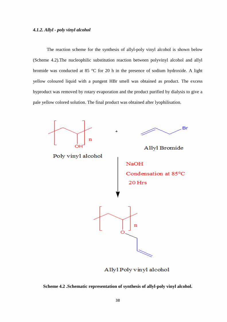
4.1.2. Allyl - poly vinyl alcohol
The reaction scheme for the synthesis of allyl-poly vinyl alcohol is shown below
(Scheme 4.2).The nucleophilic substitution reaction between polyvinyl alcohol and allyl
bromide was conducted at 85 °C for 20 h in the presence of sodium hydroxide. A light
yellow coloured liquid with a pungent HBr smell was obtained as product. The excess
byproduct was removed by rotary evaporation and the product purified by dialysis to give a
pale yellow colored solution. The final product was obtained after lyophilisation.
Scheme 4.2 .Schematic representation of synthesis of allyl-poly vinyl alcohol.
39
4.2. Characterization of pregel formulation
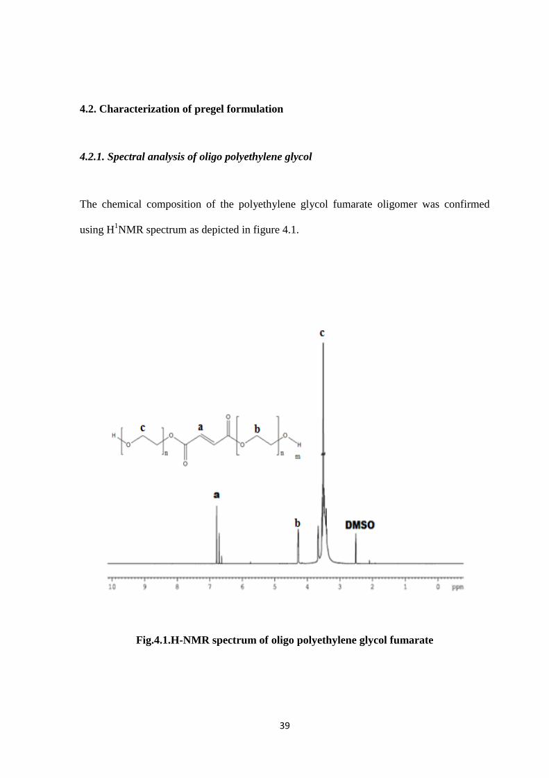
4.2.1. Spectral analysis of oligo polyethylene glycol
The chemical composition of the polyethylene glycol fumarate oligomer was confirmed
using H1NMR spectrum as depicted in figure 4.1.
Fig.4.1.H-NMR spectrum of oligo polyethylene glycol fumarate
40
The spectrum displayed peaks at 6.78 ppm and 6.706 ppm confirming the presence of
fumarate groups. The peaks at 3.40 - 4.29 ppm and 4.273 ppm correspond to the methylene
groups in PEG and fumarate group adjacent to PEG respectively.
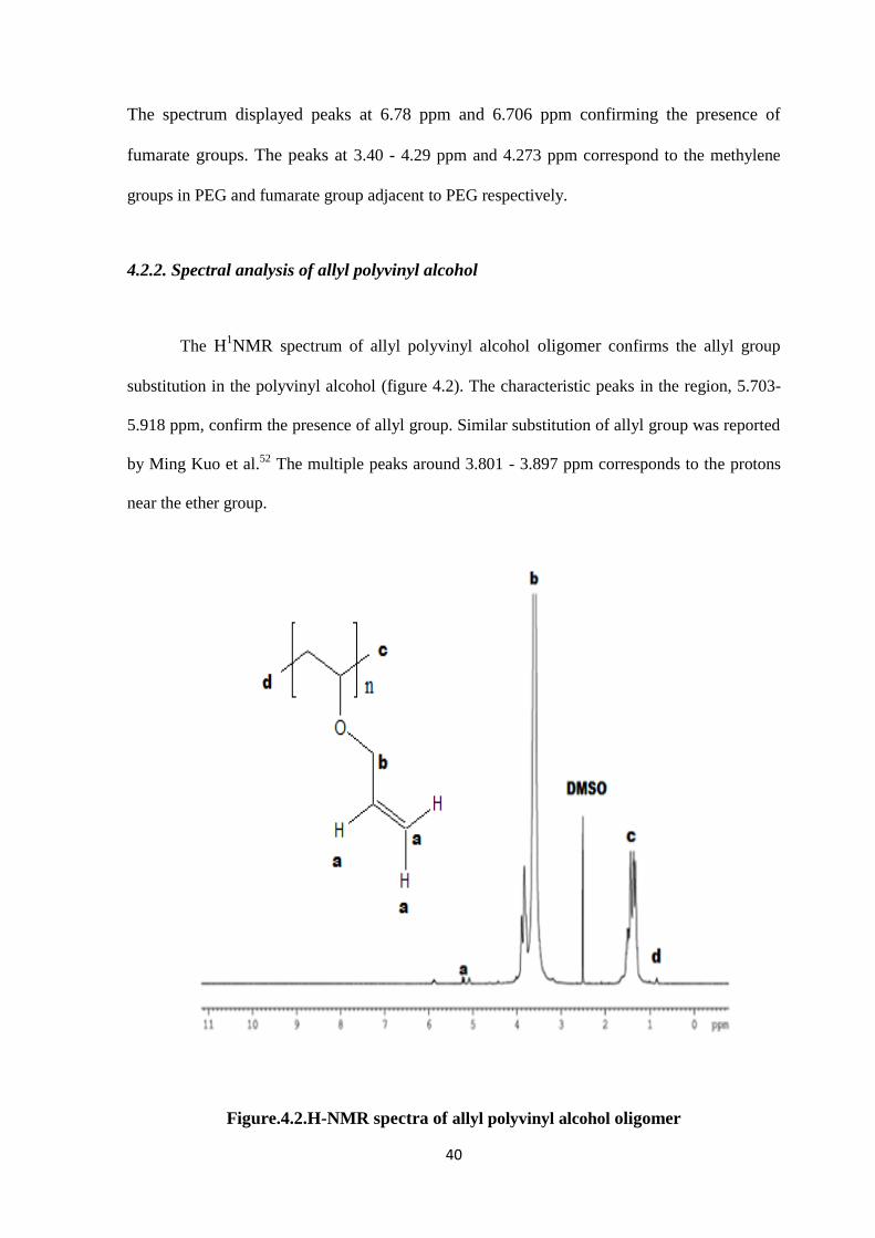
4.2.2. Spectral analysis of allyl polyvinyl alcohol
The H1NMR spectrum of allyl polyvinyl alcohol oligomer confirms the allyl group
substitution in the polyvinyl alcohol (figure 4.2). The characteristic peaks in the region, 5.703-
5.918 ppm, confirm the presence of allyl group. Similar substitution of allyl group was reported
by Ming Kuo et al.52 The multiple peaks around 3.801 - 3.897 ppm corresponds to the protons
near the ether group.
Figure.4.2.H-NMR spectra of allyl polyvinyl alcohol oligomer
41
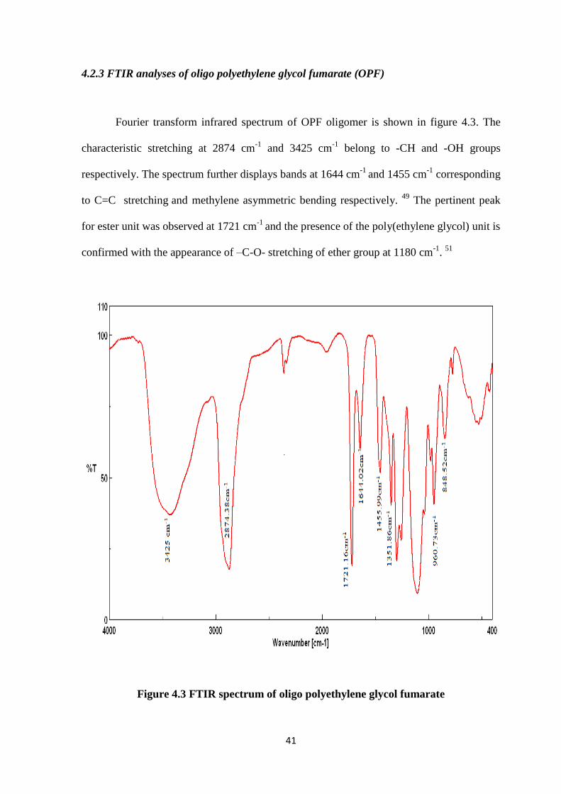
4.2.3 FTIR analyses of oligo polyethylene glycol fumarate (OPF)
Fourier transform infrared spectrum of OPF oligomer is shown in figure 4.3. The
characteristic stretching at 2874 cm-1
and 3425 cm-1
belong to -CH and -OH groups
respectively. The spectrum further displays bands at 1644 cm-1
and 1455 cm-1
corresponding
to C=C stretching and methylene asymmetric bending respectively.
49 The pertinent peak
for ester unit was observed at 1721 cm-1
and the presence of the poly(ethylene glycol) unit is
confirmed with the appearance of –C-O- stretching of ether group at 1180 cm-1
. 51
Figure 4.3 FTIR spectrum of oligo polyethylene glycol fumarate
42
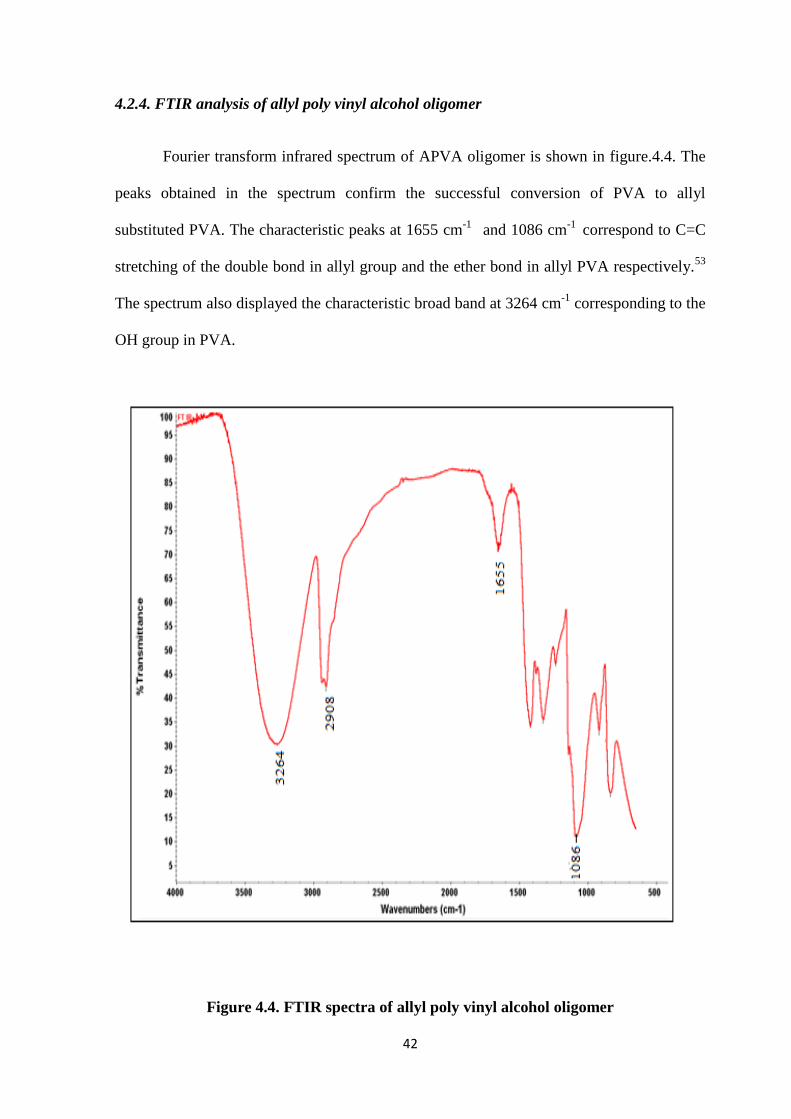
4.2.4. FTIR analysis of allyl poly vinyl alcohol oligomer
Fourier transform infrared spectrum of APVA oligomer is shown in figure.4.4. The
peaks obtained in the spectrum confirm the successful conversion of PVA to allyl
substituted PVA. The characteristic peaks at 1655 cm-1
and 1086 cm-1
correspond to C=C
stretching of the double bond in allyl group and the ether bond in allyl PVA respectively.53
The spectrum also displayed the characteristic broad band at 3264 cm-1
corresponding to the
OH group in PVA.
Figure 4.4. FTIR spectra of allyl poly vinyl alcohol oligomer
43
4.2.5. GPC analysis of oligomers
The molecular weight of oligomers was determined using GPC. The synthesized
OPF and APVA oligomers molecular weight values are given in Table 4.1. It showed
weight average molecular weight of 7438 with poly dispersity of 1.30 confirming oligomer
formation. The substitution of allyl group in the polyvinyl alcohol gives the number average
molecular weight of 11484 with poly dispersity of 2.91.
Table 4.1.Molecular weight data of oligomers
4.2.4. Viscosity of pregel formulations
The studies on viscosity of pregel formulations determined using a rotational
viscometer reveal viscosity in the range of 30-32 cp and 30-34 cp for pregel formulations of
OPF and APVA oligomers respectively.
Pregel resin Mn Mw MP Poly dispersity
Oligo PEG fumarate 5742 7438 6911 1.30
Allyl PVA 3949 11484 5791 2.91
44
4.3. Preparation of Hydrogels
4.3.1. OPF based hydrogels
OPF based hydrogels possess low protein absorption properties, minimal
inflammatory effect and good safety for in vivo uses. OPF hydrogels were prepared from
the synthesized OPF resin via free radical initiation with PEGDA crosslinker as shown in
(Table 4.2). In general, crosslinking of injectable hydrogels are initiated by physical
interaction between polymer chains or via chemical crosslinking such as disulphide bond
formation, Michael addition reaction etc. Compared to physical crosslinking, chemical
crosslinking gives improved mechanical properties and tunable degradation profiles. In this
experiment the OPF hydrogels were prepared using APS and TEMED as initiators.
Crosslinking of the hydrogels are initiated between the double bonds of fumarate groups
present in the OPF chains. Different concentrations of PEGDA crosslinker were used for
varying hydrogel characteristics (Table 4.2).
Table 4.2 OPF hydrogel formulations and setting characteristics
Hydrogels OPF
(30%solution)
PEGDA APS TEMED Setting
time
(min)
Setting
Temperature
(ºC)
OPFI 400 μL 6 mg 24
μL
8 μL 21 32
OPFII 400 μL 12 mg 24
μL
8 μL 8 32
OPFIII 400μL 16 mg 24
μL
8 μL 4 32
45
The representative image of the prepared OPF hydrogels with APS and TEMED as
initiators is shown in Figure. 4.5. The different percentage crosslinker incorporated
hydrogels showed different setting time at RT. It was observed that the setting time showed
an inverse relationship to the percentage of crosslinker used. The non-polarized double
bond of the fumarate ester group in OPF is far less reactive than acrylic ester and hence do
not undergo easily self-crosslinking. Hence, crosslinker molecules are typically added to
initiate crosslinking and reduce crosslinking time.54
The schematic representation of
crosslinking and formation of hydrogel is shown in the Scheme 4.3.
Figure 4.5. Prepared OPF hydrogels
46
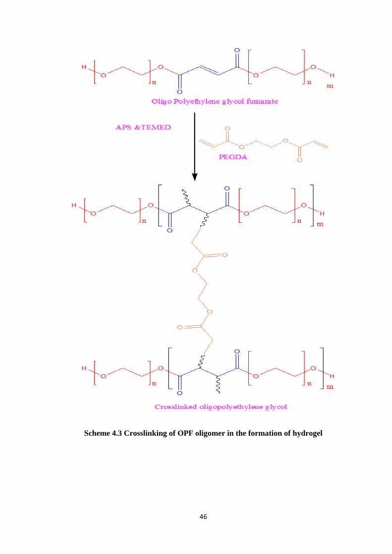
Scheme 4.3 Crosslinking of OPF oligomer in the formation of hydrogel
47
4.3.2. APVA based hydrogels
Poly vinyl alcohol based hydrogel have so many ocular applications because of its
excellent transparency property. Allyl-poly vinyl alcohol.oligomer based hydrogel
hydrogels were prepared using APVA resin by free radical polymerization with different
percentage PEGDA crosslinker (Table 4.3). The hydrogel setting time varies with different
percentage of crosslinker used. APVA hydrogels prepared with different percentage
crosslinker are represented as APVAI, APVAII, and APVAII (Table 4.3). The experimental
data revealed that the setting time showed an inverse relationship with the percentage of
crosslinker used.
Table 4.3. APVA hydrogel formulations and their setting characteristics
Chemical crosslinking gives good strength and desired gel strength. Hence APVA
hydrogels were prepared using 10% allyl PVA oligomer with different percentage of
PEGDA crosslinker (10%, 12%, 16%). APS and TEMED were used as initiators for gel
crosslinking and the hydrogels were prepared in 48 well cell culture plate. The schematic
Hydrogel Allyl PVA
(10%
Solution)
PEGDA APS TEMED Setting
time (min)
Setting
Temperature
(oC)
APVAI 400 μL 10 mg 24 μL 8 μL 3 32
APVAII 400 Μl 12 mg 24 μL 8 μL 2 32
APVAIII 400 Μl 16 mg 24 μL 8 μL 1 32
48
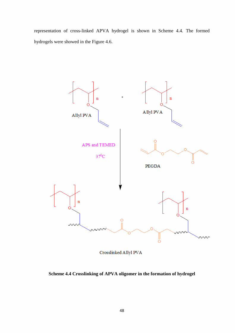
representation of cross-linked APVA hydrogel is shown in Scheme 4.4. The formed
hydrogels were showed in the Figure 4.6.
Scheme 4.4 Crosslinking of APVA oligomer in the formation of hydrogel
49
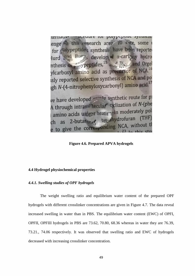
Figure 4.6. Prepared APVA hydrogels
4.4 Hydrogel physiochemical properties
4.4.1. Swelling studies of OPF hydrogels
The weight swelling ratio and equilibrium water content of the prepared OPF
hydrogels with different crosslinker concentrations are given in Figure 4.7. The data reveal
increased swelling in water than in PBS. The equilibrium water content (EWC) of OPFI,
OPFII, OPFIII hydrogels in PBS are 73.62, 70.80, 68.36 whereas in water they are 76.39,
73.21., 74.06 respectively. It was observed that swelling ratio and EWC of hydrogels
decreased with increasing crosslinker concentration.
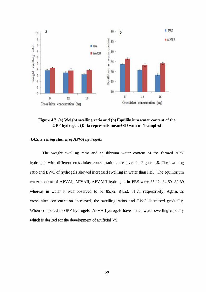
50
Figure 4.7. (a) Weight swelling ratio and (b) Equilibrium water content of the
OPF hydrogels (Data represents mean+SD with n=4 samples)
4.4.2. Swelling studies of APVA hydrogels
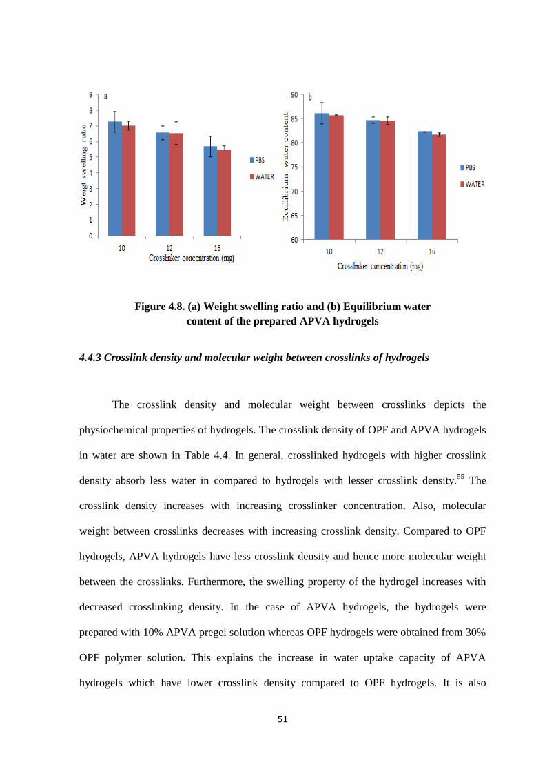
The weight swelling ratio and equilibrium water content of the formed APV
hydrogels with different crosslinker concentrations are given in Figure 4.8. The swelling
ratio and EWC of hydrogels showed increased swelling in water than PBS. The equilibrium
water content of APVAI, APVAII, APVAIII hydrogels in PBS were 86.12, 84.69, 82.39
whereas in water it was observed to be 85.72, 84.52, 81.71 respectively. Again, as
crosslinker concentration increased, the swelling ratios and EWC decreased gradually.
When compared to OPF hydrogels, APVA hydrogels have better water swelling capacity
which is desired for the development of artificial VS.
51
Figure 4.8. (a) Weight swelling ratio and (b) Equilibrium water
content of the prepared APVA hydrogels
4.4.3 Crosslink density and molecular weight between crosslinks of hydrogels
The crosslink density and molecular weight between crosslinks depicts the
physiochemical properties of hydrogels. The crosslink density of OPF and APVA hydrogels
in water are shown in Table 4.4. In general, crosslinked hydrogels with higher crosslink
density absorb less water in compared to hydrogels with lesser crosslink density.55
The
crosslink density increases with increasing crosslinker concentration. Also, molecular
weight between crosslinks decreases with increasing crosslink density. Compared to OPF
hydrogels, APVA hydrogels have less crosslink density and hence more molecular weight
between the crosslinks. Furthermore, the swelling property of the hydrogel increases with
decreased crosslinking density. In the case of APVA hydrogels, the hydrogels were
prepared with 10% APVA pregel solution whereas OPF hydrogels were obtained from 30%
OPF polymer solution. This explains the increase in water uptake capacity of APVA
hydrogels which have lower crosslink density compared to OPF hydrogels. It is also
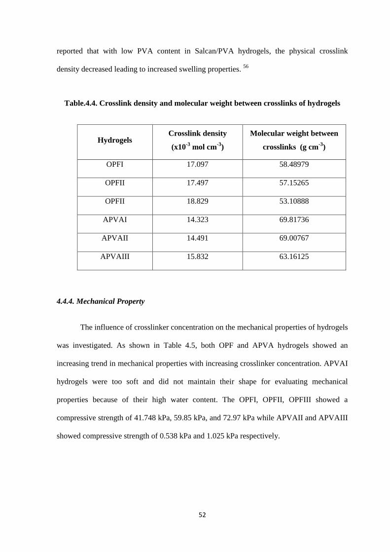
52
reported that with low PVA content in Salcan/PVA hydrogels, the physical crosslink
density decreased leading to increased swelling properties. 56
Table.4.4. Crosslink density and molecular weight between crosslinks of hydrogels
Hydrogels Crosslink density
(x10-3
mol cm-3
)
Molecular weight between
crosslinks (g cm-3
)
OPFI 17.097 58.48979
OPFII 17.497 57.15265
OPFII 18.829 53.10888
APVAI 14.323 69.81736
APVAII 14.491 69.00767
APVAIII 15.832 63.16125
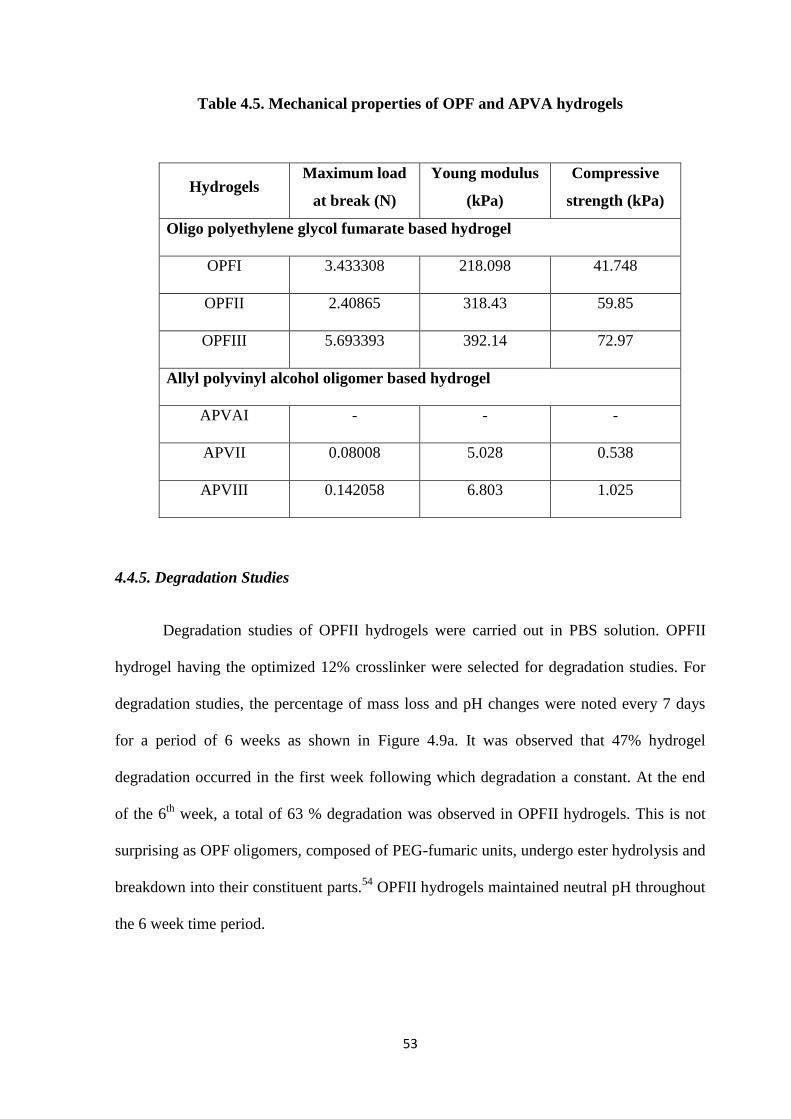
4.4.4. Mechanical Property
The influence of crosslinker concentration on the mechanical properties of hydrogels
was investigated. As shown in Table 4.5, both OPF and APVA hydrogels showed an
increasing trend in mechanical properties with increasing crosslinker concentration. APVAI
hydrogels were too soft and did not maintain their shape for evaluating mechanical
properties because of their high water content. The OPFI, OPFII, OPFIII showed a
compressive strength of 41.748 kPa, 59.85 kPa, and 72.97 kPa while APVAII and APVAIII
showed compressive strength of 0.538 kPa and 1.025 kPa respectively.
53
Table 4.5. Mechanical properties of OPF and APVA hydrogels
Hydrogels Maximum load
at break (N)
Young modulus
(kPa)
Compressive
strength (kPa)
Oligo polyethylene glycol fumarate based hydrogel
OPFI 3.433308 218.098 41.748
OPFII 2.40865 318.43 59.85
OPFIII 5.693393 392.14 72.97
Allyl polyvinyl alcohol oligomer based hydrogel
APVAI - - -
APVII 0.08008 5.028 0.538
APVIII 0.142058 6.803 1.025
4.4.5. Degradation Studies
Degradation studies of OPFII hydrogels were carried out in PBS solution. OPFII
hydrogel having the optimized 12% crosslinker were selected for degradation studies. For
degradation studies, the percentage of mass loss and pH changes were noted every 7 days
for a period of 6 weeks as shown in Figure 4.9a. It was observed that 47% hydrogel
degradation occurred in the first week following which degradation a constant. At the end
of the 6th
week, a total of 63 % degradation was observed in OPFII hydrogels. This is not
surprising as OPF oligomers, composed of PEG-fumaric units, undergo ester hydrolysis and
breakdown into their constituent parts.54
OPFII hydrogels maintained neutral pH throughout
the 6 week time period.

54
Figure 4.9. Variation of mass (a) and pH (b) of the OPFII hydrogels during aging
55
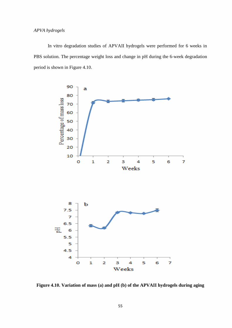
APVA hydrogels
In vitro degradation studies of APVAII hydrogels were performed for 6 weeks in
PBS solution. The percentage weight loss and change in pH during the 6-week degradation
period is shown in Figure 4.10.
Figure 4.10. Variation of mass (a) and pH (b) of the APVAII hydrogels during aging
56
The pH variation during the degradation period was around neutral pH ranging
between pH 6.3 to 7.8. Degradation profiles of APVAII hydrogels revealed 70% weight loss
during the first week following which the rate remained constant until the end of the 6th
week.
4.4.6. Surface Morphology
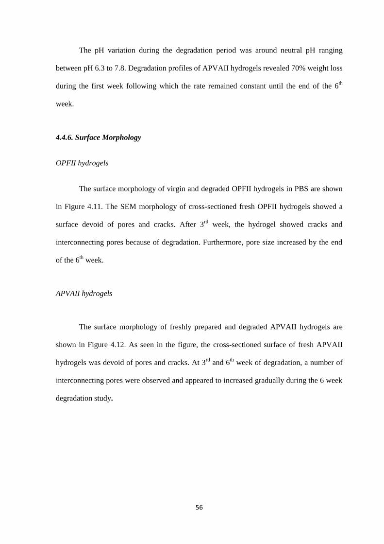
OPFII hydrogels
The surface morphology of virgin and degraded OPFII hydrogels in PBS are shown
in Figure 4.11. The SEM morphology of cross-sectioned fresh OPFII hydrogels showed a
surface devoid of pores and cracks. After 3rd
week, the hydrogel showed cracks and
interconnecting pores because of degradation. Furthermore, pore size increased by the end
of the 6th
week.
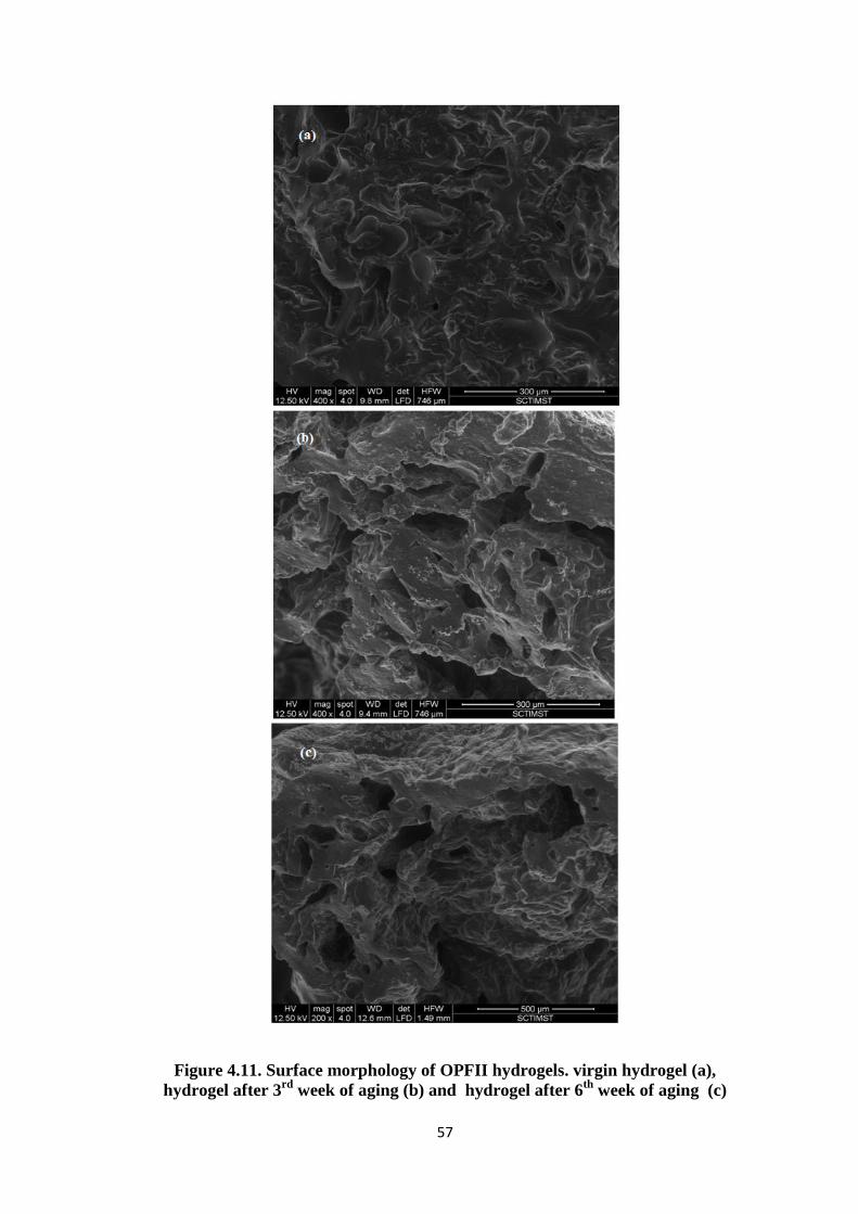
APVAII hydrogels
The surface morphology of freshly prepared and degraded APVAII hydrogels are
shown in Figure 4.12. As seen in the figure, the cross-sectioned surface of fresh APVAII
hydrogels was devoid of pores and cracks. At 3rd
and 6th
week of degradation, a number of
interconnecting pores were observed and appeared to increased gradually during the 6 week
degradation study.
57
Figure 4.11. Surface morphology of OPFII hydrogels. virgin hydrogel (a),
hydrogel after 3rd
week of aging (b) and hydrogel after 6th
week of aging (c)
58
Figure 4.12. Surface morphology of APVAII hydrogels. virgin hydrogel (a),
hydrogel after 3rd
week of aging (b) and hydrogel after 6th
week of aging (c)
59
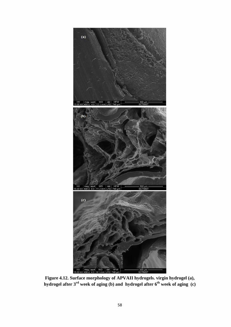
4.5 .Thermal properties of hydrogel
4.5.1.DSC analysis of hydrogels
The property of water in hydrogels was analyzed by thermal analysis. The DSC
curves of the OPFII and APVAII hydrogels are shown in Figure 4.13.
Figure 4.13. DSC curve of prepared hydrogels. (a) OPFII and (b) APVAII.
60
In the polymer network, water exists in more than two states, bound water and
unbound water. The water molecules absorbed in the hydrophilic region are present in three
forms, non-freezable bound water, freezable bound water, and free water. The non-freezable
bound water is tightly hydrogen bonded water with undetectable phase transition. Freezable
bound water interact weakly with the hydrogel molecule whereas free water does not
interact in hydrogen bonding and hence both are categorized as freezable water.57
In OPFII
hydrogels, freezable water content was 67.305 % whereas no freezable water content was
5.95% (Table 4.6). In the case of APVAII hydrogels, freezable water content was 73.35%
and non-freezable water content was 11.34%. The low free water content indicates good
hydrophilic characteristics of the prepared hydrogels.
Table 4.6. DSC analysis of hydrogels
Properties OPFII APVAII
Enthalpy of melting of freezing water (J/g) 224.8 245
On set of crystallization of freezing water (˚C) -2.44 -2.99
Equillibrium water content(%) 73.21 85.42
Freezable free water content (%) 67.305 73.35
Non-freezable water content (%) 5.95 11.34
4.5.2.TG analysis of hydrogels
TG curve of OPFII hydrogel is given in Figure 4.14. The curve; shows a two stage
decomposition with significant mass losses obtained at 130 0C and 342
0C respectively. The
decomposition temperature of OPFII was around 429 0C, as obtained in OPF/sodium
methacrylate hydrogel systems58
.
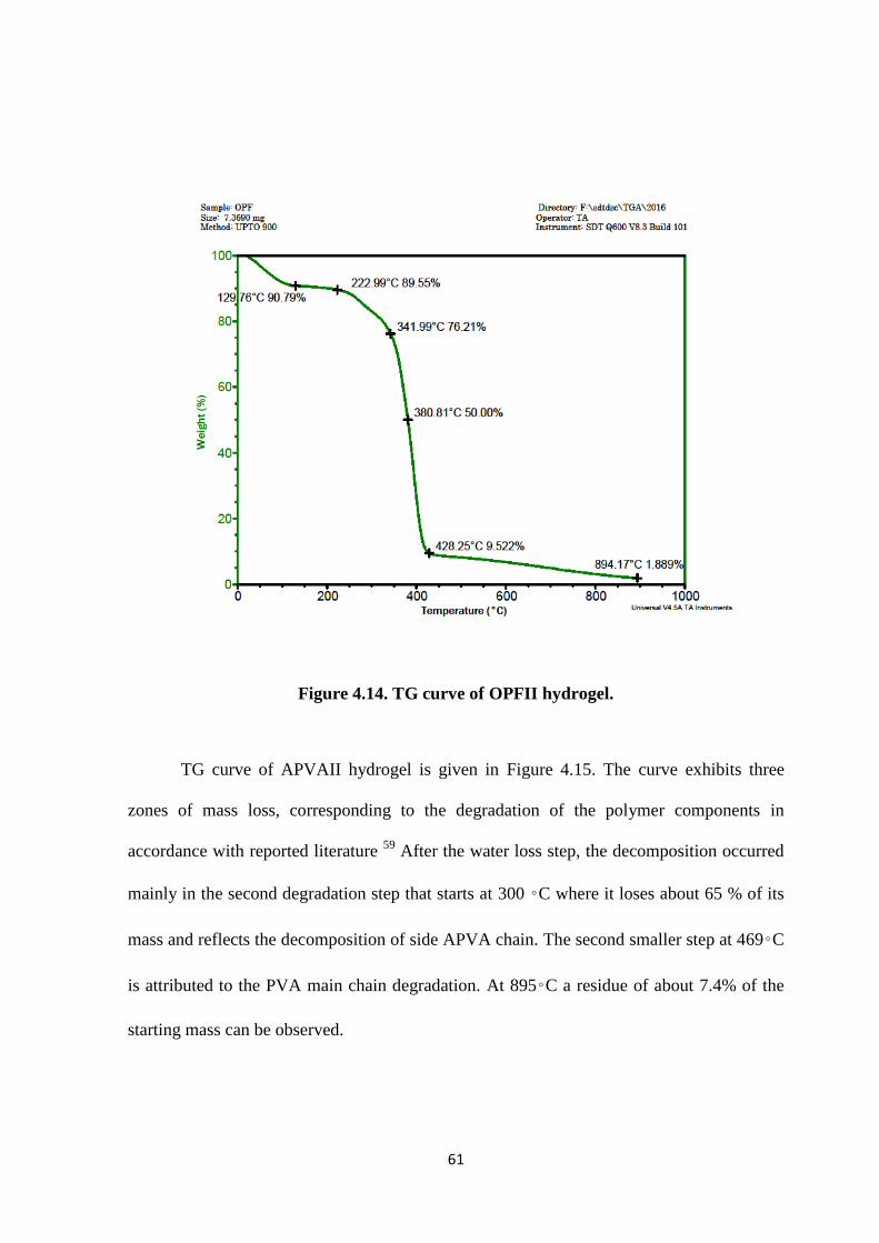
61
Figure 4.14. TG curve of OPFII hydrogel.
TG curve of APVAII hydrogel is given in Figure 4.15. The curve exhibits three
zones of mass loss, corresponding to the degradation of the polymer components in
accordance with reported literature 59
After the water loss step, the decomposition occurred
mainly in the second degradation step that starts at 300 ◦C where it loses about 65 % of its
mass and reflects the decomposition of side APVA chain. The second smaller step at 469◦C
is attributed to the PVA main chain degradation. At 895◦C a residue of about 7.4% of the
starting mass can be observed.
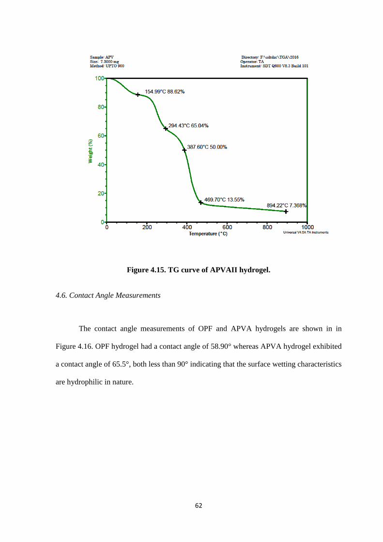
62
Figure 4.15. TG curve of APVAII hydrogel.
4.6. Contact Angle Measurements
The contact angle measurements of OPF and APVA hydrogels are shown in in
Figure 4.16. OPF hydrogel had a contact angle of 58.90° whereas APVA hydrogel exhibited
a contact angle of 65.5°, both less than 90° indicating that the surface wetting characteristics
are hydrophilic in nature.
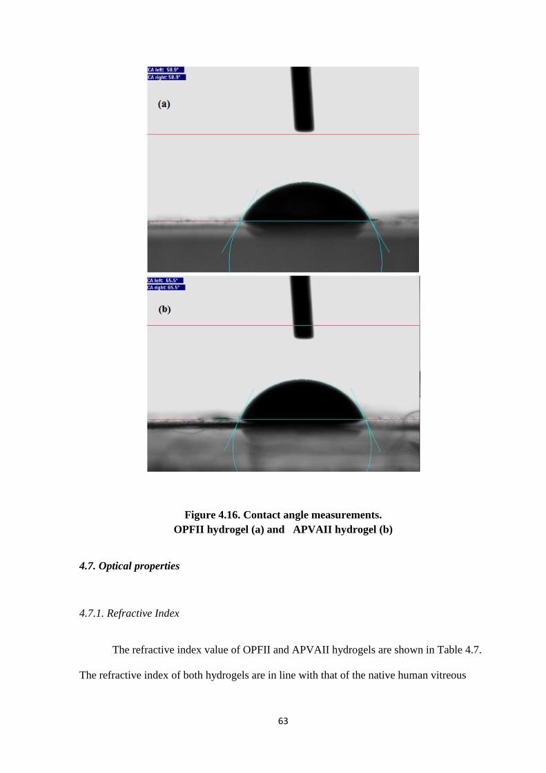
63
Figure 4.16. Contact angle measurements.
OPFII hydrogel (a) and APVAII hydrogel (b)
4.7. Optical properties
4.7.1. Refractive Index
The refractive index value of OPFII and APVAII hydrogels are shown in Table 4.7.
The refractive index of both hydrogels are in line with that of the native human vitreous
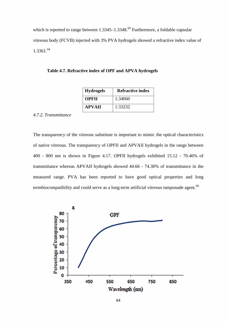
64
which is reported to range between 1.3345–1.3348.60
Furthermore, a foldable capsular
vitreous body (FCVB) injected with 3% PVA hydrogels showed a refractive index value of
1.3361.44
Table 4.7. Refractive index of OPF and APVA hydrogels
Hydrogels Refractive index
OPFII 1.34060
APVAII 1.33232
4.7.2. Transmittance
The transparency of the vitreous substitute is important to mimic the optical characteristics
of native vitreous. The transparency of OPFII and APVAII hydrogels in the range between
400 - 800 nm is shown in Figure 4.17. OPFII hydrogels exhibited 15.12 - 70.46% of
transmittance whereas APVAII hydrogels showed 44.66 - 74.30% of transmittance in the
measured range. PVA has been reported to have good optical properties and long
termbiocompatibility and could serve as a long-term artificial vitreous tamponade agent.60
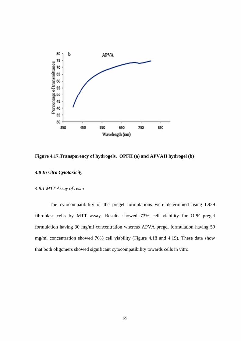
65
Figure 4.17.Transparency of hydrogels. OPFII (a) and APVAII hydrogel (b)
4.8 In vitro Cytotoxicity
4.8.1 MTT Assay of resin
The cytocompatibility of the pregel formulations were determined using L929
fibroblast cells by MTT assay. Results showed 73% cell viability for OPF pregel
formulation having 30 mg/ml concentration whereas APVA pregel formulation having 50
mg/ml concentration showed 76% cell viability (Figure 4.18 and 4.19). These data show
that both oligomers showed significant cytocompatibility towards cells in vitro.
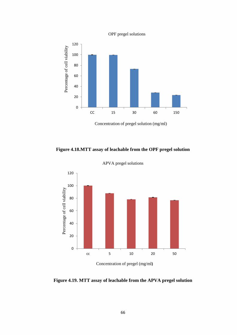
66
Figure 4.18.MTT assay of leachable from the OPF pregel solution
Figure 4.19. MTT assay of leachable from the APVA pregel solution
0
20
40
60
80
100
120
cc 5 10 20 50
Per
centa
ge
of
cell
via
bil
ity
Concentration of pregel (mg/ml)
APVA pregel solutions
0
20
40
60
80
100
120
CC 15 30 60 150
Per
cen
tage
of
cell
via
bil
ity
Concentration of pregel solution (mg/ml)
OPF pregel solutions
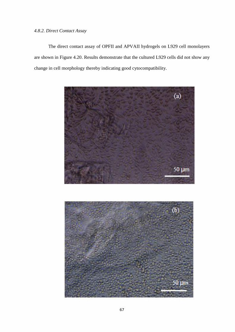
67
4.8.2. Direct Contact Assay
The direct contact assay of OPFII and APVAII hydrogels on L929 cell monolayers
are shown in Figure 4.20. Results demonstrate that the cultured L929 cells did not show any
change in cell morphology thereby indicating good cytocompatibility.
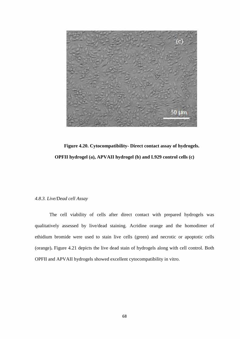
68
Figure 4.20. Cytocompatibility- Direct contact assay of hydrogels.
OPFII hydrogel (a), APVAII hydrogel (b) and L929 control cells (c)
4.8.3. Live/Dead cell Assay
The cell viability of cells after direct contact with prepared hydrogels was
qualitatively assessed by live/dead staining. Acridine orange and the homodimer of
ethidium bromide were used to stain live cells (green) and necrotic or apoptotic cells
(orange). Figure 4.21 depicts the live dead stain of hydrogels along with cell control. Both
OPFII and APVAII hydrogels showed excellent cytocompatibility in vitro.
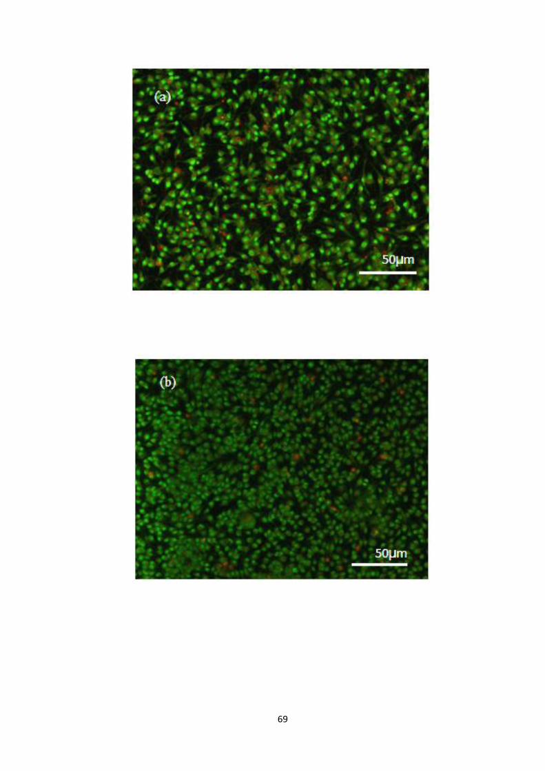
69
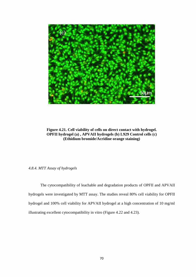
70
Figure 4.21. Cell viability of cells on direct contact with hydrogel.
OPFII hydrogel (a) , APVAII hydrogels (b) L929 Control cells (c)
(Ethidium bromide/Acridine orange staining)
4.8.4. MTT Assay of hydrogels
The cytocompatibility of leachable and degradation products of OPFII and APVAII
hydrogels were investigated by MTT assay. The studies reveal 80% cell viability for OPFII
hydrogel and 100% cell viability for APVAII hydrogel at a high concentration of 10 mg/ml
illustrating excellent cytocompatibility in vitro (Figure 4.22 and 4.23).
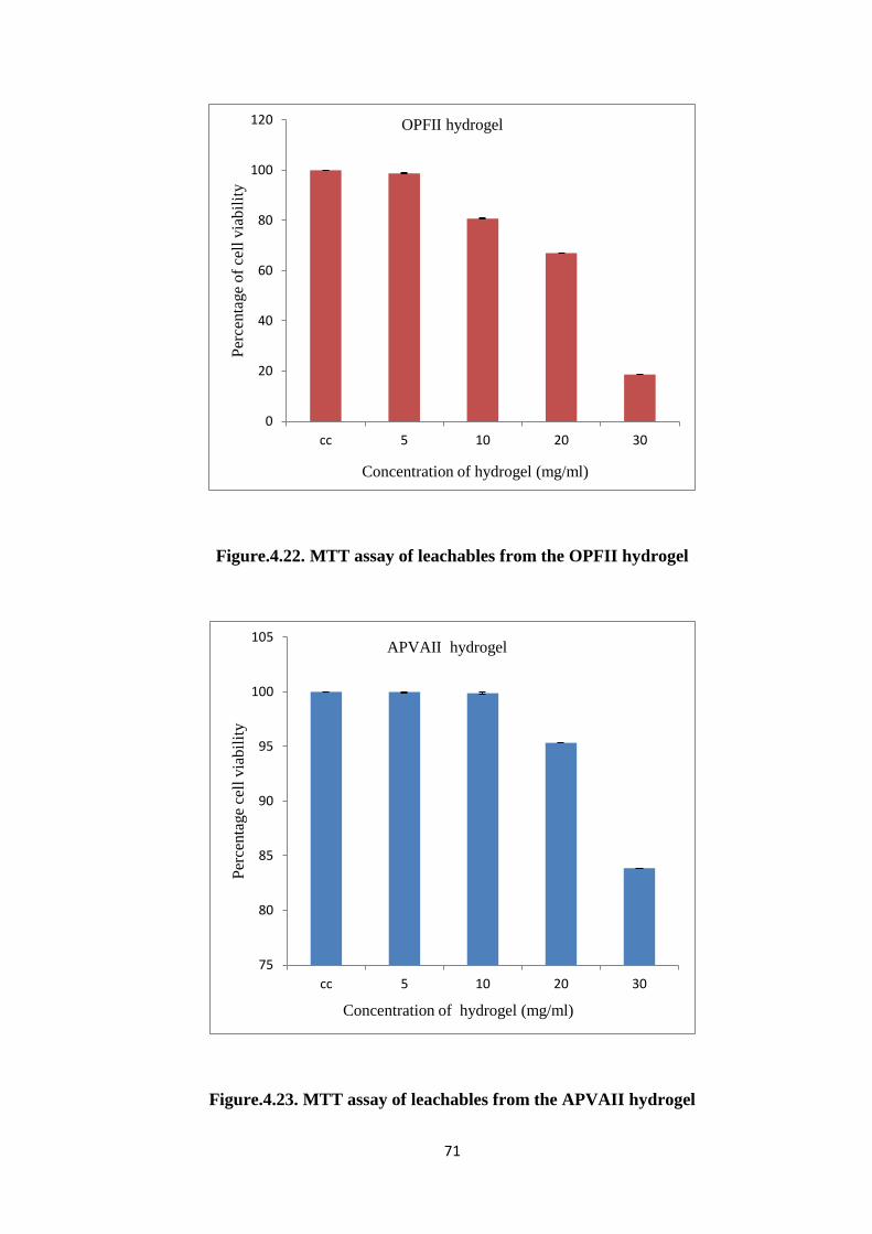
71
Figure.4.22. MTT assay of leachables from the OPFII hydrogel
Figure.4.23. MTT assay of leachables from the APVAII hydrogel
0
20
40
60
80
100
120
cc 5 10 20 30
Per
cen
tage
of
cell
via
bil
ity
Concentration of hydrogel (mg/ml)
OPFII hydrogel
75
80
85
90
95
100
105
cc 5 10 20 30
Per
centa
ge
cell
via
bil
ity
Concentration of hydrogel (mg/ml)
APVAII hydrogel
72
Chapter V
SUMMARY AND CONCLUSION
Vitreous replacement is a challenging and complex problem in clinical
ophthalmology. We report syntheses and characterization of in situ cross linkable PEG-
based and PVA-based pregel formulation and their comparison for potential use as
injectable hydrogel for potential vitreous replacement. Water soluble OPF was synthesized
by condensing PEG and fumaryl chloride. In the case of APVA synthesis, the -OH group of
PVA underwent a nucleophilic substitution reaction with allyl bromide. The FTIR and 1H
NMR analyses revealed the formation of OPF resin with functional fumarate groups that
can facilitate crosslinks to form a 3-D network. In the case of APVA gels, FTIR and 1H
NMR confirmed the successful substitution of allyl group into the PVA polymer chain
through the presence of double bonds. The determination of molecular weight and
polydispersity index of synthesized OPF and APVA via GPC confirmed the oligomer
nature.
Polymer hydrogels were prepared using different concentration of PEGDA cross-
linker with APS and TEMED as inititators. Both OPF and APVA hydrogels gave the
73
optimal setting time at RT showing great promise for in situ injectable application. The
swelling studies of OPF and APVA hydrogels in water and PBS showed comparatively high
swelling in water. The mechanical properties of OPF and APVA hydrogels showed
increasing trend of compressive strength and Young’s modulus with increasing PEGDA
concentration.
Hydrogel degradation studies showed a mass loss of around 60 - 70% for both
OPFII and APVAII hydrogels at the end of the 6-week period. ESEM images of hydrogels
aged in PBS medium confirmed the presence of interconnecting pores and increase in pore
sizes with increase in aging duration. DSC curves of both OPFII and APVAII showed free
water content of 67.302% and 73.35% demonstrating the increased hydrophilic character of
the both hydrogels. Furthermore, contact angle measurement displayed an angle < 75ᵒ
thereby confirming the surface wettability of the hydrogels. In addition, both OPFII and
APVAII hydrogels showed a refractive index close to ~1.33 which falls close to that of the
native vitreous. OPF hydrogels exhibited 15.12 - 70.46% of transmittance whereas APVAII
hydrogels showed 44.66 - 74.30% of transmittance in the measured range. The transparency
data of these hydrogels is promising for potential use in vitreous application.
The in vitro cytotoxicity of pregel formulations was performed on L929 fibroblast
cells. OPF demonstrated 73% cell viability at a high concentration of 30 mg/ml whereas
APVA showed 76% cell viability at a high concentration of 50 mg/ml after 24 h incubation.
The direct contact assay of both OPFII and APVAII hydrogels showed good cell
compatibility with no difference in morphology compared to cell controls after 24 h. Live
dead assay on cells after direct contact assay showed very few apoptotic cells for both
OPFII and APVAII hydrogels. Furthermore, MTT assay on leachables from prepared
hydrogels showed > 80% cell viability for OPFII hydrogels and 100% cell viability for
APVAII hydrogels at a high concentration of 10 mg/ml.
74
Taken together, our results show that both PEG-based and PVA-based hydrogels
prepared in this work exhibited relevant physical, optical and biological characteristics for
use as an in situ forming injectable artificial VS system.
Future studies will aim at employing non-degradable crosslinkers to reduce the
degradation of prepared hydrogels thereby prolonging its lifetime which will bring it a step
closer towards clinical application as a long-term artificial VS.
75
References
1. The Physics of Polymers - Concepts for Understanding Their | Gert R. Strobl | Springer.
Available at: http://www.springer.com/fr/book/9783540252788. (Accessed: 10th June
2016)
2. Biomaterials A. Binnaz Hazar Yoruç1 and B. Cem Şener2 1Yıldız Technical
University, Science and Technology Application and Research Center, 2Marmara
University, Faculty of Dentistry, Department of Oral and Maxillofacial Surgery,
57.795j0j8&sourceid=chrome&ie=UTF-8.
3. Polymer Science and Technology - CRC Press Book. Available at:
https://www.crcpress.com/Polymer-Science-and-
Technology/Ebewele/p/book/9780849389399. (Accessed: 10th June 2016)
4. Lyu, S. & Untereker, D. Degradability of Polymers for Implantable Biomedical
Devices. Int. J. Mol. Sci. 10, 4033–4065 (2009).
5. Ulery, B. D., Nair, L. S. & Laurencin, C. T. Biomedical Applications of Biodegradable
Polymers. J. Polym. Sci. Part B Polym. Phys. 49, 832–864 (2011).
6. Pritchard, C. D. et al. Evaluation of viscoelastic poly(ethylene glycol) sols as vitreous
substitutes in an experimental vitrectomy model in rabbits. Acta Biomater. 7, 936–943
(2011).
7. Azad, S. V. Vitreous Substitutes. Delhi J. Ophthalmol. 23, 9–13 (2012).
8. Foster, W. J. Vitreous Substitutes. Expert Rev. Ophthalmol. 3, 211–218 (2008).
9. Sebag, J. in The Vitreous and Vitreoretinal Interface (eds. M.D, C. L. S. & M.D, A. N.)
37–57 (Springer New York, 1987).
10. Coleman, D. J. Unified model for accommodative mechanism. Am. J. Ophthalmol. 69,
1063–1079 (1970).
11. Sebag, J. Imaging vitreous. Eye 16, 429–439 (2002).
76
12. Shui, Y.-B. et al. The gel state of the vitreous and ascorbate-dependent oxygen
consumption: relationship to the etiology of nuclear cataracts. Arch. Ophthalmol. Chic.
Ill 1960 127, 475–482 (2009).
13. Hogan, M. J. The vitreous,its structure, and relation to the ciliary body and retina.
proctor award lecture. Invest. Ophthalmol. 2, 418–445 (1963).
14. Heegaard, S., Jensen, O. A. & Prause, J. U. Structure and composition of the inner
limiting membrane of the retina. SEM on frozen resin-cracked and enzyme-digested
retinas of Macaca mulatta. Graefes Arch. Clin. Exp. Ophthalmol. Albrecht Von Graefes
Arch. Für Klin. Exp. Ophthalmol. 224, 355–360 (1986).
15. Ali, S. & Bettelheim, F. A. Distribution of freezable and non-freezable water in bovine
vitreous. Curr. Eye Res. 3, 1233–1239 (1984).
16. Wong, R. W., Richa, D. C., Hahn, P., Green, W. R. & Dunaief, J. L. Iron toxicity as a
potential factor in AMD. Retina Phila. Pa 27, 997–1003 (2007).
17. Bos, K. J. et al. Axial structure of the heterotypic collagen fibrils of vitreous humour
and cartilage. J. Mol. Biol. 306, 1011–1022 (2001).
18. Theocharis, D. A. et al. Hyaluronan and chondroitin sulfate proteoglycans in the
supramolecular organization of the mammalian vitreous body. Connect. Tissue Res. 49,
124–128 (2008).
19. Reddy, T. S., Birkle, D. L., Packer, A. J., Dobard, P. & Bazan, N. G. Fatty acid
composition and arachidonic acid metabolism in vitreous lipids from canine and human
eyes. Curr. Eye Res. 5, 441–447 (1986).
20. Sebag, J. Anatomy and pathology of the vitreo-retinal interface. Eye 6, 541–552 (1992).
21. Sebag, J. & Balazs, E. A. Morphology and ultrastructure of human vitreous fibers.
Invest. Ophthalmol. Vis. Sci. 30, 1867–1871 (1989).
77
22. Stay, M. S., Xu, J., Randolph, T. W. & Barocas, V. H. Computer simulation of
convective and diffusive transport of controlled-release drugs in the vitreous humor.
Pharm. Res. 20, 96–102 (2003).
23. Kleinberg, T. T., Tzekov, R. T., Stein, L., Ravi, N. & Kaushal, S. Vitreous substitutes: a
comprehensive review. Surv. Ophthalmol. 56, 300–323 (2011).
24. Nickerson, C. S., Park, J., Kornfield, J. A. & Karageozian, H. Rheological properties of
the vitreous and the role of hyaluronic acid. J. Biomech. 41, 1840–1846 (2008).
25. Los, L. I., van der Worp, R. J., van Luyn, M. J. A. & Hooymans, J. M. M. Age-related
liquefaction of the human vitreous body: LM and TEM evaluation of the role of
proteoglycans and collagen. Invest. Ophthalmol. Vis. Sci. 44, 2828–2833 (2003).
26. Colthurst, M. J., Williams, R. L., Hiscott, P. S. & Grierson, I. Biomaterials used in the
posterior segment of the eye. Biomaterials 21, 649–665 (2000).
27. Peyman, G. A., Schulman, J. A. & Sullivan, B. Perfluorocarbon liquids in
ophthalmology. Surv. Ophthalmol. 39, 375–395 (1995).
28. Kim, R. W. & Baumal, C. Anterior segment complications related to vitreous
substitutes. Ophthalmol. Clin. N. Am. 17, 569–576, vi (2004).
29. Peyman, G. A., Schulman, J. A. & Sullivan, B. Perfluorocarbon liquids in
ophthalmology. Surv. Ophthalmol. 39, 375–395 (1995).
30. Orzalesi, N., Migliavacca, L., Bottoni, F. & Miglior, S. Experimental short-term
tolerance to perfluorodecalin in the rabbit eye: a histopathological study. Curr. Eye Res.
17, 828–835 (1998).
31. Zeana, D., Becker, J., Kuckelkorn, R. & Kirchhof, B. Perfluorohexyloctane as a long-
term vitreous tamponade in the experimental animal. Experimental
perfluorohexyloctane substitution. Int. Ophthalmol. 23, 17–24 (1999).
78
32. Chirila, T. V., Tahija, S., Hong, Y., Vijayasekaran, S. & Constable, I. J. Synthetic
polymers as materials for artificial vitreous body: review and recent advances. J.
Biomater. Appl. 9, 121–137 (1994).
33. Yu, L. & Ding, J. Injectable hydrogels as unique biomedical materials. Chem. Soc. Rev.
37, 1473–1481 (2008).
34. Zhao, X. Multi-scale Multi-mechanism Design of Tough Hydrogels: Building
Dissipation into Stretchy Networks. Soft Matter 10, 672–687 (2014).
35. Tan, H. & Marra, K. G. Injectable, Biodegradable Hydrogels for Tissue Engineering
Applications. Materials 3, 1746–1767 (2010).
36. Kurt H. Stenzel, M. W. D. Collagen Gels: Design for a Vitreous Replacement. Science
164, 1282–1283 (1969).
37. Pruett RC, Calabria GA & Schepens CL. Collagen vitreous substitute: I. experimental
study. Arch. Ophthalmol. 88, 540–543 (1972).
38. Nakagawa, M., Tanaka, M. & Miyata, T. Evaluation of Collagen Gel and Hyaluronic
Acid as Vitreous Substitutes. Ophthalmic Res. 29, 409–420 (1997).
39. Lai, J.-Y. Biocompatibility of chemically cross-linked gelatin hydrogels for ophthalmic
use. J. Mater. Sci. Mater. Med. 21, 1899–1911 (2010).
40. Pruett, R. C., Schepens, C. L. & Swann, D. A. Hyaluronic acid vitreous substitute. A
six-year clinical evaluation. Arch. Ophthalmol. Chic. Ill 1960 97, 2325–2330 (1979).
41. Suri, S. & Banerjee, R. In vitro evaluation of in situ gels as short term vitreous
substitutes. J. Biomed. Mater. Res. A 79A, 650–664 (2006).
42. Baino, F. The Use of Polymers in the Treatment of Retinal Detachment: Current Trends
and Future Perspectives. Polymers 2, 286–322 (2010).
79
43. Cavalieri, F., Miano, F., D’Antona, P. & Paradossi, G. Study of gelling behavior of
poly(vinyl alcohol)-methacrylate for potential utilizations in tissue replacement and
drug delivery. Biomacromolecules 5, 2439–2446 (2004).
44. Feng, S. et al. A Novel Vitreous Substitute of Using a Foldable Capsular Vitreous Body
Injected with Polyvinylalcohol Hydrogel. Sci. Rep. 3, (2013).
45. Davidorf, F. H. et al. Ocular toxicity of vitreal pluronic polyol F-127. Retina Phila. Pa
10, 297–300 (1990).
46. Katagiri, Y. et al. Application of thermo-setting gel as artificial vitreous. Jpn. J.
Ophthalmol. 49, 491–496 (2005).
47. Su, X. et al. Recent Progress in Using Biomaterials as Vitreous Substitutes.
Biomacromolecules 16, 3093–3102 (2015).
48. Lirong He, D. S. Toward Self-Healing Hydrogels Using One-Pot Thiol–Ene Click and
Borax-Diol Chemistry. ACS Macro Lett. 4, 673–678 (2015).
49. Wang, S., Lu, L., Gruetzmacher, J. A., Currier, B. L. & Yaszemski, M. J. Synthesis and
characterizations of biodegradable and crosslinkable poly(epsilon-caprolactone
fumarate), poly(ethylene glycol fumarate), and their amphiphilic copolymer.
Biomaterials 27, 832–841 (2006).
50. Krishna, L. & Jayabalan, M. Synthesis and characterization of biodegradable poly
(ethylene glycol) and poly (caprolactone diol) end capped poly (propylene fumarate)
cross linked amphiphilic hydrogel as tissue engineering scaffold material. J. Mater. Sci.
Mater. Med. 20, 115–122 (2008).
51. Shin, H., Temenoff, J. S. & Mikos, A. G. In vitro cytotoxicity of unsaturated
oligo[poly(ethylene glycol) fumarate] macromers and their cross-linked hydrogels.
Biomacromolecules 4, 552–560 (2003).
80
52. Kuo, S.-M., Liou, C.-C., Chuang, C.-L. & Wang, Y.-J. Studies of the effects of PVA-
AE on dentinal bonding of HEMA. ResearchGate 21, (2001).
53. Kuo, S. M., Liou, C. C., Chang, S. J. & Wang, Y.-J. Synthesis and characterizations of
hydrogel based on PVA-AE and HEMA. J. Polym. Res. 8, 169–174 (2001).
54. Kinard, L. A., Kasper, F. K. & Mikos, A. G. Synthesis of oligo(poly(ethylene glycol)
fumarate). Nat. Protoc. 7, 1219–1227 (2012).
55. Balakrishnan, B., Mohanty, M., Umashankar, P. R. & Jayakrishnan, A. Evaluation of an
in situ forming hydrogel wound dressing based on oxidized alginate and gelatin.
Biomaterials 26, 6335–6342 (2005).
56. Qi, X. et al. Investigation of Salecan/poly(vinyl alcohol) hydrogels prepared by
freeze/thaw method. Carbohydr. Polym. 118, 60–69 (2015).
57. Yudianti, R., Karina, M., Sakamoto, M. & Azuma, J. DSC analysis on water state of
salvia hydrogels. Macromol. Res. 17, 1015–1020 (2009).
58. Gustafson, C. T. et al. Controlled Delivery of Vancomycin via Charged Hydrogels.
PLoS ONE 11, (2016).
59. Mbhele, Z. H. et al. Fabrication and Characterization of Silver−Polyvinyl Alcohol
Nanocomposites. Chem. Mater. 15, 5019–5024 (2003).
60. Baino, F. Towards an ideal biomaterial for vitreous replacement: Historical overview
and future trends. Acta Biomater. 7, 921–935 (2011).