Embed Size (px)
Citation preview
CeNS Workshop Venice 2017 1
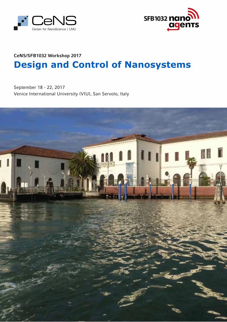
September 18 - 22, 2017Venice International University (VIU), San Servolo, Italy
CeNS/SFB1032 Workshop 2017
Design and Control of Nanosystems
organizers
Center for NanoScience (CeNS) Dr. Susanne Hennig and Claudia Leonhardt SFB 1032 "Nanoagents for the spatiotemporal control of molecular and cellular reactions" Marilena Pinto and Gabriela Milia Ludwig-Maximilians-Universität (LMU) Munich Geschwister-Scholl-Platz 1 D-80539 Munich, Germany www.cens.de [email protected]
partners
Center for the Physics of Living CellsUniversity of Illinois at Urbana-ChampaignUrbana, IL 61801 USA
program committee
Prof. Thomas Carell, LMU Munich Prof. Erwin Frey, LMU Munich Prof. Don Lamb, LMU Munich Prof. Hubert Krenner, University of Augsburg Prof. Ulrich Schollwöck, LMU Munich
venue
Venice International University (VIU) Isola di San Servolo Venezia, Italy Phone: +39-041-2719511 Fax: +39-041-2719510 www.univiu.org [email protected]

CeNS Workshop Venice 2017 3
table of contents
program page 4
invited talks page 6
posters - session I page 22
posters - session II page 36
list of participants page 54
good to know page 56
maps page 57

CeNS Workshop Venice 20174
program
Sunday, 17 September Monday, 18 September Tuesday, 19 September
Laurens Molenkamp Topological physics in HgTe-based
quantum devices
Julie BiteenSingle-molecule imaging and
plasmonics uncover nanometer-scale fundamentals of cell biology
Coffee
Peter HommelhoffLandau-Zener-Stückelberg interferometry
with electrons in graphene and other fun
coherent phenomena on (sub-)femtosecond
timescales
Alexander DeitersOptochemical control of
biological processes in cells and animals
Lunch Break
Jörn Dunkel Geometric control of microbial
fluids: From bacterial spin lattices to active matter logic
Poster Session I and Coffee
Nikta FakhriActive matters:
Probing forces and fluctuations in actomyosin cortices
9.00
9.15
10.00
10.45
11.15
11.35
12.20
14.15
15.00
15.45
17.00
9.00
9.45
10.30
11.00
12.30
17.00
Train to Venice 11.34 Arrival: 18.10
Upon arrival in Venice, participants will receive
their vaporetto tickets from CeNS staff.
Take vaporetto 2, 4.1 or 5.1 from train station to
San Zaccaria:
No. 2 (Ferrovia "B", 34 min to S. Zaccaria ):
18.22/18.34/18.46/18.58...
No. 4.1 (Ferrovia "C", 32 min to S. Zaccaria ):18.29/18.49/19.09...
No. 5.1 (Ferrovia "C", 28 min to S. Zaccaria ): 18.23/18.43/19.03...
Vaporetto 20 from San Zaccaria (M.V.E.) "B" to
San Servolo:19.10/19.50/20.30
Welcome Reception(San Servolo, Room tba)
20.15
11.45
16.15
15.00
Welcome
Daniel Müller
Studying mechanical processes of life from the cellular to
molecular scale
Mikael Rechtsman Photonic topological physics in two and three dimensions
Coffee
Stefan DatzMultifunctional mesoporous
nanoparticles for drug delivery
Nigel GoldenfeldEven parasites have parasites:
Oscillatory population dynamics of mobile genetic elements
Lunch Break
Rob PhillipsThe molecular switch and
Monod’s second secret of life
Sanford SimonAssembly of HIV-1 at the plasma membrane of cells
Coffee
Klaus KroyExact symmetries in the
velocity fluctuations of a hot Brownian swimmer
Aleksei AksimentievSensing and building with DNA
14.15

CeNS Workshop Venice 2017 5
Ronny ThomaleTopolectrical circuits
Rob PhillipsKey Challenges in biophysics
Coffee
Gil RefaelKey challenges:
The coming quantum revolution?
From new materials to new computational paradigms
Lunch and informal discussions
Wednesday, 20 September Thursday, 21 September Friday, 22 September
Ivan HucEngineering synthetic folded
organic nanoarchitectures
Peter RöttgermannTime-correlations of single cell
dual fluorescence markers
Christoph LienauProbing the motion of photo-emitted electrons by ultrafast point-projection electron mi-
croscopy
Closing remarks
Departure
Vaporetto 20 from San Ser-volo to San Zaccaria:
11.20/12.10
Take vaporetto 2, 4.2. or 5.2 from San Zaccaria to S. Lucia train station:
No. 2 (S. Zaccaria Daniele "E", 34 min to S. Lucia): 11.38/11.50/12.02...
No. 4.2 (S.Zaccaria Jolanda "C", 32 min to S. Lucia):
11.53/12.13/12.33...
No. 5.2 (S.Zaccaria Jolanda "C", 28 min to S. Lucia):
11.47/12.07/12.27...
Train to Munich 13.50 Arrival: 20.25
9.00 9.00
9.45
10.45
11.15
12.15
9.00
9.45
10.30
11.00
11.45
12.30
13.30
14.15
15.00
15.25
9.45
10.05
10.50
11.00
15.45
16.30 -
18.30
Michael StranoUsing carbon nanotechnology for the manipulation of matter
Frank PollmannMany-body localization:
Entanglement and dynamics
Coffee
L. MahadevanControlled growth and form: From precipitating microsculptures to
growing soft flowers
Oliver TrappSelf-amplification of chirality in
stereodynamic catalysts
Lunch Break
Gil RefaelFloquet quantum states: Topologi-cal transitions, steady states, and
surprising implications
Rinaldo TrottaStrain-engineered artificial
atoms for quantum nanophotonics
Coffee
Patrick VogelApplications with Traveling Wave
Magnetic Particle Imaging
Christoph Westerhausen
Fluidic hybrid systems for cell manipulation - towards neural
networks on a chip
Poster Session II and Drinks

CeNS Workshop Venice 20176
invited talks
Studying mechanical processes of life from the cellular to molecular scaleDaniel Müller . . . . . . . . . . . . . . . . . . . . . . .7
Photonic topological physics in two and three dimensionsMikael Rechtsman . . . . . . . . . . . . . . . . . . . .7
Multifunctional mesoporous nanoparticles for drug deliveryStefan Datz . . . . . . . . . . . . . . . . . . . . . . . . .8
Even parasites have parasites: Oscillatory population dynamics of mobile genetic elements in your genomeNigel Goldenfeld . . . . . . . . . . . . . . . . . . . .8
The Molecular Switch and Monod’s Second Secret of LifeRob Phillips . . . . . . . . . . . . . . . . . . . . . . . . .9
Assembly of HIV-1 at the plasma membrane of cellsSanford Simon . . . . . . . . . . . . . . . . . . . . . . .9
Exact symmetries in the velocity fluctuations of a hot Brownian swimmerKlaus Kroy . . . . . . . . . . . . . . . . . . . . . . . . .10
Sensing and building with DNAAleksei Aksimentiev . . . . . . . . . . . . . . . . .10
Topological physics in HgTe-based quantum devicesLaurens Molenkamp . . . . . . . . . . . . . . . . .11
Single-molecule imaging and plasmonics uncover nanometer-scale fundamentals of cell biologyJulie Biteen . . . . . . . . . . . . . . . . . . . . . . . .11
Landau-Zener-Stückelberg interferometry with electrons in graphene and other fun coherent phenomena on (sub-) femtosecond timescalesPeter Hommelhoff . . . . . . . . . . . . . . . . . .12
Optochemical control of biological processes in cells and animals Alexander Deiters . . . . . . . . . . . . . . . . . . .12
Geometric control of microbial fluids: From bacterial spin lattices to active matter logicJörn Dunkel . . . . . . . . . . . . . . . . . . . . . . . .13
Active Matters: Probing forces and fluctuations in actomyosin corticesNikta Fakhri . . . . . . . . . . . . . . . . . . . . . . . .13
Topolectrical circuitsRonny Thomale . . . . . . . . . . . . . . . . . . . . .14
Key challenges in biophysicsRob Phillips . . . . . . . . . . . . . . . . . . . . . . . .14
The coming quantum revolution? From new materials to new computational paradigmsGil Refael . . . . . . . . . . . . . . . . . . . . . . . . . .14
Using carbon nanotechnology for the manipulation of matterMichael Strano . . . . . . . . . . . . . . . . . . . . .15
Many-body localization: Entanglement and dynamicsFrank Pollmann . . . . . . . . . . . . . . . . . . . . .15
Controlled growth and form: From precipitating microsculptures to growing soft flowersL . Mahadevan . . . . . . . . . . . . . . . . . . . . . . .16
Self-amplification of chirality in stereodynamic catalystsOliver Trapp . . . . . . . . . . . . . . . . . . . . . . . .16
Floquet quantum states: Topological transitions, steady states, and surprising implicationsGil Refael . . . . . . . . . . . . . . . . . . . . . . . . . .17
Strain-engineered artificial atoms for quantum nanophotonicsRinaldo Trotta . . . . . . . . . . . . . . . . . . . . . .17
Applications with Traveling Wave Magnetic Particle ImagingPatrick Vogel . . . . . . . . . . . . . . . . . . . . . . .18
Fluidic hybrid systems for cell manipulation - towards neural networks on a chip Christoph Westerhausen . . . . . . . . . . . . .18
Engineering synthetic folded nanoarchitecturesIvan Huc . . . . . . . . . . . . . . . . . . . . . . . . . . .19
Time-correlations of single cell dual fluorescence markers - a kinetic finger print in nanoparticle induced apoptosisPeter Röttgermann . . . . . . . . . . . . . . . . . .19
Probing the motion of photoemitted electrons by ultrafast point-projection electron microscopyChristoph Lienau . . . . . . . . . . . . . . . . . . . .20
Ali Aghebat Rafat, Tobias Pirzer, Andrea Mückl, Friedrich C. Simmel 23
Kira Bartnik1,2, Alvaro H. Crevenna1,2,3, Mauricio Pilo-Pais2,4, Tim Liedl2,4, and Don C. Lamb1,2 23
CeNS Workshop Venice 2017 7
Monday, September 18
Studying Mechanical Processes of Life from the Cellular to Molecular ScaleDaniel Müller
ETH Zurich, Biosystems Science and Engineering, CH-4057 Basel, Switzerland; Correspondence: [email protected]
Atomic force microscopy (AFM) at the sub-nanometer resolution (Nanoscopy) is an approach that allows the mechanical charac-terization of basic cellular processes, ranging from the cellular to molecular scale. I will introduce the use of AFM-based nano-scopic assays to characterize the mechanical process guiding the drastic shape change of animal cells progressing through mitosis. We apply our assay in a massive screen to study the contribution of > 1’000 individual human genes in mitotic cell shape change. After having found the major genes responsible for regulating cell shape changes in mitosis, we apply our assay to control cancer cells progressing through mitosis. After this, we introduce high-resolution AFM-based assays to character-ize individual cellular machines (proteins) playing commanding roles in animal cells. First, we developed AFM-based imaging to observe cellular machines at sub-nanometer resolution at work. Second, we extended these imaging possibilities of AFM to im-age native membrane receptors and at the same time detect their interactions and binding steps to ligands and determine the free-energy landscape of the receptor-ligand bonds. There-by the approach can distinguish between ligands representing either agonists, inverse agonists or antagonists. Third, we apply AFM-based single-molecule force spectroscopy to image and structurally map, at amino acid accuracy, the interactions that functionally modulate a membrane receptor. Finally, I will over-view recent developments of force nanoscopy, which applied together with modern light microscopy and cell biological and genetic tools, provide unique fascinating insight into how the machinery of the cell contributes to basic processes of life.
[1] Atomic force microscopy as a multifunctional molecular toolbox in nanobiotechnology. D.J. Müller & Y. Dufrene. Nature Nanotech-nology (2008) 3, 261-269.
[2] Atomic force microscopy imaging modalities in molecular and cell biology. Y.F. Dufrêne, T. Ando, R. Garcia, D. Alsteens, D. Martinez-Martin, A. Engel, C. Gerber & D.J. Müller. Nature Nano-technology (2017) 3, 295-307.
[2] Atomic force microscopy-based characterization and design of biointerfaces. D. Alsteens, H.E. Gaub, R. Newton, M. Pfreundschuh, C. Gerber & D.J. Müller. Nature Review Materials (2017) 2, 17008.
[3] Combined activities of hydrostatic pressure and the actomyosin cortex drive mitotic cell rounding. M.P. Stewart, J. Helenius, Y. Toyoda, S.P. Ramanathan, D.J. Muller & A.A. Hyman.
Nature (2011) 469, 226-230.
[4] Cdk1 dependent mitotic enrichment of cortical myosin II promotes cell rounding against confinement. S.P. Ramanathan, J. Helenius, M.P. Stewart, C. Cattin A.A. Hyman & D.J. Muller. Nature Cell Biology (2015) 17, 148-159.
[5] Mechanical control of mitotic progression in single animal cells. C.J. Cattin, M. Düggelin, D.M. Martinez, C. Gerber, D.J. Müller & M.P. Stewart. Proc. Natl. Acad. Sci. USA (2015) 112, 11258-11263.
[6] Gating of the MlotiK1 potassium channel involves large rear-rangements of the cyclic nucleotide-binding domains. S.A. Mari, J. Pessoa, S.L. Altieri, U. Hensen, L. Thomas, J.H. Morais-Cabral & D.J. Muller Proc. Natl. Acad. Sci. USA (2011) 108, 20802-20807.
[7] Cholesterol increases kinetic, energetic and mechanical stability of the human b-adrenergic receptor. M. Zocher, C. Zhang, G.F.S. Rassmussen, B.K. Kobilka & D.J. Muller Proc. Natl. Acad. Sci. USA (2012) 109, E3463-3473.
[8] Five challenges to bringing single-molecule force spectroscopy into the living cell. Y.F. Dufrene, E. Evans, A. Engel, J. Helenius, H.E. Gaub & D.J. Muller Nature Methods (2011) 8, 123-127.
[9] Multi-parametric force mapping of biological systems to mo-lecular resolution. Y.F. Dufrene, D. Martinez-Martin, I. Medalsy, D. Alsteens & D.J. Muller Nature Methods (2013) 10, 847-854.
[10] Imaging G protein-coupled receptors while quantifying their ligand-binding free-energy landscape. D. Alsteens, M. Pfreund-schuh, C. Zhang, P. Spoerri, S.R. Coughlin, B.K. Kobilka & D.J. Müller Nature Methods (2015) 12, 845-851.
[11] Nanomechanical mapping of first binding steps of a virus to animal cells. D. Alsteens, R. Newton, R. Schubert, D. Martinez-Martin, B. Roska & D.J. Müller. Nature Nanotechnology (2017) 12, 177-183.
[12] Genome-scale single-cell mechanical phenotyping reveals disease-related genes involved in mitotic rounding. Y. Toyoda, C. Cattin, M.P. Stewart, I. Poser, M. Theis, T.V. Kurzchalia, F. Buch-holz, A.A. Hyman & D.J. Müller. Nature Communications (2017) accepted.
Photonic topological physics in two and three dimensionsMikael Rechtsman
Pennsylvania State University, Department of Physics, 104 Davey Lab, University Park, PA 16802-6300
Topological insulators are solid-state materials whose transport properties are immune to defects and disorder due to underly-ing topological order. Perhaps the first such phenomenon was the quantum Hall effect, wherein the Hall conductivity is quan-tized and hence extremely robust. In this talk, I will present the experimental observation of the topological protection of the
transport of photons (rather than electrons in the solid state) in a complex dielectric structure. I will then present the observa-tion of optical Weyl points in the context of three-dimensional photonic crystals. Applications of topological photonic devices include robust photonic networks and delay lines, and poten-tially high-power single-mode lasing.
CeNS Workshop Venice 20178
Monday, September 18
Multifunctional mesoporous nanoparticles for drug deliveryStefan Datz, Christian Argyo, Constantin v. Schirnding, Veronika Weiss, Dorothée Gößl, Michael Gattner, Korbinian Brunner, Fabio Spada, Bernhard Illes, Christoph Bräuchle, Hanna Engelke, Thomas Carell, Thomas Bein
Department of Chemistry and Center for NanoScience (CeNS), University of Munich (LMU), Butenandtstrasse 5-13(E), 81377 Munich, Germany
One of the most intriguing fields of research in this century is the development of controllable and effective drug delivery sys-tems for targeted cancer therapy. This goal is closely connected to the development of suitable and innovative nanomaterials. In addition to the design of completely new nanoparticles, the properties of already existing materials, such as mesoporous silica nanoparticles, can be improved and modified by investi-gating new stimuli-responsive release mechanisms and different cancer cell targeting strategies.1, 2
Here, we describe a novel enzyme-based cap system for meso-porous silica nanoparticles (MSNs) that is directly combined with a targeting ligand via bio-orthogonal click chemistry. The capping system is based on the pH-responsive binding of an aryl-sulfonamide-functionalized MSN and the enzyme carbonic anhydrase (CA). An unnatural amino acid (UAA) containing a norbornene moiety was genetically incorporated into CA. This UAA allowed for the site-specific bio-orthogonal attachment of even very sensitive targeting ligands such as folic acid and anandamide.3
Additionally, we report on the synthesis of a novel biocompatible material, entirely consisting of covalently crosslinked organic molecules. The β-cyclodextrin structures were crosslinked with a rigid organic linker molecule to obtain small (~150 nm) and highly water-dispersable nanoparticles. Very fast cell-uptake ki-netics were observed on HeLa cells revealing particle uptake within less than an hour due to sugar-receptor mediated endo-cytosis. This novel material expands the repertoire of powerful multifunctional nanocarrier systems.
[1] C. Argyo, V. Weiss, C. Bräuchle and T. Bein, Chemistry of Mate-rials, 2014, 26, 435-451.
[2] K. Ulbrich, K. Holá, V. Šubr, A. Bakandritsos, J. Tuček and R. Zbořil, Chemical reviews, 2016, 116, 5338-5431.
[3] S. Datz, C. Argyo, M. Gattner, V. Weiss, K. Brunner, J. Bretzler, C. von Schirnding, A. A. Torrano, F. Spada, M. Vrabel, H. Engelke, C. Brauchle, T. Carell and T. Bein, Nanoscale, 2016, 8, 8101-8110.
Even parasites have parasites: oscillatory population dynamics of mobile genetic elements in your genomeNigel Goldenfeld
Department of Physics, Loomis Laboratory, University of Illinois, 1110 W. Green, Urbana, IL 61801, USA
Transposable elements (TEs), or transposons, are a class of mo-bile genetic elements that can either move or duplicate them-selves in the genome, sometimes interfering with gene expres-sion as a result. Some TEs can code all necessary enzymes for their transposition and are thus autonomous, while non-auton-omous TEs are parasitic and must depend on the machinery of autonomous ones. We present and solve a stochastic model to describe the dynamics of non-autonomous/autonomous pairs of retrotransposons in the human genome that proliferate by a co-py-and-paste mechanism. We predict noise-induced persistent
oscillations in their copy numbers, analogous to predator-prey dynamics in an ecosystem. We discuss if it is experimentally feasible to measure these phenomena in the laboratory, using techniques recently developed in collaboration with Tom Kuhl-man to visualize transposon activity in real time in living cells. We also discuss the possibility to to observe these oscillations over evolutionary time through bioinformatics. This work shows that it is fruitful to regard the genome as an ecosystem that is host to diverse interacting populations.
CeNS Workshop Venice 2017 9
Monday, September 18
Only ten years after the discovery of the iconic structure of DNA, new questions were on biologist’s minds, namely, how are the macromolecules of the cell regulated so that they do what they are supposed to when and where they are needed. The initial resolution of the challenging question of biological regulation came in the form of the notion of “allostery”, an idea that its discoverer Jacques Monod himself referred to as "the second secret of life". We recently celebrated the 50th anniversary of the classic paper of Monod, Changeux and Jacob that intro-duced this far reaching idea. That important paper was followed shortly thereafter by a second one that revealed their musings on how simple statistical mechanical models can be used to cap-
ture how such allosteric transitions work mechanistically. In this talk, I will review the key features of the famed Monod-Wyman-Changeux (MWC) model and then describe its broad reach across many different domains of biology with special reference to the physics underlying how genes are turned on and off. One of the intriguing outcomes of this class of models is a beautiful and predictive scheme for collapsing data from entire libraries of mutants. Once we have considered some of the traditional uses of the MWC model, I will turn to more speculative recent ideas which use the MWC approach to consider the nature of kinetic proofreading.
The Molecular Switch and Monod’s Second Secret of LifeRob Phillips
California Institute of Technology, MC 128-95, 1200 California Boulevard, Pasadena, CA 91125
Assembly of HIV-1 at the plasma membrane of cellsDaniel Johnson, Marina Bleck and Sanford Simon
The Rockefeller University, New York City
The retrovirus HIV-1 assembles at the plasma membrane. Us-ing live-cell polarization fluorescence microscopy of assembling virions, we have examined the temporal sequence in which vari-ous viral and host molecules are recruited to the assembly site. The HIV-1 genome is recruited first to the plasma membrane by a sub-detectable number of molecules of the structural protein, Gag. The membrane bends as a steady accumulation of Gag ensues for 6-10 minutes. After Gag recruitment is completed, members of the ESCRT-III complex and the ATPase Vps4A are recruited transiently, for just a few minutes, to the site of assem-bly. Scission only occurs after dissociation of the ESCRT-III and ATPase from the membrane.
CeNS Workshop Venice 201710
Monday, September 18
Symmetries constrain dynamics. We test this fundamental physical principle, experimentally and by molecular dynamics simulations, for a hot Janus swimmer operating far from thermal equilibrium. Our results establish, with great precision, scalar and vectorial steady-state fluctuation theorems and a thermody-namic uncertainty relation that link the fluctuating particle cur-rent to its virtual entropy production at an effective temperature.
A Markovian minimal model elucidates the underlying non-equilibrium physics.
Exact symmetries in the velocity fluctuations of a hot Brownian swimmerKlaus Kroy
ITP, University of Leipzig, 04009 Leipzig
Sensing and Building with DNAAleksei Aksimentiev
Department of Physics, University of Illinois at Urbana-Champaign
After water and oxygen, DNA is, very likely, the most famous molecule of life. This is not surprising, as the eye-catching double helix of DNA carries instructions to manufacture and assemble all the components of a living organism. The wealth of information encoded in DNA often overshadows its unusual physical properties, for example, the possibility of effective at-traction between same-charge DNA molecules. Furthermore, the methods used to determine the informational content of DNA - its nucleotide sequence - until now relied on biological processes. In this lecture, I will describe our recent efforts to characterize the physical properties of DNA through atomis-tic simulations and demonstrate how these properties can be exploited in a physics-based technology of sequencing DNA. I will then demonstrate how DNA can be used to build synthetic biomimetic systems that outperform their biological prototypes.
CeNS Workshop Venice 2017 11
Tuesday, September 19
Topological Physics in HgTe-based Quantum DevicesLaurens Molenkamp
Physikalische Institut, Universität Würzburg, D-97074 Würzburg, Germany
Single-molecule imaging and plasmonics uncover nanometer-scale fundamentals of cell biologyJulie Biteen
University of Michigan Departments of Chemistry and Biophysics, USA
Because of the small size of bacterial cells, the mysteries of their subcellular structure, dynamics and cooperativity are well-suit-ed to single-molecule and super-resolution investigations. Our lab has been developing new methods to locate, track, and ana-lyze single molecules to answer fundamental, unanswered ques-tions in live bacterial cells. I will discuss how we are measuring and understanding the dynamical interactions essential for DNA replication and mismatch repair in living Bacillus subtilis cells, as well as our ongoing work to extend our targets from single cells to pathogens and microbial communities. Overall, our re-sults provide fundamental insight of relevance to human health and disease.
Suitably structured HgTe is a topological insulator in both 2 (a quantum well wider than some 6.3 nm) and 3 (an epilayer grown under tensile strain) dimensions.
The material has favorable properties for quantum transport studies, i.e. a good mobility and a complete absence of bulk car-riers, which allowed us to demonstrate variety of novel transport effects.
One aspect of these studies is topological superconductivity, which can be achieved by inducing superconductivity in the topological surface states of these materials. Special emphasis will be given to recent results on the ac Josephson effect. We will present data on Shapiro step behavior that is a very strong indication for the presence of a gapless Andreev mode in our Josephson junctions, both in 2- and in 3-dimensional structure.
An additional and very direct evidence for the presence of a zero mode is our observation of Josephson radiation at an energy equal to half the superconducting gap.
Controlling the strain of the HgTe layers strain opens up yet an-other line a research. We have recently optimized MBE growth of so-called virtual substrates ((Cd,Zn)Te superlattices as a buf-fer on a GaAs substrate), that allow us to vary the strain from 0.4% tensile to 1.5% compressive. While tensile strain turns 3-dimensional HgTe into a narrow gap insulator, compressive strain turns the material into a topological (Weyl) semimetal, exhibiting clear signs of the Adler-Bell-Jackiw anomaly in its magnetoresistance. In quantum wells, compressive strain allows inverted energy gaps up to 60 meV.
CeNS Workshop Venice 201712
Tuesday, September 19
Optochemical Control of Biological Processes in Cells and Animals Alexander Deiters
University of Pittsburgh, Department of Chemistry, Pittsburgh, PA 15260, USA; [email protected]; http://www.pitt.edu/~deiters/
Landau-Zener-Stückelberg interferometry with electrons in graphene and other fun coherent phenomena on (sub-) femtosecond timescalesPeter Hommelhoff
Institut für Physik der Kondensierten Materie, Friedrich-Alexander-Universität Erlangen-Nürnberg
When intense few-cycle laser pulses are focused on graphene, electrons can be strongly driven inside of the material. We ob-serve repeated coherent Landau-Zener transitions between va-lence and conduction band, establishing two different quantum excitation pathways from valence to conduction band, a process known at Landau-Zener-Stückelberg interferometry. The phase between the two quantum pathways and hence the outcome of the interference (excitation or no excitation) can be directly varied with the optical field waveform, i.e., the carrier-envelope phase of the two-cycle driving pulse. We will discuss this and related physics around optical-field-driven control of electrons in nanosystems.
Nature regulates biological processes, such as signal transduc-tion, protein function, and gene expression, with high spatial and temporal precision. In order to study and understand these processes, equally precise external control is required. Light is an excellent tool for this purpose, as it can be easily regulated in timing, location, wavelength, and amplitude, thereby enabling high-resolution control of biological processes. We are develop-ing optochemical tools to A) control protein function through genetic code expansion with unnatural amino acids that can
be activated with light, and to B) control nucleic acid function through synthetic installation of light-cleavable chromophores onto nucleobases and into phosphodiester backbones. We have applied these approaches to the conditional control of DNA re-combination, gene editing, RNA polymerization, RNA transla-tion, microRNA function, cell signaling, and other essential bio-logical processes in cells and zebrafish embryos.
CeNS Workshop Venice 2017 13
Tuesday, September 19
Geometric control of microbial fluids: From bacterial spin lattices to active matter logicJörn Dunkel
Department of Mathematics, Massachusetts Institute of Technology, 77 Massachusetts Avenue, Cambridge, MA 02139-4307, USA
Geometric constraints can profoundly affect pattern selection and topological defect formation in equilibrium and non-equilib-rium systems. In this talk, I will summarize recent experimental and theoretical work that aims to understand how confinement geometry affects the spontaneous flows of active suspensions. First, we demonstrate how collective microbial swimming can be controlled by microstructure to realize bacterial spin lattices exhibiting ferro- and antiferro-magnetic ordering.
Building on these insights, we can propose designs of active flow networks to implement logical operations in autonomous microfluidic transport devices.
Biological functions rely on ordered structures and intricately controlled collective dynamics. Such order in living systems is typically established and sustained by continuous dissipa-tion of energy. The emergence of collective patterns of motion is unique to non-equilibrium systems and is a manifestation of dynamic steady states. Mechanical resilience of animal cells is largely controlled by the actomyosin cortex. The cortex provides stability, but is at the same time highly adaptable due to rapid turnover of its components. Dynamic functions involve regulated transitions between different steady states of the cortex. In this talk, I will show model actomyosin cortices, constructed to main-tain turnover, self-organize into distinct non-equilibrium steady states when we vary crosslink density. The feedback between actin network structure and organization of stress generating
myosin motors defines the symmetries of the dynamic steady states. A marginally crosslinked state displays divergence-free long-range flow patterns. Higher crosslink density causes struc-tural symmetry breaking resulting in a stationary converging flow pattern. We track the flow patterns in the model actomyo-sin cortices using fluorescent single-walled carbon nanotubes as novel probes. The self-organization of stress patterns we have discovered in a model system has direct implications for a broad range of biological functions.
Active Matters: Probing forces and fluctuations in actomyosin corticesNikta Fakhri
400 Technology Square, NE46-611, Department of Physics, Massachusetts Institute of Technology
CeNS Workshop Venice 201714
Wednesday, September 20
Topolectrical circuitsRonny Thomale
Julius-Maximilians Universität Würzburg, Institut für Theoretische Festkörperphysik, Am Hubland, 97074 Würzburg
First developed by Alessandro Volta and Felix Savary in the early 19th century, circuits consisting of resistor, inductor and capaci-tor (RLC) components are now omnipresent in modern technol-ogy. The behavior of an RLC circuit is governed by its circuit Laplacian, which is analogous to the Hamiltonian describing the energetics of a physical system. We show that “topolectrical” boundary resonances (TBRs) appear in the impedance read-out of a circuit whenever its Laplacian bandstructure resembles that of topological semimetals - materials with extensive degenerate edge modes known as Fermi arcs that also harbor enigmatic transport properties. Such TBRs not only provide unambigu-
ous and highly robust signals for the presence of a topological phase, but also promise diverse applicability within high density electronic mode processing. Due to the versatility of electronic circuits, our topological semimetal construction can be gener-alized to topolectrical phases with any desired lattice symme-try, spatial dimension, and even quasiperiodicity. Topolectrical circuits establish a bridge between electrical engineering and topological states of matter, where the accessibility, scalability, and operability of electronics promises to synergize with the in-tricate boundary properties of topological phases.
Special Session: Key Challenges in Nanoscience
Key challenges in biophysicsRob Phillips
California Institute of Technology, MC 128-95, 1200 California Boulevard, Pasadena, CA 91125
The coming quantum revolution? From new materials to new computational paradigmsGil Refael
California Institute of Technology, MC 128-95, 1200 California Boulevard, Pasadena, CA 91125
Over the last decade, a broad array of new material classes, broadly described as topological materials, were discovered. At the same time, the quest for quantum computers dramatically accelerated. Traditional concepts of quantum computing hard-ware, e.g. the Josephson junction based qubits, started compet-ing with new paradigms such as topologically protected qubits. The giants of technology also entered the fray, with Google and Microsoft pouring resources into these two paradigms. In my talk I will try to review these discoveries and developments, and explain how they might upset our electronics industry, as well as the quest for quantum computers.

CeNS Workshop Venice 2017 15
Thursday, September 21
Our laboratory has been interested in how 1D and 2D electronic materials such as carbon nanotubes and graphene, respectively, can manipulate matter in unique ways. This presentation will fo-cus on three topics: exotic fluid phase transitions within isolated carbon nanotubes, graphene nanopores for selective molecular transport and carbon nanotube templated molecular recogni-tion for sensors. The first topic, fluid phase transitions inside single, isolated carbon nanotubes (CNT) are predicted to devi-ate substantially from classical thermodynamics and also allow the study of ice nanotube (ice-NT) properties. Herein, we mea-sure, using two different techniques, the diameter dependent phase boundaries of ice-NTs within isolated CNTs 1.05, 1.06, 1.15, 1.24, and 1.52nm in diameter using Raman spectroscopy. The results reveal both an exquisite sensitivity to diameter and substantially larger temperature elevations of the melting transi-tion than theoretically predicted by as much as 100°C. Dynamic water filling and reversible freezing transitions were marked by 2 to 5cm-1 shifts in the radial breathing mode (RBM) frequency, revealing reversible melting at 138°C and 102°C for 1.05 and 1.06nm single and double-walled CNTs, respectively. A near-ambient phase change at 15°C was observed for 1.52nm CNT, whereas freezing inside 1.24nm tube was suppressed at -30°C. These extreme phase transitions enable the study of ice-NT at high temperatures and their potential utilization as novel phase change materials. The second topic, nanopores in monolayer graphene membranes demonstrate the ability to selectively al-low molecular transport based on size and other characteristics, but at unprecedented rates due to thickness of only a single car-bon atom. We present a detailed analysis of experimental gas
permeation data through single layer graphene membranes un-der batch depletion conditions parametric in starting pressure for He, H2, Ne, and CO2 between 100 and 670 kPa. Analyzing the time dependent permeance data shows remarkably that the latter three gases exhibit discretized permeance values that are temporally repeated. Such quantized fluctuations or gating are a hallmark of isolated nanopores, since small, but rapid changes in the transport pathway necessarily influence a single detect-able flux. This is the first reported instance of gas phase gating through a nanopore, and we analyze the fluctuations using a Hidden Markov model. For the last topic, we introduce CoPh-MoRe or corona phase molecular recognition as a method for discovering what can be thought of as nanoparticle coupled syn-thetic antibodies, or recognition sites formed from a specifically designed heteropolymer library. We show that certain synthetic heteropolymers, once constrained onto a single-walled carbon nanotube by chemical adsorption, form a new corona phase that exhibits highly selective recognition for specific molecules. I will highlight recent examples allowing us to detect a wide range of challenging molecules, from neurotransmitters, to explosives, carbohydrates, and protein components in whole blood.
Using Carbon Nanotechnology for the Manipulation of MatterMichael S. Strano
Carbon P. Dubbs Professor of Chemical Engineering, Department of Chemical Engineering, 77 Massachusetts Avenue, 66-570
Cambridge, MA 02139-4307; Email: [email protected]
Many-body localization (MBL) occurs in isolated quantum sys-tems when Anderson localization persists in the presence of finite interactions. The MBL phase is characterized by a break-down of ergodicity and a slow entanglement growth following a quantum quench.
First, we show that the quantum mutual information (QMI) be-tween two small, spatially separated regions is a useful probe to study many-body localization (MBL). The QMI can in principle be used in an experimental setup to detect the MBL transition and allows to distinguish between an Anderson insulator and an MBL phase.
Second, we study the effects of local perturbations on the dy-namics of disordered fermionic systems in or- der to charac-terize time-irreversibility. We consider the dynamics of the full many-body wave-functions by measuring the Loschmidt echo (LE) and find qualitatively different behavior in localized and ex-tended phases.
Many-body localization: Entanglement and dynamicsFrank Pollmann
Physics Department, Technische Universität München, Munich, Germany
CeNS Workshop Venice 201716
Thursday, September 21
Controlled growth and form: From precipitating microsculptures to growing soft flowersL. Mahadevan
Harvard University, Pierce Hall, 29 Oxford Street, Cambridge, MA 02138, USA
Controlled self-assembly of three-dimensional shapes holds great potential for fabrication of functional materials. Their practical realization requires a theoretical framework to quan-tify and guide the dynamic sculpting of the curved structures that often arise in accretive mineralization or soft, thin growing sheets. Motivated by bioinspired coprecipitation patterns of car-bonate and silica, we develop a geometrical theory for the kinet-ics of the growth front that leaves behind thin-walled complex structures. Our theory explains the range of previously observed experimental patterns and, in addition, predicts unexplored as-sembly pathways. Similarly, motivated by the growth of flowers, we develop a geometric theory for the growth of elastic bilayers and solve the inverse problem of designing metric patterns that can take a flat sheet into a flower or a face.
Self-Amplification of Chirality in Stereodynamic CatalystsOliver Trapp
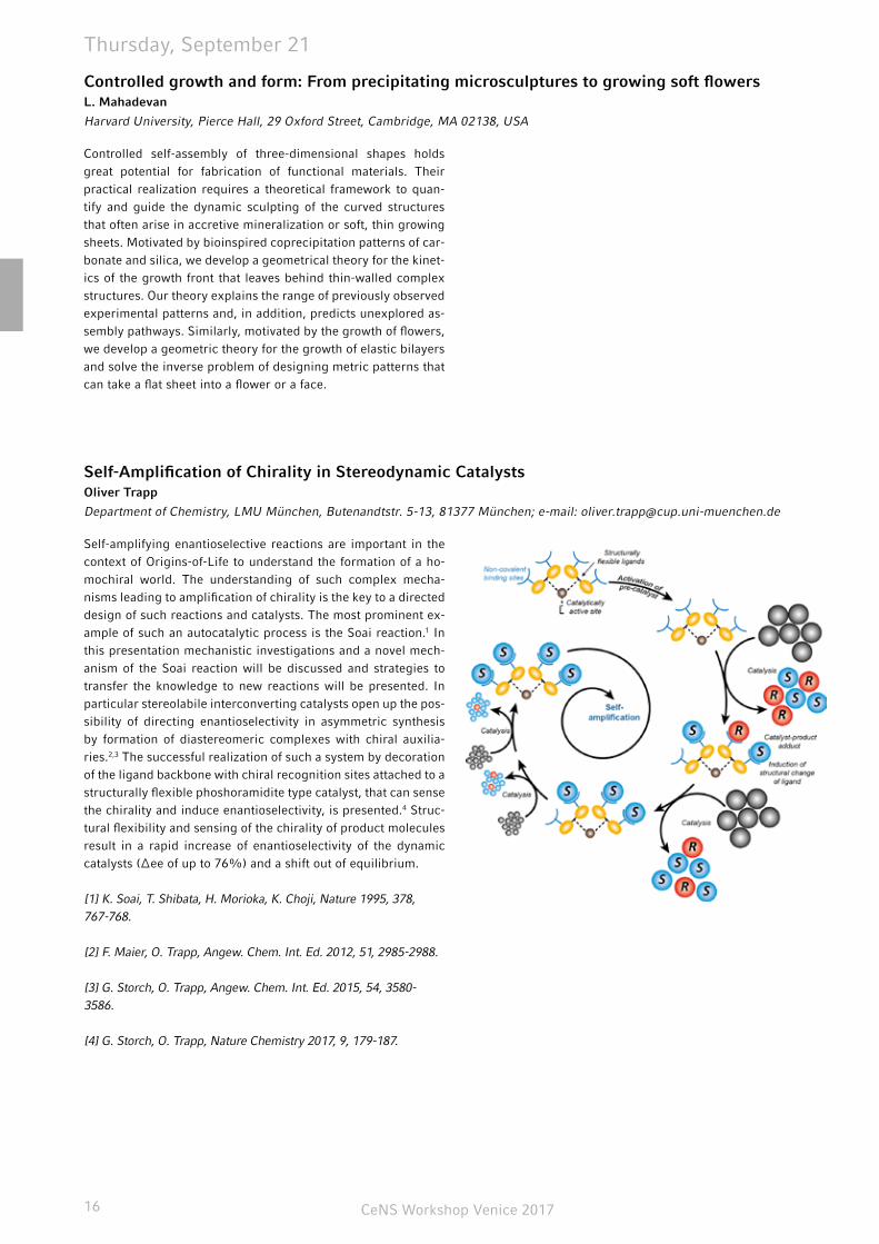
Department of Chemistry, LMU München, Butenandtstr. 5-13, 81377 München; e-mail: [email protected]
Self-amplifying enantioselective reactions are important in the context of Origins-of-Life to understand the formation of a ho-mochiral world. The understanding of such complex mecha-nisms leading to amplification of chirality is the key to a directed design of such reactions and catalysts. The most prominent ex-ample of such an autocatalytic process is the Soai reaction.1 In this presentation mechanistic investigations and a novel mech-anism of the Soai reaction will be discussed and strategies to transfer the knowledge to new reactions will be presented. In particular stereolabile interconverting catalysts open up the pos-sibility of directing enantioselectivity in asymmetric synthesis by formation of diastereomeric complexes with chiral auxilia-ries.2,3 The successful realization of such a system by decoration of the ligand backbone with chiral recognition sites attached to a structurally flexible phoshoramidite type catalyst, that can sense the chirality and induce enantioselectivity, is presented.4 Struc-tural flexibility and sensing of the chirality of product molecules result in a rapid increase of enantioselectivity of the dynamic catalysts (Δee of up to 76%) and a shift out of equilibrium.
[1] K. Soai, T. Shibata, H. Morioka, K. Choji, Nature 1995, 378, 767-768.
[2] F. Maier, O. Trapp, Angew. Chem. Int. Ed. 2012, 51, 2985-2988.
[3] G. Storch, O. Trapp, Angew. Chem. Int. Ed. 2015, 54, 3580-3586.
[4] G. Storch, O. Trapp, Nature Chemistry 2017, 9, 179-187.
CeNS Workshop Venice 2017 17
Thursday, September 21
Strain-engineered artificial atoms for quantum nanophotonicsRinaldo Trotta
Institute of Semiconductor and Solid State Physics, Johannes Kepler University Linz, Austria
The prospect of using the quantum nature of light for secure communication keeps spurring the search and investigation of suitable sources of single and entangled photons. Semiconduc-tor quantum dots (QDs), also dubbed “artificial atoms”, are ar-guably one of the most attractive. They can generate single and entangled photons on demand, with high efficiency, and they are intrinsically compatible with current photonic-integration technologies. Unlike “natural atoms”, however, no two QDs are alike. This peculiarity is a major obstacle for quantum communi-cation applications that require non-classical states of light with identical energies.
In this talk, I will first introduce a novel class of semiconductor-piezoelectric devices [1] in which strain is used to engineer the electronic structure of any arbitrary QD so that single and polar-ization-entangled photons can be generated with unprecedent-ed quality and speed [2, 3]. Then, I will show that full control of the QD in-plane strain tensor allows the energy of the entangled photons emitted by QDs to be precisely controlled without de-grading the degree of entanglement [4, 5]. This opens the pos-sibility to build up hybrid semiconductor-atomic interconnects, in which entangled photons from QDs are interfaced with clouds
of natural atoms that behave as slow-light medium [5, 6]. To conclude, I will present our recent results on novel GaAs QDs [7, 8] and discuss how they can be used to construct a QD-based quantum network.
[1] R. Trotta, et al., in “Engineering the atom-photon interaction” (Springer, Berlin, 2015).
[2] R. Trotta, et al. Nano Lett. 14, 3439 (2014).
[3] J. Zhang, et al. Nature Comm. 6, 10067 (2015).
[4] R. Trotta, et al. Phys. Rev. Lett. 114, 150502 (2015).
[5] R. Trotta, et al. Nature Comm. 7, 10375 (2016).
[6] H. Huang, et al., ACS Photonics 4, 868 (2017).
[7] D. Huber, et al., Nature Comm. 8, 15506 (2017).
[8] M. Reindl, et al., Nano Lett. 17, 4090 (2017).
Floquet quantum states: Topological transitions, steady states, and surprising implicationsGil Refael
Division of Physics, Math, and Astronomy, Caltech, Pasadena, CA-91125
Recent work has shown that manipulating a quantum system us-ing a periodic drive provides a new means for externally control-ling it. Such a periodic drive can give rise to topological states in trivial quantum wells, bulk semiconductors, and even in gra-phene, and it can also turn a quantum wire into a system which could have Majorana states - of two flavors even. Hitting a quan-tum system (more so, periodically!), may suggest complications, however, such as heating.
I will show that this can be avoided by tailoring the thermal bath in which the system is immersed. Altogether we will find that there is much rich physics in Floquet quantum states and their close relatives, and that one can work with them to obtain use-ful, finite-entropy, many-body steady states.
CeNS Workshop Venice 201718
Thursday, September 21
Cells are the building blocks of human beings. As the coopera-tive work of a body’s cells is more than the mere sum of single cells, cellular interaction is of highest interest. Among all types of cells, neurons are the most exciting ones, being responsible for sensing, thinking and acting. Thus, well-defined neural networks on a chip tunable in time and space are our goal to bridge the gap between phenomenological biological studies and those on virtual networks created by computer scientists. The requirements for such artificial networks are tunable po-sitioning, biocompatibility, controlled neurite outgrowth and a possibility to detect neural activity. Employing fluidic hybrid systems, we study cellular interaction, e.g. vesicles as protocells interacting with simple external force fields leading to fission without the need of complex scenarios. Moreover, we apply mi-crofludics to mimic cells interacting with each other in physi-ological shear flow, e.g. in the case of Malaria infection. While passive devices like micro channels allow for changing the force
fields in a static way, e.g. by adapting the geometry, the use of acoustofluidics leads to dynamically controlled forces. Surface Acoustic Waves (SAW) can drive smallest amounts of fluids to e.g. produce nanoparticles by reproducible mixing of nucleic acids and polymers. A second application is to use SAW to study cell adhesion under physiological conditions and beyond in an on-chip micro-reactor. Finally, the here presented idea is based on standing wave phenomena. Combing two or more crossed standing wave fields on a SAW-chip, we are able to manipulate hard, soft and even living objects and control their position in space and time. As we ensured biocompatibility, a possibility for detection of neural activity, and positioning of the cell bod-ies, together with our cooperation partners at the University of Santa Barbara we envision to control neurite outgrowth using tunable asymmetric force fields and present here first results supporting this assumption.
Fluidic hybrid systems for cell manipulation - towards neural networks on a chip Christoph Westerhausen
University of Augsburg, Institute for Physics, Universitätsstrasse 1, 86159 Augsburg
Applications with Traveling Wave Magnetic Particle ImagingPatrick Vogel 1,2, T.A. Bley2, V.C. Behr1, P.M. Jakob1
1 Department of Experimental Physics 5 (Biophysics), University of Würzburg, Würzburg
2 Department of Diagnostic and Interventional Radiology, University Hospital Würzburg, Würzburg
Magnetic Particle Imaging (MPI) is a novel tomographic im-aging method for the visualization of iron-oxide nanoparticles (SPIONs) in 3D. Based on the non-linear response of magnetic material on varying magnetic fields MPI provide features such as high temporal and spatial resolution as well as good sensitivity. Since the first publication in 2005 several different scanner concepts have been published. An alternative and promising approach, developed in our department, is the so-called Trav-eling Wave MPI (TWMPI) system, which uses a set of electro coils for generating the required magnetic fields in a dynamic way. The resulting pre-clinical scanner offers scanning a large field of view (FOV) with high accuracy. In proof-of-concepts experiments the performance of the TWMPI concept was validated and the scanner used for the various application fields in medicine, biology, geology and material sciences. In this talk an overview about this new technology will be given supplied with several experiments and results showing possible applications.
CeNS Workshop Venice 2017 19
Friday, September 22
Engineering synthetic folded nanoarchitecturesIvan Huc
Department of Pharmacy, LMU München, Butenandtstr. 5–13, Haus B, 81377 München
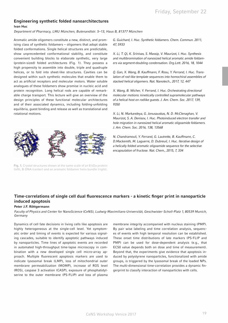
Aromatic amide oligomers constitute a new, distinct, and prom-ising class of synthetic foldamers – oligomers that adopt stable folded conformations. Single helical structures are predictable, show unprecedented conformational stability, and constitute convenient building blocks to elaborate synthetic, very large (protein-sized) folded architectures (Fig. 1). They possess a high propensity to assemble into double, triple and quadruple helices, or to fold into sheet-like structures. Cavities can be designed within such synthetic molecules that enable them to act as artificial receptors and molecular motors. Water soluble analogues of these foldamers show promise in nucleic acid and protein recognition. Long helical rods are capable of remark-able charge transport. This lecture will give an overview of the design principles of these functional molecular architectures and of their associated dynamics, including folding-unfolding equilibria, guest binding and release as well as translational and rotational motions.
Fig. 1. Crystal structures shown at the same scale of an 8 kDa protein (left), B-DNA (center) and an aromatic foldamer helix bundle (right).
G. Guichard, I. Huc. Synthetic foldamers. Chem. Commun. 2011, 47, 5933
X. Li, T. Qi, K. Srinivas, S. Massip, V. Maurizot, I. Huc. Synthesis and multibromination of nanosized helical aromatic amide foldam-ers via segment-doubling condensation. Org.Lett. 2016, 18, 1044
Q. Gan, X. Wang, B. Kauffmann, F. Rosu, Y. Ferrand, I. Huc. Trans-lation of rod-like template sequences into homochiral assemblies of stacked helical oligomers. Nat. Nanotech., 2017, 12, 447
X. Wang, B. Wicher, Y. Ferrand, I. Huc. Orchestrating directional molecular motions: kinetically controlled supramolecular pathways of a helical host on rodlike guests. J. Am. Chem. Soc. 2017, 139, 9350
X. Li, N. Markandeya, G. Jonusauskas, N. D. McClenaghan, V. Maurizot, S. A. Denisov, I. Huc. Photoinduced electron transfer and hole migration in nanosized helical aromatic oligoamide foldamers. J. Am. Chem. Soc. 2016, 138, 13568
N. Chandramouli, Y. Ferrand, G. Lautrette, B. Kauffmann, C. D.Mackereth, M. Laguerre, D. Dubreuil, I. Huc. Iterative design of a helically folded aromatic oligoamide sequence for the selective encapsulation of fructose. Nat. Chem., 2015, 7, 334
Dynamics of cell fate decisions in living cells like apoptosis are highly heterogeneous at the single-cell level. Yet symptom-atic order and timing of events is expected for various signal-ing cascades, suitable to identify apoptotic pathways induced by nanoparticles. Time lines of apoptotic events are recorded in automated high-throughput time-lapse microscopy in com-bination with a new developed single cell micro-array ap-proach. Multiple fluorescent apoptosis markers are used to indicate lysosomal break (LMP), loss of mitochondrial outer membrane permeabilization (MOMP), increase of ROS level (ROS), caspase 3 activation (CASP), exposure of phosphatidyl-serine to the outer membrane (PS-FLIP) and loss of plasma
membrane integrity accompanied with nucleus staining (PMP). By pair wise labeling and time correlation analysis, sequenc-es of events with high temporal resolution can be established. These onset time distributions of late markers (PS-FLIP and PMP) can be used for dose-dependent analysis (e.g., that EC50 value depends both on dose and time of measurement). Beyond that, the experiments give evidence that apoptosis in-duced by polystyrene nanoparticles, functionalized with amide groups, is triggered by the lysosomal break of the loaded NPs. The multi-dimensional time-correlation provides a dynamic fin-gerprint to classify interaction of nanoparticles with cells.
Time-correlations of single cell dual fluorescence markers - a kinetic finger print in nanoparticle induced apoptosisPeter J.F. Röttgermann
Faculty of Physics and Center for NanoScience (CeNS), Ludwig-Maximilians-Universität, Geschwister-Scholl-Platz 1, 80539 Munich, Germany
CeNS Workshop Venice 201720
Friday, September 22
Ultrafast optical spectroscopy is now able to track even the fastest elementary processes such as the motion of elec-trons and/or holes in biomolecules or organic solar cells. De-spite tremendous progress in sub-diffraction optical micros-copy, a direct spatially resolved imaging of such processes is still out of reach since the spatial resolution of even the most advanced near-field imaging techniques is far beyond the Angström-resolution achieved, e.g., in aberration-corrected electron microscopy. As such, numerous efforts are currently ongoing in combining ultrafast optics and electron microscopy. Recently, point-projection electron microscopy, realized by plac-ing an object directly behind a nanoscopic electron source and recording a diffraction image on a distant screen, emerged as an interesting concept for improving the time resolution in ultrafast electron microscopy into the regime of few tens of femtoseconds or possibly even beyond. It avoids the need for electron lenses, makes the experimental setup compact and simple and mini-mizes temporal dispersion of the electron pulses.
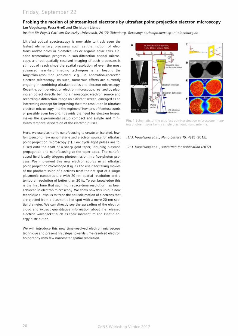
Here, we use plasmonic nanofocusing to create an isolated, few-femtosecond, few nanometer-sized electron source for ultrafast point-projection microscopy [1]. Few-cycle light pulses are fo-cused onto the shaft of a sharp gold taper, inducing plasmon propagation and nanofocusing at the taper apex. The nanofo-cused field locally triggers photoemission in a five-photon pro-cess. We implement this new electron source in an ultrafast point-projection microscope (Fig. 1) and use it for taking movies of the photoemission of electrons from the hot spot of a single plasmonic nanostructure with 20-nm spatial resolution and a temporal resolution of better than 20 fs. To our knowledge this is the first time that such high space-time resolution has been achieved in electron microscopy. We show how this unique new technique allows us to trace the ballistic motion of electrons that are ejected from a plasmonic hot spot with a mere 20-nm spa-tial diameter. We can directly see the spreading of the electron cloud and extract quantitative information about the released electron wavepacket such as their momentum and kinetic en-ergy distribution.
We will introduce this new time-resolved electron microscopy technique and present first steps towards time-resolved electron holography with few nanometer spatial resolution.
[1] J. Vogelsang et al., Nano Letters 15, 4685 (2015).
[2] J. Vogelsang et al., submitted for publication (2017)
Probing the motion of photoemitted electrons by ultrafast point-projection electron microscopyJan Vogelsang, Petra Groß and Christoph Lienau
Institut für Physik Carl von Ossietzky Universität, 26129 Oldenburg, Germany; [email protected]
Fig. 1 Schematic of the ultrafast point-projection microscope imag-ing photoemisson from a single plasmonic nanoantenna.

CeNS Workshop Venice 2017 21
notes
CeNS Workshop Venice 201722
poster abstracts - session i (a-ke)
DNA origami tiles as building blocks for various 2D patterns
Ali Aghebat Rafat, Tobias Pirzer, Andrea Mückl, Friedrich C. Simmel . . . . . . . . . . . . . . . . . . . . . . . . . . . . . . . . . . . . . . . . . . . . . . 23
Using DNA origami as a platform for single-mol-ecule fluorescence studies of DNA double-strand break Kira Bartnik, Alvaro H. Crevenna, Mauricio Pilo-Pais, Tim Liedl, and Don C. Lamb . . . . . . . . . . . . . . . . . . . . . . . . . . . . . . . . . . . . . . . . . . 23
Phase transitions in public good game modelsMarianne Bauer, Erwin Frey . . . . . . . . . . . . . . . . . . . . . . . . . . . . . . 23
Intramolecular forces in von Willebrand factor studied by AFMAchim Löf, Martin Benoit, Jan Lipfert . . . . . . . . . . . . . . . . . . . . . . . 24
A combinatorial DNA origami nanoagent for the study of tumour cell specific targetingRicarda Berger, Hans-Christian Mescheder, Jonas Helma, Heinrich Leonhardt, Tim Liedl, Joachim Rädler . . . . . . . . . . . . . . . . . . . . . . 24
Spectrally switchable photodetection with near-infrared-absorbing Covalent Organic FrameworksDerya Bessinger, Laura Ascherl, Florian Auras, Thomas Bein . . . . 25
Orchestrating cells on a chip employing standing surface acoustic waves towards neural networksManuel S. Brugger, Sarah Grundeen, Adele Doyle, Luke Theogara-jan, Achim Wixforth, and Christoph Westerhausen . . . . . . . . . . . . 25
Metal-nanoparticle enriched diamond-like carbon as an antimicrobial and wear-resistant surface modification for orthopedic implantsSascha Buchegger, Sven Fuchs, Jochen Taiber, Natascha Schuster, Caroline Vogel, Rudolf Herrmann, Achim Wixforth, Bernd Stritzker and Christoph Westerhausen . . . . . . . . . . . . . . . . . . . . . . . . . . . . . 26
Grafting of organophosphonates onto Si(111) by tethering and aggregation for biosensing applica-tionsFrancesco Casablanca, Johannes Bartl, Qi Li, Werner Schindler, Martin Stutzmann, Anna Cattani-Scholz . . . . . . . . . . . . . . . . . . . . . 26
In vitro RNA aggregationAradhana Chopra and Friedrich C. Simmel . . . . . . . . . . . . . . . . . . 27
Mutual switching as design principle for robust protein patternsJonas Denk, Simon Kretschmer, Jacob Halatek, Caroline Hartl, Petra Schwille, and Erwin Frey . . . . . . . . . . . . . . . . . . . . . . . . . . . . . . . . 27
Nickel nanoparticle deposition on (TBA,H)Ca2Nb3O10 - Insights into particle growth and CO2 reduction activityLeo Diehl, Siyuan Zhang, Geoffrey Ozin, Christina Scheu, Bettina V. Lotsch . . . . . . . . . . . . . . . . . . . . . . . . . . . . . . . . . . . . . . . . . . . . . . . 28
Tracking the source of carbon dot photolumines-cenceFlorian Ehrat, S.Bhattacharyya, R. Teves, J. Stolarczyk, A. Urban, and J. Feldmann . . . . . . . . . . . . . . . . . . . . . . . . . . . . . . . . . . . . . . . 28
Micro-structured compartment models for syn-thetic biologyHiro Eto, Jonas Mücksch, M. Heymann, N. Soga, H. Noji & P. Schwille . . . . . . . . . . . . . . . . . . . . . . . . . . . . . . . . . . . . . . . . . . . . . . 29
Movement of invasive cancer cells in predefined 3D collagen matricesFlorian Geiger, Chase Broedersz, Hanna Engelke . . . . . . . . . . . . . 29
Family-friendly zero-sum games Philipp M. Geiger, Johannes Knebel, Markus F. Weber, and Erwin Frey . . . . . . . . . . . . . . . . . . . . . . . . . . . . . . . . . . . . . . . . . . . . . . . . . 30
Progression of COFs in photocatalytic hydrogen evolution - from noble metal assisted to all-in-one systemsKerstin Gottschling, Tanmay Banerjee, Frederik Haase, Gökcen Savsci, Christian Ochsenfeld and Bettina V. Lotsch . . . . . . . . . . . . 30
Generic transport mechanisms for molecular traffic in cellular protrusionsIsabella Graf, Mareike Bojer, Erwin Frey . . . . . . . . . . . . . . . . . . . . 30
Dephasing and quantum beating of excitons in me-thylammonium lead iodide perovskite nanoplateletsBernhard J. Bohn, Moritz Gramlich, Abraham Moreno, Thomas Si-mon, Alexander F. Richter, Lakshminarayana Polavarapu, Alexander S. Urban, and Jochen Feldmann . . . . . . . . . . . . . . . . . . . . . . . . . . . 31
Mechanical properties of Leishmania Myosin XXI determined with an optical tweezers transducerAndreas Graw, Christopher Batters, and Claudia Veigel . . . . . . . . 31
Kinetic studies of the protein backbone hydrogen exchange for determination of H-bond strength and water accessibilityKristof Grohe, Kumar Tekwani Movellan, Suresh Kumar Vasa, Hi-manshu Singh, Karin Giller, Stefan Becker and Rasmus Linser . . . 32
Controlling cell functions artificially via silencing genes by delivery of synthetic microRNAsLisa Haddick, Karin Möller, Wei Zhang, Stephan Morys, Ernst Wag-ner and Thomas Bein . . . . . . . . . . . . . . . . . . . . . . . . . . . . . . . . . . . 33
Carbon templated Nb:TiO2 nanostructures as oxy-gen evolution catalyst supports for PEM electrolyz-ersSebastian Häringer, Alexander G. Hufnagel, Michael Beetz, Daniel Böhm, Dina Fattakhova-Rohlfing, and Thomas Bein . . . . . . . . . . . 33
A new LGPS-type superionic conductor – synthesis and characterization of Li7SiPS8Sascha Harm, Anna-Katharina Hatz, Igor Moudrakovski, Robert E.Usiskin, Joachim Maier, Bettina V. Lotsch . . . . . . . . . . . . . . . . . . 34
Photocontrolled nuclear translocation of mechano-transduction protein YAPBernhard Illes, Hanna Engelke . . . . . . . . . . . . . . . . . . . . . . . . . . . . 34
Enzyme activity at lipid membranes – correlation of activity and membrane state of an intrinsically water-soluble enzymeAndrej Kamenac, Matthias Schneider, Achim Wixforth, Christoph Westerhausen . . . . . . . . . . . . . . . . . . . . . . . . . . . . . . . . . . . . . . . . . 34
Oligothiophene-bridged conjugated Covalent Or-ganic FrameworksNiklas Keller, Derya Bessinger, Stephan Reuter, Mona Calik, Laura Ascherl, Fabian C. Hanusch, Florian Auras, Thomas Bein . . . . . . . 35
Folate receptor-directed siRNA lipopolyplexes for tumor-targeted gene silencing in vivoSarah Kern, Philipp Klein, Wei Zhang, Dian-Jang Lee, Ernst Wagner . . . . . . . . . . . . . . . . . . . . . . . . . . . . . . . . . . . . . . . . . . . . . . 35
CeNS Workshop Venice 2017 23
DNA origami tiles as building blocks for various 2D patterns Ali Aghebat Rafat, Tobias Pirzer, Andrea Mückl, Friedrich C. Simmel
Chair of Physics of synthetic biological systems - E14, Physics Department and ZNN/WSI, Technische Universität München, 85748 Garching, Germany
One of the main challenges in the DNA nanotechnology is the large scale ordering of DNA nanostructures. This would enable using the DNA tiles as functional materials for different applica-tions. Following the invention of the DNA origami technique, variety of 2D DNA origami structures with different symmetry was designed. These tiles can form various 2D patterns via tun-ing the tile-tile and tile-surface interactions. Depending on the geometry of the DNA origami tile, one can create crystalline and non-crystalline patterns. Moreover, there is the possibility to use more than one building block to form 2D patterns.
In order to form 2D patterns, tile-tile attractions were pro-grammed using base-stacking and/or sticky end interactions, plus with sterical repulsive interaction between structures. Fur-thermore, the tile-surface interaction was tuned via the Na+ ion concentration. Employing Na+ ions decreases the adhesion of DNA origami structures to the negatively charged mica surface.
This allows structures to diffuse on the surface and fill the sur-face according to their shape.
As it is mentioned above, DNA origami tile shape and size is a parameter, which plays an important role to form 2D patterns. Here we first create 2D crystalline patterns using single DNA origami structures as building blocks. Afterwards, we employ two DNA nanostructures to create periodic and aperiodic tiling. To create periodic tiling using two DNA origami structures (so called Archimedean tiling), an Octagon-shaped and a square-shaped DNA origami structure were used. For the formation of aperiodic pattern, two rhombi-shaped DNA origami structure (so called Penrose tiles) used to create Penrose tessellation. These two tiles must follow very specific connection rules to form this type of tessellation. In the end, these various patterns can be used as platforms for the arrangement of proteins and nanoparticles.
Using DNA origami as a platform for single-molecule fluorescence studies of DNA double-strand break Kira Bartnik1,2, Alvaro H. Crevenna1,2,3, Mauricio Pilo-Pais2,4, Tim Liedl2,4, and Don C. Lamb1,2
1 Physical Chemistry, Department of Chemistry, Ludwig-Maximilians-Universität München, Munich, Germany
2 Center for NanoScience, Ludwig-Maximilians-Universität München, Munich, Germany
3 current university: ITQB, Universidade Nova de Lisboa, Lisbon, Portugal
4 Faculty of Physics , Ludwig-Maximilians-Universität München, Munich, Germany
Custom-designed DNA nanostructures (DNA origami) are a use-ful platform for precisely arranging molecules on the nanometer scale. Thus, it is possible to create a locally high concentration of biomolecules and to investigate interactions with low affin-ity, while maintaining the overall concentration low enough to perform single-molecule experiments. We use DNA origami as a model system to explore the mechanisms and dynamics of macromolecular complexes during the repair of DNA double-strand-breaks (DSBs). DNA DSBs are considered the most cy-totoxic form of DNA damage and efficient repair is crucial to maintain genomic integrity. Although the overall process of non-homologous end joining (NHEJ) – the major pathway to re-pair DNA DSBs in higher organism – is well documented, little is currently known about the dynamics during the assembly of the repair complex. To investigate the molecular mechanism during NHEJ, we designed a DNA origami structure with attachment sites for two DNA double-strands to specifically mimic a DNA
DSB. Transmission electron microscopy (TEM) images as well as atomic force microscopy (AFM) experiments demonstrated the correct folding of our DNA origami. We next bound two fluo-rescently labeled DNA double-strands to the DNA origami and “repaired” them with the T4 DNA ligase. The successful ligation reaction was monitored by single-molecule Förster Resonance Energy Transfer (FRET) both in solution and on the surface. By testing different lengths of complementary overhangs between the two repair substrates, we showed that the T4 DNA ligase repairs sticky ends more efficiently than blunt ends. Using a construct with four nucleotides overhang, we were even able to observe the transition from unrepaired to repaired double-strands, which corresponded to dynamic and static FRET sig-nals, respectively. Thus, the presented DNA origami structure provides a useful platform for further single-molecule fluores-cence studies of DNA DSB repair.
Phase transitions in public good game modelsMarianne Bauer, Erwin Frey
Arnold-Sommerfeld-Center for Theoretical Physics and Center for NanoScience, Ludwig-Maximilians-Universität München, Theresienstraße 37, D-80333 München, Germany
Different public good game models give rise to different phe-nomenological behaviour. We explain how in one type of model, production of the public good is - despite an intrinsic cost as-sociated with it - stabilised for high mobilities. We discuss the
phenomenology of the behaviour of the dominant species on either way of the phase transition, and show how this can pos-sibly be used to distinguish different models.

CeNS Workshop Venice 201724
Poster Presentations
A combinatorial DNA origami nanoagent for the study of tumour cell specific targetingRicarda Berger1, Hans-Christian Mescheder2, Jonas Helma2, Heinrich Leonhardt2, Tim Liedl1, Joachim Rädler1
1 - Faculty of Physics and Center for NanoScience, Ludwig Maximilians University, 80539 Munich, Germany
2 - Department of Biology II, Ludwig Maximilians University, 82152 Planegg-Martinsried, Germany
Specific targeting of tumour cells plays a critical role in the de-velopment of new cancer therapeutic agents[1]. Two approach-es are heavily investigated: On the one hand, immunotherapy can be used to trigger the immune system in order to kill tu-mor cells. Hence, various new agents are being developed to enhance the immunoresponse, e.g. by recruiting effector cells to the tumor site, which then eliminate the tumor cell[2]. On the other hand, methods are being developed to achieve targeted drug delivery, resulting in tumor cell-targeted drug release[3, 4]. The common factor of both strategies is the specific target-ing of tumor cells in order to minimize damage to healthy tissue. Thus, it is crucial to thoroughly understand the binding of such agents to tumor cells. The open question to solve is: “How is the affinity of agents towards tumor cells influenced by nanoscale positioning and number density of targeting ligands.” To elu-cidate these mechanisms, we are employing DNA origami as it offers nanometer precision and thus serves as an optimal ex-perimental platform for combinatorial nanoagents. Apart from flow cytometry a new method using fluorescence correlation spectroscopy is being utilized to determine the binding affini-
ties on living cells in real time and in equilibrium conditions, al-lowing the use of less material and facilitating tests over a large range of KD values. In summary, our work presents an elaborate biophysical study aimed to gain insights into specific tumor cell targeting of hetero-multivalent agents.
[1] Sawyers, C., Targeted cancer therapy. Nature, 2004. 432(7015): p. 294-297.
[2] Weiner, L.M., J.C. Murray, and C.W. Shuptrine, Antibody-based immunotherapy of cancer. Cell, 2012. 148(6): p. 1081-4.
[3] Estanqueiro, M., et al., Nanotechnological carriers for cancer chemotherapy: the state of the art. Colloids Surf B Biointerfaces, 2015. 126: p. 631-48.
[4] Zhang, Y., H. Hong, and W. Cai, Tumor-targeted drug delivery with aptamers. Curr Med Chem, 2011. 18(27): p. 4185-94.
Intramolecular forces in von Willebrand factor studied by AFMAchim Löf, Martin Benoit, Jan Lipfert
Department of Physics and Center for NanoScience, LMU, Munich, Germany
Von Willebrand factor (vWF) is a multimeric glycoprotein in the blood plasma that contra intuitively is activated to trigger blood clotting by increased hydrodynamic forces in the blood stream, e.g., at sites of vascular injury or vasoconstrictions. By atomic force microscopy (AFM) imaging and single-molecule AFM force measurements; we could show that the structure and mechanics of VWF are governed by multiple pH-dependent interactions within vWF’s dimeric subunits. The AFM force measurements revealed beside the typical unfolding pattern of VWF’s-A2 domains a strong intermonomer interaction, which involves vWF’s-D4 domains inducing a firmly closed, compact conformation of vWF-dimers [1]. This conformation occurred with highest frequency at pH 7.4, but is essentially absent at pH values below 6.8. In contrast, single molecule AFM imaging showed that the ratio of compact vWF-dimers increased with decreasing pH below pH 6.8 [2]. Therefore, at pH values below 6.8 these interactions obviously must be weaker than the force resolution of the AFM (below 10pN). In the vasculature vWF is more sensitive to shear stress the less compact it’s conforma-tion. Since our data suggest that vWF is compacted with highest mechanical resistance at physiological pH, local deviations from physiological pH, (e.g. at sites of vascular injury) may repre-sent a means to enhance vWF’s hemostatic activity. Overall, our findings provide a force hierarchy for the mechanisms behind vWF’s force-dependend function, and thus help to understand vWF related diseases, such as bleeding disorders and thrombo-sis and may feed theoretical models of vWF [3] with new data in particular for it’s pH-dependency.
[1] Müller J.P., et al. “Force sensing by the vascular protein von Willebrand factor is tuned by a strong intermonomer interaction” 2016, PNAS; 113 (5): 1208–13
[2] Müller J.P., et al. “pH-Dependent Interactions in Dimers Govern the Mechanics and Structure of von Willebrand Factor” 2016, Biophys J; 111: 312–22
[3] Radtke M., et al. „Internal tension in a collapsed polymer under shear flow and the connection to enzymatic cleavage of von Wil-lebrand factor“ 2016, Eur. Phys. J. E; 39: 32
S S
A2
A2
Force
Force
A2
A2
D4
D4
D4
D4
Von Willebrand factor (vWF) elongates under external force. Schematics of a compact (with “Force” arrows) and an elongated vWF-dimer. VWF-domains A2 and D4 are indicated.
CeNS Workshop Venice 2017 25
Poster Presentations
Spectrally Switchable Photodetection with Near-Infrared-Absorbing Covalent Organic Frame-worksDerya Bessinger1, Laura Ascherl1, Florian Auras1,2*, Thomas Bein1*
1 Department of Chemistry and Center for NanoScience (CeNS), University of Munich (LMU), Butenandtstraße 5-13, 81377 Munich, Germany
2 Cavendish Laboratory, University of Cambridge, Cambridge CB3 0HE, United Kingdom
Covalent organic frameworks (COFs) are constructed of special-ly designed building blocks giving rise to new porous materials with tailor-made optoelectronic properties. In two-dimensional COFs covalently-linked building blocks form sheet-like layers that are π-stacked in the third dimension, creating conduc-tive columns with the ability to incorporate guest molecules. To date, photovoltaic devices with such COF-based interdigitat-ed heterojunctions can only absorb light in the blue and green spectral regions, as a result of their relatively small aromatic sub-units with limited light absorption capability. Shifting the building block absorption into the near-infrared (NIR) cannot be achieved by simply extending the length of the π-conjugated backbone, since the maximum length of COF building blocks to date is limited due to the increasing flexibility of more extended molecules. However, combining electron-rich and deficient moieties within the same building block can lead to additional
charge-transfer transitions at energies well below the funda-mental π-π* transition. We have therefore developed a series of donor-acceptor-type isoindigo- and thienoisoindigo-based building blocks and have applied them in the synthesis of highly crystalline low bandgap COFs, which are capable of absorbing light throughout the visible and NIR spectral region.1 Growth of a thienoisoindigo-COF as a vertically oriented thin film and subsequent pore infiltration with a complementary semicon-ductor allows for the construction of an ordered interdigitated COF:fullerene heterojunction. Applying this heterojunction as the photoactive component, we realized the first COF-based UV- to NIR-responsive photodetector. The spectral response of this device is furthermore reversibly voltage-switchable between blue- and red-sensitive, and green- and NIR-responsive.
[1] D. Bessinger et al., J. Am. Chem. Soc. 2017, in press
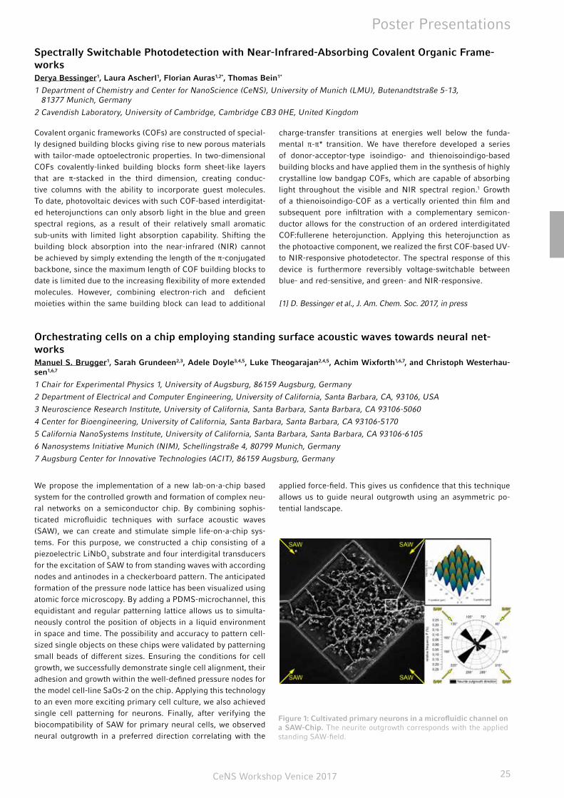
Orchestrating cells on a chip employing standing surface acoustic waves towards neural net-worksManuel S. Brugger1, Sarah Grundeen2,3, Adele Doyle3,4,5, Luke Theogarajan2,4,5, Achim Wixforth1,6,7, and Christoph Westerhau-sen1,6,7
1 Chair for Experimental Physics 1, University of Augsburg, 86159 Augsburg, Germany
2 Department of Electrical and Computer Engineering, University of California, Santa Barbara, CA, 93106, USA
3 Neuroscience Research Institute, University of California, Santa Barbara, Santa Barbara, CA 93106-5060
4 Center for Bioengineering, University of California, Santa Barbara, Santa Barbara, CA 93106-5170
5 California NanoSystems Institute, University of California, Santa Barbara, Santa Barbara, CA 93106-6105
6 Nanosystems Initiative Munich (NIM), Schellingstraße 4, 80799 Munich, Germany
7 Augsburg Center for Innovative Technologies (ACIT), 86159 Augsburg, Germany
We propose the implementation of a new lab-on-a-chip based system for the controlled growth and formation of complex neu-ral networks on a semiconductor chip. By combining sophis-ticated microfluidic techniques with surface acoustic waves (SAW), we can create and stimulate simple life-on-a-chip sys-tems. For this purpose, we constructed a chip consisting of a piezoelectric LiNbO3 substrate and four interdigital transducers for the excitation of SAW to from standing waves with according nodes and antinodes in a checkerboard pattern. The anticipated formation of the pressure node lattice has been visualized using atomic force microscopy. By adding a PDMS-microchannel, this equidistant and regular patterning lattice allows us to simulta-neously control the position of objects in a liquid environment in space and time. The possibility and accuracy to pattern cell-sized single objects on these chips were validated by patterning small beads of different sizes. Ensuring the conditions for cell growth, we successfully demonstrate single cell alignment, their adhesion and growth within the well-defined pressure nodes for the model cell-line SaOs-2 on the chip. Applying this technology to an even more exciting primary cell culture, we also achieved single cell patterning for neurons. Finally, after verifying the biocompatibility of SAW for primary neural cells, we observed neural outgrowth in a preferred direction correlating with the
applied force-field. This gives us confidence that this technique allows us to guide neural outgrowth using an asymmetric po-tential landscape.
Figure 1: Cultivated primary neurons in a microfluidic channel on a SAW-Chip. The neurite outgrowth corresponds with the applied standing SAW-field.

CeNS Workshop Venice 201726
Poster Presentations
Metal-nanoparticle enriched diamond-like carbon as an antimicrobial and wear-resistant surface modification for orthopedic implantsSascha Buchegger1, Sven Fuchs1, Jochen Taiber1, Natascha Schuster1, Caroline Vogel1, Rudolf Herrmann1, Achim Wixforth1, Bernd Stritzker1 and Christoph Westerhausen1
1 Experimental Physics I, Physics Institute, University of Augsburg, Augsburg, Germany
Accompanied by the increasing number of people of higher age, there is also a growing need for total joint replacements, e.g. total hip- or knee replacements. To lower the number of revision surgeries and to reduce stress for the patients, it is of outstanding importance to extend the lifetime of such implants and additionally reduce clinical complications. Our contribution to achieve these aims are hard and wear-resistant diamond-like carbon (DLC) surfaces in combination with various orthopedic base materials. Additional antimicrobial efficacy is achieved by adding Ag, Cu and ZnO nanoparticles to the surface modifica-tion. Our approach is to coat the components with a nanoparticle enriched polymer and subsequently transform it to DLC by plas-ma immersion ion implantation. In case of polymer components of e.g. total joint replacements it is possible to directly modify the surface to DLC and subsequently add metal nanoparticles by metal ion implantation. We investigated the ion release of DLC-modified surfaces and developed a model for the ion release
kinetics. We have shown the release of Zn2+ to be strongly pH-dependent with increasing release in acidic environment. Given that the pH can decrease within inflammations (acidosis), such functionalized surfaces can react to inflammations in an intelli-gent-like manner with a low release in healthy environment and a strongly increased release in case of inflammations. Addition-ally, we successfully increased the thickness of such coatings due to the development of multilayer coatings. Furthermore, we optimized the ion implantation processing of complex shaped prosthesis components by simulating the ion trajectories in or-der to get a homogenous surface modification and developed a novel method to measure the implantation fluence optically. In summary, we optimized the wear resistance and antimicrobial efficacy and extended the applicability to complex shaped pros-thesis components.
Grafting of organophosphonates onto Si(111) by tethering and aggregation for biosensing appli-cationsFrancesco Casablanca1,*, Johannes Bartl1, Qi Li2, Werner Schindler2, Martin Stutzmann1, Anna Cattani-Scholz1
1 Experimental Semiconductor Physics. Walter Schottky Institute and Physics Department. Technische Universität München, Am Coulombwall 4, 85748 Garching bei München, Germany
2 Chemical Physics beyond equilibrium. Physics Department. Technische Universität München, James-Franck-Straße 1, 85748 Garching bei München
Grafting of functional molecules onto semiconductor surfaces is a very active field in surface and interface science. Most of the times, such reactions are performed on hydroxylated sur-faces by means of condensation reactions. To this end, oxides are appealing as a substrate material [1, 2]. However, there is a need to optimize grafting protocols without the use of dielec-tric materials at the interface to achieve better electrical perfor-mance, especially in applications where charging effects should be avoided.
It has been shown that the atomically flat Si(111) surface can be activated to produce an oxide-free surface which is terminated by Si-OH bonds for one third of a monolayer. The remaining surface sites are passivated by hydrogen. The Si-OH termination has shown to have a low energy barrier for the condensation of phosphonic acids, allowing self assembly already at room tem-perature in solution [3]. In this context, we have investigated the grafting of 2-{2-[2-Hydroxy-ethoxy]-ethoxy}-ethyl phosphonic acid on Si(111): this class of molecules is known to be effective against physisorption and the OH-termination has already been
proven to be suitable for further reaction with specific linkers for the bio immobilization of DNA receptors [2]. The quality of the different functionalization steps has been characterized by XPS and AFM.
[1] Hanson, E.L., et al., Journal of the American Chemical Soci-ety,125 (2003): p. 16074-16080.
[2] Cattani-Scholz, A., et al., Biomacromolecules, 10 (2009): p. 489-496.
[3] Thissen, P., et al., Journal of the American Chemical Society, 134 (2012): p. 8869-8874.
CeNS Workshop Venice 2017 27
Poster Presentations
In vitro RNA aggregationAradhana Chopra1,2 and Friedrich C. Simmel1
1 Physics of Synthetic Biological Systems, Physik-Department (ZNN/WSI), Technische Universität München, Garching, Germany
2 Center for NanoScience, Ludwig Maximilians-Universität München, München, Germany
Membrane-bound organelles, such as the endoplasmic reticu-lum and Golgi apparatus, are perfect examples of intracellular organization. They partition the cell into functionally distinct compartments, ensuring that high concentrations of the right molecules localize in the right place at the right time [1]. Over the last few decades, another class of intracellular structures has been identified: organelles that are not bound by a membrane. Instead, these structures self-assemble from a pool of soluble components present in the nucleoplasm or cytoplasm, forming a type of aggregate. They usually comprise of both, RNA and proteins and are called RNA granules. A variety of RNA granules have been identified, such as P-bodies, germ granules in the cytoplasm and Cajal bodies in the nucleus [2,3]. These granules play a role in processes involving RNA metabolism.
Here, we show the formation of another kind of RNA aggregates which are produced during transcription. They are synthesized by the self-assembly of RNA and magnesium pyrophosphate produced during the transcription reaction. These aggregates compartmentalize biomolecules (T7 RNA polymerase, rNTPs, oligonucleotides) and influence the overall product formation. The size of these aggregates is dependent on the duration of transcription, presence of chelating agents and pyrophospha-tase. Transcription induced RNA aggregates provide a simple model system to study membrane-free compartmentalization and control of such aggregates in the confined volume of a cell.
[1] Brangwynne, C. P.; Eckmann, C. R.; Courson, D. S.; Rybarska, A.; Hoege, C.; Gharakhani, J.; Jülicher, F.; Hyman, A. A.; Germline, P. Granules Are Liquid Droplets That Localize by Controlled Disso-lution/ Condensation. Science, 2009, 324 (5935), 1729−1732.
[2] Mitrea, D. M.; Kriwacki, R. W. Phase separation in biology; functional organization of a higher order. Cell Commun. Signaling, 2016, 14, 1.
[3] Hyman, A. A.; Weber, C. A.; Juelicher, F. Liquid-Liquid Phase Separation in Biology. Annu. Rev. Cell Dev. Biol. 2014, 30, 39−58.
Mutual switching as design principle for robust protein patternsJonas Denk1,*, Simon Kretschmer2,*, Jacob Halatek1,*, Caroline Hartl1, Petra Schwille2, and Erwin Frey1
1 Arnold-Sommerfeld-Center for Theoretical Physics and Center for NanoScience, Ludwig-Maximilians-Universität München, Theresienstraße 37, D-80333 München, Germany.
2 Max-Planck-Institute of Biochemistry, Am Klopferspitz 18, D-82152 Martinsried, Germany.
* These authors contributed equally.
Robust protein patterning is vital for many fundamental cellular processes. An established motif of intracellular pattern-forming networks is the self-organization of nucleoside triphosphatases (NTPases), which upon interaction with a cognate NTPase acti-vating protein switch between an NTP-bound and nucleoside di-phosphate (NDP)-bound form. In the Min system, a prototypical example for pattern formation during bacterial cell division, the adenosine triphosphatase (ATPase) MinD in turn triggers a con-formational switch in its activating protein MinE from a latent to a reactive state, although the role of such mutual switching is unclear. By combining nonlinear dynamics analyses and in vitro reconstitution of mutant proteins, we show here that the MinD-dependent switch of MinE is essential for pattern formation in a broad and physiological range of protein concentrations. Our combined theoretical and experimental approach demonstrates that though simpler reaction networks can reproduce patterns, interlinking protein switches confers pattern robustness - a fun-damental prerequisite for the evolvability of organisms.
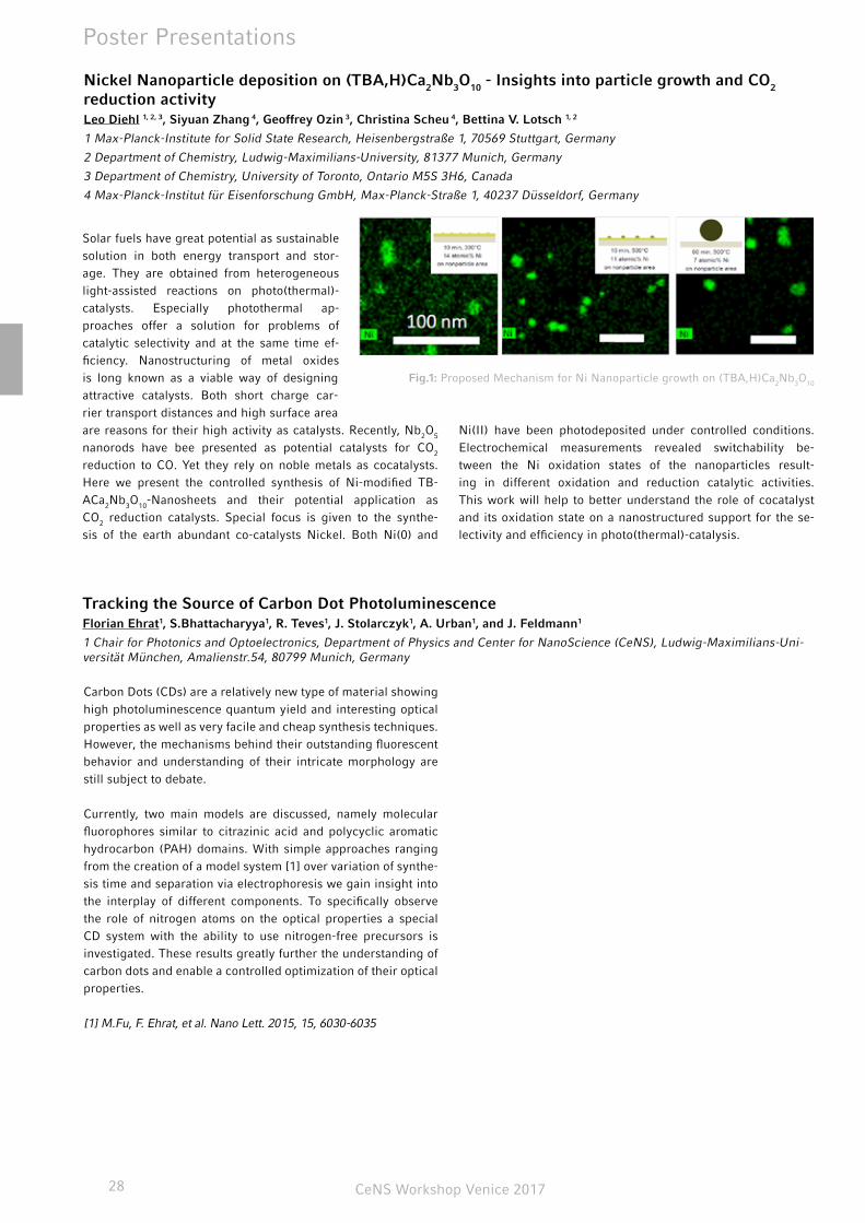
CeNS Workshop Venice 201728
Poster Presentations
Solar fuels have great potential as sustainable solution in both energy transport and stor-age. They are obtained from heterogeneous light-assisted reactions on photo(thermal)-catalysts. Especially photothermal ap-proaches offer a solution for problems of catalytic selectivity and at the same time ef-ficiency. Nanostructuring of metal oxides is long known as a viable way of designing attractive catalysts. Both short charge car-rier transport distances and high surface area are reasons for their high activity as catalysts. Recently, Nb2O5 nanorods have bee presented as potential catalysts for CO2 reduction to CO. Yet they rely on noble metals as cocatalysts. Here we present the controlled synthesis of Ni-modified TB-ACa2Nb3O10-Nanosheets and their potential application as CO2 reduction catalysts. Special focus is given to the synthe-sis of the earth abundant co-catalysts Nickel. Both Ni(0) and
Ni(II) have been photodeposited under controlled conditions. Electrochemical measurements revealed switchability be-tween the Ni oxidation states of the nanoparticles result-ing in different oxidation and reduction catalytic activities. This work will help to better understand the role of cocatalyst and its oxidation state on a nanostructured support for the se-lectivity and efficiency in photo(thermal)-catalysis.
Tracking the Source of Carbon Dot PhotoluminescenceFlorian Ehrat1, S.Bhattacharyya1, R. Teves1, J. Stolarczyk1, A. Urban1, and J. Feldmann1
1 Chair for Photonics and Optoelectronics, Department of Physics and Center for NanoScience (CeNS), Ludwig-Maximilians-Uni-versität München, Amalienstr.54, 80799 Munich, Germany
Carbon Dots (CDs) are a relatively new type of material showing high photoluminescence quantum yield and interesting optical properties as well as very facile and cheap synthesis techniques. However, the mechanisms behind their outstanding fluorescent behavior and understanding of their intricate morphology are still subject to debate.
Currently, two main models are discussed, namely molecular fluorophores similar to citrazinic acid and polycyclic aromatic hydrocarbon (PAH) domains. With simple approaches ranging from the creation of a model system [1] over variation of synthe-sis time and separation via electrophoresis we gain insight into the interplay of different components. To specifically observe the role of nitrogen atoms on the optical properties a special CD system with the ability to use nitrogen-free precursors is investigated. These results greatly further the understanding of carbon dots and enable a controlled optimization of their optical properties.
[1] M.Fu, F. Ehrat, et al. Nano Lett. 2015, 15, 6030-6035
Nickel Nanoparticle deposition on (TBA,H)Ca2Nb3O10 - Insights into particle growth and CO2 reduction activityLeo Diehl 1, 2, 3, Siyuan Zhang 4, Geoffrey Ozin 3, Christina Scheu 4, Bettina V. Lotsch 1, 2
1 Max-Planck-Institute for Solid State Research, Heisenbergstraße 1, 70569 Stuttgart, Germany
2 Department of Chemistry, Ludwig-Maximilians-University, 81377 Munich, Germany
3 Department of Chemistry, University of Toronto, Ontario M5S 3H6, Canada
4 Max-Planck-Institut für Eisenforschung GmbH, Max-Planck-Straße 1, 40237 Düsseldorf, Germany
Fig.1: Proposed Mechanism for Ni Nanoparticle growth on (TBA,H)Ca2Nb3O10
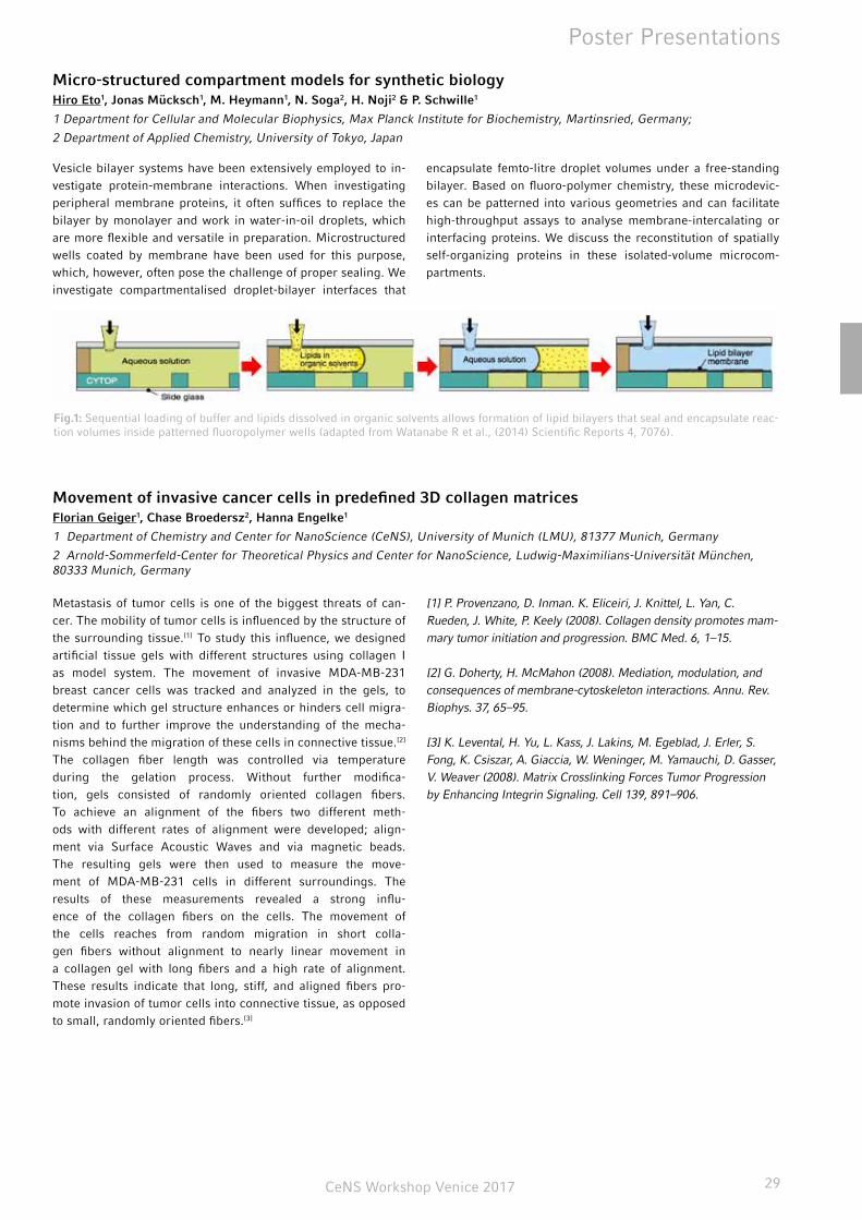
CeNS Workshop Venice 2017 29
Poster Presentations
Micro-structured compartment models for synthetic biologyHiro Eto1, Jonas Mücksch1, M. Heymann1, N. Soga2, H. Noji2 & P. Schwille1
1 Department for Cellular and Molecular Biophysics, Max Planck Institute for Biochemistry, Martinsried, Germany;
2 Department of Applied Chemistry, University of Tokyo, Japan
Vesicle bilayer systems have been extensively employed to in-vestigate protein-membrane interactions. When investigating peripheral membrane proteins, it often suffices to replace the bilayer by monolayer and work in water-in-oil droplets, which are more flexible and versatile in preparation. Microstructured wells coated by membrane have been used for this purpose, which, however, often pose the challenge of proper sealing. We investigate compartmentalised droplet-bilayer interfaces that
encapsulate femto-litre droplet volumes under a free-standing bilayer. Based on fluoro-polymer chemistry, these microdevic-es can be patterned into various geometries and can facilitate high-throughput assays to analyse membrane-intercalating or interfacing proteins. We discuss the reconstitution of spatially self-organizing proteins in these isolated-volume microcom-partments.
Fig.1: Sequential loading of buffer and lipids dissolved in organic solvents allows formation of lipid bilayers that seal and encapsulate reac-tion volumes inside patterned fluoropolymer wells (adapted from Watanabe R et al., (2014) Scientific Reports 4, 7076).
Movement of invasive cancer cells in predefined 3D collagen matricesFlorian Geiger1, Chase Broedersz2, Hanna Engelke1
1 Department of Chemistry and Center for NanoScience (CeNS), University of Munich (LMU), 81377 Munich, Germany
2 Arnold-Sommerfeld-Center for Theoretical Physics and Center for NanoScience, Ludwig-Maximilians-Universität München, 80333 Munich, Germany
Metastasis of tumor cells is one of the biggest threats of can-cer. The mobility of tumor cells is influenced by the structure of the surrounding tissue.[1] To study this influence, we designed artificial tissue gels with different structures using collagen I as model system. The movement of invasive MDA-MB-231 breast cancer cells was tracked and analyzed in the gels, to determine which gel structure enhances or hinders cell migra-tion and to further improve the understanding of the mecha-nisms behind the migration of these cells in connective tissue.[2] The collagen fiber length was controlled via temperature during the gelation process. Without further modifica-tion, gels consisted of randomly oriented collagen fibers. To achieve an alignment of the fibers two different meth-ods with different rates of alignment were developed; align-ment via Surface Acoustic Waves and via magnetic beads. The resulting gels were then used to measure the move-ment of MDA-MB-231 cells in different surroundings. The results of these measurements revealed a strong influ-ence of the collagen fibers on the cells. The movement of the cells reaches from random migration in short colla-gen fibers without alignment to nearly linear movement in a collagen gel with long fibers and a high rate of alignment. These results indicate that long, stiff, and aligned fibers pro-mote invasion of tumor cells into connective tissue, as opposed to small, randomly oriented fibers.[3]
[1] P. Provenzano, D. Inman. K. Eliceiri, J. Knittel, L. Yan, C. Rueden, J. White, P. Keely (2008). Collagen density promotes mam-mary tumor initiation and progression. BMC Med. 6, 1–15.
[2] G. Doherty, H. McMahon (2008). Mediation, modulation, and consequences of membrane-cytoskeleton interactions. Annu. Rev. Biophys. 37, 65–95.
[3] K. Levental, H. Yu, L. Kass, J. Lakins, M. Egeblad, J. Erler, S. Fong, K. Csiszar, A. Giaccia, W. Weninger, M. Yamauchi, D. Gasser, V. Weaver (2008). Matrix Crosslinking Forces Tumor Progression by Enhancing Integrin Signaling. Cell 139, 891–906.
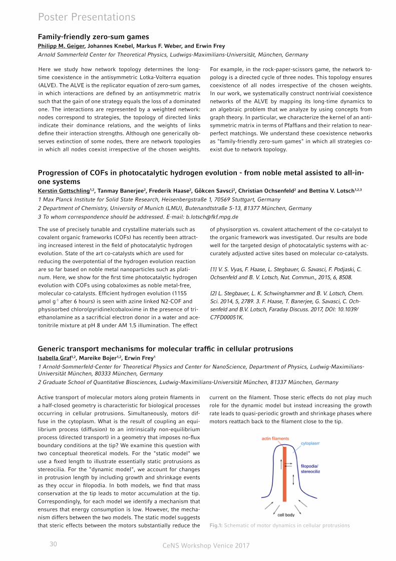
CeNS Workshop Venice 201730
Poster Presentations
Generic transport mechanisms for molecular traffic in cellular protrusionsIsabella Graf1,2, Mareike Bojer1,2, Erwin Frey1
1 Arnold-Sommerfeld-Center for Theoretical Physics and Center for NanoScience, Department of Physics, Ludwig-Maximilians-Universität München, 80333 München, Germany
2 Graduate School of Quantitative Biosciences, Ludwig-Maximilians-Universität München, 81337 München, Germany
Active transport of molecular motors along protein filaments in a half-closed geometry is characteristic for biological processes occurring in cellular protrusions. Simultaneously, motors dif-fuse in the cytoplasm. What is the result of coupling an equi-librium process (diffusion) to an intrinsically non-equilibrium process (directed transport) in a geometry that imposes no-flux boundary conditions at the tip? We examine this question with two conceptual theoretical models. For the "static model" we use a fixed length to illustrate essentially static protrusions as stereocilia. For the "dynamic model", we account for changes in protrusion length by including growth and shrinkage events as they occur in filopodia. In both models, we find that mass conservation at the tip leads to motor accumulation at the tip. Correspondingly, for each model we identify a mechanism that ensures that energy consumption is low. However, the mecha-nism differs between the two models. The static model suggests that steric effects between the motors substantially reduce the
current on the filament. Those steric effects do not play much role for the dynamic model but instead increasing the growth rate leads to quasi-periodic growth and shrinkage phases where motors reattach back to the filament close to the tip.
Fig.1: Schematic of motor dynamics in cellular protrusions
Progression of COFs in photocatalytic hydrogen evolution - from noble metal assisted to all-in-one systemsKerstin Gottschling1,2, Tanmay Banerjee2, Frederik Haase2, Gökcen Savsci2, Christian Ochsenfeld2 and Bettina V. Lotsch1,2,3
1 Max Planck Institute for Solid State Research, Heisenbergstraße 1, 70569 Stuttgart, Germany
2 Department of Chemistry, University of Munich (LMU), Butenandtstraße 5-13, 81377 München, Germany
3 To whom correspondence should be addressed. E-mail: [email protected]
The use of precisely tunable and crystalline materials such as covalent organic frameworks (COFs) has recently been attract-ing increased interest in the field of photocatalytic hydrogen evolution. State of the art co-catalysts which are used for reducing the overpotential of the hydrogen evolution reaction are so far based on noble metal nanoparticles such as plati-num. Here, we show for the first time photocatalytic hydrogen evolution with COFs using cobaloximes as noble metal-free, molecular co-catalysts. Efficient hydrogen evolution (1155 μmol g-1 after 6 hours) is seen with azine linked N2-COF and physisorbed chloro(pyridine)cobaloxime in the presence of tri-ethanolamine as a sacrificial electron donor in a water and ace-tonitrile mixture at pH 8 under AM 1.5 illumination. The effect
of physisorption vs. covalent attachement of the co-catalyst to the organic framework was investigated. Our results are bode well for the targeted design of photocatalytic systems with ac-curately adjusted active sites based on molecular co-catalysts.
[1] V. S. Vyas, F. Haase, L. Stegbauer, G. Savasci, F. Podjaski, C. Ochsenfeld and B. V. Lotsch, Nat. Commun., 2015, 6, 8508.
[2] L. Stegbauer, L. K. Schwinghammer and B. V. Lotsch, Chem. Sci. 2014, 5, 2789. 3. F. Haase, T. Banerjee, G. Savasci, C. Och-senfeld and B.V. Lotsch, Faraday Discuss. 2017, DOI: 10.1039/C7FD00051K.
Family-friendly zero-sum games Philipp M. Geiger, Johannes Knebel, Markus F. Weber, and Erwin Frey
Arnold Sommerfeld Center for Theoretical Physics, Ludwigs-Maximilians-Universität, München, Germany
Here we study how network topology determines the long-time coexistence in the antisymmetric Lotka-Volterra equation (ALVE). The ALVE is the replicator equation of zero-sum games, in which interactions are defined by an antisymmetric matrix such that the gain of one strategy equals the loss of a dominated one. The interactions are represented by a weighted network: nodes correspond to strategies, the topology of directed links indicate their dominance relations, and the weights of links define their interaction strengths. Although one generically ob-serves extinction of some nodes, there are network topologies in which all nodes coexist irrespective of the chosen weights.
For example, in the rock-paper-scissors game, the network to-pology is a directed cycle of three nodes. This topology ensures coexistence of all nodes irrespective of the chosen weights. In our work, we systematically construct nontrivial coexistence networks of the ALVE by mapping its long-time dynamics to an algebraic problem that we analyze by using concepts from graph theory. In particular, we characterize the kernel of an anti-symmetric matrix in terms of Pfaffians and their relation to near-perfect matchings. We understand these coexistence networks as "family-friendly zero-sum games" in which all strategies co-exist due to network topology.
CeNS Workshop Venice 2017 31
Poster Presentations
Dephasing and quantum beating of excitons in methylammonium lead iodide perovskite nano-plateletsBernhard J. Bohn1,2, Moritz Gramlich1,2, Abraham Moreno1,2, Thomas Simon1,2, Alexander F. Richter1,2, Lakshminarayana Polav-arapu1,2, Alexander S. Urban1,2, and Jochen Feldmann1,2
1 Chair for Photonics and Optoelectronics, Department of Physics and Center for NanoScience (CeNS), Ludwig-Maximilians-Uni-versität (LMU), Amalienstraße 54, 80799 Munich, Germany
2 Nanosystems Initiative Munich (NIM), Schellingstraße 4, 80799 Munich, Germany
Perovskite nanocrystals have emerged as an interesting material for light-emitting and other optoelectronic applications. Exci-tons are known to play an important role in determining the opti-cal properties of these nanocrystals and their energetic levels as well as quantization properties have been extensively explored. Despite this, there are still many aspects of perovskites that are still not well known, e.g. the homogeneous and inhomogeneous linewidths of the energetic transitions, quantities that cannot be directly extracted by linear absorption optical spectroscopy on nanocrystal ensembles. Here, we present temperature-depen-dent absorption and four-wave mixing (FWM) experiments on thick methylammonium lead iodide (MAPI) perovskite nano-
platelets exhibiting bulk-like absorption and emission spectra. Dephasing times T2 of excitons are determined to lie in the range of several hundreds of femtoseconds at low temperatures. This value enables us to distinguish between the homogeneous and inhomogeneous contribution to the total broadening of the excitonic transitions. These turn out to be predominantly inhomogeneously broadened at low temperatures and homoge-neously broadened at room temperature. Furthermore, we find excitonic quantum beats, which allow for the determination of the exciton binding energy and we extract EB = 25±1 meV in the low temperature regime, in good agreement with other reports.
Mechanical Properties of Leishmania Myosin XXI determined with an Optical Tweezers Trans-ducerAndreas Graw, Christopher Batters, and Claudia Veigel
Ludwig-Maximilians-Universität München, Department of Cellular Physiology, Großhaderner Str. 9, 82152 Planegg-Martinsried
Myosins form a large family of actin-based motor proteins that are involved in different forms of cellular motility. Those include muscle contraction, intracellular transport, endo- and exocytosis, cell division and locomotion. The force and movement is produced by a change in conformation of the bound actomyosin complex. Myosin XXI is the only myosin expressed in Leish-mania parasites. Therefore, it has to perform a vari-ety of motile functions. It was shown that Calmodu-lin regulates dimerization, motility, and lipid binding.1 Here we show the first mechanical measurements to determine the stiffness and working stroke of a single myosin XXI cross bridge with an optical tweezers transducer. These measure-ments are made with the “three bead” geometry originally de-vised by Finer et al.2. The movements and forces produced by actomyosin interactions are observed by detecting the position of both trapped beads with four-quadrant-photodiodes. Our first experiments on a supporting lipid layer suggest that the myosin XXI produces a small (≈2nm) working stroke, with a half lifetime of 20ms at 2mM ATP. To apply additional force on the cross-bridge one bead was driven with a sinusoidal forcing function. This leads to a further step of about 10nm.
[1] Batters et al., PNAS 111 2014
[2] Finer et al., Nature 386 1994
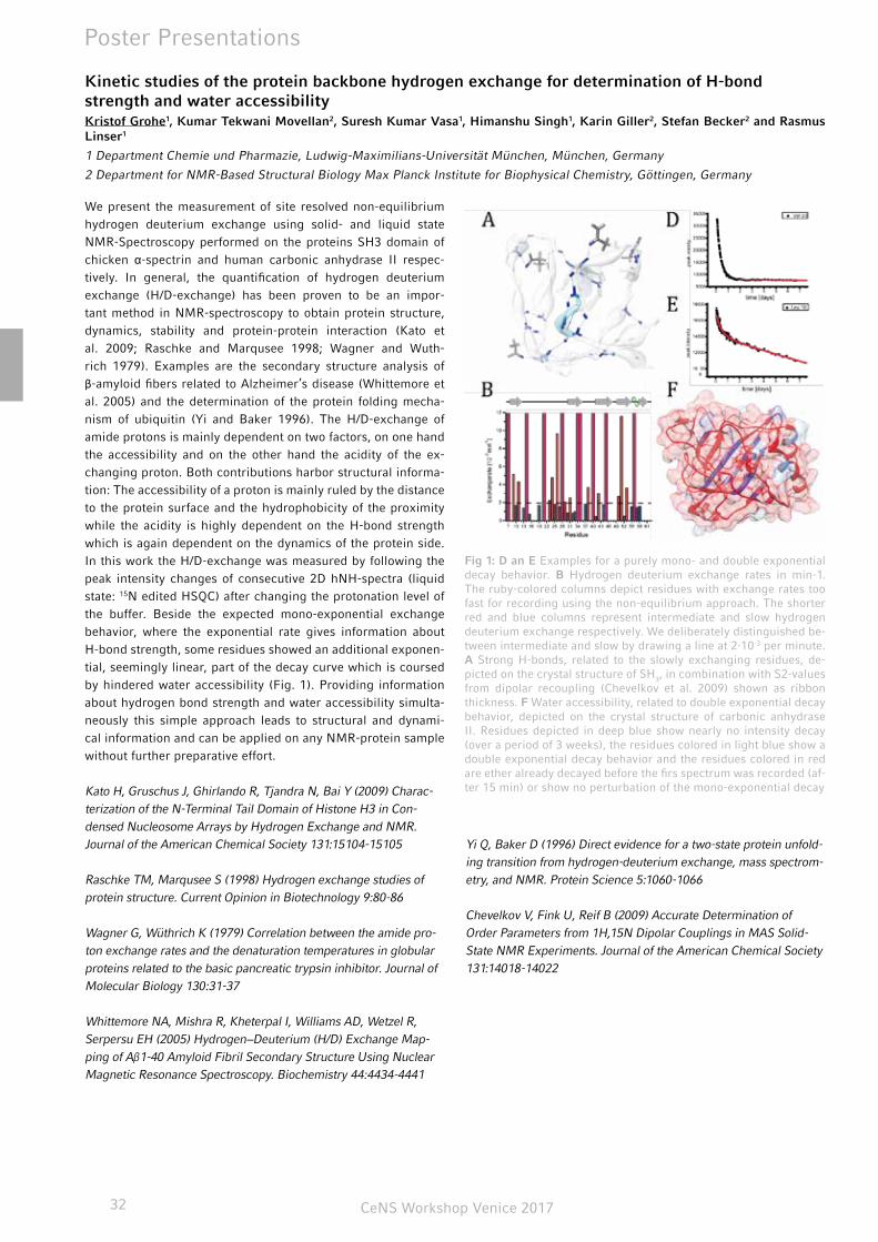
CeNS Workshop Venice 201732
Poster Presentations
Kinetic studies of the protein backbone hydrogen exchange for determination of H-bond strength and water accessibilityKristof Grohe1, Kumar Tekwani Movellan2, Suresh Kumar Vasa1, Himanshu Singh1, Karin Giller2, Stefan Becker2 and Rasmus Linser1
1 Department Chemie und Pharmazie, Ludwig-Maximilians-Universität München, München, Germany
2 Department for NMR-Based Structural Biology Max Planck Institute for Biophysical Chemistry, Göttingen, Germany
We present the measurement of site resolved non-equilibrium hydrogen deuterium exchange using solid- and liquid state NMR-Spectroscopy performed on the proteins SH3 domain of chicken α-spectrin and human carbonic anhydrase II respec-tively. In general, the quantification of hydrogen deuterium exchange (H/D-exchange) has been proven to be an impor-tant method in NMR-spectroscopy to obtain protein structure, dynamics, stability and protein-protein interaction (Kato et al. 2009; Raschke and Marqusee 1998; Wagner and Wuth-rich 1979). Examples are the secondary structure analysis of β-amyloid fibers related to Alzheimer’s disease (Whittemore et al. 2005) and the determination of the protein folding mecha-nism of ubiquitin (Yi and Baker 1996). The H/D-exchange of amide protons is mainly dependent on two factors, on one hand the accessibility and on the other hand the acidity of the ex-changing proton. Both contributions harbor structural informa-tion: The accessibility of a proton is mainly ruled by the distance to the protein surface and the hydrophobicity of the proximity while the acidity is highly dependent on the H-bond strength which is again dependent on the dynamics of the protein side. In this work the H/D-exchange was measured by following the peak intensity changes of consecutive 2D hNH-spectra (liquid state: 15N edited HSQC) after changing the protonation level of the buffer. Beside the expected mono-exponential exchange behavior, where the exponential rate gives information about H-bond strength, some residues showed an additional exponen-tial, seemingly linear, part of the decay curve which is coursed by hindered water accessibility (Fig. 1). Providing information about hydrogen bond strength and water accessibility simulta-neously this simple approach leads to structural and dynami-cal information and can be applied on any NMR-protein sample without further preparative effort.
Kato H, Gruschus J, Ghirlando R, Tjandra N, Bai Y (2009) Charac-terization of the N-Terminal Tail Domain of Histone H3 in Con-densed Nucleosome Arrays by Hydrogen Exchange and NMR. Journal of the American Chemical Society 131:15104-15105
Raschke TM, Marqusee S (1998) Hydrogen exchange studies of protein structure. Current Opinion in Biotechnology 9:80-86
Wagner G, Wüthrich K (1979) Correlation between the amide pro-ton exchange rates and the denaturation temperatures in globular proteins related to the basic pancreatic trypsin inhibitor. Journal of Molecular Biology 130:31-37
Whittemore NA, Mishra R, Kheterpal I, Williams AD, Wetzel R, Serpersu EH (2005) Hydrogen−Deuterium (H/D) Exchange Map-ping of Aβ1-40 Amyloid Fibril Secondary Structure Using Nuclear Magnetic Resonance Spectroscopy. Biochemistry 44:4434-4441
Yi Q, Baker D (1996) Direct evidence for a two-state protein unfold-ing transition from hydrogen-deuterium exchange, mass spectrom-etry, and NMR. Protein Science 5:1060-1066
Chevelkov V, Fink U, Reif B (2009) Accurate Determination of Order Parameters from 1H,15N Dipolar Couplings in MAS Solid-State NMR Experiments. Journal of the American Chemical Society 131:14018-14022
Fig 1: D an E Examples for a purely mono- and double exponential decay behavior. B Hydrogen deuterium exchange rates in min-1. The ruby-colored columns depict residues with exchange rates too fast for recording using the non-equilibrium approach. The shorter red and blue columns represent intermediate and slow hydrogen deuterium exchange respectively. We deliberately distinguished be-tween intermediate and slow by drawing a line at 2·10-3 per minute. A Strong H-bonds, related to the slowly exchanging residues, de-picted on the crystal structure of SH3, in combination with S2-values from dipolar recoupling (Chevelkov et al. 2009) shown as ribbon thickness. F Water accessibility, related to double exponential decay behavior, depicted on the crystal structure of carbonic anhydrase II. Residues depicted in deep blue show nearly no intensity decay (over a period of 3 weeks), the residues colored in light blue show a double exponential decay behavior and the residues colored in red are ether already decayed before the firs spectrum was recorded (af-ter 15 min) or show no perturbation of the mono-exponential decay
CeNS Workshop Venice 2017 33
Poster Presentations
Controlling cell functions artificially via silencing genes by delivery of synthetic microRNAsLisa Haddick1, Karin Möller1, Wei Zhang2, Stephan Morys2, Ernst Wagner2 and Thomas Bein1
1 Department of Chemistry and Center for NanoScience, University of Munich (LMU), Butenandtstrasse 5-13, 81377 Munich, Germany. E-mail: [email protected]; Fax: (+49) 89-2180-77622
2 Pharmaceutical Biotechnology and Center for NanoScience, University of Munich (LMU), Butenandtstrasse 5-13, 81377 Munich, Germany. E-mail: [email protected]
Since the discovery of RNA interference as a means to control the expression of genes, enormous research interest has arisen to exploit synthetic RNA as a therapeutic tool. In this context, microRNAs (miR) are promising candidates for achieving highly specific anti-cancer treatments via silencing oncogenes with tumor suppressor miR. miR are short, non-coding RNAs that regulate the expression level of genes by binding and cleaving mRNA and can be used to control cell functions artificially. In this work we are focusing on the cellular delivery of miR200c, which is a potent inhibitor of tumor progression and therapy resistance.
For specific medical applications of miR, challenges that need to be overcome include the delivery of plain nucleic acids while preventing premature degradation by nucleases, limited cell
uptake due to the negative charge of nucleic acids, and immu-nogenic side effects. To address these problems, mesoporous silica nanoparticles (MSNs) can be used as a delivery platform to target cancer tissue and to safeguard the miR during trans-port. Here, multifunctional core-shell MSNs were used as deliv-ery agents for targeted delivery of miR200c to cancer cells. The positively charged core of these core-shell MSNs with a pore diameter of about 4 nm and stellate pore morphology enables a high loading capacity for miR. The negatively charged MSN shell was associated with a block copolymer acting as capping and endosomal release agent. The efficient delivery of miR200c into eGFP-Luciferase reporter cells featuring a miR200c target site on the expressed mRNA was assessed by means of lucifer-ase reporter gene knock-down.
Carbon templated Nb:TiO2 nanostructures as oxygen evolution catalyst supports for PEM elec-trolyzersSebastian Häringer1, Alexander G. Hufnagel1, Michael Beetz1, Daniel Böhm1, Dina Fattakhova-Rohlfing2, and Thomas Bein1
1 Department of Chemistry and Center for NanoScience (CeNS), University of Munich (LMU)
2 Current Adress: Institute of Energy and Climate Research Jülich (IEK-1) and University of Duisburg-Essen
The steadily increasing proportion of fluctuating renewable en-ergies in the national power supply requires the development of means for the intermittent storage of energy. The storage in chemical energy via electrochemical water splitting in proton exchange membrane (PEM) electrolyzers offers a promising ap-proach.[1] So far, noble metal oxides such as iridium oxide have been identified as the most efficient way to catalyze the oxygen evolution reaction (OER). For further improvement of the effi-ciency and production costs, nanostructured conductive oxides are being developed as catalyst support due to their high stabil-ity towards oxidation at the high anodic potentials.
Here, we used thermal atomic layer deposition (ALD) to coat nanostructured carbon (soot) templates with mixed TiO2/Nb2O5 films using alkoxides and water as precursors.[2] Increasing the
amount of niobium in titania leads to a suppression of the crys-tallization during the deposition, resulting in amorphous, highly conformal films. The carbon phase stabilizes the nanostructure during annealing, yielding crystalline high-surface materials with a conductivity of up to 400 S cm-1. The resulting nanostruc-tured carrier material was loaded with an OER catalyst devel-oped in our group, consisting of ultrasmall RuIrOx nanoparticles and the OER performance and stability under operation were investigated.
[1] C. Spoeri, J. Kwan et al., Angew. Chem. Int. Ed. 2017, 56, 5994-6021.
[2] V. Pore, M. Ritala et al., Cryst. Growth Des. 2009, 9, 2974-2978.
CeNS Workshop Venice 201734
Poster Presentations
A new LGPS-type superionic conductor – synthesis and characterization of Li7SiPS8
Sascha Harm1, Anna-Katharina Hatz1,2, Igor Moudrakovski 2, Robert E.Usiskin2, Joachim Maier2, Bettina V. Lotsch1,2*
1 Department of Chemistry, University of Munich (LMU), Butenandtstraße 5-13, D-81377 Munich
2 Max Planck Institute for Solid State Research, Heisenbergstraße 1, D-70569 Stuttgart
*E-mail of the Corresponding Author: [email protected]
Tetragonal LGPS (lithium germanium thiophosphate) is one of the most promising thio-LISICON (lithium super ionic con-ductor) materials up to date. It was first introduced by Mitsui et al. in 2011 exceeding conductivity values of the best crys-talline lithium ion conductors by one order of magnitude.[1] This system can be described as Li11Me2-xP1+xS12 (LMePS) with Me = Si, Ge, Sn. Theoretical studies by Ceder et al.[2] pre-dicted decreasing activation energies for the lithium ion dif-fusion process and increasing conductivity in the series Sn → Ge → Si which is in accordance with recent findings.[3,4] We report on the facile solid-state synthesis and characterisa-tion of Li7SiPS8, a new member of the tetragonal LSiPS fam-ily. This new material shows a specific conductivity value of 2 mScm-1, which is unexpectedly low in contrast to other members of this solid solution system. Utilizing ssNMR and impedance spectroscopy we could determine that amorphous by-products limit the intergrain conductivity in such a way that a bulk con-
ductivity could not be observed, disguising a potentially much higher specific conductivity.
[1] N. Kayama, K. Homma, Y. Yamakawa, R. Kanno, M. Yonemura, T. Kamiyama, Y. Kato, S. Hama, K. Kawamoto, A. Mitsui, Nat. Mater.,10 (2011), 682; A. Kuhn, J. Köhler, B. V. Lotsch, Phys. Chem. Chem. Phys., 15 (2013), 11620.
[2] S. P. Ong, Y. Mo, W. D. Richards, L. Miara, H. S. Lee, G. Ceder, Energy Environ. Sci., 6 (2013), 148.
[3] P. Bron, S. Johansson, K. Zick, J. Schmedt a. d. Günne, S. Dehnen, B. Roling, J. Am. Chem. Soc.,135 (2013), 15694.
[4] A. Kuhn, O. Gerbig, C. Zhu, F. Falkenberg, J. Maier, B. V. Lotsch, Phys. Chem. Chem. Phys., 16 (2014), 14669.
Photocontrolled nuclear translocation of mechanotransduction protein YAPBernhard Illes, Hanna Engelke
Ludwig-Maximilians-Universität München
Yes associated Protein (YAP) is one of the proteins responsible for the mechanotransduction of the cell as well as its prolifera-tion and branching behavior. To investigate this behavior and the mechanisms behind it, controlling the YAP activity is es-sential. YAP is only active while in the nucleus, so control of its nuclear translocation is one possible path to controlling YAP activity. For this purpose we applied a previously developed op-toNLS to YAP, replacing its original nuclear localization signal, confining it to the cytoplasm until its translocation is activated through UV light. To make sure that YAP is not inhibited in its function through its usual nuclear localization pathway, a phos-phorylation of serine S127, a mutation was introduced of the Serine to Alanine was introduced (S127A), creating a perma-nently photoactive YAP. To monitor YAPs presence in the cyto-plasm and in the nucleus after photoactivation via fluorescence microscopy and to prevent its passive diffusion into the nucleus, two GFP-proteins were attached to the c-terminal end of YAP.
This construct was transfected into Heck293 and other easily transfectable cell lines and showed to permanently reside in the cytoplasm, allowing for a controlled translocation to the nucleus via UV illumination.
Enzyme Activity at Lipid Membranes – Correlation of Activity and Membrane State of an intrin-sically water-soluble enzymeAndrej Kamenac1,2, Matthias Schneider3, Achim Wixforth1,2, Christoph Westerhausen1,2
1 Chair for experimental physics I, University of Augsburg, 86159 Augsburg, Germany
2 Nanosystems Initiative Munich (NIM), 80799 Munich, Germany
3 Medizinische und Biologische Physik, Technische Universität Dortmund, 44227 Dortmund, Germany
The mechanism of catalysis in the induced fit theory has failed to explain observations where correlations of the membrane phase transitions and the activity of membrane associated enzymes have been found. We here present such an observed correlation for an intrinsically water-soluble enzyme ADAMTS13 (a disinte-
grin and metalloproteinase with a thrombospondin type 1 mo-tif, member 13) bound to phospholipid membranes. Attached to 1,2-dimyristoyl-sn-glycero-3-phosphocholine (14:0PC) vesicles the temperature-dependence of the enzyme activity with a prom-inent peak around the main phase transition temperature even
Fig.1: Schematic view of the photoactivated translocation of YAP into the nucleus and its subsequent activation leading controlled cell branching

CeNS Workshop Venice 2017 35
Poster Presentations
Oligothiophene-Bridged Conjugated Covalent Organic FrameworksNiklas Keller,1 Derya Bessinger,1 Stephan Reuter,1 Mona Calik,1 Laura Ascherl,1 Fabian C. Hanusch,1 Florian Auras,1,2* Thomas Bein1*
1 Department of Chemistry and Center for NanoScience (CeNS), University of Munich (LMU), Butenandtstraße 5-13, 81377 Munich, Germany
2 Cavendish Laboratory, University of Cambridge, Cambridge CB3 0HE, United Kingdom
2-Dimensional covalent organic frameworks (2D-COFs) are crystalline porous materials comprising aligned columns of π-stacked building blocks. Regarding potential applications of COFs in organic electronics and optoelectronics, access to oli-gothiophene-based COFs would be of great interest. However, the realization of such materials has remained a challenge, in particular concerning the laterally conjugated imine-linked COFs. We have developed a new building block design, imple-menting an asymmetric modification on an otherwise symmet-ric backbone that allows us to obtain a series of highly crystal-line quaterthiophene (4T)-derived COFs with tunable electronic properties.1 Studying the optical response of these materials, we have observed for the first time the formation of a charge transfer state between the COF subunits across the imine bond. In this work, we have developed the first quaterthiophene-based 2D covalent organic frameworks comprising ordered π-stacked columns of 4T and pyrene moieties. Applying an asymmetric functionalization strategy of the otherwise C2-symmetric 4T backbone allowed us to incorporate alkyl chains for optimized solubility while still retaining the ability of the building blocks to
stack in close-packed face-on thiophene columns. We also dem-onstrate that this approach provides a facile route for modifying the electronic properties of the 4T backbone via incorporation of electron-deficient subunits, thus forming donor-acceptor type chromophores. The absorption and emission spectra confirm that the 4T-based building blocks are electronically integrated into the framework. Spectral features below the energy of the π-π* transition and the analysis of the corresponding emis-sion decay time traces reveal the fast and efficient formation of a charge transfer state between the imine-linked pyrene and quaterthiophene subunits. We believe that our new asymmetric building block design provides a general strategy for the synthe-sis of well-ordered COFs from various extended building blocks. This will greatly expand the range of applicable molecules for realizing frameworks with tailor-made optoelectronic proper-ties.
[1] N. Keller, D. Bessinger, S. Reuter, M. Calik, L. Ascherl, F.C. Hanusch, F. Auras, T. Bein. J. Am. Chem. Soc., 2017, 139 (24), pp 8194–8199
Folate receptor-directed siRNA lipopolyplexes for tumor-targeted gene silencing in vivoSarah Kern1, Philipp Klein1, Wei Zhang1, Dian-Jang Lee1,2, Ernst Wagner1,2
1 Department of Pharmacy and Center for NanoScience, Ludwig-Maximilians-Universität, Munich, Germany
2 Nanosystems Initiative Munich, Germany
Small interfering RNA (siRNA)-based therapeutics are shown to be a promising approach in cancer therapy based on their ability to specifically down-regulate gene expression. However, siRNA by itself is incapable of reaching the tumor site and has to over-come various hurdles on its way to successful knockdown of tar-get genes. Consequently, efficient carrier systems are required.By solid phase-supported synthesis we accomplished to design sequence-defined DBCO-PEG agents with different mono- and bivalent DBCO click reagents and the shielding agent PEG to prolong in vivo circulation. Furthermore, modification of siRNA lipopolyplexes with folic acid was exploited for receptor-me-diated uptake into folate receptor-overexpressing tumor cells.The obtained formulations were compared in terms of particle formation, serum stability, binding, uptake and transfection ef-
ficiency, as well as biodistribution and tumor retention in vivo. After screening, the formulation DBCO2-ss2-PEG24-FolA was selected to evaluate gene silencing efficiency in vivo. In addi-tion, the health status of treated animals was evaluated by both monitoring body weight development and analysis of clinical biochemistry parameters.
The results demonstrate that in absence of any sanitary impair-ment, the agent combining PEG24, bis-DBCO and folic acid tar-geting ligand presents best biodistribution profile, longest re-tention (>4 h) and ~60% mRNA knockdown of target gene at the tumor site. Taken together, this study provides a promising approach for tumor-directed siRNA delivery.
shows a partial Anti-Arrhenius behavior in strong contrast to the vesicle free references. By exchanging the lipid from 14:0PC to 15:0PC again an activity peak is present, but shifted towards higher temperatures, again matching the membrane phase tran-
sition temperature. These observations are in accordance with the theory of catalysis by Kaufmann that predicts a correlation of increased system fluctuations, as present at the phase transition, and the activity of a membrane associated enzyme.
CeNS Workshop Venice 201736
poster abstracts - session ii (kie-z)
Photochemically induced electrophoresis for bind-ing quantification and all-optical mobility measure-mentsFriederike M. Möller, Michael Kieß, Dieter Braun . . . . . . . . . . . . . 37
Movement of a DNA based robotic armEnzo Kopperger, J. List, S. Madhira, F. Rothfischer, D. C. Lamb, F. C. Simmel . . . . . . . . . . . . . . . . . . . . . . . . . . . . . . . . . . . . . . . . . . . . . . 37
DNA origami pores: design and membrane interac-tionSwati Krishnan, Daniela Ziegler, Friedrich Simmel . . . . . . . . . . . . 38
Collective cell migration: Can an advancing cell sheet be compared to a hydrodynamic fluid? Matthias Zorn, Anna-Kristina Marel, Felix Kempf, Janina Lange, Erwin Frey and Joachim O. Rädler . . . . . . . . . . . . . . . . . . . . . . . . 38
Activity and effects of a human LINE-1 retrotrans-poson in bacteriaGloria Lee, Nicholas A. Sherer, Neil H. Kim, Ema Rajic, Davneet Kaur, K. Michael Martini, Chi Xue, Nigel Goldenfeld, and Thomas E. Kuhlman . . . . . . . . . . . . . . . . . . . . . . . . . . . . . . . . . . . . . . . . . . . . . 39
Chemically driven ligation chain reaction – toward protein-free hypercycles in sequence spaceStefanie Leiner, Evgeniia Edeleva, and Dieter Braun . . . . . . . . . . . 39
Long-lived direct and indirect interlayer excitons in van-der-Waals heterostructures Fabian Merbeler, Bastian Miller, Alexander Steinhoff, Borja Pano, Frank Jahnke, Alexander Holleitner, Ursula Wurstbauer . . . . . . . . 40
Dynamic structural biology of membrane transport-ers: tools and applicationsAlessandra Narducci, C. Gebhardt, R. Mächtel, T. Cordes . . . . . . . 40
Direct characterization of the evanescent field in objective-type total internal reflection microscopyChristian Niederauer, Philipp Blumhardt, Jonas Mücksch, Michael Heymann, Armin Lambacher, Petra Schwille . . . . . . . . . . . . . . . . . 41
Interactions of the motor protein myosin-VI with lipid bilayersAnastasiia B. Petrova, Michael Krzyk, Benoit Rogez, Christopher Batters and Claudia Veigel . . . . . . . . . . . . . . . . . . . . . . . . . . . . . . . 41
“In-situ” XAFS and SAXS study of the kinetics of growth of Au nanowiresJosé Martín Ramallo-López, Fernando Pschunder, Christina Hoppe, Lisandro J. Giovanetti, Christián Huck-Iriart, Felix G. Requejo . . . 41
Rotary motors made from DNALauren Quednau, Scott-Michael Slone, Christopher Maffeo, Philip Ketterer, Hendrik Dietz, Aleksei Aksimentiev . . . . . . . . . . . . . . . . . 42
Studying CXCR4 oligomerization with image cor-relation spectroscopy and fluorescence lifetime imagingChen Qian, Don C. Lamb . . . . . . . . . . . . . . . . . . . . . . . . . . . . . . . . . 42
mRNA fluorescence in situ hybridization via click-labelled oligonucleotides: a powerful tool for the detection of minimal residual diseasesNada Raddaoui, F. Geiger, L. Möckl, H. Engelke, C. Bräuchle, T. Carell . . . . . . . . . . . . . . . . . . . . . . . . . . . . . . . . . . . . . . . . . . . . . . . . 43
Influence of size and shielding of siRNA polyplexes on in vivo biodistributionSören Reinhard, P. Klein, E. Kessel, S. Kern, Ernst Wagner . . . . . . 43
Nonequilibrium diffusion and capture mechanism ensures tip localization of regulating proteins on dynamic filamentsEmanuel Reithmann, Louis Reese, and Erwin Frey . . . . . . . . . . . . 44
Expansion of the BDT-backbone for photoactive COF materialsStephan Reuter, Mona Calik, Dana Medina, Florian Auras, Thomas Bein . . . . . . . . . . . . . . . . . . . . . . . . . . . . . . . . . . . . . . . . . . . . . . . . . 44
Fluorescence lifetime imaging microscopy (FLIM) in methylammonium lead triiodide (CH3NH3PbI3, MAPI) perovskite thin films Frank Schäfer, Nicolai Hartmann, Nadja Giesbrecht, Pablo Docam-po, Thomas Bein, Achim Hartschuh . . . . . . . . . . . . . . . . . . . . . . . 45
Charge transport in solution-processed suspended organic semiconducting thin-filmsLilian Schaffroth, Jakob Lenz, R. Thomas Weitz . . . . . . . . . . . . . . 45
Helium ion modified luminescence and valley depo-larization of atomically thin MoS2F. Sigger, J. Klein, A. Kuc, A. Nolinder, M. Altzschner, J. Wierz-bowski, F. Kreupl, J. J. Finley, U. Wurstbauer, A. W. Holleitner and M. Kaniber . . . . . . . . . . . . . . . . . . . . . . . . . . . . . . . . . . . . . . . . . . . . 46
Electrical characterization of self-assembled mono-layers for on-chip self-assembly formed by Cu(i)-catalyzed alkyne-azide cycloadditionMaximilian Speckbacher, Eiman Osman, Julianne Gibbs-Davis and Marc Tornow . . . . . . . . . . . . . . . . . . . . . . . . . . . . . . . . . . . . . . . . . . 46
Self-assembling methotrexate nanopharmaceuti-cals Benjamin Steinborn, Ines Trübenbach, Patrick Hirschle, Stefan Wuttke, Ernst Wagner, Ulrich Lächelt . . . . . . . . . . . . . . . . . . . . . . . 47
Investigations of protein dynamics with single-mol-ecule FRET in vitro and in organelloVanessa Trauschke, Lena Voith von Voithenberg, Rupa Banerjee, Dejana Mokranjac, Don C. Lamb . . . . . . . . . . . . . . . . . . . . . . . . . . 47
Effect of DNA free-energy landscape on XPD heli-case activityAlice Troitskaia, Barbara Stekas, Maria Spies, and Yann R. Chemla . . . . . . . . . . . . . . . . . . . . . . . . . . . . . . . . . . . . . . . . . . . . . . 48
Towards femtosecond on-chip electronicsChristopher Trummer, Philipp Zimmermann, C. Karnetzky, C. Duque-Sierra, and A. W. Holleitner . . . . . . . . . . . . . . . . . . . . . . . . 48
Controlling Membrane Properties and Domain For-mation in Photolipid Bilayer MembranesPatrick Urban, Stefanie D. Pritzl, James A. Frank, Carla Pernpeint-ner, Christian R. Röske, Dirk Trauner, Theobald Lohmüller . . . . . . 49
“Graphene self-ironing” – a low temperature pro-cessing technique for stamped graphene Stefan Wakolbinger, S. Palmer , F. Winterer, Fabian.R. Geisenhof, R. Thomas Weitz . . . . . . . . . . . . . . . . . . . . . . . . . . . . . . . . . . . . . . . . . 49
Formulation of multi-component Polyplexes for siRNA deliveryDominik Wendel, U. Lächelt, R. Krzyszton, R. Berger, J. Rädler, E. Wagner . . . . . . . . . . . . . . . . . . . . . . . . . . . . . . . . . . . . . . . . . . . . . . 50
Topological hindrance for multi-species molecular transportPatrick Wilke, Emanuel Reithmann, Erwin Frey . . . . . . . . . . . . . . . 50
CeNS Workshop Venice 2017 37
Since the early days of nanotechnology, scientists have dreamt of nanoscale mechanical systems that resemble macroscale in-dustrial assembly lines [1]. DNA, as a tool for bottom-up cre-ation of complex molecular constructs, has been shown to hold large potential to push towards a realization of this vision. Start-ing out from simple mechanical mechanisms [2], increasingly intricate systems have been demonstrated in the past decade [3-6]. A wide collection of mechanical elements, potentially used as components for even more elaborate systems have been de-veloped over the past years [7-10]. These elements were based on a variety of different concepts to facilitate conformational changes, sliding motion and rotation. We here present a loosely tethered arm integrated on a square platform, expanding the repertoire of available building components for DNA assembled nanomachines. The flexible connection between platform and arm allows rotation of the arm with respect to the specifically addressable platform. Based on a one pot folding approach, the structure efficiently self-assembles and can be used as a versatile rapid prototyping platform for the development of components for molecular assembly lines. Our present work focuses on the detailed analysis of the arm’s movement with single molecule fluorescence techniques. Diffusive arm rotation and temporary fixation at different docking positions on the platform was ob-served in the millisecond time scale.
[1] R. P. Feynman, There’s Plenty of Room at the Bottom, 1959
[2] B. Yurke, A. J. Turberfield, A. P. Mills, F. C. Simmel, J. L. Neu-mann, A DNA-fuelled molecular machine made of DNA. Nature. 406, 605–608 (2000).
[3] J. Bath, S. J. Green, A. J. Turberfield, A Free-Running DNA Motor Powered by a Nicking Enzyme. Angewandte Chemie. 117, 4432–4435 (2005).
[4] S. Green, D. Lubrich, A. Turberfield, DNA hairpins: fuel for au-tonomous DNA devices. Biophysical journal. 91, 2966–2975 (2006).
[5] H. Gu, J. Chao, S.-J. Xiao, N. C. Seeman, A proximity-based programmable DNA nanoscale assembly line. Nature. 465, 202–205 (2010).
[6] K. Lund et al., Molecular robots guided by prescriptive land-scapes. Nature. 465, 206–210 (2010).
[7] A. E. Marras, L. Zhou, H.-J. Su, C. E. Castro, Programmable motion of DNA origami mechanisms. Proceedings of the National Academy of Sciences. 112, 713–718 (2015).
[8] P. Ketterer, E. M. Willner, H. Dietz, Nanoscale rotary apparatus formed from tight-fitting 3D DNA components. Science Advances. 2, e1501209–e1501209 (2016).
[9] J. List, E. Falgenhauer, E. Kopperger, G. U. N. Pardatscher, F. C. Simmel, Long-range movement of large mechanically interlocked DNA nanostructures. Nat Commun. 7, 1–7 (2016).
[10] J. T. Powell, B. O. Akhuetie-Oni, Z. Zhang, C. Lin, DNA Origami Rotaxanes: Tailored Synthesis and Controlled Structure Switching. Angew. Chem. Int. Ed. 55, 11412–11416 (2016).
Movement of a DNA Based Robotic ArmEnzo Kopperger1, Jonathan List1, Sushi Madhira2, Florian Rothfischer1, Don C. Lamb2, Friedrich C. Simmel1
1 Systems Biophysics and Bionanotechnology - E14, Physics-Department and ZNN, Technische Universität München, Am Coulombwall 4a, 85748 Garching, Germany
2 Department Chemie, Ludwig-Maximilians-Universität München, Butenandtstr. 5- 13, 81377 München, Germany
Photochemically induced electrophoresis for binding quantification and all-optical mobility mea-surementsFriederike M. Möller, Michael Kieß, Dieter Braun
Systems Biophysics, Physics Department, Nanosystems Initiative Munich and Center for NanoScience, Ludwig-Maximilians-Uni-versität München, Amalienstraße 54, 80799 München, Germany
Intricate spatio-temporal patterns emerge when chemical reac-tions couple to physical transport. Ion and pH gradients across membranes are common in biology and crucial for cell metabo-lism and signaling. We recreate such gradients in bulk water using local photolysis of photodissociable compounds. Similar to pattern formation in biology, differential diffusion of the pho-toproducts generates a radial electric field on the micrometer scale. Charged biomolecules move in this field by electropho-resis, which settles into a steady state within seconds propor-tionally to exp(-µ/D ϕ). On the one hand, this results in a novel and fast method to measure the electrophoretic mobility µ all-optically in bulk water and in nanoliter volumes. On the other hand, binding interactions typically alter D and/or µ. Hence, the resulting biomolecule concentration distribution is a very sensitive measure of the binding probability. We apply this approach to quantify the binding of the aptamer TBA15 to its
protein target human-α-thrombin and to probe the hybridiza-tion of DNA. Dissociation constants in the nanomolar regime were determined and match both results in literature and in control experiments using microscale thermophoresis (MST). We expect the presented photochemically induced, electroki-netic reaction-diffusion-migration system to be a versatile play-ground for further research. It can be valuable tool for the in-vestigation of electrokinetic effects and for the development of optical methods, as zeta potential measurements or isoelectric focussing. Further, it is likely that the optically controlled inter-play of electric fields with pH and ionic gradients can lead to a novel testbed for intracellular processes.
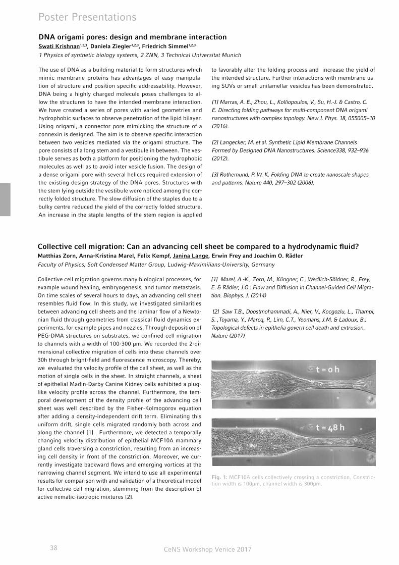
CeNS Workshop Venice 201738
Poster Presentations
DNA origami pores: design and membrane interactionSwati Krishnan1,2,3, Daniela Ziegler1,2,3, Friedrich Simmel1,2,3
1 Physics of synthetic biology systems, 2 ZNN, 3 Technical Universitat Munich
The use of DNA as a building material to form structures which mimic membrane proteins has advantages of easy manipula-tion of structure and position specific addressability. However, DNA being a highly charged molecule poses challenges to al-low the structures to have the intended membrane interaction. We have created a series of pores with varied geometries and hydrophobic surfaces to observe penetration of the lipid bilayer. Using origami, a connector pore mimicking the structure of a connexin is designed. The aim is to observe specific interaction between two vesicles mediated via the origami structure. The pore consists of a long stem and a vestibule in between. The ves-tibule serves as both a platform for positioning the hydrophobic molecules as well as to avoid inter vesicle fusion. The design of a dense origami pore with several helices required extension of the existing design strategy of the DNA pores. Structures with the stem lying outside the vestibule were noticed among the cor-rectly folded structure. The slow diffusion of the staples due to a bulky centre reduced the yield of the correctly folded structure. An increase in the staple lengths of the stem region is applied
to favorably alter the folding process and increase the yield of the intended structure. Further interactions with membrane us-ing SUVs or small unilamellar vesicles has been demonstrated.
[1] Marras, A. E., Zhou, L., Kolliopoulos, V., Su, H.-J. & Castro, C. E. Directing folding pathways for multi-component DNA origami nanostructures with complex topology. New J. Phys. 18, 055005–10 (2016).
[2] Langecker, M. et al. Synthetic Lipid Membrane Channels Formed by Designed DNA Nanostructures. Science338, 932–936 (2012).
[3] Rothemund, P. W. K. Folding DNA to create nanoscale shapes and patterns. Nature 440, 297–302 (2006).
Collective cell migration: Can an advancing cell sheet be compared to a hydrodynamic fluid?Matthias Zorn, Anna-Kristina Marel, Felix Kempf, Janina Lange, Erwin Frey and Joachim O. Rädler
Faculty of Physics, Soft Condensed Matter Group, Ludwig-Maximilians-University, Germany
Collective cell migration governs many biological processes, for example wound healing, embryogenesis, and tumor metastasis. On time scales of several hours to days, an advancing cell sheet resembles fluid flow. In this study, we investigated similarities between advancing cell sheets and the laminar flow of a Newto-nian fluid through geometries from classical fluid dynamics ex-periments, for example pipes and nozzles. Through deposition of PEG-DMA structures on substrates, we confined cell migration to channels with a width of 100-300 µm. We recorded the 2-di-mensional collective migration of cells into these channels over 30h through bright-field and fluorescence microscopy. Thereby, we evaluated the velocity profile of the cell sheet, as well as the motion of single cells in the sheet. In straight channels, a sheet of epithelial Madin-Darby Canine Kidney cells exhibited a plug-like velocity profile across the channel. Furthermore, the tem-poral development of the density profile of the advancing cell sheet was well described by the Fisher-Kolmogorov equation after adding a density-independent drift term. Eliminating this uniform drift, single cells migrated randomly both across and along the channel [1]. Furthermore, we detected a temporally changing velocity distribution of epithelial MCF10A mammary gland cells traversing a constriction, resulting from an increas-ing cell density in front of the constriction. Moreover, we cur-rently investigate backward flows and emerging vortices at the narrowing channel segment. We intend to use all experimental results for comparison with and validation of a theoretical model for collective cell migration, stemming from the description of active nematic-isotropic mixtures [2].
[1] Marel, A.-K., Zorn, M., Klingner, C., Wedlich-Söldner, R., Frey, E. & Rädler, J.O.: Flow and Diffusion in Channel-Guided Cell Migra-tion. Biophys. J. (2014)
[2] Saw T.B., Doostmohammadi, A., Nier, V., Kocgozlu, L., Thampi, S. ,Toyama, Y., Marcq, P., Lim, C.T., Yeomans, J.M. & Ladoux, B.: Topological defects in epithelia govern cell death and extrusion. Nature (2017)
Fig. 1: MCF10A cells collectively crossing a constriction. Constric-tion width is 100µm, channel width is 300µm.
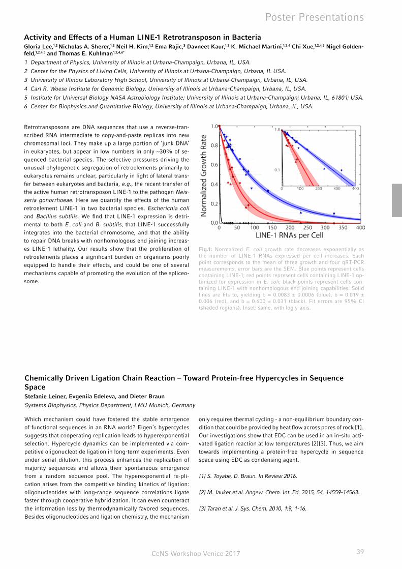
CeNS Workshop Venice 2017 39
Poster Presentations
Chemically Driven Ligation Chain Reaction – Toward Protein-free Hypercycles in Sequence SpaceStefanie Leiner, Evgeniia Edeleva, and Dieter Braun
Systems Biophysics, Physics Department, LMU Munich, Germany
Which mechanism could have fostered the stable emergence of functional sequences in an RNA world? Eigen’s hypercycles suggests that cooperating replication leads to hyperexponential selection. Hypercycle dynamics can be implemented via com-petitive oligonucleotide ligation in long-term experiments. Even under serial dilution, this process enhances the replication of majority sequences and allows their spontaneous emergence from a random sequence pool. The hyperexponential re-pli-cation arises from the competitive binding kinetics of ligation: oligonucleotides with long-range sequence correlations ligate faster through cooperative hybridization. It can even counteract the information loss by thermodynamically favored sequences. Besides oligonucleotides and ligation chemistry, the mechanism
only requires thermal cycling - a non-equilibrium boundary con-dition that could be provided by heat flow across pores of rock [1]. Our investigations show that EDC can be used in an in-situ acti-vated ligation reaction at low temperatures [2][3]. Thus, we aim towards implementing a protein-free hypercycle in sequence space using EDC as condensing agent.
[1] S. Toyabe, D. Braun. In Review 2016.
[2] M. Jauker et al. Angew. Chem. Int. Ed. 2015, 54, 14559-14563.
[3] Taran et al. J. Sys. Chem. 2010, 1:9, 1-16.
Activity and Effects of a Human LINE-1 Retrotransposon in BacteriaGloria Lee,1,2 Nicholas A. Sherer,1,2 Neil H. Kim,1,2 Ema Rajic,3 Davneet Kaur,1,2 K. Michael Martini,1,2,4 Chi Xue,1,2,4,5 Nigel Golden-feld,1,2,4,5 and Thomas E. Kuhlman1,2,4,6*
1 Department of Physics, University of Illinois at Urbana-Champaign, Urbana, IL, USA.
2 Center for the Physics of Living Cells, University of Illinois at Urbana-Champaign, Urbana, IL USA.
3 University of Illinois Laboratory High School, University of Illinois at Urbana-Champaign, Urbana, IL, USA.
4 Carl R. Woese Institute for Genomic Biology, University of Illinois at Urbana-Champaign, Urbana, IL, USA.
5 Institute for Universal Biology NASA Astrobiology Institute; University of Illinois at Urbana-Champaign; Urbana, IL, 61801; USA.
6 Center for Biophysics and Quantitative Biology, University of Illinois at Urbana-Champaign, Urbana, IL, USA.
Retrotransposons are DNA sequences that use a reverse-tran-scribed RNA intermediate to copy-and-paste replicas into new chromosomal loci. They make up a large portion of ‘junk DNA’ in eukaryotes, but appear in low numbers in only ~30% of se-quenced bacterial species. The selective pressures driving the unusual phylogenetic segregation of retroelements primarily to eukaryotes remains unclear, particularly in light of lateral trans-fer between eukaryotes and bacteria, e.g., the recent transfer of the active human retrotransposon LINE-1 to the pathogen Neis-seria gonorrhoeae. Here we quantify the effects of the human retroelement LINE-1 in two bacterial species, Escherichia coli and Bacillus subtilis. We find that LINE-1 expression is detri-mental to both E. coli and B. subtilis, that LINE-1 successfully integrates into the bacterial chromosome, and that the ability to repair DNA breaks with nonhomologous end joining increas-es LINE-1 lethality. Our results show that the proliferation of retroelements places a significant burden on organisms poorly equipped to handle their effects, and could be one of several mechanisms capable of promoting the evolution of the spliceo-some.
Fig.1: Normalized E. coli growth rate decreases exponentially as the number of LINE-1 RNAs expressed per cell increases. Each point corresponds to the mean of three growth and four qRT-PCR measurements, error bars are the SEM. Blue points represent cells containing LINE-1; red points represent cells containing LINE-1 op-timized for expression in E. coli; black points represent cells con-taining LINE-1 with nonhomologous end joining capabilities. Solid lines are fits to, yielding b = 0.0083 ± 0.0006 (blue), b = 0.019 ± 0.006 (red), and b = 0.600 ± 0.031 (black). Fit errors are 95% CI (shaded regions). Inset: same, with log y-axis.
CeNS Workshop Venice 201740
Poster Presentations
Long-lived direct and indirect interlayer excitons in van-der-Waals heterostructures Fabian Merbeler1, Bastian Miller1,2, Alexander Steinhoff3, Borja Pano1, Frank Jahnke3, Alexander Holleitner1,2, Ursula Wurst-bauer1,2
1 Walter Schottky Institut and Physics-Department, Technical University of Munich, Am Coulombwall 4a, 85748 Garching, Ger-many.
2 Nanosystems Initiative Munich (NIM), Schellingstr. 4, 80799 München, Germany.
3 Institut für Theoretische Physik, Universität Bremen, P.O. Box 330 440, 28334 Bremen, Germany
Semiconducting transition metal dichalcogenides are 2D lay-ered crystals coupled by van-der-Waals forces with strong light matter interaction.[1] Heterostructures of these 2D ma-terials form a type-II band alignment and therefore, elec-trons and holes accumulate efficiently in different layers. Interlayer excitons formed by the spatially separated elec-tron-hole pairs have a profoundly prolonged lifetime, fa-cilitating the creation of dense exciton ensembles. These bosonic ensembles are fascinating systems for the study of many-body interactions such as Bose-Einstein condensation.[2] We investigate the photoluminescence of interlayer excitons in heterostructures consisting of monolayer MoSe2 and WSe2 at low temperatures. Surprisingly, we find a doublet structure for such interlayer excitons. Both peaks exhibit long photolumines-cence lifetimes of several ten nanoseconds up to 100 ns at low temperatures, which verifies the interlayer nature of both. The
peak energy and linewidth of both show unusual temperature and power dependences. In particular, we observe a blue-shift of their emission energy for increasing excitation powers. At a low excitation power and low temperatures, the energetically higher peak shows several spikes. We explain the findings by two sorts of interlayer excitons; one that is indirect in real space but direct in reciprocal space, and the other one being indirect in both subspaces. Our results provide fundamental insights into long-lived interlayer states in van der Waals heterostruc-tures with possible bosonic many-body interactions.[3]
[1] U. Wurstbauer et al. , J. Phys. D: Appl. Phys. 50 (2017).
[2] J.M. Blatt et al., Phys. Rev. 126 1691 (1962).
[3] B. Miller et al. , arXiv:1703.09566 (2017).
Dynamic structural biology of membrane transporters: tools and applicationsAlessandra Narducci, Christian Gebhardt, Rebecca Mächtel, Thorben Cordes
Physical and Synthetic Biology, Faculty of Biology, Ludwig Maximilians-Universität München, Großhadernerstr. 2-4, 82152 Planegg-Martinsried, Germany
Membrane transporters are vital to any living system and involved in the translocation of a wide variety of substrates. Despite their importance, all proposed molecular models for transport are based on indirect evidence due to the inability of classical biophysical and biochemical techniques to visualize dynamic structural changes. Our group has recently started to use single-molecule fluorescence microscopy to character-ize conformational states and changes in active membrane transporters in vitro to directly observe how different steps in transport are coordinated.[1-3]
In this contribution we provide an overview of mechanistic studies done in the group. First we focus on the homodimeric GlnPQ complex, a bacterial ABC-importer, possessing two different substrate-binding proteins (SBDs) per single trans-locator. To decipher how conformational changes within the different subdomains drive transport, we use a combination of single-molecule methods and classical biochemical techniques (calorimetry and uptake assays). We demonstrate by single-molecule Förster resonance energy transfer (FRET) that the two SBDs intrinsically transit from open to closed ligand-free conformation, and the proteins capture their amino acid li-gands via an induced-fit mechanism. High-affinity ligands elicit transitions without changing the closed-state lifetime, whereas low-affinity ligands dramatically shorten it. We show that SBDs in the closed state compete for docking onto the translocator. We find that the rate-determining steps for translocation de-pend on the SBD and the amino acid transported. We conclude that the lifetime of the closed conformation controls both SBD docking to the translocator and substrate release.[1]
In the second part, we describe our latest developments of “enabling technology” for mechanistic studies: using photo-physical tricks, we present a simple two-colour FRET assay that allows can either monitor multiple distances within protein complexes or simultaneously reveals one FRET-based distance and the presence of a second protein.[4] Finally, we summarize our contributions towards the development of "self-healing" organic fluorophores and their applications in single-molecule FRET or super-resolution microscopy.[2]
[1] G. Gouridis et al., Nature Structural & Molecular Biology 22 (2015) 57-64
[2] J.H.M. van der Velde et al., Nature Communications 7:10144 (2016)
[3] A. A. Jazi et al., Biochemistry 56 (2017) 2031-2041
[4] E. Ploetz and E. Lerner et al., Scientific Reports 6:33257 (2016)
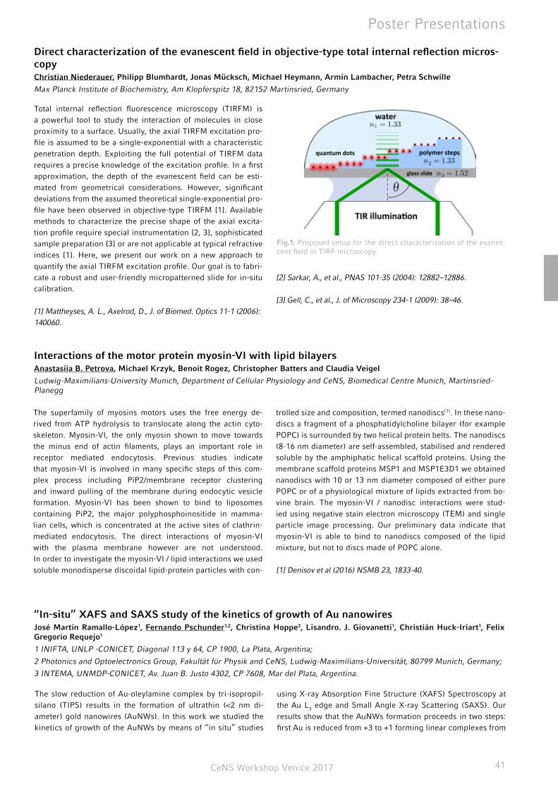
CeNS Workshop Venice 2017 41
Poster Presentations
Direct characterization of the evanescent field in objective-type total internal reflection micros-copyChristian Niederauer, Philipp Blumhardt, Jonas Mücksch, Michael Heymann, Armin Lambacher, Petra Schwille
Max Planck Institute of Biochemistry, Am Klopferspitz 18, 82152 Martinsried, Germany
Total internal reflection fluorescence microscopy (TIRFM) is a powerful tool to study the interaction of molecules in close proximity to a surface. Usually, the axial TIRFM excitation pro-file is assumed to be a single-exponential with a characteristic penetration depth. Exploiting the full potential of TIRFM data requires a precise knowledge of the excitation profile. In a first approximation, the depth of the evanescent field can be esti-mated from geometrical considerations. However, significant deviations from the assumed theoretical single-exponential pro-file have been observed in objective-type TIRFM [1]. Available methods to characterize the precise shape of the axial excita-tion profile require special instrumentation [2, 3], sophisticated sample preparation [3] or are not applicable at typical refractive indices [1]. Here, we present our work on a new approach to quantify the axial TIRFM excitation profile. Our goal is to fabri-cate a robust and user-friendly micropatterned slide for in-situ calibration.
[1] Mattheyses, A. L., Axelrod, D., J. of Biomed. Optics 11-1 (2006): 140060.
[2] Sarkar, A., et al., PNAS 101-35 (2004): 12882–12886.
[3] Gell, C., et al., J. of Microscopy 234-1 (2009): 38–46.
Interactions of the motor protein myosin-VI with lipid bilayersAnastasiia B. Petrova, Michael Krzyk, Benoit Rogez, Christopher Batters and Claudia Veigel
Ludwig-Maximilians-University Munich, Department of Cellular Physiology and CeNS, Biomedical Centre Munich, Martinsried-Planegg
The superfamily of myosins motors uses the free energy de-rived from ATP hydrolysis to translocate along the actin cyto-skeleton. Myosin-VI, the only myosin shown to move towards the minus end of actin filaments, plays an important role in receptor mediated endocytosis. Previous studies indicate that myosin-VI is involved in many specific steps of this com-plex process including PiP2/membrane receptor clustering and inward pulling of the membrane during endocytic vesicle formation. Myosin-VI has been shown to bind to liposomes containing PiP2, the major polyphosphoinositide in mamma-lian cells, which is concentrated at the active sites of clathrin-mediated endocytosis. The direct interactions of myosin-VI with the plasma membrane however are not understood. In order to investigate the myosin-VI / lipid interactions we used soluble monodisperse discoidal lipid-protein particles with con-
trolled size and composition, termed nanodiscs[1]. In these nano-discs a fragment of a phosphatidylcholine bilayer (for example POPC) is surrounded by two helical protein belts. The nanodiscs (8-16 nm diameter) are self-assembled, stabilised and rendered soluble by the amphiphatic helical scaffold proteins. Using the membrane scaffold proteins MSP1 and MSP1E3D1 we obtained nanodiscs with 10 or 13 nm diameter composed of either pure POPC or of a physiological mixture of lipids extracted from bo-vine brain. The myosin-VI / nanodisc interactions were stud-ied using negative stain electron microscopy (TEM) and single particle image processing. Our preliminary data indicate that myosin-VI is able to bind to nanodiscs composed of the lipid mixture, but not to discs made of POPC alone.
[1] Denisov et al (2016) NSMB 23, 1833-40.
Fig.1: Proposed setup for the direct characterization of the evanes-cent field in TIRF microscopy.
“In-situ” XAFS and SAXS study of the kinetics of growth of Au nanowiresJosé Martín Ramallo-López1, Fernando Pschunder1,2, Christina Hoppe3, Lisandro. J. Giovanetti1, Christián Huck-Iriart1, Felix Gregorio Requejo1
1 INIFTA, UNLP -CONICET, Diagonal 113 y 64, CP 1900, La Plata, Argentina;
2 Photonics and Optoelectronics Group, Fakultät für Physik and CeNS, Ludwig-Maximilians-Universität, 80799 Munich, Germany;
3 INTEMA, UNMDP-CONICET, Av. Juan B. Justo 4302, CP 7608, Mar del Plata, Argentina.
The slow reduction of Au-oleylamine complex by tri-isopropil-silano (TIPS) results in the formation of ultrathin (<2 nm di-ameter) gold nanowires (AuNWs). In this work we studied the kinetics of growth of the AuNWs by means of “in situ” studies
using X-ray Absorption Fine Structure (XAFS) Spectroscopy at the Au L3 edge and Small Angle X-ray Scattering (SAXS). Our results show that the AuNWs formation proceeds in two steps: first Au is reduced from +3 to +1 forming linear complexes from
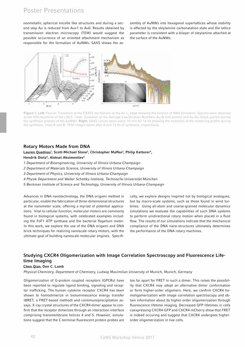
CeNS Workshop Venice 201742
Poster Presentations
Studying CXCR4 Oligomerization with Image Correlation Spectroscopy and Fluorescence Life-time ImagingChen Qian, Don C. Lamb
Physical Chemistry, Department of Chemistry, Ludwig Maximilian University of Munich, Munich, Germany
Oligomerization of G-protein coupled receptors (GPCRs) have been reported to regulate ligand binding, signaling and recep-tor trafficking. The human cytokine receptor CXCR4 has been shown to homodimerize in bioluminescence energy transfer (BRET, a FRET-based method) and coimmunoprecipitation as-says. X-ray crystal structures of the CXCR4 dimer appear to con-firm that the receptor dimerizes through an interaction interface comprising transmembrane helices 4 and 5. However, simula-tions suggest that the C-terminal fluorescent protein probes are
too far apart for FRET in such a dimer. This raises the possibil-ity that CXCR4 may adopt an alternative dimer conformation or form higher-order oligomers. Here, we confirm CXCR4 ho-moligomerization with image correlation spectroscopy and ob-tain information about its higher-order oligomerization through fluorescence lifetime imaging. Decreased GFP lifetimes in cells coexpressing CXCR4-GFP and CXCR4-mCherry show that FRET is indeed occuring and suggest that CXCR4 undergoes higher-order oligomerization in live cells.
Figure 1. Left: Fourier Transform of the EXAFS oscillations at the Au L3 edge showing the kinetics of NWs formation. Spectra were obtained at the XDS beamline of the LNLS. Inset: Evolution of the Average Coordination Numbers Au-N (red points) and Au-Au (black points) during the synthesis process of the AuNWs. Right: SAXS curves taken every 10 min for 16 hs showing the evolution of the scattering profile during the synthesis. Inset A and B: TEM images taken after 6 and 12 hs of synthesis, respectively.
nonmetallic spherical micelle like structures and during a sec-ond step Au is reduced from Au+1 to Au0. Results obtained by transmission electron microscopy (TEM) would suggest the possible occurrence of an oriented attachment mechanism as responsible for the formation of AuNWs. SAXS shows the as-
sembly of AuNWs into hexagonal superlattices whose stability is affected by the oleylamine carbonatation state and the lattice parameter is consistent with a bilayer of oleylamine attached at the surface of the AuNWs.
Rotary Motors Made from DNALauren Quednau1, Scott-Michael Slone2, Christopher Maffeo3, Philip Ketterer4,
Hendrik Dietz4, Aleksei Aksimentiev5
1 Department of Bioengineering, University of Illinois Urbana-Champaign
2 Department of Materials Science, University of Illinois Urbana-Champaign
3 Department of Physics, University of Illinois Urbana-Champaign
4 Physik Department and Walter Schottky Institute, Technische Universität München
5 Beckman Institute of Science and Technology, University of Illinois Urbana-Champaign
Advances in DNA nanotechnology, the DNA origami method in particular, enable the fabrication of three-dimensional structures at the nanometer scale, offering a myriad of potential applica-tions. Vital to cellular function, molecular motors are commonly found in biological systems, with celebrated examples includ-ing the FoF1 ATP synthase and the bacterial flagellum motor. In this work, we explore the use of the DNA origami and DNA brick techniques for realizing nanoscale rotary motors, with the ultimate goal of building nanoscale molecular engines. Specifi-
cally, we explore designs inspired not by biological analogues, but by macro-scale systems, such as those found in wind tur-bines. Using all-atom and coarse-grained molecular dynamics simulations we evaluate the capabilities of such DNA systems to perform unidirectional rotary motion when placed in a fluid flow. The results of our simulations indicate that the mechanical compliance of the DNA nano-structures ultimately determines the performance of the DNA rotary machines.

CeNS Workshop Venice 2017 43
Poster Presentations
mRNA Fluorescence in situ Hybridization via Click-Labelled Oligonucleotides: a Powerful Tool for the Detection of Minimal Residual DiseasesNada Raddaoui1,2, Florian Geiger3, L. Möckl, H. Engelke3, C. Bräuchle, T. Carell1,2,#
1 Center for Integrated Protein Science Munich CIPSM. [email protected]
2 SFB 1032, A05
3 SFB 1032, B11
#: Corresponding author. [email protected]
The average behavior of cell populations is not necessarily related to the gene expression in single cells (Larson et al., 2009;Maheshri and O’Shea, 2007; Raj and Van Oudenaarden, 2008, 2009). The detection and quantification of transcript lev-els and gene expression using RT-PCR is a common method in molecular pathology. Therefore, in cancer diagnostic is the detection of gene fusions in genetic aberrations such BCR-ABL1 expression in CML (chronic myeloid leukemia) is crucial. How-ever, RT-PCR does not give quantitative and spacial informa-tions of the transcripts in different cells within the population. The fluorescence in situ hybridization, in which labeled DNA probes can hybridize to the specific target, is the best candi-date to generate spacial informations within the cell. Howev-er, in situ hybridization using radioactive probes (Gall, 1968) or antibodies (Raap et al., 1995; Tautz and Pfeifle, 1989) can
generate false positive results due to the high background and unspecific binding. A simple method was already devel-oped to overcome these limitations (Raj et al., Nature Meth-ods 2008). It involves probing the target transcript via differ-ent (up to 48), short (22mer) and singly labeled ODN probes. Although this method is simple, the signal intensity of the probes under the microscope is remaining limited when short transcripts or fused gene transcripts should be detected. Using click chemistry we were able to increase the signal inten-sity (3 fold) using only 10 oligonucleotides instead of 48. Our method is on the way to be applied to biological studies and clinical cytogenetics. It would be a smart, efficient and cheap alternative to RT-PCR, which remains a challenge in the clinical diagnostic and is associated with huge costs, time and effort in comparison to the relatively poor throughput.
Influence of size and shielding of siRNA polyplexes on in vivo biodistributionSören Reinhard1 , Philipp Klein1, Eva Kessel, Sarah Kern1, Ernst Wagner1,2
1 Pharmaceutical Biotechnology, Department of Pharmacy, Center for NanoScience (CeNS), Ludwig-Maximilians-Universität, Butenandtstr. 5-13, 81377 München, Germany. E-mail: [email protected]
2 Nanosystems Initiative Munich (NIM), Schellingstr. 4, 80799 München, Germany
Cationic lipo-oligomers have been proven as potent siRNA car-riers based on stable electrostatic and hydrophobic polyplex formation and endosomal membrane destabilization. Although siRNA has to be readily released in the cytosol, high stability of siRNA polyplexes and shielding is desirable in the extracel-
lular space to avoid rapid decay, clearance and unspecific in-teractions. Shielding of positive charges by hydrophilic poly-mers such as polyethylene glycol (PEG) is a well-established approach to circumvent unspecific interactions of drug delivery systems. A small library of sequence-defined lipo-oligomers
containing either no PEG or PEG with 12 and 24 units were synthesized and used for siRNA poly-plex formation. Physicochemical properties and in vivo biodistribution of the polyplexes were evaluated. Using longer PEG units resulted in polyplexes with smaller hydrodynamic radius, lower zeta potential and broader biodistribu-tion, but also reduced their circulation time in tumor-bearing mice.
Fig.1: Structure of sequence-defined cationic lipo-oligomers with different PEG length and biodistribution in tumor-bearing mice 60 min after intravenous injection.
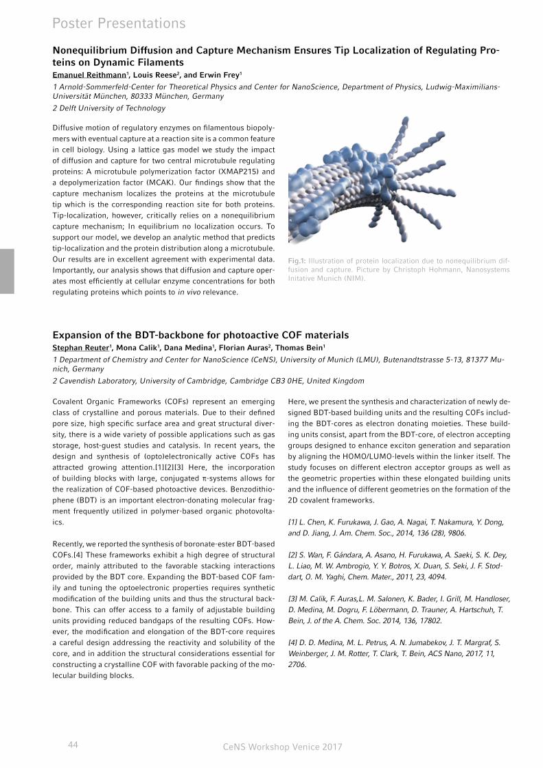
CeNS Workshop Venice 201744
Poster Presentations
Nonequilibrium Diffusion and Capture Mechanism Ensures Tip Localization of Regulating Pro-teins on Dynamic FilamentsEmanuel Reithmann1, Louis Reese2, and Erwin Frey1
1 Arnold-Sommerfeld-Center for Theoretical Physics and Center for NanoScience, Department of Physics, Ludwig-Maximilians-Universität München, 80333 München, Germany
2 Delft University of Technology
Diffusive motion of regulatory enzymes on filamentous biopoly-mers with eventual capture at a reaction site is a common feature in cell biology. Using a lattice gas model we study the impact of diffusion and capture for two central microtubule regulating proteins: A microtubule polymerization factor (XMAP215) and a depolymerization factor (MCAK). Our findings show that the capture mechanism localizes the proteins at the microtubule tip which is the corresponding reaction site for both proteins. Tip-localization, however, critically relies on a nonequilibrium capture mechanism; In equilibrium no localization occurs. To support our model, we develop an analytic method that predicts tip-localization and the protein distribution along a microtubule. Our results are in excellent agreement with experimental data. Importantly, our analysis shows that diffusion and capture oper-ates most efficiently at cellular enzyme concentrations for both regulating proteins which points to in vivo relevance.
Expansion of the BDT-backbone for photoactive COF materialsStephan Reuter1, Mona Calik1, Dana Medina1, Florian Auras2, Thomas Bein1
1 Department of Chemistry and Center for NanoScience (CeNS), University of Munich (LMU), Butenandtstrasse 5-13, 81377 Mu-nich, Germany
2 Cavendish Laboratory, University of Cambridge, Cambridge CB3 0HE, United Kingdom
Covalent Organic Frameworks (COFs) represent an emerging class of crystalline and porous materials. Due to their defined pore size, high specific surface area and great structural diver-sity, there is a wide variety of possible applications such as gas storage, host-guest studies and catalysis. In recent years, the design and synthesis of (opto)electronically active COFs has attracted growing attention.[1][2][3] Here, the incorporation of building blocks with large, conjugated π-systems allows for the realization of COF-based photoactive devices. Benzodithio-phene (BDT) is an important electron-donating molecular frag-ment frequently utilized in polymer-based organic photovolta-ics.
Recently, we reported the synthesis of boronate-ester BDT-based COFs.[4] These frameworks exhibit a high degree of structural order, mainly attributed to the favorable stacking interactions provided by the BDT core. Expanding the BDT-based COF fam-ily and tuning the optoelectronic properties requires synthetic modification of the building units and thus the structural back-bone. This can offer access to a family of adjustable building units providing reduced bandgaps of the resulting COFs. How-ever, the modification and elongation of the BDT-core requires a careful design addressing the reactivity and solubility of the core, and in addition the structural considerations essential for constructing a crystalline COF with favorable packing of the mo-lecular building blocks.
Here, we present the synthesis and characterization of newly de-signed BDT-based building units and the resulting COFs includ-ing the BDT-cores as electron donating moieties. These build-ing units consist, apart from the BDT-core, of electron accepting groups designed to enhance exciton generation and separation by aligning the HOMO/LUMO-levels within the linker itself. The study focuses on different electron acceptor groups as well as the geometric properties within these elongated building units and the influence of different geometries on the formation of the 2D covalent frameworks.
[1] L. Chen, K. Furukawa, J. Gao, A. Nagai, T. Nakamura, Y. Dong, and D. Jiang, J. Am. Chem. Soc., 2014, 136 (28), 9806.
[2] S. Wan, F. Gándara, A. Asano, H. Furukawa, A. Saeki, S. K. Dey, L. Liao, M. W. Ambrogio, Y. Y. Botros, X. Duan, S. Seki, J. F. Stod-dart, O. M. Yaghi, Chem. Mater., 2011, 23, 4094.
[3] M. Calik, F. Auras,L. M. Salonen, K. Bader, I. Grill, M. Handloser, D. Medina, M. Dogru, F. Löbermann, D. Trauner, A. Hartschuh, T. Bein, J. of the A. Chem. Soc. 2014, 136, 17802.
[4] D. D. Medina, M. L. Petrus, A. N. Jumabekov, J. T. Margraf, S. Weinberger, J. M. Rotter, T. Clark, T. Bein, ACS Nano, 2017, 11, 2706.
Fig.1: Illustration of protein localization due to nonequilibrium dif-fusion and capture. Picture by Christoph Hohmann, Nanosystems Initative Munich (NIM).
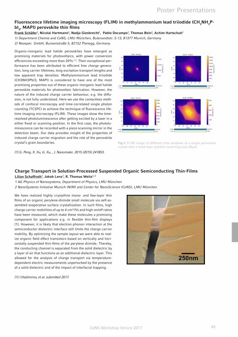
CeNS Workshop Venice 2017 45
Poster Presentations
Fluorescence lifetime imaging microscopy (FLIM) in methylammonium lead triiodide (CH3NH3P-bI3, MAPI) perovskite thin films Frank Schäfer1, Nicolai Hartmann2, Nadja Giesbrecht1, Pablo Docampo1, Thomas Bein1, Achim Hartschuh1
1) Department Chemie and CeNS, LMU München, Butenandtstr. 5-13, 81377 Munich, Germany
2) Neaspec GmbH, Bunsenstraße 5, 82152 Planegg, Germany
Organic–inorganic lead halide perovskites have emerged as promising materials for photovoltaics, with power conversion efficiencies exceeding more than 20% [1]. Their exceptional per-formance has been attributed to efficient free charge genera-tion, long carrier lifetimes, long excitation transport lengths and low apparent trap densities. Methylammonium lead triiodide (CH3NH3PbI3, MAPI) is considered to have one of the most promising properties out of these organic-inorganic lead halide perovskite materials for photovoltaic fabrication. However, the nature of the induced charge carrier behaviour, e.g. the diffu-sion, is not fully understood. Here we use the contactless meth-ods of confocal microscopy and time-correlated single photon counting (TCSPC) to achieve the technique of fluorescence life-time imaging microscopy (FLIM). These images show the time-resolved photoluminescence after getting excited by a laser in a either fixed or scanning position. In the first case, the photolu-minescence can be recorded with a piezo scanning mirror in the detection beam. Our data provides insight of the properties of induced charge carrier migration and the role of the perovskite crystal’s grain boundaries.
[1] G. Peng, X. Xu, G. Xu, , J. Nanomater. 2015 (2015) 241853.
Charge Transport in Solution-Processed Suspended Organic Semiconducting Thin-FilmsLilian Schaffroth1, Jakob Lenz1, R. Thomas Weitz1, 2
1 AG Physics of Nanosystems, Department of Physics, LMU München
2 NanoSystems Initiative Munich (NIM) and Center for NanoScience (CeNS), LMU München
We have realized highly crystalline mono- and few-layer thin films of an organic perylene-diimide small molecule via self-as-sembled evaporative surface crystallization. In such films, high charge carrier mobilities of up to 4 cm²/Vs and high on/off ratios have been measured, which make these molecules a promising component for applications e.g. in flexible thin-film displays [1]. However, it is likely that electron-phonon interaction at the semiconductor-dielectric interface still limits the charge carrier mobility. By optimizing the sample layout we were able to real-ize organic field effect transistors based on vertically and hori-zontally suspended thin-films of the perylene diimide. Thereby, the conducting channel is separated from the solid dielectric by a layer of air that functions as an additional dielectric layer. This allowed for the analysis of charge transport via temperature-dependent electric measurements unperturbed by the presence of a solid dielectric and of the impact of interfacial trapping.
[1] I.Vladimirov, et al. submitted 2017.
Fig.1: FLIM image of different time windows of a single perovskite crystal with a fixed laser position (scanning size 20µm)
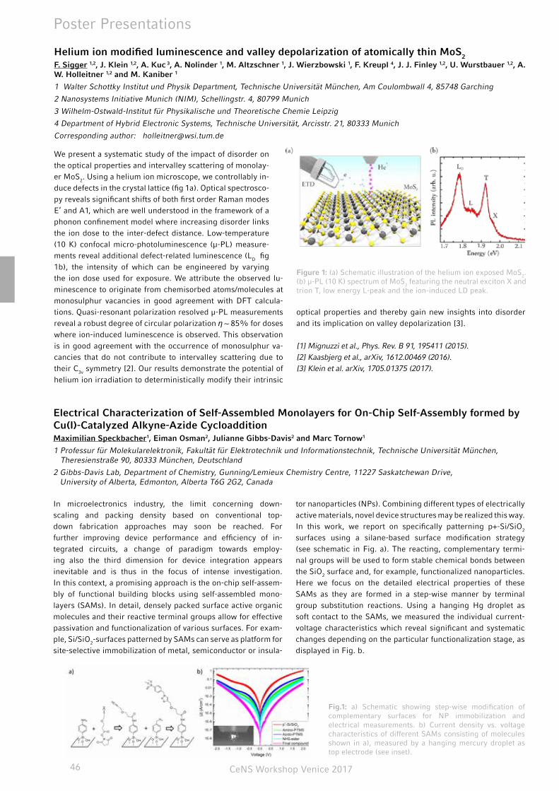
CeNS Workshop Venice 201746
Poster Presentations
Helium ion modified luminescence and valley depolarization of atomically thin MoS2
F. Sigger 1,2, J. Klein 1,2, A. Kuc 3, A. Nolinder 1, M. Altzschner 1, J. Wierzbowski 1, F. Kreupl 4, J. J. Finley 1,2, U. Wurstbauer 1,2, A. W. Holleitner 1,2 and M. Kaniber 1
1 Walter Schottky Institut und Physik Department, Technische Universität München, Am Coulombwall 4, 85748 Garching
2 Nanosystems Initiative Munich (NIM), Schellingstr. 4, 80799 Munich
3 Wilhelm-Ostwald-Institut für Physikalische und Theoretische Chemie Leipzig
4 Department of Hybrid Electronic Systems, Technische Universität, Arcisstr. 21, 80333 Munich
Corresponding author: [email protected]
We present a systematic study of the impact of disorder on the optical properties and intervalley scattering of monolay-er MoS2. Using a helium ion microscope, we controllably in-duce defects in the crystal lattice (fig 1a). Optical spectrosco-py reveals significant shifts of both first order Raman modes E’ and A1, which are well understood in the framework of a phonon confinement model where increasing disorder links the ion dose to the inter-defect distance. Low-temperature (10 K) confocal micro-photoluminescence (µ-PL) measure-ments reveal additional defect-related luminescence (LD fig 1b), the intensity of which can be engineered by varying the ion dose used for exposure. We attribute the observed lu-minescence to originate from chemisorbed atoms/molecules at monosulphur vacancies in good agreement with DFT calcula-tions. Quasi-resonant polarization resolved µ-PL measurements reveal a robust degree of circular polarization η ~ 85% for doses where ion-induced luminescence is observed. This observation is in good agreement with the occurrence of monosulphur va-cancies that do not contribute to intervalley scattering due to their C3v symmetry [2]. Our results demonstrate the potential of helium ion irradiation to deterministically modify their intrinsic
optical properties and thereby gain new insights into disorder and its implication on valley depolarization [3].
[1] Mignuzzi et al., Phys. Rev. B 91, 195411 (2015). [2] Kaasbjerg et al., arXiv, 1612.00469 (2016). [3] Klein et al. arXiv, 1705.01375 (2017).
Electrical Characterization of Self-Assembled Monolayers for On-Chip Self-Assembly formed by Cu(I)-Catalyzed Alkyne-Azide CycloadditionMaximilian Speckbacher1, Eiman Osman2, Julianne Gibbs-Davis2 and Marc Tornow1
1 Professur für Molekularelektronik, Fakultät für Elektrotechnik und Informationstechnik, Technische Universität München, Theresienstraße 90, 80333 München, Deutschland
2 Gibbs-Davis Lab, Department of Chemistry, Gunning/Lemieux Chemistry Centre, 11227 Saskatchewan Drive, University of Alberta, Edmonton, Alberta T6G 2G2, Canada
In microelectronics industry, the limit concerning down-scaling and packing density based on conventional top-down fabrication approaches may soon be reached. For further improving device performance and efficiency of in-tegrated circuits, a change of paradigm towards employ-ing also the third dimension for device integration appears inevitable and is thus in the focus of intense investigation. In this context, a promising approach is the on-chip self-assem-bly of functional building blocks using self-assembled mono-layers (SAMs). In detail, densely packed surface active organic molecules and their reactive terminal groups allow for effective passivation and functionalization of various surfaces. For exam-ple, Si/SiO2-surfaces patterned by SAMs can serve as platform for site-selective immobilization of metal, semiconductor or insula-
tor nanoparticles (NPs). Combining different types of electrically active materials, novel device structures may be realized this way. In this work, we report on specifically patterning p+-Si/SiO2 surfaces using a silane-based surface modification strategy (see schematic in Fig. a). The reacting, complementary termi-nal groups will be used to form stable chemical bonds between the SiO2 surface and, for example, functionalized nanoparticles. Here we focus on the detailed electrical properties of these SAMs as they are formed in a step-wise manner by terminal group substitution reactions. Using a hanging Hg droplet as soft contact to the SAMs, we measured the individual current-voltage characteristics which reveal significant and systematic changes depending on the particular functionalization stage, as displayed in Fig. b.
Figure 1: (a) Schematic illustration of the helium ion exposed MoS2. (b) µ-PL (10 K) spectrum of MoS2 featuring the neutral exciton X and trion T, low energy L-peak and the ion-induced LD peak.
Fig.1: a) Schematic showing step-wise modification of complementary surfaces for NP immobilization and electrical measurements. b) Current density vs. voltage characteristics of different SAMs consisting of molecules shown in a), measured by a hanging mercury droplet as top electrode (see inset).

CeNS Workshop Venice 2017 47
Poster Presentations
Self-assembling methotrexate nanopharmaceuticals Benjamin Steinborn, Ines Trübenbach, Patrick Hirschle, Stefan Wuttke, Ernst Wagner, Ulrich Lächelt
Pharmaceutical Biotechnology, Faculty of Chemistry and Pharmacy, Ludwig-Maximilian-University Munich
High-dose methotrexate (MTX) therapy is commonly used to treat a variety of adult and childhood cancers. Although be-ing a well-established treatment regime, it may lead to severe dose-limiting side effects such as acute kidney and liver toxicity in some patients which will negatively affect therapeutic out-comes. Poor drug response due to resistance formation poses another major challenge affecting approximately 25% of acute lymphoblastic leukemia (ALL) patients. Resistance is mainly based on limited drug uptake, impaired MTX polyglutamylation and retention combined with increased dihydrofolatereductase (DHFR) activity. We present nanoparticles formulated from
polyglutamylated En-MTX derivatives and multivalent metal ions as a strategy to address resistance mechanisms and reduce off-target toxicity. As impaired MTX polyglutamylation and up-take both contribute to resistance formation, direct nanoparticu-late delivery of active En-MTX is an elegant concept towards breaking MTX resistance. We show that precise control of reac-tion parameters leads to high yields of En-MTX nanoparticles in a size range of about 70nm as determined by SEM. Based on those promising nanoparticles, biological efficacy on MTX resistant cell lines will be evaluated in future studies.
Investigations of protein dynamics with single-molecule FRET in vitro and in organelloVanessa Trauschke1,2, Lena Voith von Voithenberg, Rupa Banerjee3, Dejana Mokranjac3, Don C. Lamb1,2
1 Physical Chemistry, Department of Chemistry, Ludwig-Maximilians-Universität München, Munich, Germany
2 Center for NanoScience, Ludwig-Maximilians-Universität München, Munich, Germany
3 Biomedical Center Munich, Department of Physiological Chemistry, Ludwig-Maximilians-Universität München, Munich, Ger-many
The 70 kD heat shock proteins (Hsp70) are important to prevent protein aggregation and are acting as chaperones to stabilize proteins until refolding. Additionally, the mitochondrial Hsp70 (Ssc1) is involved in translocation of proteins into the mitochon-drial matrix. The functions of proteins are to a significant extent determined by their conformational dynamics and interactions with other biomolecules. Although there are valuable techniques to obtain protein structures they often lack the time resolution to gain information about the conformational changes. We use single-molecule Förster resonance energy transfer (smFRET) as a versatile tool to study real-time dynamics of various proteins and their intermolecular interactions. Measurements can be done on freely diffusing molecules or on samples immobilized on the surface. Surface measurements allow for monitoring over longer timescales so that transitions between the conformations can be observed in real-time and the underlying kinetics can be extracted. The investigations of Ssc1 by smFRET revealed a highly dynamic ADP-bound state of the protein and gave new insights into the conformational cycle of Ssc1. Further measure-ments with different labeling positions were carried out to bet-ter understand the system. While in vitro experiments give a
rough idea of the chaperoning cycle the kinetics and the popu-lation of the various conformations can differ in vivo. To gain more information about the native conformational cycle of Ssc1 we perform in organello smFRET experiments on proteins la-beled with organic fluorophores inside isolated mitochondria.
CeNS Workshop Venice 201748
Poster Presentations
Towards femtosecond on-chip electronicsChristopher Trummer1, Philipp Zimmermann1,2, C. Karnetzky1,2, C. Duque-Sierra1, and A. W. Holleitner1,2
1 Walter Schottky Institut and Physics Department, Technical University Munich, Am Coulombwall 4a, 85748 Garching, Germany
2 Nanosystems Initiative Munich (NIM), Schellingstr. 4, 80799 Munich, Germany
For future on-chip communication schemes, it is essential to integrate nanoscale circuits with an ultrafast optoelectronic functionality into high-frequency circuits. In order to reach the highest electronic frequencies, we use light-field driven pho-toemission currents in a pair of freely suspended gold tips as well as in a nano-antenna array lying on sapphire to trigger ul-trafast electromagnetic transients in on-chip THz-circuits. The dimensions of the nano-antenna are optimized by finite element simulations to exploit plasmonic resonances and thus gain high photoemission currents due to field enhancement values up to 10. We demonstrate a superlinear increase of the photocurrent for increasing laser fields as well as dc electric fields applied across the nano-antennas. The ultrafast currents are transmitted on-chip across several millimeters as electromagnetic THz-tran-sients in the utilized stripline-circuits, before they are read-out with an ultrafast photodetector. From performing mode analy-sis and 3D simulations, we obtain the dispersion relation of the THz-circuit by which the original photocurrent pulse shapes can
be deduced. Our results are equally important for a fundamental understanding of laser-field driven photoemission processes in plasmonic devices as well as ultrafast photoswitches based on quantum tunneling, which can be integrated in THz-circuits.
Effect of DNA free-energy landscape on XPD helicase activityAlice Troitskaia1, Barbara Stekas2, Maria Spies3, and Yann R. Chemla1, 2, 4
1 Center for Biophysics and Computational Biology
2 Department of Physics
3 Department of Biochemistry, University of Iowa
4 Center for the Physics of Living Cells, University of Illinois, Urbana-Champaign
Xeroderma pigmentosum group D protein (XPD) is a Superfam-ily 2 (SF2) helicase involved in nucleotide excision repair and transcription initiation. FacXPD from the archeon Ferroplasma acidarmanus serves as a model for understanding the molecu-lar mechanism of human XPD. We investigate the mechanism through which FacXPD unwinds double-stranded DNA (dsDNA) by monitoring how a single protein unfolds a DNA hairpin us-ing high-resolution optical tweezers. Previous work has shown that XPD's unwinding activity is strongly influenced by the DNA sequence unwound and is likely a convolution of its intrinsic activity and the energetics of unfolding the hairpin. Here, we designed a DNA hairpin sequence with a uniform probability of base-pair opening along the length of the hairpin to minimize sequence-dependent effects and to detect more directly XPD's intrinsic activity. Preliminary data indicate that XPD is signifi-cantly more processive on this DNA sequence than on sequenc-es with more rugged energy landscapes. However, the observed distribution of processivities is multimodal, indicating that XPD may be affected by sequence in ways more subtle than antici-
pated. Our ability to control the energy landscape of a DNA hair-pin provides us a platform for more directly investigating XPD unwinding and the mechanism by which partner proteins, such as the single-stranded DNA binding protein FacRPA2, enhance XPD activity.
CeNS Workshop Venice 2017 49
Controlling Membrane Properties and Domain Formation in Photolipid Bilayer MembranesPatrick Urban1, Stefanie D. Pritzl1, James A. Frank2, Carla Pernpeintner1, Christian R. Röske1, Dirk Trauner2,3, Theobald Lohmül-ler1
1 Photonics and Optoelectronics Group, Department of Physics and CeNS, Ludwig-Maximilians-Universität München, Amalien-straße 54, 80799 Munich, Germany
2 Department of Chemistry and Center for Integrated Protein Science, Ludwig-Maximilians Universität München, Butenandt-straße 5-13, 81377 Munich, Germany
3 Department of Chemistry, New York University, Silver Center, 100 Washington Square East, Room 712, New York, 10003, United States
Many properties of cell membranes are governed by the com-plex interplay between lipids and other membrane components. One particular aspect is the formation and organization of mem-brane domains were lipids and other molecules concentrate or assemble into functional units. This goes hand in hand with the deformability and rigidity of the membrane itself, which are also depending on the membrane composition.
Synthetic phospholipid vesicles, made from different lipid com-positions, have long been studied as a model system to emulate properties of native bilayer membranes. The manipulation of dy-namic processes such as lipid mobility and membrane re-orga-nization with high spatio-temporal resolution, however, is often challenging as it involves drastic changes of experimental con-ditions such as temperature, ion concentration, or membrane composition, which is often non-reversible.
Here, we demonstrate an approach to control membrane prop-erties by incorporating photoswitchable lipid molecules into giant unilamellar vesicles (GUVs). The photolipids used in this study contain an azobenzene group that undergoes reversible photoisomerization upon illumination with UV and visible light. Varying the optical stimulus renders it possible to tune physical properties such as fluidity and bending rigidity of the bilayer membrane on fast time-scales. Furthermore, we show how a mixture of photolipids with other lipid molecules allows to re-versibly control membrane domain formation and phase transi-tions by means of light. Our results highlight a general approach how photolipid molecules could be employed as light sensitive constituents of native cell membranes for controlling cell func-tion and behavior in space and time.
"Graphene self-ironing" – a low temperature processing technique for stamped graphene Stefan Wakolbinger 1, S. Palmer1 , F. Winterer1, Fabian.R. Geisenhof1, R. Thomas Weitz 1, 2
1 AG Physics of Nanosystems, Department of Physics, LMU München
2 NanoSystems Initiative Munich (NIM) and Center for NanoScience (CeNS), LMU München
Since graphene was first isolated, it has been broadly studied and recently also often been used in heterostructures consisting of stacks of graphene and other van der Waals materials. Apart from graphene, also its multilayers have recently gained inter-est. Graphene multilayers (e.g. tri- or tetralayers) are found in ei-ther so called bernal (or ABA) or rhomboheral (or ABC) stacked form, both of which have distinct bandstuctures and properties. For example rhombohedrally stacked graphene is predicted to exhibit exotic electronic properties – such as surface super-conductivity [1], given that the superconductive surface of the flake is protected from the adverse effects of the widely used SiO2 substrate [2]. This can be achieved by stamping it in a sandwich-structure between hexagonal boron nitride (hBN), or other graphene multilayers. The stamping process itself– which we focus on in the poster - can be a delicate procedure, since contact between the graphene multilayers and the underly-ing material is usually not assured when stamped in ambient conditions. Moisture and residual materials from the stamping process can be trapped beneath the stamped graphene layer. This problem is often tackled by annealing the stack using high temperatures [3] or stamping in an inert environment. Here we show an alternative non-invasive, low temperature processing
technique that allows to bring two stamped layers in close con-tact (that is presumably without any contaminants in between the stamped layers) via the interaction of the tip of an atomic force microscope (AFM) even when the stamping process is per-formed in ambient air. To trigger this effect, graphene flakes are be pushed towards each other via the repulsive interaction with an AFM probe operated in contact mode whereupon the stamped flakes stick together firmly as a result of their attrac-tive van der Waals forces. During this process, the previously found dirt materials is squeezed out between the two graphene flakes. This technique -that we call “self- ironing” - presents a promising alternative to methods that require heating, since it is controllable at the local scale.
[1] N. B. Kopnin et al., Phys. Rev., 140503 (2013)
[2] Peng Zhao et al., J. Vac. Sci. Technol. 01A118-1 ,(2017)
[3] Gwanghyun Ahn et al., ACS NANO 7, 1533 (2013)
CeNS Workshop Venice 201750
Topological hindrance for multi-species molecular transportPatrick Wilke, Emanuel Reithmann, Erwin Frey
Arnold-Sommerfeld-Center for Theoretical Physics and Center for NanoScience, Department of Physics, Ludwig-Maximilians-Universität München, 80333 München, Germany
Active motion of molecules along filamentous structures is a crucial motif in cell biology and often modeled with the para-digmatic asymmetric simple exclusion process on a theoreti-cal level. Motivated by recent experimental studies addressing the stepping behavior of kinesins on microtubules, we consider collective transport of two species of particles that are distin-guished by different gaits on a cylindrical filament. We show that the collective properties critically differ from those of one species transport in a way that can not be accounted for by stan-dard models. The dynamics of both species are highly correlat-ed which is most evident in a jamming of particles far below full occupation as well as nonequilibrium pattern formation. This is
due to a global change of the network topology that significantly amplifies the impact of steric interactions as compared to single species transport. We develop a comprehensive theory for this topological hindrance and thereby provide a quantitative means to describe the corresponding two-species transport along cy-lindrical filaments. Further, our results suggest that topological hindrance becomes a key determinant of particle dynamics for a large number of lanes composing the cylinder. This is of par-ticular relevance for intracellular transport, as e.g. microtubules are typically composed of 13 filaments.
Formulation of multi-component Polyplexes for siRNA deliveryDominik Wendel, U. Lächelt, R. Krzyszton, R. Berger, J. Rädler, E. Wagner
Ludwig-Maximilians-University, Munich
Every cell in the human body holds the complete genome con-sisting of three billion base pairs. On average, three mutations are introduced per cell division. Over the lifetime of a human being, those mutations plus environmental influences can give rise to malfunctioning tissues, benign or malignant. Treatment of those cells is not trivial, since they essentially share the same origin and therefore exhibit similar interfaces making removing them selectively a considerable challenge.
One approach to tackle this problem is the application of small interfering RNA (siRNA) that can be tailored to reduce the abun-dance of any protein inside any cell by facilitating the degrada-tion of target messenger RNA via the RNA interference path-way. To introduce siRNA into a cell, several obstacles need to be overcome. Most importantly, the siRNA needs to be protected from the environment until it reaches the cytosol, for example via complexation with cationic oligo-amidoamines resulting
in nanoparticles, more precisely, polyplexes. Since the oligo-amidoamines are sequence defined, a multitude of moieties can be incorporated to overcome aforementioned barriers, namely eluding the immune system, avoiding excretion, extravasation into the diseased tissue, uptake into the cells, possibly endo-somal release and finally release of the payload.
Unfortunately, incorporating structural motifs against all the above mentioned problems into one single polymer can lead to insufficient complexation of the siRNA decreasing the effi-cacy of the polyplex. One approach to solve this conundrum is presented here. Not only is the exact sequence of the polymer important for the performance of the polyplex but also the se-quence of formulation steps. Therefore, a multi-layer polyplex is envisioned, that is reproducibly generated by a passive micro-mixer. Here, only one step is shown, namely the nanoprecipita-tion of shell polymers onto the core via solvent exchange.

CeNS Workshop Venice 2017 51
notes

CeNS Workshop Venice 201752
notes

CeNS Workshop Venice 2017 53

CeNS Workshop Venice 201754
Last name First name AffiliationAghebat Rafat Ali TU MünchenAksimentiev Aleksei University of Illinois at Urbana-ChampaignAwschalom David University of ChicagoBartnik Kira LMU MünchenBauer Marianne LMU München
Benoit Martin LMU München
Berger Ricarda LMU MünchenBessinger Derya LMU München
Bianchi David University of Illinois at Urbana-Champaign
Biteen Julie University of MichiganBraun Dieter LMU MünchenBrugger Manuel Universität AugsburgBuchegger Sascha Universität AugsburgCarell Thomas LMU MünchenCasablanca Francesco TU MünchenChopra Aradhana TU MünchenDatz Stefan LMU MünchenDeiters Alexander University of PittsburghDenk Jonas LMU MünchenDiehl Leo LMU MünchenDunkel Jörn Massachusetts Institute of TechnologyEhrat Florian LMU MünchenEmmerig Johannes LMU MünchenEngelke Hanna LMU MünchenEto Hiro LMU MünchenFakhri Nikta Massachusetts Institute of TechnologyFrey Erwin LMU MünchenFuchs Sven Universität AugsburgGaub Hermann LMU MünchenGebhardt Christian LMU MünchenGeiger Florian LMU MünchenGeiger Philipp LMU MünchenGoldenfeld Nigel University of Illinois at Urbana-ChampaignGottschling Kerstin MPI for Solid State ResearchGraf Isabella LMU MünchenGramlich Moritz LMU MünchenGraw Andreas LMU MünchenGrohe Kristof LMU MünchenHaddick Lisa LMU MünchenHäringer Sebastian LMU MünchenHarm Sascha LMU MünchenHartschuh Achim LMU MünchenHennig Susanne LMU MünchenHommelhoff Peter Universität ErlangenHuc Ivan LMU MünchenIlles Bernhard LMU MünchenKamenac Andrej Universität AugsburgKeller Niklas LMU MünchenKern Sarah LMU MünchenKieß Michael LMU MünchenKilwing Luzia LMU MünchenKopperger Enzo TU MünchenKrenner Hubert Universität AugsburgKrishnan Swati TU MünchenKroy Klaus Universität LeipzigLamb Don LMU MünchenLange Janina LMU MünchenLee Jia Gloria University of Illinois at Urbana-ChampaignLeiner Stefanie LMU MünchenLeonhardt Claudia CeNS
list of participants

CeNS Workshop Venice 2017 55
Last name First name AffiliationLiedl Tim LMU MünchenLienau Christoph Universität OldenburgLipfert Jan LMU MünchenLohmüller Theo LMU MünchenMahadevan L. Harvard UniversityMartens Kevin LMU MünchenMerbeler Fabian TU MünchenMieskes Frank LMU MünchenMilia Gabriela LMU MünchenMolenkamp Laurens Universität WürzburgMoreno Abraham LMU MünchenMüllen Klaus MPIP MainzMüller Daniel ETH Zürich, D-BSSENarducci Alessandra LMU MünchenNguyen Linh LMU MünchenNiederauer Christian MPI of BiochemistryPetrova Anastasia LMU MünchenPhillips Rob California Institute of TechnologyPinto Marilena LMU MünchenPollmann Frank TU MünchenPritzl Stefanie LMU MünchenPschunder Fernando LMU MünchenQian Chen LMU MünchenQuednau Lauren University of Illinois at Urbana-ChampaignRaddaoui Nada LMU MünchenRädler Joachim LMU MünchenRechtsman Mikael Penn State UniversityRefael Gil California Institute of TechnologyReinhard Sören LMU MünchenReithmann Emanuel LMU MünchenReuter Stephan LMU MünchenRöttgermann Peter RocheSchäfer Frank LMU MünchenSchaffroth Lilian LMU MünchenSchollwöck Ulrich LMU MünchenSchrom Eva TU MünchenSeebauer Stefan LMU MünchenSigger Florian TU MünchenSimon Sanford The Rockefeller UniversitySpeckbacher Maximilian TU MünchenStegner Andrea LMU MünchenSteinborn Benjamin LMU MünchenStrano Michael Massachusetts Institute of TechnologyThomale Ronny Julius-Maximilians-Universität WürzburgThorn-Seshold Oliver LMU MünchenTrapp Oliver LMU MünchenTrauschke Vanesa LMU MünchenTroitskaia Alice University of Illinois at Urbana-ChampaignTrotta Rinaldo JKU LinzTrummer Christopher TU MünchenUrban Patrick LMU MünchenVogel Patrick Universität WürzburgWainman Yelena LMU MünchenWakolbinger Stefan LMU MünchenWendel Dominik LMU MünchenWesterhausen Christoph Universität AugsburgWilke Patrick LMU München

CeNS Workshop Venice 201756
accommodationAccommodation for all participants is provided on San Servolo. The buildings are situated in the island’s beautiful green park-land. All bedrooms have air conditioning, television, telephone and internet access. A twenty-four hour reception service is guaranteed.
welcome receptionThe Welcome Reception for all participants will take place on Sunday, Sept. 17, at about 8:15 pm on San Servolo (room tba).
breakfast and lunchThe cafeteria is located on the ground floor of building 15 (see map). The cafeteria is open every day with the following timetable:
Breakfast 7.30 am - 9.30 am Lunch 12.00 pm - 2.30 pm Dinner 7.00 pm - 9.15 pm
For participants with accommodation on San Servolo, breakfast is included (please sign the list at the counter in the cafeteria every morning). Prices for lunch or dinner are € 11.00 and in-clude a pasta course, a main course, a side order of vegetables or salad, yoghurt, bread and water. There are also reduced menus available (pasta course, side order of vegetables or salad, water and bread, or: main course, side order of vegeta-bles or salad, water and bread).
A coffee bar offering snacks and warm and cold beverages is also available on campus and is located on the ground floor of Area 6 in the main building. The coffee bar is open from 7.50 am to 5.50 pm.
For self-catering, supermarkets are located near San Marco and close to the boat stops Piazzale de Roma and Zattere.
internetAll hotels room on San Servolo have WLAN access. In addition, there will be a conference WLAN in the lecture hall. WLAN Conference Network San Servolo: UNIVIU username: censworkshop2017 password: censworkshop2017
Internet activity will be monitored and recorded as required by Italian law.
guided toursThere will be two guided tours on San Servolo: September 19 at 6.00 pm and September 21 at 6.30 pm. To participate, please sign the lists at the conference office.
TRAIN TO VENICE AND BACK TO MUNICH
To Venice (17.09.) Back to Munich (22.09.)
Munich Main station
Venezia Santa Lucia
Venezia Santa Lucia
Munich Main station
11:34 18:10 13:50 20:25
timetables
To San Servolo Back to Venice
S. Zaccaria S. Servolo S. Servolo S. Zaccaria
6:55 7:05 7:05 7:15
7:15 7:25 7:35 7.45
8:10 8:20 8:30 8:40
8:40 8:50 8:50 9:00
9:00 9:10 9:10 9:20
9:20 9:30 9:40 9:50
9:50 10:00 10:00 10:10
10:30 10:40 10:50 11:00
11:10 11:20 11:20 11:30
11:50 12:00 12:10 12:20
12:30 12:40 12:40 12:50
13:10 13:20 13:30 13:40
13:50 14:00 14:00 14:10
14:30 14:40 14:50 15:00
15:10 15:20 15:30 15:40
15:40 15:50 16:10 16:20
16:30 16:40 16:50 17:00
17:10 17:20 17:30 17:40
17:50 18:00 18:00 18:10
18:10 18:20 18:45 18:55
19:10 19:20 19:20 19:30
19:50 20:00 20:00 20:10
20:30 20:40 20:40 20:50
21:30 21:40 21:40 21:50
22:30 22:40 22:40 22:50
23:30 23:40 23:40 23:50
0:30 0:40 0:40 0:50
1:20 1:30 1:30 1:40
BOAT LINE 20 TO WORKSHOP LOCATION (SAN SERVOLO)The boat from Venice to San Servolo leaves from the Riva degli Schiavoni at San Marco/San Zaccaria; the stop (San Zaccaria M.V.E. "B") is in front of the Londra Palace Hotel. Boat number 20 goes to San Servolo. Remember to arrive a few minutes before departure time.

alilaguna transportation (airport)
Please note that the ACTV tickets are not valid on Alilaguna lines (and vice versa).
map of san servolo
Lecture hall: Aula magna (first floor)
Breakfast and lunch
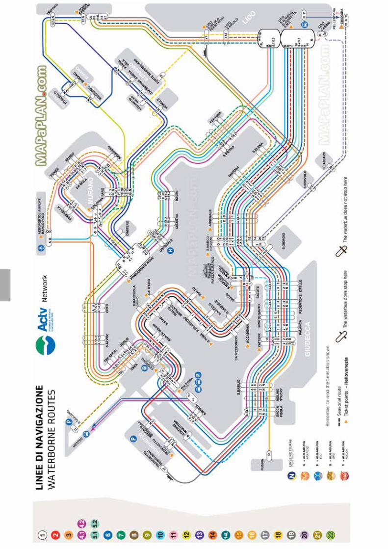
CeNS Workshop Venice 201758
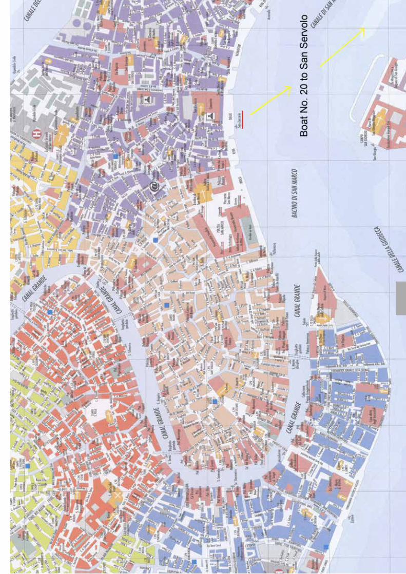
CeNS Workshop Venice 2017 59

Ce
NS
/SF
B1
03
2 W
ork
sh
op
Ve
nic
e 2
01
7:
De
sig
n a
nd
Co
ntr
ol o
f N
an
os
ys
tem
s
Su
nd
ay, S
ep
tem
be
r 1
7T
ime
Mo
nd
ay, S
ep
tem
be
r 1
8T
ime
Tu
es
da
y, S
ep
tem
be
r 1
9T
ime
We
dn
es
da
y, S
ep
tem
be
r 2
0T
ime
Th
urs
da
y, S
ep
tem
be
r 2
1T
ime
Fri
da
y, S
ep
tem
be
r 2
2
09:0
0W
elc
om
e
09:1
5
Ro
b P
hillip
s
10:0
0
10:5
0C
los
ing
re
ma
rks
Gil R
efa
el
18:3
0G
uid
ed t
our
on S
an S
erv
olo
fro
m 8
:15
pm
We
lco
me
Re
ce
pti
on
on
Sa
n S
erv
olo
18:0
0G
uid
ed t
our
on S
an S
erv
olo
Co
ffe
e b
rea
k
15:2
5
15:4
5C
off
ee
bre
ak
15:4
5
Info
rma
l d
isc
us
sio
ns
/
Sig
hts
ee
ing
13:3
0
16:1
5
Po
ste
rs s
es
sio
n II &
dri
nk
s
(16
:30
-18
:30
)
17:0
017:0
0
Lu
nc
h (
12
:30
-14
:15
)L
un
ch
(1
2:2
0-1
4:1
5)
Lu
nc
h (
fro
m 1
2:1
5)
Bo
at fr
om
Sa
n S
erv
olo
at
12
:40
an
d 1
3:3
0
15:0
0
Po
ste
rs s
es
sio
n I &
co
ffe
e (
15
:00
-17
:00
)
Sa
nfo
rd S
imo
n
Ro
b P
hillip
s14:1
514:1
514:1
5
15:0
0
11:4
5
Rin
ald
o T
rott
a
Gil R
efa
el
Olive
r T
rap
p
Jö
rn D
un
ke
l
Iva
n H
uc
Mic
ha
el S
tra
no
Ro
nn
y T
ho
ma
le
Co
ffe
e b
rea
k
10:4
5C
off
ee
bre
ak
10:4
5C
off
ee
bre
ak
11:0
011:0
011:1
5L
. M
ah
ad
eva
n
11:1
5
De
pa
rtu
re
Key
challe
nges:
The c
om
ing q
uantu
m r
evolu
tion?
Fro
m n
ew
mate
rials
to n
ew
com
puta
tional para
dig
ms
11:3
5
09:4
509:4
5
Ke
y ch
alle
ng
es
in b
iop
hys
ics
Fra
nk
Po
llm
an
n
Ch
ris
top
h L
ien
au
Pe
ter
Rö
ttg
erm
an
n
Pe
ter
Ho
mm
elh
off
09:0
009:0
0
10:0
510:3
0C
off
ee
bre
ak
10:3
0
Lu
nc
h (
12
:30
-13
:30
)
Kla
us
Kro
y
Ale
ks
ei A
ks
ime
nti
ev
Nik
ta F
ak
hri
Ch
ris
top
h W
es
terh
au
se
n
Pa
tric
k V
og
el
09:4
509:4
5
09:0
009:0
0
Nig
el G
old
en
feld
Ste
fan
Da
tz
Mik
ae
l R
ec
hts
ma
n
Da
nie
l M
ülle
r
La
ure
ns
Mo
len
ka
mp
Ju
lie
Bit
ee
n
Ale
xa
nd
er
De
ite
rs11:4
5