Embed Size (px)
Citation preview

392 | june 2013 | volume 43 | number 6 | journal of orthopaedic & sports physical therapy
[ research report ]
Altered pelvofemoral biomechanics resulting from deficits in hip muscle performance has been linked to numerous lower extremity conditions.3,8,10,13,15,20,27,28 The hip muscles, specifically the abductors and external rotators, not only
provide local structural stability to the hip joint but are also important in maintain-ing proper segmental alignment of the lower extremity during weight-bearing
tasks.5,21,26 Inability of the hip abductors and external rotators to produce adequate torque during weight-bearing activities can lead to pelvic drop, excessive hip
adduction, excessive hip internal rota-tion, and an increase in the knee valgus angle.10,16,19,20,25,27 In addition, a number of studies have shown that hip abductor muscle performance is significantly corre-lated with postural stability and locomo-tion function in older adults2 and persons who have undergone arthroplastic sur-gery of the lower extremity joints.17,31
Hip abductor muscle performance typically is quantified using a motor-driven or a handheld dynamometer in a non–weight-bearing, sidelying position. Although most investigators have con-cluded that these assessment methods are reasonably reliable,12,14,23,30,32,33 a number of limitations regarding the clinical ap-plication of the assessments have been reported.33 First, proper stabilization and orientation of the lower limb are difficult to maintain during a maximal contraction in the sidelying position. Spe-cifically, it is difficult to keep the amount of flexion/extension of the hip joint and the rotation of the pelvis consistent.23,32 Second, patients often complain that the sidelying position is uncomfortable. The discomfort resulting from compressing the contralateral hip joint against the testing table in the sidelying testing po-sition makes generating maximal abduc-tion force difficult, especially for those
TT STUDY DESIGN: Measurements, descriptive.
TT OBJECTIVES: To describe a weight-bearing method to assess bilateral hip abductor and external rotator muscle performance.
TT BACKGROUND: The hip abductors and external rotators are important in maintaining lower extremity alignment during weight-bearing tasks. As such, there is a need for a method to assess hip muscle performance in weight bearing.
TT METHODS: The weight-bearing method of assessing hip muscle performance utilized a force transducer connected to a nonstretchable fabric strap positioned around the distal ends of both thighs (proximal to the lateral epicondyles). The force generation capacity was recorded with the participants in a semi-squat position (30° of hip flexion and 50° of knee flexion). To establish the reliability of the measurement, 20 participants were tested on 2 separate days, and intraclass correlation coefficient (model 3,1) and standard error of measurement were calculated to evaluate test-retest reliability and intersession consistency. The level of agreement between the muscle perfor-
mance values obtained using the weight-bearing method and the traditional non–weight-bearing method of testing hip abduction in sidelying (dyna-mometer) was assessed using a linear correlation model.
TT RESULTS: The weight-bearing method of assessing hip muscle performance was reliable (intraclass correlation coefficient = 0.99; 95% confidence interval: 0.97, 0.99) and consistent (standard error of measurement, 0.02 N/kg). The measured strength using the weight-bearing method was moderately associated with hip abduction strength values measured in non–weight bearing (r = 0.75, P<.01).
TT CONCLUSION: The proposed weight-bearing method of assessing hip abductor and external rotator muscle performance can be used as a simple, economic, and reliable method to as-sess hip muscle strength. J Orthop Sports Phys Ther 2013;43(6):392-397. Epub 18 March 2013. doi:10.2519/jospt.2013.4412
TT KEY WORDS: gluteus maximus, gluteus medius, strength
1Department of Physical Therapy, School of Allied Health Sciences, University of Nevada, Las Vegas, Las Vegas, NV. 2Jacquelin Perry Musculoskeletal Biomechanics Research Laboratory, Division of Biokinesiology and Physical Therapy, University of Southern California, Los Angeles, CA. The study protocol was approved by the Institutional Review Board of the University of Southern California Health Sciences Campus. This work is partially supported by a grant from the International Society of Biomechanics. The authors certify that they have no affiliations with or financial involvement in any organization or entity with a direct financial interest in the subject matter or materials discussed in the article. Address correspondence to Dr Szu-Ping Lee, Department of Physical Therapy, School of Allied Health Sciences, University of Nevada, Las Vegas, 4505 South Maryland Parkway, Box 453029, Las Vegas, NV 89159. E-mail: [email protected] T Copyright ©2013 Journal of Orthopaedic & Sports Physical Therapy®
SZU-PING LEE, PT, PhD1 • CHRISTOPHER POWERS, PT, PhD2
Description of a Weight-Bearing Method to Assess Hip Abductor and External
Rotator Muscle Performance
43-06 Lee.indd 392 5/21/2013 3:21:17 PM

journal of orthopaedic & sports physical therapy | volume 43 | number 6 | june 2013 | 393
with bilateral hip pain.4 Third, motor-driven dynamometers are expensive and immobile, making them impractical in many clinical settings. Fourth, the non–weight-bearing testing position does not replicate the typical function of the hip abductor muscles during weight bearing, which work in conjunction with the hip external rotators when the hip joint is in a flexed position.11,22
Given the limitations of the currently available assessments for quantifying hip muscle performance, the primary purpose of this research report was to describe a method to assess hip abductor and external rotator muscle performance in a weight-bearing position. Further-more, 3 secondary aims were carried out. First, we investigated the test-retest reliability of the proposed method for quantifying hip abductor and external rotator muscle performance. Second, we quantified the activation levels of the primary hip abductor and external rota-tor muscles (superior portion of the glu-teus maximus [sGMAX], gluteus medius [GMED], and tensor fascia latae [TFL]) during the weight-bearing test to ensure that the muscle strength assessed by the proposed method was the result of hip abductor and external rotator activity. Third, we assessed the level of agree-ment between the hip muscle strength measured using the proposed weight-bearing method and the conventional non–weight-bearing test for quantifying hip abductor strength.
METHODS
Subjects
Twenty individuals (10 women, 10 men) between 24 and 42 years of age participated in this study
(TABLE). Participants who exhibited any of the following were excluded: (1) any his-tory of lower extremity or back surgery, (2) any concurrent condition causing pain or discomfort during physical activity, (3) neurological conditions that would influ-ence an individual’s ability to perform the required testing procedures, and (4)
any other medical conditions that would impair an individual’s ability to perform maximal force exertion. Prior to participa-tion, the objectives, procedures, and risks of the study were explained to each par-ticipant. The study protocol was approved by the Institutional Review Board of the University of Southern California Health Sciences Campus, and informed consent was obtained from each participant.
InstrumentationThe proposed muscle performance test was designed to quantify the force-gen-erating capacity of the hip abductor and external rotator musculature in a weight-bearing position (FIGURE 1A). The force was measured using a uniaxial force trans-ducer (model LCCA-1K; OMEGA Engi-neering, Inc, Stamford, CT) connected to a nonstretchable fabric strap positioned around the distal ends of both thighs, just proximal to the lateral epicondyles. The skin under the strap was protected using a thin, foam-backed, self-adhesive band (NuStim Wrap; Applied Technol-ogy International, Ltd, Exton, PA). The strap and the connectors had a maximum capacity of 2227 N, and the tensile capac-ity of the transducer was rated to 4454 N. The transducer provided force values in Newtons, with an accuracy of 0.037% and precision of 0.02% (full scale).
Care was taken to ensure that the transducer was in series with the strap, parallel to the line of force application. The signal from the force transducer was sampled digitally at 1000 Hz. Real-time feedback of force generation was dis-
played to the participant on a computer monitor throughout testing (LabVIEW Version 8.0.1; National Instruments Cor-poration, Austin, TX) (FIGURE 1B).
Electromyographic (EMG) signals were recorded for the hip musculature, including the sGMAX, GMED, and TFL. EMG data from the dominant limb, de-fined as the preferred limb to perform a single-leg jump, were collected at 1500 Hz using preamplified bipolar surface electrodes. Electrodes for each muscle consisted of two 9-mm Ag/AgCl with a 20-mm interelectrode spacing (No-rotrode 20; Myotronics-Noromed, Kent, WA). The MA-420 preamplifiers (Motion Lab Systems, Inc, Baton Rouge, LA) have a double-differential input design (com-mon-mode rejection ratio greater than 100 dB at 65 Hz, gain at 1 kHz × 20% 1%, input impedance greater than 100 000 000 Ω) and a signal bandwidth of 20 to 3000 Hz. EMG signals were transmit-ted from the first-stage preamplifier to a second-stage receiver unit attached to the back of the subject (MA-133; Motion Lab Systems, Inc). From the receiver unit, the signal was hardwired to a 16-bit analog-to-digital converter (MA-300; Motion Lab Systems, Inc).
Non–weight-bearing isometric strength of the hip abductors was as-sessed using a motor-driven dynamom-eter (CYBEX with HUMAC NORM; Computer Sports Medicine Inc, Stough-ton, MA). The dynamometer provided force values in Newtons, with a precision of 0.02% (full scale). The sampling fre-quency was 100 Hz.
TABLEParticipant Information
and Hip Muscle Performance
*Values are mean SD.†Values are mean SD (range).
Male (n = 10) Female (n = 10) Overall (n = 20)
Age, y* 30.1 4.4 30.4 4.6 30.3 4.4
Height, cm* 180.0 7.6 164.5 8.0 172.3 11.0
Weight, kg* 77.7 9.7 56.6 7.4 67.2 13.7
Normalized weight-bearing strength, N/kg† 3.0 0.6 (2.2-4.2) 2.6 0.5 (2.1-3.4) 2.8 0.6 (2.1-4.2)
Normalized non–weight-bearing strength, N/kg† 3.1 0.9 (2.1-4.3) 2.9 0.5 (2.2-3.6) 2.9 0.6 (2.1-4.3)
43-06 Lee.indd 393 5/21/2013 3:21:19 PM

394 | june 2013 | volume 43 | number 6 | journal of orthopaedic & sports physical therapy
[ research report ]ProceduresData were collected at the Jacquelin Perry Musculoskeletal Biomechanics Research Laboratory at the University of Southern California. Subjects participat-ed in 2 data-collection sessions. On the first visit, participants were tested using both the weight-bearing and the non–weight-bearing positions. The sequence of the 2 muscle performance assessments was randomized. On the second visit, participants were only tested using the weight-bearing method. The average SD interval between visits was 4.4 2.9 days. The subjects were instructed not to participate in strenuous physical activity between testing sessions. EMG activa-tion of the hip musculature during the weight-bearing test was assessed during the second testing session in a subset of 10 participants (6 women, 4 men).
Weight-Bearing Hip Abductor Muscle Performance TestWeight-bearing hip abductor and ex-ternal rotator muscle performance was assessed by a single examiner. The assess-ment was performed with the participant in a squat position (50° of knee flexion and 30° of hip flexion). These angles were established using a goniometer and were selected based on the results of a pilot work, which demonstrated that the high-est force values could be produced in this position. Participants were instructed to maintain their natural lumbar lordotic curvature; to place their feet parallel to each other, shoulder-width apart; and to fold the arms in front of the chest (FIGURE 1). The force transducer and strap connector assembly were positioned just proximal to the lateral epicondyles. Care was taken to ensure that the knee was vertically aligned over the foot. The length of the testing strap was adjusted to accommodate this position. During test-ing, participants were asked to maintain this posture without moving their head, feet, or trunk.
Prior to performing each maximum-effort contraction, participants were in-structed to maintain a baseline tension of
13 N to remove the slack from the testing strap connections. Participants were then instructed to push outward against the resistance strap “as fast and hard as pos-sible” and to maintain this maximum ef-fort for 5 seconds. Verbal encouragement was given to facilitate maximum effort, and real-time feedback of force genera-tion was provided to the subjects on a computer screen (FIGURE 1B). Prior to data collection, practice trials were provided until the participants were comfortable with the testing procedure. Data were collected for a total of 3 trials.
Non–Weight-Bearing Muscle Perfor-mance TestIsometric hip abduction strength of the participant’s dominant lower limb was assessed using a motor-driven dy-namometer in a standard position, as recommended by the manufacturer. Par-ticipants were placed sidelying on the testing table, with the tested hip placed superior and in a neutral position (0° of flexion, abduction, and rotation) (FIGURE
2). The axis of the dynamometer was aligned with the hip joint center in the frontal plane.24 The lower end of the re-sistance pad was positioned just proximal
to the participant’s lateral femoral epi-condyle and secured to the distal thigh with straps. The trunk and pelvis of the participant were strapped to the testing table to minimize motion during testing. Participants performed 2 practice trials before 3 maximum isometric contrac-tions were obtained.
For both the weight-bearing and non–weight-bearing tests, verbal encourage-ment was given to facilitate maximum effort, and force feedback was provided to the subjects on a computer screen. One minute of rest was provided between all weight-bearing and non–weight-bearing trials. The duration of each isometric exertion was 5 seconds. The force trace
FIGURE 1. (A) Hip abductor/external rotator performance assessment in the weight-bearing position (30° of hip flexion, 50° of knee flexion). (B) Testing setup with visual force production feedback interface.
FIGURE 2. Testing position for the dynamometer-based non–weight-bearing hip abductor isometric strength assessment. The hip is in neutral position for all 3 planes of movement.
43-06 Lee.indd 394 5/21/2013 3:21:20 PM
journal of orthopaedic & sports physical therapy | volume 43 | number 6 | june 2013 | 395
of each weight-bearing and non–weight-bearing trial was inspected visually to ensure that the execution of the test was adequate. The peak force produced dur-ing each of the weight-bearing and non–weight-bearing trials was then identified and used for statistical analysis.
EMG AssessmentPrior to applying the surface EMG elec-trodes, the skin was lightly abraded and cleaned with isopropyl ethanol alcohol. Electrodes for the sGMAX were placed on the most prominent portion of the muscle belly, 3 to 6 cm inferior from the posterior superior iliac spine. The elec-trodes were aligned toward the greater trochanter, following the sGMAX muscle fiber orientation. Electrode placement for the GMED was midway along the line be-tween the iliac crest and the greater tro-chanter on the muscle belly. For the TFL, the muscle belly was palpated as the par-ticipant performed resisted hip flexion, abduction, and internal rotation in the supine position. Electrodes were placed on the muscle belly 2 to 4 cm distal to the anterior superior iliac spine, follow-ing the muscle fiber direction. All ground electrodes were placed on electrically si-lent bony surfaces.
To standardize the EMG signal levels among participants, maximal voluntary isometric contractions (MVICs) were
performed. For the sGMAX, the MVIC test was conducted in a prone position, with both of the participant’s legs off the edge of the testing table. The hip was positioned in 45° of flexion and the knee was flexed to 90°. A resistance strap was attached to the femur, just proximal to the popliteal surface. During the test, participants were instructed to extend the tested hip, pushing against the resis-tance strap while keeping the knee flexed.
For the GMED, participants were placed sidelying on the testing table, with the tested hip placed superior and in a neutral position (0° of hip flexion, abduction, and rotation). A resistance strap was positioned at the participant’s lateral femoral epicondyle. Participants were instructed to push up against the strap while keeping the knee extended.
For the TFL, the MVIC trials also were performed in the sidelying position. A belt was looped around the participant’s distal thigh region and the testing table to provide resistance. In this position, the participant was instructed to push in a diagonal direction, thereby performing a combination of isometric hip flexion and abduction against the belt. Two 5-second MVIC trials were collected for each mus-cle. The participants were given at least 1 minute of rest between MVIC trials.
Data AnalysisThe average peak force values obtained from the 3 weight-bearing and non–weight-bearing trials were used for statistical analysis. For the non–weight-bearing trials performed on the dyna-mometer in a sidelying position, the force caused by the weight of the lower limb was added to the force produced during the test. As such, the gravitational influ-ence on the lower limb was accounted for in this non–weight-bearing condition.
EMG signals recorded during the weight-bearing trials were filtered using a 35- to 500-Hz digital band-pass filter and then full-wave rectified. Mean EMG signal amplitude during the third second of the 5-second testing trial was normal-ized to the mean amplitude of the high-
est 1-second EMG amplitude during the MVIC trials. The highest 1-second EMG amplitude during the MVIC trials repre-sented 100% muscle activation. The av-erage normalized muscle activation level from the 3 trials was used for analysis.
Statistical AnalysisThe test-retest reliability of obtaining the peak force using the weight-bearing method was assessed using a 2-way ran-dom intraclass correlation coefficient (ICC3,1). Intersession consistency was quantified using the standard error of measurement (SEM).18 The percentage of the SEM to the measured mean value also was calculated.
The level of agreement between the force values obtained using the weight-bearing and non–weight-bearing strength test methods was assessed using a linear correlation model (2-tailed; sig-nificance level, .05). The EMG activation levels of the 3 hip muscles were presented using descriptive statistics (ie, mean and standard deviation). All statistical analy-ses were conducted using SPSS Statistics Version 19.0 (IBM Corporation, Armonk, NY).
RESULTS
The weight-bearing muscle per-formance assessment demonstrated excellent test-retest reliability (ICC
= 0.99; 95% confidence interval: 0.97, 0.99). On average, the mean SD force measured during the first session (2.8 0.6 N/kg) was similar to the force mea-sured during the second testing session (2.8 0.6 N/kg). The SEM was 0.02 N/kg, which was 0.7% of the measured mean value. The SEM for the nonnor-malized force data was 6.8 N. During the weight-bearing test, the mean SD activation levels for the sGMAX, GMED, and TFL were 93.6% 30.8%, 77.0% 42.3%, and 37.5% 19.8% MVIC, re-spectively (FIGURE 3).
The normalized hip abduction and external rotation force with the weight-bearing method (mean SD, 2.8 0.6
0
sGMAX GMED TFL
20
40
60
80
100
120
140
Nor
mal
ized
Mus
cle
Activ
atio
n, %
MVI
C
FIGURE 3. Activation levels of the hip muscles during the weight-bearing hip abductor/external rotator muscle performance assessment. Data are mean SD. Abbreviations: GMED, gluteus medius; MVIC, maximal voluntary isometric contraction; sGMAX, superior gluteus maximus; TFL, tensor fascia latae.
43-06 Lee.indd 395 5/21/2013 3:21:21 PM

396 | june 2013 | volume 43 | number 6 | journal of orthopaedic & sports physical therapy
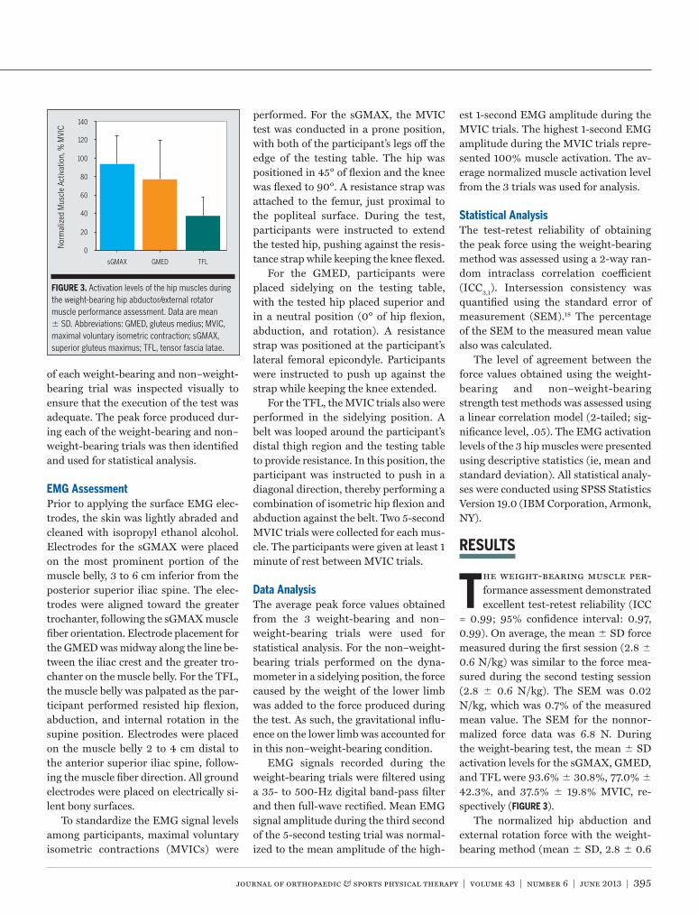
[ research report ]N/kg; range, 2.1-4.2 N/kg) was similar to the force values obtained using the non–weight-bearing method (2.9 0.6 N/kg; range, 2.1-4.3 N/kg). Additionally, hip abductor and external rotation strength measured by the weight-bearing method was found to be significantly correlated with results from the conventional, non–weight-bearing, dynamometer-based assessment for hip abduction (r = 0.75, P<.01) (FIGURE 4).
DISCUSSION
The weight-bearing method for assessing hip abductor and exter-nal rotator strength demonstrated
excellent test-retest reliability and a low SEM. The low SEM relative to the mea-sured mean value indicates that the test gave consistent results between testing sessions.29 The reliability for the weight-bearing method was comparable to that established for a hip abductor strength assessment using a handheld dyna-mometer.33 In addition, the reliability of the measurements with the weight-bearing method was slightly higher than that reported for isometric hip abduc-tor strength testing in sidelying (ICC = 0.90).32 The slightly lower test-retest reliability of the sidelying testing meth-od may be, in part, due to the fact that proper positioning and stabilization of the hip and pelvis are difficult to achieve in a non–weight-bearing position. In contrast, the testing position used for the proposed weight-bearing method placed the subject in a position that minimized the need for external stabilization. The higher test-retest reliability might have been the result of the more stable testing position.
Assessment of the hip muscle EMG signals revealed that the most active muscle during the weight-bearing test was the sGMAX. In comparison, GMED activation was 16.6% lower, and TFL activation was 56.1% lower. The gluteus minimus was not considered in the cur-rent study, as the activity of this muscle could not be evaluated using surface elec-
trodes. Although the gluteus minimus is a hip abductor, it has been shown that the primary function of this muscle is to stabilize the femoral head within the acetabulum. The contribution of its activ-ity with respect to hip abduction torque production is relatively small when com-pared to the other hip abductors.1,7,9
The fact that the sGMAX exhibited the highest activation and the TFL had the lowest may be explained by the fact that the testing position also encouraged the generation of hip external rotation torque as opposed to only hip abduction torque. Given that the sGMAX is also the primary external rotator of the hip, the higher activation observed in this muscle was expected.6 In contrast, the TFL is an internal rotator of the hip. As such, the TFL would not be expected to contrib-ute to the production of any hip external rotation torque imposed by the weight-bearing test. Thus it can be argued that the weight-bearing assessment challeng-es the gluteal muscles to a greater extent than the TFL.
The force values measured using the weight-bearing method were significant-ly correlated with values obtained for hip abduction strength measured using a dynamometer in non–weight bearing. However, it should be noted that only 56.3% of the variance in the dynamom-eter-based test could be explained by the
results obtained from the weight-bearing test. The reason that the agreement be-tween the 2 assessment methods was only moderate may be related to the dif-ferences in hip abductor muscle recruit-ment in the weight-bearing compared to the non–weight-bearing testing po-sitions.32 Additionally, the hip external rotators likely contributed to the force measured during the weight-bearing as-sessment, as subjects performed the test in 30° of hip flexion.
The simplicity of the weight-bearing method is an advantage for assessing performance of the hip musculature in clinical physical therapy practice; how-ever, a limitation of the method is that the test is performed using both lower extremities. The nature of this bilateral setup implies that the measured force is determined by the weaker side. However, it should be noted that the non–weight-bearing method of assessing hip abductor muscle performance has the same limita-tion. For example, Widler et al32 report-ed that there is considerable activation of the contralateral (not-tested) GMED during the non–weight-bearing assess-ments. Specifically, the authors reported that the contralateral-to-ipsilateral ratio of GMED activation was approximately 90% to 130% when evaluated in 3 differ-ent testing positions designed to assess unilateral hip abductor strength. This suggests that stabilization afforded by the contraction of the contralateral hip abductor is important for ipsilateral ab-ductor force production, and that bilat-eral activation is inevitable, regardless of testing position.
CONCLUSION
A weight-bearing method to as-sess hip abductor and external rotator muscle performance was
presented. The proposed weight-bearing method was shown to be reliable and exhibited a moderate level of agreement with the traditional non–weight-bearing method of measuring hip abductor force in sidelying. We propose that the as-
2.0
2.0 2.5 3.0 3.5 4.0 4.5
2.5
3.0
3.5
4.0
4.5
Non
–Wei
ght-B
earin
g H
ip A
bduc
tion
Forc
e, N
/kg
Weight-Bearing Hip Abduction and External Rotation Force, N/kg
FIGURE 4. Correlation between normalized hip force measured with the weight-bearing and non–weight-bearing positions (r = 0.75, P<.01).
43-06 Lee.indd 396 5/21/2013 3:21:22 PM

journal of orthopaedic & sports physical therapy | volume 43 | number 6 | june 2013 | 397
sessment of hip muscle performance in weight bearing may be more meaningful than conventional non–weight-bearing assessments. Future studies will be di-rected toward determining whether hip abductor and external rotator muscle performance measured in weight bear-ing can predict hip joint mechanics dur-ing functional activities. t
KEY POINTSFINDINGS: The proposed weight-bearing method to measure hip abductor/ex-ternal rotator strength was shown to be reliable and exhibited a moderate level of agreement with the traditional non–weight-bearing assessment of the hip abductors in sidelying.IMPLICATIONS: The weight-bearing meth-od can be used as a simple and econom-ic alternative for assessing hip muscle performance.CAUTION: The test was not designed to test unilateral hip abductor strength. In addition, only healthy individuals par-ticipated in this study.
ACKNOWLEDGEMENT: This study was partial-ly supported by the International Society of Biomechanics Dissertation Award.
REFERENCES
1. Beck M, Sledge JB, Gautier E, Dora CF, Ganz R. The anatomy and function of the gluteus minimus muscle. J Bone Joint Surg Br. 2000;82:358-363.
2. Chang SH, Mercer VS, Giuliani CA, Sloane PD. Relationship between hip abductor rate of force development and mediolateral stabil-ity in older adults. Arch Phys Med Rehabil. 2005;86:1843-1850. http://dx.doi.org/10.1016/j.apmr.2005.03.006
3. Cichanowski HR, Schmitt JS, Johnson RJ, Niemuth PE. Hip strength in collegiate female athletes with patellofemoral pain. Med Sci Sports Exerc. 2007;39:1227-1232. http://dx.doi.org/10.1249/mss.0b013e3180601109
4. Click Fenter P, Bellew JW, Pitts TA, Kay RE. Reli-ability of stabilised commercial dynamometers for measuring hip abduction strength: a pilot study. Br J Sports Med. 2003;37:331-334. http://dx.doi.org/10.1136/bjsm.37.4.331
5. Crossley KM, Zhang WJ, Schache AG, Bryant A, Cowan SM. Performance on the single-leg squat task indicates hip abductor muscle function. Am
J Sports Med. 2011;39:866-873. http://dx.doi.org/10.1177/0363546510395456
6. Delp SL, Hess WE, Hungerford DS, Jones LC. Variation of rotation moment arms with hip flex-ion. J Biomech. 1999;32:493-501.
7. Dostal WF, Soderberg GL, Andrews JG. Actions of hip muscles. Phys Ther. 1986;66:351-361.
8. Friel K, McLean N, Myers C, Caceres M. Ipsilateral hip abductor weakness after inversion ankle sprain. J Athl Train. 2006;41:74-78.
9. Gottschalk F, Kourosh S, Leveau B. The functional anatomy of tensor fasciae latae and gluteus me-dius and minimus. J Anat. 1989;166:179-189.
10. Ireland ML, Willson JD, Ballantyne BT, Davis IM. Hip strength in females with and without patellofemoral pain. J Orthop Sports Phys Ther. 2003;33:671-676.
11. Krause DA, Jacobs RS, Pilger KE, Sather BR, Sibunka SP, Hollman JH. Electromyographic analysis of the gluteus medius in five weight-bearing exercises. J Strength Cond Res. 2009;23:2689-2694. http://dx.doi.org/10.1519/JSC.0b013e3181bbe861
12. Laheru D, Kerr JC, McGregor AH. Assessing hip abduction and adduction strength: can greater segmental fixation enhance the reproducibility? Arch Phys Med Rehabil. 2007;88:1147-1153. http://dx.doi.org/10.1016/j.apmr.2007.05.017
13. Leetun DT, Ireland ML, Willson JD, Ballantyne BT, Davis IM. Core stability measures as risk factors for lower extremity injury in athletes. Med Sci Sports Exerc. 2004;36:926-934.
14. Nadler SF, DePrince ML, Hauesien N, Malanga GA, Stitik TP, Price E. Portable dynamometer anchoring station for measuring strength of the hip extensors and abductors. Arch Phys Med Rehabil. 2000;81:1072-1076.
15. Nadler SF, Malanga GA, DePrince M, Stitik TP, Feinberg JH. The relationship between lower extremity injury, low back pain, and hip muscle strength in male and female collegiate athletes. Clin J Sport Med. 2000;10:89-97.
16. Noehren B, Davis I, Hamill J. ASB clinical biome-chanics award winner 2006: prospective study of the biomechanical factors associated with iliotib-ial band syndrome. Clin Biomech (Bristol, Avon). 2007;22:951-956. http://dx.doi.org/10.1016/j.clinbiomech.2007.07.001
17. Piva SR, Teixeira PE, Almeida GJ, et al. Contribu-tion of hip abductor strength to physical function in patients with total knee arthroplasty. Phys Ther. 2011;91:225-233. http://dx.doi.org/10.2522/ptj.20100122
18. Portney LG, Watkins MP. Foundations of Clinical Research: Applications to Practice. 3rd ed. Upper Saddle River, NJ: Pearson/Prentice Hall; 2008.
19. Powers CM. The influence of abnormal hip me-chanics on knee injury: a biomechanical perspec-tive. J Orthop Sports Phys Ther. 2010;40:42-51. http://dx.doi.org/10.2519/jospt.2010.3337
20. Powers CM. The influence of altered lower-extremity kinematics on patellofemoral joint dysfunction: a theoretical perspective. J Orthop Sports Phys Ther. 2003;33:639-646.
21. Presswood L, Cronin J, Keogh JWL, Whatman C. Gluteus medius: applied anatomy, dysfunction, assessment, and progressive strengthening. Strength Cond J. 2008;30:41-53. http://dx.doi.org/10.1519/SSC.0b013e318187f19a
22. Sapega AA. Muscle performance evaluation in orthopaedic practice. J Bone Joint Surg Am. 1990;72:1562-1574.
23. Scott DA, Bond EQ, Sisto SA, Nadler SF. The intra- and interrater reliability of hip muscle strength assessments using a handheld versus a portable dynamometer anchoring station. Arch Phys Med Rehabil. 2004;85:598-603.
24. Seidel GK, Marchinda DM, Dijkers M, Soutas-Little RW. Hip joint center location from palpable bony landmarks—a cadaver study. J Biomech. 1995;28:995-998.
25. Sigward SM, Powers CM. Loading characteristics of females exhibiting excessive valgus moments during cutting. Clin Biomech (Bristol, Avon). 2007;22:827-833. http://dx.doi.org/10.1016/j.clinbiomech.2007.04.003
26. Sled EA, Khoja L, Deluzio KJ, Olney SJ, Culham EG. Effect of a home program of hip abductor ex-ercises on knee joint loading, strength, function, and pain in people with knee osteoarthritis: a clinical trial. Phys Ther. 2010;90:895-904. http://dx.doi.org/10.2522/ptj.20090294
27. Souza RB, Powers CM. Differences in hip kine-matics, muscle strength, and muscle activation between subjects with and without patellofemo-ral pain. J Orthop Sports Phys Ther. 2009;39:12-19. http://dx.doi.org/10.2519/jospt.2009.2885
28. Souza RB, Powers CM. Predictors of hip inter-nal rotation during running: an evaluation of hip strength and femoral structure in women with and without patellofemoral pain. Am J Sports Med. 2009;37:579-587. http://dx.doi.org/10.1177/0363546508326711
29. Stratford PW, Goldsmith CH. Use of the standard error as a reliability index of interest: an applied example using elbow flexor strength data. Phys Ther. 1997;77:745-750.
30. Thijs Y, Van Tiggelen D, Willems T, De Clercq D, Witvrouw E. Relationship between hip strength and frontal plane posture of the knee during a forward lunge. Br J Sports Med. 2007;41:723-727. http://dx.doi.org/10.1136/bjsm.2007.037374
31. Vaz MD, Kramer JF, Rorabeck CH, Bourne RB. Isometric hip abductor strength following total hip replacement and its relationship to func-tional assessments. J Orthop Sports Phys Ther. 1993;18:526-531.
32. Widler KS, Glatthorn JF, Bizzini M, et al. Assess-ment of hip abductor muscle strength. A validity and reliability study. J Bone Joint Surg Am. 2009;91:2666-2672. http://dx.doi.org/10.2106/JBJS.H.01119
33. Youdas JW, Mraz ST, Norstad BJ, Schinke JJ, Hollman JH. Determining meaningful changes in hip abductor muscle strength obtained by handheld dynamometry. Physiother Theory Pract. 2008;24:215-220. http://dx.doi.org/10.1080/03639040701429374
43-06 Lee.indd 397 5/21/2013 3:21:23 PM