Embed Size (px)
Citation preview
OPEN ACCESSHuman & Veterinary MedicineInternational Journal of the Bioflux Society Research Article
Volume 7 | Issue 4 Page 381 HVM Bioflux
http://www.hvm.bioflux.com.ro/
Descending necrotizing mediastinitis of oropharyngeal origin – a retrospective 15 years
study
1*Rares C. Roman, 2*Madalina Lazar, 3Mircea Ghergie, 4Teodora Angelescu, 4Bogdan Samachisa1 Deparment of Cranio-Maxillofacial Surgery , ”Iuliu Hatieganu” University of Medicine and Pharmacy, Cluj-Napoca, Romania; 2 Department of Oral Implantology, “Iuliu Hatieganu” University of Medicine and Farmacy, Cluj-Napoca, Cluj, Romania; 3 Department of Orthodontics and Dentofacial Orthopedics, ”Iuliu Hatieganu” University of Medicine and Pharmacy, Cluj-Napoca, Romania; 4 County Emergency Hospital, Oral and Maxillofacial Surgery Clinic, Cluj-Napoca, Romania.* Pointed authors had equal contribution.
use of CT scanning, modern antibiotic treatment, aggressive drainage, DNM has a high mortality rate, mainly as a result of rapid spread of the microbial infection and delay in diagnosis and treatment. In retrospective studies, mortality varies between 0% and 83% (Sandner&Borgermann 2011). Pearse (Mihos et al 2004) reported the first large series of 110 patients in the preantibiotic era, with mortality rates of 85% in conservatively treated patients and 35% in surgical treated patients (Sandner&Borgermann 2011).According to the literature mortality rate associated with DNM before the aggressive surgical debridement techniques was about 40-50% due to the persistence of infection with empyema, pleu-ral and pericardial effusions, pericarditis, and blood vessel ero-sion. In the presence of severe illness, the mortality rate may be as high as 67% (Sokouti&Nezafati 2009).In this article we report our experience in management of 12 patients affected by DNM.
Abstract. Background: Descending necrotizing mediastinitis is a potentially fatal complication of oral and pharyngeal infections. Rapid di-agnosis and aggressive treatment is necessary in order to successfully treat this life threatening condition. Our objective was to evaluate ret-rospectively all cases presented an treated in our clinic together with Thoracic Surgery Department. Material and methods: Following specific inclusion criteria 12 patients charts were studied regarding etiology, symptoms, radiological and laboratory exams, all patients diagnosed with descending necrotizing mediastinitis and treated in medical center Cluj-Napoca between 2000-2015. Results: The initial source of mediasti-nitis was 9 dental infection and 3 pharyngeal source. Initially patients presented with oral diffuse suppuration, lateropharyngeal abscess ret-ropharyngeal abscess and necrotizing fasciitis. Clinical and radiological confirmation of the mediastinitis were done in the first 24-48 h after admission in 58,33% cases, 3-4 days in 16,66% cases, 7 days in 25% cases. Rapid and extensive surgical debridement and drainage of the neck was performed in all patients. Surgical treatment included cervicomediastinostomy (25%) or thoracotomy (75%). Medical and surgical treatment was followed by rehabilitation of the patient in 62.5% cases. Conclusion: Once the onset of descending necrotizing mediastinitis is suspected, complex and urgent clinical, imagistic and bacteriologic investigations must be done, any delay in establishing the diagnosis or per-forming the proper therapy aggravates the prognosis. Early surgical approach (multiple drainage of cervical, mediastinal and pleural or peri-cardic collections), medical treatment (including broad spectrum antibiotics without expecting the results of antibiograms) and the complex therapy of shock may improve the prognosis.
Key Words: descending necrotizing mediastinitis, odontogenic, pharyngeal
Copyright: This is an open-access article distributed under the terms of the Creative Commons Attribution License, which permits unrestricted use, distribution, and reproduction in any medium, provided the original author and source are credited.
Corresponding Author: R. Roman, e-mail: [email protected]
IntroductionDescending necrotizing mediastinitis (DNM) is one of the most lifethreatening diseases, most frequently occur as a complication after odontogenic or pharyngeal infections with cervicofascial spreading along the deep fascial planes into the mediastinum.Many severe mediastinal infections can result from esopha-geal perforation and infections after operations through ster-notomy incisions.Also mediastinitis can arise from other source of infection such as tonsils, parotid glands, or, rarely, otitis, mastoiditis and epi-glottitis (Palma et al 2015).DNM requires a high index of suspicion, early diagnosis and a multidisciplinary approach based on intensive care support, ag-gressive antibiotherapy and surgical debridement of the initial infection site and the mediastinum, because of the poor prog-nosis of this disease from the literature.Today, the optimal management of DNM remains controversial (Sandner&Borgermann 2011, Cheng et al 2008). Even with the
Roman et al 2015
Volume 7 | Issue 4 Page 382 HVM Bioflux
http://www.hvm.bioflux.com.ro/
Material and methods This study was a retrospective analysis of 12 treated cases ad-mitted into the Department of Oral and Maxillofacial Surgery and the Department of Thoracic Surgery Cluj Napoca, between 2000 and 2015. Data of 12 patients that had DNM secondary to oropharyngeal infections or retropharyngeal abscesses were extracted from medical records of the hospital. All patients had signed informed agreements and consent of medical data use for scientific pur-poses at the time of initial hospital commitment.Patients with DNM arising from esophageal perforation were not included in this study because of the different pathogenesis and treatment.Data extracted from medical charts included demographics (age, sex), predisposing conditions, source of infection (odontogenic, cervical infections), clinical signs and symptoms, radiological and CT characteristics, laboratory exams, microbiological isola-tion, antibiotic treatment and duration, time (days) to admis-sion after diagnosis of head or neck infection, time (hours) tomediastinal drainage after the diagnosis of mediastinitis, type of surgery, complications, hospital length of stay (days), tracheostomy (number of patients and duration), mor-tality rate.All our patients were included in this study on the basis of cri-teria for diagnosis of DNM defined by Estrera (et al 1983): 1) clinical evidence of severe oropharyngeal infection; 2) char-acteristic roentgenographic features of mediastinitis; 3) docu-mentation of necrotizing mediastinal infection at the time of surgery or postmortem; and 4) establishment of the association between the descending necrotizing mediastinitis and the oro-pharyngeal infection.In all our cases the diagnosis of cervical infection was clinical-ly obvious (diffuse painful cervical swelling) and the increas-ing descending infectious symptoms suggesting mediastinitis. Diagnosis was made by cervicothoracic CT and chest radio-graphs in all patients.
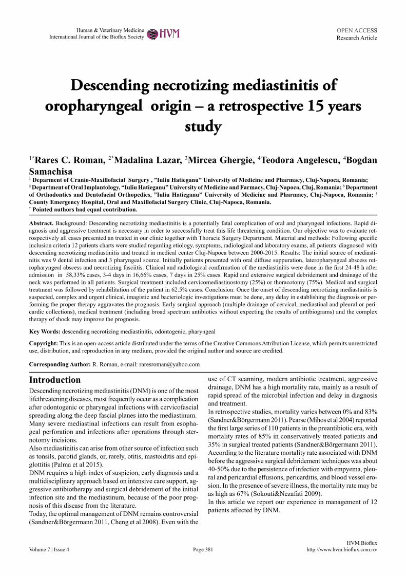
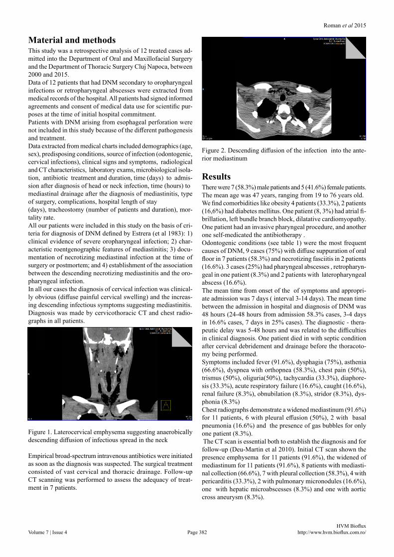
Figure 1. Laterocervical emphysema suggesting anaerobically descending diffusion of infectious spread in the neck
Empirical broad-spectrum intravenous antibiotics were initiated as soon as the diagnosis was suspected. The surgical treatment consisted of vast cervical and thoracic drainage. Follow-up CT scanning was performed to assess the adequacy of treat-ment in 7 patients.
Figure 2. Descending diffusion of the infection into the ante-rior mediastinum
ResultsThere were 7 (58.3%) male patients and 5 (41.6%) female patients. The mean age was 47 years, ranging from 19 to 76 years old.We find comorbidities like obesity 4 patients (33.3%), 2 patients (16,6%) had diabetes mellitus. One patient (8, 3%) had atrial fi-brillation, left bundle branch block, dilatative cardiomyopathy. One patient had an invasive pharyngeal procedure, and another one self-medicated the antibiotherapy . Odontogenic conditions (see table 1) were the most frequent causes of DNM, 9 cases (75%) with diffuse suppuration of oral floor in 7 patients (58.3%) and necrotizing fasciitis in 2 patients (16.6%). 3 cases (25%) had pharyngeal abscesses , retropharyn-geal in one patient (8.3%) and 2 patients with lateropharyngeal abscess (16.6%). The mean time from onset of the of symptoms and appropri-ate admission was 7 days ( interval 3-14 days). The mean time between the admission in hospital and diagnosis of DNM was 48 hours (24-48 hours from admission 58.3% cases, 3-4 days in 16.6% cases, 7 days in 25% cases). The diagnostic - thera-peutic delay was 5-48 hours and was related to the difficulties in clinical diagnosis. One patient died in with septic condition after cervical debridement and drainage before the thoracoto-my being performed. Symptoms included fever (91.6%), dysphagia (75%), asthenia (66.6%), dyspnea with orthopnea (58.3%), chest pain (50%), trismus (50%), oliguria(50%), tachycardia (33.3%), diaphore-sis (33.3%), acute respiratory failure (16.6%), caught (16.6%), renal failure (8.3%), obnubilation (8.3%), stridor (8.3%), dys-phonia (8.3%)Chest radiographs demonstrate a widened mediastinum (91.6%) for 11 patients, 6 with pleural effusion (50%), 2 with basal pneumonia (16.6%) and the presence of gas bubbles for only one patient (8.3%). The CT scan is essential both to establish the diagnosis and for follow-up (Deu-Martin et al 2010). Initial CT scan shown the presence emphysema for 11 patients (91.6%), the widened of mediastinum for 11 patients (91.6%), 8 patients with mediasti-nal collection (66.6%), 7 with pleural collection (58.3%), 4 with pericarditis (33.3%), 2 with pulmonary micronodules (16.6%), one with hepatic microabscesses (8.3%) and one with aortic cross aneurysm (8.3%).
Roman et al 2015
Volume 7 | Issue 4 Page 383 HVM Bioflux
http://www.hvm.bioflux.com.ro/
Patients should be followed up by CT because abscesses tend to reform in the early days after intervention (Ishiaga et al 2013). Some authors perform CT scan at the third, seventh, fourteenth postoperative day routinely, or whenever routine chest X-ray shows abnormal findings (Kwang et al 2012). Follow up CT scans were performed in 7 patients three days after thoracoto-my and they revealed important data regarding progression of the disease.
Laboratory exams revealed leukocytosis, high levels of procal-citonin, hyperglycemia, azotemia, hepatic cytolysis syndrome, cholestasis, hypoxemia but none of those are specific for DNM.Antibiotic therapy consisted of dual or triple intravenous therapy directed at oropharyngeal microorganism, initially ceftriaxone or imipenem when resistance to cephalosporin was found, in association with amikacin and metronidazole. Microbiogical analysis was conducted on specimens collected during surgery and revealed polymicrobial infection shown in table 2 with all cultures positive and high prevalence of Streptococcus spp. in 66.6% of cases.
Table 2. Organism found positive on initial cultures
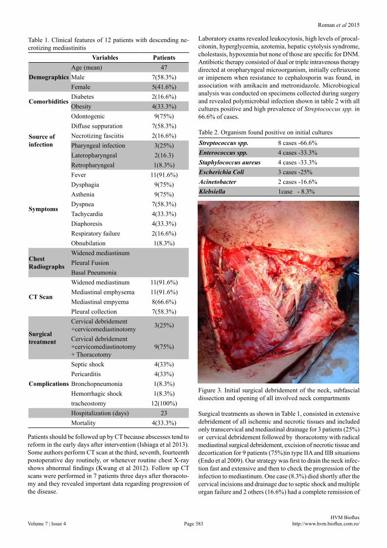
Figure 3. Initial surgical debridement of the neck, subfascial dissection and opening of all involved neck compartments
Surgical treatments as shown in Table 1, consisted in extensive debridement of all ischemic and necrotic tissues and included only transcervical and mediastinal drainage for 3 patients (25%) or cervical debridement followed by thoracotomy with radical mediastinal surgical debridement, excision of necrotic tissue and decortication for 9 patients (75%)in type IIA and IIB situations (Endo et al 2009). Our strategy was first to drain the neck infec-tion fast and extensive and then to check the progression of the infection to mediastinum. One case (8.3%) died shortly after the cervical incisions and drainage due to septic shock and multiple organ failure and 2 others (16.6%) had a complete remission of
Variables Patients
DemographicsAge (mean) 47Male 7(58.3%)Female 5(41.6%)
ComorbiditiesDiabetes 2(16.6%)Obesity 4(33.3%)
Source of infection
Odontogenic 9(75%)Diffuse suppuration 7(58.3%)Necrotizing fasciitis 2(16.6%)Pharyngeal infection 3(25%)Lateropharyngeal 2(16.3)Retropharyngeal 1(8.3%)
Symptoms
Fever 11(91.6%)Dysphagia 9(75%)Asthenia 9(75%)Dyspnea 7(58.3%)Tachycardia 4(33.3%)Diaphoresis 4(33.3%)Respiratory failure 2(16.6%)Obnubilation 1(8.3%)
Chest Radiographs
Widened mediastinumPleural FusionBasal Pneumonia
CT Scan
Widened mediastinum 11(91.6%)Mediastinal emphysema 11(91.6%)Mediastinal empyema 8(66.6%)Pleural collection 7(58.3%)
Surgical treatment
Cervical debridement +cervicomediastinotomy 3(25%)
Cervical debridement +cervicomediastinotomy + Thoracotomy
9(75%)
Complications
Septic shock 4(33%)Pericarditis 4(33%)Bronchopneumonia 1(8.3%)Hemorrhagic shock 1(8.3%)tracheostomy 12(100%)Hospitalization (days) 23Mortality 4(33.3%)
Table 1. Clinical features of 12 patients with descending ne-crotizing mediastinitis
Streptococcus spp. 8 cases -66.6%Enterococcus spp. 4 cases -33.3%Staphylococcus aureus 4 cases -33.3%Escherichia Coli 3 cases -25%Acinetobacter 2 cases -16.6%Klebsiella 1case - 8.3%
Roman et al 2015
Volume 7 | Issue 4 Page 384 HVM Bioflux
http://www.hvm.bioflux.com.ro/
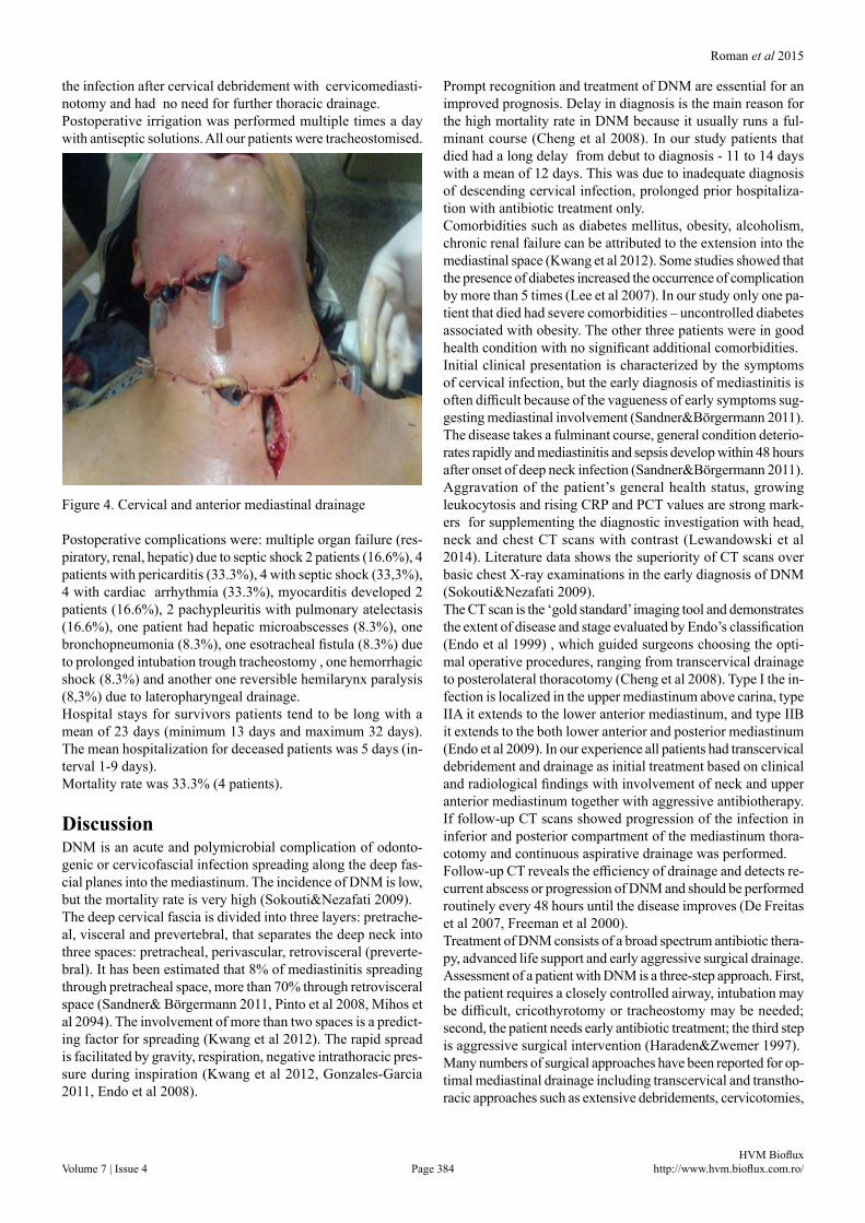
the infection after cervical debridement with cervicomediasti-notomy and had no need for further thoracic drainage.Postoperative irrigation was performed multiple times a day with antiseptic solutions. All our patients were tracheostomised.
Figure 4. Cervical and anterior mediastinal drainage
Postoperative complications were: multiple organ failure (res-piratory, renal, hepatic) due to septic shock 2 patients (16.6%), 4 patients with pericarditis (33.3%), 4 with septic shock (33,3%), 4 with cardiac arrhythmia (33.3%), myocarditis developed 2 patients (16.6%), 2 pachypleuritis with pulmonary atelectasis (16.6%), one patient had hepatic microabscesses (8.3%), one bronchopneumonia (8.3%), one esotracheal fistula (8.3%) due to prolonged intubation trough tracheostomy , one hemorrhagic shock (8.3%) and another one reversible hemilarynx paralysis (8,3%) due to lateropharyngeal drainage.Hospital stays for survivors patients tend to be long with a mean of 23 days (minimum 13 days and maximum 32 days). The mean hospitalization for deceased patients was 5 days (in-terval 1-9 days).Mortality rate was 33.3% (4 patients).
DiscussionDNM is an acute and polymicrobial complication of odonto-genic or cervicofascial infection spreading along the deep fas-cial planes into the mediastinum. The incidence of DNM is low, but the mortality rate is very high (Sokouti&Nezafati 2009).The deep cervical fascia is divided into three layers: pretrache-al, visceral and prevertebral, that separates the deep neck into three spaces: pretracheal, perivascular, retrovisceral (preverte-bral). It has been estimated that 8% of mediastinitis spreading through pretracheal space, more than 70% through retrovisceral space (Sandner& Borgermann 2011, Pinto et al 2008, Mihos et al 2094). The involvement of more than two spaces is a predict-ing factor for spreading (Kwang et al 2012). The rapid spread is facilitated by gravity, respiration, negative intrathoracic pres-sure during inspiration (Kwang et al 2012, Gonzales-Garcia 2011, Endo et al 2008).
Prompt recognition and treatment of DNM are essential for an improved prognosis. Delay in diagnosis is the main reason for the high mortality rate in DNM because it usually runs a ful-minant course (Cheng et al 2008). In our study patients that died had a long delay from debut to diagnosis - 11 to 14 days with a mean of 12 days. This was due to inadequate diagnosis of descending cervical infection, prolonged prior hospitaliza-tion with antibiotic treatment only.Comorbidities such as diabetes mellitus, obesity, alcoholism, chronic renal failure can be attributed to the extension into the mediastinal space (Kwang et al 2012). Some studies showed that the presence of diabetes increased the occurrence of complication by more than 5 times (Lee et al 2007). In our study only one pa-tient that died had severe comorbidities – uncontrolled diabetes associated with obesity. The other three patients were in good health condition with no significant additional comorbidities.Initial clinical presentation is characterized by the symptoms of cervical infection, but the early diagnosis of mediastinitis is often difficult because of the vagueness of early symptoms sug-gesting mediastinal involvement (Sandner&Borgermann 2011).The disease takes a fulminant course, general condition deterio-rates rapidly and mediastinitis and sepsis develop within 48 hours after onset of deep neck infection (Sandner&Borgermann 2011).Aggravation of the patient’s general health status, growing leukocytosis and rising CRP and PCT values are strong mark-ers for supplementing the diagnostic investigation with head, neck and chest CT scans with contrast (Lewandowski et al 2014). Literature data shows the superiority of CT scans over basic chest X-ray examinations in the early diagnosis of DNM (Sokouti&Nezafati 2009).The CT scan is the ‘gold standard’ imaging tool and demonstrates the extent of disease and stage evaluated by Endo’s classification (Endo et al 1999) , which guided surgeons choosing the opti-mal operative procedures, ranging from transcervical drainage to posterolateral thoracotomy (Cheng et al 2008). Type I the in-fection is localized in the upper mediastinum above carina, type IIA it extends to the lower anterior mediastinum, and type IIB it extends to the both lower anterior and posterior mediastinum (Endo et al 2009). In our experience all patients had transcervical debridement and drainage as initial treatment based on clinical and radiological findings with involvement of neck and upper anterior mediastinum together with aggressive antibiotherapy. If follow-up CT scans showed progression of the infection in inferior and posterior compartment of the mediastinum thora-cotomy and continuous aspirative drainage was performed.Follow-up CT reveals the efficiency of drainage and detects re-current abscess or progression of DNM and should be performed routinely every 48 hours until the disease improves (De Freitas et al 2007, Freeman et al 2000).Treatment of DNM consists of a broad spectrum antibiotic thera-py, advanced life support and early aggressive surgical drainage.Assessment of a patient with DNM is a three-step approach. First, the patient requires a closely controlled airway, intubation may be difficult, cricothyrotomy or tracheostomy may be needed; second, the patient needs early antibiotic treatment; the third step is aggressive surgical intervention (Haraden&Zwemer 1997).Many numbers of surgical approaches have been reported for op-timal mediastinal drainage including transcervical and transtho-racic approaches such as extensive debridements, cervicotomies,
Roman et al 2015
Volume 7 | Issue 4 Page 385 HVM Bioflux
http://www.hvm.bioflux.com.ro/
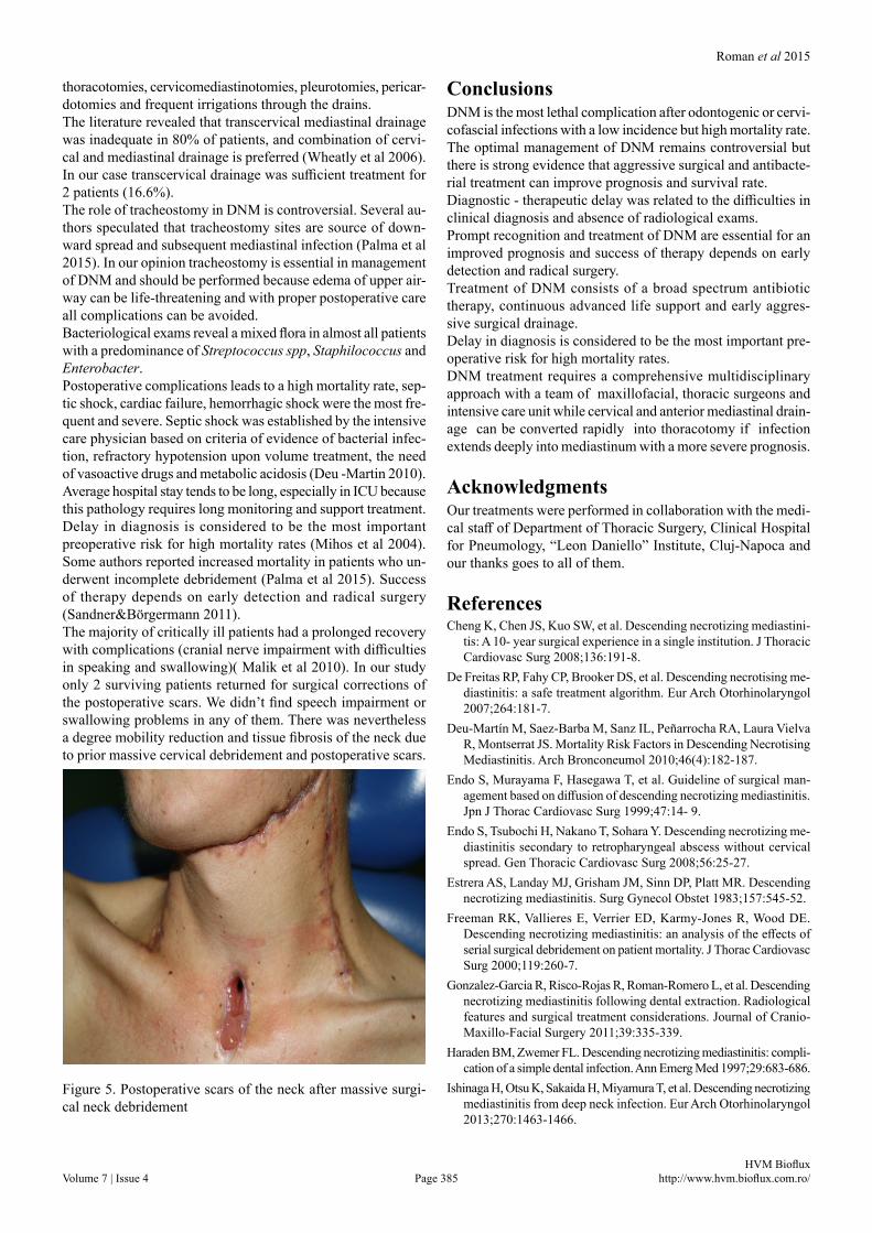
thoracotomies, cervicomediastinotomies, pleurotomies, pericar-dotomies and frequent irrigations through the drains.The literature revealed that transcervical mediastinal drainage was inadequate in 80% of patients, and combination of cervi-cal and mediastinal drainage is preferred (Wheatly et al 2006). In our case transcervical drainage was sufficient treatment for 2 patients (16.6%).The role of tracheostomy in DNM is controversial. Several au-thors speculated that tracheostomy sites are source of down-ward spread and subsequent mediastinal infection (Palma et al 2015). In our opinion tracheostomy is essential in management of DNM and should be performed because edema of upper air-way can be life-threatening and with proper postoperative care all complications can be avoided.Bacteriological exams reveal a mixed flora in almost all patients with a predominance of Streptococcus spp, Staphilococcus and Enterobacter.Postoperative complications leads to a high mortality rate, sep-tic shock, cardiac failure, hemorrhagic shock were the most fre-quent and severe. Septic shock was established by the intensive care physician based on criteria of evidence of bacterial infec-tion, refractory hypotension upon volume treatment, the need of vasoactive drugs and metabolic acidosis (Deu -Martin 2010).Average hospital stay tends to be long, especially in ICU because this pathology requires long monitoring and support treatment.Delay in diagnosis is considered to be the most important preoperative risk for high mortality rates (Mihos et al 2004). Some authors reported increased mortality in patients who un-derwent incomplete debridement (Palma et al 2015). Success of therapy depends on early detection and radical surgery (Sandner&Borgermann 2011).The majority of critically ill patients had a prolonged recovery with complications (cranial nerve impairment with difficulties in speaking and swallowing)( Malik et al 2010). In our study only 2 surviving patients returned for surgical corrections of the postoperative scars. We didn’t find speech impairment or swallowing problems in any of them. There was nevertheless a degree mobility reduction and tissue fibrosis of the neck due to prior massive cervical debridement and postoperative scars.
Figure 5. Postoperative scars of the neck after massive surgi-cal neck debridement
ConclusionsDNM is the most lethal complication after odontogenic or cervi-cofascial infections with a low incidence but high mortality rate.The optimal management of DNM remains controversial but there is strong evidence that aggressive surgical and antibacte-rial treatment can improve prognosis and survival rate.Diagnostic - therapeutic delay was related to the difficulties in clinical diagnosis and absence of radiological exams.Prompt recognition and treatment of DNM are essential for an improved prognosis and success of therapy depends on early detection and radical surgery.Treatment of DNM consists of a broad spectrum antibiotic therapy, continuous advanced life support and early aggres-sive surgical drainage.Delay in diagnosis is considered to be the most important pre-operative risk for high mortality rates.DNM treatment requires a comprehensive multidisciplinary approach with a team of maxillofacial, thoracic surgeons and intensive care unit while cervical and anterior mediastinal drain-age can be converted rapidly into thoracotomy if infection extends deeply into mediastinum with a more severe prognosis.
Acknowledgments Our treatments were performed in collaboration with the medi-cal staff of Department of Thoracic Surgery, Clinical Hospital for Pneumology, “Leon Daniello” Institute, Cluj-Napoca and our thanks goes to all of them.
ReferencesCheng K, Chen JS, Kuo SW, et al. Descending necrotizing mediastini-
tis: A 10- year surgical experience in a single institution. J Thoracic Cardiovasc Surg 2008;136:191-8.
De Freitas RP, Fahy CP, Brooker DS, et al. Descending necrotising me-diastinitis: a safe treatment algorithm. Eur Arch Otorhinolaryngol 2007;264:181-7.
Deu-Martin M, Saez-Barba M, Sanz IL, Penarrocha RA, Laura Vielva R, Montserrat JS. Mortality Risk Factors in Descending Necrotising Mediastinitis. Arch Bronconeumol 2010;46(4):182-187.
Endo S, Murayama F, Hasegawa T, et al. Guideline of surgical man-agement based on diffusion of descending necrotizing mediastinitis. Jpn J Thorac Cardiovasc Surg 1999;47:14- 9.
Endo S, Tsubochi H, Nakano T, Sohara Y. Descending necrotizing me-diastinitis secondary to retropharyngeal abscess without cervical spread. Gen Thoracic Cardiovasc Surg 2008;56:25-27.
Estrera AS, Landay MJ, Grisham JM, Sinn DP, Platt MR. Descending necrotizing mediastinitis. Surg Gynecol Obstet 1983;157:545-52.
Freeman RK, Vallieres E, Verrier ED, Karmy-Jones R, Wood DE. Descending necrotizing mediastinitis: an analysis of the effects of serial surgical debridement on patient mortality. J Thorac Cardiovasc Surg 2000;119:260-7.
Gonzalez-Garcia R, Risco-Rojas R, Roman-Romero L, et al. Descending necrotizing mediastinitis following dental extraction. Radiological features and surgical treatment considerations. Journal of Cranio-Maxillo-Facial Surgery 2011;39:335-339.
Haraden BM, Zwemer FL. Descending necrotizing mediastinitis: compli-cation of a simple dental infection. Ann Emerg Med 1997;29:683-686.
Ishinaga H, Otsu K, Sakaida H, Miyamura T, et al. Descending necrotizing mediastinitis from deep neck infection. Eur Arch Otorhinolaryngol 2013;270:1463-1466.
Roman et al 2015
Volume 7 | Issue 4 Page 386 HVM Bioflux
http://www.hvm.bioflux.com.ro/
Kang SK, Lee S, Oh HK, Kang MW, et al. Clinical Features of Deep Neck Infections and Predisposing Factors for Mediastinal Extension. Korean J Thorac Cardiovasc Surg 2012;45:171-176.
Lee JK, Kim HD, Lim SC. Predisposing factors of complicated deep neck infection: an analysis of 158 cases. Yonsei Med J 2007;48:55-62.
Lewandowski B, Pakla P, Wołek W, Jednakiewicz M, Nicpon J. A fatal case of descending necrotizing mediastinitis as a complication of od-ontogenic infection. A case report . Kardiochirurgia i Torakochirurgia Polska 2014;11(3):324-328.
Malik V, Gadepalli C, Agrawal S, et al. An algorithm for early diagno-sis of cervicofacial necrotising fasciitis. Eur Arch Otorhinolaryngol 2010;267:1169–77.
Mihos M, Potaris K, Gakidis I, Papadakis D, Rallis G. Management of descending necrotizing mediastinitis. J Oral Maxillofac Surg 2004;62:966-972.
Palma DM, Giuliano SN, Cracchiolo AN, Falcone M, Ceccarelli G, Tetamo R, Venditti M. Clinical features and outcome of patients with descending necrotizing mediastinitis: prospective analysis of 34 cases. Infection 2015 Sep 3. [Epub ahead of print].
Pearse HE. Mediastinitis following cervical suppuration . Ann Surg 1938;108:588–611.
Pinto A, Scaglione M, Scuderi MG, Tortora G, Daniele S, Romano L. Infections of the neck leading to descending necrotizing mediasti-nitis: Role of multi-detector row computed tomography. European Journal of Radiology 2008;65:389-394.
Sandner A, Borgermann J, Update on Necrotizing Mediastinitis: Causes, Approaches to Management, and Outcomes. Curr Infect Dis Rep 2011;13:278–286.
Sokouti M, Nezafati S. Descending necrotizing mediastinitis of oropharyn-geal infections. J Dent Res Dent Clin Dent Prospect 2009;3(3):82-85.
Wheatly MJ, Nakano T, Takayasu Y et al. Successful video-assisted mediastinoscopic drainage of descending mecrotizing mediastini-tis. Ann Thoracic Surg 2006;81:2279-81.
Authors•Rares C. Roman, Department of Cranio-Maxillo-Facial Surgery, ”Iuliu Hatieganu” University of Medicine and Pharmacy, 33 Motilor Street, 400001, Cluj-Napoca, Cluj, Romania, EU, email: [email protected]
•Madalina Lazar, Department of Oral Implantology, “Iuliu Hatieganu” University of Medicine and Pharmacy, 15 Victor Babes Street, 400012, Cluj-Napoca, Cluj, Romania, EU, email: [email protected]
•Mircea Ghergie, Department of Orthodontics and Dentofacial Orthopedics, ”Iuliu Hatieganu” University of Medicine and Pharmacy, 33 Motilor Street, 400001, Cluj-Napoca, Cluj, Romania, EU, email: [email protected]
•Teodora Angelescu, County Emergency Hospital, Oral and Maxillofacial Surgery Clinic , 33 Motilor Street, 400001, Cluj-Napoca, Cluj, Romania, EU, email: [email protected]
•Bogdan Samachisa, , County Emergency Hospital, Oral and Maxillofacial Surgery Clinic , 33 Motilor Street, 400001, Cluj-Napoca, Cluj, Romania, EU, email: [email protected]
CitationRoman RC, Lazar M, Ghergie M, Angelescu T, Samachisa B. Descending necrotizing mediastinitis of oropharyngeal origin – a retrospective 15 years study. HVM Bioflux 2015;7(4):381-386.
Editor Ştefan C. VesaReceived 13 November 2015Accepted 22 November 2015
Published Online 23 November 2015Funding None reported
Conflicts/ Competing
InterestsNone reported