Embed Size (px)
Citation preview
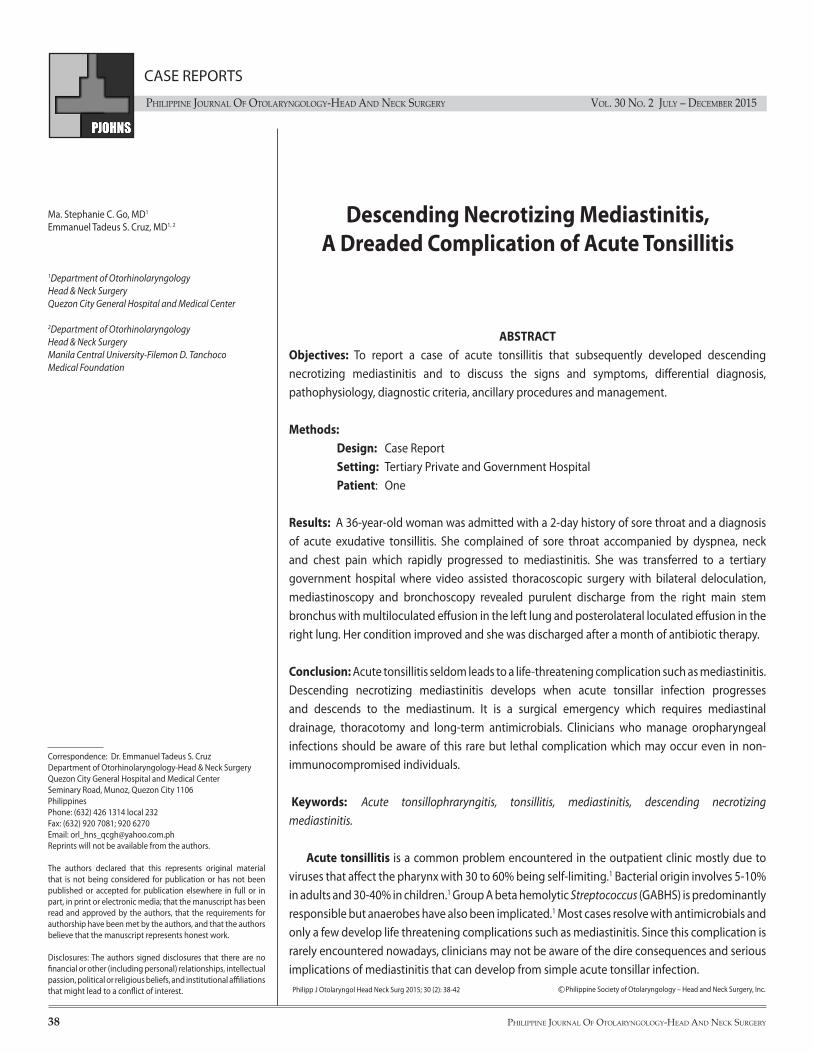
PhiliPPine Journal of otolaryngology-head and neck Surgery Vol. 30 no. 2 July – december 2015
38 PhiliPPine Journal of otolaryngology-head and neck Surgery
CASE REPORTS
Philipp J Otolaryngol Head Neck Surg 2015; 30 (2): 38-42 c Philippine Society of Otolaryngology – Head and Neck Surgery, Inc.
ABSTRACTObjectives: To report a case of acute tonsillitis that subsequently developed descending necrotizing mediastinitis and to discuss the signs and symptoms, differential diagnosis, pathophysiology, diagnostic criteria, ancillary procedures and management.
Methods:Design: Case ReportSetting: Tertiary Private and Government HospitalPatient: One
Results: A 36-year-old woman was admitted with a 2-day history of sore throat and a diagnosis of acute exudative tonsillitis. She complained of sore throat accompanied by dyspnea, neck and chest pain which rapidly progressed to mediastinitis. She was transferred to a tertiary government hospital where video assisted thoracoscopic surgery with bilateral deloculation, mediastinoscopy and bronchoscopy revealed purulent discharge from the right main stem bronchus with multiloculated effusion in the left lung and posterolateral loculated effusion in the right lung. Her condition improved and she was discharged after a month of antibiotic therapy.
Conclusion: Acute tonsillitis seldom leads to a life-threatening complication such as mediastinitis. Descending necrotizing mediastinitis develops when acute tonsillar infection progresses and descends to the mediastinum. It is a surgical emergency which requires mediastinal drainage, thoracotomy and long-term antimicrobials. Clinicians who manage oropharyngeal infections should be aware of this rare but lethal complication which may occur even in non-immunocompromised individuals.
Keywords: Acute tonsillophraryngitis, tonsillitis, mediastinitis, descending necrotizing mediastinitis.
Acute tonsillitis is a common problem encountered in the outpatient clinic mostly due to viruses that affect the pharynx with 30 to 60% being self-limiting.1 Bacterial origin involves 5-10% in adults and 30-40% in children.1 Group A beta hemolytic Streptococcus (GABHS) is predominantly responsible but anaerobes have also been implicated.1 Most cases resolve with antimicrobials and only a few develop life threatening complications such as mediastinitis. Since this complication is rarely encountered nowadays, clinicians may not be aware of the dire consequences and serious implications of mediastinitis that can develop from simple acute tonsillar infection.
Descending Necrotizing Mediastinitis, A Dreaded Complication of Acute Tonsillitis
Ma. Stephanie C. Go, MD1
Emmanuel Tadeus S. Cruz, MD1, 2
1Department of OtorhinolaryngologyHead & Neck SurgeryQuezon City General Hospital and Medical Center
2Department of OtorhinolaryngologyHead & Neck SurgeryManila Central University-Filemon D. Tanchoco Medical Foundation
Correspondence: Dr. Emmanuel Tadeus S. CruzDepartment of Otorhinolaryngology-Head & Neck SurgeryQuezon City General Hospital and Medical CenterSeminary Road, Munoz, Quezon City 1106PhilippinesPhone: (632) 426 1314 local 232Fax: (632) 920 7081; 920 6270Email: [email protected] will not be available from the authors.
The authors declared that this represents original material that is not being considered for publication or has not been published or accepted for publication elsewhere in full or in part, in print or electronic media; that the manuscript has been read and approved by the authors, that the requirements for authorship have been met by the authors, and that the authors believe that the manuscript represents honest work.
Disclosures: The authors signed disclosures that there are no financial or other (including personal) relationships, intellectual passion, political or religious beliefs, and institutional affiliations that might lead to a conflict of interest.
PhiliPPine Journal of otolaryngology-head and neck Surgery Vol. 30 no. 2 July – december 2015
CASE REPORTS
PhiliPPine Journal of otolaryngology-head and neck Surgery 39
The objective of this paper is to share the clinical experience gained in managing a case of acute tonsillitis that later developed mediastinitis and to discuss the signs and symptoms, differential diagnosis, pathophysiology, diagnostic criteria, ancillary procedures and management of such conditions.
CASe RePORTA 36-year-old woman with a 2-day history of sore throat,
odynophagia, fever and body malaise was admitted for difficulty of breathing and chest pain that developed a few hours prior to admission. The patient had no history of hypertension, diabetes or prior hospitalizations.
She was conscious and coherent with normal vital signs. Oral cavity examination showed a hyperemic pharynx and grade 1-2 tonsils with exudates. There was slight tenderness of the neck on palpation but no swelling or palpable neck nodes. She had symmetrical chest expansion with no retractions and clear breath sounds. The rest of the physical examination was normal. The admitting impression was acute exudative tonsillitis.
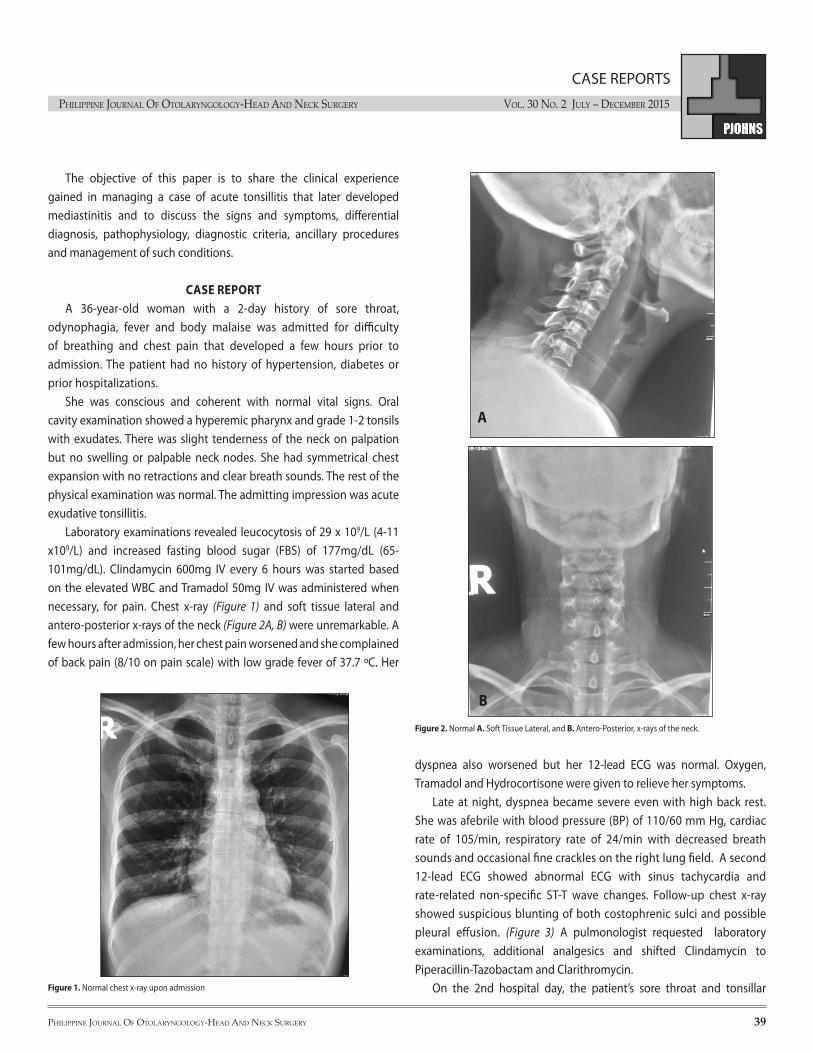
Laboratory examinations revealed leucocytosis of 29 x 109/L (4-11 x109/L) and increased fasting blood sugar (FBS) of 177mg/dL (65-101mg/dL). Clindamycin 600mg IV every 6 hours was started based on the elevated WBC and Tramadol 50mg IV was administered when necessary, for pain. Chest x-ray (Figure 1) and soft tissue lateral and antero-posterior x-rays of the neck (Figure 2A, B) were unremarkable. A few hours after admission, her chest pain worsened and she complained of back pain (8/10 on pain scale) with low grade fever of 37.7 ºC. Her
dyspnea also worsened but her 12-lead ECG was normal. Oxygen, Tramadol and Hydrocortisone were given to relieve her symptoms.
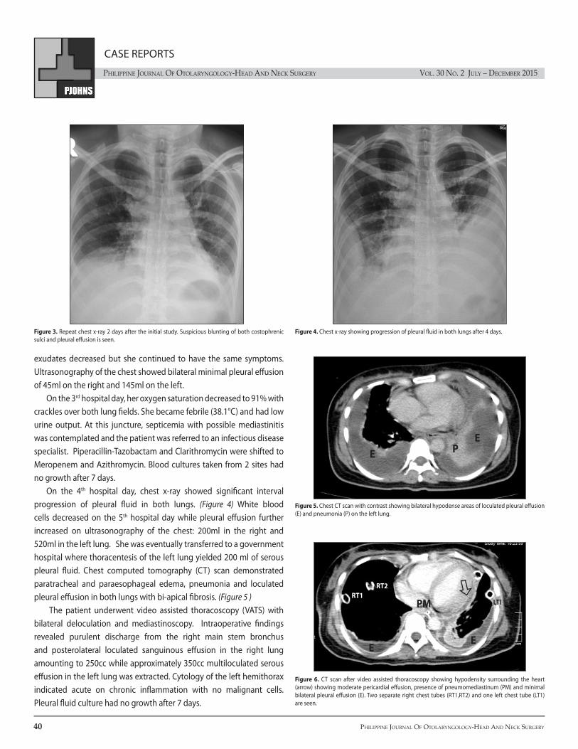
Late at night, dyspnea became severe even with high back rest. She was afebrile with blood pressure (BP) of 110/60 mm Hg, cardiac rate of 105/min, respiratory rate of 24/min with decreased breath sounds and occasional fine crackles on the right lung field. A second 12-lead ECG showed abnormal ECG with sinus tachycardia and rate-related non-specific ST-T wave changes. Follow-up chest x-ray showed suspicious blunting of both costophrenic sulci and possible pleural effusion. (Figure 3) A pulmonologist requested laboratory examinations, additional analgesics and shifted Clindamycin to Piperacillin-Tazobactam and Clarithromycin.
On the 2nd hospital day, the patient’s sore throat and tonsillar Figure 1. Normal chest x-ray upon admission
Figure 2. Normal A. Soft Tissue Lateral, and B. Antero-Posterior, x-rays of the neck.
A
B
PhiliPPine Journal of otolaryngology-head and neck Surgery Vol. 30 no. 2 July – december 2015
CASE REPORTS
40 PhiliPPine Journal of otolaryngology-head and neck Surgery
exudates decreased but she continued to have the same symptoms. Ultrasonography of the chest showed bilateral minimal pleural effusion of 45ml on the right and 145ml on the left.
On the 3rd hospital day, her oxygen saturation decreased to 91% with crackles over both lung fields. She became febrile (38.1°C) and had low urine output. At this juncture, septicemia with possible mediastinitis was contemplated and the patient was referred to an infectious disease specialist. Piperacillin-Tazobactam and Clarithromycin were shifted to Meropenem and Azithromycin. Blood cultures taken from 2 sites had no growth after 7 days.
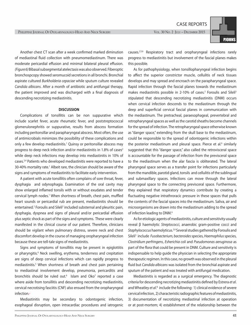
On the 4th hospital day, chest x-ray showed significant interval progression of pleural fluid in both lungs. (Figure 4) White blood cells decreased on the 5th hospital day while pleural effusion further increased on ultrasonography of the chest: 200ml in the right and 520ml in the left lung. She was eventually transferred to a government hospital where thoracentesis of the left lung yielded 200 ml of serous pleural fluid. Chest computed tomography (CT) scan demonstrated paratracheal and paraesophageal edema, pneumonia and loculated pleural effusion in both lungs with bi-apical fibrosis. (Figure 5 )
The patient underwent video assisted thoracoscopy (VATS) with bilateral deloculation and mediastinoscopy. Intraoperative findings revealed purulent discharge from the right main stem bronchus and posterolateral loculated sanguinous effusion in the right lung amounting to 250cc while approximately 350cc multiloculated serous effusion in the left lung was extracted. Cytology of the left hemithorax indicated acute on chronic inflammation with no malignant cells. Pleural fluid culture had no growth after 7 days.
Figure 3. Repeat chest x-ray 2 days after the initial study. Suspicious blunting of both costophrenic sulci and pleural effusion is seen.
Figure 4. Chest x-ray showing progression of pleural fluid in both lungs after 4 days.
Figure 5. Chest CT scan with contrast showing bilateral hypodense areas of loculated pleural effusion (E) and pneumonia (P) on the left lung.
e Pe
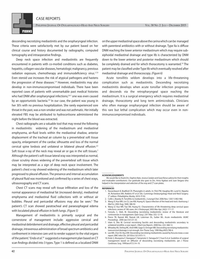
Figure 6. CT scan after video assisted thoracoscopy showing hypodensity surrounding the heart (arrow) showing moderate pericardial effusion, presence of pneumomediastinum (PM) and minimal bilateral pleural effusion (E). Two separate right chest tubes (RT1,RT2) and one left chest tube (LT1) are seen.
PM
ee
LTI
RT2RT1
PhiliPPine Journal of otolaryngology-head and neck Surgery 41
PhiliPPine Journal of otolaryngology-head and neck Surgery Vol. 30 no. 2 July – december 2015
CASE REPORTS
Another chest CT scan after a week confirmed marked diminution of mediastinal fluid collection with pneumomediastinum. There was moderate pericardial effusion and minimal bilateral pleural effusion. (Figure 6) Bibasal subsegmental atelectasis was also observed. Fiberoptic bronchospcopy showed seromucoid secretions in all bronchi. Bronchial aspirate cultured Burkholderia cepaciae while sputum culture revealed Candida albicans. After a month of antibiotic and antifungal therapy, the patient improved and was discharged with a final diagnosis of descending necrotizing mediastinitis.
DISCUSSIONComplications of tonsillitis can be non suppurative which
include scarlet fever, acute rheumatic fever, and poststreptococcal glomerulonephritis or suppurative, results from abscess formation including peritonsillar and parapharyngeal abscess. Most often, the use of antimicrobials minimizes the possibility of these complications and only a few develop mediastinitis.1 Quinsy or peritonsillar abscess may progress to deep neck infection and/or mediastinitis in 1.8% of cases2 while deep neck infections may develop into mediastinitis in 10% of cases.3,4 Patients who developed mediastinitis were reported to have a 30-40% mortality rate.1 Albeit rare, the clinician should be aware of the signs and symptoms of mediastinitis to facilitate early intervention.
A patient with acute tonsillitis often complains of sore throat, fever, dysphagia and odynophagia. Examination of the oral cavity may show enlarged inflamed tonsils with or without exudates and tender cervical lymph nodes.1 When shortness of breath, chest pain, muffled heart sounds or pericardial rub are present, mediastinitis should be entertained.2 Foroulis and Sileli5 included substernal and pleuritic pain, dysphagia, dyspnea and signs of pleural and/or pericardial effusion plus septic shock as part of the signs and symptoms. These were clearly manifested in the clinical course of our patient. Therefore, clinicians should be vigilant when pulmonary distress, severe neck and chest discomfort develop in the course of managing oropharyngeal infection because these are tell-tale signs of mediastinitis.
Signs and symptoms of tonsillitis may be present in epiglottitis or pharyngitis.6 Neck swelling, erythema, tenderness and crepitation are signs of deep cervical infections which can rapidly progress to mediastinitis.5 When shortness of breath and chest pain pertaining to mediastinal involvement develop, pneumonia, pericarditis and bronchitis should be ruled out.6 Islam and Oko7 reported a case where aside from tonsillitis and descending necrotizing mediastinitis, cervical necrotising fasciitis (CNF) also ensued from the oropharyngeal infection.
Mediastinitis may be secondary to odontogenic infection, esophageal disruption, open intracardiac procedures and iatrogenic
causes.2,5,6 Respiratory tract and oropharyngeal infections rarely progress to mediastinitis but involvement of the fascial planes makes this possible.
As for pathophysiology, when tonsillopharyngeal infection begins to affect the superior constrictor muscle, cellulitis of neck tissues develops and may spread and encroach on the parapharyngeal space. Rapid infection through the fascial planes towards the mediastinum makes mediastinitis possible in 2-10% of cases.2 Foroulis and Sileli5 stipulated that descending necrotizing mediastinitis (DNM) occurs when cervical infection descends to the mediastinum through the deep and superficial cervical fascial planes in communication with the mediastinum. The pretracheal, paraesophageal, prevertebral and retropharyngeal spaces as well as the carotid sheaths become channels for the spread of infection. The retropharyngeal space otherwise known as “danger space,” extending from the skull base to the mediastinum, could be responsible to the spread of odontogenic infections within the posterior mediastinum and pleural space. Pierce et al.6 similarly suggested that this “danger space,” also called the retrovisceral space is accountable for the passage of infection from the previsceral space to the mediastinum when the alar fascia is obliterated. The lateral pharyngeal space serves as a transfer point for infections particularly from the mandible, parotid gland, tonsils and cellulitis of the sublingual and submaxillary spaces. Infections can move through the lateral pharyngeal space to the connecting previsceral space. Furthermore, they explained that respiratory dynamics contribute by creating a fluctuating negative intrathroracic pressure in these spaces that pulls the contents of the fascial spaces into the mediastinum. Saliva, air and microorganisms are drawn into the mediastinum adding to the spread of infection leading to DNM.6
As for etiologic agents of mediastinitis, culture and sensitivity usually reveal β haemolytic Streptococci, anaerobic gram-positive cocci and Staphylococcus haemolyticus.2,6 Several studies gathered by Foroulis and Sileli5 include Fusobacterium, bacteroides species, Haemophilus species, Clostridium perfringens, Esherichia coli and Pseudomonas aeruginosa as part of the flora that could be present in DNM. Culture and sensitivity is indispensable to help guide the physician in selecting the appropriate therapeutic regimen. In this case, no growth was observed in the pleural fluid but Candida albicans was isolated from the bronchial aspirate and sputum of the patient and was treated with antifungal medication.
Mediastinitis is regarded as a surgical emergency. The diagnostic criteria for descending necrotizing mediastinitis defined by Estrera et al. and Wheatley et al.8 include the following: 1) clinical evidence of severe cervical infection, 2) characteristic radiographic features of mediastinitis, 3) documentation of necrotizing mediastinal infection at operation or at post-mortem; 4) establishment of the relationship between the
PhiliPPine Journal of otolaryngology-head and neck Surgery Vol. 30 no. 2 July – december 2015
CASE REPORTS
42 PhiliPPine Journal of otolaryngology-head and neck Surgery
descending necrotizing mediastinitis and the oropharyngeal infection. These criteria were satisfactorily met by our patient based on her clinical course and history documented by radiographs, computed tomography and intraoperative findings.
Deep neck space infection and mediastinitis are frequently encountered in patients with co-morbid conditions such as diabetes, hepatitis, collagen vascular diseases, hematologic malignancy, previous radiation exposure, chemotherapy and immunodeficiency virus.1,7,9 Even steroid use increases the risk of atypical pathogens and hastens the progression of these diseases.1,9 However, mediastinitis may also develop in non-immunocompromised individuals. There have been reported cases of patients with unremarkable past medical histories who had DNM after oropharyngeal infections;2,6,7 one was even caused by an opportunistic bacteria.2,9 In our case, the patient was young in her 30’s with no previous hospitalization. She rarely experienced sore throat in the past, was a non-smoker and was not asthmatic. Her initially elevated FBS may be attributed to hydrocortisone administered the night before the blood was extracted.
Chest radiographs are a valuable tool that may reveal the following in mediastinitis: widening of the mediastinum and mediastinal emphysema, air-fluid levels within the mediastinal shadow, anterior displacement of the tracheal air column by a prevertebral soft tissue opacity, enlargement of the cardiac silhouette and loss of the normal cervical spine lordosis and unilateral or bilateral pleural effusion.2,5 Soft tissue x-ray of the neck may reveal air or gas in the soft tissues.7 Although the patient’s soft tissue lateral xray was interpreted as normal, closer scrutiny shows widening of the prevertebral soft tissue which may be interpreted as a sign of deep neck space involvement. The patient’s chest x-ray showed widening of the mediastinum which later progressed to pleural effusion. The presence and interval accumulation of pleural fluid was monitored and confirmed by a series of chest xrays, ultrasonography and CT scans.
Chest CT scans may reveal soft tissue infiltration and loss of the normal appearance of mediastinal fat (increased density), mediastinal emphysema and mediastinal fluid collections with or without air bubbles. Pleural and pericardial effusions may also be seen.5 The patient’s CT scan showed paratracheal and paraesophageal edema with loculated pleural effusion in both lungs. (Figure 5)
Management of mediastinitis is primarily surgical and the cornerstone of management include: aggressive cervical and mediastinal debridement and drainage, effective pleural and pericardial drainage, intravenous administration of broad spectrum antibiotics and confinement in intensive care unit to render support to the vital organs of the patient.5 Endo et al.10 suggested a management plan based on CT scan findings divided into 3 types. Type 1 is defined as a localized DNM
ACKNOwLeDgeMeNTS We would like to thank Drs. Daphne Bate, Jessica Catalan and Guia Elena Ladrera for their insights
and invaluable contributions. Our gratitude also goes to Drs. Anna Agabao and Juan Vergara who helped in the interpretation and selection of the xray and CT scan plates.
ReFeReNCeS1. Nussenbaum B, Bradford CR. Pharyngitis in adults. In: Flint PW, Haughey BH, Lund VJ, Niparko
JK, Richardson MA, Robbins KT et al. Eds. Cummings Otolaryngology Head and Neck Surgery. 5th edition. Philadelphia: Mosby; 2010. P.192.
2. Collin J, Beasley N. Tonsillitis to mediastinitis. J Laryngol Otol. 2006 Nov; 120(11):963-966.3. Wang LF, Kuo WR, Lin CS, Lee KW, Huang KJ. Space infection of the head and neck. Kaohsiung J
Med Sci, 2002 Aug; 18(8): 386-92.4. Wang LF, Kuo WR, Tsai SM, Huang KJ. Characteristics of life threatening deep cervical space
infections: review of 196 cases. Am J Otolaryngol, 2003 Mar-Apr; 24(2): 111-17.5. Foroulis, C, Sileli, M. Descending necrotizing mediastinitis: review of the literature and
controversies in management. Open Surg J, 2011Mar; 5(5): 12-18.6. Pierce TB, Razzuk MA, Razzuk LM, Luterman DL, Sutker WL. Acute mediastinitis. BUMC
Proceedings. 2000 Jan;13(1):31-33.7. Islam A, Oko M. Cervical necrotising fasciitis and descending mediastinitis secondary to
unilateral tonsillitis: a case report. J Med Case Reports. 2008 Dec; 2(2): 368-12.8. Wheatley MJ, Stirling MC, Kirsh MM, Gago O, Orringer MB. Descending necrotizing mediastinitis:
transcervical drainage is not enough. Ann Thorac Surg. 1990 May;49(5):780-4.9. Lee MK, Choi SH, Ryu DW. Descending necrotizing mediastinitis caused by Kocuria rosea: a case
report. BMC Infect Dis. 2013Oct;13(1):475-11. 10. Endo S, Murayama F, Hasegawa T, Yamamoto S, Yamaguchi T, Fuse K et al. Guideline of surgical
management based on diffusion of descending necrotizing mediastinitis. Jpn J Thorac Cardiovasc Surg. 1999Jan;47(1):14–19.
on the upper mediastinal space above the carina which can be managed with parenteral antibiotics with or without drainage. Type IIa is diffuse DNM reaching the lower anterior mediastinum which may require sub-xiphoidal mediastinal drainage and type IIb is characterized by DNM down to the lower anterior and posterior mediastinum which should be completely drained and for which thoracotomy is warranted.10 The patient may be classified under Type IIb which eventually resolved after mediastinal drainage and thoracoscopy. (Figure 6)
Acute tonsillitis seldom develops into a life-threatening complication such as mediastinitis. Descending necrotizing mediastinitis develops when acute tonsillar infection progresses and descends via the retropharyngeal space reaching the mediastinum. It is a surgical emergency which requires mediastinal drainage, thoracotomy and long term antimicrobials. Clinicians who often manage oropharyngeal infection should be aware of this rare but lethal complication which may occur even in non-immunocompromised individuals.