Embed Size (px)
Citation preview
16 CUTIS® WWW.CUTIS.COM
DErmatopathology Diagnosis
PLEASE TURN TO PAGE 39 FOR DERMATOPATHOLOGY DIAGNOSIS DISCUSSION
Alyssa Miceli, DO; Nathan Cleaver, DO; Amy Spizuoco, DO
Dr. Miceli is from the College of Osteopathic Medicine, New York Institute of Technology, Old Westbury. Drs. Cleaver and Spizuoco are from Ackerman Academy of Dermatopathology, New York, New York. The authors report no conflict of interest.Correspondence: Amy Spizuoco, DO, Ackerman Academy of Dermatopathology, 145 E 32nd Street, 10th Floor, New York, NY 10016 ([email protected]).
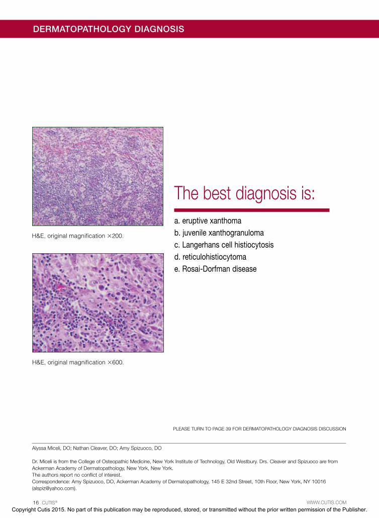
H&E, original magnification ×200.
H&E, original magnification ×600.
The best diagnosis is:a. eruptive xanthomab. juvenile xanthogranulomac. Langerhans cell histiocytosisd. reticulohistiocytomae. Rosai-Dorfman disease
Copyright Cutis 2015. No part of this publication may be reproduced, stored, or transmitted without the prior written permission of the Publisher.
CUTIS Do not c
opy
Dermatopathology Diagnosis Discussion
VOLUME 96, JULY 2015 39WWW.CUTIS.COM
Rosai-Dorfman disease (RDD), also known as sinus histiocytosis with massive lymphade- nopathy, is a rare benign histioproliferative
disorder of unknown etiology.1 Clinically, it is most frequently characterized by massive painless cervical lymphadenopathy with other systemic manifesta-tions, including fever, night sweats, and weight loss. Accompanying laboratory findings include leukocyto-sis with neutrophilia, elevated erythrocyte sedimenta-tion rate, and polyclonal hypergammaglobulinemia. Extranodal involvement has been noted in more than 40% of cases, and cutaneous lesions represent the most common form of extranodal disease.2 Cutaneous RDD is a distinct and rare entity limited to the skin without lymphadenopathy or other extracutaneous involvement.3 Patients with cutaneous RDD typically present with papules and plaques that can grow to form nodules with satellite lesions that resolve into fibrotic plaques before spontaneous regression.4
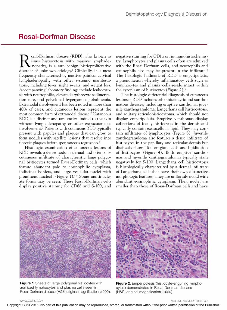
Histologic examination of cutaneous lesions of RDD reveals a dense nodular dermal and often sub-cutaneous infiltrate of characteristic large polygo-nal histiocytes termed Rosai-Dorfman cells, which feature abundant pale to eosinophilic cytoplasm, indistinct borders, and large vesicular nuclei with prominent nucleoli (Figure 1).4,5 Some multinucle-ate forms may be seen. These Rosai-Dorfman cells display positive staining for CD68 and S-100, and
negative staining for CD1a on immunohistochemis-try. Lymphocytes and plasma cells often are admixed with the Rosai-Dorfman cells, and neutrophils and eosinophils also may be present in the infiltrate.4 The histologic hallmark of RDD is emperipolesis, a phenomenon whereby inflammatory cells such as lymphocytes and plasma cells reside intact within the cytoplasm of histiocytes (Figure 2).5
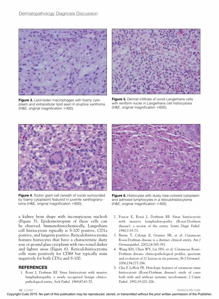
The histologic differential diagnosis of cutaneous lesions of RDD includes other histiocytic and xantho-matous diseases, including eruptive xanthoma, juve-nile xanthogranuloma, Langerhans cell histiocytosis, and solitary reticulohistiocytoma, which should not display emperipolesis. Eruptive xanthomas display collections of foamy histiocytes in the dermis and typically contain extracellular lipid. They may con-tain infiltrates of lymphocytes (Figure 3). Juvenile xanthogranuloma also features a dense infiltrate of histiocytes in the papillary and reticular dermis but distinctly shows Touton giant cells and lipidization of histiocytes (Figure 4). Both eruptive xantho-mas and juvenile xanthogranulomas typically stain negatively for S-100. Langerhans cell histiocytosis is histologically characterized by a dermal infiltrate of Langerhans cells that have their own distinctive morphologic features. They are uniformly ovoid with abundant eosinophilic cytoplasm. Their nuclei are smaller than those of Rosai-Dorfman cells and have
Figure 1. Sheets of large polygonal histiocytes with admixed lymphocytes and plasma cells seen in Rosai-Dorfman disease (H&E, original magnification ×200).
rosai-Dorfman Disease
Figure 2. Emperipolesis (histiocyte-engulfing lympho-cytes) demonstrated in Rosai-Dorfman disease (H&E, original magnification ×600).
Copyright Cutis 2015. No part of this publication may be reproduced, stored, or transmitted without the prior written permission of the Publisher.
CUTIS Do not c
opy
Dermatopathology Diagnosis Discussion
40 CUTIS® WWW.CUTIS.COM
a kidney bean shape with inconspicuous nucleoli (Figure 5). Epidermotropism of these cells can be observed. Immunohistochemically, Langerhans cell histiocytosis typically is S-100 positive, CD1a positive, and langerin positive. Reticulohistiocytoma features histiocytes that have a characteristic dusty rose or ground glass cytoplasm with two-toned darker and lighter areas (Figure 6). Reticulohistiocytoma cells stain positively for CD68 but typically stain negatively for both CD1a and S-100.
RefeRences 1. Rosai J, Dorfman RF. Sinus histiocytosis with massive
lymphadenopathy. a newly recognized benign clinico-pathological entity. Arch Pathol. 1969;87:63-70.
2. Foucar E, Rosai J, Dorfman RF. Sinus histiocytosis
with massive lymphadenopathy (Rosai-Dorfman disease): a review of the entity. Semin Diagn Pathol. 1990;7:19-73.
3. Brenn T, Calonje E, Granter SR, et al. Cutaneous Rosai-Dorfman disease is a distinct clinical entity. Am J Dermatopathol. 2002;24:385-391.
4. Wang KH, Chen WY, Lie HN, et al. Cutaneous Rosai-Dorfman disease: clinicopathological profiles, spectrum and evolution of 21 lesions in six patients. Br J Dermatol. 2006;154:277-286.
5. Chu P, LeBoit PE. Histologic features of cutaneous sinus histiocytosis (Rosai-Dorfman disease): study of cases both with and without systemic involvement. J Cutan Pathol. 1992;19:201-206.
Figure 4. Touton giant cell (wreath of nuclei surrounded by foamy cytoplasm) featured in juvenile xanthogranu-loma (H&E, original magnification ×600).
Figure 5. Dermal infiltrate of ovoid Langerhans cells with reniform nuclei in Langerhans cell histiocytosis (H&E, original magnification ×600).
Figure 6. Histiocytes with dusty rose–colored cytoplasm and admixed lymphocytes in a reticulohistiocytoma (H&E, original magnification ×400).
Figure 3. Lipid-laden macrophages with foamy cyto-plasm and extracellular lipid seen in eruptive xanthoma (H&E, original magnification ×400).
Copyright Cutis 2015. No part of this publication may be reproduced, stored, or transmitted without the prior written permission of the Publisher.
CUTIS Do not c
opy