Embed Size (px)
Citation preview
HAL Id: tel-03303079https://tel.archives-ouvertes.fr/tel-03303079
Submitted on 28 Jul 2021
HAL is a multi-disciplinary open accessarchive for the deposit and dissemination of sci-entific research documents, whether they are pub-lished or not. The documents may come fromteaching and research institutions in France orabroad, or from public or private research centers.
L’archive ouverte pluridisciplinaire HAL, estdestinée au dépôt et à la diffusion de documentsscientifiques de niveau recherche, publiés ou non,émanant des établissements d’enseignement et derecherche français ou étrangers, des laboratoirespublics ou privés.
Deregulation of genes and transposable elements andhybrid incompatibility among Drosophila mojavensis
subspecies and D. arizonaeCecilia Artico Banho
To cite this version:Cecilia Artico Banho. Deregulation of genes and transposable elements and hybrid incompatibilityamong Drosophila mojavensis subspecies and D. arizonae. Animal genetics. Université de Lyon;Universidade estadual paulista (São Paulo, Brésil). Faculdade de Ciências e Tecnologia, 2020. English.�NNT : 2020LYSE1055�. �tel-03303079�
Numéro d’ordre: 2020LYSE1055
THESE de DOCTORAT DE L’UNIVERSITE DE LYON opérée au sein de
l’Université Claude Bernard Lyon 1
Ecole Doctorale Evolution, Ecosystème, Microbiologie, Modélisation
Spécialité de doctorat :
Biomath-Bioinfo-Génomique évolutive
Soutenue publiquement/à São José do Rio Preto, São Paulo, Brésil le 13/05/2020, par : Cecília Artico Banho
Dérégulation des gènes et élément transposables et incompatibilité hybride entre les sous-espèces Drosophila mojavensis et D. arizonae
Devant le jury composé de:
CARARETO, Claudia Marcia Aparecida Professeure Université d’état Paulista São José do Rio
Preto: Directrice de thèse
GARCIA GUERREIRO, Maria del Pilar Professeure Université Autonome de Barcelone
(Espagne): Rapporteur
LORETO, Elgion Professeur Université Fédérale de Santa Maria (Brésil):
Rapporteur/Examinateur
BRANDÃO, Marcelo Mendes Chercheur Université de Campinas (Brésil):
Rapporteur/Examinateur
MADI-RAVAZZI, Lilian Professeure Université d’état Paulista São José do Rio Preto: Presidente du jury/ Examinatrice
MOUTON, Laurence Maître de Conférences, Université Lyon 1 (France) : Examinatrice
VARANI, Alessandro Chercheur Université d’état Paulista Jabotical (Brésil): Examinateur
VIEIRA, Cristina Professeure Université Lyon1: Directrice de thèse
Programa de Pós-Graduação em Biociências
Cecília Artico Banho
Desregulação de genes e elementos de transposição e incompatibilidade
híbrida entre subespécies de Drosophila mojavensis e D. arizonae
São José do Rio Preto 2020
Cecilia Artico Banho
Desregulação de genes e elementos de transposição e incompatibilidade
híbrida entre subespécies de Drosophila mojavensis e D. arizonae
Tese apresentada como parte dos requisitos para a obtenção do título de Doutor em Biociências, junto ao Programa de Pós- Graduação em Biociências, Área de Genética e Biologia Evolutiva, do Instituto de Biociências, Letras e Ciências Exatas da Universidade Estadual Paulista “Júlio de Mesquita Filho”, Câmpus de São José do Rio Preto.
Financiadores: FAPESP: processo 2016/19271-2
CNPq: processos 303455/2017-9 e 141413/2016-6
ANR: processo 14-CE19-0016
IDEX LYON
Eiffel Program (Campus France)
Orientadora: Profª. Drª. Claudia Marcia Aparecida Carareto
Orientadora: Profª. Drª. Cristina Vieira
São José do Rio Preto 2020
B216d
Banho, Cecília Artico
Desregulação de genes e elementos de transposição e
incompatibilidade híbrida entre subespécies de Drosophila mojavensis
e D. arizonae / Cecília Artico Banho. -- São José do Rio Preto, 2020
178 p.
Tese (doutorado) - Universidade Estadual Paulista (Unesp),
Instituto de Biociências Letras e Ciências Exatas, São José do Rio
Preto
Orientadora: Claudia Marcia Aparecida Carareto
1. Elementos de Transposição. 2. Híbridos. 3. Fenótipo. 4.
Expressão Gênica. 5. Drosophila mojavensis e D. arizonae. I. Título.
Sistema de geração automática de fichas catalográficas da Unesp. Biblioteca do Instituto de
Biociências Letras e Ciências Exatas, São José do Rio Preto. Dados fornecidos pelo autor(a).
Essa ficha não pode ser modificada.
Cecília Artico Banho
Desregulação de genes e elementos de transposição e incompatibilidade
híbrida entre subespécies de Drosophila mojavensis e D. arizonae
Tese apresentada como parte dos requisitos para a obtenção do título de Doutor em Biociências, junto ao Programa de Pós-Graduação em Biociências, Área de Genética e Biologia Evolutiva, do Instituto de Biociências, Letras e Ciências Exatas da Universidade Estadual Paulista “Júlio de Mesquita Filho”, Câmpus de São José do Rio Preto.
Comissão Examinadora:
Profª. Drª. Claudia Marcia Aparecida CARARETO
UNESP – Câmpus de São José do Rio Preto Orientadora
Profª. Drª Cristina VIEIRA Orientadora
Université Claude Bernard – Lyon 1 Orientadora
Profª. Drª. Lilian MADI-RAVAZZI (UNESP – Câmpus de São
José do Rio Preto)
Prof. Dr. Elgion LORETO (UFSM)
Dr. Marcelo Mendes BRANDÃO (UNICAMP)
Profª. Drª Laurence MOUTON (UCBL)
Prof. Dr. Alessandro VARANI (UNESP – Câmpus de Jaboticabal)
São José do Rio Preto 13 de maio de 2020
Este trabalho foi realizado sob convenção de co-tutela entre Universidade Estadual Paulista
(UNESP) - Brasil e l’Université Claude Bernard (Lyon1 - UCBL) – França, no laboratório de
Evolução Molecular, do Instituto de Biociências, Letras e Ciências Exatas (IBILCE/UNESP) –
São José do Rio Preto/SP, e no Laboratoire de Biométrie et Biologie Évolutive (LBBE/Lyon1).
Universidade Estadual Paulista (UNESP – IBILCE)
Laboratório de Evolução Molecular
Departamento de Biologia
Rua Cristóvão Colombo, 2265, Jardim Nazareth
15054-000 São José do Rio Preto-SP
Brasil
Université Claude Bernard – Lyon 1
Laboratoire de Biométrie et Biologie Evolutive
CNRS UMR 5558
43 Boulevard du 11 novembre 1918
69622 Cedex Villeurbanne
Cecilia ARTICO BANHO
Palavras chave - Mots Clés – Keywords
Elementos de Transposição - Eléments transposables – Transposable elements
Híbridos - Hibrides – Hybrids
Fenótipo – Phénotype – Phenotype
Expressão Gênica– Gene Expression – Gene Expression
Drosophila mojavensis
Drosophila arizonae

Aos meus pais, por toda dedicação, apoio
e exemplo de força e perseverança.
Agradecimentos
Aos meus pais, Rozangela de Fátima Artico e Claudio Banho, por todo apoio, amor e carinho ao
longo destes anos. Obrigada por serem meu exemplo de bondade, honestidade, perseverança, luta
e otimismo. Obrigada pelos valorosos ensinamentos, valores morais e por todos os sacrifícios
que fizeram para que eu pudesse ter acesso à educação de qualidade. Essa conquista eu devo a
vocês.
Ao meu namorado e companheiro Plinio Gabriel Sicuti por todo amor, carinho e paciência
nesses últimos anos. Obrigada por sempre se fazer presente, mesmo durante o período em que
estive na França, dando todo suporte e encorajamento. Obrigada por nunca me deixar desistir.
Às minhas orientadoras Claudia Marcia Aparecida Carareto e Cristina Vieira pelos valorosos
ensinamentos, pela paciência, e por toda a confiança depositada em mim na realização desse
trabalho. Sempre serei grata por todas as oportunidades que me concederam, as quais me
tornaram uma profissional melhor e que me fizeram crescer como ser humano. Vocês são meus
grandes exemplos de mulheres, de cientistas, de sucesso, e de amor à profissão. Obrigada por me
pertirem aprender com vocês.
Aos meus avós Izaura Orlando Banho, Natalino Banho, Sebastiana Caetano Artico e Denilson
Artico, por serem meu exemplo de bondade e fé. Obrigada por todo amor e carinho ao longo
desses anos. Agradeço, também, ao meu irmão Vitor Henrique Artico Banho pelo
companheirismo, e apoio durante todos esses anos.
Aos queridos amigos que a UNESP me deu e que com certeza levarei para sempre. Obrigada
Samara Videira Zorzato, Ana Letícia Guerra, Ana Beatriz Bortolozo, Fernando César Silva
Júnior, Tatiani Seni de Souza Firmino e Luis Lenin Vicente Pereira, por todos os momentos
incríveis, por todas as risadas, conversas, apoio mútuo e carinho.
Aos queridos amigos do Laboratório de Evolução Molecular, William Vilas Boas Nunes, Luis
Gustavo Galego, Marcelo Jurado, Guilherme Matheus, Edoardo Estevam Lobl, Izabella Luisa
Tambones, Lucas Moreira, Bianca Manfré, Felipe Santa-Rosa do Amaral, e em especial à
minhas queridas amigas Camila Vieira e Maryanna Cristiano Simão. Obrigada por serem minha
segunda família, por me acolherem há quatro anos e por compartilharem todo o seu
conhecimento. Muito obrigada por todos os momentos incríveis, por todas as longas conversas,
por todas as risadas e por serem pessoas maravilhosas. Eu tenho muita sorte em poder trabalhar
não com colegas, mas sim com grandes amigos.
A todos da equipe TREEP e do LBBE, Matthieu Boulesteix, Marie Fablet, Emmanuelle Lerat,
Annabelle Haudry, Justine Picarle, Hélène Henri, Nicole Lara, que me acolheram de braços
abertos, por sempre me auxiliarem e por todos os ensinamentos. À Nelly Burlet e Sonia Martinez
por toda a paciência, longas conversas e por auxilio durante os experimentos com Drosophila. E,
em especial, a Pierre Marin, Angelo Jacquet, Inessa Buch, Valentina Rodrigues Rada pela
paciência, amizade, por todo auxílio, pelas risadas e pelos almoços juntos.
À Marlène Roy, pela amizade, pelo apoio e por todos os ensinamentos. Por sempre estar disposta
a ajudar e por ser extremamente gentil. Ao Vincent Mérel, com quem tive o prazer de dividir a
sala durante toda a minha estadia no LBBE. Obrigada por ser tão gentil, atencioso, por sempre
estar disposto a ajudar, pela paciência, compreensão, pelas longas conversas, e acima de tudo,
obrigada por me ensinar as análises que apresentarei nesse trabalho, tornando-o possível.
Aos amigos que pude conhecer em Lyon e que tornaram minha estadia muito mais leve e alegre.
A todos os docentes que contribuíram para a minha formação, aos quais serei eternamente grata.
Em especial agradeço à Profa. Dra. Mary Massumi Itoyama, que me orientou durante a
graduação e mestrado, a qual transmitiu grandes ensinamentos e foi um grande exemplo.
A todos os técnicos, funcionários, e alunos que de alguma forma contribuíram para a realização
deste trabalho, em especial aos servidores do Pós-Graduação em Biociências, pelo
profissionalismo e amor à pesquisa.
À Universidade Estadual Paulista “Júlio de Mesquita Filho” – Campus de São José do Rio Preto,
que me acolheu, permitindo a realização deste sonho.
À Université Claude Bernard Lyon 1 e ao Laboratoire de Biometrie et Biologie Evolutive, que
proporcionaram maior oportunidade da minha vida e contribuíram grandemente para minha
formação acadêmica.
Aos membros da banca examinadora, pela disponibilidade e preciosas contribuições ao trabalho.
O presente trabalho foi realizado com apoio da FAPESP, sob o processo 2016/19271-2, do
CNPq, sob o processo 303455/2017-9 e da Agence Nationale de la Recherce (ANR) sob o
processo 14-CE19-0016. Agradeço ao CNPq (processo 141413/2016-6), à IDEX LYON e ao
Eiffel Program (Campus France) pela concessão de bolsas de estudo no Brasil e na França, que
possibilitaram a realização desse trabalho.

″Educação não transforma o mundo.
Educação muda pessoas. Pessoas transformam o mundo.”
Paulo Freire
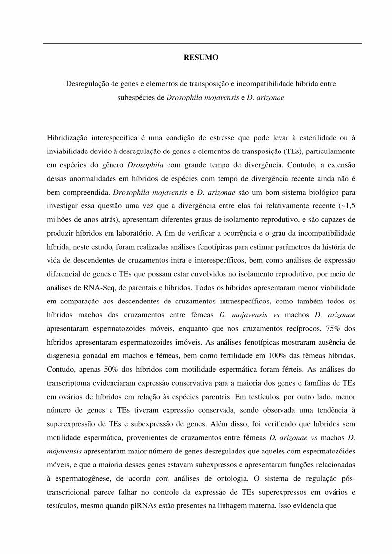
RESUMO
Desregulação de genes e elementos de transposição e incompatibilidade híbrida entre
subespécies de Drosophila mojavensis e D. arizonae
Hibridização interespecifica é uma condição de estresse que pode levar à esterilidade ou à
inviabilidade devido à desregulação de genes e elementos de transposição (TEs), particularmente
em espécies do gênero Drosophila com grande tempo de divergência. Contudo, a extensão
dessas anormalidades em híbridos de espécies com tempo de divergência recente ainda não é
bem compreendida. Drosophila mojavensis e D. arizonae são um bom sistema biológico para
investigar essa questão uma vez que a divergência entre elas foi relativamente recente (~1,5
milhões de anos atrás), apresentam diferentes graus de isolamento reprodutivo, e são capazes de
produzir híbridos em laboratório. A fim de verificar a ocorrência e o grau da incompatibilidade
híbrida, neste estudo, foram realizadas análises fenotípicas para estimar parâmetros da história de
vida de descendentes de cruzamentos intra e interespecíficos, bem como análises de expressão
diferencial de genes e TEs que possam estar envolvidos no isolamento reprodutivo, por meio de
análises de RNA-Seq, de parentais e híbridos. Todos os híbridos apresentaram menor viabilidade
em comparação aos descendentes de cruzamentos intraespecíficos, como também todos os
híbridos machos dos cruzamentos entre fêmeas D. mojavensis vs machos D. arizonae
apresentaram espermatozoides móveis, enquanto que nos cruzamentos recíprocos, 75% dos
híbridos apresentaram espermatozoides imóveis. As análises fenotípicas mostraram ausência de
disgenesia gonadal em machos e fêmeas, bem como fertilidade em 100% das fêmeas híbridas.
Contudo, apenas 50% dos híbridos com motilidade espermática foram férteis. As análises do
transcriptoma evidenciaram expressão conservativa para a maioria dos genes e famílias de TEs
em ovários de híbridos em relação às espécies parentais. Em testículos, por outro lado, menor
número de genes e TEs tiveram expressão conservada, sendo observada uma tendência à
superexpressão de TEs e subexpressão de genes. Além disso, foi verificado que híbridos sem
motilidade espermática, provenientes de cruzamentos entre fêmeas D. arizonae vs machos D.
mojavensis apresentaram maior número de genes desregulados que aqueles com espermatozóides
móveis, e que a maioria desses genes estavam subexpressos e apresentaram funções relacionadas
à espermatogênese, de acordo com análises de ontologia. O sistema de regulação pós-
transcricional parece falhar no controle da expressão de TEs superexpressos em ovários e
testículos, mesmo quando piRNAs estão presentes na linhagem materna. Isso evidencia que
outros mecanismos podem estar relacionados à desregulação dos TEs nos tecidos reprodutivos
de híbridos entre D. arizonae e D. mojavensis. Dentre esses mecanismos pode-se sugerir a
desregulação de alguns genes envolvidos na via de piRNAs, e de modifição de cromatina, como
observado em testículos. Contudo, o mesmo não pode ser sugerido para ovários, uma vez que
nessa gônada a expressão desses genes não estava desregulada. Em síntese, este estudo mostra
que testículos de híbridos entre D. arizonae e D. mojavensis apresentam maior expressão
diferencial de genes e de TEs em relação aos parentais do que em ovários e que, embora piRNAs
estejam presentes para muitos dos TEs desregulados, eles não são capazes de controlar sua
expressão, o que pode estar ligado ao fenótipo de esterilidade demostrado pelos parâmetros de
história de vida analisados nestes híbridos.
Palavras–chave: Híbridos, Isolamento pós-zigótico, Fenótipo, Expressão Diferencial, Grupo
repleta
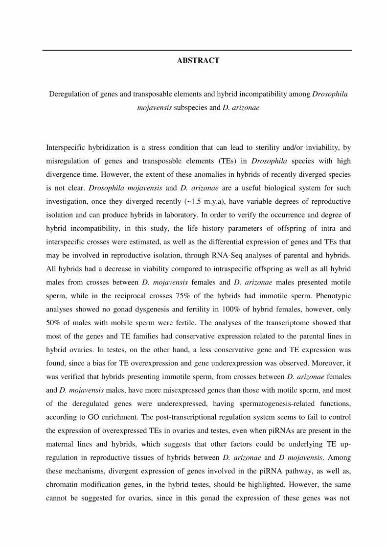
ABSTRACT
Deregulation of genes and transposable elements and hybrid incompatibility among Drosophila
mojavensis subspecies and D. arizonae
Interspecific hybridization is a stress condition that can lead to sterility and/or inviability, by
misregulation of genes and transposable elements (TEs) in Drosophila species with high
divergence time. However, the extent of these anomalies in hybrids of recently diverged species
is not clear. Drosophila mojavensis and D. arizonae are a useful biological system for such
investigation, once they diverged recently (~1.5 m.y.a), have variable degrees of reproductive
isolation and can produce hybrids in laboratory. In order to verify the occurrence and degree of
hybrid incompatibility, in this study, the life history parameters of offspring of intra and
interspecific crosses were estimated, as well as the differential expression of genes and TEs that
may be involved in reproductive isolation, through RNA-Seq analyses of parental and hybrids.
All hybrids had a decrease in viability compared to intraspecific offspring as well as all hybrid
males from crosses between D. mojavensis females and D. arizonae males presented motile
sperm, while in the reciprocal crosses 75% of the hybrids had immotile sperm. Phenotypic
analyses showed no gonad dysgenesis and fertility in 100% of hybrid females, however, only
50% of males with mobile sperm were fertile. The analyses of the transcriptome showed that
most of the genes and TE families had conservative expression related to the parental lines in
hybrid ovaries. In testes, on the other hand, a less conservative gene and TE expression was
found, since a bias for TE overexpression and gene underexpression was observed. Moreover, it
was verified that hybrids presenting immotile sperm, from crosses between D. arizonae females
and D. mojavensis males, have more misexpressed genes than those with motile sperm, and most
of the deregulated genes were underexpressed, having spermatogenesis-related functions,
according to GO enrichment. The post-transcriptional regulation system seems to fail to control
the expression of overexpressed TEs in ovaries and testes, even when piRNAs are present in the
maternal lines and hybrids, which suggests that other factors could be underlying TE up-
regulation in reproductive tissues of hybrids between D. arizonae and D mojavensis. Among
these mechanisms, divergent expression of genes involved in the piRNA pathway, as well as,
chromatin modification genes, in the hybrid testes, should be highlighted. However, the same
cannot be suggested for ovaries, since in this gonad the expression of these genes was not
deregulated. In summary, this study shows that testes of hybrids between D. arizonae and D.
mojavensis have greater differential expression of genes and TEs in relation to parental species
than in ovaries. In addition, it shows that although piRNAs are present for many of the
unregulated TEs, they are not able to control their expression, which may be linked to the
sterility phenotype shown by the life history parameters estimated in these hybrids.
Keywords: Hybrids, Postzygotic isolation, Phenotype, Differential expression, repleta group

RÉSUMÉ
Dérégulation des gènes et éléments transposables et incompatibilité hybride entre les sous-
espèces Drosophila mojavensis et D. arizonae
L'hybridation interspécifique est une condition de stress qui peut conduire à des hybrides stériles
ou non viables, en raison de la dérégulation des gènes et des éléments transposables (ET), en
particulier chez les hybrides entre espèces du genre Drosophila ayant un long temps de
divergence. Cependant, l’ampleur de ces anomalies chez les hybrides d'espèces ayant un temps
de divergence récent n'est pas encore bien comprise. Drosophila mojavensis et D. arizonae
constituent un bon système biologique pour étudier cette question car le temps de divergence est
relativement récent (~1,5 m. a.). En plus, ces espèces présentent différents degrés d'isolement
reproducteur et sont capables de produire des hybrides en laboratoire. Afin de vérifier
l'occurrence et le degré d'incompatibilité des hybrides, nous avons mesuré les traits d’histoire de
vie des descendants de croisements intra et interspécifiques, ainsi que l'expression différentielle
des gènes et des ET qui peuvent être impliqués dans l'isolement reproducteur. Les analyses
phénotypiques ont montré que tous les hybrides présentaient une viabilité inférieure aux
descendants des croisements intraspécifiques. Les analyses de la motilité des spermatique ont
montré que tous les hybrides mâles issus de croisement entre femelle D. mojavensis et mâle D.
arizonae croisés avaient des spermatozoïdes mobiles, alors que dans le croisement réciproque,
75% des hybrides avaient des spermatozoïdes immobiles. Les analyses de fertilité des hybrides
n'ont montré aucune dysgénésie gonadique chez les mâles et les femelles et une fertilité de 100%
chez les hybrides femelles. Cependant, seulement 50% des hybrides males ayant la motilité des
spermatozoïdes étaient fertiles. Les analyses RNASeq ont montré que la plupart des gènes et des
familles d'ET présentaient une expression conservée par rapport aux espèces parentales dans les
ovaires des hybrides. Dans les testicules, en revanche, moins de gènes et de ET ont une
expression de type conservé et on observe une tendance à la surexpression des ET et à la sous-
expression des gènes. En outre, il a été observé que les hybrides sans motilité des spermatozoïdes
(issus de croisements entre les femelles D. arizonae et les mâles D. mojavensis) présentaient un
nombre plus élevé de gènes dérégulés que ceux avec des spermatozoïdes mobiles, et que la
majorité de ces gènes étaient sous-exprimés et présentaient des fonctions liées à la
spermatogenèse. Le système de régulation post-transcriptionnel semble ne pas être efficace dans
le contrôle de l'expression des ET qui sont surexprimées dans les ovaires et les testicules, même
quand des piRNA sont présents dans la lignée maternelle. Cela montre que d'autres mécanismes
peuvent être liés à la dérégulation des ET dans les tissus reproducteurs des hybrides entre D.
arizonae et D. mojavensis. Parmi ces mécanismes, on peut suggérer la dérégulation de certains
gènes impliqués dans la voie des RNAi, et la modification de la chromatine, telle qu'observée
dans les testicules. Cependant, on ne peut pas en dire autant des ovaires, car dans ce tissu,
l'expression de ces gènes n'a pas été dérégulée. En résumé, cette étude montre que les testicules
des hybrides entre D. arizonae et D. mojavensis présentent une expression différentielle des
gènes et des ET plus importante par rapport à celle des parents que les ovaires et que, bien que
des piARN soient présents pour un grand nombre des ET dérégulés, ils ne semblent pas être
capables de contrôler leur expression. Ceci qui peut être associé au phénotype de stérilité observé
pour les traits d'histoire de vie analysés chez les hybrides.
Mots-clés: Hybrides, Isolement postzygotique, Phénotype, Expression différentielle, groupe
repleta
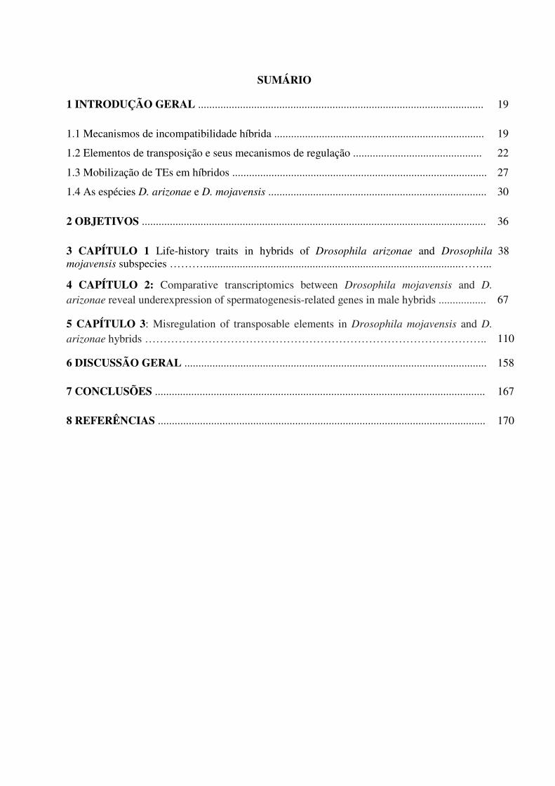
SUMÁRIO
1 INTRODUÇÃO GERAL ...................................................................................................... 19
1.1 Mecanismos de incompatibilidade híbrida ........................................................................... 19
1.2 Elementos de transposição e seus mecanismos de regulação .............................................. 22
1.3 Mobilização de TEs em híbridos ........................................................................................... 27
1.4 As espécies D. arizonae e D. mojavensis .............................................................................. 30
2 OBJETIVOS ........................................................................................................................... 36
3 CAPÍTULO 1 Life-history traits in hybrids of Drosophila arizonae and Drosophila
mojavensis subspecies ………............................................................................................……... 38
4 CAPÍTULO 2: Comparative transcriptomics between Drosophila mojavensis and D.
arizonae reveal underexpression of spermatogenesis-related genes in male hybrids .................
67
5 CAPÍTULO 3: Misregulation of transposable elements in Drosophila mojavensis and D.
arizonae hybrids ……………………………………………………………………………….. 110
6 DISCUSSÃO GERAL ............................................................................................................ 158
7 CONCLUSÕES ...................................................................................................................... 167
8 REFERÊNCIAS ..................................................................................................................... 170

INTRODUÇÃO GERAL
19
1 INTRODUÇÃO GERAL
Como novas espécies surgem e quais são os fatores genéticos envolvidos com o processo
de especiação são duas das grandes questões em Biologia Evolutiva que, até os dias atuais, não
são completamente esclarecidas. O conceito biológico define espécie como grupos de
populações naturais intercruzantes que são reprodutivamente isoladas de outros grupos
semelhantes (MAYR, 1942; 1963). Sendo assim, especiação pode ocorrer quando o fluxo de
informação genética entre populações é inibido pela formação de barreiras que levam ao
isolamento reprodutivo (DOBZHANSKY, 1937; 1940). Dessa forma, a magnitude e a taxa com
que as barreiras de isolamento reprodutivo evoluem, em diferentes grupos de espécies, pode ser
um fator chave na origem de novas espécies (TURISSINI et al., 2018).
1.1 Mecanismos de incompatibilidade híbrida
Sabe-se, atualmente, que o processo de hibridização interespecífica, que já foi
considerado um evento raro, é relativamente comum na natureza, ocorrendo em cerca de 25%
das espécies vegetais e em 10% das animais (MALLET, 2005) e que, em espécies incipientes, as
barreiras que levam à restrição ao fluxo gênico podem ser incompletas, levando à produção de
descendentes híbridos. De modo geral, os mecanismos de isolamento reprodutivo podem ser
classificados em diferentes tipos, dependendo do momento em que ocorrem durante o ciclo
reprodutivo, podendo ser pré-zigótico, pré-zigótico pós-cópula, ou pós-zigótico. O isolamento
pré-zigótico resulta no impedimento de cruzamentos interespecíficos, e pode ocorrer em
decorrência de especificidade de nicho, de preferências de hábitats e no período reprodutivo,
como também devido a fatores comportamentais (padrões de corte e preferência de
acasalamento) e mecânicos (morfologia das genitálias externas) (TURISSINI et al., 2018). Os
mecanismos de isolamento pré-zigótico pós-cópula, por sua vez, são aqueles que envolvem
incompatibilidades entre os gametas, ou mesmo, entre proteínas do trato reprodutivo feminino e
fluido seminal masculino (MARKOW; HOCUTT, 1998; KNOWLES; MARKOW, 2001;
20
COYNE; ORR, 2004; REED; MARKOW, 2008; TURISSINI et al., 2018). Por outro lado,
mecanismos de isolamento pós-zigóticos, incluem incompatibilidades que levam à redução do
valor adaptativo de híbridos interespecíficos em relação às espécies puras, podendo ocasionar
anormalidades no desenvolvimento (inviabilidade) e na reprodução (esterilidade) dos híbridos F1
ou F2, e mesmo no comportamento, como o padrão de corte dos híbridos (ORR; PRESGRAVES,
2000; PRESGRAVES, 2010; MCBRIDE; SINGER, 2010; TURISSINI et al., 2018).
Embora os mecanismos de isolamento pós-zigótico sejam extensivamente estudados,
principalmente em espécies de Drosophila, ainda não se tem amplo conhecimento dos fatores
genéticos que influenciam a fertilidade e/ou viabilidade híbrida. Coyne et al. (1997) mostraram,
pela análise de dados de 171 cruzamentos interespecíficos em Drosophila, que os mecanismos
de isolamento pós-zigótico evoluem primeiramente em machos que em fêmeas, e que o grau da
inviabilidade ou esterilidade está diretamente relacionado à distância genética entre as espécies.
Além disso, ao analisarem o tempo de divergência das espécies, os autores constataram que
hibridizações entre espécies com tempo de divergência recente produziam principalmente
híbridos machos inviáveis ou estéreis, ao passo que apenas intercruzamentos entre espécies com
maior tempo de divergência produziam fêmeas inviáveis. Estes resultados corroboram a regra de
Haldane (1922), que postula que em eventos de hibridização, quando há esterilidade híbrida, essa
afetará primeiramente o sexo heterogamético.
Com o avanço das técnicas de Biologia Molecular e sequenciamento genômico, diversos
estudos têm buscado compreender as bases genéticas da especiação. Esses estudos têm mostrado
que mudanças na expressão gênica são importantes fontes de variação em características
morfológicas adaptativas (MCGIRR; MARTIN, 2019), e, em especial para o processo de
especiação. A incompatibilidade decorrente da divergência genética entre os parentais pode
causar desregulação de genes específicos em híbridos, os quais podem ser expressos em nível
superior ou inferior ao das espécies parentais, resultando na redução do valor adaptativo do
híbrido, aumentando, portanto, as barreiras de isolamento pós-zigótico.
21
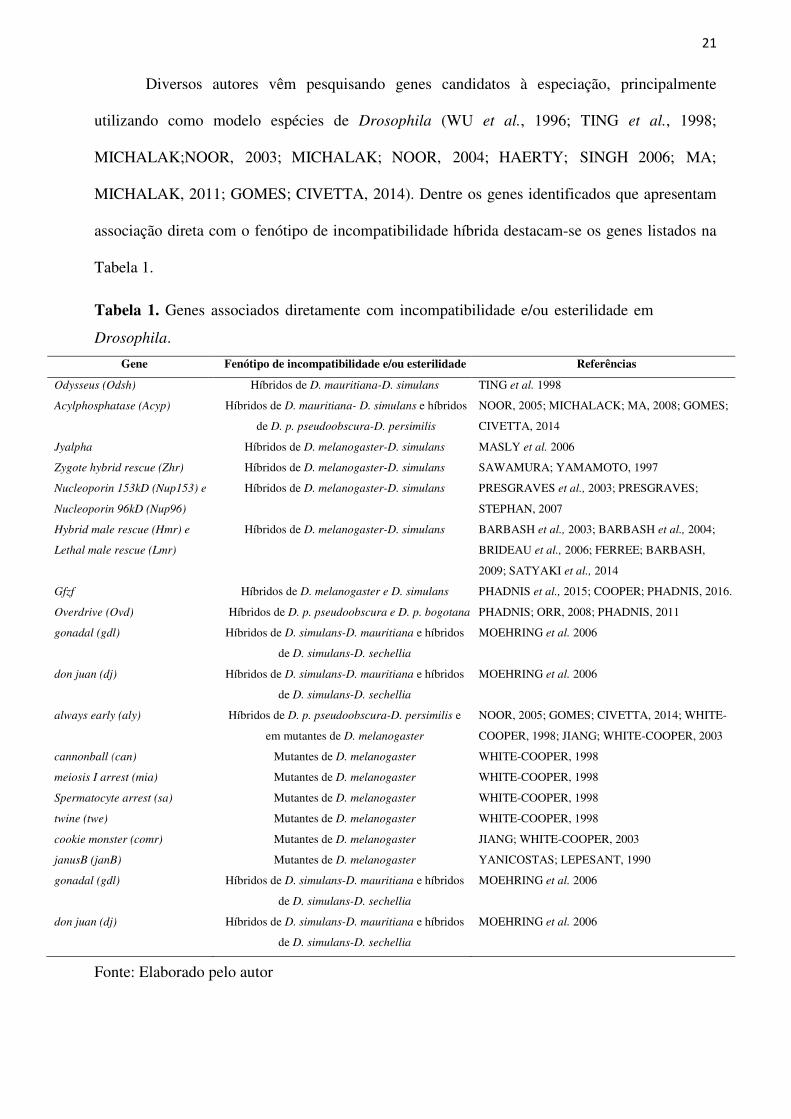
Diversos autores vêm pesquisando genes candidatos à especiação, principalmente
utilizando como modelo espécies de Drosophila (WU et al., 1996; TING et al., 1998;
MICHALAK;NOOR, 2003; MICHALAK; NOOR, 2004; HAERTY; SINGH 2006; MA;
MICHALAK, 2011; GOMES; CIVETTA, 2014). Dentre os genes identificados que apresentam
associação direta com o fenótipo de incompatibilidade híbrida destacam-se os genes listados na
Tabela 1.
Tabela 1. Genes associados diretamente com incompatibilidade e/ou esterilidade em
Drosophila.
Gene Fenótipo de incompatibilidade e/ou esterilidade Referências
Odysseus (Odsh) Híbridos de D. mauritiana-D. simulans TING et al. 1998
Acylphosphatase (Acyp) Híbridos de D. mauritiana- D. simulans e híbridos
de D. p. pseudoobscura-D. persimilis
NOOR, 2005; MICHALACK; MA, 2008; GOMES;
CIVETTA, 2014
Jyalpha Híbridos de D. melanogaster-D. simulans MASLY et al. 2006
Zygote hybrid rescue (Zhr) Híbridos de D. melanogaster-D. simulans SAWAMURA; YAMAMOTO, 1997
Nucleoporin 153kD (Nup153) e
Nucleoporin 96kD (Nup96)
Híbridos de D. melanogaster-D. simulans PRESGRAVES et al., 2003; PRESGRAVES;
STEPHAN, 2007
Hybrid male rescue (Hmr) e
Lethal male rescue (Lmr)
Híbridos de D. melanogaster-D. simulans BARBASH et al., 2003; BARBASH et al., 2004;
BRIDEAU et al., 2006; FERREE; BARBASH,
2009; SATYAKI et al., 2014
Gfzf Híbridos de D. melanogaster e D. simulans PHADNIS et al., 2015; COOPER; PHADNIS, 2016.
Overdrive (Ovd) Híbridos de D. p. pseudoobscura e D. p. bogotana PHADNIS; ORR, 2008; PHADNIS, 2011
gonadal (gdl) Híbridos de D. simulans-D. mauritiana e híbridos
de D. simulans-D. sechellia
MOEHRING et al. 2006
don juan (dj) Híbridos de D. simulans-D. mauritiana e híbridos
de D. simulans-D. sechellia
MOEHRING et al. 2006
always early (aly) Híbridos de D. p. pseudoobscura-D. persimilis e
em mutantes de D. melanogaster
NOOR, 2005; GOMES; CIVETTA, 2014; WHITE-
COOPER, 1998; JIANG; WHITE-COOPER, 2003
cannonball (can) Mutantes de D. melanogaster WHITE-COOPER, 1998
meiosis I arrest (mia) Mutantes de D. melanogaster WHITE-COOPER, 1998
Spermatocyte arrest (sa) Mutantes de D. melanogaster WHITE-COOPER, 1998
twine (twe) Mutantes de D. melanogaster WHITE-COOPER, 1998
cookie monster (comr) Mutantes de D. melanogaster JIANG; WHITE-COOPER, 2003
janusB (janB) Mutantes de D. melanogaster YANICOSTAS; LEPESANT, 1990
gonadal (gdl) Híbridos de D. simulans-D. mauritiana e híbridos
de D. simulans-D. sechellia
MOEHRING et al. 2006
don juan (dj) Híbridos de D. simulans-D. mauritiana e híbridos
de D. simulans-D. sechellia
MOEHRING et al. 2006
Fonte: Elaborado pelo autor
22
Embora grande quantidade de genes envolvidos com isolamento reprodutivo tenha sido
identificada em híbridos de diferentes espécies, ainda é difícil encontrar uma associação entre a
desregulação de genes específicos em diferentes híbridos e o processo de isolamento pós-
zigótico, isso porque, diversos genes codificantes de proteínas e reguladores podem estar
envolvidos nesse processo. Além disso, a desregulação de muitos genes pode ser espécie-
específica, devido ao maior ou menor grau de divergência das espécies, ou mesmo pode haver a
influência de outras sequências genéticas nesse processo, como os elementos de transposição (do
Inglês, Transposable Elements (TEs)).
1.2 Elementos de Transposição e seus mecanismos de regulação
Os elementos de transposição são sequências de DNA repetitivas capazes de se
movimentar de um local para outro no genoma, replicando-se a si mesmos, sendo essa
capacidade a essência de seu sucesso evolutivo. Essas sequências estão presentes em grandes
proporções e diversidade nos genomas de quase todos os eucariotos (exceto em Plasmodium
falciparum) (WICKER et al., 2007), sendo que em fungos 3 a 20% do genoma é composto por
TEs, ao passo que em metazoários essa proporção varia de 3 a 45% (DABOUSSI; CAPY, 2003;
WICKER et al., 2007). Finnegan (1989) pioneiramente propôs que os TEs fossem classificados
em duas classes, com base no seu mecanismo de transposição: elementos de Classe I, que se
transpõem por transcrição reversa de um intermediário de RNA usando um mecanismo DNA-
RNA-DNA, e elementos de Classe II que se transpõem diretamente de DNA para DNA. A
classificação de Finnegan foi objeto de duas grandes atualizações que têm sido debatidas
ativamente; uma delas, o sistema hierárquico de classificação de Wicker (WICKER et al., 2007)
e a outra a utilizada pelo Repbase, que é o banco de dados de elementos repetitivos de DNA mais
comumente usado (JURKA et al., 2005; KAPITONOV; JURKA, 2008).
Como não há consenso para um sistema universal de classificação dos TEs (PIEGU et al.
2015), descreve-se a seguir o sistema hierárquico de classificação de TEs em eucariotos proposto
23
por Wicker et al. (2007), que mantém a divisão dos TEs em duas classes, mas aplicando
critérios enzimáticos e mecanicistas, e inclui em ordem hierárquica os níveis subclasse, ordem,
superfamília, família e subfamília. Na Classe I estão inclusos os elementos que utilizam uma
etapa de transcrição reversa, mobilizando-se por meio de um mecanismo denominado copy and
paste (elementos de RNA, conhecidos como retrotransposons). Essa classe é composta por cinco
ordens, sendo denominadas LTR (Long terminal repeat), DIRS (Dictyostelium intermediate
repeat sequence), PLE (Penelope-like elements), LINE (Long Interspersed Nuclear Element) e
SINE (Short Interspersed Nuclear Element), as quais são distinguidas pela organização de seus
constituintes internos, filogenia da enzima transcriptase reversa, bem como pelo seu mecanismo
de transposição (WICKER et al., 2007). Por outro lado, a Classe II é composta por elementos de
DNA, ou seja, aqueles que não utilizam uma etapa intermediária de RNA para mobilização,
possuindo um mecanismo de transposição conhecido como cut and paste, uma vez que ocorre a
excisão do elemento do genoma antes de sua reinserção em outra região (FINNEGAN, 1989;
WICKER et al., 2007). Esta classe contém duas subclasses, que são distinguidas pelo número de
cadeias de DNA que são cortadas durante a transposição. A Subclasse I é composta por
elementos das ordens TIR (terminal inverted repeat) e Crypton, e a Subclasse II por elementos
das ordens Maverick e Helitron (WICKER et al., 2007).
É interessante ressaltar que os elementos de Classe I apresentam cópias adicionais em
cada novo evento de mobilização, enquanto que em elementos de Classe II, a transposição é, na
maior parte das vezes, conservativa (WICKER et al., 2007). Considerando esses aspectos e o
fato de que TEs são entidades dinâmicas, durante eventos de mobilização essas sequências
podem ter efeitos relevantes sobre o genoma hospedeiro. Esses efeitos podem ser verificados em
nível da linhagem celular germinativa ou em estados precoces de desenvolvimento, assim como
em nível somático, resultando em mudanças fenotípicas no hospedeiro. Além disso, eventos
transposição podem desencadear modificações estruturais no genoma, como translocações,
duplicações segmentais e deleções capazes de induzir profundas alterações genômicas, levando à
24
sua contração ou expansão, além do estabelecimento de novas relações de ligação entre genes
(KIDWELL; LISCH, 2001). Tem-se reportado, também, que os TEs desempenham papel
importante na regulação de diversos processos celulares e diversificação das famílias gênicas,
sendo capazes de promover a transdução e amplificação de fragmentos de genes do genoma
hospedeiro (VAN de LAGEMAAT et al., 2003); bem como, de influenciar a expressão e função
gênica, ampliando a variabilidade do repertório transcricional e proteico (CARARETO et al.,
2013; LOPES et al., 2008, 2013). Em humanos, estima-se que 25% das regiões promotoras
contêm sequências derivadas de TEs, e aproximadamente 20% dos genes em humanos e ratos
contêm esses elementos em suas regiões UTRs (Untranslated Regions) (WONG; CHOO, 2004).
A relação dinâmica dos TEs com o genoma no qual estão inseridos explica, em alguns
pontos, os processos evolutivos que ocorrem nos organismos. Em geral, a intensa mobilização de
TEs, em organismos bem adaptados, pode ocasionar efeitos deletérios aos hospedeiros. Sendo
assim, o controle da sua expressão é fundamental para estabelecer um balanço de efeitos
positivos e negativos, garantindo a viabilidade das funções do genoma nos organismos. Esse
controle é realizado por meio do silenciamento pós-transcricional (utilizando RNAs de
interferências) e do silenciamento transcricional (metilação do DNA e modificação de histonas)
(BRENNECKE et al., 2007; RIGAL; MATHIEU, 2011). Os mecanismos de regulação pós-
transcricionais envolvem a maquinaria de RNA de interferência (RNAi). Em células somáticas,
os TEs são controlados por meio de siRNAs (small interfering RNA), que ao reconhecerem e se
ligarem ao mRNA promovem sua degradação (HIRAKATA; SIOMI, 2019; SATO; SIOMI,
2020). Em células germinativas, o mecanismo de regulação pós-transcricional, descrito em
Drosophila e humanos, possui algumas semelhanças com o que ocorre em células somáticas,
contudo, neste caso o RNA de interferência é denominado de piRNA (piwi-interacting RNAs),
devido ao grupo de proteínas que participam do mecanismo de silenciamento, pertencentes à
família das Argonautas, Ago3, Aubergina (Aub) e Piwi (revisado em HIRAKATA; SIOMI,
2019; SATO; SIOMI, 2020).
25
A produção de piRNAs acontece por duas vias em Drosophila. A via de biogênese de
piRNAs primários ocorre a partir de transcritos provenientes de clusters de piRNAs presentes
nos genomas. Esse processo envolve principalmente as proteínas Piwi e Zucchini (Zuc), entre
outras (BRENNECKE et al., 2007; IPSARO et al., 2012; SATO; SIOMI, 2020). Por outro lado,
a via de piRNAs secundários é produzida apenas em células germinativas, por meio de um
mecanismo de amplificação chamado de ping-pong (BRENNECKE et al., 2007). Nesse
mecanismo, piRNAs primários ou de origem materna são necessários para iniciar a via, uma vez
que, piRNAs anti-senso precisam ser ligados às proteínas Piwi ou Aub. Quando esse complexo
(piRNA-proteína) é formado, ele é capaz de degradar transcritos complementares (no sentido
senso) de TEs que provavelmente estavam ativos no genoma. Essa clivagem produz, então,
transcritos de piRNA senso, os quais são ligados à proteína Ago3, que degrada transcritos de TEs
anti-senso, levando então ao mecanismo de amplificação ping-pong (Figura 1). Esse mecanismo,
geralmente, permite uma eficiente resposta contra cópias de TEs ativas nos genomas
(BRENNECKE et al., 2007; FABLET, 2014). Adicionalmente, em tecidos germinativos de
Drosophila foi verificado que os complexos Piwi-piRNA são capazes de reprimir a transcrição
de TEs por modificação do estado da cromatina (IWASAKI et al., 2016). Em gônadas animais,
muitas inserções de TEs são metiladas nos resíduos H3K9 de maneira dependente de PIWI-
piRNA e problemas nessa via podem resultar em perda seletiva da marca repressiva H3K9me3
em inserções de TEs alvo e, portanto, na sua ativação (SIENSKI et al., 2015).
O impacto causado pela instabilidade genética proveniente da hibridização interespecífica
pode gerar diversas modificações no genoma hospedeiro. Dentre elas se encontram a
desregulação de TEs, bem como de seus elementos regulatórios, podendo influenciar diretamente
a expressão gênica. De acordo com Senti et al. (2015), o aumento da expressão de TEs no
genoma pode gerar maiores quantidades de mRNAs de TEs disponíveis no citoplasma. Esse
processo, consequentemente, influencia o complexo nuclear Piwi-piRNA, aumentando a
repressão em nível transcricional, pelo recrutamento de marcas inativadoras da cromatina, como
26
H3K9me3. De fato, diversos estudos analisaram a influência de TEs na expressão de genes
vizinhos, por meio da análise de marcas repressoras ou ativadoras da cromatina. Sienski et al.
(2012) observaram que, em pelo menos 88% das regiões genômicas em D. melanogaster que
continham a marca inativadora H3K9me3 na eucromatina haviam inserções de TEs, com pelo
menos 5 kb de distância, à jusante ou à montante de onde estavam localizadas. Essas inserções
são capazes de influenciar a expressão de genes vizinhos, como foi verificado para o locus ex
(expanded) no qual o retrotransposon Gypsy é normalmente inserido. Nesse loco, Gypsy está,
aproximadamente, a 1,2 kb à jusante do sítio de iniciação da transcrição (TSS) e, em condições
normais, esse TE tem sua expressão controlada pelo espalhamento da marca de inativação da
cromatina H3K9me3, em aproximadamente 10 a 12 kb, cobrindo, portanto, o TSS de ex.
Contudo, em mutantes para a proteína Piwi, a qual participa das vias de silenciamento
transcricional e pós- transcricional, foi verificada a perda completa de marcas H3K9me3,
desencadeando a expressão desse elemento. Além disso, também foi reportado que em mutantes
para essa proteína, 34% dos genes com inserções de TEs próximas a eles foram superexpressos,
sendo que 80% desses estavam associados com inserções de TEs com até 5 kb de distância
(SIENSKI et al., 2012).
Esses resultados evidenciam o impacto do aumento da expressão de TEs nos processos de
regulação e expressão de genes vizinhos, favorecendo indagações sobre a existência de
associação desses elementos com genes diferencialmente expressos em tecidos germinativos de
híbridos, e consequentemente, com o surgimento de barreiras reprodutivas pós-zigóticas que
podem levar ao processo de especiação. Embora atualmente não existam evidências diretas sobre
a influência da desregulação dos TEs sobre os genes da especiação, tem sido demonstrado que
híbridos interespecíficos apresentam diversos genes desregulados, bem como aumento nas taxas
de transposição (KELEHER et al., 2012; SATYAKI et al., 2014; ROMERO-SORIANO et al.,
2017; LOPEZ-MAESTRE et al., 2017), contudo, associação de ambos fatores ainda não é
completamente entendida.
27
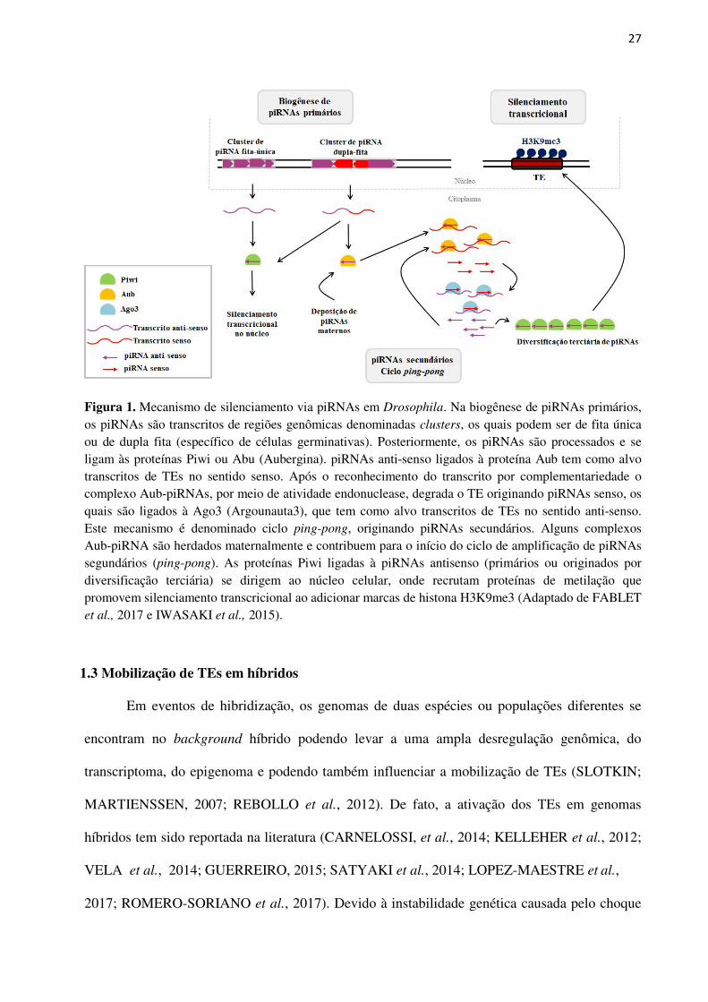
Figura 1. Mecanismo de silenciamento via piRNAs em Drosophila. Na biogênese de piRNAs primários, os piRNAs são transcritos de regiões genômicas denominadas clusters, os quais podem ser de fita única ou de dupla fita (específico de células germinativas). Posteriormente, os piRNAs são processados e se ligam às proteínas Piwi ou Abu (Aubergina). piRNAs anti-senso ligados à proteína Aub tem como alvo transcritos de TEs no sentido senso. Após o reconhecimento do transcrito por complementariedade o complexo Aub-piRNAs, por meio de atividade endonuclease, degrada o TE originando piRNAs senso, os quais são ligados à Ago3 (Argounauta3), que tem como alvo transcritos de TEs no sentido anti-senso. Este mecanismo é denominado ciclo ping-pong, originando piRNAs secundários. Alguns complexos Aub-piRNA são herdados maternalmente e contribuem para o início do ciclo de amplificação de piRNAs segundários (ping-pong). As proteínas Piwi ligadas à piRNAs antisenso (primários ou originados por diversificação terciária) se dirigem ao núcleo celular, onde recrutam proteínas de metilação que promovem silenciamento transcricional ao adicionar marcas de histona H3K9me3 (Adaptado de FABLET et al., 2017 e IWASAKI et al., 2015).
1.3 Mobilização de TEs em híbridos
Em eventos de hibridização, os genomas de duas espécies ou populações diferentes se
encontram no background híbrido podendo levar a uma ampla desregulação genômica, do
transcriptoma, do epigenoma e podendo também influenciar a mobilização de TEs (SLOTKIN;
MARTIENSSEN, 2007; REBOLLO et al., 2012). De fato, a ativação dos TEs em genomas
híbridos tem sido reportada na literatura (CARNELOSSI, et al., 2014; KELLEHER et al., 2012;
VELA et al., 2014; GUERREIRO, 2015; SATYAKI et al., 2014; LOPEZ-MAESTRE et al.,
2017; ROMERO-SORIANO et al., 2017). Devido à instabilidade genética causada pelo choque
28
genômico, eventos de transposição foram relatados em híbridos de plantas e animais (KIDWELL
et al., 1977; PETROV et al., 1985; BAACK et al., 2005; PARISOD et al., 2010; KAWAKAMI
et al., 2011; KELLEHER et al., 2012; VELA et al., 2014; GUERREIRO et al., 2015; HILL et
al., 2016; ROMERO-SORIANO et al., 2017; CASTILLO; MOYLE, 2019). Em plantas,
explosões de transposição foram registradas em três diferentes híbridos de girassol (gênero
Helianthus), as quais estavam relacionadas ao aumento do número de cópias de elementos da
ordem LTR (Ty1/copia-like e Ty3/gypsy-like), os quais foram responsáveis pelo aumento do
genoma híbrido em aproximadamente 50% (BAACK et al., 2005; KAWAKAMI et al., 2011).
Em animais, aumento nas taxas de transposição em decorrência de hibridização foi verificado em
híbridos de espécies de cangurus, Macropus eugenii e Wallabia bicolor (O’NEILL et al., 1998;
METCALFE et al., 2007). Nesses híbridos as consequências de eventos de transposição foram
detectadas como grandes alterações cromossômicas, cariotípicas e no estado de metilação da
cromatina, contribuindo para o menor va[lor adaptativo desses híbridos (O’NEILL et al., 1998;
METCALFE et al., 2007).
Drosophila é um grupo extensivamente estudado quanto aos efeitos de eventos de
mobilização de TEs em decorrência de eventos de hibridização. Nessas espécies, a ativação de
TEs pode ser verificada em híbridos provenientes de cruzamentos intraespecíficos, bem como
interespecíficos. Em nível intraespecífico, diversos estudos mostraram o fenômeno de disgenesia
híbrida, caracterizado por atrofia gonadal ou inviabilidade larval, o qual está relacionado à
mobilização de elementos específicos em determinadas espécies (PICARD, 1976; BLACKMAN
et al., 1987; YANNOPOULOS et al., 1987; KIDWELL et al., 1977; HILL et al., 2016). Como
exemplo, o sistema de disgenesia P-M, que ocorre em D. melanogaster e D. simulans, está
relacionado à presença do elemento P ativo em apenas uma das populações submetidas a
cruzamentos intraespecíficos. Quando populações maternas que não possuem o elemento P ativo
(denominadas M) se cruzam com machos provenientes de populações que apresentam este
elemento ativo em seus genomas (denominados P), as fêmeas híbridas geradas apresentam
29
diversos efeitos disgênicos como atrofia gonadal, e consequentemente ausência ou redução de
fertilidade, aberrações cromossômicas e mutações espontâneas (KIDWELL et al., 1977;
BINGHAM et al., 1982; HILL et al., 2016). Contudo, é interessante ressaltar que essas
consequências são verificadas em apenas uma direção de cruzamento, na qual as fêmeas não
possuem os elementos ativos em seus genomas (KIDWELL et al., 1977; BINGHAM et al.,
1982). Estudos posteriores mostraram que a mobilização desses elementos na linhagem
germinal, e consequente prejuízo para os híbridos estavam relacionadas à falhas no sistema de
regulação pós-transcricional, associado à via de piRNAs (revisado em LUO; LU, 2017). Mais
especificamente, quando a linhagem materna não possui em seu genoma elementos que estão
ativo na linhagem paterna, piRNAs primários provenientes de clusters genômicos e de deposição
materna estarão ausentes, e portanto, a via de biogênese de piRNAs secundários, ping-pong
loop, será prejudicada, resultando na ausência de controle pós-transcricional desse elemento
(LUO; LU, 2017).
Em nível interespecifico foi mostrado que em híbridos de D. buzzatii e D. koepferae
(espécies irmãs do grupo repleta, subgênero Drosophila) houve aumento na transposição do
retrotransposon Osvaldo, que se encontra reprimido nos genomas parentais (LABRADOR et al,
1994), bem como, uma explosão de transposição associada a três elementos: Osvaldo, Helena e
Galileo (GUERREIRO, 2015; VELA et al., 2014). Um estudo mais recente em híbridos desse
par de espécies evidenciou que a desregulação, e consequente ativação de determinados TEs
estava associada à divergência de genes que participavam do sistema de regulação pós-
transcricional, via piRNAs, entre as espécies parentais (ROMERO-SORIANO et al., 2017).
Dessa forma, nos híbridos, a regulação desses elementos não era realizada de forma eficiente,
resultando no aumento de sua expressão Similarmente, análises de transcriptomas de híbridos de
D. melanogaster e D. simulans mostraram ativação global de famílias de TEs, herdados tanto
maternalmente, quanto paternalmente, e que a desrepressão poderia ter sido ocasionada pela
grande divergência adaptativa de genes da via de piRNA ao invés de diferenças espécies-
30
específicas de piRNAs derivados de TEs (KELLEHER et al., 2012). Assim, a hibridização ou
introgressão em populações podem contribuir para eventos de mobilização de TEs e
instabilidade genômica que pode ser benéfica ao híbrido ao lhe conferir a capacidade de
adaptação e especiação, ou ser prejudicial, podendo levá-lo à extinção (FONTDEVILA, 2005).
Embora, ainda não existam evidências se o aumento de transposição influencia a
regulação gênica em híbridos, e vice versa, contribuindo para as barreiras de isolamento
reprodutivo (REBOLLO et al., 2010), um estudo em híbridos de D. melanogaster e D. simulans
mostrou uma associação direta de efeitos epistáticos deletérios entre dois genes específicos, Hmr
(Hybrid male rescue) e Lmr (Lethal male rescue), levando à desregulação de genes de
heterocromatina, e culminando no aumento de expressão TEs e sequências satélites,
principalmente em regiões centroméricas (SATYAKI et al., 2014). Considerando esses fatores,
espécies que divergiram recentemente e que apresentam barreiras de isolamento pré e pós-
zigóticas incompletas, que propiciam a produção de híbridos, são bons modelos para o estudo
das bases genéticas que influenciam o isolamento reprodutivo, bem como a dinâmica dos TEs
nos genomas híbridos e sua influência nos passos iniciais do processo de especiação.
1.4 As espécies Drosophila mojavensis e D. arizonae
Dentre as espécies do gênero Drosophila, D. mojavensis e D. arizonae (grupo repleta,
subgênero Drosophila) são espécies irmãs, cactofílicas, que constituem populações alopátricas e
simpátricas, capazes de produzir híbridos em laboratório (RUIZ et al., 1990; MASSIE;
MARKOW, 2005; JENNINGS; ETGES, 2009). Populações de D. arizonae são mais
generalistas, utilizando como sítios de alimentação e reprodução cactos colunares ou do gênero
Opuntia, dependendo de sua distribuição geográfica (RUIZ; HEED 1988; REED et al., 2006).
Essa espécie é encontrada a partir do sul da Guatemala, México e sudoeste dos Estados Unidos
da América, onde podem constituir populações simpátricas com D. mojavensis (HEED, 1982;
RUIZ; HEED, 1988; REED et al., 2006). D. mojavensis, por sua vez, foi dividida em quatro
31
subespécies, sendo elas D. m. mojavensis, encontrada no Deserto de Mojave, D. m. baja, presente
na Península de Baja Califórnia, D. m. sonorensis, localizada no deserto de Sonora e sul do
Arizona e D. m. wrigleyi, endêmica da ilha de Santa Catalina, na costa da Califórnia (REED et
al., 2006), e em cada uma dessas regiões, as diferentes subespécies utilizam diferentes cactos
colunares como sítio de alimentação e reprodução (REED et al., 2006; JENNINGS; ETGES,
2009). Reed et al. (2006) também reportaram que as populações pertencentes às quatro
principais áreas de distribuição de D. mojavensis apresentam-se estruturadas e com diferenças
genéticas significativas, em relação aos haplótipos de DNA mitocondrial. Dados semelhantes
foram reportados por Ross et al. (2006) utilizando locos microssatélites. Esses dados evidenciam
que as populações de D. mojavensis apresentam restrição do fluxo gênico entre as quatro
principais regiões geográficas.
Embora existam poucos estudos sobre as barreiras de isolamento reprodutivo pós-
zigótico entre D. arizonae e D. m. mojavensis, muitos estudos têm evidenciado a complexidade
de suas barreiras de isolamento pré-zigótico. Isto é, essas espécies apresentam isolamento pré-
zigótico e isolamento pré-zigótico pós-copula variável e dependente da direção de cruzamento,
bem como da subespécie de D. mojavensis considerada (PATTERSON, 1946; RUIZ et al., 1990;
MARKOW; HOCUTT, 1998; KNOWLES; MARKOW, 2001; REED; MARKOW, 2004,
ETGES et al., 2006; KELLEHER; MARKOW, 2007). Alguns estudos reportaram que fêmeas de
D. mojavensis provenientes de populações simpátricas apresentam isolamento reprodutivo pré-
zigótico quase completo em relação a machos de D. arizonae, ao passo que fêmeas de D.
mojavensis que vivem em alopatria com D. arizonae mostraram menor índice de isolamento
reprodutivo (RUIZ et al., 1990; WARSEMAN; KOEPFER, 1977; MASSIE; MARKOW, 2005),
o que se ajusta à Teoria o Reforço (DOBZHANSKY, 1937). Por outro lado, Markow et al.
(1998), ao analisarem fêmeas de D. mojavensis provenientes da península de Baja California,
encontraram maiores taxas de isolamento reprodutivo em relação à D. arizonae em comparação
com populações de D. mojavensis do sul do Arizona (USA), evidenciando que índices de
32
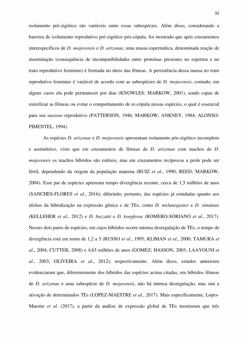
isolamento pré-zigótico são variáveis entre essas subespécies. Além disso, considerando a
barreira de isolamento reprodutivo pré-zigótico pós-cópula, foi mostrado que após cruzamentos
interespecíficos de D. mojavensis e D. arizonae, uma massa espermática, denominada reação de
inseminação (consequência de incompatibilidades entre proteínas presentes no esperma e no
trato reprodutivo feminino) é formada no útero das fêmeas. A persistência dessa massa no trato
reprodutivo feminino é variável de acordo com as subespécies de D. mojavensis, contudo, em
alguns casos ela pode permanecer por dias (KNOWLES; MARKOW, 2001), sendo capaz de
esterilizar as fêmeas ou evitar o comportamento de re-cópula nessas espécies, o qual é essencial
para seu sucesso reprodutivo (PATTERSON, 1946; MARKOW; ANKNEY, 1984; ALONSO-
PIMENTEL, 1994).
As espécies D. arizonae e D. mojavensis apresentam isolamento pós-zigótico incompleto
e assimétrico, visto que em cruzamentos de fêmeas de D. arizonae com machos de D.
mojavensis os machos híbridos são estéreis, mas em cruzamentos recíprocos a prole pode ser
fértil, dependendo da origem da população materna (RUIZ et al., 1990; REED; MARKOW,
2004). Esse par de espécies apresenta tempo divergência recente, cerca de 1,5 milhões de anos
(SANCHES-FLORES et al., 2016); diferindo, portanto, das espécies já estudadas quanto aos
efeitos da hibridização na expressão gênica e de TEs, como D. melanogaster e D. simulans
(KELLEHER et al., 2012) e D. buzzatti e D. koepferae (ROMERO-SORIANO et al., 2017).
Nesses dois pares de espécies, em cujos híbridos ocorre intensa desregulação de TEs, o tempo de
divergência está em torno de 1,2 a 5 (RUSSO et al., 1995; KLIMAN et al., 2000; TAMURA et
al., 2004; CUTTER, 2008) e 4,63 milhões de anos (GOMEZ; HASSON, 2003; LAAYOUNI et
al., 2003; OLIVEIRA et al., 2012), respectivamente. Além disso, estudos anteriores
evidenciaram que, diferentemente dos híbridos das espécies acima citadas, em híbridos fêmeas
de D. arizonae e uma subespécie de D. mojavensis, não há intensa desregulação, mas sim a
ativação de determinados TEs (LOPEZ-MAESTRE et al., 2017). Mais especificamente, Lopes-
Maestre et al. (2017), a partir da análise de expressão global de TEs mostraram que três
33
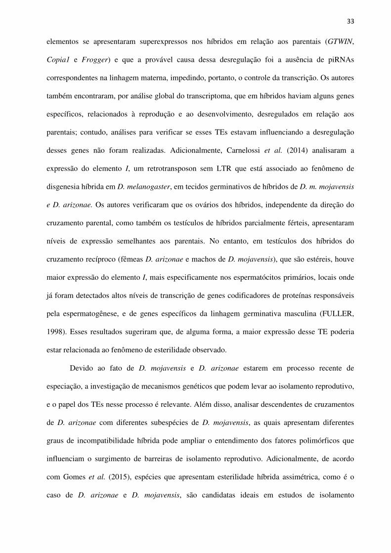
elementos se apresentaram superexpressos nos híbridos em relação aos parentais (GTWIN,
Copia1 e Frogger) e que a provável causa dessa desregulação foi a ausência de piRNAs
correspondentes na linhagem materna, impedindo, portanto, o controle da transcrição. Os autores
também encontraram, por análise global do transcriptoma, que em híbridos haviam alguns genes
específicos, relacionados à reprodução e ao desenvolvimento, desregulados em relação aos
parentais; contudo, análises para verificar se esses TEs estavam influenciando a desregulação
desses genes não foram realizadas. Adicionalmente, Carnelossi et al. (2014) analisaram a
expressão do elemento I, um retrotransposon sem LTR que está associado ao fenômeno de
disgenesia híbrida em D. melanogaster, em tecidos germinativos de híbridos de D. m. mojavensis
e D. arizonae. Os autores verificaram que os ovários dos híbridos, independente da direção do
cruzamento parental, como também os testículos de híbridos parcialmente férteis, apresentaram
níveis de expressão semelhantes aos parentais. No entanto, em testículos dos híbridos do
cruzamento recíproco (fêmeas D. arizonae e machos de D. mojavensis), que são estéreis, houve
maior expressão do elemento I, mais especificamente nos espermatócitos primários, locais onde
já foram detectados altos níveis de transcrição de genes codificadores de proteínas responsáveis
pela espermatogênese, e de genes específicos da linhagem germinativa masculina (FULLER,
1998). Esses resultados sugeriram que, de alguma forma, a maior expressão desse TE poderia
estar relacionada ao fenômeno de esterilidade observado.
Devido ao fato de D. mojavensis e D. arizonae estarem em processo recente de
especiação, a investigação de mecanismos genéticos que podem levar ao isolamento reprodutivo,
e o papel dos TEs nesse processo é relevante. Além disso, analisar descendentes de cruzamentos
de D. arizonae com diferentes subespécies de D. mojavensis, as quais apresentam diferentes
graus de incompatibilidade híbrida pode ampliar o entendimento dos fatores polimórficos que
influenciam o surgimento de barreiras de isolamento reprodutivo. Adicionalmente, de acordo
com Gomes et al. (2015), espécies que apresentam esterilidade híbrida assimétrica, como é o
caso de D. arizonae e D. mojavensis, são candidatas ideais em estudos de isolamento

34
reprodutivo, visto que é possível comparar a expressão de sequências genéticas de híbridos
estéreis com férteis e seus respectivos parentais.

35
OBJETIVOS
36
2 OBJETIVOS
Esta tese foi dividida em três partes principais, sendo que, a primeira delas teve por
objetivo estimar os parâmetros da história de vida de híbridos interespecíficos entre D. arizonae
e quatro subespécies de D. mojavensis, com ênfase em componentes relacionados ao isolamento
pós-zigótico, com viabilidade, motilidade espermática e atrofia de gônadas. A segunda parte
buscou analisar o impacto do fenômeno de hibridização interespecífica na expressão gênica em
tecidos reprodutivos de machos e fêmeas híbridas provenientes de cruzamentos entre D. arizonae
e duas subespécies de D. mojavensis. Por fim, na terceira parte, foi investigada a extensão da
desregulação de Tes em ovários e testículos de híbridos, e sua associação com os mecanismos de
regulação pós-transcricional, por meio da via de piRNAs.

37
CAPÍTULO 1
38
3 CAPÍTULO 1 Life-history traits in hybrids of Drosophila arizonae and Drosophila mojavensis subspecies
Cecilia Artico Banho1.2, Felipe Santa Rosa do Amaral1, Cristina Vieira2, Claudia Marcia
Aparecida Carareto1
1UNESP - São Paulo State University, Department of Biology, São José do Rio Preto, São Paulo
State (SP), Brazil 2 Université de Lyon, Université Lyon 1, CNRS, Laboratoire de Biométrie et Biologie Evolutive
UMR 5558, F-69622 Villeurbanne, France
ABSTRACT Drosophila arizonae and D. mojavensis are recently diverged (~1.5 million years ago (m.y.a))
species widely used in speciation studies due to their ability to produce hybrids in the laboratory;
however, no evidence of introgression has been found in nature, despite favourable conditions
for the hybridization of the species due to their overlapping habitats. The prezygotic isolation of
these species has been well characterized, but few studies have demonstrated the consequences
of interspecific hybridization on their postzygotic isolation. To verify the occurrence and degree
of hybrid incompatibility in D. arizonae and D. mojavensis, we evaluated the life-history
parameters of their inter- and intraspecific offspring. Phenotypic analyses showed that all hybrids
presented a decrease in viability compared to intraspecific offspring. Sperm motility analyses
showed that all hybrid males of one cross direction presented mobile sperm, while in reciprocal
crosses, 75% of the hybrids presented immobile sperm. The fertility analyses showed 100% of
fertile hybrid females, which had no gonad dysgenesis, however, only 25% of the male hybrids
with mobile sperm were fertile. These findings suggests the presence of polymorphic factors
dependent on the D. mojavensis subspecies that can influence the fitness of F1 hybrids and that
several genetic mechanisms can influence the reproductive isolation between these sibling
species.
Keywords: male sterility, repleta group, reproductive isolation
39
INTRODUCTION
Drosophila mojavensis and D. arizonae are recently diverged (approximately 1.5 m.y.a)
sibling species (SANCHEZ-FLORES et al., 2016) that belong to the mulleri complex of the
repleta group. The area of D. mojavensis distribution extends from Arizona to southern
California, Sonora, Sinaloa, and the peninsula of Baja California, while D. arizonae has a more
diffuse population distribution in Central America, with populations reported as far south as Guatemala,
through Mexico and in the USA, where D. arizonae can be found in California and Arizona. Although
widely distributed, D. arizonae does not have populations differentiated enough to be classified as
subspecies. On the other hand, D. mojavensis is classified into four subspecies based on morphological
features (METTLER, 1963), allozymes (ADH) (MATZKIN; EANES, 2003), genetic population
differences (ROSS; MARKOW, 2006), chromosomal inversions and the degree of reproductive isolation
(WARSEMAN; KOEPFER, 1977; RUIZ et al. 1990; REED; MARKOW, 2004). Drosophila arizonae
and D. mojavensis are sympatric populations in an area that includes northern Sinaloa and the whole of
Sonora, where both species use a specific columnar cactus (Stenocereus sp.) as a host; in contrast, in the
other areas, the species constitute allopatric populations (MASSIE; MARKOW, 2005; JENNINGS;
ETGES, 2009). The four geographically isolated subspecies of D. mojavensis are found in the
Mojave Desert (D. m. mojavensis), Baja California peninsula (D. m. baja), Sonoran Desert (D. m.
sonorensis) and Catalina Island on the coast of southern California (D. m. wrigleyi). In each of these
regions, D. mojavensis uses a specific host cactus for feeding and breeding sites, with no evidence of gene
flow, and thus comprises well-structured populations (KNOWLES; MARKOW, 2001; PITNICK et al.,
2003; REED; MARKOW, 2004; MASSIE; MARKOW, 2005; REED et al., 2007; ETGES et al., 2010).
These sibling species are widely used in speciation studies due to their ability to produce
hybrids in the laboratory; however, no evidence of introgression has been found in nature,
despite the favourable ecological conditions for hybridization between D. arizonae and D.
mojavensis, mainly because of their sympatric populations (JENNINGS; ETGES, 2009). As they
are incipient species, some reports have shown incomplete and asymmetric postzygotic isolation
in their interspecific hybrids. It was demonstrated that in crosses between D. arizonae mothers
and D. mojavensis fathers, the male offspring are often sterile, while in reciprocal crosses, the
40
offspring are fertile, depending on the source of the D. mojavensis populations (RUIZ et al.,
1990; REED; MARKOW 2004; CARNELOSSI et al., 2014).
It is important to emphasize that despite several studies analysing the degree of
prezygotic and postmating prezygotic reproductive isolation in D. arizonae and D. mojavensis,
few studies have evaluated the levels of postzygotic isolation in hybrids of these species,
particularly between the four D. mojavensis subspecies and D. arizonae, probably because of the
difficulty in obtaining a large number of offspring due to the well-characterized mechanisms of
prezygotic isolation between them (WARSEMAN; KOEPFER, 1977; RUIZ et al., 1990).
Therefore, in this study, we aimed to evaluate components of the life-history traits in
hybrids from reciprocal crosses between D. arizonae and four subspecies of D. mojavensis.
Despite obtaining a low offspring number, we were able to record the productivity, viability,
sperm motility, fertility and gonadal dygenesis in descendants of all intra- and interespecific
crosses, which allows us to better understand the complexity of the mechanisms involved in the
postzygotic reproductive isolation process between these sibling species.
MATERIAL AND METHODS
Stock flies
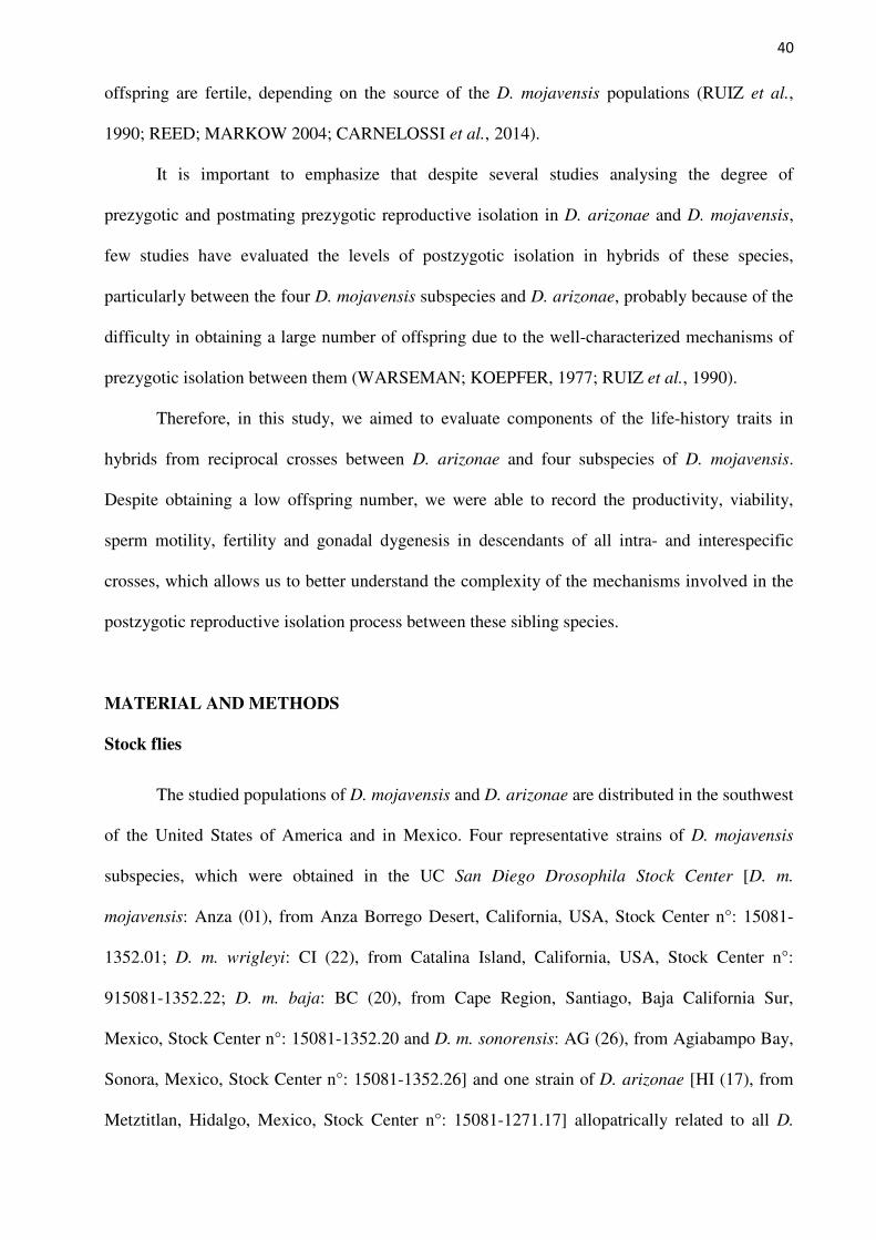
The studied populations of D. mojavensis and D. arizonae are distributed in the southwest
of the United States of America and in Mexico. Four representative strains of D. mojavensis
subspecies, which were obtained in the UC San Diego Drosophila Stock Center [D. m.
mojavensis: Anza (01), from Anza Borrego Desert, California, USA, Stock Center n°: 15081-
1352.01; D. m. wrigleyi: CI (22), from Catalina Island, California, USA, Stock Center n°:
915081-1352.22; D. m. baja: BC (20), from Cape Region, Santiago, Baja California Sur,
Mexico, Stock Center n°: 15081-1352.20 and D. m. sonorensis: AG (26), from Agiabampo Bay,
Sonora, Mexico, Stock Center n°: 15081-1352.26] and one strain of D. arizonae [HI (17), from
Metztitlan, Hidalgo, Mexico, Stock Center n°: 15081-1271.17] allopatrically related to all D.
41
mojavensis lines, were used (Figure 1).
Figure 1. Geographic distribution of the four subspecies of D. mojavensis and D. arizonae analysed.
Fecundity and viability analyses
Virgin males and females of each strain were separated by sex 10 hours after eclosion
and stored separately in yeasted cactus-banana vials, with 10 flies per vial, until they were
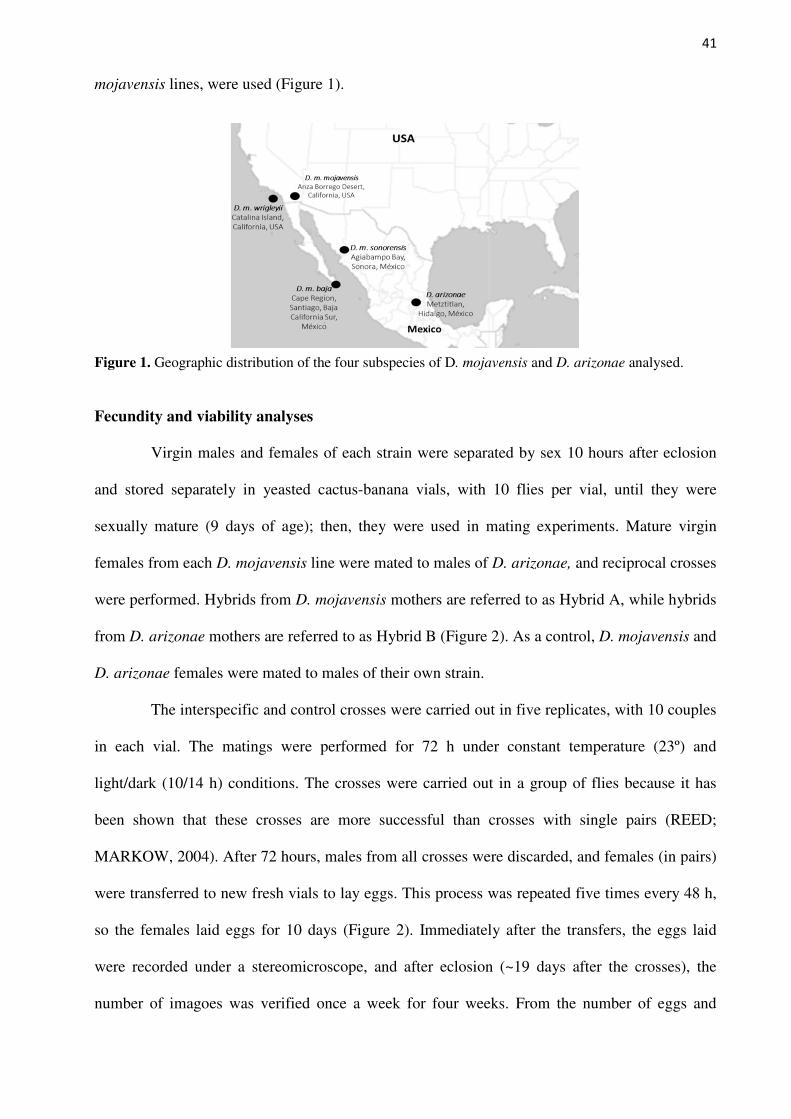
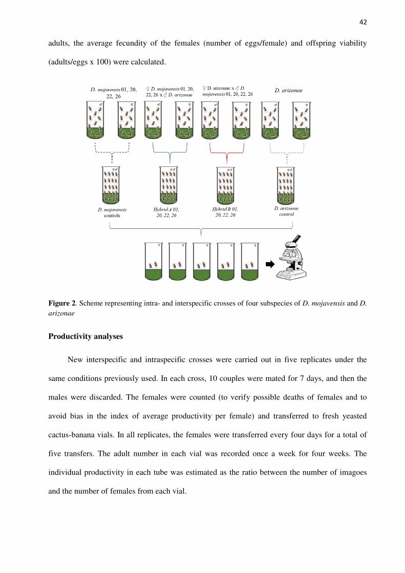
sexually mature (9 days of age); then, they were used in mating experiments. Mature virgin
females from each D. mojavensis line were mated to males of D. arizonae, and reciprocal crosses
were performed. Hybrids from D. mojavensis mothers are referred to as Hybrid A, while hybrids
from D. arizonae mothers are referred to as Hybrid B (Figure 2). As a control, D. mojavensis and
D. arizonae females were mated to males of their own strain.
The interspecific and control crosses were carried out in five replicates, with 10 couples
in each vial. The matings were performed for 72 h under constant temperature (23º) and
light/dark (10/14 h) conditions. The crosses were carried out in a group of flies because it has
been shown that these crosses are more successful than crosses with single pairs (REED;
MARKOW, 2004). After 72 hours, males from all crosses were discarded, and females (in pairs)
were transferred to new fresh vials to lay eggs. This process was repeated five times every 48 h,
so the females laid eggs for 10 days (Figure 2). Immediately after the transfers, the eggs laid
were recorded under a stereomicroscope, and after eclosion (~19 days after the crosses), the
number of imagoes was verified once a week for four weeks. From the number of eggs and
42
adults, the average fecundity of the females (number of eggs/female) and offspring viability
(adults/eggs x 100) were calculated.
Figure 2. Scheme representing intra- and interspecific crosses of four subspecies of D. mojavensis and D.
arizonae
Productivity analyses
New interspecific and intraspecific crosses were carried out in five replicates under the
same conditions previously used. In each cross, 10 couples were mated for 7 days, and then the
males were discarded. The females were counted (to verify possible deaths of females and to
avoid bias in the index of average productivity per female) and transferred to fresh yeasted
cactus-banana vials. In all replicates, the females were transferred every four days for a total of
five transfers. The adult number in each vial was recorded once a week for four weeks. The
individual productivity in each tube was estimated as the ratio between the number of imagoes
and the number of females from each vial.
43
Sterility analyses
Two traits that can result in sterility were studied: gonadal dysgenesis and sperm motility.
For these analyses, new interspecific and control crosses were performed in yeasted cactus-
banana vials. To obtain as many hybrids as possible, three-day-old virgin flies were used because
in previous tests, we noticed an increased production of hybrids when the two species were kept
together before they reached sexual maturity. All crosses were performed in five replicates under
the same temperature and light/dark conditions for 12 days. After that, the parents were
discarded, and the imagoes were separated by sex daily. The descendants were maintained in
yeasted food vials until they reached 10 days of age (sexually mature). Then, they were used in
gonadal dysgenesis and sperm motility experiments.
Gonadal dysgenesis, a morphological component of hybrid dysgenesis, is characterized
by several degrees of abnormalities in the reproductive organs (ALMEIDA; CARARETO,
2002). To verify this component, we analysed 10 F1 males and females from each reciprocal
cross, as well as the control flies. The reproductive organs were dissected in phosphate-buffered
saline (PBS) and then checked and photographed under a stereomicroscope. Statistical analyses
were not performed because all flies presented normal testes and ovaries.
Sperm motility analyses were carried out in 20 F1 male testes and seminal vesicles of
each control and interspecific cross, according to Reed et al. (2008). No statistical analyses were
performed because for each cross, all males presented the same phenotype or presented motility
or immobility, which differs from the results verified by Reed et al. (2004) and Reed et al.
(2008). However, following the method described in Reed et al. (2008), we noticed differences
among the degrees of motility in hybrids from some specific crosses.
Fertility analyses
Three-day-old female and male hybrids from reciprocal interspecific crosses were
backcrossed with their respective parents, D. arizonae and D. mojavensis (from their respective
44
subspecies). Crosses were performed with five couples per replicate in five replicates by cross
direction. To ensure that the absence of offspring was due to possible prezygotic, postmating
prezygotic or postzygotic isolation mechanisms, we increased the crossing time and allowed the
couples to mate for 15 days. After that, all parents were discarded, and fertility was evaluated
based on the presence or absence of offspring, as reported by Carnelossi et al. (2014). F1 x F1
crosses were also performed using offspring of each interspecific cross under the same
conditions as the backcrosses. To certify that tubes containing only eggs would not produce
offspring, they were maintained for 20 days after parent removal and then discarded.
Hybrid Status
To test the hybrid status in offspring of each interspecific cross, amplification of the ITS-
1 (internal transcribed spacer 1) region was performed. For this analysis, DNA from five random
individuals of each replicate of the interspecific crosses was extracted, and the ITS-1 sequence
was amplified using a forward primer that hybridizes to the 3’-end of the 18S rDNA gene and a
reverse primer that hybridizes at the beginning of the 5.8S rDNA gene [NCBI Reference
Sequence: EU306666.1] (BAFFI; CERON, 2002). It was expected that male and female hybrids
would present two fragments of 500 and 550 bp corresponding to the amplified ITS-1 region of
the rDNA of their parents D. arizonae and D. mojavensis, respectively.
Statistical analyses
Statistical analyses were performed for average fecundity by female, average productivity
by female and viability for each replicate of intraspecific and interspecific crosses by using R
software R v. 3.6.1 (The R Core Team). Normality and variance tests (Shapiro-Wilk and
Levene’s test, respectively) were carried out, and when we obtained significant p-values (non-
normal distribution), a nonparametric Kruskal-Wallis test was performed. Then, a post hoc
Wilcoxon test was performed to determine significant differences between the treatments. For
45
results with no significant p-values for normality and variance tests, one-way ANOVA was
performed using Tukey’s post hoc test.
RESULTS
Fecundity and viability analyses
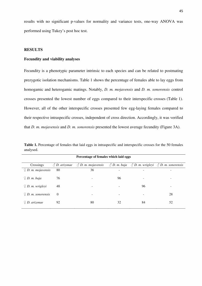
Fecundity is a phenotypic parameter intrinsic to each species and can be related to postmating
prezygotic isolation mechanisms. Table 1 shows the percentage of females able to lay eggs from
homogamic and heterogamic matings. Notably, D. m. mojavensis and D. m. sonorensis control
crosses presented the lowest number of eggs compared to their interspecific crosses (Table 1).
However, all of the other interspecific crosses presented few egg-laying females compared to
their respective intraspecific crosses, independent of cross direction. Accordingly, it was verified
that D. m. mojavensis and D. m. sonorensis presented the lowest average fecundity (Figure 3A).
Table 1. Percentage of females that laid eggs in intraspecific and interspecific crosses for the 50 females analysed.
Percentage of females which laid eggs
Crossings ♂ D. arizonae ♂ D. m. mojavensis ♂ D. m. baja ♂ D. m. wrigleyi ♂ D. m. sonorensis
♀ D. m. mojavensis 80 36 - - -
♀ D. m. baja 76 - 96 - -
♀ D. m. wrigleyi 48 - - 96 -
♀ D. m. sonorensis 0 - - - 28
♀ D. arizonae 92 80 32 84 52
46
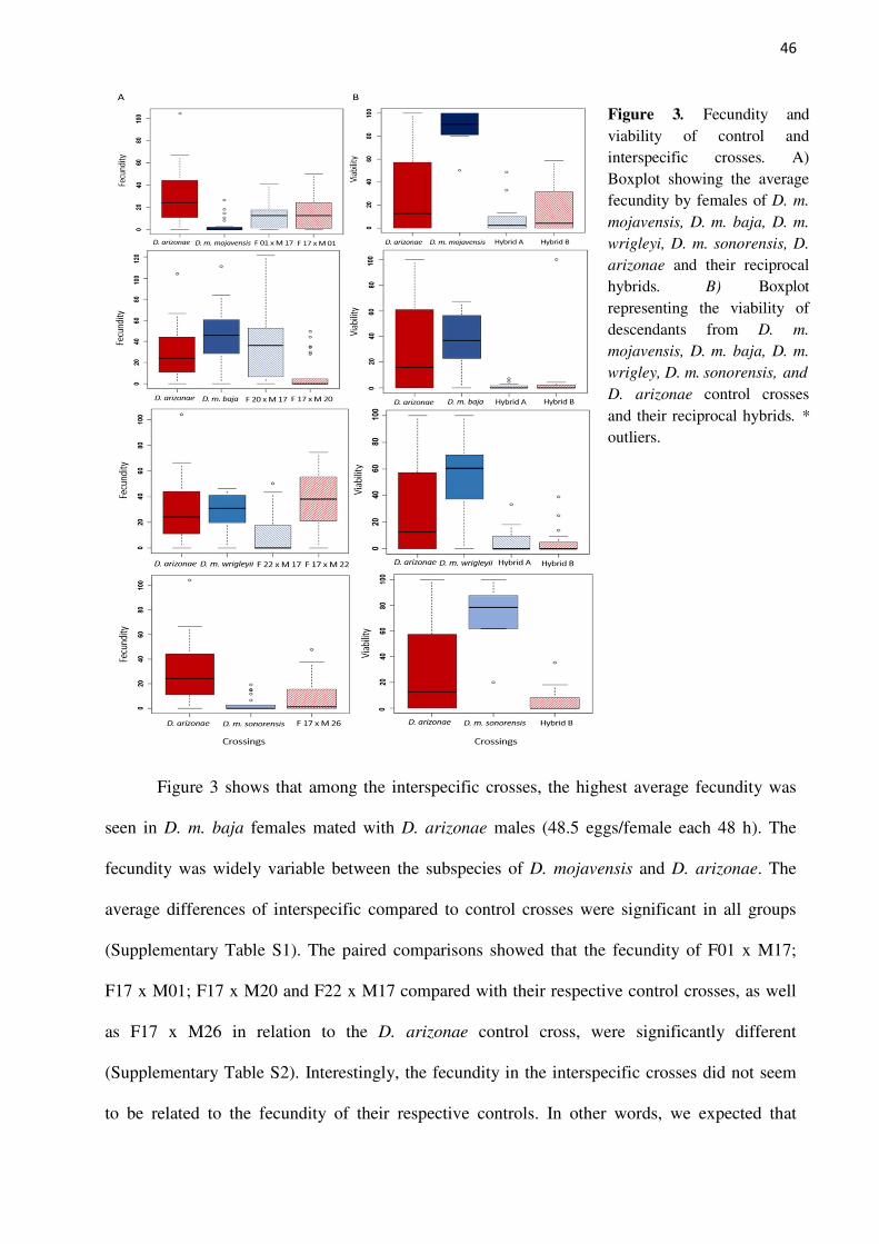
Figure 3. Fecundity and viability of control and interspecific crosses. A) Boxplot showing the average fecundity by females of D. m.
mojavensis, D. m. baja, D. m.
wrigleyi, D. m. sonorensis, D.
arizonae and their reciprocal hybrids. B) Boxplot representing the viability of descendants from D. m.
mojavensis, D. m. baja, D. m.
wrigley, D. m. sonorensis, and
D. arizonae control crosses and their reciprocal hybrids. *
outliers.
Figure 3 shows that among the interspecific crosses, the highest average fecundity was
seen in D. m. baja females mated with D. arizonae males (48.5 eggs/female each 48 h). The
fecundity was widely variable between the subspecies of D. mojavensis and D. arizonae. The
average differences of interspecific compared to control crosses were significant in all groups
(Supplementary Table S1). The paired comparisons showed that the fecundity of F01 x M17;
F17 x M01; F17 x M20 and F22 x M17 compared with their respective control crosses, as well
as F17 x M26 in relation to the D. arizonae control cross, were significantly different
(Supplementary Table S2). Interestingly, the fecundity in the interspecific crosses did not seem
to be related to the fecundity of their respective controls. In other words, we expected that
47
interspecific crosses using D. mojavensis mothers would show a similar fecundity as their
control, with the same expectation for interspecific crosses with D. arizonae mothers and control
crosses, because fecundity is an intrinsic characteristic of the specific strains; therefore, we used
the same lines to perform the crosses. However, this was not observed in our experiments,
evidencing that other factors may be behind this characteristic.
The averages of viability showed significant differences in all groups of comparisons
(Supplementary Table S3). Interestingly, although D. m. mojavensis and D. m. sonorensis
exhibited the lowest fecundity, they had the highest viability (86.8% and 70.9%, respectively).
The other control lines, D. m. baja, D. m. wrigleyi and D. arizonae, showed large variations in
the average viability of 38.67%, 51.93% and 29.58%, respectively. In all interspecific crosses, a
decrease in viability compared to the intraspecific crosses was verified (Supplementary Table
S4). The average hybrid viability ranged from 1.16 to 14.2%. The avarege of viability in hybrids
was, at least, seven times as small as the avarage viability of control crosses. Among the
interspecific crosses, Hybrid A 20 presented the lowest viability (Figure 3B, Table 2). This is an
interesting observation, since the heterospecific crosses between D. m. baja females and D.
arizonae males presented the highest average fecundity. On the other hand, Hybrid B 01
presented the highest viability compared to other interspecific crosses (Figure 3B, Table 2).
In the paired comparisons, not significant differences were found in all reciprocal hybrids
from the crosses between the four subspecies of D. mojavensis and D. arizonae. This result
indicates there are similar deleterious effects in the hybrids regardless of cross direction. We
were not able to analyse the fecundity and viability of crosses between D. m. sonorensis (26)
females from the Sonoran Desert mated with D. arizonae males because there was no egg
production.
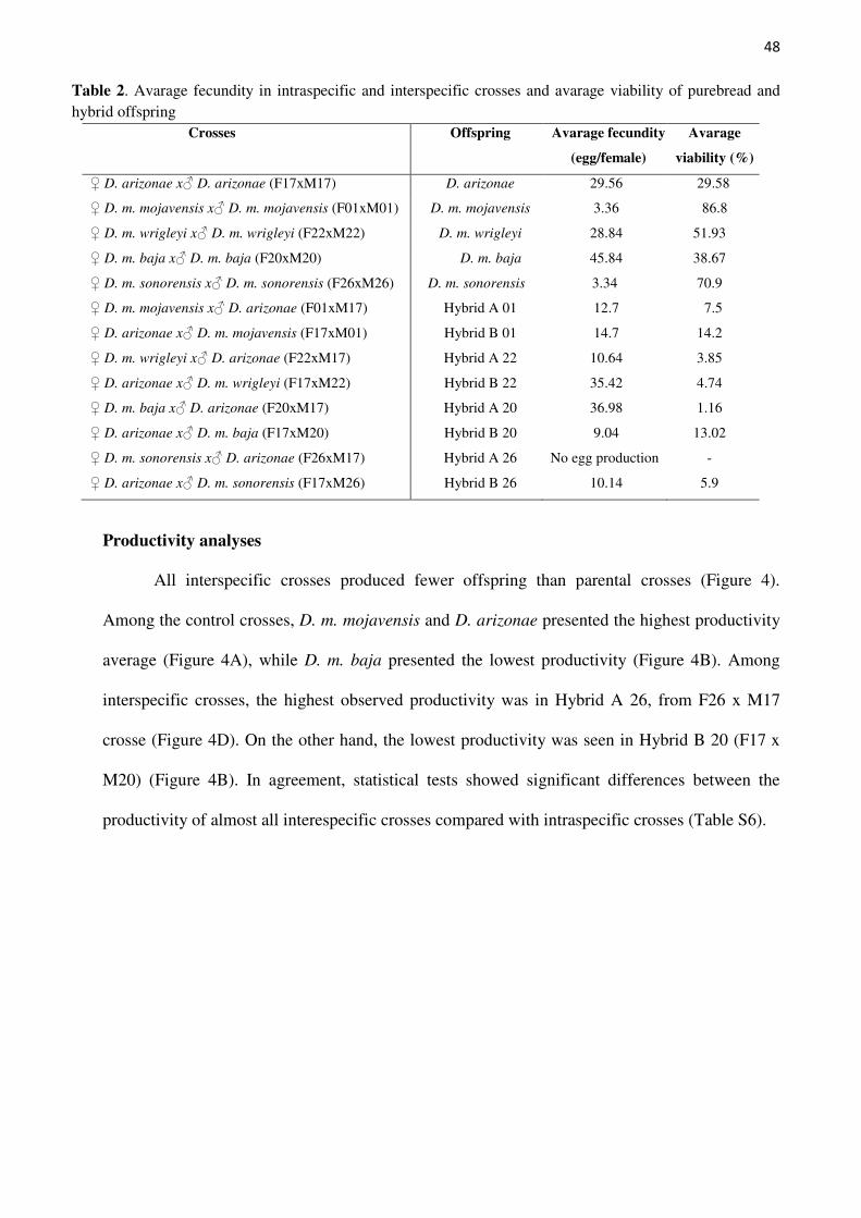
48
Table 2. Avarage fecundity in intraspecific and interspecific crosses and avarage viability of purebread and hybrid offspring
Crosses Offspring Avarage fecundity
(egg/female)
Avarage
viability (%)
♀ D. arizonae x♂ D. arizonae (F17xM17) D. arizonae 29.56 29.58
♀ D. m. mojavensis x♂ D. m. mojavensis (F01xM01) D. m. mojavensis 3.36 86.8
♀ D. m. wrigleyi x♂ D. m. wrigleyi (F22xM22) D. m. wrigleyi 28.84 51.93
♀ D. m. baja x♂ D. m. baja (F20xM20) D. m. baja 45.84 38.67
♀ D. m. sonorensis x♂ D. m. sonorensis (F26xM26) D. m. sonorensis 3.34 70.9
♀ D. m. mojavensis x♂ D. arizonae (F01xM17) Hybrid A 01 12.7 7.5
♀ D. arizonae x♂ D. m. mojavensis (F17xM01) Hybrid B 01 14.7 14.2
♀ D. m. wrigleyi x♂ D. arizonae (F22xM17) Hybrid A 22 10.64 3.85
♀ D. arizonae x♂ D. m. wrigleyi (F17xM22) Hybrid B 22 35.42 4.74
♀ D. m. baja x♂ D. arizonae (F20xM17) Hybrid A 20 36.98 1.16
♀ D. arizonae x♂ D. m. baja (F17xM20) Hybrid B 20 9.04 13.02
♀ D. m. sonorensis x♂ D. arizonae (F26xM17) Hybrid A 26 No egg production -
♀ D. arizonae x♂ D. m. sonorensis (F17xM26) Hybrid B 26 10.14 5.9
Productivity analyses
All interspecific crosses produced fewer offspring than parental crosses (Figure 4).
Among the control crosses, D. m. mojavensis and D. arizonae presented the highest productivity
average (Figure 4A), while D. m. baja presented the lowest productivity (Figure 4B). Among
interspecific crosses, the highest observed productivity was in Hybrid A 26, from F26 x M17
crosse (Figure 4D). On the other hand, the lowest productivity was seen in Hybrid B 20 (F17 x
M20) (Figure 4B). In agreement, statistical tests showed significant differences between the
productivity of almost all interespecific crosses compared with intraspecific crosses (Table S6).
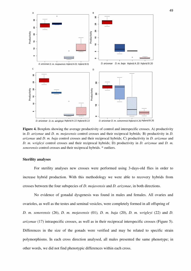
49
Figure 4. Boxplots showing the average productivity of control and interspecific crosses. A) productivity in D. arizonae and D. m. mojavensis control crosses and their reciprocal hybrids; B) productivity in D.
arizonae and D. m. baja control crosses and their reciprocal hybrids; C) productivity in D. arizonae and D. m. wrigleyi control crosses and their reciprocal hybrids; D) productivity in D. arizonae and D. m.
sonorensis control crosses and their reciprocal hybrids. * outliers.
Sterility analyses
For sterility analyses new crosses were performed using 3-days-old flies in order to
increase hybrid production. With this methodology we were able to recovery hybrids from
crosses between the four subspecies of D. mojavensis and D. arizonae, in both directions.
No evidence of gonadal dysgenesis was found in males and females. All ovaries and
ovarioles, as well as the testes and seminal vesicles, were completely formed in all offspring of
D. m. sonorensis (26), D. m. mojavensis (01), D. m. baja (20), D. m. wrigleyi (22) and D.
arizonae (17) intraspecific crosses, as well as in their reciprocal interspecific crosses (Figure 5).
Differences in the size of the gonads were verified and may be related to specific strain
polymorphisms. In each cross direction analysed, all males presented the same phenotype; in
other words, we did not find phenotypic differences within each cross.
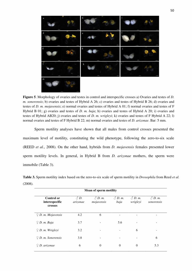
50
Figure 5. Morphology of ovaries and testes in control and interspecific crosses a) Ovaries and testes of D.
m. sonorensis; b) ovaries and testes of Hybrid A 26; c) ovaries and testes of Hybrid B 26; d) ovaries and testes of D. m. mojavensis; e) normal ovaries and testes of Hybrid A 01; f) normal ovaries and testes of F Hybrid B 01; g) ovaries and testes of D. m. baja; h) ovaries and testes of Hybrid A 20; i) ovaries and testes of Hybrid AB20; j) ovaries and testes of D. m. wrigleyi; k) ovaries and testes of F Hybrid A 22; l) normal ovaries and testes of F Hybrid B 22; m) normal ovaries and testes of D. arizonae. Bar: 5 mm.
Sperm motility analyses have shown that all males from control crosses presented the
maximum level of motility, constituting the wild phenotype, following the zero-to-six scale
(REED et al., 2008). On the other hand, hybrids from D. mojavensis females presented lower
sperm motility levels. In general, in Hybrid B from D. arizonae mothers, the sperm were
immobile (Table 3).
Table 3. Sperm motility index based on the zero-to-six scale of sperm motility in Drosophila from Reed et al.
(2008).
Mean of sperm motility
Control or interespecific
crosses
♂ D.
arizonae
♂ D. m.
mojavensis
♂ D. m.
baja
♂ D. m.
wrigleyi
♂ D. m.
sonorensis
♀ D. m. Mojavensis 4.2 6 - - -
♀ D. m. Baja 3.7 - 5.6 - -
♀ D. m. Wrigleyi 3.2 - - 6 -
♀ D. m. Sonorensis 3.8 - - - 6
♀ D. arizonae 6 0 0 0 5.3
51
The only exception was Hybrid B 26, from crosses between ♂ D. m. sonorensis (26) x ♀
D. arizonae (17). They presented high sperm motility similar to the wild phenotype. In addition,
their motility level was higher than that of their reciprocal hybrids (Hybrid A 26). It is important
to highlight that D. m. sonorensis is a sympatric population. In addition, among the hybrids
presenting mobile sperm, we can found great variation in the motility levels. This evidence
indicates the presence of some specific polymorphisms that may contribute to the evolution of
postzygotic isolation mechanisms in each line of D. mojavensis.
Fertility analyses
Hybrid sterility was analysed by backcrosses and F1 x F1 crosses for all male and female
offspring from interspecific crosses. The results show that all hybrid females were fertile,
regardless of the cross direction.
In contrast, we observed that males from crosses between D. m. mojavensis (01) and D.m.
wrigleyi with D. arizonae were sterile in both directions (Hybrids A 01 and 22; Hybrids B 01
and 22), although they presented mobile sperm (Table 2). On the other hand, Hybrid A 20, from
crosses between D. m. baja females and D. arizonae males, were fertile despite low sperm
motility since their F1 x F1 crosses produced offspring; however, in backcrosses, no offspring
were verified. Nevertheless, Hybrid B 20 (from ♀ D. arizonae x ♂ D. m. baja) were sterile,
which was expected, as they presented no sperm motility (Table 3). Interspecific crosses between
D.m. sonorensis and D. arizonae were the only crosses that presented male and female hybrids,
regardless of cross direction (Supplementary table S7).
DISCUSSION
To better understand the speciation process, it is important to analyse and identify the
reproductive barriers that contribute to reproductive isolation. Among them, postzygotic
reproductive barriers can be responsible for reducing hybrid fitness compared to the parental
52
lines. According to Coyne and Orr (2004), postzygotic isolation is split into two categories:
intrinsic and extrinsic. Intrinsic barriers are those that can cause inviability or sterility, while
extrinsic barriers are those that allow normal hybrid development but can present problems for
attracting mates (behavioural sterility). According to Orr (2009), differences in fitness caused by
stress conditions, such as hybridization, can arise in any stage of the life cycle and affect the
fitness components (e.g., viability, mating success, and fecundity) contributing to differences in
total fitness among individuals.
To identify some factors acting on postzygotic mechanisms, we characterized the life-
history traits of offspring from interspecific crosses between D. arizonae and four subspecies of
D. mojavensis, such as fecundity, productivity, viability, hybrid dysgenesis, sperm motility and
fertility.
Fecundity and viability analyses
The low egg production observed in the D. m. mojavensis and D. m. sonorensis crosses
was an unexpected result, as they were control crosses. This low egg production may have
occurred because D. mojavensis needs to mate to lay eggs, a necessity possibly related to male
investment in egg production (KELLEHER; MARKOW, 2007). It has been proposed that
because D. mojavensis females live in a harsh environment often with limited resources, they
need to incorporate male-derived molecules into somatic tissues and oocytes (MARKOW;
ANKNEY, 1984). Therefore, in this specific case, D. m. mojavensis and D. m. sonorensis control
crosses may not have mated, since no eggs were seen. Regarding interspecific crosses, the small
number of females laying eggs might have been related to the strong prezygotic isolation
between D. mojavensis and D. arizonae, which prevented mating for most females. Accordingly,
it has been shown that 50% of the females from interspecific crosses between three subspecies of
D. mojavensis and D. arizonae did not lay eggs (KELLEHER; MARKOW, 2007).
According to Massie and Markow (2005), D. arizonae females rarely can cross with D.
53
mojavensis males, regardless of the origin of the population, while D. mojavensis females from
sympatric populations (Sonoran Desert) exhibit stronger prezygotic isolation related to D.
arizonae males than D. mojavensis females from allopatric regions. This pattern of strong
prezygotic isolation influencing fecundity was verified, mainly in crosses with female D. m.
sonorensis and D. arizonae males derived from sympatric populations, corroborating the
Wasserman and Koepfer (1977) observations and the reinforcement of prezygotic reproductive
isolation. In addition, Jennings and Etges (2009) found that factors related to the maintenance of
flies in the laboratory, such as the experimental crossing conditions, can increase the prezygotic
isolation between D. arizonae and D. mojavensis, and this may be a factor interfering in the
frequency of mating in our crosses.
However, prezygotic isolation cannot be the only factor influencing oviposition in
interspecific crosses. Postmating prezygotic isolation (PZPM) has been extensively studied, and
evidence shows that it can influence oviposition levels, mainly in heterospecific matings
(PATTERSON, 1946; MARKOW; HOCUTT, 1998; KNOWLES; MARKOW, 2001; BONO et
al., 2011). According to Markow and Hocutt (1998), females from the repleta group, such as D.
arizonae and D. mojavensis, present an insemination reaction (PATTERSON, 1946; ALONSO-
PIMENTEL et al., 1994), which consists of the enlargement of the vagina produced by the male
ejaculate in intraspecific crossings, forming an opaque mass that prevents remating behaviour
temporarily (PATTERSON, 1946). D. arizonae and D. mojavensis normally present a mating
system characterized by frequent female remating relative to D. melanogaster. This can be
linked to the fact that females use nutrients from the male ejaculate, and males transfer less
material at each copulation to take advantage of the increased mating opportunities in their
population (MARKOW; ANKEY 1984; ALONSO-PIMENTEL et al., 1994; BONO et al., 2011;
BONO et al., 2015). On the other hand, in heterospecific crosses, the insemination reaction can
be stronger, and this mass can last days in the uterus of flies, preventing regular remating
behaviour and oviposition, and can even sterilize females (KELLEHER et al., 2007; BONO et
54
al., 2011, BONO et al., 2015).
According to Bono et al. (2011), heterospecifically mated D. mojavensis females exhibit
perturbations in a number of processes occurring within their reproductive tract that result in a
high incidence of failed fertilization, a reduced rate of oviposition, and ultimately the production
of few hybrid offspring, as verified in our crosses. These problems are associated with
deficiencies in the heterospecifically mated female’s storage and retention of sperm and in the
degradation of the insemination reaction (BONO et al., 2011; PATTERSON 1946).
Additionally, Bono et al. (2011) found that PMPZ isolation was strong in all crosses involving
D. mojavensis females, suggesting that this barrier was the earliest to evolve. However, in our
results, the average fecundity in interspecific crosses did not seem to be stronger in D.
mojavensis females than in D. arizonae females.
The reduction in hybrid viability found in this study agrees with the results of Kelleher
and Markow (2007), who found that viability decreased from 60-70% in homospecific crosses to
1-4% in heterospecific crosses. Additionally, in interspecific crosses between D. virilis and D.
lummei, species from the repleta group, it was verified that the low viability of hybrids was due
to early embryo lethality, which differed with cross direction, since in D. virilis female x D.
lummei male crosses, progeny production was reduced by 62%, while in the reciprocal cross,
progeny production was reduced by 50% (CASTILLO; MOYLE, 2019). Therefore, the low
hybrid survival in our interspecific hybrids indicates that there are several levels of isolation
between the parental species. The results reinforce that despite the evident prezygotic isolation
between D. mojavensis and D. arizonae, there are several barriers to postzygotic isolation,
decreasing the fitness of reciprocal hybrids and influencing the initial steps of the speciation
process.
Productivity analyses
Our results showed a decrease in productivity in all interspecific crosses, in agreement
55
with Kelleher and Markow (2007) and Ruiz et al. (1990). This fitness component can be
influenced by prezygotic, postmating-prezygotic and postzygotic isolation, such as fecundity and
viability. Regarding prezygotic isolation, the different subspecies of D. mojavensis present
different degrees of isolation related to D. arizonae. In general, D. m. sonorensis is almost
completely isolated from D. arizonae, followed by D. m. baja and by the least isolated
subspecies D. m. mojavensis (WASSERMAN; KOEPFER, 1977; MASSIE; MARKOW, 2005).
In addition, postmating prezygotic isolation can influence productivity; however, it does not
seem to be related to maternal origin. Therefore, we consider postzygotic isolation to be the
reproductive barrier that has the greatest influence on the average productivity.
Sterility analyses
Hybrid dysgenesis corresponds to abnormal phenotypes in F1 hybrid gonads, which can
influence the speciation process (FONTDEVILA, 2005; MICHALAK, 2009). Gonadal
dysgenesis is a rare phenomenon in purebred lines under optimal developmental conditions, such
as constant temperature and nutritional availability. However, this phenomenon can occur due to
germline development problems, heat shock, transposable element mobilization, and genomic
shock as hybridization. In the analysis of male and female gonads from four subspecies of D.
mojavensis, D. arizonae and their reciprocal hybrids, no evidence of gonadal dysgenesis was
observed, consistent with the results of Reed and Markow (2004) for hybrids of D. m. baja, D.
m. sonorensis and D. m. wrigleyi mated with D. arizonae. However, our results showed different
ovary sizes, which may be related to the strain characteristics because the number of ovarioles
can vary within and between species (MARKOW; O'GRADY, 2007). Because each ovariole
produces eggs autonomously, the number of ovarioles is an important determinant of fecundity,
thereby influencing evolutionary fitness (ORR, 2009).
In species of the Drosophila genus, where the male is the heterogametic sex, sterile
hybrid males have mostly atrophied seminal vesicles with normal testes morphology, although a
56
small proportion of hybrids might also show whole or partial testes atrophy (Ferguson et al.,
2013). It was shown the testes and seminal vesicles of D. mojavensis males (11 days old) were
longer than those of D. arizonae, which was expected, since the sperm of the former are slightly
longer than those of the latter (HARDY et al., 2011), and their hybrids presented different testes
and seminal vesicle sizes compared to both parents. In our study, all males from intraspecific and
interspecific crosses presented normal testes and seminal vesicles, even males that presented
sterility confirmed by sperm motility or fertility analyses. However, we could not affirm that the
parents and hybrids presented no differences in gonad size, as no measures were performed.
In our analyses of sperm motility and fertility, all female hybrids (homogametic sex) were
fertile, regardless of the cross direction. On the other hand, almost all male hybrids were sterile,
depending on the cross direction and the D. mojavensis subspecies, in agreement with Haldane’s
(1922) rule and findings of Turissini et al. (2018), who showed that in interspecific crosses of
recently diverged Drosophila species, the hybrid males usually are sterile while the femeles
remain fertile. We also observed that all hybrids from D. mojavensis females and D. arizonae
males had mobile sperm, although the sperm motility level was lower in hybrids than in their
respective controls and was variable among the hybrids from different D. mojavensis subspecies.
Different levels of motility in hybrids might be related to intraspecific polymorphisms in the
different populations of D. mojavensis, as also observed by Reed and Markow (2004). On the
other hand, almost all males from intercrosses of D. arizonae females presented immobile sperm.
Although the factors responsible for sperm immobility in these hybrids are not completely
known, somes studies have reported that it could be related to incompatibilities between the D.
mojavensis Y chromosome and D. arizonae IV chromosome (ZOUROS et al., 1981, 1988;
PANTAZIDIS, 1988).
Among hybrids from D. arizonae mothers, we found one exception regarding sperm
immotility. Hybrids from D. arizonae females and D. m. sonorensis males presented high sperm
motility, which was approximately equal to the motility of males of pure lineages. These hybrids

57
are from crosses between D. arizonae females and males from the Sonora Desert, one of the
sympatric populations analysed. From this result, we can verify that although the prezygotic
isolation between these species is very strong (MASSIE; MARKOW, 2005), when this barrier is
overcome, several offspring are produced, and they present lower postzygotic isolation than
interspecific offspring from allopatric populations.
Fertility analyses
Sterility is a complex trait that can have multiple influences and be controlled genetically
at different phenotypic levels (REED; MARKOW, 2004). In general, Drosophila sterility is
measured by the presence or absence of sperm motility (COYNE; ORR, 1997). However, this
may not necessarily always be correct because males presenting motile sperm may be unable to
produce offspring. Due to this factor, the fertility of the hybrids with mobile sperm was tested by
backcrosses with both parental lines and F1 x F1 crosses. The results verified that the hybrid
males from the crosses between D. arizonae males and D. m. baja females were fertile, since
they produced offspring. In addition, hybrids from D. arizonae females and D. m. sonorensis
males, the only males that presented sperm motility in this cross direction, were also fertile,
evidencing weak postzygotic isolation in hybrids from sympatric populations. On the other hand,
male hybrids from D. m. mojavensis and D. m. wrigleyi females crosses with D. arizonae males,
which showed sperm motility, were unable to produce offspring, in agreement with Reed and
Markow (2004). The authors found that some males displaying sperm motility were able to
produce offspring, while others were sterile. Therefore, motility is not a factor that always
indicates fertility, which suggests that this phenotypic trait has a complex genetic base, which
needs to be better understood.
CONCLUSION
Here, we verified that all hybrids presented reduced fitness compared to their parental
58
lines, regardless of the cross direction and the source of the parental population. Moreover, male
sterility was shown in almost all hybrids, while all female hybrids were fertile, corroborating
Haldane’s rule. Regarding male sterility, it was observed that some hybrids presented mobile
sperm, and when this occurred, the motility exhibited different levels compared to the parental
males and between the hybrids. This feature suggests the presence of polymorphic factors
dependent on the D. mojavensis subspecies that can influence the fitness of F1 hybrids. In
addition, an interesting result was observed: D arizonae and D. m. sonorensis, which are
sympatric populations, present strong prezygotic isolation, however, when this barrier is
overcome, they are able to produce a large number of fertile male hybrids, in both cross
directions, differently from the male hybrid offspring of allopatric populations.
Therefore, these results suggest the presence of several genetic mechanisms influencing
reproductive isolation between these sibling species, which are species-specific. Considering
this, the next steps will be to analyse the expression of some candidate genes involved in the
reproductive isolation process between D. arizonae and all subspecies of D. mojavensis, as well
as the possible influence of transposable elements in the first steps of speciation.
REFERENCES
ALMEIDA, L. M.; CARARETO, C. M. A. Gonadal Hybrid Dysgenesis in Drosophila
sturtevanti (Diptera, Drosophilidae). Iheringia, SÈr. Zool., Porto Alegre, V. 92, n. 2, p. 71-79, 2002. ALONSO-PIMENTEL, H.; TOLBERT, L. P.; HEED, W. B. Ultrastructural examination of the insemination reaction in Drosophila. Cell Tissue Res., v. 275, p. 467-479, 1994. BAFFI, M. A.; CERON, C. R. Molecular analysis of the rDNA ITS-1 intergenic spacer in Drosophila mulleri, D. arizonae, and their hybrids. Biochemical Genetics, v. 40, n. 11-12, p. 411-421, 2002. BONO, J. M.; MATZKIN, L. M. et al. Postmating transcriptional changes in reproductive tracts of con- and heterospecifically mated Drosophila mojavensis females. PNAS, v. 108, n. 19, p. 7878, 2011. BONO, J. M.; MATZKIN, L. M.; HOANG, K.; BRANDSMEIER, L. Molecular evolution of candidate genes involved in post-mating-Prezygotic reproductive isolation. J. Evol. Biol. v. 28,
59
n. 2, p. 403–414. 2015. CARNELOSSI, E. A. et al. Specific activation of anI-Like element in Drosophila interspecific hybrids. Genome Biol Evol. v. 6, p. 1806-1817, 2014. CASTILLO D. M., MOYLE, L. C. Transposable elements that cause dysgenesis also contribute to postzygotic isolation in the Drosophila virilis clade. bioRxiv preprint first posted online. Doi: http://dx.doi.org/10.1101/753814, 2019. COYNE J. A., ORR, H. A. ‘‘Patterns of speciation in Drosophila’’ revisited. Evolution, v. 51, n. 295–303, 1997. COYNE, J. A.; ORR, H. A. Speciation. Sunderland, MA: Sinauer, 2004. ETGES, W. J. et al. Genetics of incipient speciation in Drosophila mojavensis. iii. Life-history divergence in allopatry and reproductive isolation. The Society for the Study of Evolution, v. 64, n. 12, p. 3549–3569, 2010. ETGES, W. J. et al. Inheritance of courtship song variation among geographically isolated populations of Drosophila mojavensis. Animal Behaviour, v. 71, p. 1205–1214, 2006.
FERGUSON, J.; GOMES, S.; CIVETTA, A. Rapid male-specific regulatory divergence and down regulation of spermatogenesis genes in Drosophila species hybrids. PLOS ONE, v. 8, n. 4, 2013. FONTDEVILA, A. Hybrid genome evolution by transposition. Cytogenetic and Genome Research, v. 110, n. 1-4, p. 49-55, 2005. HALDANE, J. B.S. Sex ratio and unisexual sterility in animal hybrids. Journal of Genetics v. 12, n.101–109, 1922. HARDY, R. W.; LOUGHEED, A.; MARKOW, T. A. Reproductive tract and spermatid abnormalities of hybrid males from reciprocal crosses between Drosophila mojavensis and D.
arizonae. Fly, v. 5, n. 2, p. 1-5, 2011. JENNINGS, J. H.; ETGES. W. J. Species hybrids in the laboratory but not in nature: a reanalysis of premating isolation between Drosophila arizonae and D. mojavensis. The Society for the Study of Evolution. v. 64, n. 2, p. 587–598, 2009. KELLEHER, E. S.; MARKOW, T. A. Reproductive Tract Interactions Contribute to Isolation in Drosophila. Fly, v. 1, n. 1, p. 33-37, 2007. KNOWLES, L. L.; MARKOW, T. A. Sexually antagonistic coevolution of a postmating- prezygotic reproductive character in desert Drosophila. PNAS, v. 98, n. 15, p. 8692–8696, 2001. MARKOW, T. A.; HOCUTT, G. D. Reproductive Isolation in Sonoran Desert Drosophila: Testing the Limits of the Rules. P. 234–244, 1998 in D. J. Howard and S. H. Berlocher, eds. Endless forms: species and speciation. Oxford Univ. Press, New York, 1998. MARKOW, T. A.; O’GRADY, P. M. Drosophila Biology in the Genomic Age. Genetics, v. 177, p. 1269–1276, 2007.
60
MARKOW, T.; ANKNEY, P. Drosophila males contribute to oogenesis in a multiple mating species. Science, v. 224, p. 302—303, 1984. MASSIE, K. R.; MARKOW, T. A. Sympatry, allopatry and sexual isolation between Drosophila
mojavensis and D. arizonae. Hereditas, v. 142, p. 51-55, 2005. MATZKIN, L. M.; EANES, W. F. Sequence variation of Alcohol dehydrogenase (Adh) paralogs in cactophilic Drosophila. Genetics. v. 163, p. 181–194, 2003.
METTLER, L. E. D. mojavensis baja a new form in the mulleri complex. Dros Infor Serv. v. 38,
p. 57-58, 1963.
MICHALACK, P. EPIGENETIC, transposon and small RNA determinants of hybrid dysfunctions. Heredity, v. 102, p. 45–50, 2009. ORR, H. A. Fitness and its role in evolutionary genetics. Nat Rev Genet. v. 10, n. 8, p. 531–539, 2009. PANTAZIDIS A. C., ZOUROS, E. Location of an Autosomal Factor Causing Sterility in Drosophila-Mojavensis Males Carrying the Drosophila-Arizonensis Y-Chromosome. Heredity, v. 60, n. 299–304, 1988. PATTERSON, J. T. A new type of isolating mechanism in Drosophila. Proceedings of the National Academy of Sciences of the United States of America. v. 32, p. 202-208, 1946.
PITNICK, S.; MILLER, G. T.; Schneider, K.; Markow, T. A. Ejaculate–female coevolution in Drosophila mojavensis. Proc. R. Soc. Lond. B, 2003 R Development Core Team. R: A language and environment for statistical computing. R Foundation for Statistical Computing, Vienna, Austria. ISBN 3-900051-07-0, URL http://www.R-project.org. 2008. REED, L. K.; LAFLAMME, B. A.; MARKOW, T. A. Genetic Architecture of Hybrid Male Sterility in Drosophila: Analysis of Intraspecies Variation for Interspecies Isolation. Plos One. v. 3, n. 8, p. e3076, 2008. REED, L. K.; MARKOW, T. A. Early events in speciation: polymorphism for hybrid male sterility in Drosophila. Proceedings of the National Academy of Sciences of the United States of America, v. 101, n. 24, p. 9009-9012, 2004. REED, L. K.; NYBOER, M.; MARKOW, T. A. Evolutionary relationships of Drosophila
mojavensis geographic host races and their sister species Drosophila arizonae. Molecular Ecology, Oxford, v. 16, n. 5, p. 1007-1022, 2007. ROSS, C. L.; MARKOW, T. A. Microsatellite variation among diverging populations of Drosophila mojavensis. European Society for Evolutionary Biology, v.19, p. 1691–1700, 2006. RUIZ, A.; HEED, W. B.; WASSERMAN, M. Evolution of the mojavensis cluster of cactophilic Drosophila with descriptions of two new species. Journal of Heredity, v. 81, n. 1, p.30-42,
61
1990. SANCHES-FLORES, A. et al. Genome evolution in three species of cactophilic Drosophila. G3, Genes, Genomes, Genetics, v. 6, 2016. TURISSINI D. A.; MCGIRR J. A.; PATEL S. S.; DAVID J. R.; MATUTE D. R. The Rate of Evolution of Postmating-Prezygotic Reproductive Isolation in Drosophila. Mol Biol Evol. v. 35, p. 312–334, 2018. WARSSEMAN, M.; KOEPFER, R. Character displacement for sexual isolation between Drosophila mojavensis and Drosophila arizonensis. Evolution, v. 31, n. 4, 1977. ZOUROS E., LOFDAHL, K., MARTIN, P. A. Male hybrid sterility in Drosophila - Interactions between autosomes and sex-chromosomes in crosses of Drosophila mojavensis and Drosophila
arizonensis. Evolution, v. 42, n. 1321–1331, 1988. ZOUROS, E An autosome-Y chromosome combination that causes sterility in D. mojavensis and D. arizonensis hybrids. Drosophila Information Service, v. 56, n. 167–168, 1981.
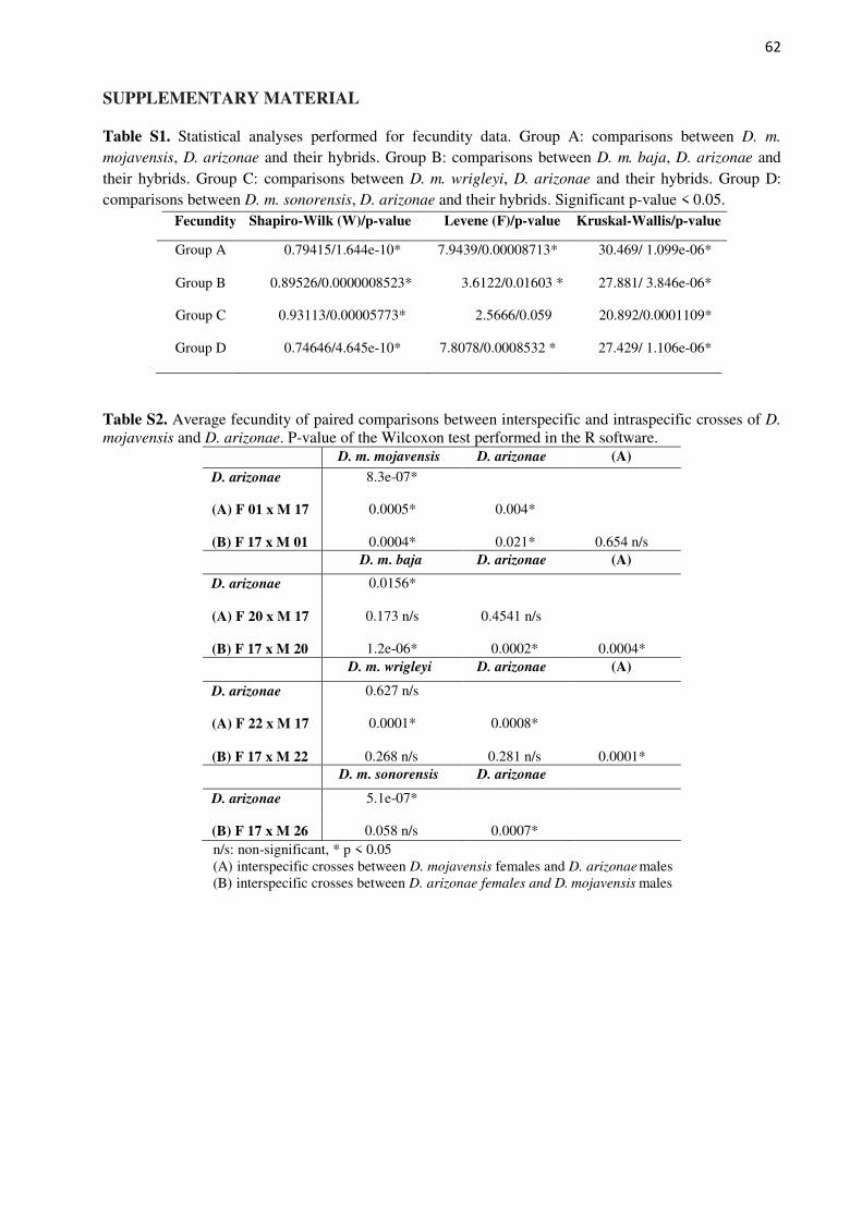
62
SUPPLEMENTARY MATERIAL Table S1. Statistical analyses performed for fecundity data. Group A: comparisons between D. m.
mojavensis, D. arizonae and their hybrids. Group B: comparisons between D. m. baja, D. arizonae and their hybrids. Group C: comparisons between D. m. wrigleyi, D. arizonae and their hybrids. Group D: comparisons between D. m. sonorensis, D. arizonae and their hybrids. Significant p-value < 0.05.
Fecundity Shapiro-Wilk (W)/p-value Levene (F)/p-value Kruskal-Wallis/p-value
Group A 0.79415/1.644e-10* 7.9439/0.00008713* 30.469/ 1.099e-06*
Group B 0.89526/0.0000008523* 3.6122/0.01603 * 27.881/ 3.846e-06*
Group C 0.93113/0.00005773* 2.5666/0.059 20.892/0.0001109*
Group D 0.74646/4.645e-10* 7.8078/0.0008532 * 27.429/ 1.106e-06*
Table S2. Average fecundity of paired comparisons between interspecific and intraspecific crosses of D.
mojavensis and D. arizonae. P-value of the Wilcoxon test performed in the R software. D. m. mojavensis D. arizonae (A)
D. arizonae 8.3e-07*
(A) F 01 x M 17 0.0005* 0.004*
(B) F 17 x M 01 0.0004* 0.021* 0.654 n/s D. m. baja D. arizonae (A)
D. arizonae 0.0156*
(A) F 20 x M 17 0.173 n/s 0.4541 n/s
(B) F 17 x M 20 1.2e-06* 0.0002* 0.0004* D. m. wrigleyi D. arizonae (A)
D. arizonae 0.627 n/s
(A) F 22 x M 17 0.0001* 0.0008*
(B) F 17 x M 22 0.268 n/s 0.281 n/s 0.0001* D. m. sonorensis D. arizonae
D. arizonae 5.1e-07*
(B) F 17 x M 26 0.058 n/s 0.0007*
n/s: non-significant, * p < 0.05 (A) interspecific crosses between D. mojavensis females and D. arizonae males (B) interspecific crosses between D. arizonae females and D. mojavensis males
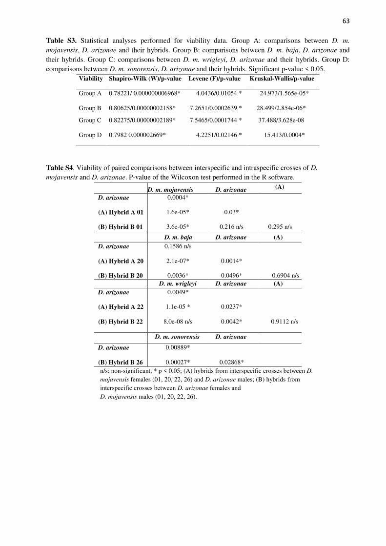
63
Table S3. Statistical analyses performed for viability data. Group A: comparisons between D. m.
mojavensis, D. arizonae and their hybrids. Group B: comparisons between D. m. baja, D. arizonae and their hybrids. Group C: comparisons between D. m. wrigleyi, D. arizonae and their hybrids. Group D: comparisons between D. m. sonorensis, D. arizonae and their hybrids. Significant p-value < 0.05.
Viability Shapiro-Wilk (W)/p-value Levene (F)/p-value Kruskal-Wallis/p-value
Group A 0.78221/ 0.000000006968* 4.0436/0.01054 * 24.973/1.565e-05*
Group B 0.80625/0.00000002158* 7.2651/0.0002639 * 28.499/2.854e-06*
Group C 0.82275/0.00000002189* 7.5465/0.0001744 * 37.488/3.628e-08
Group D 0.7982 0.000002669* 4.2251/0.02146 * 15.413/0.0004*
Table S4. Viability of paired comparisons between interspecific and intraspecific crosses of D.
mojavensis and D. arizonae. P-value of the Wilcoxon test performed in the R software.
D. m. mojavensis D. arizonae (A)
D. arizonae 0.0004*
(A) Hybrid A 01 1.6e-05* 0.03*
(B) Hybrid B 01 3.6e-05* 0.216 n/s 0.295 n/s
D. m. baja D. arizonae (A) D. arizonae 0.1586 n/s
(A) Hybrid A 20 2.1e-07* 0.0014*
(B) Hybrid B 20 0.0036* 0.0496* 0.6904 n/s D. m. wrigleyi D. arizonae (A)
D. arizonae 0.0049*
(A) Hybrid A 22 1.1e-05 * 0.0237*
(B) Hybrid B 22 8.0e-08 n/s 0.0042* 0.9112 n/s
D. m. sonorensis D. arizonae
D. arizonae 0.00889*
(B) Hybrid B 26 0.00027* 0.02868*
n/s: non-significant, * p < 0.05; (A) hybrids from interspecific crosses between D.
mojavensis females (01, 20, 22, 26) and D. arizonae males; (B) hybrids from interspecific crosses between D. arizonae females and D. mojavensis males (01, 20, 22, 26).
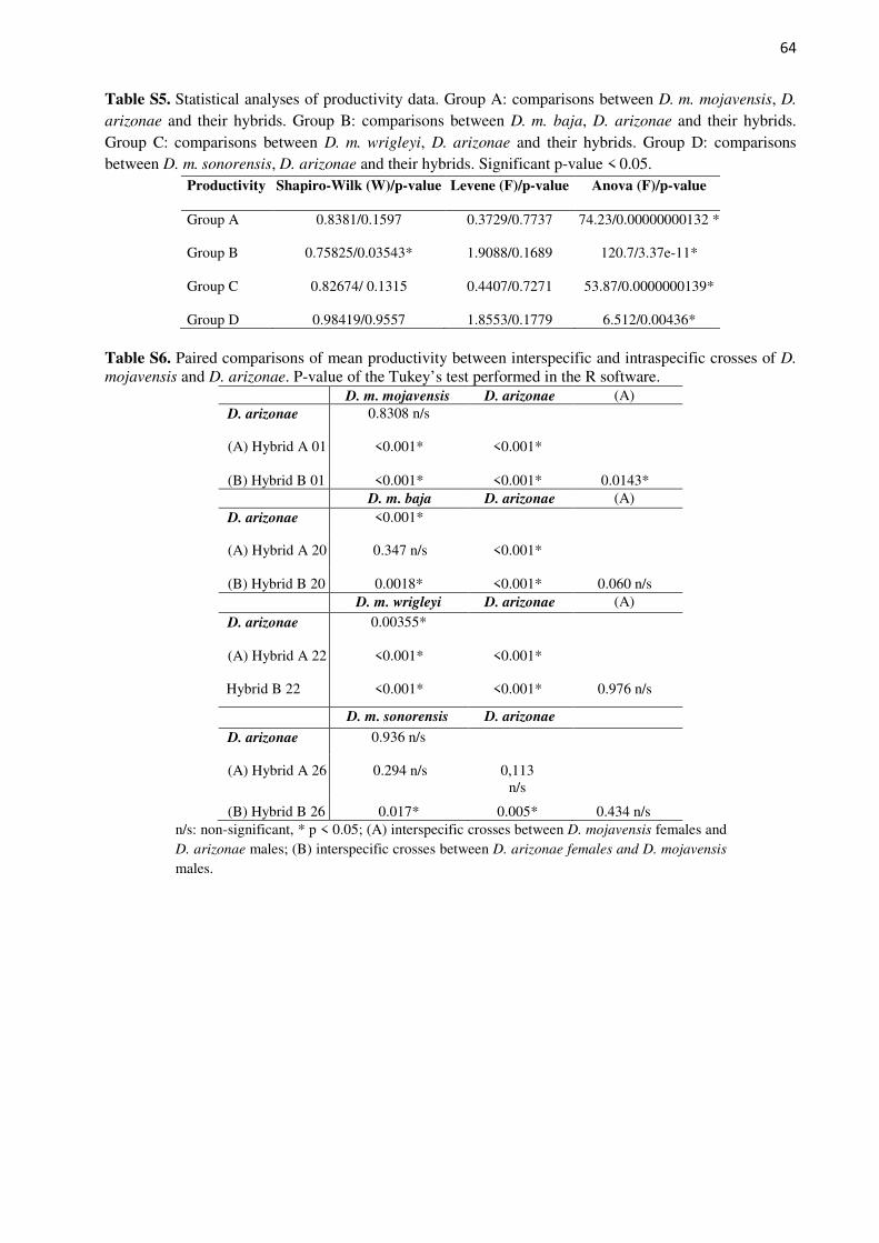
64
Table S5. Statistical analyses of productivity data. Group A: comparisons between D. m. mojavensis, D.
arizonae and their hybrids. Group B: comparisons between D. m. baja, D. arizonae and their hybrids. Group C: comparisons between D. m. wrigleyi, D. arizonae and their hybrids. Group D: comparisons between D. m. sonorensis, D. arizonae and their hybrids. Significant p-value < 0.05.
Productivity Shapiro-Wilk (W)/p-value Levene (F)/p-value Anova (F)/p-value
Group A 0.8381/0.1597 0.3729/0.7737 74.23/0.00000000132 *
Group B 0.75825/0.03543* 1.9088/0.1689 120.7/3.37e-11*
Group C 0.82674/ 0.1315 0.4407/0.7271 53.87/0.0000000139*
Group D 0.98419/0.9557 1.8553/0.1779 6.512/0.00436* Table S6. Paired comparisons of mean productivity between interspecific and intraspecific crosses of D.
mojavensis and D. arizonae. P-value of the Tukey’s test performed in the R software. D. m. mojavensis D. arizonae (A)
D. arizonae 0.8308 n/s
(A) Hybrid A 01 <0.001* <0.001*
(B) Hybrid B 01 <0.001* <0.001* 0.0143* D. m. baja D. arizonae (A)
D. arizonae <0.001*
(A) Hybrid A 20 0.347 n/s <0.001*
(B) Hybrid B 20 0.0018* <0.001* 0.060 n/s D. m. wrigleyi D. arizonae (A)
D. arizonae 0.00355*
(A) Hybrid A 22 <0.001* <0.001*
Hybrid B 22 <0.001* <0.001* 0.976 n/s
D. m. sonorensis D. arizonae
D. arizonae 0.936 n/s
(A) Hybrid A 26 0.294 n/s 0,113 n/s
(B) Hybrid B 26 0.017* 0.005* 0.434 n/s n/s: non-significant, * p < 0.05; (A) interspecific crosses between D. mojavensis females and D. arizonae males; (B) interspecific crosses between D. arizonae females and D. mojavensis
males.
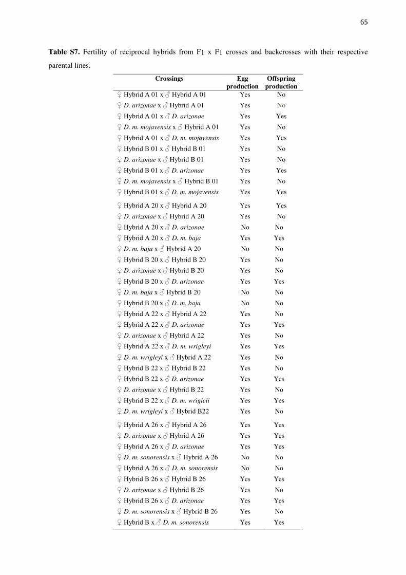
65
Table S7. Fertility of reciprocal hybrids from F1 x F1 crosses and backcrosses with their respective
parental lines.
Crossings Egg production
Offspring production
♀ Hybrid A 01 x ♂ Hybrid A 01 Yes No
♀ D. arizonae x ♂ Hybrid A 01 Yes No
♀ Hybrid A 01 x ♂ D. arizonae Yes Yes
♀ D. m. mojavensis x ♂ Hybrid A 01 Yes No
♀ Hybrid A 01 x ♂ D. m. mojavensis Yes Yes
♀ Hybrid B 01 x ♂ Hybrid B 01 Yes No
♀ D. arizonae x ♂ Hybrid B 01 Yes No
♀ Hybrid B 01 x ♂ D. arizonae Yes Yes
♀ D. m. mojavensis x ♂ Hybrid B 01 Yes No
♀ Hybrid B 01 x ♂ D. m. mojavensis Yes Yes
♀ Hybrid A 20 x ♂ Hybrid A 20 Yes Yes
♀ D. arizonae x ♂ Hybrid A 20 Yes No
♀ Hybrid A 20 x ♂ D. arizonae No No
♀ Hybrid A 20 x ♂ D. m. baja Yes Yes
♀ D. m. baja x ♂ Hybrid A 20 No No
♀ Hybrid B 20 x ♂ Hybrid B 20 Yes No
♀ D. arizonae x ♂ Hybrid B 20 Yes No
♀ Hybrid B 20 x ♂ D. arizonae Yes Yes
♀ D. m. baja x ♂ Hybrid B 20 No No
♀ Hybrid B 20 x ♂ D. m. baja No No
♀ Hybrid A 22 x ♂ Hybrid A 22 Yes No
♀ Hybrid A 22 x ♂ D. arizonae Yes Yes
♀ D. arizonae x ♂ Hybrid A 22 Yes No
♀ Hybrid A 22 x ♂ D. m. wrigleyi Yes Yes
♀ D. m. wrigleyi x ♂ Hybrid A 22 Yes No
♀ Hybrid B 22 x ♂ Hybrid B 22 Yes No
♀ Hybrid B 22 x ♂ D. arizonae Yes Yes
♀ D. arizonae x ♂ Hybrid B 22 Yes No
♀ Hybrid B 22 x ♂ D. m. wrigleii Yes Yes
♀ D. m. wrigleyi x ♂ Hybrid B22 Yes No
♀ Hybrid A 26 x ♂ Hybrid A 26 Yes Yes
♀ D. arizonae x ♂ Hybrid A 26 Yes Yes
♀ Hybrid A 26 x ♂ D. arizonae Yes Yes
♀ D. m. sonorensis x ♂ Hybrid A 26 No No
♀ Hybrid A 26 x ♂ D. m. sonorensis No No
♀ Hybrid B 26 x ♂ Hybrid B 26 Yes Yes
♀ D. arizonae x ♂ Hybrid B 26 Yes No
♀ Hybrid B 26 x ♂ D. arizonae Yes Yes
♀ D. m. sonorensis x ♂ Hybrid B 26 Yes No
♀ Hybrid B x ♂ D. m. sonorensis Yes Yes

66
CAPÍTULO 2
67
4 CAPÍTULO 2
Article - Discoveries
Comparative transcriptomics between Drosophila mojavensis and D. arizonae reveal
underexpression of spermatogenesis-related genes in male hybrids
Cecilia A. Banho,1,2 Vincent Mérel,2 Thiago Y. K. Oliveira,3 Claudia M. A. Carareto,1 and
Cristina Vieira2*
1Department of Biology, UNESP - São Paulo State University, São José do Rio Preto, São Paulo
State (SP), Brazil.
2 Laboratoire de Biométrie et Biologie Evolutive, Université Lyon 1, CNRS, UMR 5558,
Villeurbanne F-69622, France.
3Laboratory of Molecular Immunology, The Rockefeller University, New York, NY, USA.
*Author for Correspondence: Cristina Vieira, Laboratoire de Biométrie et Biologie Evolutive,
Université Lyon 1, CNRS, UMR 5558, Villeurbanne F-69622, France, telephone number +33 6
08 91 58 15, e-mail: [email protected].
Keywords: repleta group, gene expression, hybrid male sterility
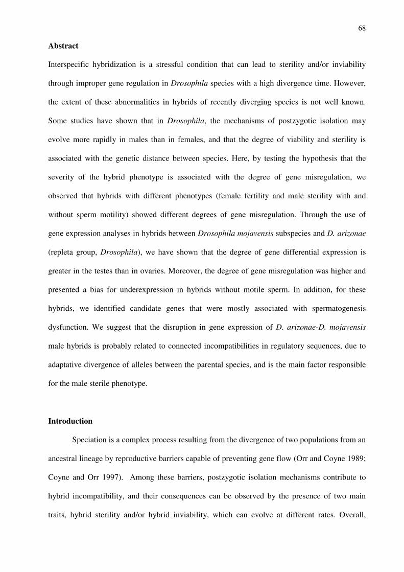
68
Abstract
Interspecific hybridization is a stressful condition that can lead to sterility and/or inviability
through improper gene regulation in Drosophila species with a high divergence time. However,
the extent of these abnormalities in hybrids of recently diverging species is not well known.
Some studies have shown that in Drosophila, the mechanisms of postzygotic isolation may
evolve more rapidly in males than in females, and that the degree of viability and sterility is
associated with the genetic distance between species. Here, by testing the hypothesis that the
severity of the hybrid phenotype is associated with the degree of gene misregulation, we
observed that hybrids with different phenotypes (female fertility and male sterility with and
without sperm motility) showed different degrees of gene misregulation. Through the use of
gene expression analyses in hybrids between Drosophila mojavensis subspecies and D. arizonae
(repleta group, Drosophila), we have shown that the degree of gene differential expression is
greater in the testes than in ovaries. Moreover, the degree of gene misregulation was higher and
presented a bias for underexpression in hybrids without motile sperm. In addition, for these
hybrids, we identified candidate genes that were mostly associated with spermatogenesis
dysfunction. We suggest that the disruption in gene expression of D. arizonae-D. mojavensis
male hybrids is probably related to connected incompatibilities in regulatory sequences, due to
adaptative divergence of alleles between the parental species, and is the main factor responsible
for the male sterile phenotype.
Introduction
Speciation is a complex process resulting from the divergence of two populations from an
ancestral lineage by reproductive barriers capable of preventing gene flow (Orr and Coyne 1989;
Coyne and Orr 1997). Among these barriers, postzygotic isolation mechanisms contribute to
hybrid incompatibility, and their consequences can be observed by the presence of two main
traits, hybrid sterility and/or hybrid inviability, which can evolve at different rates. Overall,
69
hybrid sterility evolves fasterthan hybrid inviability, mainly when considering the different
sexes, since these barriers evolve faster in males than in females (Turissini et al. 2018). Indeed,
several studies considering intraspecific and interspecific hybridization have shown that the
germline is primarily affected, and sterility is often detected (Ruiz et al. 1990; Ranz et al. 2004;
Moerhring et al. 2007; Llopart 2012; Brill et al. 2016; Hill et al. 2016; Li et al. 2016).Although
the process of speciation has been widely studied, the causes of postzygotic incompatibility in
hybrids are not fully understood. According to the model of Dobzhansky–Muller (1936, 1942),
genetic variation could lead to deleterious epistatic interactions, disrupting regulatory networks
and leading to serious consequences in the hybrids (Orr and Coyne 1989; McManus et al. 2010;
Kelleher et al. 2012; Romero-Soriano et al. 2017). In addition, according to Haldane’s rule
(Haldane 1922), sterility in hybrids is more likely to affect the heterogametic sex, and the degree
and extent of these genetic incompatibilities are related to the time of divergence and are likely
the result of divergent regulatory sequences (McManus et al. 2010; Kelleher et al. 2012;
Ryazansky et al. 2014; Llopart et al. 2018). Many studies have searched for genes involved with
postzygotic isolation, particularly in Drosophila. So far, few genes that may play a role in hybrid
incompatibility have been identified. Among these genes, Odisseus (OdsH), which is located on
the X-chromosome and encodes a transcription factor, has been found to be one of the
responsible factors contributing to hybrid sterility between Drosophila simulans and D.
mauritiana (Ting et al. 1998). In addition, the genes Hybrid male-rescue (Hmr) and Lethal male-
rescue (Lmr) are known to be related to hybrid unviability in D. melanogaster-D. simulans.
These genes encode centromeric proteins, and the interaction of the alleles of D. melanogaster
Hmr with D. simulans Lmr leads to hybrid breakdown (Barbash et al. 2004; Barbash and Lorigan
2007; Satyaki et al. 2014). Nup96, a gene located on the X chromosome that encodes a nuclear
pore protein, also seems to contribute to D. melanogaster-D. simulans hybrid unviability, likely
due the deleterious interaction between the alleles D. simulans Nup96 and D. melanogaster
Nup153, located on the X chromosome (Presgraves et al. 2003; Tang and Presgraves 2015).
70
Overdrive (Ovd) is another gene responsible for male sterility and segregation distortion in F1
hybrids of D. p. pseudoobscura and D. p. bogotana by epistatic X-autosomal interactions
(Phadnis and Orr, 2009; Phadnis 2011). Moreover, multiple studies have shown that in sterile
hybrids, several differentially expressed genes (DEG) have a bias for underexpression (Michalak
and Noor 2003; Haerty and Singh 2006) and function in spermatogenesis, such as Acyp,
spermatocyte arrest, bag of marbles, always early, twine, cookie monster, meiosis I arrest,
janusB, gonadal and don juan (White-Cooper et al. 1998; Jiang and White-Cooper 2003;
Michalak and Noor 2003, 2004; Ma and Michalak 2011; Gomes and Civetta 2014).
Most of the hybrid incompatibility genes identified so far seems to be species-specific,
indicating that hybrid incompatibility is a complex polygenic trait that has different epistatic
effects related to the rapid accumulation of genetic changes over time. Studies of recently
diverged species presenting different phenotypes regarding the postzygotic isolation mechanisms
can help to clarify early disruptions in gene regulation and expression that may influence the
speciation process. Drosophila mojavensis and D. arizonae are appropriate for such a study.
They are sibling species, recently diverged (~1.5 mya, Sanches-Flores et al. 2016), and widely
used in speciation studies due to their ability to produce hybrids in the laboratory; however,
introgression evidence has not yet been found in nature despite of the favourable ecological
conditions for hybridization, mainly in sympatric populations (Jennings and Etges 2009). D.
mojavensis is composed of four ecologically distinct subspecies, which are distributed in four
different geographic regions, and each one uses a specific host cactus as a feeding and breeding
site, with no evidence of gene flow, constituting well-structured populations (Knowles and
Markow 2001; Reed and Markow 2004; Massie and Markow 2005; Reed et al. 2007; Etges et al.
2010). In crosses between D. mojavensis subspecies and D. arizonae, hybrids are produced in
both directions. Nevertheless, they present incomplete and asymmetric postzygotic isolation,
since in crosses with D. mojavensis females the sterility of the male hybrids is variable, whereas
in crosses with D. arizonae females the male hybrids are always sterile because they do not have
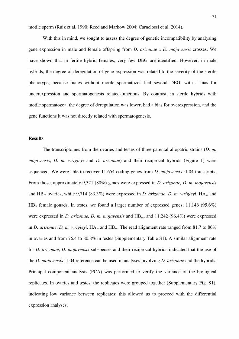
71
motile sperm (Ruiz et al. 1990; Reed and Markow 2004; Carnelossi et al. 2014).
With this in mind, we sought to assess the degree of genetic incompatibility by analysing
gene expression in male and female offspring from D. arizonae x D. mojavensis crosses. We
have shown that in fertile hybrid females, very few DEG are identified. However, in male
hybrids, the degree of deregulation of gene expression was related to the severity of the sterile
phenotype, because males without motile spermatozoa had several DEG, with a bias for
underexpression and spermatogenesis related-functions. By contrast, in sterile hybrids with
motile spermatozoa, the degree of deregulation was lower, had a bias for overexpression, and the
gene functions it was not directly related with spermatogenesis.
Results
The transcriptomes from the ovaries and testes of three parental allopatric strains (D. m.
mojavensis, D. m. wrigleyi and D. arizonae) and their reciprocal hybrids (Figure 1) were
sequenced. We were able to recover 11,654 coding genes from D. mojavensis r1.04 transcripts.
From those, approximately 9,321 (80%) genes were expressed in D. arizonae, D. m. mojavensis
and HBm ovaries, while 9,714 (83.3%) were expressed in D. arizonae, D. m. wrigleyi, HAw and
HBw female gonads. In testes, we found a larger number of expressed genes; 11,146 (95.6%)
were expressed in D. arizonae, D. m. mojavensis and HBm, and 11,242 (96.4%) were expressed
in D. arizonae, D. m. wrigleyi, HAw and HBw. The read alignment rate ranged from 81.7 to 86%
in ovaries and from 76.4 to 80.8% in testes (Supplementary Table S1). A similar alignment rate
for D. arizonae, D. mojavensis subspecies and their reciprocal hybrids indicated that the use of
the D. mojavensis r1.04 reference can be used in analyses involving D. arizonae and the hybrids.
Principal component analysis (PCA) was performed to verify the variance of the biological
replicates. In ovaries and testes, the replicates were grouped together (Supplementary Fig. S1),
indicating low variance between replicates; this allowed us to proceed with the differential
expression analyses.
72
Fig. 1. Crosses performed between D. arizonae and D. mojavensis subspecies and
differential gene expression between parental lines and hybrids. Geographic distribution of
D. m. wrigleyi, D. m. mojavensis and D. arizonae lines used in this study and their respective
cross direction with offspring identification. HAw and HBw are offspring from crosses between
D. arizonae and D. m. wrigleyi, HBm are hybrids from crosses between D. arizonae and D. m.
mojavensis. A and B represent the cross direction (A = crosses with D. mojavensis mothers; B =
crosses with D. arizonae mothers).
Differential expression of genes in D. arizonae and D. mojavensis gonads
We compared the transcriptomes of D. arizonae and D. m. mojavensis and D. arizonae
and D. m. wrigleyi and we identified 501 (5.3%) and 621 (6.3%) DEG in ovaries, respectively,
with the majority of the genes overexpressed in D. arizonae (Figure 2A). In testes, there was a
higher number of DEG, which reached 16% when comparing D. arizonae with each D.
mojavensis subspecies; however, there was a similar distribution between the over- and
underexpressed genes (Fig. 2E). Gene ontology (GO) analyses of DEG betweem parental lines
showed enrichment in the ovaries of genes related to response to stimulus, multicellular
organismal process, multicellular organismal development, develomental process, cellular
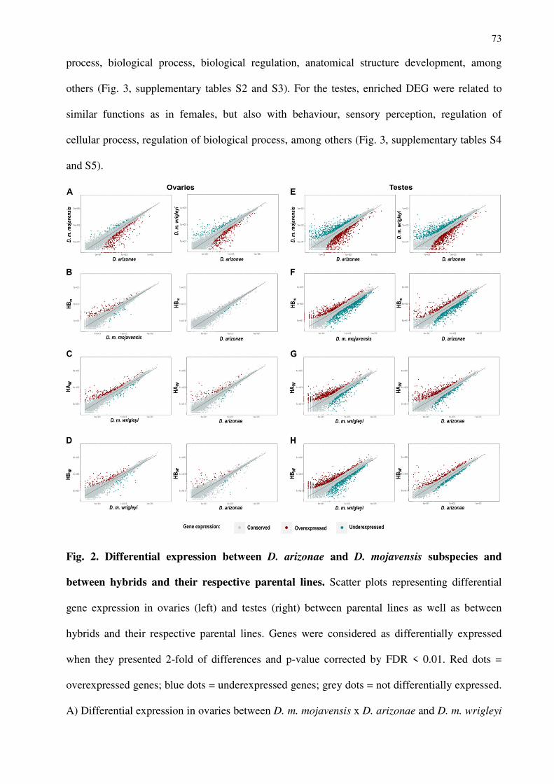
73
process, biological process, biological regulation, anatomical structure development, among
others (Fig. 3, supplementary tables S2 and S3). For the testes, enriched DEG were related to
similar functions as in females, but also with behaviour, sensory perception, regulation of
cellular process, regulation of biological process, among others (Fig. 3, supplementary tables S4
and S5).
Fig. 2. Differential expression between D. arizonae and D. mojavensis subspecies and
between hybrids and their respective parental lines. Scatter plots representing differential
gene expression in ovaries (left) and testes (right) between parental lines as well as between
hybrids and their respective parental lines. Genes were considered as differentially expressed
when they presented 2-fold of differences and p-value corrected by FDR < 0.01. Red dots =
overexpressed genes; blue dots = underexpressed genes; grey dots = not differentially expressed.
A) Differential expression in ovaries between D. m. mojavensis x D. arizonae and D. m. wrigleyi
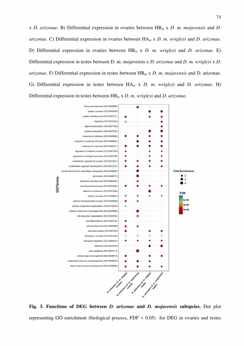
74
x D. arizonae. B) Differential expression in ovaries between HBm x D. m. mojavensis and D.
arizonae. C) Differential expression in ovaries between HAw x D. m. wrigleyi and D. arizonae.
D) Differential expression in ovaries between HBw x D. m. wrigleyi and D. arizonae. E)
Differential expression in testes between D. m. mojavensis x D. arizonae and D. m. wrigleyi x D.
arizonae. F) Differential expression in testes between HBm x D. m. mojavensis and D. arizonae.
G) Differential expression in testes between HAw x D. m. wrigleyi and D. arizonae. H)
Differential expression in testes between HBw x D. m. wrigleyi and D. arizonae.
Fig. 3. Functions of DEG between D. arizonae and D. mojavensis subspcies. Dot plot
representing GO enrichment (biological process, FDF < 0.05) for DEG in ovaries and testes
75
between D. arizonae and D. mojavensis subspecies (FDR < 0.01, FC >2).
Gene expression in the hybrid female gonads is very similar to the parental lines
We compared the expression of hybrids to their parental lines. The expression profile of
the HBm ovaries revealed that 1.93% of DEG between the hybrid and D. m. mojavensis, in
contrast to the comparison with D. arizonae, which showed almost no differences (0.26%) (Fig.
2B, supplementary table S6). Gene expression in HAw and HBw ovaries showed a larger number
of DEG, mostly overexpressed in the hybrids when compared to D. m. wrigleyi (4.2% and 2.7%,
respectively), regardless of the direction of the crossing (Fig. 2C and D, suplementary table S7).
To identify the genes that were over- or underexpressed relative to both parental lines in
all crosses, we filtered those that presented a greater than 2-fold difference and significant
adjusted p-values (< 0.01) in HBm, HAw and HBw ovaries. This reduced greatly the number of
genes. The HBm ovaries presented no overexpressed genes and only three underexpressed genes.
These included FBgn014602, which corresponds to alpha-Est5 and has carboxylic ester
hydrolase activity, FBgn028050, corresponding to Maverick gene, which has a role in signalling
pathways, the regulation of neuromuscular junctions, dendrite development and imaginal disc-
derived wing size, and FBgn0146651, which has an unknown function (From Flybase database,
available at: http://flybase.org). By contrast, HAw ovaries showed 16 overexpressed genes
(supplementary table S8), however no significant enrichment was found due to the few number
of DEG, and none underexpressed genes was found in relation to both parental lines. The HBw
ovaries showed 13 overexpressed genes (supplementary table S9) and only one underexpressed
gene (FBgn0135298, with unknown function) when compared with both parental lines.
Additionaly, HAw and HBw ovaries did not show any common overexpressed genes.
Gene expression in hybrid male germline is divergent from the parental lines
Compared to ovaries, hybrid testes presented a larger number of DEG in comparison with
76
the parental lines. HBm, the offspring from crosses with D. arizonae females, displayed 11.9%
and 16% of DEG compared with D. arizonae and D. m. mojavensis, respectively, and most of
them were underexpressed (Fig. 2F, supplementary table S6). In HAw testes (offspring from D.
mojavensis females), 7.9% and 9.7% DEG compared with D. arizonae and D. m. wrigleyi were
found, respectively, and presented a bias for overexpression (Fig. 2G, supplementary table S7).
Similarly, hybrids from reciprocal cross (HBw) also exhibited a larger number of DEG compared
with D. m. wrigleyi than when compared with D. arizonae. However, in these hybrids, as was
observed for HBm, most them were underexpressed (Fig. 2H, supplementary table S7).
Regarding the deregulated genes in HAw testes, 130 were overexpressed and 60
underexpressed compared with both parental species (supplementary tables S10 and S11), while
in HBw testes 79 and 172 genes were commonly over- and underexpressed, respectively,
compared with D. arizonae and D. m. wrigleyi (supplementary tables S12 and S13). As with
HBw, the common deregulated genes in the testes of HBm compared to the parental species also
showed an underexpression bias, as 57 genes were overexpressed and 519 were underexpressed
(supplementary tables S14 and S15).
Spermatogenesis-related functions of DEG in testes
In the male gonads, the deregulated gene expression pattern differed depending on the
direction of crossing. In HAw testes, from the 190 deregulated genes, 160 were orthologous to D.
melanogaster, allowing us to find their functions. Most of the DEG were significantly enriched
for functions associated with plasma membrane (Fig. 4). On the other hand, in HBw testes, 251
genes were deregulated, and the D. melanogaster orthologues were recovered for 185 of them.
From enrichment analysis we detected that, unlike HAw, the function of most of these genes are
related with axoneme, cilium, dynein and microtubule (Fig. 4). In HBm testes, 598 genes were
identified as differentially expressed, and 462 had a D. melanogaster orthologue. In these
hybrids, most of the over- and underexpressed genes were enriched for some functions similar to
77
HAw and HBw (Fig. 4).
Globally, hybrid testes presented a larger number of deregulated genes. However, the
over- and underexpressed gene ratio depended on the direction of crossing. HAw testes showed a
larger number of overexpressed genes, while HBw and HBm exhibited a larger number of
underexpressed genes. We searched for shared deregulated genes in the different hybrids in both
gonad tissues and found 31 overexpressed and 8 underexpressed genes shared in the HAw and
HBw males (suplementary table S16).
Comparisons of HBm and HBw male gonads, corresponding to hybrids from crosses of
different D. mojavensis subspecies, but in the same direction, showed 31 shared overexpressed
genes (suplementary table S17) and 146 underexpressed genes (suplementary table S18).
Interestingly, many of these genes exhibited enrichment for spermatogenesis-related functions,
as cilium movement (Fig. 3, Table 1), which are linked to the hybrid sterile phenotype. Looking
specifically these genes it is remarkable that some of them might have a direct role in
reproduction, since they participate in male meiosis, male courtship, mating behaviour, as well as
playing a role in gene silencing and pre-miRNA processing (Table 1). Additionally, for many of
these shared DEG genes, the mutant phenotype already described for other Drosophila
corresponds to male sterility (Table 1). To test our hypothesis that genes with specific expression
in testes would show signatures of positive selection, as already reported in some studies
(Meiklejohn et al. 2003; Artieri and Haerty 2007, Llopart 2012) we calculated Ka/Ks for a set of
DEG testis-specific. However, no signatures of positive selection were observed in these genes,
although several of them present evidence of having undergone relaxed selection.
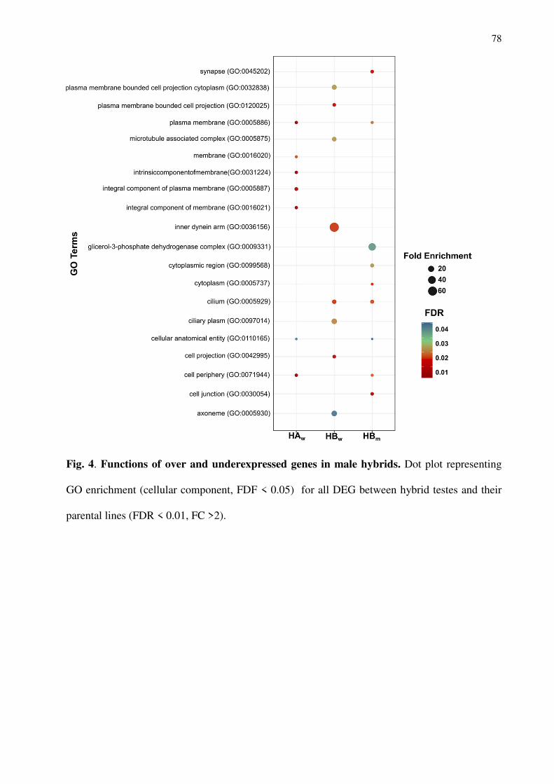
78
Fig. 4. Functions of over and underexpressed genes in male hybrids. Dot plot representing
GO enrichment (cellular component, FDF < 0.05) for all DEG between hybrid testes and their
parental lines (FDR < 0.01, FC >2).
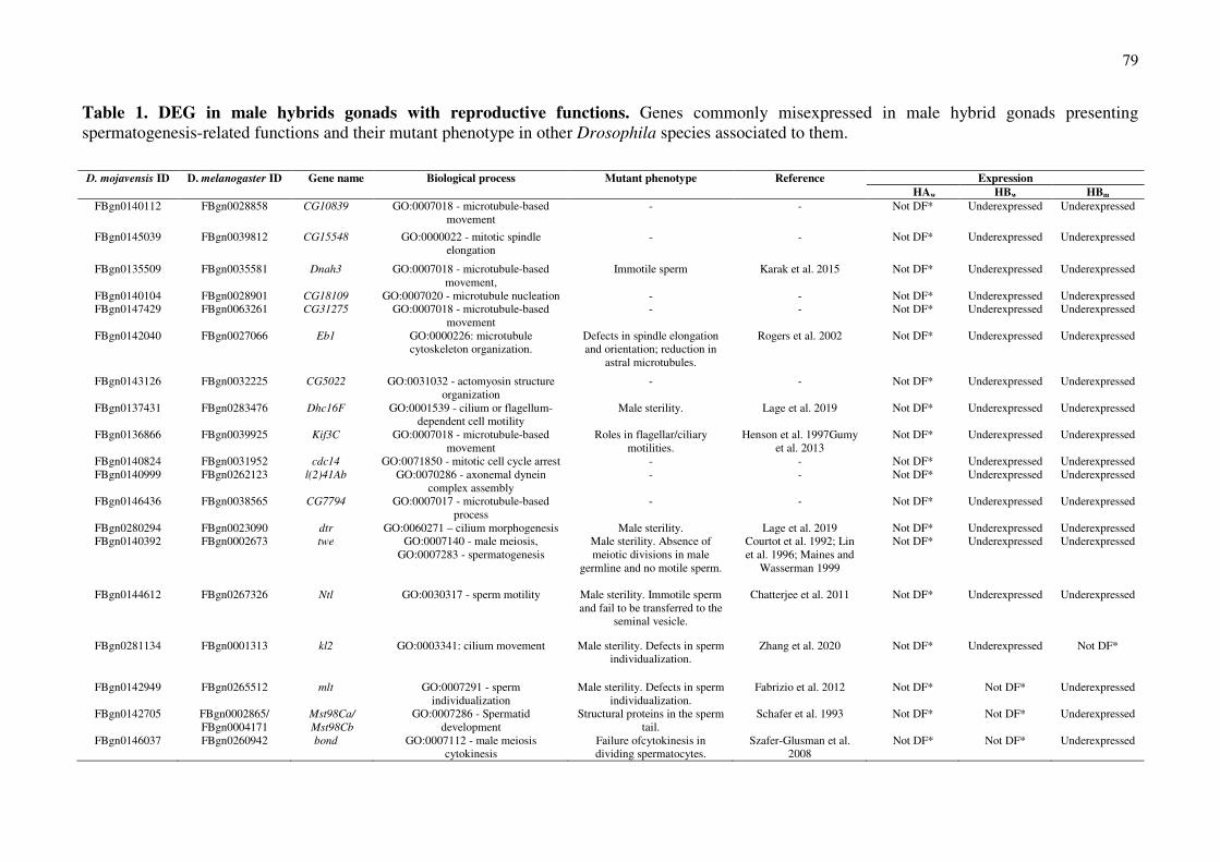
79
Table 1. DEG in male hybrids gonads with reproductive functions. Genes commonly misexpressed in male hybrid gonads presenting spermatogenesis-related functions and their mutant phenotype in other Drosophila species associated to them.
D. mojavensis ID D. melanogaster ID Gene name Biological process Mutant phenotype Reference Expression HAw HBw HBm
FBgn0140112 FBgn0028858 CG10839 GO:0007018 - microtubule-based movement
- - Not DF* Underexpressed Underexpressed
FBgn0145039 FBgn0039812 CG15548 GO:0000022 - mitotic spindle elongation
- - Not DF* Underexpressed Underexpressed
FBgn0135509 FBgn0035581 Dnah3 GO:0007018 - microtubule-based movement,
Immotile sperm Karak et al. 2015 Not DF* Underexpressed Underexpressed
FBgn0140104 FBgn0028901 CG18109 GO:0007020 - microtubule nucleation - - Not DF* Underexpressed Underexpressed FBgn0147429 FBgn0063261 CG31275 GO:0007018 - microtubule-based
movement - - Not DF* Underexpressed Underexpressed
FBgn0142040 FBgn0027066 Eb1 GO:0000226: microtubule cytoskeleton organization.
Defects in spindle elongation and orientation; reduction in
astral microtubules.
Rogers et al. 2002 Not DF* Underexpressed Underexpressed
FBgn0143126 FBgn0032225 CG5022 GO:0031032 - actomyosin structure organization
- - Not DF* Underexpressed Underexpressed
FBgn0137431 FBgn0283476 Dhc16F GO:0001539 - cilium or flagellum-dependent cell motility
Male sterility. Lage et al. 2019 Not DF* Underexpressed Underexpressed
FBgn0136866 FBgn0039925 Kif3C GO:0007018 - microtubule-based movement
Roles in flagellar/ciliary motilities.
Henson et al. 1997Gumy et al. 2013
Not DF* Underexpressed Underexpressed
FBgn0140824 FBgn0031952 cdc14 GO:0071850 - mitotic cell cycle arrest - - Not DF* Underexpressed Underexpressed FBgn0140999 FBgn0262123 l(2)41Ab GO:0070286 - axonemal dynein
complex assembly - - Not DF* Underexpressed Underexpressed
FBgn0146436 FBgn0038565 CG7794 GO:0007017 - microtubule-based process
- - Not DF* Underexpressed Underexpressed
FBgn0280294 FBgn0023090 dtr GO:0060271 – cilium morphogenesis Male sterility. Lage et al. 2019 Not DF* Underexpressed Underexpressed FBgn0140392 FBgn0002673 twe GO:0007140 - male meiosis,
GO:0007283 - spermatogenesis Male sterility. Absence of meiotic divisions in male
germline and no motile sperm.
Courtot et al. 1992; Lin et al. 1996; Maines and
Wasserman 1999
Not DF* Underexpressed Underexpressed
FBgn0144612 FBgn0267326 Ntl GO:0030317 - sperm motility Male sterility. Immotile sperm and fail to be transferred to the
seminal vesicle.
Chatterjee et al. 2011 Not DF* Underexpressed Underexpressed
FBgn0281134 FBgn0001313 kl2 GO:0003341: cilium movement Male sterility. Defects in sperm individualization.
Zhang et al. 2020 Not DF* Underexpressed Not DF*
FBgn0142949 FBgn0265512 mlt GO:0007291 - sperm individualization
Male sterility. Defects in sperm individualization.
Fabrizio et al. 2012 Not DF* Not DF* Underexpressed
FBgn0142705 FBgn0002865/ FBgn0004171
Mst98Ca/
Mst98Cb
GO:0007286 - Spermatid development
Structural proteins in the sperm tail.
Schafer et al. 1993 Not DF* Not DF* Underexpressed
FBgn0146037 FBgn0260942 bond GO:0007112 - male meiosis cytokinesis
Failure ofcytokinesis in dividing spermatocytes.
Szafer-Glusman et al. 2008
Not DF* Not DF* Underexpressed
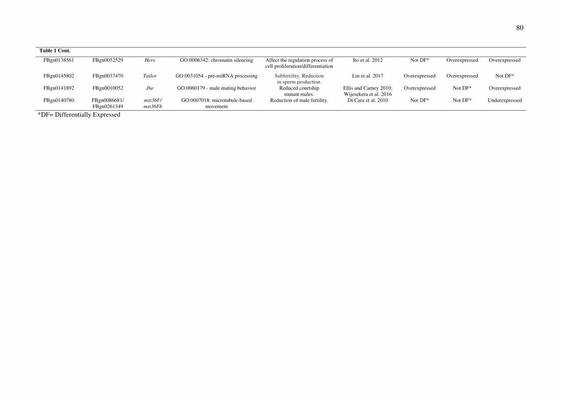
80
*DF= Differentially Expressed
Table 1 Cont.
FBgn0138561 FBgn0052529 Hers GO:0006342: chromatin silencing Affect the regulation process of cell proliferation/differentiation
Ito et al. 2012 Not DF* Overexpressed Overexpressed
FBgn0145802 FBgn0037470 Tailor GO:0031054 - pre-miRNA processing Subfertility. Reduction in sperm production.
Lin et al. 2017 Overexpressed Overexpressed Not DF*
FBgn0141892 FBgn0010052 Jhe GO:0060179 - male mating behavior Reduced courtship mutant males.
Ellis and Carney 2010; Wijesekera et al. 2016
Overexpressed Not DF* Overexpressed
FBgn0140780 FBgn0086681/ FBgn0261349
mst36F/
mst36Fb
GO:0007018: microtubule-based movement
Reduction of male fertility. Di Cara et al. 2010 Not DF* Not DF* Underexpressed
81
Inheritance of gene expression
We compared the level of gene expression of HBm, HAw and HBw with each parental
line, following the six inheritance categories of McManus et al. (2010). Most of the genes in the
ovaries and testes of the interspecific hybrids showed conserved expression (Fig. 5;
suplementary tables S19 and S20).
HBm showed conserved expression for 97.8% of the genes in ovaries and for 78.5% in
testes (Fig. 5; suplementary table S19). Few genes were classified having additive expression in
the female and male gonads (0.05 and 1.45%, respectively). In the dominant categories, HBm
exhibited an overrepresentation of genes with D. arizonae-like expression (1.8%, in ovaries and
9.5% in testes), and few genes were D. mojavensis-dominant (Fig. 5; suplementary table S19). In
the over-dominant and under-dominant categories, no genes were found for ovaries (0.03% of
DEG). However, the testes showed several genes in these categories, with 0.5% over-dominant
and 4.6% under-dominant (Fig. 5; suplementary table S19).
Similar to HBm, most of the expressed genes in HAw and HBw female and male gonads
had conserved expression, but in testes the conserved inheritance was lower than that in ovaries
(Fig. 5; suplementary table S20). Additionally, very few genes in ovaries of HAw and HBw (0.3
and 0.22%) and testes (1.1 and 1.2%) displayed additive inheritance expression (Fig. 5;
suplementary table S20), as it was observed for HBm. An interesting finding is that most of the
genes classified in dominant categories displayed a D. arizonae-like pattern of expression,
regardless of crossing direction or gonad tissue (Fig. 5; suplementary table S20). Considering the
over-dominant and under-dominant categories, few genes were in these categories in ovaries
(0.16% in HAw and 0.13% in HBw) (Fig. 5; suplementary table S20). In hybrid testes, however, a
large number of genes were classified as over- (1.1% in HAw and 0.7% in HBw) or under-
dominant (0.53% in HAw and 1.5% in HBw) (Fig. 5; suplementary table S20).
Comparing all inheritance categories of HAw and HBw, signficative differences in the
expression profile of reciprocal hybrids were observed in ovaries (X2 = 146.67, P <0.001), which
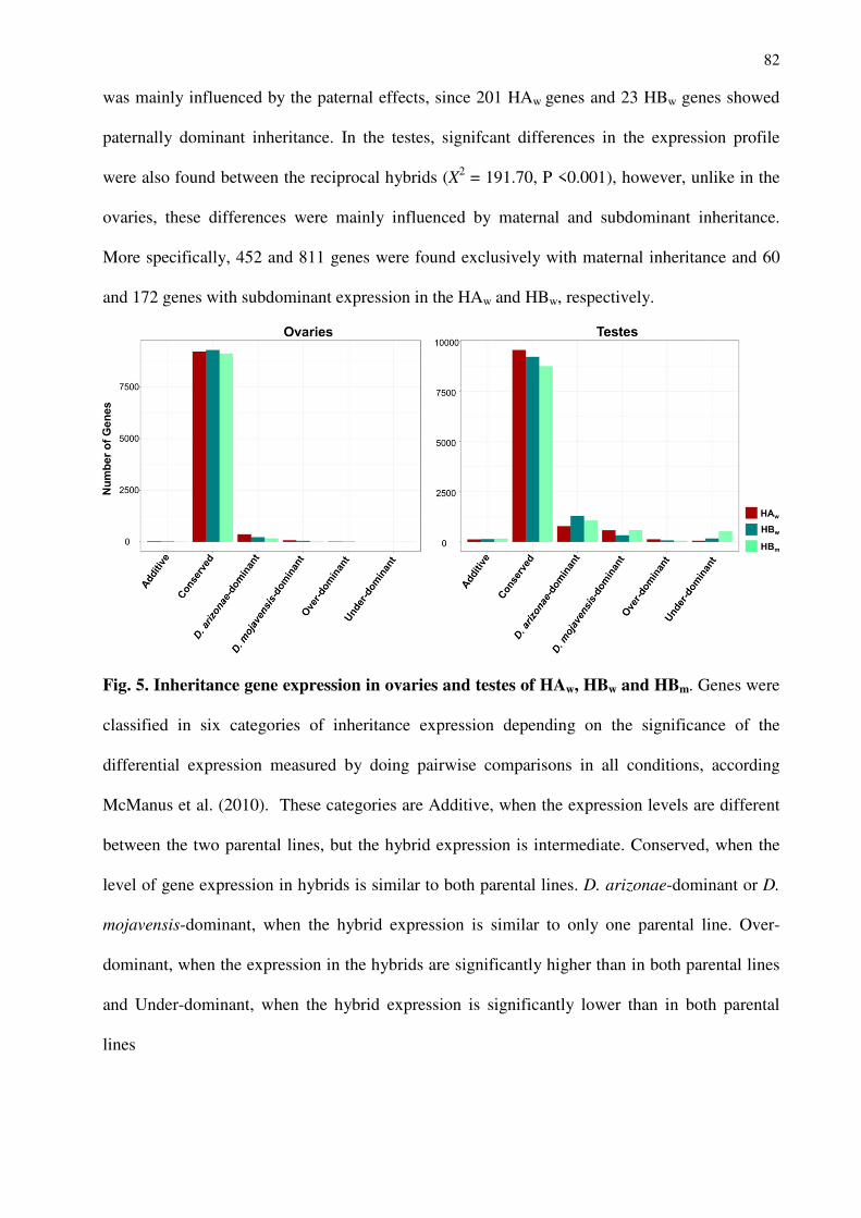
82
was mainly influenced by the paternal effects, since 201 HAw genes and 23 HBw genes showed
paternally dominant inheritance. In the testes, signifcant differences in the expression profile
were also found between the reciprocal hybrids (X2 = 191.70, P <0.001), however, unlike in the
ovaries, these differences were mainly influenced by maternal and subdominant inheritance.
More specifically, 452 and 811 genes were found exclusively with maternal inheritance and 60
and 172 genes with subdominant expression in the HAw and HBw, respectively.
Fig. 5. Inheritance gene expression in ovaries and testes of HAw, HBw and HBm. Genes were
classified in six categories of inheritance expression depending on the significance of the
differential expression measured by doing pairwise comparisons in all conditions, according
McManus et al. (2010). These categories are Additive, when the expression levels are different
between the two parental lines, but the hybrid expression is intermediate. Conserved, when the
level of gene expression in hybrids is similar to both parental lines. D. arizonae-dominant or D.
mojavensis-dominant, when the hybrid expression is similar to only one parental line. Over-
dominant, when the expression in the hybrids are significantly higher than in both parental lines
and Under-dominant, when the hybrid expression is significantly lower than in both parental
lines
83
Discussion
In our study, the target species D. mojavensis and D. arizonae showed approximately 6%
DEG in the ovaries and approximately 16% in the testes, agreeing with our previous findings of
hybrid female transcriptomes (Lopez-Maestre et al. 2017) and with results obtained for male
hybrid gonads (Llopart 2012). The divergence time of the studied species appeared to influence
the rate of DEG, since D. arizonae and D. mojavensis presented levels of differential expression
in the range of what was previously shown for other Drosophila species. It was reported that the
pairs D. yakuba and D. melanogaster (~ 6.1 mya divergence time, Russo et al. 1995), D.
melanogaster and D. simulans (~1.2 to ~5 mya divergence time, Russo et al. 1995; Kliman et al.
2000; Tamura et al. 2004; Cutter 2008), and D. melanogaster and D. sechellia (~1.2 to ~5 mya
divergence time, Russo et al. 1995; Kliman et al. 2000; Tamura et al. 2004; Cutter 2008)
exhibited 29-2%, 18-24 (Rifkin et al. 2003) and 78% DEG (McManus et al. 2010), respectively
In more recently diverged species, fewer DEG are observed, sinceIn males of D. yakuba and D.
santomea (~0.4 mya, Caricou et al. 2001) and D. p. bogotana and D. p. pseudoobscura (~0.25
mya, Wang et al. 1997), 19% (Llopart 2012) and 14.6% (Gomes and Civetta 2015) DEG were
found, respectively.
The differences in the number of DEG between ovaries and testes could be explained by
two main factors. First, there is a larger number of genes expressed in the male germline than
that in the ovaries. Second, there is faster evolution for male-specific genes, such as male-biased
genes, which has been previously reported in other Drosophila species. A previous study found
that 83% of DEG in hybrids of D. melanogaster and D. simulans showed a sex-specific pattern,
with a greater differential expression in males, indicating that some specific genes can evolve
faster than others (Ranz et al. 2003). Moreover, other studies have shown that during the
divergence process, male-biased genes display higher evolutionary rates, driven by positive
selection (Artieri and Haerty 2007), and most of them are preferentially expressed in gonad
tissues (Meiklejohn et al. 2003; Civetta et al. 2006; Haerty and Singh et al. 2006). Additionally,
84
gene ontology analyses showed that in testes, several DEG were related to mating and courtship
behaviours, indicating that prezygotic reproductive barriers evolve faster among species, as was
observed by Turissini et al. (2018).
Therefore, as was observed for other Drosophila species, the number of differentially
expressed genes found in both gonad tissues of D. arizonae and D. mojavensis could be related
to the divergence time, and hence, might be driven by regulatory differences, as was previously
reported in drosophilids (McManus et al. 2010). The higher deregulation observed in the testes
of D. arizonae and D. mojavensis subspecies can indicate that male-biased genes evolve faster
than female-biased genes or non-sexual genes, agreeing with the faster-male hypothesis (Wu and
Davis 1993) and with other results previously reported for Drosophila (Meiklejohn et al. 2003;
Artieri and Haerty 2007; Llopart et al. 2018).
Differential expression in hybrid ovaries
Female hybrid gonads exhibited few DEG when compared with both parental lines, and
most of them presented overexpression. Previous studies in hybrids of D. arizonae-D. m.
mojavensis (Lopez-Maestre et al. 2017) and D. melanogaster-D. simulans (Kelleher et al. 2012)
showed similar patterns. However, this result was not observed in D. buzzatii and D. koepferae
(~4.49 mya) female hybrids, since no bias for over- or underexpression was observed (Romero-
Soriano et al. 2017). We suggest that in hybrid ovaries from recently diverged species, the DEG
tend to be overexpressed, but over time the global deregulation will increase due to the
accumulation of genetic changes, and the number of over- and underexpressed genes will be
symmetric.
Overall, no massive deregulation was observed in hybrid ovaries, since few genes were
over- or underexpressed compared with the parental lines. One explanation for this finding is the
higher stability of the female genome, which shows a slower evolution of gene expression,
mainly for female-biased genes, as was reported in other Drosophila species (Meiklejohn et al.

85
2003; Llopart et al. 2018). This could have an important role in the fertile phenotype observed in
these hybrids.
Differential expression in hybrid testes
Overall, male gonads showed a higher proportion of DEG compared with ovaries in all
hybrids analysed. Moreover, hybrids from different crossing directions presented very different
numbers of DEG compared with one parental line, agreeing with results obtained with hybrid
males of D. simulans–D. mauritiana (Mohering et al. 2007) and of fire ants (Ommeto et al.
(2012). Thus, in D. mojavensis x D. arizonae male hybrids, one specific parental line could
display a greater dominance of genetic elements, leading to higher divergence of the expression
profile.
In general, hybrids from different crossing directions exhibited distinct proportions of
over- or underexpressed genes. HAw showed twice as many overexpressed genes than
underexpressed genes in comparison to parental lines, while HBw exhibited twice as many
underexpressed genes than overexpressed genes. Similarly, HBm exhibited nine times more
underexpressed genes than overexpressed genes. Two main factors could influence these
differences in the over- and underexpressed genes regarding the crossing direction. These
hybrids differ only in a few genetic characteristics, such as the origin of the X chromosome and
the genetic background; thus, it is possible that some specific epistatic factors among sexual
chromosomes and autosomes play a role in the differential expression profile, as was found by
Reed et al. (2008). Moreover, there is evidence that in hybrids of D. arizonae (females) x D.
mojavensis (males) crosses, type HBm in this study, the immotile sperm observed can be an
outcome from interactions between the D. mojavensis Y chromosome and the 3rd autosome or X
chromosome from D. arizonae (Vigneault and Zouros 1986). Regarding the reciprocal cross, it
was previously shown that incompatibilities leading to hybrid sterility were related to
interactions between the Y chromosome of D. arizonae and the 4th autosome of D. mojavensis
86
(Pantazidis and Zouros 1988). In addition, in previous analyses of sperm motility and fertility
that we have performed (data not shown), we found that HBw and HBm have no motile sperm
and are therefore sterile. On the other hand, the sperm of HAw was motile, although no progeny
were produced in F1xF1 crosses or backcrosses; therefore, they were also sterile indicating that
the disruption of spermatogenesis was weaker than in HBw and HBm males. These findings are in
agreement with Hardy et al. (2011), who observed that D. arizonae-D. m. mojavensis reciprocal
hybrids display abnormal spermatid development. However, the disruption of spermatid
formation is lower in hybrids carrying D. mojavensis X chromosomes, as they exhibit sperm
with normal tails; in contrast, hybrids with D. arizonae X chromosomes do not have
individualized spermatids and their tails are severely disrupted. Considering these results, it can
be suggested that the level of gene over- or underexpression can be related to the degree of
spermatogenesis disturbance in recently diverged species. Consistent with these findings, it was
reported that in male hybrids between D. santomea x D. yakuba presented less severe sterility
(Moehring et al. 2006) and exhibited more overexpression of male-biased genes, than those
presenting greater severity in sterility (Moehring et al. 2006) which showed more deregulated
genes that were underexpressed (Llopart 2012), as it was observed for HBm and HBw.
Although a large number of misexpressed genes is probably related to the degree of the
sterile phenotype, other factors can also influence this feature. Many studies have shown that
genes involved in spermatogenesis are usually underexpressed in hybrids and display rapid
sequence divergence driven by positive selection (Ranz et al. 2003; Michalak and Noor 2003;
Haerty and Singh 2006; Moehring et al. 2007; Llopart 2012; Llopart et al. 2018). In agreement
with these findings, one study observed that the substitution rate in male-biased DEG of hybrids
was higher than that in non-sexual DEG and that the substitution rate of underexpressed genes
was greater than that of overexpressed genes (Artieri and Haerty 2007). Therefore, we suggest
that the higher level of differential expression in D. arizonae-D. mojavensis hybrids, displaying
stronger sterility mechanisms and exhibiting a higher number of underexpressed genes, can be
87
linked to the divergence of regulatory elements between the species, causing a breakdown in
gene regulation during spermatogenesis, as was previously reported for hybrids of other
Drosophila species (Wu and Davis 1993; Meiklejohn et al. 2003; Civetta et al. 2006; Haerty and
Singh 2006; DuMont et al. 2007; Sundararajan and Civetta 2011).
The majority of DEG in testes of hybrids have functions related to spermatogenesis.
In male hybrids, the functionally enriched categories of DEG in HAw testes were mainly
related to components of plasma membrane, including cell adhesion functions. In agreement
with these results, Gomes and Civetta (2015) also reported enrichment in overexpressed genes of
D. p. pseudoobscura-D. p. bogotana male hybrids for similar categories. Cell adhesion genes are
thought to play a role in the hybrid male sterility phenotype, since fertilization requires cell–cell
recognition (Go et al. 2019). In fact, studies in other organisms, such as mammals and sea
urchins, have shown that cell-adhesion proteins are fundamental for sperm-egg binding and that
they can rapidly evolve to be species-specific (Palombi et al. 1992; Gao and Garbers 1998).
Thus, the genes classified in these categories can have a direct role in the reproduction of HAw
males. However, we cannot discard the influence of DEG with other functions, such as those
involved in regulatory processes and silencing, wich can also have an impact on reproduction.
Unlike, in HBw and HBm testes most of DEG (over- and underexpressed) present
enrichment for spermatogenesis and sperm movement function. Indeed, several studies have
reported that in male hybrids most of the DEG are underexpressed and their functions are
associated with spermatogenesis (Moehring et al. 2007; Brill et al. 2016; Li et al. 2016). A
previous work that investigated DEG in male hybrids of D. simulans and D. mauritiana showed
that the underexpressed genes presented functions related to the pre-meiotic stages of
spermatogenesis, contributing to their sterile phenotype (Michalak and Noor 2003). Our findings
of underexpressed genes in HBw and HBm primarily showed enrichment for sperm movement
categories, as cilium, corroborating Hardy et al. (2011), and showed an association between
88
DEG and the immotile sperm phenotype.
Among the DEG shared by HBm and HBw males, several were directly linked to
reproductive functions such as meiosis, spindle formation and sperm tail movement. This finding
confirms the specific sterile phenotype of hybrids from crosses with D. arizonae females (Ruiz et
al. 1990; Reed et al. 2008). Additionally, several studies have reported that males presenting the
underexpression of specific genes, such as those involved in meiotic arrest and male-specific
genes related to spermatogenesis and spermiogenesis, are often observed in sterile hybrids
(White-Cooper et al. 1998; Maines and Wasseman 1999; Fossela et al. 2000; Jiang and White-
Cooper 2003; Moehring et al. 2006; Fabian and Brill 2012; Lindsley et al. 2013).
Inheritance of expression
Our analyses showed that most of the genes had conserved inheritance of expression in
hybrid ovaries and testes. This result is similar to previous findings for D. arizonae-D. m.
mojavensis female hybrids (Lopez-Maestre et al. 2017) but is quite different from that found in
D. sechellia-D. melanogaster and D. simulans-D. melanogaster hybrids, since only 6% and 11%
of their genes had conserved expression, respectively (Ranz et al. 2004; McManus et al. 2010).
Furthermore, in D. sechellia-D. melanogaster (Cutter 2008) and D. arizonae-D. mojavensis
(Sanches-Flores et al. 2016), which present similar divergence times, the amount of genes
classified as conserved is quite different, indicating that the interactions of different genetic
changes accumulated in distinct species might have stronger or weaker effects on hybrids,
causing disturbances in gene expression to variable degrees.
Among the DEG in the dominant categories in ovaries and testes of all hybrids, most
showed D. arizonae-like expression, regardless of the crossing direction. These findings could
indicate that in hybrid gonads of these species, the effect of D. arizonae genome is more
important than the maternal effects. Therefore, we could speculate that for hybrid ovaries, D.
arizonae has a stronger effect on the hybrid gene expression, likely due to regulatory sequences.
89
This result corroborates the findings of (Lopez-Maestre et. 2017), who verified that among the
genes classified in the dominant category, for both crossing directions, D. arizonae-dominant
inheritance was stronger. Likewise, the expression profile in ovaries of hybrids between D.
melanogaster females x D. sechellia males showed that 49% of the genes were classified as
dominant, and among these, 84% showed D. sechellia-like expression (McManus et al. 2010),
indicating that the maternal species does not influence the expression profile of the hybrids. In
the target species of the current study, the hybrid female gonads showed very few genes
classified as over- or under-dominant, agreeing with previously reported results (Lopez-Maestre
et al. 2017) and demonstrating the greater stability of their gene expression. In testes, on the
other hand, the number of differentially expressed genes classified in these categories was
higher, with a higher number of under-dominant genes for one specific cross direction. In male
hybrid gonads, the underexpression of genes, mainly related to reproduction, has often been
observed (Gomes and Civetta 2014; Haerty and Singh 2006; Li et al. 2016; McManus et al.
2010; Ometto et al. 2012), indicating that this can be related to disruptions in regulatory
networks driven by the rapid divergence of male-biased genes leading to sterility (Haerty and
Singh 2006; Llopart 2012).
Conclusion
Here, we showed that in hybrids of recently diverged species, as D. arizonae and D.
mojavensis, the degree of gene misexpression is related with the severity of the sterile
phenotype. Female hybrids, which are fertile, displayed few DEG, which probably have no
influence on reproductive features. In contrast, in males hybrids, the degree of gene
misexpression was dependent on the subspecies of D. mojavensis and, most importantly, the
cross direction. Hybrids carrying the X chromosome of D. mojavensis, which are sterile but with
motile sperm, displayed a smaller number of misexpressed genes, with a bias to overexpression,
which are not directly related to sterile phenotype, since few genes acting on reprodution were
90
found. Nevertheless, in hybrids carrying the X chromosome of D. arizonae, wich are sterile with
immotile sperm, the disruption of gene expression was higher and presented a bias towads
underexpression. Surprisingly, most of these genes were directly related with spermatogenesis-
related functions, as well as, sperm movement. Therefore, our data suggest that the disruption in
gene expression in hybrid males, from recently diverged species, could be connected
incompatibilities in regulatory sequences, due to adaptative divergence of alleles between the
parental species, leading to misregulation of gene expression in hybrids, which could be the main
factor responsible for the male sterile phenotype. However, more analyses must be undertaken to
clarify the regulatory differences between this pair of species. These include investigating the
divergence of male-biased gene sequences, regulatory studies (cis-trans regulatory changes and
the impact of micro-RNAs in gene expression), and functional analyses of the genes identified in
the current study. Thus, we can obtain a better understanding of their relationship with the sterile
phenotype in the initial steps of hybrid incompatibility.
Materials and Methods
Drosophila strains and RNA sequencing
Intraspecific and interspecific reciprocal crosses were performed between D. arizonae,
from Metztitlan, Hidalgo, México (stock number: 15081-1271.17), and two subspecies of D.
mojavensis: D. m. mojavensis, from Anza Borrego Desert, California, USA (stock number:
15081-1352.01) and D. m. wrigleyi, from Catalina Island, California, USA (stock
number: 15081-1352.22). Crosses were performed with 3-day-old flies, ten males and ten
females, in 2.3 × 9.5 cm vials containing a standard Drosophila medium supplemented with
yeast under the same temperature (23ºC) and humidity conditions. One-day-old virgin female
and male offspring (control and F1 hybrids) were collected after hatching and were isolated until
they reached sexual maturity. The male and female reproductive tracts of 9-12-day-old flies were
dissected in PBS (phosphate-buffered saline) and stored at -80ºC until used for RNA extraction.
91
To verify the hybrid status of the F1 offspring of interspecific crosses, 10 individuals of
each cross were randomly collected for DNA extraction, and PCR for the ribosomal ITS-1
(internal transcribed spacer 1) from the 18S gene region (NCBI Reference Sequence:
EU306666.1) (Baffi and Ceron 2002) was performed. The oligonucleotide primer for ITS-1
amplified 500 bp and 550 bp amplicons, respectively, in D. arizonae and in D. mojavensis.
Therefore, in hybrids, two different fragments corresponding to D. arizonae and D. mojavensis
alleles were expected.
After confirming the hybrid status, 30 pairs of ovaries and 50 pairs of testes were used to
perform total RNA extraction, in two biological replicates, using the RNeasy kit (Qiagen). The
samples were treated with DNase (DNA-free Kit, Ambion) and stored at -80°C. The samples
were quantified by fluorescence in a Bioanalyzer 2100 (Agilent). Sequencing was performed
using the GenomEast platform by a member of the France Génomique consortium (ANR-10-
INBS-0009) with an Illumina HiSeq 4000. The samples were sequenced in 2 × 100 paired-end
reads, and the average size of the inserts was 300 base pairs.
Twelve transcriptomes were sequenced with two biological replicates, corresponding to
D. arizonae, D. m. mojavensis, D. m. wrigleyi (controls, ovaries and testes) and hybrids from
crosses between D. arizonae and both D. mojavensis subspecies (ovaries and testes). The hybrid
transcriptomes from D. m. mojavensis female and D. arizonae male crosses were not sequenced
because the hybrid incompatibility in this direction of the cross is very high and it was not
possible to obtain enough material to perform RNA extraction.
Mapping and quantification of expression
The sequenced transcriptomes were trimmed using UrQt (Modolo and Lerat 2015) to
remove polyA tails (from RNA-Seq reads) and low-quality nucleotides. The sequence quality
was then checked with FastQC software. The transcriptomes were aligned against all annotated
coding sequences (CDS) of the D. mojavensis r1.04 public genome (Clark et al. 2007) (available
92
at http://flybase.org/). Overall, 21,915 RefSeq sequences were downloaded from
https://www.ncbi.nlm.nih.gov/refseq/. From those sequences, 20,110 corresponding to mRNA
were used as a reference to perform the alignments. This approach was used because the public
genome of D. mojavensis presents the best quality of sequences and, because D. mojavensis and
D. arizonae are recently diverged species, a large divergence in their coding protein genes was
not expected. Kallisto (Bray et al. 2016) was used to map the reads from parental and hybrid
transcriptomes against the D. mojavensis r1.04 reference transcripts. Kallisto is able to perform
rapid pseudoalignment to quickly determine the compatibility of the reads with their respective
targets. The pseudoalignment of reads preserves the key information needed for quantification
and is robust against error, presenting a similar accuracy as other alignment tools (Bray et al.
2016). After the mapping procedure, biomaRt (Durinck et al. 2005), an R (3.6.1) (R Core Team
2018) Bioconductor package, was used to recover the gene names corresponding to each
transcript from the reference. This was possible because the biomaRt database is maintain by
Ensembl, providing direct access to a diverse set of data and enabling a wide range of powerful
online queries, from gene annotation to database mining. Subsequently, due to several genes
displaying different isoforms, the package tximport (Soneson et al. 2015) was used to summarize
the transcript level estimation for the gene level analysis, allowing us to use these data for the
differential expression of gene-level counts.
Differential expression analyses
Differential expression analyses were performed using DESeq2 (Love et al. 2014), an R
(3.6.1) package [85], using raw read counts to identify differential gene expression in the hybrids
compared to the parental species (controls lines) for each gonad tissue. This package normalizes
counts using size factors that are estimated according to the median counts taken for all genes.
Additionally, DESeq2 estimates the means and variances of raw read counts and tests for
differential expression based on a model using a negative binomial distribution and uses
93
Benjamini–Hochberg multiple test correction (FDR level of 0.1) (Benjamini and Hochberg
1995). Genes were classified as significantly differentially expressed when the p-value adjusted,
by FDR level, was below 0.01 and an at least 2-fold change in differential expression was
identified. Transcripts that presented less than ten mapped counts, in all conditions tested, were
excluded from the analyses.
Gene ontology enrichment analyses
GO terms (Ashburner et al. 2000) were investigated using a list of DEG in ovaries and
testes of hybrids. For this, an orthologous gene table for Drosophila species was downloaded
from http://flybase.org/. The D. melanogaster ortholog genes corresponding to DEG in hybrids
were identified and submited to the PANTHER Classification System platform (Mi et al. 2013;
Thomas et al. 2003) available at http://geneontology.org/, in order to verify the gene function by
biological processes and cellular components. In this analysis, a set of genes (target gene list)
were compared with a reference list, which corresponds to all the genes in the PANTHER
dataase for a selected organism, using as statistical test Fisher’s exact test with FDR correction.
After that, we selected all the GO terms, from our target gene list, presentin FDR < 0.05 and
submited them to REVIGO web server (Supek et al. 2011). By using REVIGO we were able to
summarize and remove redundant GO terms.
Inheritance classifications
The R (3.6.1) package (R Core Team 2018) was used to sort genes in terms of differences
in their expression levels between each parental line and their respective reciprocal hybrids,
according to McManus et al. (2010). The expression data was transformed into log percentage,
and a threshold of 2-fold change and adjusted p-value < 0.01 was set to determine the
significance of differential gene expression. Genes not differentially expressed were considered
to have the same expression level as the parental lines, being conserved. Genes considered
94
differentially expressed were classified as additive, conservative, dominant, under-dominant or
over-dominant. Chi-square statistical tests were performed in R (3.6.1) package.
Data Availability Statement:
The datasets used and/or analysed during the current study are available from the corresponding
author on reasonable request.
Competing interests
The authors have no competing interests.
Acknowledgements
We thank Nelly Burlet and Sonia Martinez for the technical support. This work was
supported by the Agence Nationale de la Recherche (grant number 14-CE19-0016); by Brazilian
agency FAPESP—Fundação de Amparo à Pesquisa do Estado de São Paulo (grant number
2016/19271-2); and CNPq-Conselho Nacional de Desenvolvimento Científico e Tecnológico
(grant numbers 303455/2017-9, 141413/2016-6); by fellowships from the Eiffel program; and
Idex Lyon.
References
Andrews, S. FastQC: A quality control tool for high throughput sequence data [Online].
Available online at:http://www.bioinformatics.babraham.ac.uk/projects/fastqc/, 2010.
Artieri CG, Haerty W. 2007. Association between levels of coding sequence divergence and
gene misregulation in Drosophila male hybrids. J Mol Evol. 65:697–704.
Baffi MA, Ceron CR. 2002. Molecular analysis of the rDNA ITS-1 intergenic spacer in
Drosophila mulleri D arizonae and their hybrids. Biochemical Genetics. 2002; 40: 411-21.
Barbash DA, Awadalla P, Tarone AM. 2004. Functional divergence caused by ancient positive

95
selection of a Drosophila hybrid incompatibility locus. PLoS Biol. 2 :e142.
Barbash DA, Lorigan JG. 2007. Lethality in Drosophila melanogaster/Drosophila simulans species
hybrids is not associated with substantial transcriptional misregulation. J Exp Zool B Mol
Dev Evol. 308:74-84.
Benjamini Y, Hochberg Y. 1995. Controlling the false discovery rate: a practical and powerful
approach to multiple testing. Journal of the Royal Statistical Society 57:289-300.
Bray NL, Pimentel H, Melsted P, Pachter L. 2016. Near-optimal probabilistic RNA-seq
quantification. Nature Biotechnology 34:525–7.
Brill E, Kang L, Michalak K, Michalak P, Price D K. 2016. Hybrid sterility and evolution in
Hawaiian Drosophila: differential gene and allele-specific expression analysis of backcross
males. Heredity 17:100–108.
Caricou ML, Silvain JF, Daubin V, Da Lage JL, Lachaise D. 2001. Divergence between
Drosophila santomea and allopatric or sympatric populations of D. yakuba using paralogous
amylase genes and migration scenarios along the Cameroon volcanic line. Mol Ecol.
10:649-60.
Carnelossi EAG, Lerat E, Henri H, Martinez S, Carareto CMA, Vieira C. 2014. Specific
activation of an I-like element in Drosophila interspecific hybrids.Genome Biol Evol.
6:1806–17.
Chatterjee N, Rollins J, Mahowald AP, Bazinet C. 2011. Neurotransmitter transporter-like: a
male germline-specific slc6 transporter required for Drosophila spermiogenesis. PLoS ONE
6:e16275.
Civetta A, Rajakumar SA, Brouwers B, Bacik JP. 2006. Rapid evolution and gene-specific
patterns of selection for three genes of spermatogenesis in Drosophila. Mol Biol Evol.
23:655–662.
Clark AG, Eisen MB, Smith DR, Bergman CM, Oliver B, Markow TA, Kaufman TC, Kellis M,
Gelbart W, Iyer VN, et al. 2007. Evolution of genes and genomes on the Drosophila
96
phylogeny - Drosophila 12 Genomes Consortium. Nature 450:203-18.
Coutot C, Fankhauser C, Simanis V, Lehner CF. 1992. The Drosophila cdc25 homolog twine is
required for meiosis. Development 116:405-416.
Coyne J A, Orr HA. 1997. ‘‘Patterns of speciation in Drosophila’’ revisited. Evolution
51: 295–303.
Cutter AD. 2008. Divergence times in Caenorhabditis and Drosophila inferred from direct
estimates of the neutral mutation rate. Mol Biol Evol. 25:778–786.
Di Cara F, Cavaliere D, Galliero V, Polito LC, Digilio FA. 2010. Expressional and functional
analysis of the male-specific cluster mst36F during Drosophila spermatogenesis. Insect
Molecular Biology 19:807–13.
Dobzhansky T. 1936. Studies on hybrid sterility II localization of sterility factors in Drosophila
pseudoobscura hybrids. Genetics 21:113—135.
DuMont VLB, Flores HA, Wright MH, Aquadro CF. 2007. Recurrent positive selection at bgcn
a key determinant of germ line differentiation does not appear to be driven by simple
coevolution with its partner protein bam. Mol Biol Evol. 24:182–191.
Durinck S, Moreau Y, Kasprzyk A, Davis S, De Moor B, Brazma A, Huber W. 2005. “BioMart
and Bioconductor: a powerful link between biological databases and microarray data
analysis”. Bioinformatics 21:3439–40.
Ellis LL, Carney GE. 2010. Mating alters gene expression patterns in Drosophila melanogaster
male heads. BMC Genomics 11:558.
Etges WJ, Oliveira CC, Noor MAF, Rithcie MG. 2010. Genetics of incipient speciation in
Drosophila mojavensis. III. Life-history divergence in allopatry and reproductive isolation.
The Society for the Study of Evolution 64:3549–69.
Fabian L; Brill J A. 2012. Drosophila spermiogenesis: Big things come from little packages.
Spermatogenesis 2:197-212.
Fabrizio JJ, Aqeel N, Cote J, Estevez J, Jongoy M, Mangal V, Tema W, Rivera A, Wnukowski J,
97
Bencosme Y. 2012. mulet (mlt) encodes a tubulin-binding cofactor E-like homolog required
for spermatid individualization in Drosophila melanogaster. Fly 6:261-272.
Fossela J, Samant SA, Silver LM, King SM, Vaughan KT, Olds-Clarke P, Johnson KA, Mikami
A, Vallee RB, Pilder SH. 2000. An axonemal dynein at the Hybrid Sterility 6 locus:
implications for T haplotype-specific male sterility and the evolution of species barriers
Mammalian. Genome 11:8–15.
Gao Z, Garbers DL. 1998. Species diversity in the structure of zonadhesin a sperm-specific
membrane protein containing multiple cell adhesion molecule-like domains. J Biol Chem.
273:3415-21.
Go A, Alhazmi D, Civetta A. 2019. Altered expression of cell adhesion genes and hybrid male
sterility between subspecies of Drosophila pseudoobscura. Genome 62:657-663.
Gomes S, Civetta A. 2014. Misregulation of spermatogenesis genes in Drosophila hybrids is
lineage-specific and driven by the combined effects of sterility and fast male regulatory
divergence. J Evol Biol. 27:1775–1783.
Gomes S, Civetta A. 2015. Hybrid male sterility and genome-wide misexpression of male
reproductive proteases. Scientific Reports 101038/srep11976.
Gumy LF, Chew DJ, Tortosa E, Katrukha EA, Kapitein LC, Tolkovsky AM, Hoogenraad CC,
Fawcett JW. 2013. The Kinesin-2 Family Member KIF3C Regulates Microtubule Dynamics
and Is Required for Axon Growth and Regeneration. The Journal of Neuroscience 33:11329
–11345.
Haerty W, Singh RS. 2006. Gene regulation divergence is a major contributor to the evolution of
Dobzhansky–Muller incompatibilities between species of Drosophila. Mol Biol Evol. 23:
1707–1714.
Haldane JBS. 1922. Sex ratio and unisexual sterility in animal hybrids. Journal of Genetics
12:101–109.
Hardy RW, Lougheed A, Markow TA. 2011. Reproductive tract and spermatid abnormalities of
98
hybrid males from reciprocal crosses between Drosophila mojavensis and D arizonae. Fly
5:76-80.
Henson JH, Cole DG, Roesener CD, Capuano S, Mendola RJ, Scholey JM. 1997. The
Heterotrimeric Motor Protein Kinesin-II Localizes to the Midpiece and Flagellum of Sea
Urchin and Sand Dollar Sperm. Cell Motility and the Cytoskeleton 38:29–37.
Hill T, Schlotterer C, Betancout AJ. 2016. Hybrid dysgenesis in Drosophila simulans associated
with a rapid Invasion of the P-Element. PLoS Genet. 12:e1005920.
Ito S, Fujiyama-Nakamura S, Kimura S, Lim J, Kamoshida Y, Shiozaki-Sato Y, Sawatsubashi
S, Suzuki E, Tanabe M, Ueda T, et al. 2012. Epigenetic silencing of core histone genes by
HERS in Drosophila. Molecular Cell 45:494–504.
Jennings JH, Etges WJ. 2009. Species hybrids in the laboratory but not in nature: a reanalysis of
premating isolation between Drosophila arizonae and D mojavensis. The Society for the
Study of Evolution 64: 587–598.
Jiang J, White-Cooper H. 2003. Transcriptional activation in Drosophila spermatogenesis
involves themutually dependent function of aly and a novel meiotic arrest gene cookie
monster. Development 130:563-573.
Karak S, Jacobs JS, Kittelmann M, Spalthoff C, Katana, R, Sivan-Loukianova, E, Schon MA,
Kernan MJ, Eberl DF, Gopfert MC. 2015. Diverse roles of axonemal dyneins in Drosophila
auditory neuron function and mechanical amplification in hearing. Scientific Reports
5:17085.
Karpova N, Bobinnec Y, Fouix S, Huitorel P, Debec A. 2006. Jupiter a new Drosophila protein
associated with microtubules. Cell Motility and the Cytoskeleton 63:301–312.
Kelleher ES, Edelman NB, Barbash DA. 2012. Drosophila interspecific hybrids phenocopy
piRNA-pathway mutants. PLoS Biology 10: e1001428.
Kliman RM, Andolfatto P, Coyne JA, Depaulis F, Kreitman M, Berry AJ, McCarter J, Wakeley
J, Hey J. 2000. The population genetics of the origin and divergence of the Drosophila
99
simulans complex species. Genetics. 156:1913–31.
Knowles LL, Markow TA. 2001. Sexually antagonistic coevolution of a postmating- prezygotic
reproductive character in desert Drosophila. PNAS 98:8692–6.
Li R, Ren X, Bi Y, Ho VWS, Hsieh CL, Young A, Zhang Z, Lin T, Zhao Y, Miao L. et al. 2016.
Specific down-regulation of spermatogenesis genes targeted by 22G RNAs in hybrid sterile
males associated with an X-Chromosome introgression. Genome Research 26:1219–1232.
Lin CJ, Wen JY, Bejarano F, Fuquhu, Bortolamiol-Becet D, Lijuankan, Sanfilippo P, Shukondo,
Lai EC. 2017. Characterization of a TUTase/RNase complex required for Drosophila
gametogenesis. RNA 23:284–296.
Lin TY, Viswanathan S, Wood C, Wilson PG, Wolf N, Fuller MT. 1996. Coordinate
developmental control of the meiotic cell cycle and spermatid differentiation in Drosophila
males. Development 122:1331-41.
Lindsley DL, Roote J, Kennison JA. 2013. Anent the genomics of spermatogenesis in
Drosophila melanogaster. Plos One. 8:e55915.
Llopart A, Brud E, Pettie N, Comeron JM. 2018. Support for the dominance theory in
Drosophila transcriptomes. Genetics 210:703–718.
Llopart A. 2012. The rapid evolution of X-linked male-biased gene expression and the large-X
effect in Drosophila yakuba, D. santomea, and their hybrids. Mol. Biol. Evol. 29: 3873–
3886.
Lopez-Maestre H, Carnelossi EAG, Lacroix V, Burlet N, Mugat B, Chambeyron S, Carareto
CMA, Vieira C. 2017. Identification of misexpressed genetic elements in hybrids between
Drosophila-related species. Scientific Reports 7:40618.
Love MI, Huber W, Anders S. 2014. Moderated estimation of fold change and dispersion for
RNA-seq data with DESeq2. Genome Biology 15:550.
Ma D, Michalak P. 2011. Ephemeral association between gene CG5762 and hybrid male sterility
in Drosophila sibling species. J Mol Evol. 73:181–187.
100
Maines JZ, Wasseman SA. 1999. Post-transcriptional regulation of the meiotic Cdc25 protein
Twine by Dazl orthologue Boule. Nature Cell Biology 1:171-4.
Massie KR, Markow TA. 2005. Sympatry allopatry and sexual isolation between Drosophila
mojavensis and D arizonae. Hereditas. 142:51-55.
McManus CJ, Coolon JD, Duff MO, Eipper-Mains J, Graveley BR, Wittkopp PJ. 2010.
Regulatory divergence in Drosophila revealed by mRNA-seq. Genome Research 20:816–
825.
Meiklejohn CD, Parsch J, Ranz JM, Hartl DL. 2003. Rapid evolution of male-biased gene
expression in Drosophila. PNAS 100:9894-9899.
Mi H, Muruganujan A, Thomas PD. 2013. PANTHER in 2013: Modeling the evolution of gene
function, and other gene attributes, in the context of phylogenetic trees. Nucleic Acids Res.
41:D377–D386.
Michalak P, Noor MA. 2003. Genome-wide patterns of expression in Drosophila pure species
and hybrid males. Mol Biol Evol. 20:1070–1076.
Michalak P, Noor MA. 2004. Association of misexpression with sterility in hybrids of
Drosophila simulans and D mauritiana. J Mol Evol. 59:277–282.
Modolo L, Lerat E. 2015. UrQt: an efficient software for the unsupervised quality trimming of
NGS data. BMC Bioinformatics 16:137.
Moehring AJ, Llopart A, Elwyn S, Coyne JA, Mackay TFC. 2006. The genetic basis of
postzygotic reproductive isolation between Drosophila santomea and D yakuba Due to
hybrid male sterility. Genetics 173:225–33.
Moehring AJ, Teeter KC, Noor MAF. 2007. Genome-wide patterns of expression in Drosophila
pure species and hybrid males. ii. examination of multiple-species hybridizations, platforms,
and life cycle stages. Mol. Biol. Evol. 24:137–145.
Muller HJ. 1942. Isolating mechanisms evolution and temperature. Biol Symp. 6:71–125.
Ometto L, Ross KG, Shoemaker W, Keller L. 2012. Disruption of gene expression in hybrids of
101
the fire ants Solenopsis invicta and Solenopsis richteri. Molecular Ecology. 21:2488–501.
Orr H, Coyne JA. 1989. The genetics of postzygotic isolation in the Drosophila virilis group.
Genetics 121: 527—537.
Palombi F, Salanova M, Tarone G, Farini D, Stefanini M. 1992. Distribution of beta 1 integrin
subunit in rat seminiferous epithelium. Biol Reprod. 47:1173–82.
Pantazidis AC, Zouros E. 1988. Location of an autosomal factor causing sterility in Drosophila
mojavensis males carrying the Drosophila arizonensis Y-chromosome. Heredity 60:299–
304.
Phadnis N, Orr HA. 2009. A single gene causes both male sterility and segregation distortion in
Drosophila hybrids. Science 323:376–379.
Phadnis N. 2011. Genetic architecture of male sterility and segregation distortion in Drosophila
pseudoobscura Bogota–USA Hybrids. Genetics 189:1001–1009.
Presgraves D, Balagopalan L, Abmayr S, Orr A. 2003. Adaptive evolution drives divergence of a
hybrid inviability gene between two species of Drosophila. Nature 423:715–719.
R Core Team R: A language and environment for statistical computing R Foundation for
Statistical Computing Vienna Austria. 2018: Available online at https://www.R-projectorg/.
Ranz JM, Castillo-Davos CI, Meiklejohn CD, Hartl DL. 2003. Sex-dependent gene expression
and evolution of the Drosophila transcriptome. Science 300:1742-5.
Ranz JM, Namgyal K, Gibson G, Hartl DL. 2004. Anomalies in the expression profile of
interspecific hybrids of Drosophila melanogaster and Drosophila simulans. Genome
Res.14:373–379.
Reed LK, Laflamme BA, Markow TA. 2008. Genetic architecture of hybrid male sterility in
drosophila: analysis of intraspecies variation for interspecies isolation. Plos One 3:e3076.
Reed LK, Markow TA. 2004. Early events in speciation: polymorphism for hybrid male sterility
in Drosophila. Proc Natl Acad Sci USA. 101:9009-12.
Reed LK, Nyboer M, Markow TA. 2007. Evolutionary relationships of Drosophila mojavensis
102
geographic host races and their sister species Drosophila arizonae. Mol Ecol. 16:1007-22.
Rifkin SA, Kim J, White KP. 2003. Evolution of gene expression in the Drosophila
melanogaster subgroup. Nat Genet. 33:138–144.
Rogers SL, Rogers GC, Sharp DJ, Vale RD. 2002. Drosophila EB1 is important for proper
assembly, dynamics, and positioning of the mitotic spindle. The Journal of Cell Biology
158:873–884.
Romero-Soriano V, Modolo L, Lopez-Maestre H, Mugat B, Pessia E, Chambeyron S, Vieira C,
Guerreiro MPG. 2017. Transposable element misregulation is linked to the divergence
between parental pirna pathways in Drosophila hybrids. Genom. Biol Evol. 9:1450–1470.
Rotkopf S, Hanberg Y, Aigaki T, Snappers SB, Shilo BZ, Schejter ED. 2011. The WASp-based
actin polymerization machinery is required in somatic support cells for spermatid maturation
and Release. Development 138:2729-2739.
Ruiz A, Heed WB, Wasserman M. 1990. Evolution of the mojavensis cluster of cactophilic
Drosophila with descriptions of two new species. J. Hered. 81:30–42.
Russo CAM, Takezaki N, Nei M.1995. Molecular phylogeny and divergence times of
Drosophilid species. Mol Biol Evo. 12:391–404.
Ryazansky S, Mikhaleva E, Akulenko N, Olenkina O. 2014. Testis-expressed cluster of
microRNAs 959-964 controls spermatid differentiation in Drosophila. bioRxiv.
Sanches-flores A, Peñaloza F, Carinteyro-Ponce J, Nazario-Yepiz N, Abreu-Goodger C,
Machado CA, Markow TA. 2016. Genome evolution in three species of cactophilic
Drosophila. G3 Genes Genomes Genetics 6:3097-3105.
Satyaki PRV, Cuykendall TN, Wei KHC, Brideau NJ, Kwak H, Aruna S, Ferree PM, Ji S,
Barbash DA. 2014. The Hmr and Lhr hybrid incompatibility genes suppress a broad range
of heterochromatic repeats. PLoS Genet. 10:e1004240.
Schafer M, Borsch D, Hulster A, Schafer U. 1993. Expression of a gene duplication encoding
conserved sperm tail proteins is translationally regulated in Drosophila melanogaster.
103
Molecular and Cellular Biology 13:1708-18.
Soneson C, Love MI, Robinson MD. 2015. Differential analyses for RNA-seq: transcript-level
estimates improve gene-level inferences. F1000Research.
Sundararajan V, Civetta A. 2011. Male sex interspecies divergence and down regulation of
expression of spermatogenesis genes in Drosophila sterile hybrids. J Mol Evol. 72:80–89.
Szafer-Glusman E, Giansanti MG, Nishihama R, Bolival B, Pringle J, Gatti M, Fuller MT. 2008.
A role for very-long-chain fatty acids in furrow ingression during cytokinesis in Drosophila
spermatocytes. Current Biology 18:1426-1431.
Tamura K, Subramanian S, Kumar S. 2004. Temporal patterns of fruit fly (Drosophila) evolution
revealed by mutation clocks. Mol Biol Evol. 21:36–44.
Tang S, Presgraves DC. 2015. Lineage-specific Evolution of the complex Nup160 hybrid
incompatibility between Drosophila melanogaster and its sister species. Genetics 200:1245–
1254.
Thomas PD, Campbell MJ, Kejariwal A, Mi H, Karlak B, Daverman R, Diemer K, Muruganujan
A, Narechania A. 2003. PANTHER: A Library of protein families and subfamilies indexed
by function. Genome Res. 13:2129–2141.
Ting CT, Tsaur SC, Wu ML, Wu CI. 1998. A rapidly evolving homeobox at the site of a hybrid
sterility gene. Science 228: 1501-1504.
Turissini DA, McGirr JA, Patel SS, David JR, Matute DR. 2018. The rate of evolution of
postmating-prezygotic reproductive isolation in Drosophila. Mol Biol Evol. 35:312–334.
Vigneault G, Zouros E. 1986. The genetics of asymmetrical male sterility in Drosophila
mojavensis and Drosophila arizonensis hybrids: interactions between the Y-chromosome
and autosomes. Evolution 40:1160-1170.
Wang RL, Wakeley J, Hey J. 1997. Gene flow and natural selection in the origin of Drosophila
pseudoobscura and close relatives. Genetics 147:1091-106.
White-Cooper H, Schäfer MA, Alphey LS, Fuller MT. 1998. Transcriptional and post-
104
transcriptional control mechanisms coordinate the onset of spermatid differentiation with
meiosis I in Drosophila. Development 125:125-134.
Wijesekera TP, Saurabh S, Dauwalder B. 2016. Juvenile Hormone Is Required in Adult Males
for Drosophila Courtship. PLoS ONE 11:e0151912.
Wu CI, Davis AW. 1993. Evolution of postmating reproductive isolation: the composite nature
of Haldane’s rule and its genetic bases. The American Naturalist 142:187–212.
Zur Lage P, Newton FG, Jarman AP. 201.) Survey of the ciliary motility machinery of
drosophila sperm and ciliated echanosensory neurons reveals unexpected cell-type specific
variations: a model for motile ciliopathies. Front. Genet. 10:24.
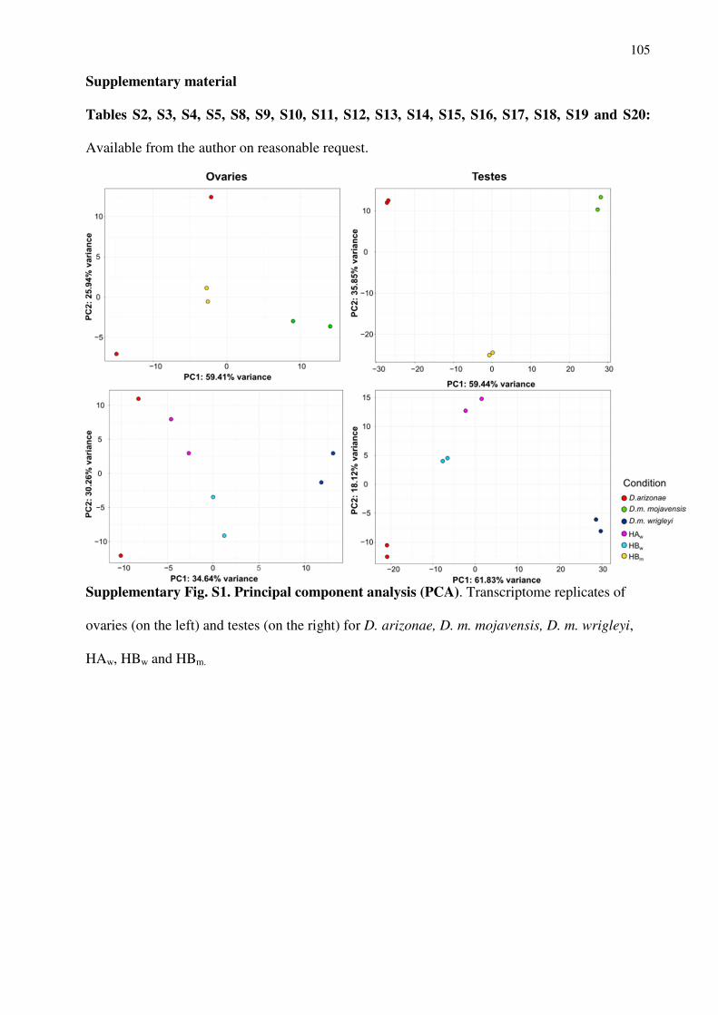
105
Supplementary material Tables S2, S3, S4, S5, S8, S9, S10, S11, S12, S13, S14, S15, S16, S17, S18, S19 and S20:
Available from the author on reasonable request.
Supplementary Fig. S1. Principal component analysis (PCA). Transcriptome replicates of
ovaries (on the left) and testes (on the right) for D. arizonae, D. m. mojavensis, D. m. wrigleyi,
HAw, HBw and HBm.
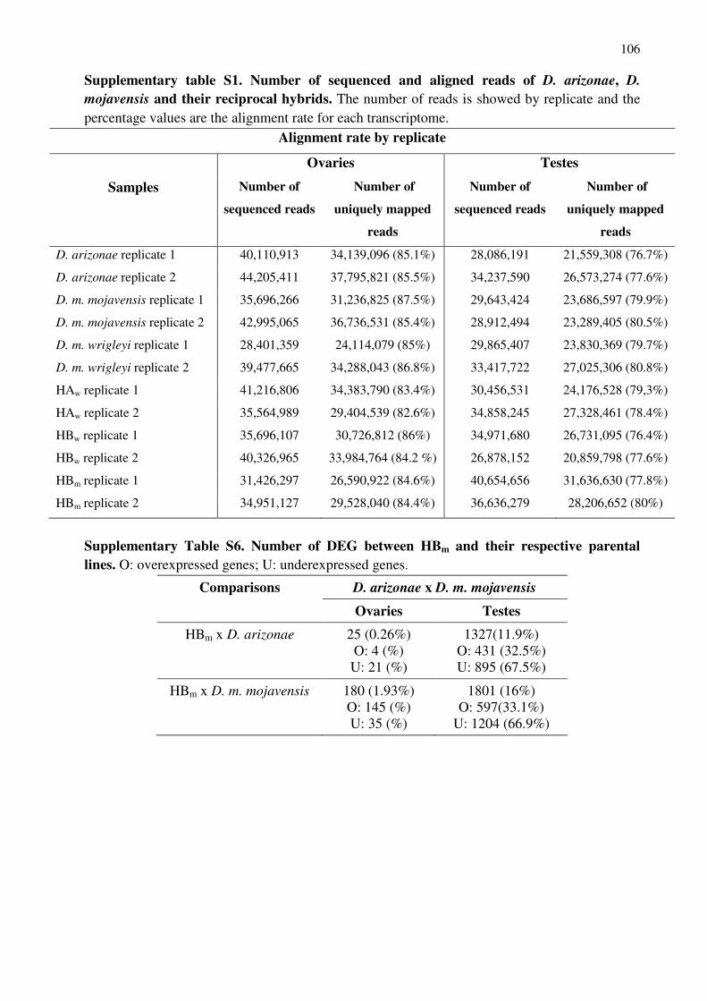
106
Supplementary table S1. Number of sequenced and aligned reads of D. arizonae, D.
mojavensis and their reciprocal hybrids. The number of reads is showed by replicate and the percentage values are the alignment rate for each transcriptome.
Alignment rate by replicate
Ovaries Testes
Samples Number of
sequenced reads
Number of
uniquely mapped
reads
Number of
sequenced reads
Number of
uniquely mapped
reads
D. arizonae replicate 1 40,110,913 34,139,096 (85.1%) 28,086,191 21,559,308 (76.7%)
D. arizonae replicate 2 44,205,411 37,795,821 (85.5%) 34,237,590 26,573,274 (77.6%)
D. m. mojavensis replicate 1 35,696,266 31,236,825 (87.5%) 29,643,424 23,686,597 (79.9%)
D. m. mojavensis replicate 2 42,995,065 36,736,531 (85.4%) 28,912,494 23,289,405 (80.5%)
D. m. wrigleyi replicate 1 28,401,359 24,114,079 (85%) 29,865,407 23,830,369 (79.7%)
D. m. wrigleyi replicate 2 39,477,665 34,288,043 (86.8%) 33,417,722 27,025,306 (80.8%)
HAw replicate 1 41,216,806 34,383,790 (83.4%) 30,456,531 24,176,528 (79,3%)
HAw replicate 2 35,564,989 29,404,539 (82.6%) 34,858,245 27,328,461 (78.4%)
HBw replicate 1 35,696,107 30,726,812 (86%) 34,971,680 26,731,095 (76.4%)
HBw replicate 2 40,326,965 33,984,764 (84.2 %) 26,878,152 20,859,798 (77.6%)
HBm replicate 1 31,426,297 26,590,922 (84.6%) 40,654,656 31,636,630 (77.8%)
HBm replicate 2 34,951,127 29,528,040 (84.4%) 36,636,279 28,206,652 (80%)
Supplementary Table S6. Number of DEG between HBm and their respective parental lines. O: overexpressed genes; U: underexpressed genes.
Comparisons D. arizonae x D. m. mojavensis
Ovaries Testes
HBm x D. arizonae 25 (0.26%) O: 4 (%)
U: 21 (%)
1327(11.9%) O: 431 (32.5%) U: 895 (67.5%)
HBm x D. m. mojavensis 180 (1.93%) O: 145 (%) U: 35 (%)
1801 (16%) O: 597(33.1%)
U: 1204 (66.9%)
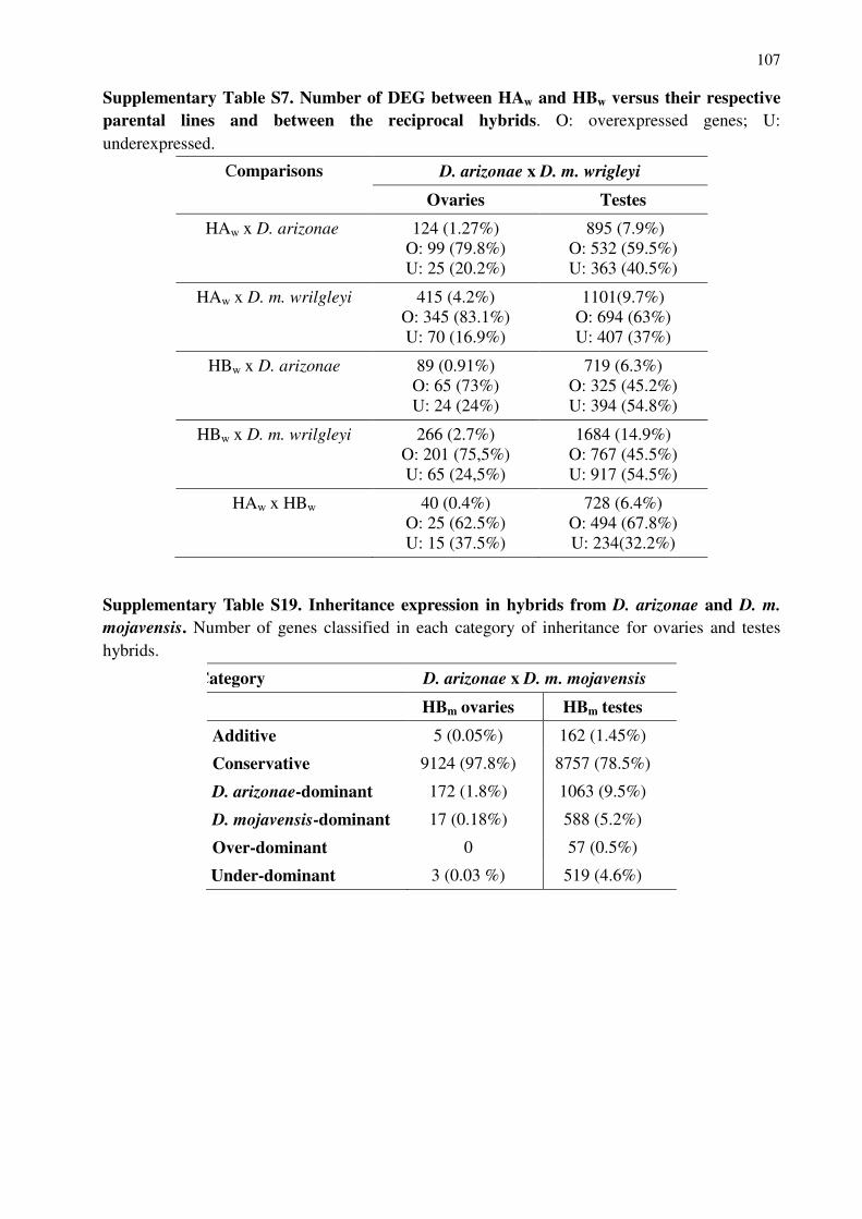
107
Supplementary Table S7. Number of DEG between HAw and HBw versus their respective parental lines and between the reciprocal hybrids. O: overexpressed genes; U: underexpressed.
Comparisons D. arizonae x D. m. wrigleyi
Ovaries Testes
HAw x D. arizonae 124 (1.27%) O: 99 (79.8%) U: 25 (20.2%)
895 (7.9%) O: 532 (59.5%) U: 363 (40.5%)
HAw x D. m. wrilgleyi 415 (4.2%) O: 345 (83.1%) U: 70 (16.9%)
1101(9.7%) O: 694 (63%) U: 407 (37%)
HBw x D. arizonae 89 (0.91%) O: 65 (73%) U: 24 (24%)
719 (6.3%) O: 325 (45.2%) U: 394 (54.8%)
HBw x D. m. wrilgleyi 266 (2.7%) O: 201 (75,5%) U: 65 (24,5%)
1684 (14.9%) O: 767 (45.5%) U: 917 (54.5%)
HAw x HBw 40 (0.4%) O: 25 (62.5%) U: 15 (37.5%)
728 (6.4%) O: 494 (67.8%) U: 234(32.2%)
Supplementary Table S19. Inheritance expression in hybrids from D. arizonae and D. m.
mojavensis. Number of genes classified in each category of inheritance for ovaries and testes hybrids.
Category D. arizonae x D. m. mojavensis
HBm ovaries HBm testes
Additive 5 (0.05%) 162 (1.45%)
Conservative 9124 (97.8%) 8757 (78.5%)
D. arizonae-dominant 172 (1.8%) 1063 (9.5%)
D. mojavensis-dominant 17 (0.18%) 588 (5.2%)
Over-dominant 0 57 (0.5%)
Under-dominant 3 (0.03 %) 519 (4.6%)
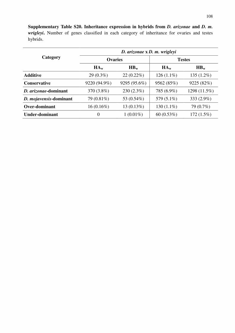
108
Supplementary Table S20. Inheritance expression in hybrids from D. arizonae and D. m.
wrigleyi. Number of genes classified in each category of inheritance for ovaries and testes hybrids.
Category
D. arizonae x D. m. wrigleyi
Ovaries Testes
HAw HBw HAw HBw
Additive 29 (0.3%) 22 (0.22%) 126 (1.1%) 135 (1.2%)
Conservative 9220 (94.9%) 9295 (95.6%) 9562 (85%) 9225 (82%)
D. arizonae-dominant 370 (3.8%) 230 (2.3%) 785 (6.9%) 1298 (11.5%)
D. mojavensis-dominant 79 (0.81%) 53 (0.54%) 579 (5.1%) 333 (2.9%)
Over-dominant 16 (0.16%) 13 (0.13%) 130 (1.1%) 79 (0.7%)
Under-dominant 0 1 (0.01%) 60 (0.53%) 172 (1.5%)

109
CAPÍTULO 3
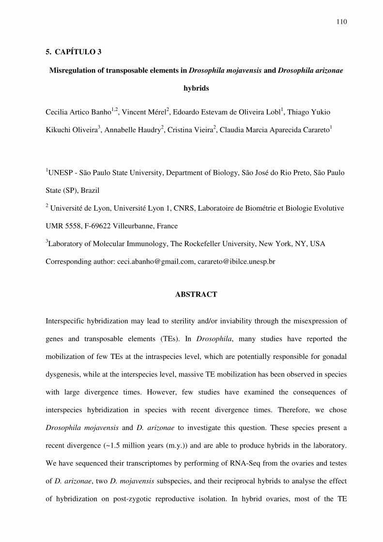
110
5. CAPÍTULO 3 Misregulation of transposable elements in Drosophila mojavensis and Drosophila arizonae
hybrids
Cecilia Artico Banho1,2, Vincent Mérel2, Edoardo Estevam de Oliveira Lobl1, Thiago Yukio
Kikuchi Oliveira3, Annabelle Haudry2, Cristina Vieira2, Claudia Marcia Aparecida Carareto1
1UNESP - São Paulo State University, Department of Biology, São José do Rio Preto, São Paulo
State (SP), Brazil
2 Université de Lyon, Université Lyon 1, CNRS, Laboratoire de Biométrie et Biologie Evolutive
UMR 5558, F-69622 Villeurbanne, France
3Laboratory of Molecular Immunology, The Rockefeller University, New York, NY, USA
Corresponding author: [email protected], [email protected]
ABSTRACT
Interspecific hybridization may lead to sterility and/or inviability through the misexpression of
genes and transposable elements (TEs). In Drosophila, many studies have reported the
mobilization of few TEs at the intraspecies level, which are potentially responsible for gonadal
dysgenesis, while at the interspecies level, massive TE mobilization has been observed in species
with large divergence times. However, few studies have examined the consequences of
interspecies hybridization in species with recent divergence times. Therefore, we chose
Drosophila mojavensis and D. arizonae to investigate this question. These species present a
recent divergence (~1.5 million years (m.y.)) and are able to produce hybrids in the laboratory.
We have sequenced their transcriptomes by performing of RNA-Seq from the ovaries and testes
of D. arizonae, two D. mojavensis subspecies, and their reciprocal hybrids to analyse the effect
of hybridization on post-zygotic reproductive isolation. In hybrid ovaries, most of the TE
111
families are not deregulated (98-99.6%). However, in testes, TE deregulation was observed at a
rate of 11.4% and presented a bias towards overexpression. We have sequenced the piRNAs
from the ovaries and testes of hybrids and parental lines to obtain a better understanding of the
causes of TE upregulation. Here, several deregulated TEs in male and female gonads have had
complementary piRNAs mapped onto them; however, the control of their expression was not
observed. In males, some genes, such as chromatin genes and piRNA pathway genes, showed
divergent expression in hybrids compared with that in the parental lines, potentially indicating
that TE deregulation is associated with the divergence of regulatory genes that play a role in
modulating transcriptional and post-transcriptional mechanisms.
Keywords: TEs, differential expression, piRNA, repleta group, hybridization.
INTRODUCTION
Speciation is a complex process resulting from the split of one ancestral lineage in two
reproductively isolated populations (ORR; COYNE, 1989; COYNE; ORR, 1997). However,
under specific conditions, species present incomplete reproductive isolation and are able to cross;
nevertheless, their hybrids might exhibit a decrease in fitness that is often due to inviability and
sterility. According to the Dobzhansky–Muller model (1936, 1942), the presence of allele-
specific genetic variation in different species or populations may lead to deleterious epistatic
interactions, disrupting regulatory networks and causing severe consequences in hybrids (ORR;
COYNE, 1997; KELLEHER et al., 2012, ROMERO-SORIANO et al., 2017). Although
numerous efforts have focused on obtaining a better understanding of the mechanisms
underlying reproductive isolation, few studies have specifically analysed deregulated genes or
other factors directly associated with reproductive barriers (TING et al., 1998; MICHALAK;
NOOR, 2003; RANZ et al., 2004; HAERTY; SINGH, 2006; MA; MICHALAK, 2011; GOMES;
CIVETTA, 2014; LI et al., 2016; HILL et al., 2016). Moreover, the deregulation of gene
expression may not be the only or the main cause of hybrid breakdown, but a consequence of
112
other factors, such as divergence in regulatory sequences, mobilization of transposable elements
or epigenetic changes (MCMANNUS et al., 2010; REBOLLO et al., 2012; AKKOUCHE et al.,
2013; IWASAKI et al., 2016; FABLET et al., 2019).
Transposable elements (TEs), which are repetitive DNA sequences capable of moving
from one place to another in the genome and between genomes, are important sources of genetic
variability and genomic evolution (KIDWELL; LISCH, 2001; VAN de LAGEMAAT et al.,
2003; WONG; CHOO, 2004). Several studies have reported the effect of TEs on reproductive
isolation, due to their ability to modify regulatory networks and gene expression and lead to
structural rearrangements (KIDWELL; LISCH, 2001; SERRATO-CAPUCHINA; MAMUTE,
2018). TE activation is induced by environmental stress, such as temperature and pathogens
(CAPY et al., 2000), and, as proposed by Barbara McClintock (1980), by genomic shock, such
as interspecific hybridization (O’NEILL et al., 1998; LABRADOR et al., 1999; FONTDEVILA,
2005; GUERREIRO, 2012). The disruption of genome stability resulting from hybridization is
attributed to the divergence of regulatory sequences and/or the content of TEs (SESSEGOLO et
al., 2016). The effects of TEs on interspecific hybrids may be beneficial or deleterious,
depending on the species. In successful hybrids of Helianthus annuus and Helianthus petiolaris
(BAACK et al., 2005; KAWAKAMI et al., 2011), a burst of transposition was identified
involving two main TE families (Ty1/Copia-like and Ty3/Gypsy-like), which contributed to
increase the size of the hybrid genome by ~50%. In animals, on the other hand, the consequences
of hybridization are potentially more severe. Hybrids of marsupials in the Macropodidae family
have shown centromeric expansion and chromosomal instability, which was associated with the
amplification of KerV-1 transposable elements and satellite repeats, decreasing the fitness of the
hybrids (O’NEILL et al., 1998).
Several studies have described the consequences of TE mobilization in Drosophila
species at different levels of hybridization. In intraspecies hybrids, some TEs are responsible for
hybrid dysgenesis syndrome, such as the I/R system (PICARD, 1976) and hobo element
113
(BLACKMAN et al., 1987; YANNOPOULOS et al., 1987) in D. melanogaster, the P/M system
(KIDWELL et al., 1977; HILL et al., 2016) in D. melanogaster and D. simulans, and the
gonadal dysgenesis caused by the mobilization of some specific TEs in D. virilis, such as the
Penelope element (PETROV et al., 1995). This syndrome is characterized by gonadal atrophy
and generally affects offspring obtained from crosses in only one direction. The deregulation of
some TEs in one specific direction of the cross is associated with differences in the TE content
between the genomes of the parental populations and their regulatory system, in which the
piRNAs (a class of small RNAs named piwi-interacting RNAs) play the main role (ARAVIN et
al., 2007; MALONE et al., 2009; SENTI; BRENNECKE, 2010). These small RNAs function in
the Drosophila germline at post-transcriptional and transcriptional levels together with a
complex of Argonaute proteins, which recognize homologous TEs and induce their degradation
(KLATTENHOFF; THEURKAUF, 2008; SENTI; BRENNECKE, 2010; ROZHKOV et al.,
2013; LUO; LU, 2017). In intraspecies or interspecies crosses, females lacking one or a few TE
families are not able to produce specific piRNAs, which are maternally deposited, and hence,
silence TE expression (LUO; LU, 2017). As an example, in interspecies hybrids obtained from
D. virilis and D. lummei crosses, the magnitude of prezygotic and postzygotic isolation depends
on the presence of the transposon Penelope. The lack of this element in the female parent is
responsible for hybrid inviability, likely because of parental mismatch between the number or
identity of TEs and the machinery responsible for Penelope repression (CASTILLO; MOYLE,
2019). This potential to cause negative genetic interactions via TE derepression in hybrids places
TEs within the classical Dobzhansky-Muller model (1936, 1942) for the evolution of
incompatibilities. Overall, the role of repetitive elements, such as TEs and satellite sequences, in
postzygotic reproductive isolation has been suggested by some authors that correlate differences
in their content between species with increased transcription and dysfunction in hybrids
(SATYAKI et al., 2014; ROMERO-SORIANO et al., 2017; KOTOV et al., 2019). Moreover, in
interspecies Drosophila hybrids obtained from highly divergent species, high levels of TE
114
expression have been observed (SATYAKI et al., 2014). In hybrids of D. melanogaster x D.
simulans, which diverged between 1.2 to 5 m.y. (RUSSO et al., 1995; KLIMAN et al., 2000;
TAMURA et al., 2004; CUTTER, 2008), and D. buzzatii x D. koepferae, which diverged
between 4.02 to 4.63 mya (GOMEZ; HASSON 2003; LAAYOUNI et al., 2003; OLIVEIRA et
al., 2012), bursts of transposition were observed. This massive TE mobilization was associated
with the adaptive divergence of piRNA pathway genes, leading to the disruption of the system
regulating TEs and contributing to hybrid sterility to some extent (LABRADOR et al., 1999;
KELLEHER et al., 2012; VELA et al., 2014; GUERREIRO, 2015; ROMERO-SORIANO et al.,
2017).
In addition, some studies have reported the causes and consequences of mobilization of
TEs at the intra- and interspecies level. The focus of the present study is the role of TEs in the
early stages of hybrid incompatibility. Thus, we chose D. arizonae and D. mojavensis (repleta
group, Drosophila genus) to investigate this topic because they are sibling species with a shorter
divergence time (1.5 m.y., SANCHES-FLORES et al., 2016). They produce hybrids in the
laboratory with asymmetrical sterility, because the females are fertile and the males, most of
which are sterile, have different phenotypes, with the sperm of the sterile males being mobile or
immobile, depending on the direction of crossing (REED; MARKOW, 2004; REED et al., 2008;
JENNINGS; ETGES, 2009). Moreover, the TE contents of these species have been
characterized, enabling a study of differential expression in hybrids and both parental lines. In
the first publication of the D. mojavensis genome (r 1.04), the annotation of TEs showed that
8.92% of the genome is composed of TEs, 45% of which are LTRs, ~28% are LINEs and 13%
are DNA transposons (CLARK et al., 2007). However, using a new approach to annotate TEs
(the pipeline TEdenovo present in the suite of applications REPET, FLUTRE et al., 2011), we
showed that the D. mojavensis r 1.04 public genome is composed of 16.6% TEs (data not
shown), the same content reported by Rius et al. (2016) and twice as many TE families that were
previously identified by Clark et al. (2007). However, fewer TEs were annotated in the genomes
115
of two D. arizonae lines, 10% (SANCHEZ-FLORES et al., 2016) and 6.4% (data not shown),
which is explained by the underestimation of TE sequences in genomes obtained using NGS
compared to genomes obtained using Sanger sequencing. In the latter species, 3.5% of TEs are
Class I elements, known as retrotransposons, and 2.8% are Class II elements, known as DNA
transposons (data not shown).
Considering the characteristics mentioned above, this pair of species is interesting as a
model to study differences in the expression of TEs in the reproductive organs of hybrids, as well
as their regulation in different gonad tissues by analysing the piRNA pathway. At the same time,
they allow us to test the hypothesis that the rate of TE deregulation in hybrids depends on the
time of divergence of the parental species and may exert a direct effect on the male phenotype.
Indeed, two previous studies have analysed the expression of TEs in female and male hybrids of
D. arizonae and D. mojavensis (CARNELOSSI et al., 2014; LOPEZ-MAESTRE et al., 2017).
According to the findings reported by Lopez-Maestre et al. (2017), no global burst of
transposition is detected in the female germline of hybrids, but the transposition of specific TE
families is detected in male and female gonad tissues (CARNELOSSI et al., 2014). In addition,
studies of the expression profile of TEs in male hybrids of these species, as well as their
associations with the TE regulatory system in the testes are lacking.
Therefore, in this study, we aimed to investigate the profile of TE expression in the
ovaries and testes of hybrids obtained from crosses between D. arizonae and two subspecies of
D. mojavensis (D. m. mojavensis and D. m. wrigleyi), as well as the association of deregulated
TE families with the systems regulating the piRNA and siRNA pathways. A higher level of TE
deregulation is observed in the male gonads compared with the ovaries. Moreover, in ovaries and
testes, we identified complementary piRNAs that are able to degrade and start the ping-pong
amplification cycle for most of the deregulated TEs. However, TE expression is not completely
controlled by the piRNA pathway, suggesting that other mechanisms might control TEs in
reproductive tissues.
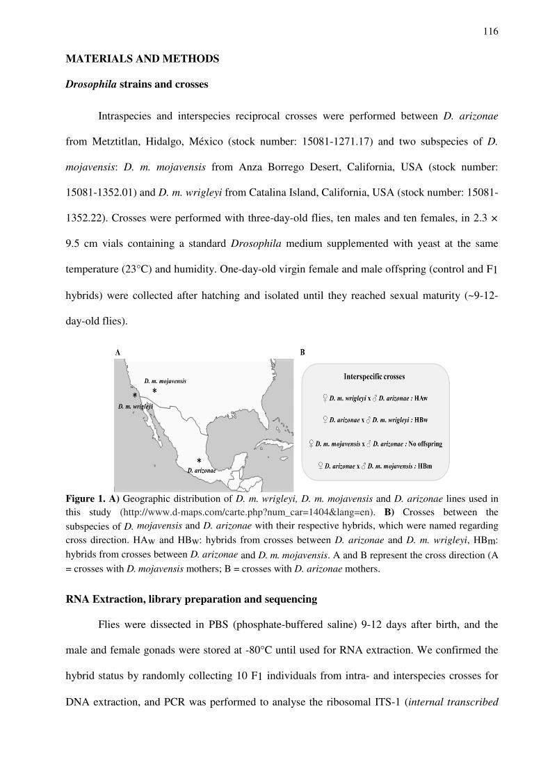
116
MATERIALS AND METHODS
Drosophila strains and crosses
Intraspecies and interspecies reciprocal crosses were performed between D. arizonae
from Metztitlan, Hidalgo, México (stock number: 15081-1271.17) and two subspecies of D.
mojavensis: D. m. mojavensis from Anza Borrego Desert, California, USA (stock number:
15081-1352.01) and D. m. wrigleyi from Catalina Island, California, USA (stock number: 15081-
1352.22). Crosses were performed with three-day-old flies, ten males and ten females, in 2.3 ×
9.5 cm vials containing a standard Drosophila medium supplemented with yeast at the same
temperature (23°C) and humidity. One-day-old virgin female and male offspring (control and F1
hybrids) were collected after hatching and isolated until they reached sexual maturity (~9-12-
day-old flies).
Figure 1. A) Geographic distribution of D. m. wrigleyi, D. m. mojavensis and D. arizonae lines used in this study (http://www.d-maps.com/carte.php?num_car=1404&lang=en). B) Crosses between the
subspecies of D. mojavensis and D. arizonae with their respective hybrids, which were named regarding
cross direction. HAw and HBw: hybrids from crosses between D. arizonae and D. m. wrigleyi, HBm:
hybrids from crosses between D. arizonae and D. m. mojavensis. A and B represent the cross direction (A = crosses with D. mojavensis mothers; B = crosses with D. arizonae mothers.
RNA Extraction, library preparation and sequencing
Flies were dissected in PBS (phosphate-buffered saline) 9-12 days after birth, and the
male and female gonads were stored at -80°C until used for RNA extraction. We confirmed the
hybrid status by randomly collecting 10 F1 individuals from intra- and interspecies crosses for
DNA extraction, and PCR was performed to analyse the ribosomal ITS-1 (internal transcribed
117
spacer 1) sequence from the 18S gene region, NCBI Reference Sequence: EU306666.1 (BAFFI;
CERON, 2002). The oligonucleotide primers for ITS-1 amplify an amplicon with a size of 500
bp and 550 bp in D. arizonae and in D. mojavensis, respectively. Therefore, in the hybrids, two
different fragments corresponding to the D. arizonae and D. mojavensis alleles are expected.
After confirming the hybrid status, 30 pairs of ovaries and 50 pairs of testes were selected to
perform total RNA extraction using the RNeasy kit (Qiagen). The samples were treated with
DNase (DNA-free Kit, Ambion) and stored at -80°C. The samples were quantified by measuring
the fluorescence in a Bioanalyser 2100 (Agilent). Two Illumina libraries of 2 × 100 paired-end
reads containing inserts of 300 bp on average were prepared for each sample (ovaries and testes
of D. arizonae, D. m. mojavensis, D. m. wrigleyi and their respective F1 hybrids) using 1 µg of
purified RNA. Sequencing was performed using the GenomEast platform, a member of the
‘France Génomique’ consortium (ANR-10-INBS-0009)”, with Illumina technology in an
Illumina HiSeq 4000 instrument. The hybrid transcriptomes from D. m. mojavensis females and
D. arizonae males were not sequenced, because the hybrid incompatibility in this direction of the
cross is very high and we were unable to obtain a sufficient amount of material to perform RNA
extraction.
TE library construction
The new TEs were identified using the TEdenovo pipeline in the REPET v 2.5 package
(available at https://urgi.versailles.inra.fr/Tools/REPET) (FLUTRE et al., 2011). The consensus
sequences obtained were classified up to the family level by performing phylogenetic analyses of
the conserved domains of each TE order. The conserved domains of each consensus sequence
were aligned with the corresponding domains of reference sequences from Drosophila available
at Repbase (BAO et al., 2015), along with new reference sequences of LTR elements described
by Bargues and Lerat (2017), and Tc1-Mariner elements listed in the study by Hernandez-
Hernandez et al. (2017) using the MAFFT 7 software (KATOH; STANDLEY, 2013). The non-
118
informative locations in each alignment were removed using the trimAL tool v1.3 (CAPELLA-
GUTIÉRREZ et al., 2009). The amino acid evolution model for the phylogenetic reconstructions
was created using the Smart Model Selection software (LEFORT et al., 2017). The
reconstructions of the trees were performed using the maximum likelihood method and PhyML
3.0 software (GUINDON et al., 2010). For the TE annotation, Repbase repeats from Drosophila
species (BAO et al., 2015) were added to the custom library obtained from the TEdenovo step.
The genomes (D. arizonae, which was previously sequenced and assembled by our research
group, and the D. mojavensis r 1.04 public genome) were masked with RepeatMasker (version
4.0.7) (SMIT et al., 2015) and RMBlast engine (version 2.2.27+) using the standard options,
except for the cutoff (score 250), no low and no RNA. The “one code to find them all” pipeline
(available at http://doua.prabi.fr/software/one-code-to-find-them-all) was used to correctly
estimate the number of copies of the TEs identified using RepeatMasker.
Small RNA extraction, library Preparation, and sequencing
Small RNAs were extracted from the ovaries (70 pairs) and testes (100 pairs) of D.
arizonae, D. m. mojavensis, D. m. wrigleyi, and their interspecific hybrids using HiTrap Q HP
anion exchange columns, as described in the study by Grentzinger et al. (2014). Two Illumina
libraries were prepared for each sample from 1-6 ng of the purified small RNA fraction using the
TruSeq Small RNA Library Prep Kit (Illumina, San Diego, CA) and the GenomEast platform, a
member of the ‘France Génomique’ consortium (ANR-10-INBS-0009)”, and sequencing was
performed using Illumina technology and an Illumina HiSeq 4000 instrument (1x50 bases).
Overall, 729 million reads were obtained, and reads with a length of 23–30 nt were retained as
piRNAs, whereas reads with a length of 21 nt were considered siRNAs.
119
TE and gene read mapping and the analysis of differential expression
The sequenced transcriptomes were trimmed using UrQt (MODOLO; LERAT, 2015) to
remove polyA tails (from RNA-Seq reads) and low-quality nucleotides, and then the sequence
quality was assessed using FastQC software. TE expression analyses were performed with the
module TEcount from the TEtools pipeline (LERAT et al., 2017; https://github.com/l-
modolo/TEtools) to ensure the accuracy of the map of the RNA-Seq data. RNA-Seq reads were
aligned to our homemade TE library containing approximately 894 candidate TE families from
the D. arizonae and D. mojavensis r 1.04 genomes using Bowtie2. The read count step was
computed for each TE family by adding all reads mapped on copies of the same family. The
parental and hybrid transcriptomes were aligned to all annotated (20,110 mRNAs) Coding
Sequence (CDS) of D. mojavensis r1.04 public genome (CLARK et al., 2007) available on
http://flybase.org/ using Kallisto, to verify the expression of genes regulating the piRNA
pathway (BRAY et al., 2016). After the mapping procedure was complete, biomaRt (DURINCK
et al., 2005), an R 3.6.1 (R Core Team, 2018) Bioconductor package, was used to recover the
gene names corresponding to each transcript from the reference genome. Afterwards, because
several genes are expressed in different isoforms, the package tximport (SONESON et al., 2015)
was used to summarize the estimated transcript levels of each gene analysed, allowing us to
recover 11,654 coding genes from D. mojavensis r 1.04 transcripts. We have used these data in
the differential expression analyses. Gene ontology was accessed using an orthologous gene
table from Drosophila melanogaster downloaded from http://flybase.org/, allowing us to identify
the genes involved in the piRNA pathway.
Differential expression analyses were performed with the R Biocondutor package
DESeq2 (LOVE et al., 2014; https://www.R-project.org) using raw read counts to identify
differential TE and gene expression in the reproductive tissues from the hybrids compared to the
parental species (control lines). This package normalizes counts using size factors that are
estimated according to the median counts from all TE families. Additionally, DESeq2 estimates
120
the means and variances of raw read counts and tests for differential expression based on a
model using the negative binomial distribution and the Benjamini-Hochberg correction for
multiple tests (FDR level of 0.1, BENJAMINI; HOCHBERG, 1995). TE families and genes
were classified as differentially expressed when the adjusted p-value (FDR level) was less
than0.01 and an at least 2-fold difference in expression was identified. TE families and genes
that showed ≤ 10 aligned reads mapped under all conditions tested were excluded from the
analyses.
Small RNA and ping-pong analyses
The small RNA-Seq analyses were performed using the method described by Fablet et al.
(2019). Thus, the data were initially cleaned using Cutadapt (MARTIN, 2011) to remove the
adapter sequences (-a TGGAATTCTCGGGTGCCAAGGAACTCCAGTCACTTA). PRINSEQ
lite version 0.20.4 (SCHMIEDER; EDWARDS, 2011) was used to filter the reads with a size
ranging from 23 to 30 nucleotides to ensure that only piRNAs were analysed. The TEcount
module from TEtools (LERAT et al., 2017) was used to map the sense and antisense piRNA
reads to the same TE library that was previously used as a reference in the analysis of differential
TE expression. Read counts were normalized to miRNA read counts obtained from the
alignment created using Bowtie (with –S and –best arguments to obtain the most sensitive option
and retaining a single alignment for reads mapping to multiple positions) (LANGMEAD et al.,
2009) of the cleaned small RNA data (before the filtering) against the miRNA sequences of D.
mojavensis (Flybase: dmoj-all-miRNA-r1.04.fasta.gz). The ping-pong signature was analysed
using signature.py with the options min_size = 23 and max_size = 30 (ANTONIEWSKI, 2014).
SAM alignments of cleaned small RNA-seq reads with complete TE copies that were identified
as differentially expressed in ovaries and testes were used as input files when the hybrids were
compared with the parental lines.
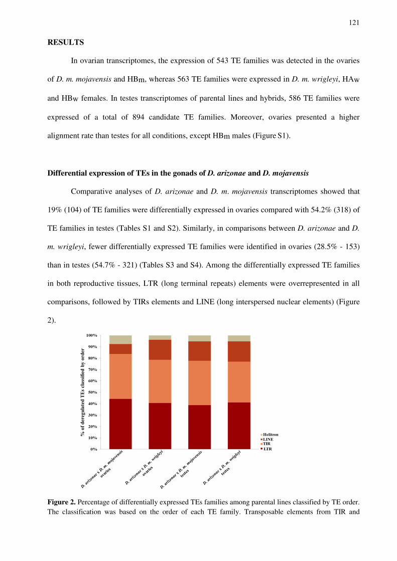
121
RESULTS
In ovarian transcriptomes, the expression of 543 TE families was detected in the ovaries
of D. m. mojavensis and HBm, whereas 563 TE families were expressed in D. m. wrigleyi, HAw
and HBw females. In testes transcriptomes of parental lines and hybrids, 586 TE families were
expressed of a total of 894 candidate TE families. Moreover, ovaries presented a higher
alignment rate than testes for all conditions, except HBm males (Figure S1).
Differential expression of TEs in the gonads of D. arizonae and D. mojavensis
Comparative analyses of D. arizonae and D. m. mojavensis transcriptomes showed that
19% (104) of TE families were differentially expressed in ovaries compared with 54.2% (318) of
TE families in testes (Tables S1 and S2). Similarly, in comparisons between D. arizonae and D.
m. wrigleyi, fewer differentially expressed TE families were identified in ovaries (28.5% - 153)
than in testes (54.7% - 321) (Tables S3 and S4). Among the differentially expressed TE families
in both reproductive tissues, LTR (long terminal repeats) elements were overrepresented in all
comparisons, followed by TIRs elements and LINE (long interspersed nuclear elements) (Figure
2).
Figure 2. Percentage of differentially expressed TEs families among parental lines classified by TE order. The classification was based on the order of each TE family. Transposable elements from TIR and
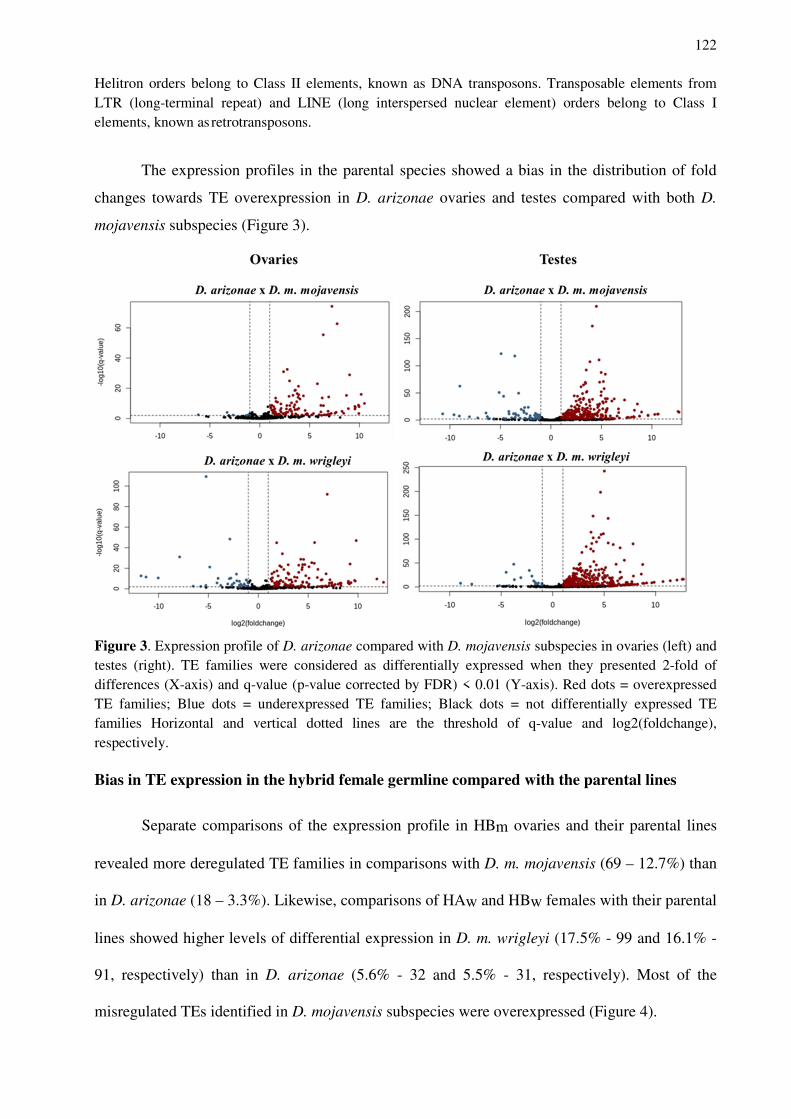
122
Helitron orders belong to Class II elements, known as DNA transposons. Transposable elements from LTR (long-terminal repeat) and LINE (long interspersed nuclear element) orders belong to Class I elements, known as retrotransposons.
The expression profiles in the parental species showed a bias in the distribution of fold
changes towards TE overexpression in D. arizonae ovaries and testes compared with both D.
mojavensis subspecies (Figure 3).
Figure 3. Expression profile of D. arizonae compared with D. mojavensis subspecies in ovaries (left) and testes (right). TE families were considered as differentially expressed when they presented 2-fold of differences (X-axis) and q-value (p-value corrected by FDR) < 0.01 (Y-axis). Red dots = overexpressed TE families; Blue dots = underexpressed TE families; Black dots = not differentially expressed TE families Horizontal and vertical dotted lines are the threshold of q-value and log2(foldchange), respectively. Bias in TE expression in the hybrid female germline compared with the parental lines
Separate comparisons of the expression profile in HBm ovaries and their parental lines
revealed more deregulated TE families in comparisons with D. m. mojavensis (69 – 12.7%) than
in D. arizonae (18 – 3.3%). Likewise, comparisons of HAw and HBw females with their parental
lines showed higher levels of differential expression in D. m. wrigleyi (17.5% - 99 and 16.1% -
91, respectively) than in D. arizonae (5.6% - 32 and 5.5% - 31, respectively). Most of the
misregulated TEs identified in D. mojavensis subspecies were overexpressed (Figure 4).
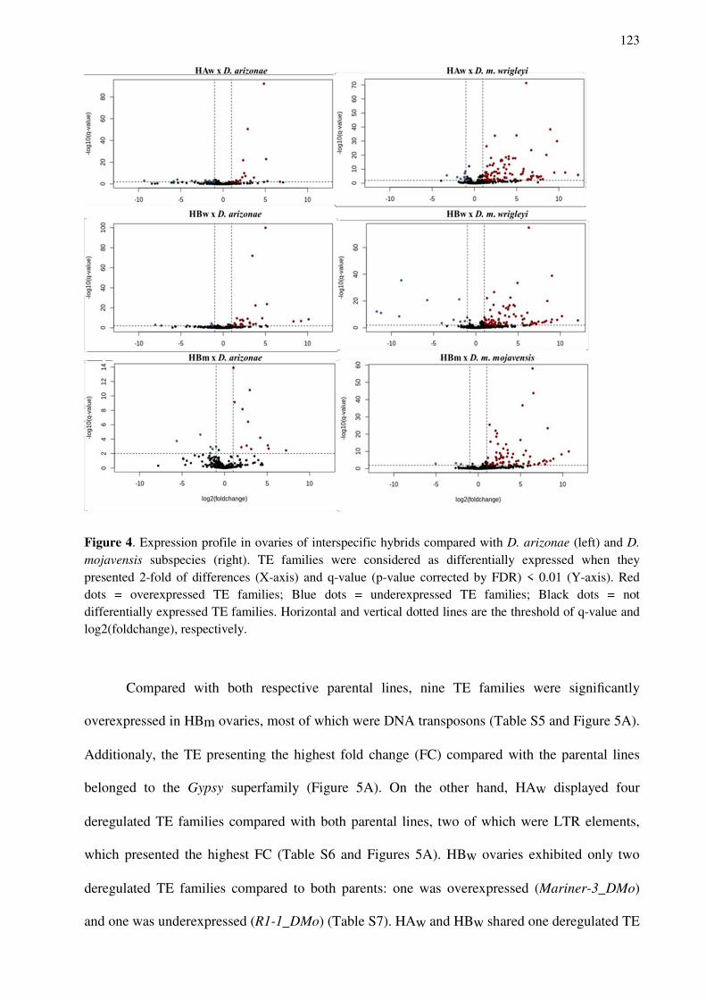
123
Figure 4. Expression profile in ovaries of interspecific hybrids compared with D. arizonae (left) and D.
mojavensis subspecies (right). TE families were considered as differentially expressed when they presented 2-fold of differences (X-axis) and q-value (p-value corrected by FDR) < 0.01 (Y-axis). Red dots = overexpressed TE families; Blue dots = underexpressed TE families; Black dots = not differentially expressed TE families. Horizontal and vertical dotted lines are the threshold of q-value and log2(foldchange), respectively.
Compared with both respective parental lines, nine TE families were significantly
overexpressed in HBm ovaries, most of which were DNA transposons (Table S5 and Figure 5A).
Additionaly, the TE presenting the highest fold change (FC) compared with the parental lines
belonged to the Gypsy superfamily (Figure 5A). On the other hand, HAw displayed four
deregulated TE families compared with both parental lines, two of which were LTR elements,
which presented the highest FC (Table S6 and Figures 5A). HBw ovaries exhibited only two
deregulated TE families compared to both parents: one was overexpressed (Mariner-3_DMo)
and one was underexpressed (R1-1_DMo) (Table S7). HAw and HBw shared one deregulated TE
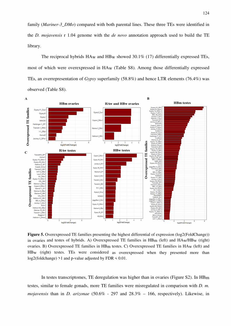
124
family (Mariner-3_DMo) compared with both parental lines. These three TEs were identified in
the D. mojavensis r 1.04 genome with the de novo annotation approach used to build the TE
library.
The reciprocal hybrids HAw and HBw showed 30.1% (17) differentially expressed TEs,
most of which were overexpressed in HAw (Table S8). Among those differentially expressed
TEs, an overrepresentation of Gypsy superfamily (58.8%) and hence LTR elements (76.4%) was
observed (Table S8).
Figure 5. Overexpressed TE families presenting the highest differential of expression (log2(FoldChange))
in ovaries and testes of hybrids. A) Overexpressed TE families in HBm (left) and HAw/HBw (right)
ovaries. B) Overexpressed TE families in HBm testes. C) Overexpressed TE families in HAw (left) and
HBw (right) testes. TEs were considered as overexpressed when they presented more than log2(foldchange) >1 and p-value adjusted by FDR < 0.01.
In testes transcriptomes, TE deregulation was higher than in ovaries (Figure S2). In HBm
testes, similar to female gonads, more TE families were misregulated in comparison with D. m.
mojavensis than in D. arizonae (50.6% - 297 and 28.3% – 166, respectively). Likewise, in
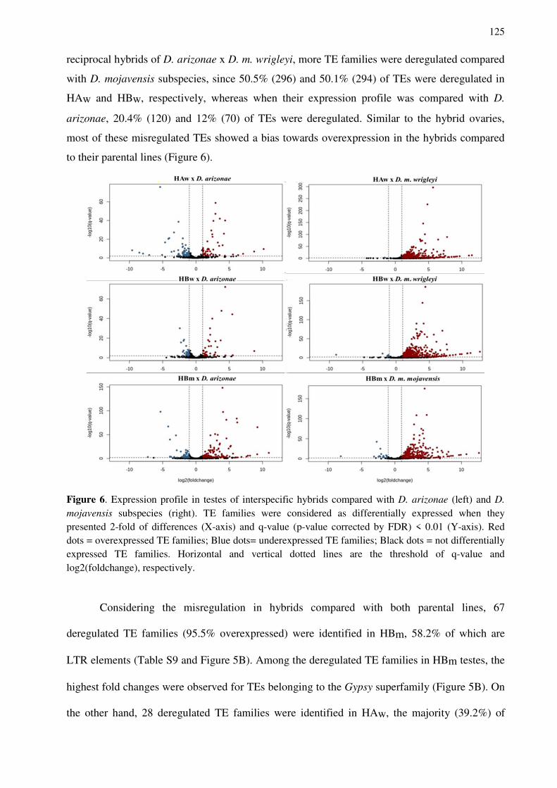
125
reciprocal hybrids of D. arizonae x D. m. wrigleyi, more TE families were deregulated compared
with D. mojavensis subspecies, since 50.5% (296) and 50.1% (294) of TEs were deregulated in
HAw and HBw, respectively, whereas when their expression profile was compared with D.
arizonae, 20.4% (120) and 12% (70) of TEs were deregulated. Similar to the hybrid ovaries,
most of these misregulated TEs showed a bias towards overexpression in the hybrids compared
to their parental lines (Figure 6).
Figure 6. Expression profile in testes of interspecific hybrids compared with D. arizonae (left) and D.
mojavensis subspecies (right). TE families were considered as differentially expressed when they presented 2-fold of differences (X-axis) and q-value (p-value corrected by FDR) < 0.01 (Y-axis). Red dots = overexpressed TE families; Blue dots= underexpressed TE families; Black dots = not differentially expressed TE families. Horizontal and vertical dotted lines are the threshold of q-value and log2(foldchange), respectively.
Considering the misregulation in hybrids compared with both parental lines, 67
deregulated TE families (95.5% overexpressed) were identified in HBm, 58.2% of which are
LTR elements (Table S9 and Figure 5B). Among the deregulated TE families in HBm testes, the
highest fold changes were observed for TEs belonging to the Gypsy superfamily (Figure 5B). On
the other hand, 28 deregulated TE families were identified in HAw, the majority (39.2%) of
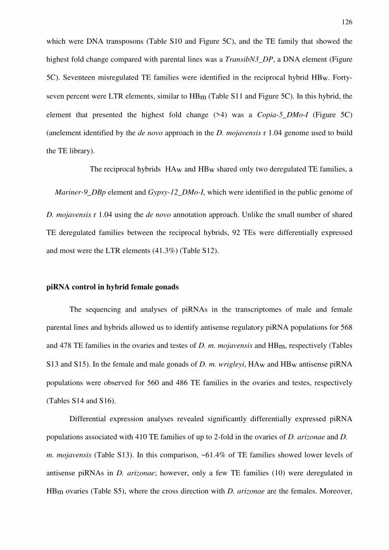
126
which were DNA transposons (Table S10 and Figure 5C), and the TE family that showed the
highest fold change compared with parental lines was a TransibN3_DP, a DNA element (Figure
5C). Seventeen misregulated TE families were identified in the reciprocal hybrid HBw. Forty-
seven percent were LTR elements, similar to HBm (Table S11 and Figure 5C). In this hybrid, the
element that presented the highest fold change (>4) was a Copia-5_DMo-I (Figure 5C)
(anelement identified by the de novo approach in the D. mojavensis r 1.04 genome used to build
the TE library).
The reciprocal hybrids HAw and HBw shared only two deregulated TE families, a
Mariner-9_DBp element and Gypsy-12_DMo-I, which were identified in the public genome of
D. mojavensis r 1.04 using the de novo annotation approach. Unlike the small number of shared
TE deregulated families between the reciprocal hybrids, 92 TEs were differentially expressed
and most were the LTR elements (41.3%) (Table S12).
piRNA control in hybrid female gonads
The sequencing and analyses of piRNAs in the transcriptomes of male and female
parental lines and hybrids allowed us to identify antisense regulatory piRNA populations for 568
and 478 TE families in the ovaries and testes of D. m. mojavensis and HBm, respectively (Tables
S13 and S15). In the female and male gonads of D. m. wrigleyi, HAw and HBw antisense piRNA
populations were observed for 560 and 486 TE families in the ovaries and testes, respectively
(Tables S14 and S16).
Differential expression analyses revealed significantly differentially expressed piRNA
populations associated with 410 TE families of up to 2-fold in the ovaries of D. arizonae and D.
m. mojavensis (Table S13). In this comparison, ~61.4% of TE families showed lower levels of
antisense piRNAs in D. arizonae; however, only a few TE families (10) were deregulated in
HBm ovaries (Table S5), where the cross direction with D. arizonae are the females. Moreover,
127
almost all deregulated TE families detected using RNA-Seq in HBm ovaries displayed lower
levels of piRNA expression in D. arizonae than in D. m. mojavensis and HBm (Table S13 and
Figure 7).
Figure 7. Expression of TE families in ovaries of D. m. mojavensis versus D. arizonae and the piRNA
expression for the correspondent TE families. In red triangles are the TE families overexpressed in HBm ovaries. TE families were considered as upregulated when they presented log2(2) > 1 and p-value adjusted < 0.01 in HBm ovaries compared with both parental lines.
Similarly, in the comparisons between D. arizonae and D. m. wrigleyi ovaries,
approximately 405 TE families showed antisense piRNA populations with differences in
expression greater than 2-fold (Table S14). Among these populations, ~64% were
underexpressed in D. arizonae; however, in the ovaries of HBw, only two TE families were
deregulated (Table S7). On the other hand, the ovaries of HAw, where D. m. wrigleyi is the
maternal source, the three TEs that were uniquely deregulated in this cross showed lower levels
of piRNA expression in D. m. wrigleyi than in D. arizonae, which was also verified by
comparing the expression levels of HAw and D. arizonae (Table S14 and Figure 8).
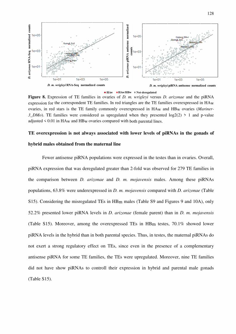
128
Figure 8. Expression of TE families in ovaries of D. m. wrigleyi versus D. arizonae and the piRNA
expression for the correspondent TE families. In red triangles are the TE families overexpressed in HAw ovaries, in red stars is the TE family commonly overexpressed in HAw and HBw ovaries (Mariner-
3_DMo). TE families were considered as upregulated when they presented log2(2) > 1 and p-value adjusted < 0.01 in HAw and HBw ovaries compared with both parental lines.
TE overexpression is not always associated with lower levels of piRNAs in the gonads of
hybrid males obtained from the maternal line
Fewer antisense piRNA populations were expressed in the testes than in ovaries. Overall,
piRNA expression that was deregulated greater than 2-fold was observed for 279 TE families in
the comparison between D. arizonae and D. m. mojavensis males. Among these piRNAs
populations, 63.8% were underexpressed in D. m. mojavensis compared with D. arizonae (Table
S15). Considering the misregulated TEs in HBm males (Table S9 and Figures 9 and 10A), only
52.2% presented lower piRNA levels in D. arizonae (female parent) than in D. m. mojavensis
(Table S15). Moreover, among the overexpressed TEs in HBm testes, 70.1% showed lower
piRNA levels in the hybrid than in both parental species. Thus, in testes, the maternal piRNAs do
not exert a strong regulatory effect on TEs, since even in the presence of a complementary
antisense piRNA for some TE families, the TEs were upregulated. Moreover, nine TE families
did not have show piRNAs to controll their expression in hybrid and parental male gonads
(Table S15).
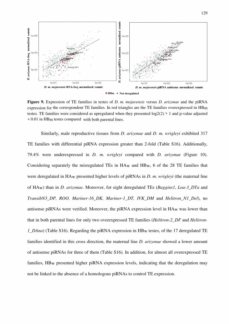
129
Figure 9. Expression of TE families in testes of D. m. mojavensis versus D. arizonae and the piRNA
expression for the correspondent TE families. In red triangles are the TE families overexpressed in HBm testes. TE families were considered as upregulated when they presented log2(2) > 1 and p-value adjusted < 0.01 in HBm testes compared with both parental lines.
Similarly, male reproductive tissues from D. arizonae and D. m. wrigleyi exhibited 317
TE families with differential piRNA expression greater than 2-fold (Table S16). Additionally,
79.4% were underexpressed in D. m. wrigleyi compared with D. arizonae (Figure 10).
Considering separately the misregulated TEs in HAw and HBw, 6 of the 28 TE families that
were deregulated in HAw presented higher levels of piRNAs in D. m. wrigleyi (the maternal line
of HAw) than in D. arizonae. Moreover, for eight deregulated TEs (Baggins1, Loa-3_DTa and
TransibN3_DP, ROO, Mariner-16_DK, Mariner-1_DT, IVK_DM and Helitron_N1_Del), no
antisense piRNAs were verified. Moreover, the piRNA expression level in HAw was lower than
that in both parental lines for only two overexpressed TE families (Helitron-2_DF and Helitron-
1_DAna) (Table S16). Regarding the piRNA expression in HBw testes, of the 17 deregulated TE
families identified in this cross direction, the maternal line D. arizonae showed a lower amount
of antisense piRNAs for three of them (Table S16). In addition, for almost all overexpressed TE
families, HBw presented higher piRNA expression levels, indicating that the deregulation may
not be linked to the absence of a homologous piRNAs to control TE expression.
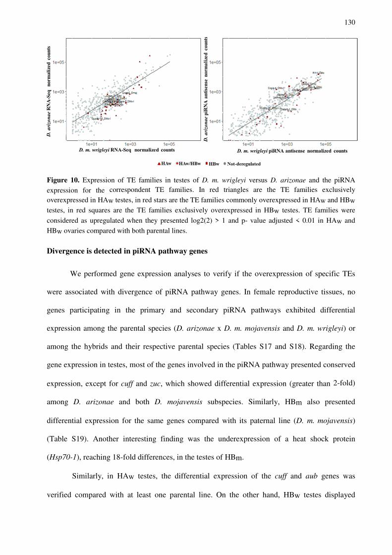
130
Figure 10. Expression of TE families in testes of D. m. wrigleyi versus D. arizonae and the piRNA
expression for the correspondent TE families. In red triangles are the TE families exclusively
overexpressed in HAw testes, in red stars are the TE families commonly overexpressed in HAw and HBw testes, in red squares are the TE families exclusively overexpressed in HBw testes. TE families were
considered as upregulated when they presented log2(2) > 1 and p- value adjusted < 0.01 in HAw and
HBw ovaries compared with both parental lines.
Divergence is detected in piRNA pathway genes
We performed gene expression analyses to verify if the overexpression of specific TEs
were associated with divergence of piRNA pathway genes. In female reproductive tissues, no
genes participating in the primary and secondary piRNA pathways exhibited differential
expression among the parental species (D. arizonae x D. m. mojavensis and D. m. wrigleyi) or
among the hybrids and their respective parental species (Tables S17 and S18). Regarding the
gene expression in testes, most of the genes involved in the piRNA pathway presented conserved
expression, except for cuff and zuc, which showed differential expression (greater than 2-fold)
among D. arizonae and both D. mojavensis subspecies. Similarly, HBm also presented
differential expression for the same genes compared with its paternal line (D. m. mojavensis)
(Table S19). Another interesting finding was the underexpression of a heat shock protein
(Hsp70-1), reaching 18-fold differences, in the testes of HBm.
Similarly, in HAw testes, the differential expression of the cuff and aub genes was
verified compared with at least one parental line. On the other hand, HBw testes displayed
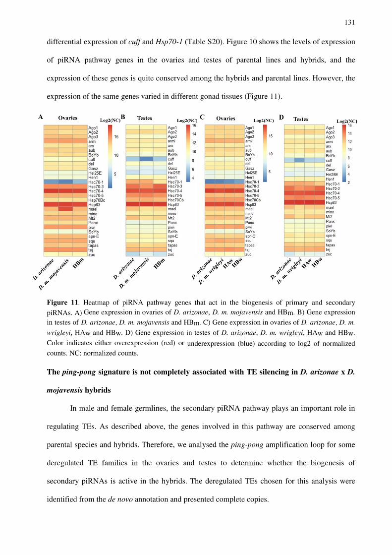
131
differential expression of cuff and Hsp70-1 (Table S20). Figure 10 shows the levels of expression
of piRNA pathway genes in the ovaries and testes of parental lines and hybrids, and the
expression of these genes is quite conserved among the hybrids and parental lines. However, the
expression of the same genes varied in different gonad tissues (Figure 11).
Figure 11. Heatmap of piRNA pathway genes that act in the biogenesis of primary and secondary
piRNAs. A) Gene expression in ovaries of D. arizonae, D. m. mojavensis and HBm. B) Gene expression
in testes of D. arizonae, D. m. mojavensis and HBm. C) Gene expression in ovaries of D. arizonae, D. m.
wrigleyi, HAw and HBw. D) Gene expression in testes of D. arizonae, D. m. wrigleyi, HAw and HBw.
Color indicates either overexpression (red) or underexpression (blue) according to log2 of normalized counts. NC: normalized counts.
The ping-pong signature is not completely associated with TE silencing in D. arizonae x D.
mojavensis hybrids
In male and female germlines, the secondary piRNA pathway plays an important role in
regulating TEs. As described above, the genes involved in this pathway are conserved among
parental species and hybrids. Therefore, we analysed the ping-pong amplification loop for some
deregulated TE families in the ovaries and testes to determine whether the biogenesis of
secondary piRNAs is active in the hybrids. The deregulated TEs chosen for this analysis were
identified from the de novo annotation and presented complete copies.
132
In HBm ovaries, we analysed the piRNA signature for Transib-1_DMo and R1-1_DMo
elements, and in both cases, the hybrids showed the ping-pong signature (Figure S3). Regarding
the other TEs overexpressed in HBm ovaries, the elements Gypsy11_Dya, Gypsy6, Harbinger-
1_DF, I- 1_DBp, Minos showed very few or no antisense and sense piRNAs mapped to them in
HBm and in D. arizonae (the maternal line) (Table S21). Thus, for those TEs, the ping-pong
amplification is likely absent in hybrid ovaries and is responsible for their overexpression.
Similarly, in the ovaries of HAw and HBw, the three TEs (Mariner-3_DMo, Copia-
4_DMo-I and R1-1_DMo) for which the ping-pong signature was analysed showed strong ping-
pong signals (Figure S4). Additionally, for all overexpressed TEs identified using RNA-Seq in
HAw and HBw ovaries, antisense and sense pools of mapping piRNAs were detected at high
levels (Table S22).
In testes, the biogenesis of secondary piRNAs generated by ping-pong amplification does
not appear to be directly associated with TE regulation. In HBm male gonads, the ping-pong
signature was analysed for 9 of 67 deregulated TEs. Among these 9 TEs, only three (hAT-
1_DMo, Gypsy-11_DMo-I and Gypsy-14_DMo-I) did not show ping-pong amplification in the
hybrids, which was also absent in D. arizonae, but present in D. m. mojavensis testes (Figure
S5). Similarly, in ovaries, several of these deregulated TEs that showed the ping-pong signature
displayed sense and antisense piRNA-mapping TEs, explaining the secondary piRNA biogenesis
in the germline (Table S23). Additionally, nine of the 67 misregulated TE families detected in
HBm testes did not show homologous antisense or sense piRNAs (Table S23).
In HAw testes, four of the 28 deregulated TE families were analysed for the ping-pong
cycle. One showed no ping-pong signature for both the parental species and the reciprocal
hybrids (Mariner-3_DMo) and was also deregulated in HAw and HBw ovaries. One showed the
ping-pong signature only for HAw testes, in which this TE was overexpressed (Transib-1_DMo),
and two TE families presented the ping-pong signature in the parental lines and the reciprocal
hybrids (Gypsy-12_DMo-I, also upregulated in HBw testes, and Copia-5_DMo-I) (Figure S6). In
133
HBw testes, the ping-pong signature was present for the two TEs tested (Figure S7). Consistent
with these findings, an analysis of the number of antisense and sense piRNAs mapping to these
deregulated TEs revealed sense and antisense piRNAs for most of these deregulated TEs (Table
S24). Notably, piRNA expression was not detected for 10 of the TEs overexpressed in HAw and
HBw testes (Table S24).
Importantly, some overexpressed TE families that do not have complementary antisense
or sense piRNAs to regulate their expression are shared among HBm, HAw and HBw testes.
More specifically, the TE families that shared a lack of complementary piRNAs in hybrid and
parental testes are Gyspsy-38_DWil, Helitron-1_DT, Helitron-N1_DEl, IVK_DM and Mariner-
16_DK (Tables S23 and S24).
siRNA control in somatic cells of gonad tissues
We have analysed the siRNA populations for the male and female gonads of the parental
lines and hybrids to verify the role of siRNAs in the reproductive tissues. In the ovaries of HBm,
siRNAs associated with 291 TE families were identified. Among these siRNAs, 192 presented
differential expression between D. arizonae and D. m. mojavensis, and most (86%) were
expressed at an at least 2-fold lower level in D. arizonae (Table S25). Complementary siRNAs
were identified for most of the TE families that were overexpressed in HBm ovaries, and the
hybrids generally contained more siRNAs than the parental lines (Table S25). However, four
upregulated TE families (Gypsy11_Dya, Gypsy6, Harbinger-1_DF and I-1_DBp) identified in
the HBm female ovaries did not have complementary siRNAs. On the other hand, in the ovaries
of HAw and HBw, siRNAs associated with 267 TE families were detected. Of those TE families,
207 display differential expression of siRNAs between D. arizonae and D. m. wrigleyi, and most
(92.7%) were underexpressed up to 2-fold in D. arizonae (Table S26). Considering only the
upregulated TE families in HAw and HBw ovaries, siRNAs for almost all of these TE families
were expressed at higher levels in hybrids, regardless of the cross direction, than in parental
134
lines. The only exception was verified for the Gypsy6_Dya family, for which the levels of
siRNAs were lower in HAw ovaries than in both parental lines and in D. m. wrigleyi (maternal
source) compared with D. arizonae (Table S26). However, this difference was less than 2-fold,
and hence, not significant.
In testes, siRNA-mediated control is more complex. In HBm male gonads, 213 TE
families mapped to siRNAs. Comparing the parental species, 134 presented differential siRNA
expression, and, unlike ovaries, most of these siRNAs (79%) were overexpressed in D. arizonae.
Considering the 67 overexpressed TE families detected in the HBm male reproductive tissue,
lower amounts of siRNAs for 15 TE families were detected in hybrids than in both parental lines
(Table S27). Interestingly, the majority of these TE families belong to the Gypsy superfamily,
and ping-pong amplification was analysed in three of these 15 TE families. Only Gypsy-
14_DMo-I did not show the ping-pong signature, indicating that this element is either
exclusively expressed in the soma or it is absent in D. arizonae, since D. m. mojavensis showed
ping-pong amplification. The elements Gypsy4_Dmoj and R1-1_DMo exhibited the ping-pong
signature and lower amounts of siRNAs, suggesting that they are expressed in both the germline
and soma, but the overexpression likely occurs in the somatic cells. Another important finding
was that 35 of the 67 upregulated TE families showed no associated siRNAs, including eight TE
families for which a lack of piRNAs was also detected in this tissue (Table S27). Moreover,
some TEs that exhibited ping-pong amplification also showed larger amounts of siRNAs in the
hybrids than in both parental lines, such as BEL-7_Dmoj and Copia-5_DMo-I, indicating that
these elements may be expressed in somatic cells in the testes, although they are not controlled
by siRNA or piRNA pathways.
Similarly, to the HBm testes, 236 TE families showed associated siRNAs in HAw and
HBw testes. One hundred sixty-two families displayed differential siRNA expression between D.
arizonae and D. m. wrigleyi testes, band most were expressed at higher levels in D. arizonae
(79.6%), as it was previously observed in D. arizonae x D. m. mojavensis comparisons for the
135
same tissue. Considering the upregulated TE families in HAw and HBw testes, none of TE
families with associated siRNAs showed lower expression of siRNAs in the hybrids than in both
parental lines (Table S28). However, for some TEs, differences greater than 2-fold were verified
in hybrid testes than in one of the parental lines (Table S28). Additionally, 17 deregulated TE
families did not present homologous siRNAs, indicating that these elements are not expressed in
the soma, since this pattern was observed for hybrids and both parental lines. Moreover, among
these 17 TE families without associated siRNAs, 9 TE families lacking piRNAs were identified
(Baggins1, Helitron-1_DT, Helitron-N1_DEl, IVK_DM, Loa-3_Dta, Mariner-1_DT, Mariner-
16_DK, ROO and TransibN3_DP) (Table S28). Notably, almost all TE families assessed for the
ping-pong signature in this tissue showed mapping siRNAs, except Transib-1_DMo.
DISCUSSION
Differential expression of TE families in the gonads of D. arizonae and D. mojavensis
We have detected a higher alignment rate of RNA-Seq reads in the TE library in ovary
transcriptomes than in testes. However, a greater number of differentially expressed TE families
was detected in males, similar to the male and female transcriptomes of D. buzzatii and D.
koepferae (ROMERO-SORIANO et al., 2017). Differences in the alignment rate might be
related to difference in the genome size, which is driven by differences in repetitive sequences
between sexes. Indeed, males and females presented differences in genome size, such as D.
buzzatii, D. koepferae, D. mauritiana and D. hydei, in previous studies (GIRARD; HANNON,
2008; ROMERO-SORIANO et al., 2016). Moreover, in males, the dynamic changes in the
number of gene and repetitive sequences may be highly variable due to the Y chromosome
(CARVALHO et al., 2009), which modulates the differential expression among species.
Based on the number of differentially expressed TEs in each tissue, 19% of TEs were
deregulated between D. arizonae and D. mojavensis ovaries, which was lower than the value
reported by Lopez-Maestre et al. (2017) (29%) in the comparison D. arizonae and D. m.

136
mojavensis transcriptomes sequenced at a different time point. Differences in the number of
misregulated TE families between the species of the present study and the study by Lopez-
Maestre et al. (2017) are likely related to the use of different TE libraries. Here, we have used a
library that contains TE sequences derived from the D. mojavensis r 1.04 public genome, as well
as from D. arizonae genome that was recently sequenced, assembled and annotated by our
research group using a de novo approach.
Furthermore, most of the deregulated TEs detected in the ovaries and testes of D.
arizonae and the two D. mojavensis subspecies were LTR retrotransposons, followed by DNA
transposons and LINEs. Interestingly, these proportions of deregulated TE classes for these
species are similar to those reported by Clark et al. (2007), the 12 Genomes Consortium and by
Rius et al. (2016) for LTR elements.
Differential expression of TE families and the relationship with post-transcriptional control
by piRNA and siRNA pathways in hybrid female gonads
Few TE families showed dysregulated expression in the hybrid female reproductive
tissues compared with both parental lines. This result concurs with the findings reported by
Lopez-Maestre et al. (2017), who analysed ovaries of hybrids from the same parental species.
However, it is quite different from the results described by Romero-Soriano et al. (2017) for D.
buzzatii and D. koepferae female hybrids (4.96 m.y. of divergence), since ~35% (234 of 658 TE
families) of differentially expressed TE families were identified. Similarly, our results differ
from those reported for female hybrids of D. melanogaster and D. simulans (KELLEHER et al.,
2012; SATYAKI et al., 2014), where a larger number of deregulated TEs was verified. These
results corroborate our hypothesis that a longer divergence time of two genomes results in a
greater rate of TE deregulation.
We sequenced and analysed the piRNA pool of parental species and their respective
hybrids to obtain a better understanding of the factors that lead to higher levels of TE
137
transcription in hybrids. In ovaries of HBm derived from crosses of D. arizonae females x D. m.
mojavensis males, nine TEs were overexpressed, most belonging to the Gypsy superfamily. The
maternal source showed fewer sense and antisense piRNAs mapping to TEs, and some of them,
such as the Gypsy11_Dya, Gypsy6, Harbinger-1_DF, I-1_DBp and MINOS families, showed
very few or no antisense and sense mapping piRNAs in D. arizonae and HBm. Thus, these
elements may be absent in the maternal species D. arizonae, contributing to the upregulation of
these TE families in the hybrid genome through a lack of primary piRNAs, maternal deposition
or transcription from single-strand piRNA clusters, initiating the ping-pong loop. However,
analyses of the ping-pong signature must be performed to confirm this hypothesis. In fact, the
contribution of the maternal cytotype in post-transcriptional silencing mechanisms mediated by
piRNAs has been reported in hybrids of D. arizonae and D. m. mojavensis by Lopez-Maestre et
al. (2017) for two LTR retrotransposons. Considering other Drosophila species, a similar pattern
has been reported in intraspecies hybrids of D. melanogaster and D. simulans and D. virilis
intraspecies hybrids (PICARD, 1976; BLACKMAN et al., 1987; YANNOPOULOS et al., 1987;
KIDWELL et al., 1977; PETROV et al., 1995; HILL et al., 2016; FUNICOV et al., 2018).
Nonetheless, the overexpressed MINOS TE family presented a different pattern, since no
antisense and sense piRNAs were detected in hybrids and both parental lines, suggesting that the
expression of this element is exclusively somatic.
Additionally, in ovaries of HBm, HAw and HBw hybrids, some upregulated TEs
displayed strong ping-pong signals and antisense and sense piRNAs in the maternal species, thus
failures in silencing the paternally inherited TE family mediated by the secondary biogenesis of
piRNAs do not explain their deregulation. Likewise, the element Frogger displays a similar
pattern in D. arizonae x D. m. mojavensis female hybrids (LOPEZ-MAESTRE et al., 2017),
since its upregulation was verified in one cross direction and strong ping-pong amplification was
observed. The authors claimed that the main explanation for this phenomenon was related to the
diversity of copies expressed in the parental genome, which limits the capacity to attribute

138
transcripts and piRNAs to specific insertions (LOPEZ-MAESTRE et al., 2017). Similar results
were also observed in female hybrids of D. melanogaster x D. simulans and D. buzzatii x D.
koepferae (KELLEHER et al., 2012; ROMERO-SORIANO et al., 2017), since in both cases, TE
derepression in hybrids is not always associated with the disruption of the ping-pong cycle for all
misregulated TEs, as was verified in the present study. Nevertheless, in hybrids from those more
divergent species, the failure to regulate TEs is widely associated with adaptive divergence in
piRNA pathway genes (KELLEHER et al., 2012; ROMERO-SORIANO et al., 2017). However,
divergence in piRNA pathway genes does not explain the TE dysregulation in hybrids from more
recently diverged species, such as D. arizonae and D. mojavensis, since no differential
expression was detected. Similar results were reported by Lopez-Maestre et al. (2017), since
they observed less than 2% of divergence between orthologous genes of D. mojavensis and D.
arizonae for more than 95% of the genes.
Because some upregulated TE families did not show failures in the secondary biogenesis
of piRNAs or divergence of piRNA genes in hybrid ovaries, we speculated that two main
reasons may underlie TE deregulation. The first reason is potentially related to the expression of
these TEs in somatic cells, which are present in Drosophila ovaries and have a specific post-
transcriptional regulation system driven by siRNAs (21 nt in length) (reviewed in SATO;
SIOMI, 2020). Therefore, failures in this type of silencing might induce TE overexpression in
ovaries. We analysed the siRNA population associated with TE families in ovaries of the
parental lines and hybrids to confirm this hypothesis. Interestingly, no associated siRNAs were
identified for some elements that were upregulated in HBm ovaries and lacked antisense or sense
complementary piRNAs in hybrids and the maternal line (Gypsy11_Dya, Gypsy6, Harbinger-
1_DF and I-1_DBp), suggesting that they are exclusively expressed in the germline.
Furthermore, their overexpression is likely linked to the lack of primary piRNAs able to start the
ping-pong amplification cycle. On the other hand, the transposon Minos, which was deregulated
(up to 2-fold) in HBm and D. arizonae compared with D. m. mojavensis, did not display any

139
antisense or sense complementary piRNAs, but had associated siRNAs. Based on this result, the
overexpression of this TE occurs in the somatic cells of the ovary and is related to lower amounts
of siRNAs that control its expression in hybrids. Despite the positive correlation between siRNA
levels and TE expression, some upregulated TE families identified in the ovaries of HBm, HAw
and HBw exhibited larger amounts of associated siRNAs and piRNAs, as well as a strong ping-
pong signature, suggesting that these TEs are expressed in somatic and germline cells. Indeed,
piRNAs and siRNAs have been shown to originate from the same genomic clusters, indicating
that they may arise from the same single precursor transcript (GHILDIYAL et al., 2008).
Moreover, functional interactions between piRNA and siRNA pathways have been identified in
C. elegans (BAGIJN et al., 2012), confirming that they may function cooperatively in TE
silencing. However, TEs overexpressed in ovaries presented associated piRNA and siRNAs,
indicating that other mechanisms might be influencing their deregulation. Importantly, although
we postulate that the expression of some specific TEs occurs in somatic and/or germ cells, an
analysis designed to locate TE transcript expression in gonad tissues must be performed to
confirm these findings.
The second reason underlying TE deregulation might be related to epigenetic changes
and failure of the transcriptional regulatory mechanism. Some stressful events, such as heat
shock, colonization of new environments and interspecific hybridization, have been shown to
activate transposable elements in male and female germlines, which is associated, at some level,
with the piRNA regulation mechanism (LABRADOR et al., 1999; FONTDEVILA, 2005;
KELEHER et al., 2012; VELA et al., 2014; GUERREIRO, 2014; SATYAKI et al., 2014;
SAINT-LEANDRE et al., 2015; CAPPUCCI et al., 2019). In Drosophila ovaries, the Piwi
protein plays a major role in heterochromatin modification by interacting with several proteins,
including HP1 and H1, leading to heterochromatin formation (YIN; LIN, 2007;
MOSHKOVICH; LEI, 2010; OLOVNIKOV et al., 2012; SATO; SIOMI, 2020). Moreover, for
some specific TEs, maternally deposited piRNAs have an important role in silencing TEs in the
140
somatic ovarian cells (AKKOUCHE et al., 2013), and they are responsible for chromatin
modification and TE regulation. Thus, for some TEs, the failure of maternal piRNA deposition
might contribute to the TE activation observed in the D. arizonae-D. mojavensis hybrids that did
not show antisense piRNAs in the maternal lines. Nonetheless, in hybrids of D. melanogaster
and D. simulans, interspecies hybridization is responsible for TE activation in germ cells and in
somatic follicle cells of ovaries (THOMAE et al., 2013; SATYAKI et al., 2014). In these
hybrids, although they have normal amounts of piRNAs, their heterochromatin genes are
downregulated, which is associated with Hmr (Hybrid male rescue) and Lmr (Lethal male
rescue) genes that are divergent among the parental lines. These genes, which are responsible for
hybrid incompatibility in D. melanogaster x D. simulans, exert a positive effect on
heterochromatin genes, such as HP1a. Thus, in the D. arizonae-D. mojavensis hybrids, which
present regular amounts of piRNAs and conserved expression of genes that participate in the
piRNA pathway, other factors, such as chromatin changes induced by gene misregulation, may
alter TE expression.
Differential expression in the hybrid male reproductive tissues and TE post-transcriptional
control by piRNA and siRNA pathways
Overall, male gonads showed higher differential expression of TEs than ovaries. Hybrid
males obtained from D. arizonae x D. mojavensis crosses are sterile, although they show
different phenotypes in sperm motility, depending on the cross direction (RUIZ et al., 1990;
REED; MARKOW, 2004).
One of the potential mechanisms affecting the hybrid incompatibility is increased TE
expression. TEs are thought to contribute to reproductive isolation through transcriptional
misregulation, leading to sterility or inviability (CASTILLO; MOYLE, 2019). Here, hybrids
obtained from the same cross direction (HBm and HBw) that exhibit the same phenotype of
immotile sperm displayed different amounts of deregulated TEs, with very few being equally
141
misregulated between them. On the other hand, HAw, hybrids obtained from crosses between
D.m. wrigleyi females x D. arizonae males that are sterile and have motile sperm, showed more
deregulated TE families than HBw. Thus, the two subspecies of D. mojavensis likely have
different TE contents, and when interspecies crosses occur, TE misregulation is detected in their
hybrids at different levels. Differences in TE contents are often observed in Drosophila, even for
different populations of the same species or for different species (SONG et al., 2014; FABLET
et al., 2019), and the consequences of these differences under hybridization events is often
observed as hybrid dysgenesis (PICARD, 1976; KIDWELL et al., 1977; BLACKMAN et al.,
1987; YANNOPOULOS et al., 1987; PETROV et al., 1995; HILL et al., 2016; FUNICOV et al.,
2018). According to Castillo and Moyle (2019), the occurrence of hybrid sterility and hybrid
inviability might result from the misregulation of TEs at different developmental stages, and as
shown in the present study, it may be related to the misregulation in different tissues. Therefore,
the rapid evolution of TEs in different lineages potentially explains the presence of polymorphic
incompatibilities in many systems (CASTILLO; MOYLE, 2019).
In our hybrids, most of the deregulated TEs were overexpressed, in contrast to the results
reported by Romero-Soriano et al. (2017), who described underexpression of TEs in hybrid
males from D. buzzatii and D. koepferae. Some studies have examined the location of
overexpressed TEs in hybrid testes and found that the new copies were expressed in the primary
spermatocytes near the apical and basal regions of the testes (CARNELOSSI et al., 2014;
GUERREIRO, 2015). The apical region is composed of germline stem cells and primary and
secondary spermatocytes, while the basal region contains transcripts for many spermatogenesis
genes (FULLER, 1998; VEDELECK et al., 2018). Therefore, we speculated that in D.
mojavensis-D. arizonae hybrids, the higher expression of TE families observed in testes might
influence the hybrid incompatibility, which is stronger in males. Additionally, higher TE
expression may be a product of divergence of some specific regulatory genes, such as piRNA
pathway genes or genes involved in the transcriptional control of TEs (histone markers), or
142
differences in the TE content in the parental genomes, leading to a lack of piRNAs for some
specific TE families.
In testes, the regulation of TEs by piRNA post-transcriptional silencing is complex. For
some deregulated TE families in HAw, HBw and HBm, the relationship between the TE
overexpression and lack of ping-pong amplification was verified. However, for several TEs,
regardless of the presence of sense and antisense mapping piRNAs, upregulation was verified. In
Drosophila testes, the piRNA pathway functions is differently from ovaries (NAGAO et al.,
2010), and it may be related to a specific developmental stage and hybrid sterility (KOTOV et
al., 2019). In fact, according to Quénerch’du et al (2016), different populations of piRNA are
present in the spermatogonia or primary spermatocytes. In spermatogonia, piRNAs are
predominantly transposon-mapping piRNAs, and the ping-pong signature has been verified for
several of these sequences. However, in testes enriched in primary spermatocytes, Ago3 is not
expressed, but the ping-pong signature is observed, indicating that a noncanonical ping-pong
cycle functions in spermatogenesis. In our analyses of piRNA-related genes, no misregulation or
absence of Ago3 was observed in testes; nevertheless, this protein was expressed at lower levels
in testes than in ovaries. Moreover, in testes enriched in primary spermatocytes, most of the
piRNAs function to suppress the repeats and TEs (QUÉNERCH’DU et al., 2016). However, in
the testes of aub (Aubergine) mutants, where Ago3 expression is also perturbed, the number of
transposon-mapping piRNAs was reduced, while piRNA biogenesis was affected in aub
heterozygous testes (QUÉNERCH’DU et al., 2016).
Unlike ovaries, three genes involved in the piRNA pathway displayed differential
expression in the testes: cuff, aub and zuc. The Cutoff (cuff) gene has an essential role in piRNA
production from dual-strand clusters (PANE et al., 2011). Moreover, their interactions with other
piRNA proteins, such as Rhino (rhi) and Deadlock (del), form the RDC complex, which licenses
the transcription of dual-strand piRNA clusters (MOHN et al., 2014). On the other hand, the
gene Zucchini (zuc) is necessary for the biogenesis of Piwi-bound piRNAs (HAN et al., 2015),

143
whereas Aubergine (aub) participates in the secondary biogenesis of piRNAs, together with
Ago3, by binding mature piRNAs and initiating the ping-pong amplification cycle (review in
LUO; LU, 2017). In the HAw males, two genes (cuff and aub) presented differential expression
compared with at least one parental line, which might affect the piRNA silencing mechanism to
some extent. Interestingly, differential Aub expression was also verified in hybrids of D. buzaatti
x D. koepferae, but only for one cross direction (GÁMEZ-VISAIRAS et al., 2020). Moreover,
differential expression was verified for cuff and zuc in D. arizonae, the two D. mojavensis
subspecies, and in the HBm and HBw hybrids. Indeed, some genes with functions in genome
maintenance, such as piRNA or siRNA pathway genes, display signatures of positive selection
(OBBARD et al., 2009; KOLACZKOWSKI et al., 2011; FABLET, 2014), and among these
genes, aub and zuc present evidence of adaptations, since deviations in neutrality have been
verified (OBBARD et al., 2009; GÁMEZ-VISAIRAS et al., 2020). Thus, differential expression
of piRNA pathway genes may affect the large amount of deregulated TEs in hybrid testes.
Based on our data, the siRNA control pathway is quite complex in the male reproductive
tissues, since several TEs overexpressed in HBm, HAw and HBw testes exhibited larger amounts
siRNAs mapping to deregulated TE families. Surprisingly, most of these TEs also showed
complementary piRNAs and the ping-pong signature. Thus, they are not efficiently regulated by
the siRNA or piRNA pathway. A potential explanation for the higher amount of homologous
siRNAs for these TEs is a compensatory mechanisms, as was previously reported in D.
melanogaster. When problems in the piRNA pathway were detected, an increase in siRNA
production has been reported for numerous elements (ROZHKOV et al., 2013). However, for
these specific TEs, no decrease in piRNA production was detected, suggesting that epigenetic
changes may play a role in TE derepression. At the same time, several TEs that were upregulate
in male reproductive tissues presented lower amounts of complementary siRNAs than the
parental lines. Based on this result, the overexpression of these elements likely occurs in the
soma. Surprisingly, most of these elements belong to the Gypsy superfamily. Gypsy
144
retrotransposons are endogenous retroviruses (ERVs), and their expression in Drosophila is
mainly controlled by siRNAs (WEBER et al., 2006; GHILDIYAL et al., 2008), suggesting that
the lack of control by siRNAs in the somatic cells of the testes might promote the overexpression
of this type of TE. Furthermore, in the present study, some TEs did not present complementary
siRNAs or piRNAs in hybrids or the parental lines. These elements were likely transcriptionally
regulated in the parental lines, possibly in heterochromatin regions, but due to divergence of
regulatory sequences, they lost their regulatory mechanism in hybrids and were upregulated,
since post-transcriptional regulatory mechanisms are absent. However, additional analyses must
be performed to confirm this hypothesis.
Similar to the situation in ovaries, we are unable to exclude other factors that play a
potential role in TE derepression in testes. In fact, the hybrids HBm and HBw showed decreased
expression of a heat shock protein, Hsp70-1. Proteins of this family have been shown to play
roles in the siRNA, miRNA and piRNA regulatory pathways (IWASAKI et al., 2010; KARAM
et al., 2017; CAPPUCCI et al., 2019). The Hsp70 chaperone has recently been shown to interact
with components of chaperone machinery involved in piRNA biogenesis, and the disruption of
these proteins decrease the efficacy of TE repression (CAPPUCCI et al., 2019). Therefore, in
testes, the post-transcriptional and transcriptional silencing mechanisms are likely more complex
and prevent TE derepression, which in hybrid testes, is disturbed due to gene misexpression and
regulatory incompatibilities.
CONCLUSIONS
The findings from this study make several contributions to the current literature. Here, the
genome of female hybrids of D. arizonae and D. mojavensis is likely to be more stable than the
genome of males. In hybrid ovaries, a few TEs showed dysregulated expression compared to that
in both parental lines, which might be related to the fertile phenotypes of these hybrids. In male
hybrids, substantial TE derepression was verified, regardless of the cross direction, indicating
145
that TE overexpression may play an important role in determining the sterile hybrid phenotype.
Furthermore, in these hybrids, the silencing mechanism in ovaries and testes is complex, since
some TEs were upregulated even in the presence of complementary piRNAs and ping-pong
amplification, indicating that hybrid incompatibility might affect other components of piRNA
biogenesis and result in TE derepression.
REFERENCES AKKOUCHE, A. et al. Maternally deposited germ-line piRNAs silence the tirant
retrotransposon in somatic cells. EMBO Rep, v.14, p. 458–464, 2013. ANTONIEWSK, C. Computing siRNA and piRNA overlap signatures In: Animal Endo- SiRNAs: Methods and Protocols. Cap. 12, v. 1173, p. 135-146, 2014. ARAVIN, A. A.; HANNON, G. J.; BRENNECKE, J. The Piwi-PiRNA pathway provides an adaptive defense in the transposon arms race. Science, v. 318, p. 761–764, 2007. BAACK, E. J.; WHITNEY, K. D.; RIESEBERG, L. H. Hybridization and genome size evolution: timing and magnitude of nuclear DNA content increases in Helianthus homoploid hybrid species, New Phytol, v.167, n.2, p. 623–630, 2005. BAFFI, M. A.; CERON, C. R. Molecular analysis of the rDNA ITS-1 intergenic spacer in Drosophila mulleri, D. arizonae, and their hybrids. Biochemical Genetics, v. 40, p. 411-421, 2002. BAGIJN, M. P. et al. Function, targets, and evolution of Caenorhabditis elegans piRNAs. Science, v. 337, p. 574–578, 2012. BAO, W.; KOJIMA, K. K.; KOHANY, O. Repbase Update, a database of repetitive elements in eukaryotic genomes. Mobile DNA, v. 6, n. 1, p. 11, 2 dez. 2015. BENJAMINI, Y.; HOCHBERG, Y. Controlling the false discovery rate: A practical and powerful approach to multiple testing. Journal of the Royal Statistical Society, v. 57, n. 1, p. 289-300, 1995. BERNARDO CARVALHO, A.; KOERICH, L. B.; CLARK, A. G. Origin and evolution of Y chromosomes: Drosophila tales. Trends Genet. v. 25, p. 270–277, 2009. BLACKMAN, R. K. et al. Mobilization of hobo elements residing within the decapentaplegic gene complex: suggestion of a new hybrid dysgenesis system in Drosophila melanogaster. Cell, v. 49, p. 497–505, 1987. BRAY, N. L. et al. Near-optimal probabilistic RNA-seq quantification, Nature Biotechnology, v. 34, p. 525–527, 2016. CAPELLA-GUTIÉRREZ, S.; SILLA-MARTÍNEZ, J. M.; GABALDÓN, T. trimAl: a tool for automated alignment trimming in large-scale phylogenetic analyses. Bioinformatics
146
applications note, v. 25, n. 15, p. 1972–1973, 2009. CAPPUCCI, U.; NORO, F.; CASALE, A. S. The Hsp70 chaperone is a major player in stress- induced transposable element activation. PNAS, p. 1-8, 2019. CAPY, P.; GASPERI, G. BIÉMONT, C.; BAZIN, C. Stress and transposable elements: co- evolution or useful parasites? Herediy, v. 85, p. 101-106, 2000. CARNELOSSI, E. A. G. et al. Specific activation of an I-like element in Drosophila
interspecific hybrids. Genome Biology Evolution. v. 6, n. 7, p. 1806–1817, 2014.
CASTILLO D. M.; MOYLE, L. C. Transposable elements that cause dysgenesis also contribute to postzygotic isolation in the Drosophila virilis clade. bioRxiv preprint first posted online. Doi: http://dx.doi.org/10.1101/753814, 2019. CLARK, A. G. et al. Evolution of genes and genomes on the Drosophila phylogeny - Drosophila 12 Genomes Consortium. Nature, v. 450, n. 8, 2007. COYNE, J. A., ORR, H. A. ‘‘Patterns of speciation in Drosophila’’ revisited. Evolution, v. 51, n.1, p. 295–303, 1997. CUTTER A. D. Divergence times in Caenorhabditis and Drosophila inferred from direct estimates of the neutral mutation rate. Mol Biol Evol, v. 25, n. 4, p. 778–786, 2008. DOBZHANSKY, T. Studies on hybrid sterility. II. Localization of sterility factors in Drosophila
pseudoobscura hybrids. Genetics, v. 21, p. 113—135, 1936. DURINCK, S. et al. “BioMart and Bioconductor: a powerful link between biological databases and microarray data analysis.” Bioinformatics, v. 21, p. 3439–3440, 2005. FABLET, M. et al. Dynamic interactions between the genome and an endogenous retrovirus: Tirant in Drosophila simulans Wild-Type Strains. G3: Genes, Genomes, Genetics, v. 9, 2019. FABLET, M. Host Control of insect endogenous retroviruses: Small RNA silencing and immune response. Viruses, v. 6, n. 11, p. 4447-64, 2014. FABLET, M. Host Control of Insect Endogenous Retroviruses: Small RNA Silencing and Immune Response. Viruses, v. 6, p. 4447-4464, 2014. FLUTRE, T. et al. Considering transposable element diversification in de novo annotation approaches. PLoS ONE, v. 6, n. 1, 2011. FONTDEVILA, A. Hybrid genome evolution by transposition. Cytogenet Genome Res. v. 110, p. 49–55, 2005. FULLER, M. T. Genetic control of cell proliferation and differentiation in Drosophila
spermatogenesis. Semin Cell Dev Biol. v. 9, p. 433–444, 1998. FUNIKOV, S. Y. et al. Spontaneous gain of susceptibility suggests a novel mechanism of resistance to hybrid dysgenesis in Drosophila virilis. PLOS Genetics, p. 1-20, 2018. Genome Res. v. 110, p. 462–467, 2005. GÁMEZ-VISAIRA, V. et al. Drosophila interspecific hybridization causes a deregulation of the
147
piRNA pathway genes. Genes, v.11, n. 215, p. 2-15, 2020. GHILDIYAL, M. et al. Endogenous siRNAs derived from transposons and mRNAs in Drosophila somatic cells. Science, v. 320, p. 1077–1081, 2008. GIRARD, A.; HANNON, G. J. Conserved themes in small-RNA-mediated transposon control. Trends Cell Biol. V. 18, p. 136–148, 2008.
GOMES S.; CIVETTA A. Misregulation of spermatogenesis genes in Drosophila hybrids is lineage-specific and driven by the combined effects of sterility and fast male regulatory divergence. J Evol Biol. v. 27, n. 9, p. 1775–1783. 2014. GOMEZ, G.A.; HASSON, E. Transpecific polymorphisms in an inversion linked esterase locus in Drosophila buzzatii. Mol Biol Evol. v. 20, n. 3, p.410–423, 2003. GRETZINGER, T. et al. A user-friendly chromatographic method to purify small regulatory RNAs. Methods, v. 67, p. 91–101, 2014. GUERREIRO, M. P. Changes of Osvaldo expression patterns in germ-line of male hybrids between the species Drosophila buzzatii and Drosophila koepferae. Mol. Genet. Genomics, v. 290, n. 4, p.1471-83, 2015. GUERREIRO, M. G. What makes transposable elements move in the Drosophila genome? Heredity (Edinb), v. 108, p. 461–468, 2012. GUINDON, S. et al. New algorithms and methods to estimate Maximum-Likelihood phylogenies: Assessing the performance of PhyML 3.0. Systematic Biology, n. 59, p. 307–21, 2010. HAERTY, W.; SINGH, R. S. Gene regulation divergence is a major contributor to the evolution of Dobzhansky–Muller incompatibilities between species of Drosophila. Mol. Biol. Evol., v. 23, n. 9, p. 1707–14, 2006. HAN, B. W. et al. piRNA-guided transposon cleavage initiates Zucchini-dependent, phased piRNA production. Science, v. 348, p. 817–21, 2015.
HERNANDEZ-HERNANDEZ, E.M. et al. Genome-wide analysis of transposable elements in the coffee berry borer Hypothenemus hampei (Coleoptera: Curculionidae): description of novel families. Mol Genet Genomics. V. 292, p. 565–583, 2017. HILL, T.; SCHLOTTERER, C.; BETANCOUT, A. J. Hybrid dysgenesis in Drosophila simulans
associated with a rapid Invasion of the P-Element. PLoS Genet. v. 12, n. 3, p. e1005920, 2016. IWASAKI, Y. W. et al., Hsc70/Hsp90 chaperone machinery mediates ATP-dependent RISC loading of small RNA duplexes. Mol. Cell, v. 39, p. 292–299, 2010. IWASAKI, Y. W. et al. Piwi modulates chromatin accessibility by regulating multiple factors including Histone H1 to repress transposons. Molecular Cell, v. 63, n. 3, p. 408–419, 2016. JENNINGS, J. H.; ETGES. W. J. Species hybrids in the laboratory but not in nature: a reanalysis of premating isolation between Drosophila arizonae and D. mojavensis. The Society for the
148
Study of Evolution. v. 64, n. 2, p. 587–598, 2009. KARAM, J. A. et al. Co-chaperone Hsp70/Hsp90-organizing protein (Hop) is required for transposon silencing and Piwi-interacting RNA (piRNA) biogenesis. J. Biol. Chem. v. 292, p. 6039–6046, 2017.
KATOH, K.; STANDLEY, D. M. MAFFT multiple sequence alignment software version 7: Improvements in performance and usability. Molecular Biology and Evolution, v. 50 30, n. 4, p. 772–780, 2013. KAWAKAMI, T. et al. Different scales of Ty1/copia-like retrotransposon proliferation in the genomes of three diploid hybrid sunflower species. Heredity, v. 104, n. 4, p. 341-50, 2010. KELLEHER, E. S.; MARKOW, T. A. Reproductive tract interactions contribute to isolation in Drosophila. Fly, v. 1, n. 1, p. 33-37, 2007. KIDWELL, M. G.; KIDWELL, J. F.; SVED, J. A. Hybrid dysgenesis in Drosophila
melanogaster: A syndrome of aberrant traits including mutation, sterility and male recombination. Genetics, v. 86, p. 813–833, 1977. KIDWELL, M. G.; LISCH, D. R. Perspective: transposable elements, parasitic DNA, and genome evolution. Evolution, v. 55, n. 1, p. 1–24, 2001. KLATTENHOFF, C.; THEURKAUF, W. Biogenesis and germline functions of piRNAs. Development, v. 135, n. 1, p. 3-9, 2008. KLIMAN, R. M. et al. The population genetics of the origin and divergence of the Drosophila
simulans complex species. Genetics, v. 156, n. 4, p. 1913–31, 2000. KOLACZKOWSKI, B.; HUPALO, D. N.; KERN, A. D. Recurrent adaptation in RNA interference genes across the Drosophila phylogeny. Mol. Biol. Evol. v. 28, n. 2, 1033–1042, 2011. KOTOV, A. A. et al. piRNA silencing contributes to interspecies hybrid sterility and reproductive isolation in Drosophila melanogaster. Nucleic Acids Research, v. 47, N. 8, p. 4255–4271, 2019. LAAYOUNI, H. et al. The evolutionary history of Drosophila buzzatii. XXXV. Inversion polymorphism and nucleotide variability in different regions of the second chromosome. Mol Biol Evol. v. 20, n. 6, p. 931–944, 2003. LABRADOR M. et al. Interspecific hybridization increases transposition rates of Osvaldo. Mol Biol Evol. v. 16, p. 931–937, 1999. LANGMEAD, B. et al. Ultrafast and memory-efficient alignment of short DNA sequences to the human genome. Genome Biol. v.10, p. R25, 2009. LEFORT, V.; LONGUEVILLE, J. E.; Gascuel, O. SMS: Smart model selection in PhyML supplementary material. Mol. Biol. Evol, v. 34, n. 9, p. 2422–2424, 2017. LERAT, E. et al. TEtools facilitates big data expression analysis of transposable elements and
149
reveals an antagonism between their activity and that of piRNA genes. Nucleic Acids Res, v. 45, n. 4, p. e17, 2017. LI, R. et al. Specific downregulation of spermatogenesis genes targeted by 22G RNAs in hybrid sterile males associated with an X-Chromosome introgression. Genome Research, 26:1219– 1232, 2016. LOPEZ-MAESTRE, H. et al. Identification of misexpressed genetic elements in hybrids between Drosophila-related species. Scientific Report, v. 7, n. 40618, 2017. LOVE, M. I.; HUBER, W.; ANDERS, S. Moderated estimation of fold change and dispersion for RNA-seq data with DESeq2. Genome Biology. V. 15, p. 550, 2014. LUO, S. ; LU, J. Silencing of transposable elements by piRNAs in Drosophila: An evolutionary perspective. Genomics Proteomics Bioinformatics, v. 15, n. 3, p. 164–176, 2017. MA, D.; MICHALAK, P. Ephemeral association between gene CG5762 and hybrid male sterility in Drosophila sibling species. J. Mol. Evol., v. 73, n. 3-4, p.181–187, 2011. MALONE C. D. et al. Specialized piRNA pathways act in germline and somatic tissues of the Drosophila ovary. Cell, v. 137, p. 522–535, 2009. MARTIN, M. Cutadapt Removes Adapter Sequences from HighThroughput Sequencing Reads. EMBnet.journal. v. 17, p. 10–12, 2011. MCMANUS, C. J. et al. Regulatory divergence in Drosophila revealed by mRNA-seq. Genome Res. 20, 816–25, 2010. MICHALAK, P.; NOOR, M. A. Genome-wide patterns of expression in Drosophila pure species and hybrid males. Mol. Biol. Evol, v. 20, n. 7, p. 1070–6, 2003. MODOLO, L.; LERAT, E. UrQt: an efficient software for the Unsupervised Quality trimming of NGS data. BMC Bioinformatics, v. 16, n. 137, 2015. MOHN, F. The Rhino-Deadlock-Cutoff complex licenses noncanonical transcription of dual- strand piRNA clusters in Drosophila. Cell, v. 157, p. 1364-1379, 2014. MOSHKOVICH N, N. HP1 recruitment in the absence of argonaute proteins in Drosophila. PLoS Genet, v. 6, p. e1000880 28, 2010. MULLER, H. J. Isolating mechanisms, evolution, and temperature. Biol. Symp., v. 6, 71–125, 1942. NAGAO, A. et al. Biogenesis pathways of piRNAs loaded onto AGO3 in the Drosophila testis. RNA, v.16, p. 2503–2515, 2010. O’NEILL, R. J.; O’NEILL, M. J.; GRAVES, J. A. Undermethylation associated with retroelement activation and chromosome remodeling in an interspecific mammalian hybrid. Nature, v. 393, n. 6680, p. 68-72, 1998. OBBARD, D. J., et al., Quantifying adaptive evolution in the Drosophila immune system. PLoS Genet. v. 5, n. 10, e1000698, 2009.
150
OLIVEIRA, D. C.S. G; et al. Monophyly, divergence times, and evolution of host plant use inferred from a revised phylogeny of the Drosophila repleta species group. Mol Phylogenet Evol. v. 64, n. 3, p. 533–544, 2012.
OLOVNIKOV, I.; ARAVIN, A. A.; FEJES, T. K. Small RNA in the nucleus: the RNA- chromatin ping-pong. Curr Opin Genet Dev, v. 22, p. 164–171, 2012. PANE, A. et al. The Cutoff protein regulates piRNA cluster expression and piRNA production in the Drosophila germline. EMBO J., v. 30, p. 4601–4615, 2011. PETROV, D. A. et al. Diverse transposable elements are mobilized in hybrid dysgenesis in Drosophila virilis. Proc. Natl. Acad. Sci. U S A. 92, 17:8050-4, 1995. PICARD, G. Non-mendelian female sterility in Drosophila melanogaster: hereditary transmission of I factor. Genetics, v. 83, p. 107–123, 1976. QUÉNERCH’DU, E.; ANAND, A.; KAI, T. The piRNA pathway is developmentally regulated during spermatogenesis in Drosophila. RNA, v. 22, n. 7, p.1044–1054, 2016. R Core Team. R: A language and environment for statistical computing. R Foundation for Statistical Computing, Vienna, Austria, 2018. Available online at https://www.R-project.org/. RANZ, J. M. et al. Anomalies in the expression profile of interspecific hybrids of Drosophila
melanogaster and Drosophila simulans. Genome Res. 14, 373–9, 2004. REBOLLO, R. et al. A snapshot of histone modifications within transposable elements in Drosophila wild type strains. Plos One, v. 7, n. 9, p. e44253, 2012. REED, L. K.; LAFLAMME, B. A.; MARKOW, T. A. Genetic architecture of hybrid male sterility in Drosophila: Analysis of intraspecies variation for interspecies isolation. Plos One, v. 3, n. 8, p. e3076, 2008. REED, L. K.; MARKOW, T. A. Early events in speciation: polymorphism for hybrid male sterility in Drosophila. Proc Natl Acad Sci U S A, v. 101, n. 24, 2004. RIUS, N. et al. Exploration of the Drosophila buzzatii transposable element content suggests underestimation of repeats in Drosophila genomes. BMC Genomics, v. 17, p. 344, 2016. ROMERO-SORIANO, V. et al. Transposable element misregulation is linked to the divergence between parental piRNA pathways in Drosophila Hybrids. Genome Biol. Evol. v. 9, n.6, p.1450–1470, 2017. ROMERO-SORIANO, V. et al. Drosophila females undergo genome expansion after interspecific hybridization. Genome Biol. Evol., v. 8, n. 3, p. 556–561, 2016. ROZHKOV, N. V.; HAMMELL, M.; HANNON, G. J. Multiple roles for Piwi in silencing Drosophila transposons. Genes Dev,v. 27, p. 400–412, 2013. RUIZ, A.; HEED, W. B.; WASSERMAN, M. Evolution of the mojavensis cluster of cactophilic Drosophila with descriptions of two new species. Journal of Heredity, v. 81, n. 1, p.30-42, 1990.
151
RUSSO, C. A. M.; TAKEZAKI, N.; NEI, M. Molecular phylogeny and divergence times of drosophilid species. Molecular Biology and Evolution, v. 12, p. 391–404, 1995.
SAINT-LEANDRE, B. et al. Transcriptional polymorphism of piRNA regulatory genes underlies the mariner activity in Drosophila simulans testes. Molecular Ecology, v.26, p.3715– 3731, 2017. SANCHES-FLORES, A. et al. Genome evolution in three species of cactophilic Drosophila. G3, Genes, Genomes, Genetics, v. 6, 2016. SATO, K.; SIOMI, M. C. The piRNA pathway in Drosophila ovarian germ and somatic cells. The Japan Academy, v. 96, n. 1, p. 32-42, 2020. SATYAKI, P. R. V. et al. The Hmr and Lhr hybrid incompatibility genes suppress a broad range of heterochromatic repeats. PLOS Genetics, v. 10, e1004240, 2014. SCHMIEDER, R.; EDWARDS, R. Quality control and preprocessing of metagenomic datasets. Bioinformatics, v. 27, p. 863–864, 2011. SENTI, K. A.; BRENNECKE, J. The piRNA pathway: A fly’s perspective on the guardian of the genome. Trends Genet. v. 26, p. 499–509, 2010. SERRATO-CAPUCHINA, A.; MATUTE, D. R. The role of transposable elements in speciation. Genes. v. 9, n. 5, 2018. SESSEGOLO, C., BURLET, N., HAUDRY, A. Strong phylogenetic inertia on genome size and transposable elemento content among 26 species of flies. Biol. Lett. v. 12, p. 20160407, 2016. SMIT, A. et al. RepeatMasker Open-4.0 http://www.repeatmasker.org, 2015. SONESON, C.; LOVE, M. I.; ROBINSON, M. D. “Differential analyses for RNA-seq: transcript-level estimates improve gene-level inferences.” F1000Research, 2015. SONG, J. et al. Variation in piRNA and transposable element content in strains of Drosophila
melanogaster. Genome Biol. Evol., v. 6, n. 10, p. 2786–2798 2014. TAMURA, K.; SUBRAMANIAN, S.; KUMAR, S. Temporal patterns of fruit fly (Drosophila) evolution revealed by mutation clocks. Mol Biol Evol., v. 21, p. 36–44, 2004. THOMAE, A. W. et al. A pair of centromeric proteins mediates reproductive isolation in Drosophila species. Developmental Cell, v. 27, p. 412–424, 2013. TING, C. T. et al. A rapidly evolving homeobox at the site of a hybrid sterility gene. Science, 282, 5393:1501-1504, 1998. VAN DE LAGEMAAT, L. N. et al. Transposable elements in mammals promote regulatory variation and diversification of genes with specialized functions. Trends in Genetics, v. 19, n. 10, p. 530–536, 2003. VEDELEK, V. et al. Analysis of Drosophila melanogaster testis transcriptome. BMC Genomics, v. 19, n. 697, 2018.
152
VELA, D. et al. A genome-wide survey of genetic instability by transposition in Drosophila
hybrids. PloS One, v. 9, p. e88992, 2014.
WEBER, F. et al. Double-stranded RNA is produced by positive-strand RNA viruses and DNA viruses but not in detectable amounts by negative-strand RNA viruses. J. Virol. v. 80, p. 5059– 5064, 2006. WONG, L. H.; CHOO, K. H. A. Evolutionary dynamics of transposable elements at the centromere. Trends in Genetics, v. 20, n. 2, p. 611-616, 2004. YANNOPOULOS, G. et al. hobo is responsible for the induction of hybrid dysgenesis by strains of Drosophila melanogaster bearing the male recombination factor 23.5MRF. Cell. v. 49, p. 487–495, 1987. YIN H.; LIN, H. An epigenetic activation role of Piwi and a Piwi-associated piRNA in Drosophila melanogaster. Nature. V. 450, p. 304–308, 2007.
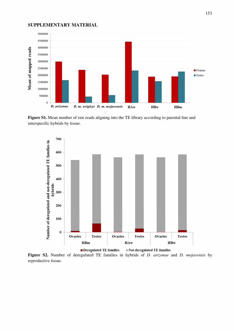
153
SUPPLEMENTARY MATERIAL
Figure S1. Mean number of raw reads aligning into the TE library according to parental line and interspecific hybrids by tissue.
Figure S2. Number of deregulated TE families in hybrids of D. arizonae and D. mojavensis by reproductive tissue.
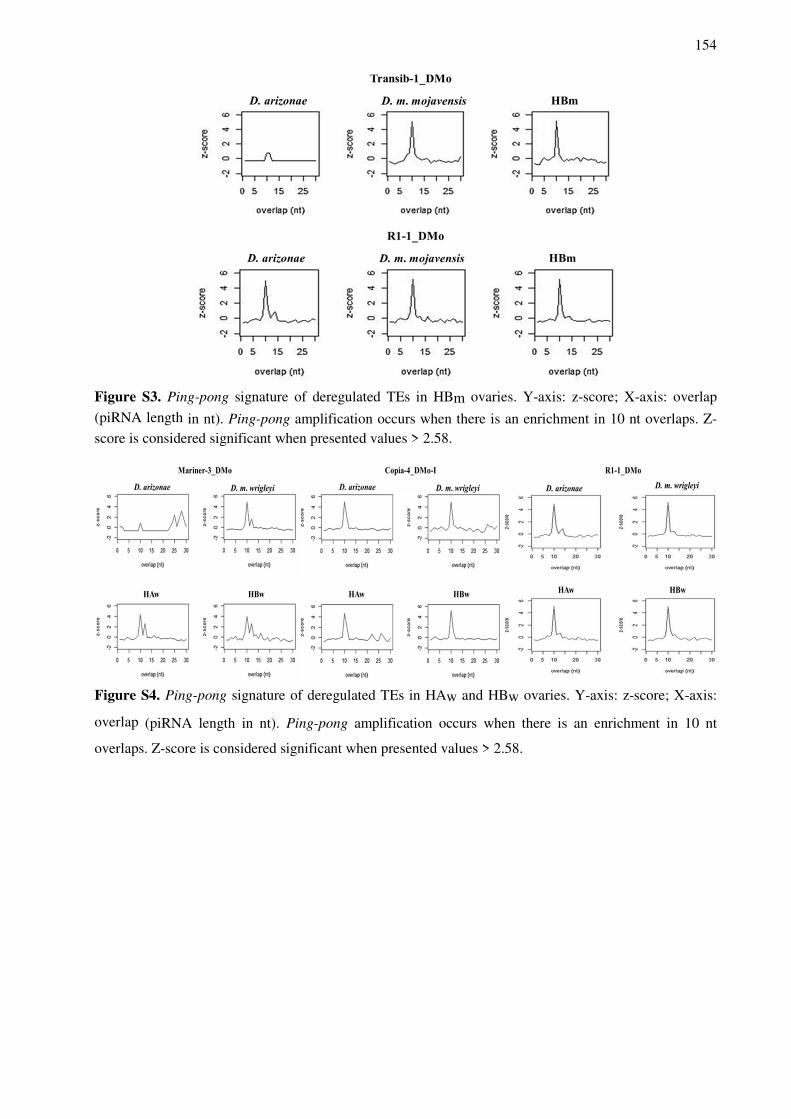
154
Figure S3. Ping-pong signature of deregulated TEs in HBm ovaries. Y-axis: z-score; X-axis: overlap
(piRNA length in nt). Ping-pong amplification occurs when there is an enrichment in 10 nt overlaps. Z-score is considered significant when presented values > 2.58.
Figure S4. Ping-pong signature of deregulated TEs in HAw and HBw ovaries. Y-axis: z-score; X-axis:
overlap (piRNA length in nt). Ping-pong amplification occurs when there is an enrichment in 10 nt
overlaps. Z-score is considered significant when presented values > 2.58.
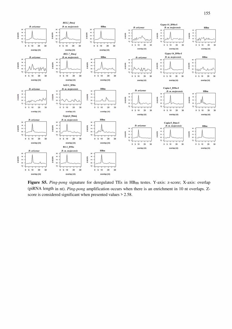
155
Figure S5. Ping-pong signature for deregulated TEs in HBm testes. Y-axis: z-score; X-axis: overlap
(piRNA length in nt). Ping-pong amplification occurs when there is an enrichment in 10 nt overlaps. Z-score is considered significant when presented values > 2.58.
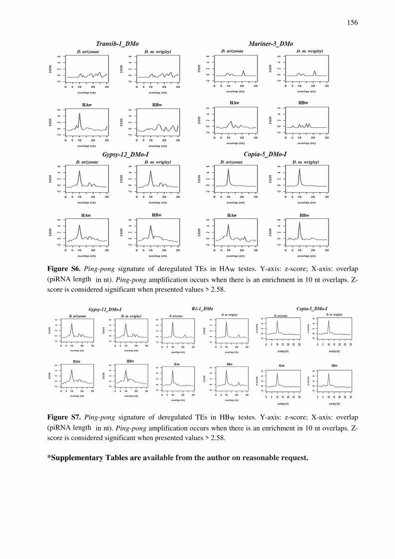
156
Figure S6. Ping-pong signature of deregulated TEs in HAw testes. Y-axis: z-score; X-axis: overlap
(piRNA length in nt). Ping-pong amplification occurs when there is an enrichment in 10 nt overlaps. Z-score is considered significant when presented values > 2.58.
Figure S7. Ping-pong signature of deregulated TEs in HBw testes. Y-axis: z-score; X-axis: overlap
(piRNA length in nt). Ping-pong amplification occurs when there is an enrichment in 10 nt overlaps. Z-score is considered significant when presented values > 2.58.
*Supplementary Tables are available from the author on reasonable request.

157
DISCUSSÃO GERAL
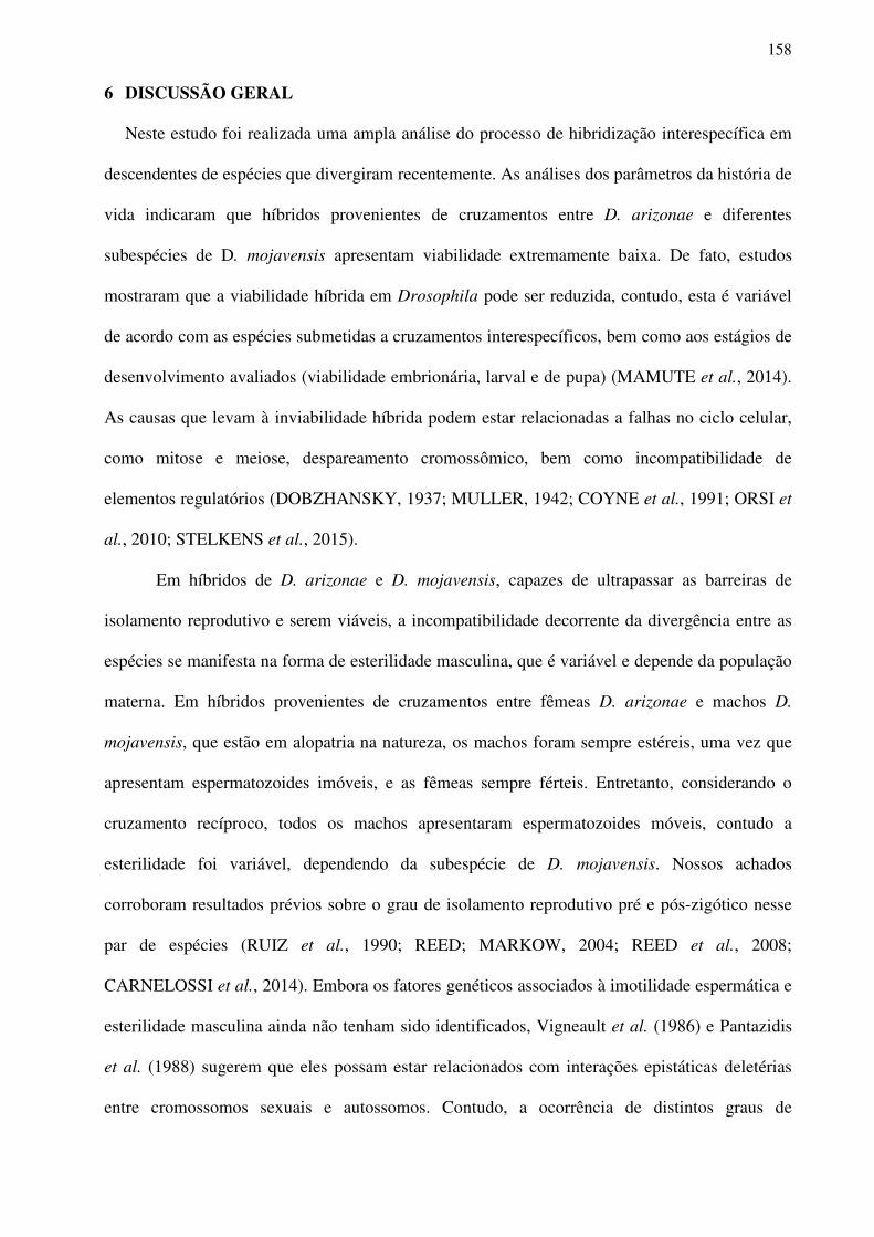
158
6 DISCUSSÃO GERAL
Neste estudo foi realizada uma ampla análise do processo de hibridização interespecífica em
descendentes de espécies que divergiram recentemente. As análises dos parâmetros da história de
vida indicaram que híbridos provenientes de cruzamentos entre D. arizonae e diferentes
subespécies de D. mojavensis apresentam viabilidade extremamente baixa. De fato, estudos
mostraram que a viabilidade híbrida em Drosophila pode ser reduzida, contudo, esta é variável
de acordo com as espécies submetidas a cruzamentos interespecíficos, bem como aos estágios de
desenvolvimento avaliados (viabilidade embrionária, larval e de pupa) (MAMUTE et al., 2014).
As causas que levam à inviabilidade híbrida podem estar relacionadas a falhas no ciclo celular,
como mitose e meiose, despareamento cromossômico, bem como incompatibilidade de
elementos regulatórios (DOBZHANSKY, 1937; MULLER, 1942; COYNE et al., 1991; ORSI et
al., 2010; STELKENS et al., 2015).
Em híbridos de D. arizonae e D. mojavensis, capazes de ultrapassar as barreiras de
isolamento reprodutivo e serem viáveis, a incompatibilidade decorrente da divergência entre as
espécies se manifesta na forma de esterilidade masculina, que é variável e depende da população
materna. Em híbridos provenientes de cruzamentos entre fêmeas D. arizonae e machos D.
mojavensis, que estão em alopatria na natureza, os machos foram sempre estéreis, uma vez que
apresentam espermatozoides imóveis, e as fêmeas sempre férteis. Entretanto, considerando o
cruzamento recíproco, todos os machos apresentaram espermatozoides móveis, contudo a
esterilidade foi variável, dependendo da subespécie de D. mojavensis. Nossos achados
corroboram resultados prévios sobre o grau de isolamento reprodutivo pré e pós-zigótico nesse
par de espécies (RUIZ et al., 1990; REED; MARKOW, 2004; REED et al., 2008;
CARNELOSSI et al., 2014). Embora os fatores genéticos associados à imotilidade espermática e
esterilidade masculina ainda não tenham sido identificados, Vigneault et al. (1986) e Pantazidis
et al. (1988) sugerem que eles possam estar relacionados com interações epistáticas deletérias
entre cromossomos sexuais e autossomos. Contudo, a ocorrência de distintos graus de
159
esterilidade em híbridos de diferentes subespécies de D. mojavensis cruzadas com D. arizonae
indicam a presença de fatores polimórficos entre as espécies que estão envolvidos no processo de
isolamento reprodutivo, como também proposto por Reed et al. (2004). É interessante ressaltar
que as diferenças observadas na fertilidade entre híbridos machos e fêmeas está de acordo com a
Regra de Haldane (1922), a qual postula que em cruzamentos interespecíficos os híbridos
primeiramente afetados por inviabilidade ou esterilidade são os que possuem sexo
heterogamético. Bono et al. (2009) ao analisarem híbridos de D. arizonae e D. mojavensis,
quanto a alguns parâmetros de história de vida, demonstraram que as fêmeas híbridas viáveis
possuem desempenho similar às espécies puras, ao passo que os machos híbridos, além de serem
estéreis, apresentam menor resistência à dessecação, o que é um importante fator seletivo,
considerando que essas espécies vivem em ambientes desérticos. Portanto, é possível inferir que
em híbridos desse par de espécies, os fatores que levam à incompatibilidade híbrida são mais
severos, e provavelmente evoluem mais rapidamente em machos do que em fêmeas, o que pode
ser verificado fenotipicamente. Confirmando esse fato, Turissini et al. (2018) mostraram que em
machos híbridos, provenientes de cruzamentos de espécies do grupo melanogaster, a
inviabilidade e a esterilidade evoluem mais rapidamente do que nas fêmeas híbridas.
Considerando as diferenças fenotípicas, e na tentativa de identificar alguns dos fatores
que estão influenciando a esterilidade híbrida, análises de expressão gênica e de elementos de
transposição foram realizadas. Confirmando o fenótipo fértil de fêmeas híbridas de D. arizonae e
das duas subspécies de D. mojavensis, poucos genes apresentaram expressão divergente em
relação às linhagens parentais. Contudo, em testículos de híbridos o cenário foi muito diferente,
isso porque, dependendo da direção de cruzamento, e, portanto, da linhagem materna, maior ou
menor grau de desregulação da expressão gênica foi observado. Resultados similares foram
obtidos por Llopart et al. (2018) e Ranz et al. (2003) que demonstraram que híbridos machos de
D. santomea–D. yakuba e D. melanogaster-D. simulans, respectivamente, possuem mais genes
desregulados em comparação às fêmeas híbridas.
160
Nosso estudo mostrou a ocorrência de muitos genes subexpressos em híbridos com
espermatozoides imóveis (provenientes de cruzamentos entre fêmeas D. arizonae e machos D.
m. mojavensis e D. m. wrigleyi), e que a maior parte deles apresentava funções relacionadas à
espermatogênese e a motilidade espermática, o que pode ser associado ao fenótipo estéril.
Híbridos de outras espécies de Drosophila, como os provenientes de cruzamentos entre D.
simulans e D. mauritiana, mostraram o mesmo padrão, isto é, super-representação de genes
subexpressos com funções relacionadas à espermatogênese em híbridos estéreis (MICHALAK;
NOOR, 2003, 2004; MA; MICHALAK, 2011), evidenciando a associação entre o fenótipo e a
desregulação da expressão gênica. Dentre esses genes podemos destacar alguns diretamente
relacionados com o ciclo celular como Jupiter e twe (twine), bem com funções diretamente
relacionadas à motilidade espermática, como genes de dineína (Dnah3, Dhc16F CG10839,
CG31275 l(2)41Ab), entre outros. Jupiter é um gene que codifica uma proteína localizada na
rede de microtubulos, com importante função no ciclo celular (KARPOVA et al., 2006). O gene
twe, por sua vez, codifica uma proteína necessária para o início da meiose em machos. Em
híbridos machos sem a expressão da proteína twine foram verificadas falhas na transição G2/M,
dessa forma a meiose não é completa e podem ser encontradas espermátides 4N (WHITE-
COOPER et al., 1998; MAINES; WASSEMAN, 1999; KARPOVA et al., 2006; LINDSLEY et
al., 2013; JIANG et al., 2013). Além disso, o correto funcionamento de twine depende da
regulação por meio de outros genes envolvidos nas fases iniciais da meiose (JIANG et al., 2013;
WHITE-COOPER et al., 1998). Genes relacionados com o movimento dos espermatozoides,
cílios e montagem dos microtúbulos, como genes de dineína, exibiram grande enriquecimento
entre os genes subexpressos de híbridos estéreis originados do cruzamento entre fêmeas D.
arizonae e machos D. mojavensis. Esses genes normalmente são expressos em altos níveis em
testículos (RASMUSSON et al., 1994). Sendo assim, a falta dessas proteínas podem estar
relacionadas com a esterilidade masculina, como previamente foi mostrado em ratos e moscas da
fruta (FOSSELLA et al., 2000; FABIAN; BRILL, 2012; KARAK et al., 2015). Adicionalmente,
161
de acordo com Wu et al. (1993), híbridos que não apresentam espermatozoides móveis, mas que
têm meiose completa, provavelmente apresentam falhas pós-meioticas associadas à
espermiogênese, ao invés de anormalidades em larga escala no desenvolvimento dos órgãos
reprodutivos.
Ao contrário do cruzamento acima descrito, em híbridos do cruzamento recíproco
(fêmeas D. m. wrigleyi x machos D. arizonae), que possuem espematozoides móveis, embora
sejam estéreis, menor grau de desregulação gênica foi verificado, principalmente envolvendo
genes que atuam na espermatogênese. Contudo, alguns genes envolvidos com reprodução,
processamento de micro-RNA e em mecanismos de silenciamento foram identificados,
mostrando que outros fatores podem estar contribuindo para a esterilidade dos híbridos
provenientes de cruzamentos com fêmeas D. mojavensis. Hardy et al. (2016), ao analisarem a
ultraestrutura de testículos de híbridos recíprocos de D. arizonae e D. m. mojavensis, mostraram
que híbridos de cruzamentos com fêmeas D. mojavensis apresentavam defeitos no
desenvolvimento do núcleo das espermátides, mas os espermatozoides tinham caudas normais, o
que explica o baixo número de genes desregulados envolvidos com movimento da cauda nesses
híbridos.
Considerando os TEs, foi observada uma tendência à superexpressão, em menor grau em
ovários e em maior grau em testículos. Níveis diferentes de desregulação de TEs entre tecidos
reprodutivos masculinos e femininos já foram identificados em espécies de Drosophila
(CARNELOSSI et al., 2014; VELA et al., 2014; ROMERO-SORIANO et al., 2016). Neste
trabalho, verificamos ainda que a expressão diferencial de TEs não estava completamente
relacionada com a direção de cruzamento, como foi observado para expressão gênica, mas sim
relacionada com a subespécie de D. mojavensis utilizada nos cruzamentos interespecíficos. Isso
porque os diferentes híbridos apresentaram números muito diferentes de TEs desregulados,
sendo a maioria deles unicamente superexpressos em híbridos específicos. Isso indica que as
diferentes subespécies de D. mojavensis provavelmente possuem famílias de TEs distintas com

162
expressão controlada em seus genomas, as quais, em face do choque genômico decorrente da
hibridização interespecífica, podem ser diferencialmente ativadas devido à perda de mecanismos
de regulação. De fato, diferenças no conteúdo de TEs em genomas são frequentemente
observadas para diversas espécies, sendo que essas variações podem ocorrer também em nível
populacional, como para D. simulans (SONG et al., 2014; FABLET et al., 2019).
Ademais, um fato interessante a ser destacado é que os híbridos machos exibem maior
número de família de TEs desreguladas do que as fêmeas do mesmo cruzamento. Isso sugere que
os mecanismos de regulação em níveis transcricional e pós-transcricional são sexo-específicos,
podendo ser mais eficientes nas fêmeas que em machos. Confirmando esses resultados, Malone
et al. (2009) sugerem que a regulação de determinados TEs podem ser tecido-específica e estar
realcionada aos clusters de piRNAs presentes nos genomas parentais.
A análise da via de piRNAs mostrou que em ovários, assim como em testículos, alguns TEs não
apresentavam piRNAs correspondentes na linhagem maternal. Isso poderia levar à falha de
regulação desses elementos nos híbridos, devido à falta de deposição de piRNAs maternos, os
quais são essenciais para iniciar o processo de regulação pós-transcricional, denominado ciclo
ping-pong, como verificado previamente para híbridos de outras espécies de Drosophila
(BRENNECKE et al., 2008; AKKOUCHE et al., 2013; LOPEZ-MAESTRE et al., 2017;
FUNIKOV et al., 2018). Contudo, diversos TEs, em ambos tecidos reprodutivos de híbridos,
apresentaram piRNAs complementares, os quais não foram capazes de controlar sua expressão.
Isso poderia se explicado por um aumento de transposição em células somáticas associadas aos
testículos extraídos (vesículas seminais), como proposto para o retrotransposon Osvaldo que
tende aumentar a taxa de transposição em machos híbridos de cruzamentos entre D. buzzatii e D.
koepferae (VELA et al., 2014). Contudo, em nossas análises de siRNAs verificou-se que
determinados TEs, que apresentam piRNAs correspondentes, também possuiam siRNAs
complementares, os quais deveriam ser capazes de controlar sua expressão em células somáticas;
entretanto, apesar da presença de siRNAs, esses TEs estavam superexpressos nos híbridos. Isso
163
significa que, de alguma forma, os mecanismos de regulação nos híbridos entre D. arizonae e D.
mojavensis estão falhando, seja na via de piRNAs ou em possíveis mecanismos de regulação
alternativos. Quénerch’du et al. (2016) mostraram que em testículos provavelmente há uma via
de controle e amplificação de piRNAs não canônica, uma vez que em mutantes de proteínas
essenciais para a via ping-pong, amplificação de piRNAs foi observada.
Em fêmeas de cruzamentos entre D. arizonae e D. mojavensis, genes que fazem parte da
via de piRNAs e alguns genes associados a modificações de cromatina apresentam expressão
conservada em híbridos em relação aos parentais, bem como entre as diferentes espécies
parentais. Todavia, em testículos o mesmo não foi verificado para determinados genes. Isso pode
ter um impacto direto na superexpressão desses elementos nas gônadas masculinas, bem como
influenciar a regulação de outros genes, ou mesmo ter um papel importante no fenótipo de
esterilidade observado. Desregulação na expressão gênica em híbridos masculinos é
frequentemente observada em espécies de Drosophila e já foi associada com taxas de evolução
mais rápidas em machos, que em fêmeas (MEIKLEJOHN et al., 2003; RANZ et al., 2003;
ARTIERI et al., 2007; MOEHRING et al., 2007; LLOPART, 2012). Sendo assim, sugerimos
que, em híbridos de D. arizonae e D. mojavensis, provavelmente diferenças de sequências
regulatórias que agem de forma cis e trans, e interações epistáticas deletérias, levam à
desregulação de diversos genes, incluindo aqueles relacionados aos sistemas de regulação pós-
transcricional e transcricional. Como consequência, a expressão de muitos TEs não é capaz de
ser controlada, o que pode ter um efeito negativo no genoma híbrido, podendo levar à
incompatibilidade e ser associada ao isolamento reprodutivo. Esse efeito pode ser mais
acentuado em determinadas direções de cruzamento, devido a diferentes interações espistáticas,
ou dependendo da subespécie, devido a polimorfismos existentes entre elas, bem como ter maior
impacto em um sexo que em outro. Isso porque, em machos, determinados genes podem
apresentar maior taxa de evolução, ou mesmo devido a herança dominante de determinados
alelos no cromossomo X em hemizogose em machos, que não ocorre em fêmeas (LLOPART et
164
al., 2018).
De maneira geral, ainda não se sabe se interações epistáticas deletérias que levam à
desregulação de genes de vias específicas são responsáveis pela superexpressão de TEs em
híbridos ou se o choque genômico que leva à mobilização de TEs afeta a expressão gênica.
Contudo, podemos sugerir que ambos processos são intimamente ligados e diversos estudos vêm
demonstrando isso. Satyaki et al. (2014) mostraram que a alelos dos genes Lmr (Lethal male
rescue) e Hmr (Hybrid male rescue) interagem e controlam a expressão de sequências repetitivas
e satélites. Contudo, em híbridos de D. simulans e D. melagaster, inviabilidade híbrida e
aumento da expressão de TEs ocorre devido à divergência adaptativa dessas sequências nas
diferentes espécies (SATYAKI et al., 2014). Moehring et al. (2007) sugerem que a regulação
transcricional pode estar associada com incompatibilidade em híbridos, como a esterilidade. Isso
porque em muitas espécies de Drosophila, a transcrição de genes envolvidos na espermatogênese
ocorre na fase anterior à meiose (FULLER, 1998). Superexpressão e eventos de transposição já
foram verificados para alguns TEs específicos em tecidos reprodutivos masculinos. Dentre eles
pode-se destacar o elemento Copia, que possui transposição limitada a espematócitos masculinos
(PASYUKOVA et al., 1997; KALMYKOVA e al., 2005) e os elementos Osvaldo e I
(GUERREIRO, 2015; CARNELOSSI et al., 2014), os quais são expressos nas regiões apicais
dos testículos, que contém células da linhagem germinativa, espermatócitos primários e
secundários (VEDELECK et al., 2018). Mostrou-se que híbridos intraespecíficos de D.
melanogaster com expressão do elemento I, devido a falhas na via de piRNAs, apresentaram
meiose anormal, sugerindo que a atividade desse elemento perturba a organização cromossômica
durante o ciclo celular (ORSI et al., 2010). Esse fato pode indicar que, de alguma forma, a
grande desregulação de TEs (principalmente superexpressos) pode estar relacionada à
esterilidade híbrida nos machos híbridos de cruzamentos entre D. arizonae e D. mojavensis. O
estudo de Iwasaki et al. (2016), sobre as marcas repressivas da cromatina em células somáticas
de ovários de Drosophila mutantes para a proteína Piwi, auxiliam a compreensão desses
165
resultados. Os autores verificaram que falhas no sistema de regulação influenciam a expressão de
um subconjunto de genes codificadores de proteínas próximos a inserções de TEs. Dentre esses
genes, a maioria apresentava funções relacionadas ao desenvolvimento das características
sexuais primárias e diferenciação sexual. Além disso, segundo Girrard et al. (2006), piRNAs
podem ter um importante papel relacionado ao controle da expressão gênica durante a
gametogênese, uma vez que durante esse processo muitas modificações epigenéticas ocorrem.
Sendo assim, os resultados obtidos neste trabalho e diversas evidências da literatura
indicam que existe uma grande complexidade e interações de mecanismos que permeiam as
barreiras do isolamento reprodutivo. Dentre esses mecanismos, podemos sugerir que, em
híbridos, prejuízps nos sistemas de regulação, devido à divergência adaptativa de allos, levam à
desregulação da expressão gênica e de TEs, originando uma cascata de sucessivos eventos de
desregulação que contribuem para mecanismos de incompatibilidade híbrida. Os resultados
apresentados nesta tese trazem contribuições inéditas para a compreensão dos efeitos da
hibridização interespecífica em descendentes de espécies que divergiram recentemente, contudo,
análises adicionais precisam ser realizadas para melhor entender esse processo. Dentre elas,
podemos destacar estudos de regulação cis e trans, bem como análises de taxa de evolução e
funcionais para determinados genes regulatórios.

166
CONCLUSÕES
167
7 CONCLUSÕES
Este trabalho permitiu elaborar as seguintes conclusões:
1. Redução de viabilidade nos dois sexos em híbridos recíprocos provenientes de cruzamentos
entre D. arizonae e quatro subespécies de D. mojavensis e esterilidade variável dos machos
híbridos dependente da população materna de D. mojavensis, sugerem que os fatores que
levam à incompatibilidade híbrida nesse par de espécies são polimórficos, os quais podem
evoluir a diferentes taxas em cada população de D. mojavensis, dependendo de sua origem
geográfica, isto é, se estão em simpatria ou em alopatria.
2. Machos híbridos sem espermatozoides móveis apresentaram diversos genes diferencialmente
expressos, os quais eram, em sua maioria, subexpressos em relação à expressão gênica nos
parentais, e tinham funções reprodutivas e de motilidade dos espermatozoides. No entanto,
híbridos machos que possuíam espermatozoides móveis apresentaram menos genes
diferencialmente expresssos, os quais tinham diversas funções, como regulação e transcrição
gênica. Esses resultados indicam que a motilidade espermática está relacionada ao grau de
desregulação da expressão gênica que ocorre nesses híbridos.
3. Fêmeas híbridas apresentaram poucos genes diferencialmente expressos, concordando com
fenótipo fértil, e sugerindo menor disrupção de elementos regulatórios. Em machos híbridos, a
maior expressão diferencial de genes pode estar relacionada com a evolução mais rápida de
sequências regulatórias, principalmente para genes exclusivamente expressos em testículos,
bem como a incompatibilidade decorrente de alelos localizados no cromossomo X, uma vez
que em machos, alelos que normalmente se expressam em condições de recessividade podem
se comportar como dominantes devido ao fato de estarem em hemizigose.
168
4. Em fêmeas híbridas ocorre menor desregulação de TEs do que em machos híbridos, o que
também pode ter um papel importante no fenótipo fértil e estéril, respectivamente, verificados
nesses híbridos.
5 Diferentes mecanismos que envolvem a via de regulação de TEs atuam em ovários e
testículos, uma vez que híbridos machos e fêmeas provenientes dos mesmos cruzamentos
interespecíficos não apresentaram os mesmos TEs superexpressos.
6. Os mecanismos de silenciamento por meio das vias de piRNA e siRNAs se mostraram
complexos em fêmeas e machos híbridos, uma vez que TEs que tinham piRNAs e siRNAs
complementares não estavam regulados, estando superexpressos, sugerindo ocorrência de
falhas em outros mecanismos de regulação, tais como divergência de expressão de genes
envolvidos nas vias de regulação em machos híbridos. Isso indica que, em machos, assim
como verificado em análises de expressão gênica, interações epistáticas de alelos com maior
taxa de evolução ou expressão dominante podem contribuir para a desregulação de TEs.

169
REFERÊNCIAS
170
8 REFERÊNCIAS AKKOUCHE, A. et al. Maternally deposited germ-line piRNAs silence the tirant
retrotransposon in somatic cells. EMBO Rep, v.14, p. 458–464, 2013. ALONSO-PIMENTEL, H.; TOLBERT, L. P.; HEED, W. B. Ultrastructural examination of the insemination reaction in Drosophila. Cell Tissue Res., v. 275, p. 467-479, 1994. ANTONIO SERRATO-CAPUCHINA, A.; MATUTE, D. R. The Role of Transposable Elements in Speciation. Genes, v. 9, n. 254, p. 1-29, 2018. ARTIERI, C. G., HAERTY, W. Association Between Levels of Coding Sequence Divergence and Gene Misregulation in Drosophila Male Hybrids. J Mol Evol, v. 65, n. 6, p. 697-704, 2007. BAACK, E. J.; WHITNEY, K. D.; RIESEBERG, L. H. Hybridization and genome size evolution: timing and magnitude of nuclear DNA content increases in Helianthus homoploid hybrid species, New Phytol, v.167, n.2, p. 623–630, 2005. BARBASH, D. A. et al. A rapidly evolving MYB-related protein causes species isolation in Drosophila. Proc. Natl Acad. Sci. USA, v. 100, n. 9, p. 5302–5307, 2003. BARBASH, D. A.; AWADALLA, P.; TARONE, A. M. Functional divergence caused by ancient positive selection of a Drosophila hybrid incompatibility locus. PLoS Biol. v. 2, p. 839– 848, 2004. BARBASH, D. A.; LORIGAN, J. G. Lethality in Drosophila melanogaster/Drosophila simulans
species hybrids is not associated with substantial transcriptional misregulation. J Exp Zool B Mol Dev Evol. v. 308, n. 11, p. 74-84, 2007. BINGHAM, P. M.; KIDWELL. M. G.; RUBIN, G. M. The molecular basis of P-M hybrid dysgenesis: the role of the P element, a Pstrain-specific transposon family. Cell, v. 29, n. 3, p. 995-1 004, 1982. BONO, J. M.; MARKOW, T. A. Post-zygotic isolation in cactophilic Drosophila: larval viability and adult life-history traits of D. mojavensis/D. arizonae hybrids. J Evol Biol., v. 22, n. 7, p. 1387–1395, 2009. BRENNECKE J. et al. An epigenetic role for maternally inherited piRNAs in transposon silencing. Science, v. 322, p. 1387–1392, 2008. BRENNECKE, J. et al. Discrete small RNA-generating loci as master regulators of transposon activity in Drosophila. Cell, v. 128, n. 6, 1089–103, 2007. BRIDEAU, N. J. et al. Two Dobzhansky–Muller genes interact to cause hybrid lethality in Drosophila. Science, v. 314, n. 5803, p. 1292–5, 2006. CARARETO, C. M. A.; HERNANDEZ, E. H; VIEIRA, C. Genomic regions harboring insecticide resistance associated Cyp genes are enriched by transposable element fragments carrying putative transcription factor binding sites in two sibling Drosophila species. Gene, v. 537, n.1, p. 93-99, 2013.
171
CARNELOSSI, E. A. G. et al. Specific activation of an I-like element in Drosophila
interspecific hybrids. Genome Biology Evolution. v. 6, n. 7, p. 1806–1817, 2014. CASTILLO D. M. ; MOYLE, L. C. Transposable elements that cause dysgenesis also contribute to postzygotic isolation in the Drosophila virilis clade. bioRxiv preprint first posted online. Doi: http://dx.doi.org/10.1101/753814, 2019. COYNE, J. A., ORR, H. A. ‘‘Patterns of speciation in Drosophila’’ revisited. Evolution, v. 51, n.1, p. 295–303, 1997. COYNE, J. A.; ORTH, B. C.; ORR, H. Notes and comments Haldane's Rule revisited. Evolution, v. 45, n. 7, p. 1710—1714, 1991. CUTTER A. D. Divergence times in Caenorhabditis and Drosophila inferred from direct estimates of the neutral mutation rate. Mol Biol Evol, v. 25, n. 4, p. 778–786, 2008. DABOUSSI, M.; CAPY, P. Transposable elements in filamentous fungi. Annu. Rev. Microbiol. V. 57, p. 275–299, 2003. DOBZHANSKY T. Speciation as a stage in evolutionary divergence. Am Nat., v. 74, p. 312– 321, 1940. DOBZHANSKY, T. Genetics and the Origin of Species. Columbia Univ. Press, New York, 1937. ETGES, W. J. et al. Inheritance of courtship song variation among geographically isolated populations of Drosophila mojavensis. Animal Behaviour, v. 71, p. 1205–1214, 2006. FABIAN, L.; BRILL, J. A. Drosophila spermiogenesis: Big things come from little packages. Spermatogenesis, v 2, n. 3, p. 197-212, 2012. FABLET, M. et al. Dynamic interactions between the genome and an endogenous retrovirus: Tirant in Drosophila simulans Wild-Type Strains. G3: Genes, Genomes, Genetics, v. 9, 2019. FABLET, M. Host Control of insect endogenous retroviruses: Small RNA silencing and immune response. Viruses, v. 6, n. 11, p. 4447-64, 2014b. FABLET, M. et al. Self and Nonself from a Genomic Perspective: Transposable Elements, p. 111-128, 2017 In: PONTAROTTI, P. Evolutionary Biology: Self/Nonself Evolution, Species and Complex Traits Evolution, Methods and Concepts, Springer, 2017.
FERREE, P. M.; BARBASH, D. A. Species-specific heterochromatin prevents mitotic chromosome segregation to cause hybrid lethality in Drosophila. PLoS Biol., v. 7, n. 10, e1000234, 2009. FOSSELA, J. et al. An axonemal dynein at the Hybrid Sterility 6 locus: implications for T haplotype-specific male sterility and the evolution of species barriers Mammalian. Genome, v. 11, n. 1, p. 8-15, 2000.
FULLER, M. T. Genetic control of cell proliferation and differentiation in Drosophila
spermatogenesis. Semin Cell Dev Biol. v. 9, p. 433–444, 1998.
172
FUNIKOV, S. Y. et al. Spontaneous gain of susceptibility suggests a novel mechanism of resistance to hybrid dysgenesis in Drosophila virilis. PLOS Genetics, p. 1-20, 2018. GIRARD, A. et al. A germline-specific class of RNAs binds mammalian Piwi proteins. Nature, v. 442, p. 199-202, 2006. GOMES S.; CIVETTA A. Misregulation of spermatogenesis genes in Drosophila hybrids is lineage-specific and driven by the combined effects of sterility and fast male regulatory divergence. J Evol Biol. v. 27, n. 9, p. 1775–1783. 2014. GOMES, S.; CIVETTA, A. Hybrid male sterility and genome-wide misexpression of male reproductive proteases. Scientific Reports, v. 5: 11976, 2015. GOMEZ, G.A.; HASSON, E. Transpecific polymorphisms in an inversion linked esterase locus in Drosophila buzzatii. Mol Biol Evol. v. 20, n. 3, p.410–423, 2003. GUERREIRO, M. P. Changes of Osvaldo expression patterns in germ-line of male hybrids between the species Drosophila buzzatii and Drosophila koepferae. Mol. Genet. Genomics, v. 290, n. 4, p.1471-83, 2015. GUERREIRO, M. P. Interspecific hybridization as a genomic stressor inducing mobilization of transposable elements in Drosophila. Mob Genet Elements. v. 4, p. e34394-1-e34394-1, 2014. HAERTY, W.; SINGH, R. S. Gene Regulation Divergence Is a Major Contributor to the Evolution of Dobzhansky–Muller Incompatibilities between Species of Drosophila. Mol. Biol. Evol., v. 23, n. 9, p. 1707–14, 2006. HALDANE, J. B. S. Sex ratio and unisexual sterility in hybrid animals. Journal of Genetics. v.12, p. 101–109, 1922. HARDY, R. W.; LOUGHEED, A. ; MARKOW, T. A. Reproductive tract and spermatid abnormalities of hybrid males from reciprocal crosses between Drosophila mojavensis and D
arizonae. Fly, v. 5, n. 2, p. 76-80, 2011. HEED, W. B. The origin of Drosophila in the Sonoran Desert. In: Ecological Genetics and Evolution: The Cactus-Yeast-Drosophila Model System. Ed: Academic Press, Sydney; p. 65– 80, 1982. HILL T.; SCHLOTTERER C.; BETANCOUT A. J. Hybrid Dysgenesis in Drosophila simulans
associated with a rapid Invasion of the P-Element. PLoS Genet. v. 12, n. 3, p. e1005920, 2016. HIRAKATA, S.; SIOMI, M. C. Assembly and Function of Gonad-Specific Non-Membranous Organelles in Drosophila piRNA Biogenesis. Non-coding RNA , v. 5, n. 52, 2019. IPSARO, J. J. et al. The structural biochemistry of Zucchini implicates it as a nuclease in piRNA biogenesis. Nature, v. 491, n. 7423, p. 279–283, 2012.
IWASAKI, Y. W.; SIOMI, M. C.; SIOMI, H. PIWI-Interacting RNA: Its Biogenesis and Functions. Annu. Rev. Biochem, v. 84, p. 405–33, 2015.
173
IWASAKI, Y. W. et al. Piwi Modulates Chromatin Accessibility by Regulating Multiple Factors Including Histone H1 to Repress Transposons. Molecular Cell, v. 63, n. 3, p. 408–419, 2016. JENNINGS, J. H.; ETGES. W. J. Species hybrids in the laboratory but not in nature: a reanalysis of premating isolation between Drosophila arizonae and D. mojavensis. The Society for the Study of Evolution. v. 64, n. 2, p. 587–598, 2009. JIANG, J.; WHITE-COOPER, H. Transcriptional activation in Drosophila spermatogenesis involves the mutually dependent function of aly and a novel meiotic arrest gene cookie monster. Development, v. 130, n. 3, p. 563-573, 2003. JURKA, J. et al. Repbase Update, a database of eukaryotic repetitive elements. Cytogenet. Genome Res. v. 110, p. 462–467, 2005. KALMYKOVA, A. I.; KLENOV, M. S.; GVOZDEV, V. A. Argonaute protein PIWI controls mobilization of retrotransposons in the Drosophila male germline. Nucleic Acids Research, v. 33, n. 6, p. 2052–2059, 2005. KAPITONOV, V. V.; JURKA, J. A universal classification of eukaryotic transposable elements implemented in Repbase. Nat. Rev. Genet. v. 9, p. 411–412, 2008. KARAK, S. et al. Diverse Roles of Axonemal Dyneins in Drosophila Auditory Neuron Function and Mechanical Amplification in Hearing. Scientific Reports, v. 5, p. 17085, 2015. KARPOVA N. et al. Jupiter a New Drosophila Protein Associated With Microtubules. Cell Motility and the Cytoskeleton, v. 63, n. 5, p. 301-312, 2006. KAWAKAMI, T. et al. Different Scales of Ty1/copia-like Retrotransposon Proliferation in the Genomes of Three Diploid Hybrid Sunflower Species. Heredity, v. 104, n. 4, p. 341-50, 2010. KELLEHER, E. S.; EDELMAN, N. B.; BARBASH, D. A. Drosophila interspecific hybrids phenocopy piRNA-pathway mutants. PLoS biology. v. 10, n. 11, 2012. KELLEHER, E. S.; MARKOW, T. A. Reproductive Tract Interactions Contribute to Isolation in Drosophila. Fly, v. 1, n. 1, p. 33-37, 2007. KIDWELL, M. G.; KIDWELL, J. F.; SVED, J. A. Hybrid dysgenesis in Drosophila
melanogaster: A syndrome of aberrant traits including mutation, sterility and male recombination. Genetics, v. 86, p. 813–833, 1977. KIDWELL, M. G.; LISCH, D. R. Perspective: transposable elements, parasitic DNA, and genome evolution. Evolution, v. 55, n. 1, p. 1–24, 2001. KLIMAN, R. M. et al. The population genetics of the origin and divergence of the Drosophila
simulans complex species. Genetics, v. 156, n. 4, p. 1913–31, 2000. KNOWLES, L. L.; MARKOW, T. A. Sexually antagonistic coevolution of a postmating- prezygotic reproductive character in desert Drosophila. PNAS, v. 98, n. 15, p. 8692–8696, 2001.
LAAYOUNI, H. et al. The evolutionary history of Drosophila buzzatii. XXXV. Inversion polymorphism and nucleotide variability in different regions of the second chromosome. Mol
174
Biol Evol. v. 20, n. 6, p. 931–944, 2003. LABRADOR M. et al. Interspecific hybridization increases transposition rates of Osvaldo. Mol Biol Evol. v. 16, p. 931–937, 1999. LACHAISE, D. et al. Historical biogeography of the Drosophila melanogaster species subgroup. P. 159-225, 1998 in HECHT, M. K.; WALLACE, B.; PRANCE, G. T. E. Evolutionary Biology, Springer, Boston, MA, v. 22, 1978. LINDSLEY, D. L.; ROOTE, J.; KENNISON, J. A. Anent the genomics of spermatogenesis in Drosophila melanogaster. Plos One. v. 8, n. 2, p. e55915, 2013. LLOPART, A. et al. Support for the dominance theory in Drosophila Transcriptomes. Genetics, v. 210, n. 2, p. 703–718, 2018. LLOPART A. The rapid evolution of X-linked male-biased gene expression and the large-X effect in Drosophila yakuba, D. santomea, and their hybrids. Mol. Biol. Evol, v. 29, n. 12, p. 3873–3886, 2012. LOPES, F. R. et al. Transcriptional activity, chromosomal distribution and expression effects of transposable elements in Coffea genomes. Plos One, v. 8, n. 11, p. e78931, 2013. LOPES, F. R. et al. Transposable elements in Coffea (Gentianales: Rubiacea) transcripts and their role in the origin of protein diversity in flowering plants. Molecular Genetics and Genomics. v. 279, n. 4, p. 385-401, 2008. LOPEZ-MAESTRE, H. et al. Identification of misexpressed genetic elements in hybrids between Drosophila-related species. Scientific Report, v. 7, n. 40618, 2017. LUO, S. ; LU, J. Silencing of transposable elements by piRNAs in Drosophila: An evolutionary perspective. Genomics Proteomics Bioinformatics, v. 15, n. 3, p. 164–176, 2017. MA, D.; MICHALAK, P. Ephemeral association between Gene CG5762 and hybrid male Sterility in Drosophila sibling species. J. Mol. Evol., v. 73, n. 3-4, p.181–187, 2011. MAINES, J. Z.; WASSEMAN, S. A. Post-transcriptional regulation of the meiotic Cdc25 protein Twine by Dazl orthologue Boule. Nature Cell Biology, v. 1, n. 3, p. 171-9, 1999. MALLET, J. Hybridization as an Invasion of the Genome. Trends Ecol. Evol., v. 20, n. 5, p. 229-37, 2005. MALONE C. D. et al. Specialized piRNA pathways act in germline and somatic tissues of the Drosophila ovary. Cell, v. 137, p. 522–535, 2009. MAMUTE, D. R.; GAVIN-SMYTH, J.; LIU, G. Variable post-zygotic isolation Drosophila
melanogaster/D. simulans hybrids. J Evol Biol, v. 27, n. 8, p. 1691-705, 2014.
MARKOW, T. A.; HOCUTT, G. D. Reproductive Isolation in Sonoran Desert Drosophila: Testing the Limits of the Rules. p. 234–244, 1998 in HOWARD, D. J.; BERLOCHER, S. H. Endless forms: species and speciation. Oxford Univ. Press, New York, 1998. MARKOW, T.; ANKNEY, P. Drosophila males contribute to oogenesis in a multiple mating

175
species. Science, v. 224, p. 302—303, 1984. MASLY, J. P. et al. Gene transposition as a cause of hybrid sterility in Drosophila. Science, v. 313, n. 5792, p. 1448–1450, 2006. MASSIE, K. R.; MARKOW, T. A. Sympatry, allopatry and sexual isolation between Drosophila
mojavensis and D. arizonae. Hereditas, v. 142, p. 51-55, 2005. MAYR, E. Animal species and evolution. Harvard Univ. Press, Cambridge, MA, 1963. MAYR, E. Systematics and the Origin of Species. Columbia Univ. Press, New York, 1942. MCBRIDE, C. S.; SINGER, M. C. Field Studies Reveal Strong Postmating Isolation between Ecologically Divergent Butterfly Populations. PloS Biol. v. 8, n. 10, 2010. MCGIRR, J. A.; MARTIN, C. H. Hybrid gene misregulation in multiple developing tissues within a recent adaptive radiation of Cyprinodon pupfishes. PLoS One, v. 14, n. 7, p. e0218899, 2019. MEIKLEJOHN, C. D. et al. Rapid Evolution of Male-Biased Gene Expression in Drosophila.
PNAS, v. 100, n. 17, p. 9894-9899, 2003. METCALFE, C. J. et al. Genomic instability within centromeres of interspecific marsupial hybrids. Genetics, v. 177, n. 4, p. 2507-2517, 2007 MICHALAK, P.; MA, D. The acylphosphatase (Acyp) alleles associate with male hybrid sterility in Drosophila. Gene, v. 416, n. 1-2, p. 61-65, 2008. MICHALAK, P.; NOOR, M. A. Association of misexpression with sterility in hybrids of Drosophila simulans and D. mauritiana. J. Mol. Evol., v. 59, n. 2, p. 277–282, 2004. MICHALAK, P.; NOOR, M. A. Genome-wide patterns of expression in Drosophila pure species and hybrid males. Mol. Biol. Evol, v. 20, n. 7, p. 1070–6, 2003. MOEHRING, A. J.; TEETER, K. C. ; Noor, M. A. F. Genome-wide patterns of expression in Drosophila pure species and hybrid males. II. Examination of multiple-species hybridizations, platforms, and life cycle stages. Mol. Biol. Evol, v. 24, n. 1, p. 137-145, 2007. MULLER, H. J. Isolating mechanisms, evolution, and temperature. Biol. Symp., v. 6, 71–125, 1942. O’NEILL, R. J.; O’NEILL, M. J.; GRAVES, J. A. Undermethylation associated with retroelement activation and chromosome remodeling in an interspecific mammalian hybrid. Nature, v. 393, n. 6680, p. 68-72, 1998.
OLIVEIRA, D. C.S. G; et al. Monophyly, divergence times, and evolution of host plant use inferred from a revised phylogeny of the Drosophila repleta species group. Mol Phylogenet Evol. v. 64, n. 3, p. 533–544, 2012. ORR, H. A.; MASLY, J. P.; PRESGRAVES, D. C. Speciation Genes. Curr Opin Genet Dev. v. 14, n. 6, p. 675-9, 2004.
176
ORR, H. A.; PRESGRAVES, D. C. Speciation by Postzygotic Isolation: Forces, Genes and Molecules. Bioessays, v. 22, n. 12, p. 1085-94, 2000. ORSI, G. A. et al. Drosophila I-R hybrid dysgenesis is associated with catastrophic meiosis and abnormal zygote formation. Journal of Cell Science, v. 123, p. 3515-3524, 2010. PANTAZIDIS, A. C., ZOUROS E. Location of an autosomal factor causing sterility in Drosophila-mojavensis males carrying the Drosophila-arizonensis Y-chromosome. Heredity, v. 60, p. 299–304, 1988. PARISOD, C. et al. Impact of transposable elements on the organization and function of allopolyploid genomes. The New Phytologist, v. 186, n. 1, p. 37-45, 2010. PASYUKOVA E. et al. Germ line transposition of the copia retrotransposon in Drosophila
melanogaster is restricted to males by tissue-specific control of copia RNA levels. Mol Gen Genet, v. 255, p. 115–124, 1997. PATTERSON, J. T. A new type of isolating mechanism in Drosophila. Proceedings of the National Academy of Sciences of the United States of America. v. 32, p. 202-208, 1946. PHADNIS, N. Genetic architecture of male sterility and segregation distortion in Drosophila
pseudoobscura Bogota–USA hybrids. Genetics, v. 189, n. 3, p. 1001–1009, 2011. PHADNIS, N.; ORR, H. A. A single gene causes both male sterility and segregation distortion in Drosophila hybrids. Science, v. 323, n. 5912, p. 376–379, 2008. PICARD, G. Non-mendelian female sterility in Drosophila melanogaster: hereditary transmission of I factor. Genetics, v. 83, p. 107–123, 1976. PIÉGU B.; BIRE S.; ARENSBURGER P. A survey of transposable element classification systems—a call for a fundamental update to meet the challenge of their diversity and complexity. Mol Phylogenet Evol., v. 86, p. 90–109, 2015. PRESGRAVES, D. C. The molecular evolutionary basis of species formation. Nature Reviews Genetics. v. 11, n. 3, p. 175-80, 2010. PRESGRAVES, D. C.; STEPHAN, W. Pervasive adaptive evolution among interactors of the Drosophila hybrid inviability gene, Nup96. Mol. Biol. Evol. v. 24, n. 1, p. 306–314, 2007. PRESGRAVES, D. et al. Adaptive evolution drives divergence of a hybrid inviability gene between two species of Drosophila. Nature, v. 423, n. 6941, p. 715–719, 2003.
QUÉNERCH’DU, E.; ANAND, A.; KAI, T. The piRNA pathway is developmentally regulated during spermatogenesis in Drosophila. RNA, v. 22, n. 7, p.1044–1054, 2016.
RANZ J. M. et al. Sex-Dependent Gene Expression and Evolution of the Drosophila
Transcriptome. Science, v. 300, n. 5626, p. 1742-5, 2003. RASMUSSON, K. et al. Family of Dynein Genes in Drosophila melanogaster. Molecular Biology of the Cell, v. 5, n. 1, p. 45-55, 1994.
177
REBOLLO, R. et al. A Snapshot of histone modifications within transposable elements in Drosophila wild type strains. Plos One, v. 7, n. 9, p. e44253, 2012. REBOLLO, R. et al. Jumping genes and epigenetics: towards new species. Gene, 454, n. 1/2, p. 1-7, 2010. REED, L. K.; LAFLAMME, B. A.; MARKOW, T. A. Genetic architecture of hybrid male sterility in Drosophila: analysis of intraspecies variation for interspecies isolation. Plos One, v. 3, n. 8, p. e3076, 2008. REED, L. K.; MARKOW, T. A. Early events in speciation: polymorphism for hybrid male sterility in Drosophila. Proc Natl Acad Sci U S A, v. 101, n. 24, 9009-9012, 2004. REED, L. K.; NYBOER, M.; MARKOW, T. A. Evolutionary relationships of Drosophila
mojavensis geographic host races and their sister species Drosophila arizonae. Molecular Ecology, v. 16, n. 5, p. 1007-1022, 2006. RIESEBERG, L. H.; WILLIS, J. H. Plant speciation. Science, v. 317, p. 910–914, 2007. RIGAL, M.; MATHIEU, O. A “mille-feuille” of silencing: Epigenetic control of transposable elements. Biochimica et Biophysica Acta, v. 1809, n. 8, p. 452–458, 2011. ROMERO-SORIANO, V. et al. Transposable element misregulation is linked to the divergence between parental piRNA pathways in Drosophila Hybrids. Genome Biol. Evol. v. 9, n.6, p.1450–1470, 2017. ROMERO-SORIANO, V.; GARCIA GUERREIRO, M. P. Expression of the retrotransposon Helena reveals a complex pattern of TE deregulation in Drosophila hybrids. PLoS ONE, v. 11, n. 1, p. e0147903, 2016. ROSS, C. L.; MARKOW T. A. Microsatellite variation among diverging populations of Drosophila mojavensis. Journal Compilation European Society For Evolutionary Biology, p. 1691–1700, 2006. RUIZ, A.; HEED, W. B. Host-plant specificity in the cactophilic Drosophila mulleri species complex. Journal of Animal Ecology. V. 57,n. 237–249, 1988. RUIZ, A.; HEED, W. B.; WASSERMAN, M. Evolution of the mojavensis cluster of cactophilic Drosophila with descriptions of two new species. Journal of Heredity, v. 81, n. 1, p.30-42, 1990. RUSSO, C. A. M.; TAKEZAKI, N.; NEI, M. Molecular phylogeny and divergence times of drosophilid species. Molecular Biology and Evolution, v. 12, p. 391–404, 1995.
SANCHES-FLORES, A. et al. Genome evolution in three species of cactophilic Drosophila. G3, Genes, Genomes, Genetics, v. 6, 2016. SATO, K.; SIOMI, M. C. The piRNA pathway in Drosophila ovarian germ and somatic cells. The Japan Academy, v. 96, n. 1, p. 32-42, 2020. SATYAKI, P. R. V. et al. The Hmr and Lhr hybrid incompatibility genes suppress a broad range
178
of heterochromatic repeats. PLOS Genetics, v. 10, e1004240, 2014. SAWAMURA, K. ; YAMAMOTO, M. T. Characterization of a reproductive isolation gene, zygotic hybrid rescue, of Drosophila melanogaster by using minichromosomes. Heredity,
v. 79, p. 97–103, 1997. SENTI, K. A.; JUCZAK, D.; SACHIDANANDAM, R.; BRENNECKE, J. piRNA-guided slicing of transposon transcripts enforces their transcriptional silencing via specifying the nuclear piRNA repertoire. Genes & Development, v. 29, p. 1747–176, 2015. SIENSKI, G.; BATKI, J. SENTI, K. A. Silencio/CG9754 connects the Piwi–piRNA complex to the cellular heterochromatin machinery. Genes e Development, v. 14, n. 2258–2271, 2015. SIENSKI, G.; DONERTAS, D.; BRENNECKE, J. Transcriptional silencing of transposons by Piwi and Maelstrom and its Impact on chromatin state and gene expression. Cell, v. 151, p. 964– 980, 2012. SLOTKIN, R.; MARTIENSSEN, R. Transposable elements and the epigenetic regulation of the genome. Nature Reviews Genetics, v. 8, p. 272-285, 2007. SONG, J. et al. Variation in piRNA and transposable element content in strains of Drosophila
melanogaster. Genome Biol. Evol., v. 6, n. 10, p. 2786–2798 2014. STELKENS, R. B.; SCHMID C.; SEEHAUSEN O. Hybrid breakdown in cichlid fish. PLoS ONE, v. 10, n.5, p.e0127207, 2015. TAMURA, K.; SUBRAMANIAN, S.; KUMAR, S. Temporal patterns of fruit fly (Drosophila) evolution revealed by mutation clocks. Mol Biol Evol., v. 21, p. 36–44, 2004. TING, C. T. et al. A rapidly evolving homeobox at the site of a hybrid sterility gene. Science, 282, 5393:1501-1504, 1998. TURISSINI, D. A. et al. The rate of evolution of postmating-prezygotic reproductive isolation in Drosophila. Mol Biol Evol., v. 35, n. 2, p. 312–334, 2018. VAN DE LAGEMAAT, L. N. et al. Transposable elements in mammals promote regulatory variation and diversification of genes with specialized functions. Trends in Genetics, v. 19, n. 10, p. 530–536, 2003. VEDELEK, V. et al. Analysis of Drosophila melanogaster testis Transcriptome. BMC Genomics, v. 19, n. 697, 2018. VELA, D. et al. A genome-wide survey of genetic instability by transposition in Drosophila
hybrids. PloS One, v. 9, p. e88992, 2014.