Embed Size (px)
Citation preview

1521-0103/363/2/164–175$25.00 https://doi.org/10.1124/jpet.117.242164THE JOURNAL OF PHARMACOLOGY AND EXPERIMENTAL THERAPEUTICS J Pharmacol Exp Ther 363:164–175, November 2017Copyright ª 2017 by The American Society for Pharmacology and Experimental Therapeutics
Depletion of Progranulin Reduces GluN2B-Containing NMDAReceptor Density, Tau Phosphorylation, and DendriticArborization in Mouse Primary Cortical Neurons s
Francesca Longhena, Michela Zaltieri, Jessica Grigoletto, Gaia Faustini, Luca La Via,Roberta Ghidoni, Luisa Benussi, Cristina Missale, PierFranco Spano, and Arianna BellucciDepartment of Molecular and Translational Medicine, University of Brescia, Brescia, Italy (F.L, M.Z., J.G., G.F., L.L.V., C.M., P.S.,A.B.) and Molecular Markers Laboratory, IRCCS Instituto Centro San Giovanni di Dio Fatebenefratelli, Brescia, Italy (R.G., L.B.)
ABSTRACTLoss-of-function mutations in the progranulin (PGRN) gene are acommon cause of familial frontotemporal lobar degeneration(FTLD). This age-related neurodegenerative disorder, character-ized by brain atrophy in the frontal and temporal lobes and suchtypical symptoms as cognitive and memory impairment, pro-found behavioral abnormalities, and personality changes isthought to be related to connectome dysfunctions. Recently,PGRN reduction has been found to induce a behavioralphenotype reminiscent of FTLD symptoms in mice by affectingneuron spine density and morphology, suggesting that theprotein can influence neuronal structural plasticity. Here, weevaluated whether a partial haploinsufficiency-like PGRN de-pletion, achieved by using RNA interference in primary mousecortical neurons, could modulate GluN2B-containing N-methyl-
D-aspartate (NMDA) receptors and tau phosphorylation, whichare crucially involved in the regulation of the structural plasticity ofthese cells. In addition, we studied the effect of PGRNdecrease onneuronal cell arborization both in the presence and absence ofGluN2B-containing NMDA receptor stimulation. We found thatPGRN decline diminished GluN2B-containing NMDA receptorlevels and density as well as NMDA-dependent tau phosphory-lation. These alterations were accompanied by a marked drop inneuronal arborization that was prevented by an acute GluN2B-containing NMDA receptor stimulation. Our findings supportthat PGRN decrease, resulting from pathogenic mutations, mightcompromise the trophism of cortical neurons by affecting GluN2B-contaning NMDA receptors. These mechanisms might be impli-cated in the pathogenesis of FTLD.
IntroductionProgranulin (PGRN), also known as granulin (GRN)-epithelin
precursor, is a secreted pleiotropic growth factor controlling themaintenance and regulation of the homeostatic dynamics ofnormal tissue development, proliferation, regeneration, immu-nity, and inflammation (Jian et al., 2013; Petkau and Leavitt,2014). Its role has been widely studied in the field of infectiousdiseases, wound healing, tumorigenesis, and most recently,neurodegenerative diseases. Indeed, GRN nonsense mu-tations, producing aberrant mRNA transcripts undergoingnon-sense–mediated decay, have been found to be respon-sible for the onset of familial forms of frontotemporal lobardegeneration (FTLD) (Baker et al., 2006; Cruts et al., 2006;Gijselinck et al., 2008; Skoglund et al., 2011). All thepathogenic GRN mutations identified thus far cause thedisease through a uniform mechanism, i.e., loss of functionalprogranulin or haploinsufficiency with FTLD resulting fromprogranulin depletion rather than from the accumulation ofthe mutant protein (Ghidoni et al., 2008, 2012) (Finch et al.,
2009; Sleegers et al., 2009). FTLD is a neurodegenerativedisorder characterized by prominent atrophy of the frontaland temporal lobes of the brain and occurring in mid-life andlater. The typical symptoms encompass profound behavioralabnormalities, personality changes, progressive aphasia, aswell as other types of cognitive and memory impairment(McKhann et al., 2001).In FTLD patients carrying a GRN mutation, the main
pathologic signature on magnetic resonance imaging is theinvolvement of the fronto-temporo-parietal circuits, with prom-inent parietal and asymmetric atrophy (Whitwell et al., 2007;Rohrer et al., 2010; Bozzali et al., 2013), impaired connectivityin long-distance intrahemispheric tracts (Rohrer et al., 2013),salience network disruption (Borroni et al., 2012), and impairedbrain oscillatory activity (Moretti et al., 2016).Cortical atrophyandwhitematter tract abnormalities in frontal-
parietal circuits can be detected at least a decade before theestimated onset of symptoms in asymptomatic mutation carriers(Pievani et al., 2014; Rohrer et al., 2015), suggesting thatconnectomedysfunctions couldbe crucial for diseasemanifestation.In the brain, PGRN is expressed in both microglial cells and
neurons but not in astrocytes. Although GRN expression inneurons increases with cell maturation, its expression in micro-glia varies in relation to the activation state of the cells and canbe enhanced in response to excitotoxic injury (Petkau et al.,
This work was supported by the Fondazione Cariplo Project 2009-2633, andby the Italian Ministry of Education, Universities, and Research (Ex 60%).
doi.org/10.1124/jpet.117.242164.s This article has supplemental material available at jpet.aspetjournals.org.
ABBREVIATIONS: ANOVA, analysis of variance; BSA, bovine serum albumin; DIV, day(s) in vitro; FTLD, frontotemporal lobar degeneration; GRN,granulin; ICC, immunocytochemistry; IHC, immunohistochemistry; KRS, Krebs-Ringer solution; LDH, lactate dehydrogenase; NMDA, N-methyl-D-aspartate; P0, newborn; PBS, phosphate-buffered saline; PRGN, progranulin; RNAi, RNA interference; SCR, scramble.
164
http://jpet.aspetjournals.org/content/suppl/2017/09/12/jpet.117.242164.DC1Supplemental material to this article can be found at:
at ASPE
T Journals on February 15, 2019
jpet.aspetjournals.orgD
ownloaded from

2010). Complete deficiency of PGRN has been found to dysregu-late microglial activation, contribute to increased neuron losswith injury, and promote circuit-specific synaptic pruning viacomplement activation (Lui et al., 2016; Martens et al., 2012).However,more recent studies reported differences in the effect ofprotein deficiency inmultiple inbredmouse strains in relation toneuron spine density and morphology (Petkau et al., 2016), thussuggesting a complex role for PGRN in the regulation of neuronalresilience. Consistently, a biased lack of PGRN in mice caninduce behavioral abnormalities that are reminiscent of FTLDsymptoms such as age-dependent social and emotional deficits.These alterations occur in the absence of gliosis or increasedexpression of tumor necrosis factor-a, thus suggesting thatFTLD-related deficits can develop in the absence of detectableneuroinflammatory changes, and supporting an important effectof PGRNdeficiency onneurons (Filiano et al., 2013).On the otherhand, both full length PGRN and GRN-epithelin peptide pro-mote neuronal survival and neurite outgrowth in primary ratcortical andmotor neurons (VanDamme et al., 2008; Ryan et al.,2009). PGRN has also been implicated in the regulation of motorneuron development as well as neurite outgrowth and branching(Chitramuthu et al., 2010; Laird et al., 2010), and gene silencingof PGRN in primary hippocampal neurons affects neuronalconnectivity (Tapia et al., 2011). In line with this evidence, wedemonstrated (Benussi et al., 2016) that the presence of GRNnull mutations strongly reduces the number of released exo-somes, microvesicles serving as intercellular communicationtools that can be released by neurons following synapticactivation (Laulagnier et al., 2017).Collectively, these studies support the conclusion that PGRN
deficiency can affect neuronal structural plasticity.Of note, this latter process can be finely regulated by the
activity of glutamate N-methyl-D-aspartate (NMDA) receptors(Carpenter-Hyland and Chandler, 2007; Mony et al., 2009;Wyllie et al., 2013; Stein et al., 2015), those containing theGluN2B subunit in particular (Williams et al., 2003; El Gaa-mouch et al., 2012). GluN2B-containing NMDA receptors havebeen found to be implicated in the control of tau phosphorylationin cortical andhippocampal neurons (Laurier-Laurin et al., 2014;Arendt et al., 2015) and NMDA receptors can mediate tau-dependent neuronal degeneration (Tackenberg et al., 2013).Therefore, the resilience of cortical and hippocampal neurons,which are vulnerable in FTLD, may be particularly sensitive tosubtle homeostatic changes in NMDA receptor function and taupost-translational modifications.On this basis, we aimed at studying whether and how
PGRN reduction could modulate the structural plasticity ofprimary cortical neurons by affecting the expression ofNMDA receptors and tau phosphorylation. We found thatPGRN gene silencing decreased GluN2B-containing NMDAreceptor density and activity as well as NMDA-dependenttau phosphorylation without affecting cell viability. Theseevents were accompanied by the onset of a marked re-duction in dendritic arborization of cortical neurons thatcould be prevented by an acute stimulation of GluN2B-containing NMDA receptors.
Materials and MethodsAnimals. C57BL/6J mice were used to prepare primary neuronal
cell cultures and to characterize PGRN expression in the adult brain.Animals were bred and housed in the Animal House facility of the
Department of Molecular and Translational Medicine of the Univer-sity of Brescia with food and water and maintained on a 12-hourlight/dark cycle at a room temperature 23°C. All experiments andsurgical procedures conformed to the National Research Guide for theCare and Use of Laboratory Animals and were approved by theAnimal Research Committees of the University of Brescia (protocolpermit nos. 03/12 and 04/12). All effortsweremade tominimize animalsuffering and to reduce the number of animals used.
Primary Cortical and Hippocampal Neuronal Cultures.Primary neuronal cultures from cortical and hippocampal tissueswere dissected from C57BL/6J newborn pups at day 0. Briefly, aftermechanical dissociation the single cells were resuspended in Neuro-basal-A medium (Gibco/Thermo Fisher Scientific, Milan, Italy), con-taining 100 mg/ml penicillin, 100 mg/ml streptomycin (Sigma-Aldrich,Milan, Italy), 0.5 mM glutamine (EuroClone, Milan, Italy), and 1%B27 supplement (Gibco). Cells were then centrifuged, and cell countand viability assays were performed using the Trypan blue exclusiontest. Cells were seeded onto poly-D-lysine-coated glass coverslides in24-well plates (14 mg/ml) for immunocytochemistry (70,000 cells/well),or onto poly-D-lysine-coated Petri dishes (10 mg/ml) for biochemicalanalyses (800,000 cells/dish). Cells were maintained at 37°C under ahumidified atmosphere of 5% CO2 and 95% O2 for at least 10 daysin vitro (DIV) to allow their maturation prior to their use. Character-ization experiments on cell cultures showed that they contained95% neuronal cells and 5% astrocytes (results not shown).
RNA Interference. Cortical neurons were subjected to RNAinterference (RNAi) at DIV 10 with four different siRNA sequencesfor PGRN gene silencing (Dharmacon, Chicago, IL). A nonsilencingRNA sequence, scramble (SCR), was used as negative control. Theoptimal targeting and SCR RNA concentrations for gene silencingwere estimated to be 25 nM.
All the sequences were diluted in Opti-MEM (Gibco) and thentransfected into cells using INTERFERin (Polyplus-Transfection,Illkirch, France) according to the manufacturer’ s instructions.
Lactate Dehydrogenase Activity–Based Cytotoxicity Assay.Cell culture media from either control primary neuronal cultures orcultures treated with siRNA or SCR were centrifuged at 250g for4 minutes to remove the cells debris. Lactate dehydrogenase (LDH)activity measurements were performed using a commercially avail-able assay (Sigma-Aldrich) according to the manufacturer’s instruc-tions. The LDH activity was measured at 490 and 600 nm with aspectrophotometer.
Measurement of Intracellular Calcium Concentrations.Regulation of cytosolic free Ca21 concentration by NMDA in primarycortical neuronal cells was investigated by microfluorimetry insingle cells according to Navarria et al. (2015). The cells were platedonto 50-ng/ml poly-L-lysine–coated glass coverslips at a density of0.5 � 103 cells/cm2 and cultured as described above. At DIV 10, cellswere loaded with the Ca21-sensitive fluorescent dye Fura-2 AM(Sigma-Aldrich). Incubation was carried out for 60 minutes at 37°Cin Krebs-Ringer solution (KRS) (125 mM NaCl, 5 mM KCl, 1.2 mMKH2PO4, 2 mMCaCl2, 25 mMHEPES–NaOH, 6mM glucose, pH 7.4)containing 1.3 mg/ml bovine serum albumin (BSA) and 4 mM Fura-2AM. They were then mounted in a 22-mm holder creating a chamberwith the coverslips on the bottom. Fura-2 emission wasmonitored byusing an inverted fluorescence microscope (Nikon Diaphot) associ-ated with an intensified charge-coupled device camera (MIRA-100TE; Applied Imaging, Gateshead, UK), which recorded the 510-nmfluorescence emission in neurons excited through narrow band-passfilters (340 and 380 nm). The background was subtracted and theamount of free Ca21 within the cell bodies was calculated from theratio of 340/380 nm obtained every 3 2 4 seconds. Calibration wasmade according to external standards of Ca21 and Fura-2. Fluores-cent image acquisition and analysis were performed using theMIRAcal (Multiple Image Ratioing and Analysis with CalibrationSystem; Applied Imaging, Gateshead, UK). Cells were exposed to500 mM NMDA and 100 mM glycine for 200 seconds in the chambercontainingMg21-free KRS. To check whether Ca21 release was dependent
PGRN Affects GluN2B-Containing NMDA Receptors 165
at ASPE
T Journals on February 15, 2019
jpet.aspetjournals.orgD
ownloaded from
on GluN2B-containing NMDA receptors, cells were exposed to 1 mMifenprodil, which was added 30 seconds before glutamate and glycinestimulation and maintained for the entire experiment. Cells were thenwashed with Ca21-free solution and returned to the KRS. To checkwhether analyzed cells were sensitive to depolarizing stimuli, at the endof each experiment, 100 mM KCl stimulation was performed. Plateauvalues of [Ca21] increase were calculated from the mean of fourdeterminations taken at 20, 40, 60, 80, and 120 seconds after applicationof stimulating agents.
Cell Culture Treatments. To evaluate the effect of NMDA re-ceptor stimulation on neuronal arborization and tau phosphorylation insiRNA-exposed neurons, at 24 hours from gene silencing the cells wereexposed to 500 mM NMDA and 100 mM glycine in Mg21-free KRS for10 minutes. Pretreatment with the GluN2B antagonist ifenprodil wasperformed in the same conditions with the addition of 10 mM ifenprodiltoMg21-free KRS 20minutes prior to NMDA1 glycine. Cells were thenincubated again with their original cell culture media. A fraction ofsiRNA-exposed cells were subjected to media changes without theaddition of NMDA and glycine as a control.
Immunocytochemistry. For immunocytochemistry (ICC) cellswere fixed by incubation for 15 minutes in 3% paraformaldehyde/3%sucrose made up in phosphate-buffered saline (PBS) 1 M, pH 7.4, andthen stored in PBS containing 0.05% sodium azide. Slides wereincubated for 10 minutes at 25°C in a permeabilization solutioncomposed by 20%methanol in PBS containing 0.1%TritonX-100, then30 minutes in blocking solution composed by 2% (v/v) normal goatserum (Sigma-Aldrich) plus 3% (w/v) BSA (Sigma-Aldrich) in PBScontaining 0.1% Triton X-100, and then overnight at 4°C with primaryantibody at the optimalworking dilutionmade up in blocking solution.On the following day, cells were incubated for 1 hour at 25°C with thefluorescent secondary antibody diluted in PBS containing 0.1% TritonX-100 with 0.1% BSA. For double-labeling, at the end of this in-cubation cells underwent another cycle of staining. Cell nuclei werecounterstained with Hoechst 2495 (Sigma-Aldrich). Coverslips werethen mounted on glass slides with Vectashield mounting medium(Vector Laboratories, Burlingame, CA).
Immunohistochemistry. For immunohistochemistry (IHC), micewere anesthetized with chloral hydrate (400 mg/kg, i.p.) and wereperfused transcardially with 4% ice-cold paraformaldehyde in 0.1 Mphosphate buffer, pH 7.2. For newborn (P0) pups, mice weredecapitated and the whole head was fixed in 4% ice-cold paraformal-dehyde overnight. At 4 hours postfixation, brains were put in 18%sucrose, and the day after, 30-mm coronal sections were cut with acryostat (Leica Biosystems, Nußloch, Germany).
Free-floating sections were washed with PBS containing 0.3% TritonX-100 and incubated for 30 minutes at 25°C in a permeabilizationsolution composed by 20% methanol in PBS containing 0.3% TritonX-100, then 1 hour in blocking solution composed by 2% (v/v) normalgoat serum (Sigma-Aldrich) plus 3% (w/v) bovine serum albumin (BSA;Sigma-Aldrich) in PBS containing 0.3% Triton X-100, and then over-night at 4°C with primary antibody at the optimal working dilutionmade up in blocking solution. On the following day, cells were incubatedfor 1 hour at 25°C with the fluorescent secondary antibody diluted inPBS containing 0.3% Triton X-100 with 0.1% BSA. For double-labeling,at the end of this incubation, cells underwent another cycle of staining.Cell nuclei were counterstained with Hoechst 2495 (Sigma-Aldrich).Coverslips were then mounted on glass slides with the Vectashieldmounting medium (Vector Laboratories).
Antibodies. A list of the primary antibodies used for this study issummarized in Table 1. Secondary antibodies used for IHC and ICCwere anti-mouseCy3-conjugated, anti-rabbit Cy3- or FITC-conjugated(Jackson ImmunoResearch, Baltimore, MD), and anti-sheep ALEXA-488-conjugated (Molecular Probes, Eugene, OR).
Secondary antibodies used for Western blot were anti-mouse, anti-rabbit (Santa Cruz Biotechnology, Santa Cruz, CA) and anti-sheep(Southern Biotech, Birmingham, AL) horseradish peroxidase–conjugated.
Confocal and Fluorescence Microscopy. Fixed cells andmouse brain sections were observed by means of an inverted
light/epifluorescence microscope (Olympus IX50; Olympus, Milan,Italy) or by means of a Zeiss confocal laser microscope (Carl ZeissS.p.A., Milan, Italy), with the laser set on l5 405–488–543 nm and theheight of the sections scanning5 1mm. Images (512� 512 pixels) werethen reconstructed using LSM Image Examiner (Carl Zeiss S.p.A) andAdobe Photoshop 7.0 (Adobe Systems, Mountain View, CA) software.
Sholl Analysis. A Sholl analysis of single neurons in the primarycortical neuronal cell cultures was performedmanually on the basis ofImageJ software (NIH, Bethesda, MD). After MAP-2 ICC, randomlychosen neurons were analyzed. The number of intersections of theneurite tree with increasing circular perimeters from the center of thesoma was counted every 30 mm by using a calibrated concentric circlemask. The collected data were plotted by using GraphPad Prism 4 andanalyzed as described below.
Western Blot. For total protein extraction, either cell pellets ormouse brain tissue was lysed in a buffer containing 50mMTris-HCl,pH 7.4, 150 mM NaCl, 0.5% Na deoxycholate, 0.1% Na dodecylsulfate, 2 mM EDTA, 0.1 mM phenylmethylsulfonyl fluoride, 1 mMN-ethylmaleimide, 1 mM Na orthovanadate, 1% Nonidet P-40, mMNaF 1, and protease inhibitor cocktail (Roche Diagnostics, Man-nheim, Germany). For membrane protein extraction, cell pelletswere homogenized in Nonidet P-40 buffer (NaCl 150 mM, 50 mMHEPES, pH 7.4, 1% Nonidet P-40) and nuclei and cell debris wereeliminated by centrifugation of the homogenate at 500g for 10 min-utes at 4°C. Pellets were discarded and supernatants were thencentrifuged at 100,000g for 1 hour at 4°C. The resulting pellet wasthen dissolved in lysis buffer and centrifuged at 100,000g at 4°C for1 hour. This final pellet containing membrane fractions was used forWestern blot analysis. Protein concentrations in the samples weremeasured by using the Bradford assay (Pierce, Rockford, IL). Equalamounts of proteins (30 mg) were run on 4–12% Invitrogen NovexNuPAGE 4–12% Bis-Tris Protein Gels (Fisher Scientific, Waltham,MA) and blotted on a polyvinylidene difluoride Immobilon-P mem-brane (Millipore, Milan, Italy). Densitometric analysis of bands wasperformed by means of Gel Pro Analyzer version 6.0. (MediaCybernetics, Bethesda, MD). Band densities were normalized toeither glyceraldehyde-3-phosphate dehydrogenase or a-tubulin lev-els as a control of equal loading of samples. For membrane proteinextracts GluN1 and GluN2B, bands were normalized to the mem-brane protein amyloid precursor protein levels as a control for equalloading.
Analysis of GluN2B, GluN1 and PSD-95 Density. Imageswereobtained by using a Zeiss LSM510 Laser scanning confocalMicroscope(Ziess, Oberkochen, Germany) with a 63� oil-immersion objective. Allimages within a single experiment were acquired using equivalentsettings by an individual who was blind to treatment conditions.Neurons were selected at random from each quadrant of the coverslipby PGRN staining. For each neuron, three dendrites were chosen andtheir length was measured from phase contrast images. To quantifythe ICC data from the three dendrites of each neuron, the neuronswere chosen randomly for image acquisition and processed usingImage J software (10 cells from each condition from three independentexperiments were acquired). For each experiment, the efficiency ofPGRN gene silencing was confirmed by the analysis of the ICC signal,and images in each channel were captured using the same exposuretime across all fixed cells. Images were acquired as grayscale fromindividual channels and pseudocolor overlays were prepared usingAdobe Photoshop. To quantify GluN1, GluN2B, and PSD-95 densityper dendrite length in ICC photomicrographs, we selected regionsusing Image J software, subjected the digital images to a user-definedintensity threshold to select clusters (the same threshold sets wereused for each independent experiment), and measured for clusterintensity, number, and area. All imaging and analysis were blind togene-silencing and treatment condition. All the measurements werenormalized to control-cell values.
Statistical Analysis. Each experiment was replicated at leastthree times, with each experimental condition produced either induplicate or triplicate. Differences between 88- and 62-kDa PGRN
166 Longhena et al.
at ASPE
T Journals on February 15, 2019
jpet.aspetjournals.orgD
ownloaded from
levels assayed by Western blot in cortical and hippocampal samplesfrom control mice were analyzed by two-way analysis of variance(ANOVA) 1 Bonferroni’s multiple comparison test. Differences in62-kDa PGRN, GluN1, and GluN2B levels between cortical andhippocampal neurons were analyzed by Student’s t test. The effect ofPGRN gene silencing on PGRN levels and LDH release was analyzedby one-way ANOVA followed by Newman-Keuls postcomparison test.The same analysis was used to evaluate differences in GluN1 andGluN2B levels, intracellular Ca21 load, as well as GluN1, GluN2B,and PSD-95 distribution in control, SCR-treated, and siRNA-exposedcells. Two-way ANOVA 1 Bonferroni’s postcomparison test was usedto analyze the data from Sholl analysis.
ResultsAge-Related Differences in the Expression of the Mature62-kDa and Glycosylated 88-kDa Form of PGRN in Corticaland Hippocampal Extracts of C57BL/6J Mice
Since the aim of this work was to estimate the effect ofPGRN reduction in primary neurons, we first probed theexpression of PGRN in the cortex and hippocampus ofC57BL/6J mice during aging (Fig. 1, A–C). By Western blotanalysis we found that newborn mice only express themature nonglycosylated form of the 62-kDa protein. Theexpression of this form of the protein was significantly higherin cortical extract compared with hippocampal extracts(Fig. 1, A and C). However, the expression of nonglycosy-lated PGRN was significantly reduced both in the hippocam-pus and cortex of 1-, 8-, and 12-month-old mice comparedwith newborn mice (Fig. 1, A and C). In parallel, the levels ofthe glycosylated 88-kDa PGRN form, that was low at P0,strongly increased in the cortical and hippocampal extractsfrom C57BL6J mice along with aging (Fig. 1, A and B). Ofnote, the levels of 88-kDa glycosylated PGRN in the hippo-campal extracts from 12-month-old mice were found to besignificantly higher than those observed in 1- and 8-month-oldmice (Fig. 1, A andB). Conversely, the levels of 88-kDaPGRN inthe cortical extracts of 1-, 8-, and 12-month-old mice werecomparable (Fig. 1, A and B).
Characterization of Primary Cortical and HippocampalNeuronal Cell Cultures
To definitively choose whether to work on cortical orhippocampal neurons we probed PGRN levels in the cellcultures prepared from newborn P0 mice by Western blot. Inlinewith the above observations we found that at 10DIV thesecells only expressed the 62-kDa PGRN form and its levels weresignificantly higher in cortical neurons compared with hippo-campal neurons (Fig. 2A). These findings indicated thatcortical neurons may constitute a more representative modelto study the effects of PGRN gene silencing in vitro.Since one of our aims was to probe the effect of PGRN gene
silencing on the GluN2B-containing NMDA receptor, we alsostudied the levels of GluN1 and GluN2B in primary corticaland hippocampal neuronal protein extracts at DIV 10. ByWestern blot, we found that the expression of GluN1 andGluN2Bwas significantly higher in cortical neurons comparedwith hippocampal neurons (Fig. 2B). These changes were notthe result of differences in cell maturation, as confirmed by theanalysis of neuronal morphology in phase contrast images(Fig. 2C) and by the fact that both cortical and hippocampalneurons showed a good degree of expression of matureneuronal markers such as NeuN (Fig. 2D), MAP-2 (Fig. 2E),and GAD-67 (Fig. 2F).
Experimental Design and Evaluation of PGRN GeneSilencing and Cell Viability
In this study we aimed at evaluating whether PGRNreduction could modulate neuronal structural plasticitythrough the modulation of GluN2B-containing NMDA re-ceptor and tau phosphorylation. For this reason we usedprimary cortical neurons cultured for 10 DIV prepared fromnewborn P0 mice (Fig. 3A). Cells were subjected to silencingRNA (siRNA)–based RNA interference at DIV 10. Fourdifferent siRNA sequences (Fig. 3B) were tested. Efficacy ofPGRN gene silencing and NMDA and tau phosphorylationwere evaluated at 72 hours from RNAi, cell viability wasprobed at 96 hours from RNAi, whereas neuronal morphology
TABLE 1List of the primary antibodies used for Western blot, ICC, and IHC
Antibody Specificity SourceDilution
HostWestern blot ICC IHC
PGRN AA 18-589 R&D System 1:500 1:400 1:500 SheepAPP N-terminal Millipore 1:500 — — MouseCD11b — AbD Serotec — — 1:1000 RatGAD-67 — Millipore — 1:1000 — MouseGAPDH Millipore 1:5000 — MouseGFAP — DAKO — — 1:500 RabbitMAP-2 Millipore — 1:300 — RabbitGluN1 C-terminal Santa Cruz Biotechnology 1:1500 — — Rabbit
GluN2B C-terminal Santa Cruz Biotechnology 1:1500 — — Rabbit
PHF1 TaupS396/pS404 P. Davies 1:1000 — —PSD-95 Cell Signaling Technology — 1:1000 — Mouse
p-TAU(Ser262) TaupS262 Santa Cruz Biotechnology 1:1000 — — Rabbit
Tau 46 C-terminal Cell Signaling Technology 1:1000 — — Mouse
a-Tubulin Sigma-Aldrich 1:5000 — — Mouse
APP, amyloid precursor protein; GADPH, glyceraldehyde 3-phosphate dehydrogenase; GFAP, glial fibrillary acidic protein.
PGRN Affects GluN2B-Containing NMDA Receptors 167
at ASPE
T Journals on February 15, 2019
jpet.aspetjournals.orgD
ownloaded from

and GluN2B and PGRN expression were analyzed at120 hours from gene silencing. A schematic representation ofour experimental design is showed in Fig. 3A.
ByWestern blot we found that siRNA sequence 4was able toinduce an efficient 60% reduction in PGRN levels that wasevident at 72 hours from RNAi (Fig. 3C), without affecting cell
Fig. 1. Levels and distribution of PGRN in the cortex and hippocampus of C57BL/6J mice during aging. (A) Representative Western blot imagesshowing the levels of 62- and 88-kDa PGRN in the hippocampus and cortex of newborn (P0), 1-month-, 8-month-, and 12-month-old mice.Glyceraldehyde-3-phosphate dehydrogenase (GAPDH) bands are shown as a control of sample loading. (B) The histogram shows the mean + S.E.M.optical density (o.d.) of the 88-kDa PGRN-immunopositive bands when normalized to GAPDH bands’ o.d. Please note the statistically significantincrease of 88-kDa hippocampal and cortical PGRN duringmouse aging. *P, 0.05 vs. P0, **P, 0.01 vs. P0, •P, 0.05 vs. 1- and 8-month-old, ♦P, 0.001vs. P0, two-way ANOVA plus Bonferroni’s postcomparison test (N = 4 triplicates for each of the experimental conditions analyzed). (C) Histogramshowing the relative o.d. (mean + S.E.M.) of 62-kDa PGRN-immunopositive bands when normalized to o.d of GAPDH bands. A statistically significantdecrease in the protein in cortical and hippocampal neurons duringmouse aging was evident. *P, 0.001 vs. P0; ♦P, 0.001 vs. P0, two-way ANOVA plusBonferroni’s postcomparison tests (N = 4 triplicates for each of the experimental conditions analyzed).
Fig. 2. PGRN- and GluN2B-containing NMDA receptor levels and maturation of primary cortical and hippocampal neuronal cells at 10 DIV. (A)Histogram showing the levels of 62-kDa PGRN (mean +S.E.M.) in primary mouse cortical and hippocampal neurons. Representative immunoblottingbands are shown below the bars. Please note that cortical neurons express higher levels of the protein; **P , 0.01 Student’s t test (N = 5 triplicates foreach group). (B) GluN1 and GluN2B levels in primary mouse cortical and hippocampal neuronal cell cultures. Representative immunoblottings areshown on the left. The histograms show that GluN1 and GluN2B levels (mean + S.E.M.) were significantly higher in cortical neurons than inhippocampal neurons; *P , 0.001 Student’s t test (N = 4 triplicates for each experimental condition analyzed). (C) Phase contrast images showing themorphology of cortical and hippocampal neuronal cells at 10 DIV. Scale bar, 280 mm. (D) NeuN ICC shows the presence of mature NeuN-positive neuronsin both primary cortical and hippocampal neuronal cell cultures. Scale bar, 120 mm. (E) MAP-2 immunopositivity in cortical and hippocampal neurons.Scale bar, 50 mm. (F) GAD-67 ICC shows the presence of mature GABAergic neurons. Scale bar, 100 mm.
168 Longhena et al.
at ASPE
T Journals on February 15, 2019
jpet.aspetjournals.orgD
ownloaded from

viability assayed by LDH release assay at 96 hours (Fig. 3D).The reduction in PGRN by siRNA 4 was confirmed byimmunocytochemical analysis (Fig. 3E).
PGRN Gene Silencing Reduces GluN2B-Containing NMDARDensity and Expression
To evaluate whether PGRN gene silencing could influenceNMDA receptors we probed whether this could change themembrane levels and density of GluN2B-containing NMDAreceptors. The choice to analyze GluN2B-containing NMDAreceptors was mostly related to the fact that their expressionand clustering in primary cortical neurons appears to beestablished from DIV 3, whereas GluN2A developmentalexpression and clustering are delayed and became detectable
betweenDIV 12 andDIV 18 (Li et al., 1998;Mizuta et al., 1998;Desai et al., 2002). In line with these findings, we found thatGluN2Awas not detectable in primary cortical neurons at DIV15 (Supplemental Fig. 1A). The levels of GluN1 andGluN2B inthemembrane protein extracts were analyzed byWestern blot(Fig. 4A). We found that PGRN gene silencing induced astatistically significant reduction in both GluN1 (** 222.8%;P, 0.01) and GluN2B levels (**239%; P, 0.01), as indicatedby the reduced density of the immunopositive bands comparedwith control neurons. Exposure to SCRRNA sequences did notalter GluN1 and GluN2B levels (Fig. 4A). Likewise, GluN1and GluN2B levels were found to be reduced in total proteinextracts produced from siRNA-exposed primary cortical neu-rons compared with either control or SCR RNA-treated cells(Supplemental Fig. 1B). We also probed whether silenced
Fig. 3. Evaluation of PGRNgene silencing efficiency. (A) Schematic diagram showing the time-course of the study addressing the effect of PGRNgene silencingin primary cortical neurons. (B) siRNA sequences tested to induce PGRNgene silencing inmouse primary cortical neurons. (C)Histogram showing the efficiencyof PGRN gene silencing evaluated as percentage decrease in protein levels in relation to control cells as measured by Western blot. Please note that siRNAsequence number 4was able to induce the highest degree of PGRNgene silencing comparedwith both control (CTR) and SCR-treated neurons; **P, 0.01260%vs. CTR, ••P , 0.01%–55% vs. SCR. This difference was also statistically significant against siRNA 1 (♦P , 0.01) and siRNA 2 and 3 (mP , 0.05). Indeed,siRNA1 did not significantly reduce PGRN levels, and siRNA 2 and siRNA 3 induced only a modest although significant reduction in PGRN levels comparedwith either control or SCR-treated cells, respectively (*P , 0.05 vs. CTR; •P , 0.01 vs. SCR, one-way ANOVA + Newman-Keuls postcomparison test withN5 3 triplicates for each experimental condition analyzed. (D)Histogram showing the percentageLDH release in relation to control samples as ameasure of cellviability and showing that siRNA sequence 4 did not significantly increase LDH release from primary neuronal cells compared with either control or SCR-treated cells (one-way ANOVA + Newman-Keuls postcomparison test with N = 3 triplicates for each experimental condition analyzed). (E) Representativephotomicrographs showing fluorescence PGRN immunoreactivity in primary mouse cortical neurons in basal condition (CTR) after exposure to nonsilencingSCR RNA or siRNA4. Please note the decrease in PGRN immunopositivity in the cells exposed to siRNA4, which is indicative of the specificity of the PGRNsignal and the efficiency of silencing of this specific siRNA. Scale bar, 100 mm.
PGRN Affects GluN2B-Containing NMDA Receptors 169
at ASPE
T Journals on February 15, 2019
jpet.aspetjournals.orgD
ownloaded from
Fig. 4. GluN2B-containing NMDA levels and distribution in control, SCR-, and siRNA-exposed primary mouse cortical neurons at 72 hours from genesilencing. (A) Representative Western blot images showing GluN1, GluN2B, and amyloid precursor protein (APP)-immunopositive Western blot bandsfrom membrane protein extracts of control (CTR), SCR-, or siRNA-exposed neurons are shown on the left. The histograms show the quantification ofWestern blot immunopositive bands (mean + S.D.) for GluN1 and GluN2B proteins when normalized against APP as a reference membrane protein.**P , 0.01 vs. ctr, one-way ANOVA + Newman-Keuls postcomparison test (N = 5 triplicates for each experimental condition analyzed). (B) Histogramshowing the percentage changes (mean % vs. CTR + S.D.) in intracellular Ca2+ concentration observed in control, SCR-, or siRNA-exposed cortical
170 Longhena et al.
at ASPE
T Journals on February 15, 2019
jpet.aspetjournals.orgD
ownloaded from

neuronsmay display decreased activation of GluN2B-containingNMDA receptors after NMDA1 glycine exposure. By using theGluN2B selective antagonist ifenprodil we found that NMDA 1glycine-induced Ca21 release from primary cortical neurons wasGluN2B-dependent (Fig. 4B). Moreover, we observed that cellsexposed to PGRN gene silencing showed a decreased Ca21
overflow upon NMDA stimulation (Fig. 4B). We also evaluatedwhether PGRN gene silencing could decrease the levels ofa-amino-3-hydroxy-5-methyl-4-isoxazolepropionic acid (AMPA)receptors in primary cortical neurons. We found that the cellsthat were exposed to siRNA did not exhibit a decrease in GluA1or GluA2 levels (Supplemental Fig. 1C).We investigated next the colocalization between NMDA
receptors with either the presynaptic protein marker synapto-physin or the postsynaptic protein PSD-95 in primary controlcortical neurons at DIV 10 (Fig. 4C). The results showed thatGluN2B and GluN1 mostly colocalized with the postsynapticmarker PSD-95, whereas NR2B only showed a modest colocal-ization with synaptophysin, thus indicating that GluN2B-containing NMDA receptors were mostly localized at extrasy-naptic sites (Fig. 4C). We then probed at 72 hours from genesilencing the density of GluN1, GluN2B, and PSD-95 ICC inneurons that were double-labeled with NMDA and PGRN toensure that we could measure the changes in relation of PGRNlevels (Fig. 4D). The densitometric analysis showed that cellsthat were exposed to PGRN gene silencing displayed a statisti-cally significant reduction in density of GluN1 (Fig. 4E), GluN2B(Fig. 4F), and PSD-95 (Fig. 4G) compared with control or SCR-treated cells. Finally, since it has been previously described thatPGRN depletion can cause a decrease in spine density (Petkauet al., 2012), we also assayed whether the decrease in NR1,NR2B, and PSD-95 might coincide with a reduction in dendriticspines. We found that at 72 hours from siRNA exposure primaryneuronal cells showed a statistically significant reduction inspine density compared with either control (P , 0.001) or SCRRNA-treated cells (P , 0.01) (Supplemental Fig. 1D). Theseresults support the conclusion that the decrease in GluN2B-containing NMDA receptors observed after PGRN depletion wasassociated with a reduction in spine density.
PGRN Gene Silencing Reduces Neuronal Arborization
Neuronal arborization was analyzed by Sholl analysis (Chenand Firestein, 2007; Kutzing et al., 2010) by evaluating thedifferences in the number of dendritic intersections betweenMAP-2-positive control, SCR-exposed and siRNA-exposed neu-rons at 120 hours from gene silencing. To assess whetherGluN2B-containing NMDA receptor stimulation could be
involved in the onset of the PGRN gene silencing–dependentdecrease in neuronal arborization, we also analyzed siRNA-exposed cells that were subjected to acute stimulation witheitherNMDA1 glycine orNMDA1 glycine following ifenprodilpretreatment. We found that cells exposed to PGRN genesilencing showed a statistically significant decrease in dendriticintersections (Fig. 5A) at 60, 90, 120, and 150mm from the soma(Fig. 5B). To corroborate the specificity of the effect of PGRNgene silencing on neuronal arborization, we compared the effectof siRNA1, siRNA2, siRNA3, and siRNA4 on this parameter. Inline with the data deriving from the evaluation of the genesilencing efficiency of these sequences, we found that siRNA2and siRNA3 induced a modest increase in neuronal arboriza-tion, whereas siRNA4 significantly reduced the number ofintersections at 30, 60, 90, 120, and 150 mm from the somacompared with siRNA1 (Supplemental Fig. 2, A and B). Theseobservations were corroborated by data showing that theefficiency rate of PGRN gene silencing achieved by using thedifferent siRNAsequenceswas inversely correlatedwith the totalnumber of intersections counted for siRNA1, siRNA2, siRNA3, orsiRNA4 (Supplemental Fig. 2C).We then wanted to probe the effect of NMDA stimulation on
neuronal arborization in siRNA4-exposed cells. Of note, anacute stimulation of cortical neurons with NMDA1 glycine at24 hours from gene silencing could prevent the effect of PGRNgene silencing on neuronal arborization. Indeed, the NMDA1glycine treated PGRN-silenced neurons showed a weaker,although significant difference in the number of dendriticintersections only at 60 and 90 mm from the soma comparedwith siRNA-exposed cells. Of note, a pretreatment with theGluN2B selective antagonist ifenprodil before the acutestimulation with NMDA 1 glycine in siRNA-exposed cellswas able to significantly prevent the effect of NMDA stimu-lation on neuronal arborization in the cells exposed to PGRNgene silencing. Finally, we evaluated the persistence ofGluN2B decrease in MAP-2 positive cells at 120 hours fromgene silencing to ensure they effectively displayed GluN2Breduction. As shown in Fig. 5C we observed a decrease inGluN2B in the siRNA-exposed MAP-2-positive cells but not incontrol or SCR-treated neurons.PGRN Gene Silencing Affects NMDA Receptor-
Dependent Tau Phosphorylation. To evaluate whetherPGRN gene silencing could modulate NMDA-dependent tauphosphorylation, we assayed byWestern blot tau phosphorylationof Ser 262, 396, and 404 normalized against total tau levelsprobed by using the tau 46 antibody (Fig. 6A).We found that PGRN gene silencing significantly reduced the
levels of tau phosphorylation in cortical neurons exposed to
neurons at 72 hours from RNAi after NMDA + glycine stimulation. PGRN-deficient cells show a statistically significant reduction in NMDA-induced Ca2+
overflow compared with both control or SCR-treated neurons (**P, 0.01 vs. CTR; ••P, 0.01 vs. SCR, one-way ANOVA + Newman-Keuls postcomparisontestwithN = 5 triplicates for each experimental condition analyzed). (C) Representative photomicrographs showingGluN2B/synaptophysin, GluN1/PSD-95,and GluN2B/PSD-95 double-labeling in primary cortical neurons in basal condition. Please note that GluN2B mis-localizes with the presynaptic markersynaptophysin at the same time it colocalizes with the postsynaptic protein PSD-95. Scale bars, upper panel 40 mm, lower panel 20 mm. (D) Representativephotomicrographs showing PGRN/GluN2B double fluorescence immunolabeling in control SCR- or siRNA-exposed primary mouse cortical neurons. Pleasenote the decrease in both proteins in the neurons exposed to siRNA. Scale bar, 90 mm. (E) GluN1 subunit distribution in primary cortical neurons in thedifferent experimental conditions analyzed at 72 hours from gene silencing. Please note the statistically significant reduction in GluN1 density alongneuronal processes in the siRNA-exposed cells compared with both control or SCR-treated neurons that is evident in the histogram showing thedensitometric analysis (**P, 0.01 vs. CTR and ••P, 0.01 vs. SCR, one-way ANOVA + Newman-Keuls,N = 10 triplicates for each experimental conditionanalyzed). Scale bar, 20 mm. (F) Density of GluN2B along neuronal processes. Please note the statistically significant decrease in the cells exposed to PGRNgene silencing compared with control or SCR-exposed neurons. (**P , 0.01 vs. CTR and ••P , 0.01 vs. SCR, one-way ANOVA + Newman-Keuls, N =10 triplicates for each experimental condition analyzed). Scale bar, 20mm. (G) PSD-95 distribution. Please note the significant decrease in PSD-95 density inthe cells exposed to PGRN gene silencing compared with either CTR or SCR-treated cells (*P , 0.05 vs. CTR and •P , 0.05 vs. SCR, one-way ANOVA +Newman-Keuls, N = 10 triplicates for each experimental condition analyzed). Scale bar, 20 mm.
PGRN Affects GluN2B-Containing NMDA Receptors 171
at ASPE
T Journals on February 15, 2019
jpet.aspetjournals.orgD
ownloaded from
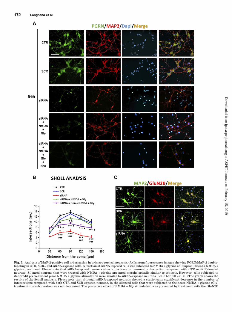
Fig. 5. Analysis of MAP-2-positive cell arborization in primary cortical neurons. (A) Immunofluorescence images showing PGRN/MAP-2 double-labeling in CTR, SCR-, and siRNA-exposed cells. A fraction of siRNA exposed cells was subjected to NMDA + glycine or ifenprodil (ifen) + NMDA +glycine treatment. Please note that siRNA-exposed neurons show a decrease in neuronal arborization compared with CTR or SCR-treatedneurons. Silenced neurons that were treated with NMDA + glycine appeared morphologically similar to controls. However, cells subjected toifenprodil pretreatment prior NMDA + glycine stimulation were similar to siRNA-exposed neurons. Scale bar, 90 mm. (B) The graph shows theresults of the Scholl analysis. Please note that although siRNA-exposed neurons showed a statistically significant decrease in the number ofintersections compared with both CTR and SCR-exposed neurons, in the silenced cells that were subjected to the acute NMDA + glycine (Gly)treatment the arborization was not decreased. The protective effect of NMDA + Gly stimulation was prevented by treatment with the GluN2B
172 Longhena et al.
at ASPE
T Journals on February 15, 2019
jpet.aspetjournals.orgD
ownloaded from
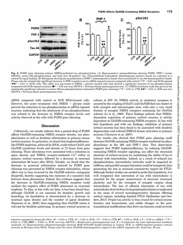
siRNA compared with control or SCR RNA-treated cells.However, the acute treatment with NMDA 1 glycine couldprevent the reduction in tau phosphorylation in siRNA-exposedneurons, indicating that the abatement of tau phosphorylationwas related to the decrease in NMDA receptor levels andactivity observed in the cells with PGRN gene silencing.
DiscussionCollectively, our results indicate that a partial drop of PGRN
affects GluN2B-containing NMDA receptor density, tau phos-phorylation as well as dendritic arborization in primary mousecortical neurons. In particular, we found that haploinsufficiency-likePGRNdepletion, achieved byRNAi, could reduceGluN1 andGluN2B membrane levels and density at 72 hours from genesilencing. These alterations were associated with a reduction inspine density and NMDA receptor-mediated Ca21-influx inprimary cortical neurons, followed by a decrease in neuronalarborization 96 hours after RNAi. Notably, we found that thedecrease in neuronal arborization induced by PGRN genesilencing was prevented by NMDA receptor stimulation, whoseeffect was in turn reversed by the GluN2B selective antagonistifenprodil, thereby supporting the existence of a causative linkbetween these phenomena. Indeed, this evidence suggests thatthe decrease in GluN2B-containing NMDA receptors couldmediate the negative effect of PGRN abatement on neuronaltrophism. To date, in line with our data, it has been found that,although GluN2B is not essential for dendrite growth andbranching, its knockdown is characterized by a decrease inneuronal spine density and the number of apical dendrites(Espinosa et al., 2009), thus suggesting that GluN2B action isrelevant for dendrite patterning. In primary cortical neurons in
culture at DIV 10, NMDA activity at excitatory synapses isassured by the coupling ofGluN1andGluN2B that can cluster atboth synaptic and extrasynaptic sites, with only a very smallfraction of synaptic NMDA receptors containing the GluN2Asubunit (Li et al., 1998). These findings indicate that NMDA-dependent regulation of primary cortical neurons is strictlydependent on GluN2B-containing NMDA receptors. In line withthis hypothesis and with our findings, inhibition of primarycortical neurons has been found to be associated with dendritedegeneration and reduced ERK1/2 kinase activation in primaryneurons (Chernova et al., 2007).Our results also showed that PGRN gene silencing could
decreaseGluN2B-containingNMDAreceptor-mediated tauphos-phorylation at Ser 262 and PHF-1 sites. This observationsuggests that PGRN haploinsufficiency, by reducing GluN2B-containing NMDA receptor signaling, can affect the structuralplasticity of cortical neurons by modulating the ability of tau tointeract with microtubules. Indeed, as a result of reduced tauphosphorylation, microtubular networks could be impaired bystiffness, andparallel synapse formation could behampered, thusgenerating the loss in neuronal connectivity typical for FTLD.Although further studies are needed to probe this hypothesis, it iswell recognized that interaction of tau with microtubules isessential for the proper maintenance of neuronal structuralplasticity and for the transport of synaptic cargoes alongmicrotubules. The loss of efficient interaction of tau withmicrotubulesderived from itshyperphosphorylation is implicatedin the onset of several neurodegenerative diseases affectingcortical regions, including familial FTLD (Spillantini and Goe-dert, 2013). Proper tau activity is thus crucial for cortical neuronfunction and homeostasis, and subtle changes in the post-translational modifications that drive tau function could severely
selective antagonist ifenprodil (Ifen). •P , 0.05 vs. CTR, xP , 0.05 vs. SCR, sP , 0.05 vs. CTR, ♦P , 0.05 vs. SCR, + P , 0.05 vs. siRNA, ***P ,0.001 vs. CTR, ###P , 0.001 vs. SCR; two-way ANOVA + Bonferroni’s postcomparison test, N = 6 triplicates for each experimental conditionanalyzed. (C) Representative photomicrographs showing MAP-2/GluNB double-labeling in control, SCR, or siRNA-exposed primary corticalneurons at 96 hours from RNAi. Please note the lower GluN2B-immunopositive signal in the siRNA exposed cells. Scale bar, 90 mm.
Fig. 6. PGRN gene silencing reduces NMDA-mediated tau phosphorylation. (A) Representative immunoblotting showing PGRN, PHF-1 (serine396/404), serine 262 phosphorylated, and total (tau 46-positive) tau. Glyceraldehyde-3-phosphate dehydrogenase–positive bands are reported as acontrol for equal loading. (B) Histogram showing the quantification of PHF-1-immunopositive bands when normalized vs. tau 46-immunopositive bands.Please note the statistically significant decrease in PHF-1-positive tau in siRNA exposed neurons compared with control (**P , 0.01) and SCR-exposed(♦♦P , 0.01 vs. SCR) cells. This effect was prevented in NMDA + glycine–treated cells that showed a significant increase of tau phosphorylationcompared with SCR-exposed neurons (dP, 0.05 one-way ANOVA + Newman-Keuls postcomparison test). (C) NMDA treatment could also prevent thestatistically significant reduction in serine 262 tau phosphorylation induced by PGRN gene silencing (**P, 0.01 vs. CTR; ♦♦P, 0.01 vs. SCR, one-wayANOVA + Newman-Keuls postcomparison test).
PGRN Affects GluN2B-Containing NMDA Receptors 173
at ASPE
T Journals on February 15, 2019
jpet.aspetjournals.orgD
ownloaded from
impinge on cortical neuron resilience. In this scenario, tauhyperphosphorylation, which generates neurofibrillarytangles in Microtubule-Associated Protein Tau-associated FTLD,and tau hypophosphorylation, which results fromPGRN reductioninFTLDwithPGRNmutations, couldbe twooppositemechanismsthat drive neuronal cells toward degeneration by compromisingtau-microtubular interaction. Our results support the conclusionthat the reduced activity of NMDA receptors deriving from lowPGRN levels could pivotally control the capacity of tau to interactwith microtubules in FTLD with PGRN mutations. Notably,several previous research reports have described the occurrenceof NMDA receptor-mediated tau phosphorylation in neuronal cells(Zhou et al., 2009; Sava et al., 2012), suggesting that this processcould be part of a physiologic intracellular signaling cascaderegulating synaptic NMDA receptor-dependent transmission dur-ing structural plasticity changes. In particular, NMDA receptorscan increase the phosphorylation of tau on specific sites thatmediate its interaction with synaptic proteins. Moreover, thephosphorylation of tau controls the interaction of tau with thepostsynaptic PSD-95-Fyn-NMDA receptor complex, which regu-lates GluN2B-containing NMDA-dependent synaptic plasticity,suggesting that physiologically occurring phosphorylation of taucould serve as a regulatorymechanism to prevent NMDA receptoroverexcitation (Mondragon-Rodriguez et al., 2012). These findings,coupled to our results, hint that a decrease in PGRN increasesneuronal vulnerability by leading to a reduction in GluN2B-containing NMDA receptors, which in turn affects the rate of tauphosphorylation and might perturb the ability of neuronal cells toeasily display structural plasticity changes. Structural changesoccur in the brain throughout life, including the generation of newneurons and other brain cells and connections between and amongneurons, and structural plasticity provides the mechanism for thebrain to repair itself (Gage, 2004). In addition, subtle changes infunctional plasticity in brain cortical areas can contribute tobehavioral impairments in the absence of significant pathology(Burke and Barnes, 2006). This implies that in PGRN mutationcarriers, cortical neurons holding reduced PGRN levels maypresent a lower ability to display structural plasticity changes.This phenomenon may also significantly decrease the resilience ofthese cells to aging and stressors.Consistent with our conclusions are recent reports that
PGRN-deficient mice display reduced synaptic connectivityand plasticity impairment occurring before neuropathologicalabnormalities (Petkau et al., 2012). Moreover, PGRN de-ficiency reduces synaptic pruning in the thalamus by dysre-gulating microglial cells (Lui et al., 2016). Our findings showthat the PGRN-deficiency–related decrease in neuronal ar-borization is attributable to a decrease in NR2B-containingNMDA receptors that is paralleled by a decrease in spinedensity. This evidence is in tune with previous findingsshowing that, although PGRN reduction can enhance trans-mission at individual synapses, it decreases gross neuronalconnectivity (Tapia et al., 2011), and confirms that importantneuronal plasticity changes occur in the early stages ofdisease. Remarkably, both NMDA receptor activity and tauphosphorylation have been found to be highly involved in thecontrol of microtubule dynamic changes during neuronalstructural plasticity (Caceres and Kosik, 1990; Knops et al.,1991; Biernat and Mandelkow, 1999; Dawson et al., 2001).Worthy of note, a recent finding indicates that PGRN
reduction is associated with increased tau phosphorylationin P301L transgenic mice (Hosokawa et al., 2015). Although in
part these observations are not in agreement with our results,which indicate that a decrease in PGRN reduces tau phos-phorylation, it is feasible that other mechanisms may beinvolved in the opposite phenomenon in the transgenic P301Ltau model. This is supported by the fact that PGRN-deficientmice do not show tau hyperphosphorylation (Hosokawa et al.,2015). In addition, we cannot exclude that a partial reductionin PGRN resulting in the case of haploinsufficiency may affectneuronal homeostasis in a manner completely different fromthe complete absence of the protein, as supported by thedifferent pathologic phenotypes described for PGRN-knockoutand PGRN-insufficient mice (Yin et al., 2010; Martens et al.,2012; Petkau et al., 2012; Filiano et al., 2013; Arrant et al.,2015). Data in human cerebrospinal fluids further support ourexperimental evidence, as we previously described that tauphosphorylation is increased in Alzheimer’s disease patientsand in FTLD patients comparedwith controls but not in FTLDpatients carrying a GRN mutation that causes progranulinhaploinsufficiency (Carecchio et al., 2011).The results of this study indicate that an haploinsufficiency-
like decrease in PGRN can reduce the density and activity ofGluN2B-containing NMDA receptors. This phenomenon isassociated with a reduction in NMDA receptor-mediated tauphosphorylation as well as a loss of neuronal dendritic arbor-ization, which is reversible by an acute GluN2B-containingNMDA receptor stimulation at 24 hours from gene silencing.These findings support the conclusions that GluN2B-containing NMDA receptors could be crucial mediators for thecontrol of PGRN-dependent cortical neuron neurotrophism.Collectively, these observations suggest that an aberrant
regulation of GluN2B-containing NMDA receptors resultingfrom PGRN loss of function mutations could impinge oncortical neuron resilience by perturbing their ability to displaystructural plasticity changes. The resulting progressive de-cline of neuronal trophism could probably initiate neuro-degeneration in FTLD with GRN mutations.These observations have relevant implications for understand-
ing the molecular mechanisms underlying the onset of FTLD.
Acknowledgments
The authors are grateful to Alessandro Barbon for providing theGluN2A, GluA1, and GluA2 antibodies.
Authorship Contributions
Participated in research design: Bellucci, Spano, Zaltieri.Conducted experiments: Bellucci, Longhena, Zaltieri, Grigoletto,
Faustini, La Via.Performed data analysis: Longhena, Zaltieri, Grigoletto.Wrote or contributed to the writing of the manuscript: Bellucci,
Longhena, Ghidoni, Benussi, Spano, Missale.
References
Arendt T, Stieler J, and Holzer M (2015) Brain hypometabolism triggers PHF-likephosphorylation of tau, a major hallmark of Alzheimer’s disease pathology. JNeural Transm (Vienna) 122:531–539.
Arrant AE, Patel AR, and Roberson ED (2015) Effects of exercise on progranulinlevels and gliosis in progranulin-insufficient mice. eNeuro 2:1–12 DOI: https://doi.org/10.1523/ENEURO.0061-14.2015.
BakerM,Mackenzie IR, Pickering-BrownSM,Gass J, Rademakers R, LindholmC, SnowdenJ, Adamson J, Sadovnick AD, Rollinson S, et al. (2006) Mutations in progranulin causetau-negative frontotemporal dementia linked to chromosome 17. Nature 442:916–919.
Benussi L, Ciani M, Tonoli E, Morbin M, Palamara L, Albani D, Fusco F, Forloni G,Glionna M, Baco M, et al. (2016) Loss of exosomes in progranulin-associatedfrontotemporal dementia. Neurobiol Aging 40:41–49.
Biernat J and Mandelkow EM (1999) The development of cell processes induced by tauprotein requires phosphorylation of serine 262 and 356 in the repeat domain and isinhibited by phosphorylation in the proline-rich domains. Mol Biol Cell 10:727–740.
174 Longhena et al.
at ASPE
T Journals on February 15, 2019
jpet.aspetjournals.orgD
ownloaded from
Borroni B, Alberici A, Cercignani M, Premi E, Serra L, Cerini C, CossedduM, Pettenati C,Turla M, Archetti S, et al. (2012) Granulin mutation drives brain damage and re-organization from preclinical to symptomatic FTLD. Neurobiol Aging 33:2506–2520.
Bozzali M, Battistoni V, Premi E, Alberici A, Giulietti G, Archetti S, Turla M, Gas-parotti R, Cercignani M, Padovani A, et al. (2013) Structural brain signature ofFTLD driven by Granulin mutation. J Alzheimers Dis 33:483–494.
Burke SN and Barnes CA (2006) Neural plasticity in the ageing brain. Nat RevNeurosci 7:30–40.
Caceres A and Kosik KS (1990) Inhibition of neurite polarity by tau antisense oli-gonucleotides in primary cerebellar neurons. Nature 343:461–463.
Carecchio M, Fenoglio C, Cortini F, Comi C, Benussi L, Ghidoni R, Borroni B, De RizM, Serpente M, Cantoni C, et al. (2011) Cerebrospinal fluid biomarkers in Pro-granulin mutations carriers. J Alzheimers Dis 27:781–790.
Carpenter-Hyland EP and Chandler LJ (2007) Adaptive plasticity of NMDA recep-tors and dendritic spines: implications for enhanced vulnerability of the adolescentbrain to alcohol addiction. Pharmacol Biochem Behav 86:200–208.
Chen H and Firestein BL (2007) RhoA regulates dendrite branching in hippocampalneurons by decreasing cypin protein levels. J Neurosci 27:8378–8386.
Chernova T, Steinert JR, Guerin CJ, Nicotera P, Forsythe ID, and Smith AG (2007) Neuritedegeneration induced by heme deficiency mediated via inhibition of NMDA receptor-dependent extracellular signal-regulated kinase 1/2 activation. J Neurosci 27:8475–8485.
Chitramuthu BP, Baranowski DC, Kay DG, Bateman A, and Bennett HP (2010) Pro-granulin modulates zebrafish motoneuron development in vivo and rescues truncationdefects associated with knockdown of Survival motor neuron 1. Mol Neurodegener 5:41.
Cruts M, Gijselinck I, van der Zee J, Engelborghs S, Wils H, Pirici D, Rademakers R,Vandenberghe R, Dermaut B, Martin JJ, et al. (2006) Null mutations in pro-granulin cause ubiquitin-positive frontotemporal dementia linked to chromosome17q21. Nature 442:920–924.
Dawson HN, Ferreira A, Eyster MV, Ghoshal N, Binder LI, and Vitek MP (2001)Inhibition of neuronal maturation in primary hippocampal neurons from tau de-ficient mice. J Cell Sci 114:1179–1187.
Desai A, Turetsky D, Vasudevan K, and Buonanno A (2002) Analysis of transcrip-tional regulatory sequences of the N-methyl-D-aspartate receptor 2A subunit genein cultured cortical neurons and transgenic mice. J Biol Chem 277:46374–46384.
El Gaamouch F, Buisson A, Moustié O, Lemieux M, Labrecque S, Bontempi B, DeKoninck P, and Nicole O (2012) Interaction between aCaMKII and GluN2B con-trols ERK-dependent plasticity. J Neurosci 32:10767–10779.
Espinosa JS, Wheeler DG, Tsien RW, and Luo L (2009) Uncoupling dendrite growth andpatterning: single-cell knockout analysis of NMDA receptor 2B. Neuron 62:205–217.
Filiano AJ, Martens LH, Young AH, Warmus BA, Zhou P, Diaz-Ramirez G, Jiao J,Zhang Z, Huang EJ, Gao FB, et al. (2013) Dissociation of frontotemporal dementia-related deficits and neuroinflammation in progranulin haploinsufficient mice.J Neurosci 33:5352–5361.
Finch N, Baker M, Crook R, Swanson K, Kuntz K, Surtees R, Bisceglio G, Rovelet-Lecrux A, Boeve B, Petersen RC, et al. (2009) Plasma progranulin levels predictprogranulin mutation status in frontotemporal dementia patients and asymp-tomatic family members. Brain 132:583–591.
Gage FH (2004) Structural plasticity of the adult brain. Dialogues Clin Neurosci 6:135–141.
Ghidoni R, Benussi L, Glionna M, Franzoni M, and Binetti G (2008) Low plasmaprogranulin levels predict progranulin mutations in frontotemporal lobar de-generation. Neurology 71:1235–1239.
Ghidoni R, Paterlini A, Albertini V, Binetti G, and Benussi L (2012) Losing protein inthe brain: the case of progranulin. Brain Res 1476:172–182.
Gijselinck I, van der Zee J, Engelborghs S, Goossens D, Peeters K, Mattheijssens M,Corsmit E, Del-Favero J, De Deyn PP, Van Broeckhoven C, et al. (2008) Pro-granulin locus deletion in frontotemporal dementia. Hum Mutat 29:53–58.
Hosokawa M, Arai T, Masuda-Suzukake M, Kondo H, Matsuwaki T, Nishihara M,Hasegawa M, and Akiyama H (2015) Progranulin reduction is associated withincreased tau phosphorylation in P301L tau transgenic mice. J Neuropathol ExpNeurol 74:158–165.
Jian J, Konopka J, and Liu C (2013) Insights into the role of progranulin in immu-nity, infection, and inflammation. J Leukoc Biol 93:199–208.
Knops J, Kosik KS, Lee G, Pardee JD, Cohen-Gould L, and McConlogue L (1991)Overexpression of tau in a nonneuronal cell induces long cellular processes. J CellBiol 114:725–733.
Kutzing MK, Langhammer CG, Luo V, Lakdawala H, and Firestein BL (2010) AutomatedSholl analysis of digitized neuronal morphology at multiple scales. J Vis Exp 45: e2354.
Laird AS, Van Hoecke A, De Muynck L, Timmers M, Van den Bosch L, Van DammeP, and Robberecht W (2010) Progranulin is neurotrophic in vivo and protectsagainst a mutant TDP-43 induced axonopathy. PLoS One 5:e13368.
Laulagnier K, Javalet C, Hemming FJ, and Sadoul R (2017) Purification and analysisof exosomes released by mature cortical neurons following synaptic activation.Methods Mol Biol 1545:129–138.
Laurier-Laurin ME, De Montigny A, Attiori Essis S, Cyr M, and Massicotte G (2014)Blockade of lysosomal acid ceramidase induces GluN2B-dependent tau phosphor-ylation in rat hippocampal slices. Neural Plast 2014:196812.
Li JH, Wang YH, Wolfe BB, Krueger KE, Corsi L, Stocca G, and Vicini S (1998)Developmental changes in localization of NMDA receptor subunits in primarycultures of cortical neurons. Eur J Neurosci 10:1704–1715.
Lui H, Zhang J, Makinson SR, Cahill MK, Kelley KW, Huang HY, Shang Y, OldhamMC, Martens LH, Gao F, et al. (2016) Progranulin deficiency promotes circuit-specificsynaptic pruning by microglia via complement activation. Cell 165:921–935.
Martens LH, Zhang J, Barmada SJ, Zhou P, Kamiya S, Sun B, Min SW, Gan L, Fink-beiner S, Huang EJ, et al. (2012) Progranulin deficiency promotes neuroinflammationand neuron loss following toxin-induced injury. J Clin Invest 122:3955–3959.
McKhann GM, Albert MS, Grossman M, Miller B, Dickson D, and Trojanowski JQ;Work Group on Frontotemporal Dementia and Pick’s Disease (2001) Clinical andpathological diagnosis of frontotemporal dementia: report of the Work Group onFrontotemporal Dementia and Pick’s Disease. Arch Neurol 58:1803–1809.
Mizuta I, Katayama M, Watanabe M, Mishina M, and Ishii K (1998) Developmentalexpression of NMDA receptor subunits and the emergence of glutamate neuro-toxicity in primary cultures of murine cerebral cortical neurons. Cell Mol Life Sci54:721–725.
Mondragón-Rodríguez S, Trillaud-Doppia E, Dudilot A, Bourgeois C, Lauzon M,Leclerc N, and Boehm J (2012) Interaction of endogenous tau protein with synapticproteins is regulated by N-methyl-D-aspartate receptor-dependent tau phosphor-ylation. J Biol Chem 287:32040–32053.
Mony L, Kew JN, Gunthorpe MJ, and Paoletti P (2009) Allosteric modulators ofNR2B-containing NMDA receptors: molecular mechanisms and therapeutic po-tential. Br J Pharmacol 157:1301–1317.
Moretti DV, Benussi L, Fostinelli S, Ciani M, Binetti G, and Ghidoni R (2016) Pro-granulin mutations affects brain oscillatory activity in fronto-temporal dementia.Front Aging Neurosci 8:35.
Navarria L, et al. (2015) Alpha-synuclein modulates NR2B-containing NMDA receptorsand decreases their levels after rotenone exposure. Neurochem Int 85-86:14–23.
Petkau TL, Hill A, and Leavitt BR (2016) Core neuropathological abnormalities inprogranulin-deficient mice are penetrant on multiple genetic backgrounds. Neu-roscience 315:175–195.
Petkau TL and Leavitt BR (2014) Progranulin in neurodegenerative disease. TrendsNeurosci 37:388–398.
Petkau TL, Neal SJ, Milnerwood A, Mew A, Hill AM, Orban P, Gregg J, Lu G,Feldman HH, Mackenzie IR, et al. (2012) Synaptic dysfunction in progranulin-deficient mice. Neurobiol Dis 45:711–722.
Petkau TL, Neal SJ, Orban PC, MacDonald JL, Hill AM, Lu G, Feldman HH,Mackenzie IR, and Leavitt BR (2010) Progranulin expression in the developing andadult murine brain. J Comp Neurol 518:3931–3947.
Pievani M, Paternicò D, Benussi L, Binetti G, Orlandini A, Cobelli M, Magnaldi S,Ghidoni R, and Frisoni GB (2014) Pattern of structural and functional brain ab-normalities in asymptomatic granulin mutation carriers. Alzheimers Dement 10(Suppl 5)S354–S363, 363.e1.
Rohrer JD, Geser F, Zhou J, Gennatas ED, Sidhu M, Trojanowski JQ, Dearmond SJ,Miller BL, and Seeley WW (2010) TDP-43 subtypes are associated with distinctatrophy patterns in frontotemporal dementia. Neurology 75:2204–2211.
Rohrer JD, Nicholas JM, Cash DM, van Swieten J, Dopper E, Jiskoot L, van Min-kelen R, Rombouts SA, Cardoso MJ, Clegg S, et al. (2015) Presymptomatic cogni-tive and neuroanatomical changes in genetic frontotemporal dementia in theGenetic Frontotemporal dementia Initiative (GENFI) study: a cross-sectionalanalysis. Lancet Neurol 14:253–262.
Rohrer JD, Warren JD, Fox NC, and Rossor MN (2013) Presymptomatic studies ingenetic frontotemporal dementia. Rev Neurol (Paris) 169:820–824.
Ryan CL, Baranowski DC, Chitramuthu BP, Malik S, Li Z, Cao M, Minotti S, Dur-ham HD, Kay DG, Shaw CA, et al. (2009) Progranulin is expressed within motorneurons and promotes neuronal cell survival. BMC Neurosci 10:130.
Sava A, Formaggio E, Carignani C, Andreetta F, Bettini E, and Griffante C (2012)NMDA-induced ERK signalling is mediated by NR2B subunit in rat cortical neu-rons and switches from positive to negative depending on stage of development.Neuropharmacology 62:925–932.
Skoglund L, Matsui T, Freeman SH, Wallin A, Blom ES, Frosch MP, Growdon JH,Hyman BT, Lannfelt L, Ingelsson M, et al. (2011) Novel progranulin mutationdetected in 2 patients with FTLD. Alzheimer Dis Assoc Disord 25:173–178.
Sleegers K, Brouwers N, Van Damme P, Engelborghs S, Gijselinck I, van der Zee J,Peeters K, Mattheijssens M, Cruts M, Vandenberghe R, et al. (2009) Serum biomarkerfor progranulin-associated frontotemporal lobar degeneration. Ann Neurol 65:603–609.
Spillantini MG and Goedert M (2013) Tau pathology and neurodegeneration. LancetNeurol 12:609–622.
Stein IS, Gray JA, and Zito K (2015) Non-ionotropic NMDA receptor signaling drivesactivity-induced dendritic spine shrinkage. J Neurosci 35:12303–12308.
Tackenberg C, Grinschgl S, Trutzel A, Santuccione AC, Frey MC, Konietzko U, GrimmJ, Brandt R, and Nitsch RM (2013) NMDA receptor subunit composition determinesbeta-amyloid-induced neurodegeneration and synaptic loss. Cell Death Dis 4:e608.
Tapia L, Milnerwood A, Guo A, Mills F, Yoshida E, Vasuta C, Mackenzie IR, Raymond L,Cynader M, Jia W, et al. (2011) Progranulin deficiency decreases gross neural connec-tivity but enhances transmission at individual synapses. J Neurosci 31:11126–11132.
Van Damme P, Van Hoecke A, Lambrechts D, Vanacker P, Bogaert E, van Swieten J,Carmeliet P, Van Den Bosch L, and Robberecht W (2008) Progranulin functions asa neurotrophic factor to regulate neurite outgrowth and enhance neuronal sur-vival. J Cell Biol 181:37–41.
Whitwell JL, Jack CR, Jr, Baker M, Rademakers R, Adamson J, Boeve BF, KnopmanDS, Parisi JF, Petersen RC, Dickson DW, et al. (2007) Voxel-based morphometry infrontotemporal lobar degeneration with ubiquitin-positive inclusions with andwithout progranulin mutations. Arch Neurol 64:371–376.
Williams JM, Guévremont D, Kennard JT, Mason-Parker SE, Tate WP, and Abraham WC(2003) Long-term regulation of N-methyl-D-aspartate receptor subunits and associatedsynaptic proteins following hippocampal synaptic plasticity. Neuroscience 118:1003–1013.
Wyllie DJ, Livesey MR, and Hardingham GE (2013) Influence of GluN2 subunitidentity on NMDA receptor function. Neuropharmacology 74:4–17.
Yin F, Banerjee R, Thomas B, Zhou P, Qian L, Jia T, Ma X, Ma Y, Iadecola C, BealMF, et al. (2010) Exaggerated inflammation, impaired host defense, and neuro-pathology in progranulin-deficient mice. J Exp Med 207:117–128.
Zhou X, Moon C, Zheng F, Luo Y, Soellner D, Nuñez JL, and Wang H (2009) N-methyl-D-aspartate-stimulated ERK1/2 signaling and the transcriptionalup-regulation of plasticity-related genes are developmentally regulated followingin vitro neuronal maturation. J Neurosci Res 87:2632–2644.
Address correspondence to: Dr. Arianna Bellucci, Division of Pharmacology,Department of Molecular and Translational Medicine, University of Brescia,Viale Europa no. 11, 25123, Brescia, Italy. E-mail: [email protected]
PGRN Affects GluN2B-Containing NMDA Receptors 175
at ASPE
T Journals on February 15, 2019
jpet.aspetjournals.orgD
ownloaded from