Embed Size (px)
Citation preview

Optogenetics: 10 years of microbial opsins in neuroscience
Karl DeisserothDepartments of Bioengineering and of Psychiatry and Behavioral Sciences and the Howard Hughes Medical Institute, Stanford University, Stanford, California, USA.
Karl Deisseroth: [email protected]
Abstract
Over the past 10 years, the development and convergence of microbial opsin engineering, modular
genetic methods for cell-type targeting and optical strategies for guiding light through tissue have
enabled versatile optical control of defined cells in living systems, defining modern optogenetics.
Despite widespread recognition of the importance of spatiotemporally precise causal control over
cellular signaling, for nearly the first half (2005–2009) of this 10-year period, as optogenetics was
being created, there were difficulties in implementation, few publications and limited biological
findings. In contrast, the ensuing years have witnessed a substantial acceleration in the application
domain, with the publication of thousands of discoveries and insights into the function of nervous
systems and beyond. This Historical Commentary reflects on the scientific landscape of this
decade-long transition.
Optogenetics is the combination of genetic and optical methods to cause or inhibit well-
defined events in specific cells of living tissue and behaving animals1. This technology, as
employed today to study the neural circuit underpinnings of behavior, most commonly
involves three core features: (i) microbial opsins, members of an ancient, but uniquely well-
suited, gene family adapted from evolutionarily distant organisms such as algae and
archaebacteria, with each gene encoding a distinct protein that directly elicits electrical
current across cellular membranes in response to light, (ii) general methods for targeting
sufficiently strong and specific opsin gene expression to well-defined cellular elements in
the brain, and (iii) general methods for guiding sufficiently strong and precisely timed light
to specific brain regions, cells or parts of cells while the experimental subject carries out
behaviors of interest.
None of these three components was enabled for general optogenetic discovery in
neuroscience 10 years ago2, or even when we suggested the new word to describe this
emerging process a year later3, but each has scientific origins dating back decades. The
cross-integration that enabled optogenetics over the past 10 years only became possible
under the right conditions. Each component also continues to rapidly evolve for greater
precision and complexity. Indeed, these fundamental elements of optogenetics are
increasingly recruiting and bringing to bear more branches of science and engineering,
Any Supplementary Information and Source Data files are available in the online version of the paper.
COMPETING FINANCIAL INTERESTSThe author declares no competing financial interests.
Published as: Nat Neurosci. 2015 September ; 18(9): 1213–1225.
HH
MI A
uthor Manuscript
HH
MI A
uthor Manuscript
HH
MI A
uthor Manuscript

ranging from computational tools for system identification to automated optical readouts of
behavior and neural activity to high-content anatomical data extraction methods for
discovering structural and wiring relationships4.
The most recent technological developments, along with experimental guidelines, challenges
and limitations, have already been reviewed in detail this year4. In the present Historical
Commentary, I focus on the optogenetic transition itself over the past 10 years, from
scientific conditions surrounding the early work to the major discoveries that have arisen
with application of this technology over the same time period, all set in the context of the
development of three converging disciplines that could hardly be more disparate in origin
and tradition.
Developing and assembling the components of optogenetics
The first of these three components has been part of the fabric of biochemistry and
physiology for many decades: the microbial opsin genes and the microbial rhodopsin
proteins they encode5, a family of molecules (Fig. 1a) functionally completely distinct from
(and unrelated in primary sequence to) the better-known rhodopsins that mediate
phototransduction in the vertebrate eye6. Indeed, the initial evidence from Oesterhelt and
Stoeckenius, beginning in 1971, that microbial organisms might also produce and use
rhodopsin-like proteins7 was surprising and intriguing. Instead of coupling to intracellular
second-messenger cascades to indirectly influence ion channels, like their vertebrate
counterparts, these microbial proteins for the most part directly transduce photons into
electrical current (Fig. 1a,b). This molecular feat provoked intense curiosity and has inspired
thousands of investigations over the decades5. Indeed, within a few years, this discovery had
given birth to a vibrant community that produced a steady output of about 100 papers
annually for four decades extending to the present day, spanning genomic, functional and
structural investigations into the photocycles and mechanisms of these microbial proteins,
and defining part of the textbook training of biologists8.
Three branches of this family tree have found utility in optogenetics: the bacteriorho-
dopsins, the halorhodopsins and the channel-rhodopsins (Fig. 1a). The naturally occurring
bacteriorhodopsins (the first-discovered members of this family, which pump protons out of
the cell) and halorhodopsins (which pump chloride ions into the cell) are typically inhibitory
in neural systems, as both of these types of hyperpolarizing current make it harder for
neurons to fire action potentials; in contrast, the naturally occurring channel-rhodopsins for
the most part allow positively charged ions to flow freely through the opsin pore and so tend
to be depolarizing and excit-atory5 (Fig. 1a). This pattern held for many years until the high-
resolution crystal structure of channelrhodopsin9 allowed structure-guided engineering of
the opsin channel pore (Fig. 1c)10 to create inhibitory chloride-conducting channels10,11 in
2014, followed by identification of a natural chloride-conducting channelrhodopsin12,13 in
2015. Over the years, more variants in these protein families have been discovered in nature
(or engineered in the laboratory) to have faster kinetics, bistable properties, altered ion
conductances and shifted color-response properties; this diversity is now leveraged to
powerful effect in optogenetic experimentation.
Deisseroth Page 2
Nat Neurosci. Author manuscript; available in PMC 2016 March 14.
HH
MI A
uthor Manuscript
HH
MI A
uthor Manuscript
HH
MI A
uthor Manuscript

The key functional properties of these proteins were widely known for decades, and many
investigators had sought to create strategies for controlling neurons with light. So why did it
take time to develop and apply methods for placing these proteins into different classes of
neurons in behaving animals? As mentioned above, the development of optogenetics was a
biological three-body problem in which it was hard to resolve (or, even more importantly, to
motivate attempts to resolve) any one of the three challenges without first addressing the
other components. For example, microbial rhodopsin photocurrents were predicted to be
exceedingly small, suggesting a difficult path forward even if efficient delivery and
incorporation of the all-trans retinal chromophore were possible in adult non-retinal brain
tissue, and even in the event of safe and correct trafficking of these evolutionarily remote
proteins to the surface membrane of complex metazoan neurons. For these weak membrane
conductance regulators to work, high gene-expression and light-intensity levels would have
to be attained in living nervous systems while simultaneously attaining cell-type specificity
and minimizing cellular toxicity. All of this would have to be achieved even though neurons
were well known to be highly vulnerable to (and often damaged or destroyed by)
overexpression of membrane proteins, as well as sensitive to side effects of heat and light.
Motivating dedicated effort to exploration of microbial opsin-based optical control was
difficult in the face of these multiple unsolved problems, and the dimmest initial sparks of
hope would turn out to mean a great deal.
Outside neuroscience, several examples of functional heterologous expression of opsins for
light-activated ion flow had been published in non-neural isolated-cell systems for microbial
opsins14–16 (beginning14 in the early 1990s) or vertebrate opsins17 (beginning in the late
1980s), although neuroscience or behavior applications were not suggested. It is not known
how many investigators actually did attempt to transduce microbial opsins into neurons
before 2005, but even in this one step, among the many steps required for optogenetics,
much can go wrong18 (Fig. 1d). Meanwhile, over the years leading up to 2005, several other
strategies for optical control of targeted neurons, involving multiple simultaneously
delivered metazoan genes or coordinated delivery of both a metazoan gene and a light-
sensitive synthesized chemical, were devised19–23, perhaps by their very elegance reducing
enthusiasm for another approach based entirely on a family of far more foreign microbial
proteins that would seem much less likely to work.
Worthiness for allocation of time and effort was not a trivial consideration, as my laboratory
group in 2004 had limited material resources; moreover, there was still a great deal of self-
doubt, with the realization that many more steps of equal or greater magnitude and risk
would be needed to reach even the most basic initial goal. However, once neural membrane
expression with appreciable light-activated functionality of a microbial opsin had been seen
(Fig. 2a) and it first became possible to report “I think it worked,” the landscape changed
from speculation to action. The ensuing 2 years indeed saw action on many levels:
constructing and concentrating the crucial expression vectors for stable, well-tolerated
expression (Fig. 2b), testing real-time readouts using electrophysiology and behavior in vitro
(Fig. 2b,c) and then, crucially, in vivo; and design (Fig. 2d) and implementation (Fig. 2e) of
neural interfaces for in vivo light delivery and behavior2,3,24–26. Many investigators were
discussing the possibilities and working on related efforts, and although a number of papers
Deisseroth Page 3
Nat Neurosci. Author manuscript; available in PMC 2016 March 14.
HH
MI A
uthor Manuscript
HH
MI A
uthor Manuscript
HH
MI A
uthor Manuscript
were published in 2005 and 2006 from pioneering laboratories around the world2,3,27–30, it
was not for two more difficult years that the journey to mammalian behavioral control was
ultimately completed25,26 (Fig. 3a,b and Supplementary Video 1), finally allowing
confidence that the microbial opsin approach was going to be generally useful.
Some issues that had seemed formidable for in vivo work turned out to be readily addressed.
For example, invertebrates could make use of dietary all-trans retinal as the
chromophore28,31–33, immature and developing vertebrate nervous systems and the retina
required no supplementation27,29,30, and the adult mammalian brain required no exogenous
retinal or other component3,24–26. With these discoveries, along with the long-known single-
gene logic of microbial opsin transduction of light into ion flow7,8,14, single componency for
optogenetics had been achieved—as with green fluorescent protein (GFP), a property crucial
for utility. But still much more challenging discovery and development were required, as
evidenced by the fact that broad adoption of optogenetics with microbial opsins did not
occur until 2009. The transition that followed was enabled only with the convergence of
optics and genetics components of optogenetics, the second and third fields that would have
to come together with microbial opsin genes. Between 2004 and 2009, discovery along these
key dimensions proceeded rapidly.
Only a few new biological findings using microbial opsin optogenetics had emerged by
early 2007, chiefly from small, optically accessible invertebrate systems. For example, in
Drosophila larvae, the effects of activity in distinct neuromodulator (dopaminergic versus
octopaminergic/tyraminergic) systems had been tested in classical odorant conditioning31,
and a role for nociceptive neurons in defensive behaviors protecting larvae from wasps had
been defined32. In addition, opsin-expressing mammalian brain surfaces had been acutely
exposed and illuminated with spots of light to drive and map responses3,24,34–36. However,
control of mammalian behavior had not been achieved, and indeed the intact brain seemed
largely inaccessible to optogenetics. There was therefore a compelling need to safely,
focally and flexibly deliver visible light via a neural interface deep into the brain of a freely
behaving mammal at a high-intensity: ~100 mW mm‒2 at the interface output, ~100× greater
than needed at the opsin-expressing cells themselves because of expected scattering losses
over the effective brain volume25 and much more intense than needed for imaging. LEDs at
the time were underpowered for coupling to optical fibers, and so necessity rapidly drove
development of optogenetic interfaces based on laser diode–coupled fiber optics25,26.
Among other features, including heat isolation and activity feedback, these interfaces
crucially also registered virus injection to illumination site (Fig. 2d,e), which opened a new
realm of experimental possibilities for targeted control and readout during behavior.
By mid-2007 (Fig. 3a,b) it was possible26 to selectively target a microbial opsin gene with
high specificity and penetrance to a defined population of neurons deep in the brain of adult
mice (in this case, hypocretin/orexin neurons in the hypothalamus), to play in a broad range
of spike patterns through an optical fiber to those cells, to collect simultaneous multimodal
system readouts during freely moving behavior (in this case, describing sleep/wake status
via electroencephalography (EEG) and electromyography), and to demonstrate a causal role
for defined activity patterns in specific brain cells in a natural behavior (in this case, sleep-
wake transitions)26. It is worth noting that, although optogenetic control of behavior through
Deisseroth Page 4
Nat Neurosci. Author manuscript; available in PMC 2016 March 14.
HH
MI A
uthor Manuscript
HH
MI A
uthor Manuscript
HH
MI A
uthor Manuscript

the intact skull37 or a cra-niotomy38 using LEDs directly apposed to the cranium was
reported later that year, this approach is rarely taken today, as LEDs generate substantial
local heating, the light delivered in this way is not as readily targeted to defined circuit
elements and, in contrast, the fiber-optic method also allows imaging light to be collected
back through the same interface to report on local neural activity.
This example also illustrates how genetic targeting of circuit components had to advance in
a fundamental way that was adapted to the high expression levels needed for the microbial
opsins. In these first 5 years, the field accordingly saw development of versatile, high-titer
cell-targeting opsin viruses3,24–26,39–42 and the creation of the initial broadly expressed35,36
and specific43 transgenic opsin mouse lines. These advances were complemented by the
initial reports of in vivo behavioral effects resulting from genetic targeting of cells in the
worm (for excitation28 and inhibition33), fly31, fish44 and rodent25,26. Cell-type targeting of
opsin genes was not only achieved with genetics, however. It was the fiber-optic hardware
method that enabled what is now the one of most widely used and generalizable approaches
for targeting cells in behaving animals on the basis of anatomy or wiring. Termed projection
targeting, this involves the use of the fiber-optic neural interface directed to axonal
projections of the opsin-expressing cells to recruit cells defined by wiring for control in
behavior4,45.
Dovetailing with this genetics and optics development in those first few years were advances
in opsin genomics and engineering. In a different sort of reverse translation situated entirely
in the basic science realm, new opportunities in optogenetics were in turn now driving the
basic study of microbial opsins, including the development of new structural models,
photocycle analyses, and opsin gene discovery with modern genomic methods. This field
was thriving before opto-genetics; for example, two of the sparks that ultimately led to
optogenetics arose from the identification of rhodopsins in microbes7 by Stoeckenius and
Oesterhelt in 1971 and the identification of rhodopsin-regulated currents in
Chlamydomonas46 by Hegemann and Harz in 1991. But optogenetics returned the favor,
providing a new spark to the field of microbial opsin biology. Very much relevant to the
early optical hardware development occurring at the same time, the new proteins discovered
and engineered in these first few years were able to take advantage of red-shifted light47,
bistable ‘step-function’ photocurrents that could be stably turned on with a pulse of one
wavelength of light and off with another48,49, much higher cellular light sensitivity48,49, and
higher safe expression levels in vivo (further contributing to improved cellular light
sensitivity) based on membrane trafficking50,51 and chimerization52,53 innovations. These
opsin-engineering advances thus guided and defined constraints for initial engineering of the
optical hardware elements (Fig. 2d,e).
In this way, the microbial opsins, in vivo optics and expression-targeting genetics finally
came together as a mutually interdependent and complementary whole. Although genetic
targeting for optogenetics has also advanced further for multiple-feature targeting in the
second half of the decade54 (Fig. 3c,d), even by 2009 it was already possible to achieve cell-
type targeting not just for isolated tractable cases, but also in a generalizable fashion, with
recombinase-dependent viruses39–42 that were strong and specific enough to be used to
control behavior and with projection-targeting behavioral control enabled by the fiber-optic
Deisseroth Page 5
Nat Neurosci. Author manuscript; available in PMC 2016 March 14.
HH
MI A
uthor Manuscript
HH
MI A
uthor Manuscript
HH
MI A
uthor Manuscript

interface that began to move beyond the need for genetic targeting reagents45. This
experimental and conceptual framework for optogenetics allowed adoption, application and
discovery by investigators around the world.
Discoveries with optogenetics
Optogenetic methods have now enabled acquisition of insights into a broad range of
questions in behavior, physiology and pathology, spanning domains of sensation, cognition
and action. Although many of these studies have been conducted in mammals (typically rats
and mice), optogenetic methods have also become a standard resource for scientific
communities studying neural circuit foundations of behavior in invertebrates. Beyond simple
observation of evoked electrical activity, numerous fascinating biological findings on
regulation of complex behavioral states have been revealed in the nematode (for example,
ref. 55) and the fruit fly (for example, ref. 56), while more gradually, scientific insights from
optogenetics in species such as the songbird (for example, ref. 57), the zebrafish (for
example, ref. 58) and the nonhuman primate (for example, ref. 59) have also emerged. The
capabilities that have come along with the emergence of single-component optogenetics as a
standard research tool displaying speed, simplicity and versatility were cited for justifying,
in part, the timing and scale of national-scale neuroscience research initiatives launched in
2013, including the BRAIN initia-tive60. Although the full scope of the findings that have
resulted in the field can no longer be reviewed in detail, it is interesting to take note of key
examples in the different categories of investigation that have emerged.
Broadly speaking, optogenetic methods have now illuminated the causal role of defined cell
types and projections in natural as well as disease-related physiology and behaviors, ranging
from the most basic homeostasis to advanced cognitive functions. For example, optogenetic
methods have now been used to illuminate the causal neuronal underpinnings of movement
regulation43,61, including the identification of surprising bottom-up circuit mechanisms by
which the spinal cord and cerebellum regulate forebrain control of skilled and voluntary
movements62–64. Patterns of activity arising from genetically and anatomically defined cells
have been identified, even when these cells are intermixed with other cell types, that
specifically drive or inhibit the most fundamental of organismal functions: hunger, thirst and
energy balance65–69; respiration70,71; and arousal, sleep and circadian rhythm26,72–75. And
the transmission of primary sensory information to the brain has also been studied
extensively with optogenetics, including in the domains of olfactory76–78, auditory79,
visual80,81 and tactile82,83 processing.
Many studies have employed optogenetics to discover and map the pathways along which
such information flows in the brain, including analysis of physical circuit connectivity
itself 84,85, tagging cells defined by type or connectivity for use in other analyses86, and
integrating with fMRI or PET imaging to generate brain-wide maps of activity patterns
recruited by defined neural cells or projections87,88. Optogenetic methods have also yielded
insight into the dynamics of information transmission in neural circuits more generally—for
example, regarding the modulation of activity by oscillatory rhythms41,89–97, the causal
significance of phase timing within the theta rhythm98, the influence of gamma rhythmicity
on information propagation41,89,90 and (dovetailing with parallel insights from genetic
Deisseroth Page 6
Nat Neurosci. Author manuscript; available in PMC 2016 March 14.
HH
MI A
uthor Manuscript
HH
MI A
uthor Manuscript
HH
MI A
uthor Manuscript
interventions99,100) the real-time regulation of information flow via the dynamic balance of
excitation and inhibition90,101–110.
Circuit activity patterns have also been identified, using microbial opsin genes expressed in
conditional viruses and targeted with the fiber-optic interface, that control and modulate
many motivated behaviors. Social behavior, which places intense demand on circuitry across
the brain involving sensation, motivation, reward, cognition and memory, has been studied
under a range of conditions, leading to identification of cells and projections that modulate
same-sex social patterns90,111, as well as parental112, mating-related113,114, aggression-
related113–116 and defensive115,116 interactions. Studies of tradeoffs between active and
passive coping in response to challenge117 and tradeoffs between avoiding risk and seeking
reward118 have also proven readily accessible to optogenetic identification of the cells and
circuits involved. Indeed, insights have been derived into the causal circuit underpinnings of
reward itself 42,119–124, as well as into the circuit implementations of fear and
anxiety125–135. Optogenetics in these cases has enabled the delineation of different cell types
that, even though juxtaposed and intertwined, can have fundamentally oppositional roles in
these complex behaviors. Principles of brain functional organization have emerged along the
way; for example, revealing the potency and adaptability of brain-wide connections, defined
by origin and target and so not previously accessible to investigators for specific control, in
the precise regulation of behaviors and behavioral states136. And long-term action and
electrical patterns have been illuminated as well, with resulting insights into the cell
activity–specific causation of stable brain states underlying behavior137.
Beyond the dynamics of brain and behavioral states, the mechanisms underlying information
storage in the brain have also been illuminated by optogenetic methods138–140. One of the
most exciting developments in recent years has been the testing of long-held and much-
debated models of neural information representation. Use of clever activity-guided
expression of microbial opsin genes has led to causal identification of sparse and distributed
population representations (engrams)139–142 underlying memory states and mediating
linkages among features of these memory states. Learning itself has been studied with
optogenetics in ventral striatum and amygdala143,144, as well as in the hippocampus138, and
inputs to the hippocampal formation have been studied in some detail with regard to causal
roles in learning, navigation and information flow145–147. Finally, precisely timed activity
patterns in diverse well-defined cell types, projections and populations have been causally
implicated in synaptic plasticity148,149, wiring and microcircuit plasticity150,151, and in long-
timescale devel-opmental152,153 and adaptive changes such as activity-dependent
myelination154 and adult neurogenesis155,156.
Many discoveries have also emerged regarding the neural circuitry of symptoms related to
disease states; indeed, the precise circuit-level mechanisms and causal tissue-level
manifestations of neuropsychiatric disease have been mysterious for many of the same
reasons that made optogenetics useful for basic neuroscience. For example, optogenetic
methods have been applied to study the cellular activity underpinnings of seizure
propagation and termination157–159; these studies and those that followed have included
some of the first closed-loop approaches in behaving animals (Fig. 4), as well as the
surprising identification of highly focal sites for immediate seizure termination remote from
Deisseroth Page 7
Nat Neurosci. Author manuscript; available in PMC 2016 March 14.
HH
MI A
uthor Manuscript
HH
MI A
uthor Manuscript
HH
MI A
uthor Manuscript

the initiation site158. The latter finding is still being explored, but, if the percolation of
activity imbalance through the brain occurs not generally, but instead chiefly through
surprisingly well-defined and separated nodes where highly effective point control (as with a
fiber optic or electrode; Fig. 4) may be exerted, the implications are substantial not just for
understanding epilepsy but for understanding the brain as a dynamical system.
Also on the neurological front, optogenetic investigations have led to the determination of
the cells and pathways that promote or inhibit normal and parkinsonian movement
patterns45,61. The latter study included the initial specific and causal demonstration of the
oppositional roles of the striatal direct and indirect pathways in action initiation and
suppression, respectively, under either healthy or parkinsonian conditions61—in this case,
resolving a longstanding question that had not been susceptible to direct testing, with clear
implications for understanding the disease but also for understanding adaptive movement
regulation.
A full discussion of whether or not optogenetics will be directly applied in a therapeutic
sense (such as through direct viral approaches160 or introduction of light-sensitive cells161)
is beyond the scope of this Historical Commentary. Laboratory-derived circuit-level insights
into maladaptive behavior are important in the scientific sense, however, and will in turn
feed back and enhance the basic study of natural circuits and processes that go awry in
disease states. Optogenetics has been used to identify neoplastic and degenerative processes
such as activity-dependent double-strand DNA breaks in neurons162, amyloid-β pathology
augmentation by chronic moderate activity in Alzheimer’s model mice163, altered glioma
growth mediated by activity-driven neuroligin-3 s ecretion164 and altered striatal inhibition
in Huntington’s disease mouse models165, all tested and modulated by precisely initiated
activity in defined cell populations expressing microbial opsin genes. Regenerative
processes have been studied as well, including restoration of light responses in retinitis
pigmentosa via halorhodopsin166, altered activity-dependent myelination of long-range
projections from neocortex154 and recovery and rewiring processes after stroke167,168.
Despite the seemingly intractable brainwide complexity of addictive drug effects and our
clinical difficulty in treating these disorders pharmacologically, optogenetic methods have
been employed for the discovery of cells and synapses that can powerfully and specifically
control the expression of cocaine-related behaviors119,120,169–171. For example, optogenetic
methods were used to resolve a controversy on the causal role of a small (~1%) population
of cholinergic cells in the nucleus accumbens that controls acute cocaine condi-tioning119.
Additionally, a surprising series of papers revealed the potency of focally initiated control
from the frontal cortex over long-term cocaine-initiated behavioral patterns169–171, with
potential relevance for understanding how stable patterns of behavior can be put in place and
specifically modulated in a top-down manner (as also seen in a pair of papers on optogenetic
modulation of corticostriatal control over obsessive-compulsive behavior
mechanisms172,173).
Psychiatric diseases in general have been illuminated136 through the determination of
mechanisms by which features that contribute to behavioral states, such as those of altered
motivation, reward, social interaction and anxiety, are recruited, assembled and coordinated
Deisseroth Page 8
Nat Neurosci. Author manuscript; available in PMC 2016 March 14.
HH
MI A
uthor Manuscript
HH
MI A
uthor Manuscript
HH
MI A
uthor Manuscript
across the brain. Each of these symptom domains is shared, though with different qualities
and in different combinations, across diverse clinical conditions117,128–130,133. When a
projection is targeted, symptom domain– specific behavioral consequences can manifest in a
manner that is more specific and/or potent than when activity is modulated in either the
originating structure or the target structure of this projection alone4, as in the transition from
active-coping to passive-coping behavioral strategies in the context of insuperable
challenge117. In particular, it was found that a specific long-range projection from prefrontal
cortex to the dorsal raphe was able to promote (when optogenetically resolved and excited)
or suppress (when optogenetically resolved and inhibited) swimming and climbing behavior
in an automated forced-swim test117, with implications for understanding the assessment and
assignment of cost/effort relationships that may vary with affective state in health and
disease. Less specific, absent, or opposite-direction effects were seen with control of the
projection target, the projection origin, or a differently targeted projection to habenula,
respectively117. A common theme in such optogenetic studies is the identification of defined
cell populations (or projections arising from these cells) with mechanistic contributions to
individual symptom domains, revealing causal mapping with explanatory power for both
adaptive and maladaptive states. Findings from the clinically inspired side, together with
those from the basic science side, have resulted in both the emergence of these broad
concepts as well as the resolution of longstanding questions.
Current experimental considerations and challenges
As with any scientific method, there are important technical limitations and potential
confounding factors, known to practitioners and addressed over the years4,37,136,174 and
discussed here for completeness. Rigorous optogenetics requires careful consideration of (i)
controls to account for effects of the experimental system itself, (ii) definition of the
stimulus pattern delivered, and (iii) definition of the directly modulated tissue element.
These are general considerations for any intervention in biology, although optogenetics
affords new opportunities for both specificity and means of validating specificity.
First, accounting for the experimental system requires considering the influence of light
delivery in itself and of the opsin gene in itself. Regarding the light delivery, the flexible
fiber-optic interface is comparable to, though smaller and lighter than, the electrical
interfaces that have been a staple of freely moving systems neurobiology for
decades25,26,174. This optical interface further enables the two new capabilities of
projection-targeting for behavioral control45,121,128 and readout of activity in cells and
projections during behavior4,111. Nevertheless, the careful behaviorist must carry out
baseline behavioral testing with the interface in place to validate the task for each cohort and
each experimental subject. It is also standard practice for behavioral experiments to include
no-opsin arms, in which animals receive every other aspect of the preparation, including
viral transduction and light delivery; these controls address many theoretical concerns,
ranging from toxic side effects of the surgery to distracting or heating effects of the light
itself174. Notably, it would be much harder to build in such controls for electrical
stimulation, providing the same energy in such a way that the tissue could not respond
functionally. Optogenetic experimentation both requires and makes possible such controls.
Deisseroth Page 9
Nat Neurosci. Author manuscript; available in PMC 2016 March 14.
HH
MI A
uthor Manuscript
HH
MI A
uthor Manuscript
HH
MI A
uthor Manuscript
Regarding the opsin gene expression itself, as with any foreign or native gene (especially
those encoding membrane proteins), genes for optical actuation and for readout of structure
and function bring a risk of possible toxic effects in the setting of high, long-term
expression. This was addressable45,50,51, and now every major class of overexpressed
microbial opsin (proton pumps, chloride pumps and channels) in mammals has been shown
to benefit from the provision of mammalian membrane trafficking signals18,45,50,51,90 to
facilitate efficient movement through protein production pathways to the cell surface
membrane. Avoiding long-term expression with certain viruses (for example, rabies and
herpesviruses) or with certain promoter/enhancer combinations that are too strong (such as
CMV-based promoters in many settings174) is also important, and optogenetics experiments
should include, where practical, histological and/or electrophysiological validation of cell
health, as well as baseline comparisons with non-opsin-expressing cells or animals. Of
course, in most cases, each animal can also serve as its own light-off/light-on/light-off
control to verify that the experiment is truly reading out an effect of actuation rather than of
preparation or changes over time—controls that are harder to achieve in other approaches
such as lesion studies.
Second, defining the stimulus pattern delivered is of substantial importance for fully
capitalizing on the capabilities of optogenetic control. Light pulse patterns can be mapped
across a broad range from undetectable to pathological in outcome while reading out the
behavior and physiology of interest (for example, ref. 26) or can be tuned to match various
distinct patterns observed from recording studies (for example, ref. 42). Moreover, when
optogenetics is used as a circuit-level tool (as is usually the case)—for example, to study the
influence of one population of cells on another population in a different brain region—the
train of action potentials optogenetically elicited in the presynaptic cell population is created
by native voltage-gated sodium channels and propagates as a spike to the axon terminal,
where it will be susceptible to the same mechanisms of presynaptic plasticity (for example,
via releasable vesicle pool dynamics) and post-synaptic plasticity (for example, via receptor
internalization and phosphorylation) as native action potential trains with similar rates and
timing. Of course, optogenetics may encounter more challenges for within-cell studies,
where the region studied is also the region illuminated and so nonphysiological distribution
of light-activated channels and abnormal relative timing of voltage changes across the cell
could cause artifactual patterns of intracellular plasticity and cell signaling; hence, this
approach is usually not recommended.
Relative timing of activity among the cells of the targeted population is also of interest,
although experimental data describing such relationships are not usually available in a
meaningful form. However, much like the amplitude of population activity, relative
synchrony of the population is also titratable with optogenetics. At one extreme,
subthreshold modulation of excitability within the population (without directly driving
spikes) is readily achieved using bistable step-function optogenetic tools48,49,90 or with
weak steady light delivery to conventional microbial opsin–expressing cells; in these cases,
spike timing is asynchronous and depends on each cell’s native synaptic input pattern48,90.
At the other extreme, synchrony to the level that can be observed in vivo during local field
potential or EEG oscillations can be achieved with very intense but brief light flashes,
Deisseroth Page 10
Nat Neurosci. Author manuscript; available in PMC 2016 March 14.
HH
MI A
uthor Manuscript
HH
MI A
uthor Manuscript
HH
MI A
uthor Manuscript
although (just as with native synaptic drive) variation in cell and synapse history will
inevitably still give rise to variation in spike latency. As capabilities in the field move
toward independent control of multiple single cells in a population (Fig. 5; recently achieved
with two-photon illumination-optimized opsins175 and spatial light targeting176 during
behavior177,178) and to optogenetic control of sparse, distributed ensembles of cells defined
by activity history139,142, relative intrapopulation timing will become an increasingly
interesting avenue of experimentation. Experimental leverage provided by optogenetics in
this domain extends to facilitating the use of native activity patterns detected in the very
same animal by optical or electrical means177–180, wherein these activity patterns can then
be recapitulated with optical control. For example, the initial neural demonstrations of red
light excitation and inhibition with microbial opsins18,90 have begun to enable integration of
optogenetic control with blue light–responsive, genetically encoded Ca2+ indicators4,177,178,
even through the same optical fiber interface4. Further optogenetic experimental leverage
regarding definition of the activity pattern provided arises from avoiding interpopulation
synchrony confounds to which electrical stimulation is susceptible, such as inadvertently
driving nontargeted fibers of passage or afferent fibers to a region136.
Third, defining and validating the directly modulated tissue element is crucial— attainable
in optogenetics to a level not previously possible but therefore all the more in need of proper
experimental design and interpretation4. First and most fundamentally, any strategy to target
a cell type or population—by genetics, anatomy, guided light or other means—must be
validated in the same preparation wherein the behavior or physiology is conducted,
especially since promoters and driver lines may not have specificity properties that translate
exactly across precise subregions or developmental stages spanning different laboratories,
animals and scientific questions4. To address this issue, expression of opsins is tracked (via
GFP or related fusions, or staining for coexpressed tags) and correlated with endogenous
cell type markers (in turn labeled or described with antibodies, transgenic fluorescent labels,
anatomy and morphology). Sparing of nontargeted populations (specificity) is of course
even more important than successful access to the targeted population (penetrance) and must
be quantified.
Targeting based on anatomy rather than (or in addition to) genetics is also widely practiced
in optogenetics, and it requires similar care with validation; some approaches involve
various forms of retrograde tracing for targeting actuator expression to neurons that project
to the injected brain area, while other strategies involve anterograde targeting136. One
version of the latter approach, called projection targeting, involves optogenetic tool
expression in an upstream neuronal population followed by selection of an output-defined
subset of these neurons by restricting light delivery to target brain regions where light-
sensitive axons defined by projection origin and target will reside, in a location that
distinguishes the pathway in vivo during behavior128. Excitation of opsin-containing axons
can lead to anti-dromic spikes spreading to somata and axon collaterals across the brain45;
with the field’s movement toward increasingly realistic input of activity patterns, fully
recruiting a wiring-defined cell type as a functional unit becomes increasingly preferred,
since isolated excitation of one axonal collateral alone is unlikely to occur naturally.
However, for those cases where it may be experimentally desired to isolate a specific
Deisseroth Page 11
Nat Neurosci. Author manuscript; available in PMC 2016 March 14.
HH
MI A
uthor Manuscript
HH
MI A
uthor Manuscript
HH
MI A
uthor Manuscript

collateral, inhibition is valuable for remaining local in direct effect128,136, and with
excitation, pharmacological activity blockade at the cell body can provide more specificity if
necessary. Excitation and inhibition are both widely used forms of projection targeting for
testing sufficiency and necessity of wiring-defined cells; as noted above, improved
resolution of behavioral effects is often seen as compared with simple regional
targeting4,117,136.
The future of optogenetics and companion technologies
Optogenetics as a field has in some cases driven and advanced technological developments
that later became useful for non-optogenetic approaches; for example, the same fiber-optic-
based neural interfaces developed for optogenetic control are now also used to record natural
activity signals111, the membrane trafficking signals used to help opsins express safely at
high levels have turned out to be useful for other classes of heterologous protein
expression179 and the recombinase-dependent viruses originally developed for targeting
microbial opsins39–42 are now in use for targeting diverse genes in a general-purpose fash-
ion69,111. But optogenetic technology is also now able to leverage technological advances
arising from other fields. For example, two-photon microscopy helped enable temporally
precise optogenetic control of single cells in living mammalian brains (Fig. 5)175–178, and
liquid crystal–based holography allowed the use of spatial light modulators to control
multiple individually defined cells176—both long-held goals not possible in behaving
animals with electrical or pharmacological methods.
The fullest imaginable promise of this approach—controlling separately all of the cells in a
mammalian brain with diffraction-limited spatial and temporal resolution during behavior—
is not likely to be realizable, in part owing to fundamental physical challenges including
light scattering and power deposition requirements associated with targeting large numbers
of individually specified cells. But an important conclusion from the optogenetic memory
engram studies139 has been that not only optics, but also genetics, can be used to achieve
control of multiple individual cells in optogenetics; of course, powerful intersections of the
two are also possible and are certain to be part of the future of optogenetics. When working
together with opsin-targeting genetic methods, light-guidance optical methods now enable
cells of interest to be readily controlled in a general way to study physiology and behavior in
freely behaving animals, and development of new classes of optics, light sources and
computational methods will continue to open up new avenues for optogenetic control4.
Employing data streams for use in optogenetic experiments that were collected with non-
optogenetic technologies will also become increasingly common, and indeed crucial136,180.
It is of substantial value to deliver patterns of activity with optogenetics that are guided by
native patterns of activity, whether at the level of cells, projections, ensembles, type-defined
populations or combinations thereof4. Recent advances in readout methods for neural
activity (both in the biological sensor and the hardware domains)177–180 and closed-loop
methods for detecting and feeding back this activity information in real time as desired for
activity-guided control4 have provided avenues for new kinds of experiments that
substantially augment the utility of opto-genetics4 (Fig. 5).
Deisseroth Page 12
Nat Neurosci. Author manuscript; available in PMC 2016 March 14.
HH
MI A
uthor Manuscript
HH
MI A
uthor Manuscript
HH
MI A
uthor Manuscript

Though electrophysiological readouts were available and used during optogenetic control of
behavior even in 2007 (ref. 26), early studies were nevertheless all open-loop in nature. But
the halfway point of the decade in 2009 also marked the advent of closed-loop opto-
genetics41 (Fig. 4); in this study, feedback was used to guide stimulation in a principled way
as parvalbumin interneurons were driven in a manner triggered by observed pyramidal
neuron spikes to achieve, and study the causal consequences of, well-defined circuit-level
feedback-type inhibition41. Such precise timing of optogenetic control relative to neural
activity events has become one of the fastest-growing methods in the field4, and among the
most important; for example, optogenetic interventions have been found to exert different
effects depending both on endogenous oscillation phase and on sensory information
availability98. However, increasing the speed and precision of feedback in closed-loop
control, conditional on brain and behavioral state, will be an important engineering
challenge for years to come, proceeding hand in hand with the development of new
technologies for detecting neural activity (as with genetically encoded fluorescent sensors)
and behavioral activity (as with machine vision methods), as well as with the development
of computational tools for extracting these meaningful readout motifs in real time4.
Like the advances that led to these new cellular-resolution activity data streams, recent
advances in anatomical methods resulting in the availability of local and brainwide wiring
data sets now confer interpretability and power on a broad range of other experiments—for
example, in terms of identifying the components observed to have specific activity patterns
during behavior136. Linking those structural and anatomical data sets with knowledge of the
causal impact of the cells and ensembles involved is now in principle possible. Each aspect
of acquisition, storage, analysis, morphing and registration of these large data sets is no
small feat, which in turn is driving advances in data sharing, cluster computing and cross-
validation of manual and automated tools for data set segmentation and annotation.
The drive to develop optogenetic tools has also advanced, and will continue to advance, the
study of the microbial opsins themselves. For example, understanding of the mysterious
light-activated pore of channelrhodopsin was enabled in the course of a long-term effort to
create inhibitory channelrhodopsins, by first obtaining the high-resolution crystal structure
(in this case of a high-expressing chimeric channelrhodopsin engineered for this purpose,
C1C2)9 followed by structure-guided pore engineering10 to convert the protein from a cation
channel into a chloride channel10,11; along with subsequent discovery of naturally occurring
chloride channels12,13, this body of work on the one hand has broadened the inhibitory
channel toolkit and on the other has advanced understanding of the pore. Likewise,
discovery of the red-shifted channelrhodopsin VChR1 (ref. 47), which gave rise to the initial
red light-activated channelrhodopsin C1V1 (ref. 90), led to both deeper understanding of the
spectral diversity of channelrhodopsins and new technological capability for in vivo single-
cell two-photon control and integration with genetically encoded activity-imaging
readouts175–178.
The discovery and engineering of new single-component optogenetic tools proceeds on
many fronts not limited to the microbial opsins, as with the engineering of potassium
channels using a light-sensing LOV (naturally occurring light-oxygen-voltage protein)
domain181. Indeed, the development of optogenetics with microbial opsins also created tools
Deisseroth Page 13
Nat Neurosci. Author manuscript; available in PMC 2016 March 14.
HH
MI A
uthor Manuscript
HH
MI A
uthor Manuscript
HH
MI A
uthor Manuscript
that turned out to be useful in non-microbial opsin strategies, and this process is likely to
continue (Fig. 6). Beginning in 2009, single-component optogenetics in neurons moved
beyond the microbial opsins while still taking advantage of the newly assembled optogenetic
toolkit. First came all-in-one approaches for creating light-activated regulators of defined
intracellular molecular signaling pathways. These strategies initially provided for
recruitment of heterotrimeric G proteins, small GTPases, cyclic nucleotides or direct
transcriptional regulators. The first of these to be tested were the optoXRs182: single-protein
chimeras between G protein– coupled rhodopsins for light responsivity and G protein–
coupled signaling receptors for signaling specificity183, designed and tested for expression
in neurons of behaving ani-mals182. The initial optoXRs were mammalian expression–
optimized light-activated a2 adrenergic receptors and b1 adrenergic receptors for control of
behavior182; subsequent work by many groups extended the optoXR toolkit to the m-opioid
receptor184,185, the ade-nosine 2A receptor186, the 5HT1A receptor187, the metabotropic
glutamate type 6 receptor188 and the dopamine type 1 receptor111, with in vivo control and
new behavioral findings typically achieved using the very same fiberoptic interface and
recombinase-dependent viral targeting that had been developed for microbial opsins174,182.
Just as with chemo-genetics189, in which overexpressed GPCRs are driven with synthetic
ligands, the optoXR approach could rightly be criticized for driving G protein–mediated
signaling pathways at levels that might be either too weak or too strong relative to native
pathway recruitment mechanisms. In principle, the rapid actuation and reversibility of the
optoXR approach could facilitate parametric mapping from weak to strong, as with
microbial opsin optogenetics; with development of the right concomitant readouts for
signaling pathway recruitment and activity modulation, as occurred with electrical control,
biochemical optogenetics may move toward activity-guided and closed-loop control to attain
a new level of precision.
After the single-component optoXR approach was shown to modulate behavior in a
behaviorally closed-loop real-time place preference task by recruiting heterotrimeric G-
protein pathways182, single-component control of a small GTPase (Rac1) pathway was
achieved later in 2009 using a LOV domain to create conformationally altered protein-
protein interactions and allow activation of the small GTPases by recruitment to the plasma
membrane190. Subsequent identification of microbial light-activated nucleotide cyclases
followed191,192, and direct light-activated regulation of transcriptional and epigenetic state
was achieved with cryptochrome-derived tools called LITEs193 (Fig. 6a).
Though not conducted with microbial opsins, these subsequent single-component
optogenetic strategies now complement microbial opsin-based optogenetics in several
important ways. First, as single-component methods, these reagents also deliver temporally
precise and reproducible effects that capitalize on the genetic-targeting and fiberoptic-
interface tools developed for the microbial opsin approach (for example, ref. 111; Fig. 6).
Second, the slower timescale of these tools, along with the previously identified step-
function bistable variants of the microbial opsins, provides functionality on longer
timescales that complement the fast action of the typical microbial opsins. Third, even
though the microbial opsins have demonstrated their utility in non-neuronal contexts ranging
from stem cells161 to cardiac cells194 to astroglia45,70, the biochemical single-component
Deisseroth Page 14
Nat Neurosci. Author manuscript; available in PMC 2016 March 14.
HH
MI A
uthor Manuscript
HH
MI A
uthor Manuscript
HH
MI A
uthor Manuscript

optogenetic approaches began in 2009 to truly open the door to light-mediated control of
any cell. The central concept of optogenetics, optically providing or deleting well-defined
and precisely timed events in targeted cell types of complex functioning biological systems,
is useful throughout biology, and the second half of the last decade has witnessed the
increasing growth of non-neuronal and nonelectrical single-component optogenetics (Fig. 6).
Other candidate interventional methods are now sometimes contrasted directly: for example,
as optogenetics without light195 or optogenetics without genetics196,197. Nonoptical
genetically targeted routes to control will continue to be explored for other modalities of
information delivery—including thermal, ultrasonic and magnetic, as they have for
chemical189—and nongenetic interventional methods will continue to be explored by
accessing cell types for control not by exogenous gene expression but by targeting surface
proteins197. These efforts are intriguing, and they will be pursued further in efforts to
improve speed, signal-to-noise ratio, compatibility with free behavior, and minimal
invasiveness. One major class of such efforts involves thermal activation, in which
nanoparticles that serve as antennas for visible-light, magnetic or radio-frequency energy are
used as sources for local heating, which is converted into neural activity using genetically
delivered heat-responsive ion channels such as those of the TRPV1 family (Fig. 6b,c). These
methods, though innovative, face challenges associated with nanoparticle diffusion
properties in dense tissue, extremely high energy requirements that can be many orders of
magnitude greater than for microbial opsins, substantial basal or unstimulated activity, and
challenges associated with multicomponency (including speed relative to neural coding and
dynamics, temporal precision and targetability). Yet even when performance in one or
another of these important parameters falls short, such additional strategies will always be of
value in complementing the optogenetic approach by adding orthogonality for
multidimensional combinatorial control, where limits are encountered regarding the number
of control channels available with light alone due to the broad action spectra of the opsins4,5.
Reflecting on the past 10 years brings up a final point, always worth making in the context
of optogenetics, regarding a concept that could be conveyed more widely to the public: the
essential value of exploratory basic science research, even for investigators, institutions and
funding agencies primarily interested in health and translational research. It seems unlikely
that the initial experiments described here would have been fundable, as such, by typical
grant programs focusing on a disease state, on a translational question, or even on solidly
justified basic science. Though clinical and commercial applications are not addressed here,
the advances brought by microbial opsin-based optogenetics may inform the
pathophysiology and treatment of neurological and psychiatric disease states, as well as
other clinical conditions, in addition to the broad basic science discoveries described above
that have from the beginning constituted the core motivation of optogenetics. In this way,
progress over the last ten years (Fig. 7) has revealed not only much about the brain, but also
something about the scientific process.
It is sometimes noticed that the thrill of patch clamping, of gaining access and listening
directly to the inner workings of a living, processing, responding neuron, never fully
habituates for electrophysiologists—over any number of years or neurons. With
optogenetics, a similar connection is felt, as investigators see the subject’s behavior and
Deisseroth Page 15
Nat Neurosci. Author manuscript; available in PMC 2016 March 14.
HH
MI A
uthor Manuscript
HH
MI A
uthor Manuscript
HH
MI A
uthor Manuscript

physiology change in real time while specific cells and projections are controlled—a
connection still thrilling after all these years. The connection with light links experimenter
and subject, but also in some sense spans the tree of life: microbial DNA has yet again
returned to eukaryotic cells, a recurrent and curious theme of life on earth over billions of
years, to provide another symbiosis, this one scientific. Such relationships coevolve and
persist once formed, and though biology as a whole in the coming years will continue to
move in unexpected directions, the ancient microbial opsins now seem inextricably part of
our journey.
Supplementary Material
Refer to Web version on PubMed Central for supplementary material.
Acknowledgments
I am deeply grateful to all of our collaborators and lab members from the first 5 years of optogenetics development (for more on these scientists, see http://www.scientificamerican.com/article/optogenetics-controlling/) to the present (http://web.stanford.edu/group/dlab/group_members.html), and to the funders who supported the lab from the earliest time, corresponding to Figure 2a (http://optogenetics.org/funding/), to the present. From those early years there were wonderful collaborations with P. Hegemann and L. de Lecea, and I am notably grateful to G. Nagel for promptly sending the initial channelrhodopsin clone in response to my e-mail request. I am also grateful for discussions, experiments and analysis with all of the students and postdoctoral fellows, including, for ref. 2, E. Boyden and F. Zhang; for Figure 1, F. Zhang, A. Berndt, V. Gradinaru, S.Y. Lee, C. Ramakrishnan and L. Stryer; and for Figure 2b–e, F. Zhang as well as A. Adamantidis, A. Aravanis, E. Boyden, L. Grosenick, S.Y. Lee and L. Wang. Tools and reagents developed are freely available (http://www.optogenetics.org/, http://addgene.org/).
References
1. Deisseroth K. Nat. Methods. 2011; 8:26–29. [PubMed: 21191368]
2. Boyden ES, Zhang F, Bamberg E, Nagel G, Deisseroth K. Nat. Neurosci. 2005; 8:1263–1268. [PubMed: 16116447]
3. Deisseroth K, et al. J. Neurosci. 2006; 26:10380–10386. [PubMed: 17035522]
4. Grosenick L, Marshel JH, Deisseroth K. Neuron. 2015; 86:106–139. [PubMed: 25856490]
5. Zhang F, et al. Cell. 2011; 147:1446–1457. [PubMed: 22196724]
6. Stryer L. Proc. Natl. Acad. Sci. USA. 1996; 93:557–559. [PubMed: 9254392]
7. Oesterhelt D, Stoeckenius W. Nat. New Biol. 1971; 233:149–152. [PubMed: 4940442]
8. Stryer, L. Biochemistry. 3rd edn. Freeman; 1988. p. 962
9. Kato HE, et al. Nature. 2012; 482:369–374. [PubMed: 22266941]
10. Berndt A, Lee SY, Ramakrishnan C, Deisseroth K. Science. 2014; 344:420–424. [PubMed: 24763591]
11. Wietek J, et al. Science. 2014; 344:409–412. [PubMed: 24674867]
12. Govorunova EG, Sineshchekov OA, Janz R, Liu X, Spudich JL. Science. 2015; 349:647–650. [PubMed: 26113638]
13. Berndt A, Deisseroth K. Science. 2015; 349:590–591. [PubMed: 26250674]
14. Hoffmann A, Hildebrandt V, Heberle J, Büldt G. Proc. Natl. Acad. Sci. USA. 1994; 91:9367–9371. [PubMed: 7937771]
15. Nagel G, et al. Science. 2002; 296:2395–2398. [PubMed: 12089443]
16. Nagel G, et al. Proc. Natl. Acad. Sci. USA. 2003; 100:13940–13945. [PubMed: 14615590]
17. Khorana HG, Knox BE, Nasi E, Swanson R, Thompson DA. Proc. Natl. Acad. Sci. USA. 1988; 85:7917–7921. [PubMed: 3141920]
18. Gradinaru V, et al. Cell. 2010; 141:154–165. [PubMed: 20303157]
19. Zemelman BV, Lee GA, Ng M, Miesenböck G. Neuron. 2002; 33:15–22. [PubMed: 11779476]
Deisseroth Page 16
Nat Neurosci. Author manuscript; available in PMC 2016 March 14.
HH
MI A
uthor Manuscript
HH
MI A
uthor Manuscript
HH
MI A
uthor Manuscript
20. Zemelman BV, Nesnas N, Lee GA, Miesenbock G. Proc. Natl. Acad. Sci. USA. 2003; 100:1352–1357. [PubMed: 12540832]
21. Banghart M, Borges K, Isacoff E, Trauner D, Kramer RH. Nat. Neurosci. 2004; 7:1381–1386. [PubMed: 15558062]
22. Lima SQ, Miesenböck G. Cell. 2005; 121:141–152. [PubMed: 15820685]
23. Volgraf M, et al. Nat. Chem. Biol. 2006; 2:47–52. [PubMed: 16408092]
24. Zhang F, Wang L-P, Boyden ES, Deisseroth K. Nat. Methods. 2006; 3:785–792. [PubMed: 16990810]
25. Aravanis AM, et al. J. Neural Eng. 2007; 4:S143–S156. [PubMed: 17873414]
26. Adamantidis AR, Zhang F, Aravanis AM, Deisseroth K, de Lecea L. Nature. 2007; 450:420–424. [PubMed: 17943086]
27. Li X, et al. Proc. Natl. Acad. Sci. USA. 2005; 102:17816–17821. [PubMed: 16306259]
28. Nagel G, et al. Curr. Biol. 2005; 15:2279–2284. [PubMed: 16360690]
29. Ishizuka T, Kakuda M, Araki R, Yawo H. Neurosci. Res. 2006; 54:85–94. [PubMed: 16298005]
30. Bi A, et al. Neuron. 2006; 50:23–33. [PubMed: 16600853]
31. Schroll C, et al. Curr. Biol. 2006; 16:1741–1747. [PubMed: 16950113]
32. Hwang RY, et al. Curr. Biol. 2007; 17:2105–2116. [PubMed: 18060782]
33. Zhang F, et al. Nature. 2007; 446:633–639. [PubMed: 17410168]
34. Petreanu L, Huber D, Sobczyk A, Svoboda K. Nat. Neurosci. 2007; 10:663–668. [PubMed: 17435752]
35. Wang H, et al. Proc. Natl. Acad. Sci. USA. 2007; 104:8143–8148. [PubMed: 17483470]
36. Arenkiel BR, et al. Neuron. 2007; 54:205–218. [PubMed: 17442243]
37. Gradinaru V, et al. J. Neurosci. 2007; 27:14231–14238. [PubMed: 18160630]
38. Huber D, et al. Nature. 2008; 451:61–64. [PubMed: 18094685]
39. Zhang, F. Larry Katz Memorial Lecture, Cold Spring Harbor Laboratory Meeting on Neuronal Circuits. Cold Spring Harbor Laboratory Press; 2008.
40. Atasoy D, Aponte Y, Su HH, Sternson SMA. J. Neurosci. 2008; 28:7025–7030. [PubMed: 18614669]
41. Sohal VS, Zhang F, Yizhar O, Deisseroth K. Nature. 2009; 459:698–702. [PubMed: 19396159]
42. Tsai H-C, et al. Science. 2009; 324:1080–1084. [PubMed: 19389999]
43. Hägglund M, Borgius L, Dougherty KJ, Kiehn O. Nat. Neurosci. 2010; 13:246–252. [PubMed: 20081850]
44. Douglass AD, et al. Curr. Biol. 2008; 18:1133–1137. [PubMed: 18682213]
45. Gradinaru V, Mogri M, Thompson KR, Henderson JM, Deisseroth K. Science. 2009; 324:354–359. [PubMed: 19299587]
46. Harz H, Hegemann P. Nature. 1991; 351:489–491.
47. Zhang F, et al. Nat. Neurosci. 2008; 11:631–633. [PubMed: 18432196]
48. Berndt A, Yizhar O, Gunaydin LA, Hegemann P, Deisseroth K. Nat. Neurosci. 2009; 12:229–234. [PubMed: 19079251]
49. Bamann C, Gueta R, Kleinlogel S, Nagel G, Bamberg E. Biochemistry. 2010; 49:267–278. [PubMed: 20000562]
50. Gradinaru V, Thompson KR, Deisseroth K. Brain Cell Biol. 2008; 36:129–139. [PubMed: 18677566]
51. Zhao S, et al. Brain Cell Biol. 2008; 36:141–154. [PubMed: 18931914]
52. Wang H, et al. J. Biol. Chem. 2009; 284:5685–5696. [PubMed: 19103605]
53. Lin JY, Lin MZ, Steinbach P, Tsien RY. Biophys. J. 2009; 96:1803–1814. [PubMed: 19254539]
54. Fenno LE, et al. Nat. Methods. 2014; 11:763–772. [PubMed: 24908100]
55. Flavell SW, et al. Cell. 2013; 154:1023–1035. [PubMed: 23972393]
56. Ramdya P, et al. Nature. 2015; 519:233–236. [PubMed: 25533959]
57. Roberts TF, Gobes SMH, Murugan M, Ölveczky BP, Mooney R. Nat. Neurosci. 2012; 15:1454–1459. [PubMed: 22983208]
Deisseroth Page 17
Nat Neurosci. Author manuscript; available in PMC 2016 March 14.
HH
MI A
uthor Manuscript
HH
MI A
uthor Manuscript
HH
MI A
uthor Manuscript
58. Thiele TR, Donovan JC, Baier H. Neuron. 2014; 83:679–691. [PubMed: 25066082]
59. Lu Y, et al. J. Neurophysiol. 2015; 113:3574–3587. [PubMed: 25761956]
60. Jorgenson LA, et al. Phil. Trans. R. Soc. Lond. B. 2015 Mar 30.
61. Kravitz AV, et al. Nature. 2010; 466:622–626. [PubMed: 20613723]
62. Proville RD, et al. Nat. Neurosci. 2014; 17:1233–1239. [PubMed: 25064850]
63. Azim E, Jiang J, Alstermark B, Jessell TM. Nature. 2014; 508:357–363. [PubMed: 24487617]
64. Chen CH, Fremont R, Arteaga-Bracho EE, Khodakhah K. Nat. Neurosci. 2014; 17:1767–1775. [PubMed: 25402853]
65. Domingos AI, et al. Nat. Neurosci. 2011; 14:1562–1568. [PubMed: 22081158]
66. Yang Y, Atasoy D, Su HH, Sternson SM. Cell. 2011; 146:992–1003. [PubMed: 21925320]
67. Kong D, et al. Cell. 2012; 151:645–657. [PubMed: 23101631]
68. Oka Y, Ye M, Zuker CS. Nature. 2015; 520:349–352. [PubMed: 25624099]
69. Jennings JH, et al. Cell. 2015; 160:516–527. [PubMed: 25635459]
70. Gourine AV, et al. Science. 2010; 329:571–575. [PubMed: 20647426]
71. Abbott SBG, Stornetta RL, Coates MB, Guyenet PG. J. Neurosci. 2011; 31:16410–16422. [PubMed: 22072691]
72. Carter ME, et al. Nat. Neurosci. 2010; 13:1526–1533. [PubMed: 21037585]
73. Jego S, et al. Nat. Neurosci. 2013; 16:1637–1643. [PubMed: 24056699]
74. Anaclet C, et al. Nat. Neurosci. 2014; 17:1217–1224. [PubMed: 25129078]
75. Jones JR, Tackenberg MC, McMahon DG. Nat. Neurosci. 2015; 18:373–375. [PubMed: 25643294]
76. Smear M, Shusterman R, O’Connor R, Bozza T, Rinberg D. Nature. 2011; 479:397–400. [PubMed: 21993623]
77. Choi GB, et al. Cell. 2011; 146:1004–1015. [PubMed: 21925321]
78. Lepousez G, Lledo P-M. Neuron. 2013; 80:1010–1024. [PubMed: 24139818]
79. Znamenskiy P, Zador AM. Nature. 2013; 497:482–485. [PubMed: 23636333]
80. Lee S-H, et al. Nature. 2012; 488:379–383. [PubMed: 22878719]
81. Zhao X, Liu M, Cang J. Neuron. 2014; 84:202–213. [PubMed: 25220812]
82. O’Connor DH, et al. Nat. Neurosci. 2013; 16:958–965. [PubMed: 23727820]
83. Maksimovic S, et al. Nature. 2014; 509:617–621. [PubMed: 24717432]
84. Kress GJ, et al. Nat. Neurosci. 2013; 16:665–667. [PubMed: 23666180]
85. Hunnicutt BJ, et al. Nat. Neurosci. 2014; 17:1276–1285. [PubMed: 25086607]
86. Cohen JY, Haesler S, Vong L, Lowell BB, Uchida N. Nature. 2012; 482:85–88. [PubMed: 22258508]
87. Lee JH, et al. Nature. 2010; 465:788–792. [PubMed: 20473285]
88. Thanos PK, et al. J. Neurosci. 2013; 33:6343–6349. [PubMed: 23575833]
89. Cardin JA, et al. Nature. 2009; 459:663–667. [PubMed: 19396156]
90. Yizhar O, et al. Nature. 2011; 477:171–178. [PubMed: 21796121]
91. Pastoll H, Solanka L, van Rossum MW, Nolan MF. Neuron. 2013; 77:141–154. [PubMed: 23312522]
92. Beltramo R, et al. Nat. Neurosci. 2013; 16:227–234. [PubMed: 23313909]
93. Stark E, et al. Neuron. 2014; 83:467–480. [PubMed: 25033186]
94. Siegle JH, Pritchett DL, Moore CI. Nat. Neurosci. 2014; 17:1371–1379. [PubMed: 25151266]
95. Halassa MM, et al. Cell. 2014; 158:808–821. [PubMed: 25126786]
96. Barthó P, et al. Neuron. 2014; 82:1367–1379. [PubMed: 24945776]
97. Kim T, et al. Proc. Natl. Acad. Sci. USA. 2015; 112:3535–3540. [PubMed: 25733878]
98. Siegle JH, Wilson MA. Elife. 2014; 3:e03061. [PubMed: 25073927]
99. Canty AJ, et al. J. Neurosci. 2009; 29:10695–10705. [PubMed: 19710321]
100. Chao H-T, et al. Nature. 2010; 468:263–269. [PubMed: 21068835]
Deisseroth Page 18
Nat Neurosci. Author manuscript; available in PMC 2016 March 14.
HH
MI A
uthor Manuscript
HH
MI A
uthor Manuscript
HH
MI A
uthor Manuscript

101. Pfeffer CK, Xue M, He M, Huang ZJ, Scanziani M. Nat. Neurosci. 2013; 16:1068–1076. [PubMed: 23817549]
102. Jackson J, et al. Nat. Neurosci. 2014; 17:1362–1370. [PubMed: 25174002]
103. Kohara K, et al. Nat. Neurosci. 2014; 17:269–279. [PubMed: 24336151]
104. Pi H-J, et al. Nature. 2013; 503:521–524. [PubMed: 24097352]
105. Poulet JFA, Fernandez LMJ, Crochet S, Petersen CCH. Nat. Neurosci. 2012; 15:370–372. [PubMed: 22267163]
106. Basu J, et al. Neuron. 2013; 79:1208–1221. [PubMed: 24050406]
107. Melzer S, et al. Science. 2012; 335:1506–1510. [PubMed: 22442486]
108. Lien AD, Scanziani M. Nat. Neurosci. 2013; 16:1315–1323. [PubMed: 23933748]
109. Markopoulos F, Rokni D, Gire DH, Murthy VN. Neuron. 2012; 76:1175–1188. [PubMed: 23259952]
110. Gentet LJ, et al. Nat. Neurosci. 2012; 15:607–612. [PubMed: 22366760]
111. Gunaydin LA, et al. Cell. 2014; 157:1535–1551. [PubMed: 24949967]
112. Wu Z, Autry AE, Bergan JF, Watabe-Uchida M, Dulac CG. Nature. 2014; 509:325–330. [PubMed: 24828191]
113. Lin D, et al. Nature. 2011; 470:221–226. [PubMed: 21307935]
114. Lee H, et al. Nature. 2014; 509:627–632. [PubMed: 24739975]
115. Kunwar PS, et al. Elife. 2015; 4
116. Wang L, Chen IZ, Lin D. Neuron. 2015; 85:1344–1358. [PubMed: 25754823]
117. Warden MR, et al. Nature. 2012; 492:428–432. [PubMed: 23160494]
118. Jennings JH, et al. Nature. 2013; 496:224–228. [PubMed: 23515155]
119. Witten IB, et al. Science. 2010; 330:1677–1681. [PubMed: 21164015]
120. Lobo MK, et al. Science. 2010; 330:385–390. [PubMed: 20947769]
121. Stuber GD, et al. Nature. 2011; 475:377–380. [PubMed: 21716290]
122. Witten IB, et al. Neuron. 2011; 72:721–733. [PubMed: 22153370]
123. Steinberg EE, et al. Nat. Neurosci. 2013; 16:966–973. [PubMed: 23708143]
124. McDevitt RA, et al. Cell Reports. 2014; 8:1857–1869. [PubMed: 25242321]
125. Haubensak W, et al. Nature. 2010; 468:270–276. [PubMed: 21068836]
126. Ciocchi S, et al. Nature. 2010; 468:277–282. [PubMed: 21068837]
127. Letzkus JJ, et al. Nature. 2011; 480:331–335. [PubMed: 22158104]
128. Tye KM, et al. Nature. 2011; 471:358–362. [PubMed: 21389985]
129. Kim S-Y, et al. Nature. 2013; 496:219–223. [PubMed: 23515158]
130. Felix-Ortiz AC, et al. Neuron. 2013; 79:658–664. [PubMed: 23972595]
131. Wolff SBE, et al. Nature. 2014; 509:453–458. [PubMed: 24814341]
132. Amo R, et al. Neuron. 2014; 84:1034–1048. [PubMed: 25467985]
133. Anthony TE, et al. Cell. 2014; 156:522–536. [PubMed: 24485458]
134. Kvitsiani D, et al. Nature. 2013; 498:363–366. [PubMed: 23708967]
135. Senn V, et al. Neuron. 2014; 81:428–437. [PubMed: 24462103]
136. Deisseroth K. Nature. 2014; 505:309–317. [PubMed: 24429629]
137. Lee AM, et al. Neuron. 2014; 83:455–466. [PubMed: 25033185]
138. Goshen I, et al. Cell. 2011; 147:678–689. [PubMed: 22019004]
139. Liu X, et al. Nature. 2012; 484:381–385. [PubMed: 22441246]
140. Xu W, Südhof TC. Science. 2013; 339:1290–1295. [PubMed: 23493706]
141. Cowansage KK, et al. Neuron. 2014; 84:432–441. [PubMed: 25308330]
142. Redondo RL, et al. Nature. 2014; 513:426–430. [PubMed: 25162525]
143. Johansen JP, et al. Proc. Natl. Acad. Sci. USA. 2010; 107:12692–12697. [PubMed: 20615999]
144. Brown MTC, et al. Nature. 2012; 492:452–456. [PubMed: 23178810]
145. Kheirbek MA, et al. Neuron. 2013; 77:955–968. [PubMed: 23473324]
Deisseroth Page 19
Nat Neurosci. Author manuscript; available in PMC 2016 March 14.
HH
MI A
uthor Manuscript
HH
MI A
uthor Manuscript
HH
MI A
uthor Manuscript
146. Zhang S-J, et al. Science. 2013; 340:1232627. [PubMed: 23559255]
147. Buetfering C, Allen K, Monyer H. Nat. Neurosci. 2014; 17:710–718. [PubMed: 24705183]
148. Wu Y-W, et al. Cell Reports. 2015; 10:75–87. [PubMed: 25543142]
149. Larsen RS, et al. Neuron. 2014; 83:879–893. [PubMed: 25144876]
150. Jensen M, et al. Cell. 2012; 149:173–187. [PubMed: 22464329]
151. MacAskill AF, Cassel JM, Carter AG. Nat. Neurosci. 2014; 17:1198–1207. [PubMed: 25108911]
152. Suárez R, et al. Neuron. 2014; 82:1289–1298. [PubMed: 24945772]
153. Duan X, Krishnaswamy A, De la Huerta I, Sanes JR. Cell. 2014; 158:793–807. [PubMed: 25126785]
154. Gibson EM, et al. Science. 2014; 344:1252304. [PubMed: 24727982]
155. Song J, et al. Nature. 2012; 489:150–154. [PubMed: 22842902]
156. Temprana SG, et al. Neuron. 2015; 85:116–130. [PubMed: 25533485]
157. Ledri M, et al. Eur. J. Neurosci. 2012; 36:1971–1983. [PubMed: 22512307]
158. Paz JT, et al. Nat. Neurosci. 2013; 16:64–70. [PubMed: 23143518]
159. Krook-Magnuson E, Armstrong C, Oijala M, Soltesz I. Nat. Commun. 2013; 4:1376. [PubMed: 23340416]
160. Iyer SM, et al. Nat. Biotechnol. 2014; 32:274–278. [PubMed: 24531797]
161. Steinbeck JA, et al. Nat. Biotechnol. 2015; 33:204–209. [PubMed: 25580598]
162. Suberbielle E, et al. Nat. Neurosci. 2013; 16:613–621. [PubMed: 23525040]
163. Yamamoto K, et al. Cell Reports. 2015; 11:859–865. [PubMed: 25937280]
164. Venkatesh HS, et al. Cell. 2015; 161:803–816. [PubMed: 25913192]
165. Cepeda C, et al. J. Neurosci. 2013; 33:7393–7406. [PubMed: 23616545]
166. Busskamp V, et al. Science. 2010; 329:413–417. [PubMed: 20576849]
167. Lim DH, LeDue JM, Mohajerani MH, Murphy TH. J. Neurosci. 2014; 34:16455–16466. [PubMed: 25471583]
168. Cheng MY, et al. Proc. Natl. Acad. Sci. USA. 2014; 111:12913–12918. [PubMed: 25136109]
169. Chen BT, et al. Nature. 2013; 496:359–362. [PubMed: 23552889]
170. Lucantonio F, et al. Nat. Neurosci. 2014; 17:1092–1099. [PubMed: 25042581]
171. Creed M, Pascoli VJ, Lüscher C. Science. 2015; 347:659–664. [PubMed: 25657248]
172. Ahmari SE, et al. Science. 2013; 340:1234–1239. [PubMed: 23744948]
173. Burguière E, Monteiro P, Feng G, Graybiel AM. Science. 2013; 340:1243–1246. [PubMed: 23744950]
174. Yizhar O, Fenno L, Davidson T, Mogri M, Deisseroth K. Neuron. 2011; 71:9–34. [PubMed: 21745635]
175. Prakash R, et al. Nat. Methods. 2012; 9:1171–1179. [PubMed: 23169303]
176. Packer AM, et al. Nat. Methods. 2012; 9:1202–1205. [PubMed: 23142873]
177. Rickgauer JP, et al. Nat. Neurosci. 2014; 17:1816–1824. [PubMed: 25402854]
178. Packer AM, Russell LE, Dalgleish HW, Häusser M. Nat. Methods. 2015; 12:140–146. [PubMed: 25532138]
179. Mutoh H, Akemann W, Knöpfel T. ACS Chem. Neurosci. 2012; 3:585–592. [PubMed: 22896802]
180. Buzsáki G, et al. Neuron. 2015; 86:92–105. [PubMed: 25856489]
181. Cosentino C, et al. Science. 2015; 348:707–710. [PubMed: 25954011]
182. Airan RD, Thompson KR, Fenno LE, Bernstein H, Deisseroth K. Nature. 2009; 458:1025–1029. [PubMed: 19295515]
183. Kim JM, et al. Biochemistry. 2005; 44:2284–2292. [PubMed: 15709741]
184. Barish PA, et al. Eur. J. Pharmacol. 2013; 705:42–48. [PubMed: 23454521]
185. Siuda ER, et al. Neuron. 2015; 86:923–935. [PubMed: 25937173]
186. Li P, et al. Mol. Psychiatry. 2015 Feb 17.
Deisseroth Page 20
Nat Neurosci. Author manuscript; available in PMC 2016 March 14.
HH
MI A
uthor Manuscript
HH
MI A
uthor Manuscript
HH
MI A
uthor Manuscript

187. Oh E, Maejima T, Liu C, Deneris E, Herlitze S. J. Biol. Chem. 2010; 285:30825–30836. [PubMed: 20643652]
188. van Wyk M, et al. PLoS Biol. 2015; 13:e1002143. [PubMed: 25950461]
189. Urban DJ, Roth BL. Annu. Rev. Pharmacol. Toxicol. 2015; 55:399–417. [PubMed: 25292433]
190. Wu YI, et al. Nature. 2009; 461:104–108. [PubMed: 19693014]
191. Stierl M, et al. J. Biol. Chem. 2011; 286:1181–1188. [PubMed: 21030594]
192. Kim T, Folcher M, Baba MD-E, Fussenegger M. Angew. Chem. Int. Edn. Engl. 2015; 54:5933–5938.
193. Konermann S, et al. Nature. 2013; 500:472–476. [PubMed: 23877069]
194. Arrenberg AB, Stainier DYR, Baier H, Huisken J. Science. 2010; 330:971–974. [PubMed: 21071670]
195. Chen R, Romero G, Christiansen MG, Mohr A, Anikeeva P. Science. 2015; 347:1477–1480. [PubMed: 25765068]
196. Carvalho-de-Souza JL, et al. Neuron. 2015; 86:207–217. [PubMed: 25772189]
197. Tochitsky I, et al. Neuron. 2014; 81:800–813. [PubMed: 24559673]
198. Matsuno-Yagi A, Mukohata Y. Biochem. Biophys. Res. Commun. 1977; 78:237–243. [PubMed: 20882]
199. Schobert B, Lanyi JK. J. Biol. Chem. 1982; 257:10306–10313. [PubMed: 7107607]
200. Foster KW, et al. Nature. 1984; 311:756–759. [PubMed: 6493336]
Deisseroth Page 21
Nat Neurosci. Author manuscript; available in PMC 2016 March 14.
HH
MI A
uthor Manuscript
HH
MI A
uthor Manuscript
HH
MI A
uthor Manuscript

Figure 1. The biochemical foundations of the study of microbial light-activated proteins. (a) The three
major classes of microbial proteins used for single-component optogenetics (adapted from
ref. 5, Elsevier). (b) Light-activated transmembrane current mechanism of the proton pump
bacteriorhodopsin (BR)5. Photon (hv) absorption initiates a conformational switch, leading
to discontinuous proton transfers involving Asp85, Asp96, Asp212, Arg82 and the proton
release complex (PRC), and net charge movement across the membrane. The core concept
of single-component light-activated transmembrane ion conductance had become textbook
material by the 1980s8 (reproduced from ref. 5, Elsevier). (c) Elucidation of channel-type
conductance. The channelrhodopsin crystal structure9 revealed positioning of
transmembrane helices (green), the binding pocket of all-trans retina (purple), and
angstrom-scale positioning of residues lining the pore (left). In the course of testing the pore
model, structure-guided mutagenesis10 of the residues in orange (left) shifted expected pore
electrostatics from largely negative (red, center) to largely positive (blue, right) and
switched ion selectivity from cation to anion (chloride) conductance13. (d) All three classes
of microbia opsin-derived proteins suffer to some degree from formation of aggregations
within metazoan host cells18,50,51, but in all cases this can be addressed with membrane
trafficking motifs borrowed from mammalian channels18,50,51,90. Shown: original BR fused
to enhanced yellow fluorescent protein (EYFP); upper left depicts accumulations seen with
Deisseroth Page 22
Nat Neurosci. Author manuscript; available in PMC 2016 March 14.
HH
MI A
uthor Manuscript
HH
MI A
uthor Manuscript
HH
MI A
uthor Manuscript

wild-type BR expression in mammalian neurons, upper right shows the effect on surface
membrane expression of adding a neurite targeting motif (TS), and the lower row shows the
effect of combined TS and ER (endoplasmic reticulum export) motif provision (reproduced
from ref. 18, Elsevier).
Deisseroth Page 23
Nat Neurosci. Author manuscript; available in PMC 2016 March 14.
HH
MI A
uthor Manuscript
HH
MI A
uthor Manuscript
HH
MI A
uthor Manuscript
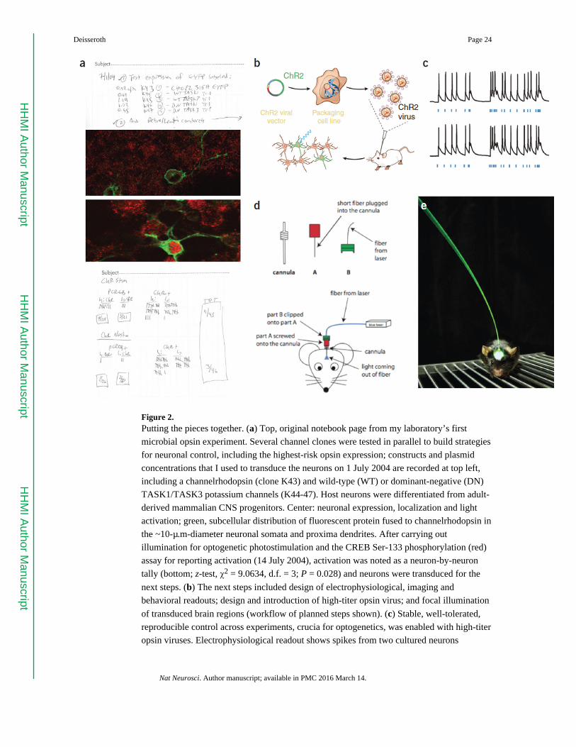
Figure 2. Putting the pieces together. (a) Top, original notebook page from my laboratory’s first
microbial opsin experiment. Several channel clones were tested in parallel to build strategies
for neuronal control, including the highest-risk opsin expression; constructs and plasmid
concentrations that I used to transduce the neurons on 1 July 2004 are recorded at top left,
including a channelrhodopsin (clone K43) and wild-type (WT) or dominant-negative (DN)
TASK1/TASK3 potassium channels (K44-47). Host neurons were differentiated from adult-
derived mammalian CNS progenitors. Center: neuronal expression, localization and light
activation; green, subcellular distribution of fluorescent protein fused to channelrhodopsin in
the ~10-µ.m-diameter neuronal somata and proxima dendrites. After carrying out
illumination for optogenetic photostimulation and the CREB Ser-133 phosphorylation (red)
assay for reporting activation (14 July 2004), activation was noted as a neuron-by-neuron
tally (bottom; z-test, χ2 = 9.0634, d.f. = 3; P = 0.028) and neurons were transduced for the
next steps. (b) The next steps included design of electrophysiological, imaging and
behavioral readouts; design and introduction of high-titer opsin virus; and focal illumination
of transduced brain regions (workflow of planned steps shown). (c) Stable, well-tolerated,
reproducible control across experiments, crucia for optogenetics, was enabled with high-titer
opsin viruses. Electrophysiological readout shows spikes from two cultured neurons
Deisseroth Page 24
Nat Neurosci. Author manuscript; available in PMC 2016 March 14.
HH
MI A
uthor Manuscript
HH
MI A
uthor Manuscript
HH
MI A
uthor Manuscript

receiving the same light pattern (adapted from ref. 2, Springer Nature). (d) Initial
engineering sketch of the fiber-optic neural interface for spatially registering viral
transduction with focal high-intensity illumination (drawings courtesy F. Zhang, Stanford).
(e) Instantiation of the interface that ultimately allowed depth-targeted control26 and
observation4,111 of population-level activity in cells and projections of freely moving
mammals (photo courtesy I. Goshen, Stanford). Supplementary Video 1 shows initial
mammalian behavioral control.
Deisseroth Page 25
Nat Neurosci. Author manuscript; available in PMC 2016 March 14.
HH
MI A
uthor Manuscript
HH
MI A
uthor Manuscript
HH
MI A
uthor Manuscript
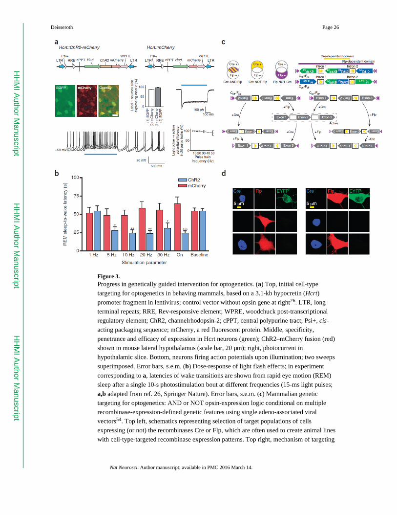
Figure 3. Progress in genetically guided intervention for optogenetics. (a) Top, initial cell-type
targeting for optogenetics in behaving mammals, based on a 3.1-kb hypocretin (Hcrt)
promoter fragment in lentivirus; control vector without opsin gene at right26. LTR, long
terminal repeats; RRE, Rev-responsive element; WPRE, woodchuck post-transcriptional
regulatory element; ChR2, channelrhodopsin-2; cPPT, central polypurine tract; Psi+, cis-
acting packaging sequence; mCherry, a red fluorescent protein. Middle, specificity,
penetrance and efficacy of expression in Hcrt neurons (green); ChR2–mCherry fusion (red)
shown in mouse lateral hypothalamus (scale bar, 20 µm); right, photocurrent in
hypothalamic slice. Bottom, neurons firing action potentials upon illumination; two sweeps
superimposed. Error bars, s.e.m. (b) Dose-response of light flash effects; in experiment
corresponding to a, latencies of wake transitions are shown from rapid eye motion (REM)
sleep after a single 10-s photostimulation bout at different frequencies (15-ms light pulses;
a,b adapted from ref. 26, Springer Nature). Error bars, s.e.m. (c) Mammalian genetic
targeting for optogenetics: AND or NOT opsin-expression logic conditional on multiple
recombinase-expression-defined genetic features using single adeno-associated viral
vectors54. Top left, schematics representing selection of target populations of cells
expressing (or not) the recombinases Cre or Flp, which are often used to create animal lines
with cell-type-targeted recombinase expression patterns. Top right, mechanism of targeting
Deisseroth Page 26
Nat Neurosci. Author manuscript; available in PMC 2016 March 14.
HH
MI A
uthor Manuscript
HH
MI A
uthor Manuscript
HH
MI A
uthor Manuscript

based on recombination followed by removal of recombination sites in introns54. (d)
Neurons transfected with combinations of Cre (blue), Flp (red) and the vectors that
implement Cre AND NOT Flp (Con/Foff) or Flp AND NOT Cre (Coff/Fon). ChR2-YFP is
only expressed in cells marked by one or the other, but not both, recombinases (c,d adapted
from ref. 54, Springer Nature).
Deisseroth Page 27
Nat Neurosci. Author manuscript; available in PMC 2016 March 14.
HH
MI A
uthor Manuscript
HH
MI A
uthor Manuscript
HH
MI A
uthor Manuscript

Figure 4. Progress in activity-guided intervention for optogenetics. (a–d) Closed-loop targeting of
thalamocortical neurons in epileptic cortex (reproduced from ref. 158, Springer Nature).
Yellow light terminates seizures defined by EEG and behavior, detected and interrupted in
real time with closed-loop optogenetic inhibition using eNpHR3.0, an engineered inhibitory
halorhodopsin (a,c). Without yellow light, native epileptic events follow an unmodified time
course (b,d). Thalamocortical activity was thus, surprisingly, identified as necessary for
poststroke epileptic events in this context. (e) Optogenetic closed-loop control in 2009.
Optogenetic stimulation of ChR2-expressing inhibitory (FS) neurons was made conditional
on spike detection in pyramidal (PY) neurons, implementing feed-forward inhibition under
experimenter control (adapted from ref. 41, Springer Nature). Black and red traces show
action potentials in PY cells without or with, respectively, use of closed-loop optogenetic
excitation of FS cells. (f) Closed-loop, all-optical control could be implemented using deep
brain fluorescence (for example, GCaMP) detection of genetically specified activity signals,
via techniques such as fiber photometry (shown here detecting in real time the activity of
ventral tegmental area (VTA) dopamine neuron projections to the nucleus accumbens (NAc)
during appetitive social interaction)111. Red bars indicate interaction episodes (social or
object). (g) With considerable potential for closed-loop control of projection dynamics, the
Deisseroth Page 28
Nat Neurosci. Author manuscript; available in PMC 2016 March 14.
HH
MI A
uthor Manuscript
HH
MI A
uthor Manuscript
HH
MI A
uthor Manuscript

same fiber-optic interface25,26 can be used not only to observe activity in defined deep-brain
projections, but also to control activity in deep-brain projections, as shown here modulating
social behavior (f,g adapted from ref. 111, Elsevier). These specified methods for activity-
guided opsin expression139.
Deisseroth Page 29
Nat Neurosci. Author manuscript; available in PMC 2016 March 14.
HH
MI A
uthor Manuscript
HH
MI A
uthor Manuscript
HH
MI A
uthor Manuscript
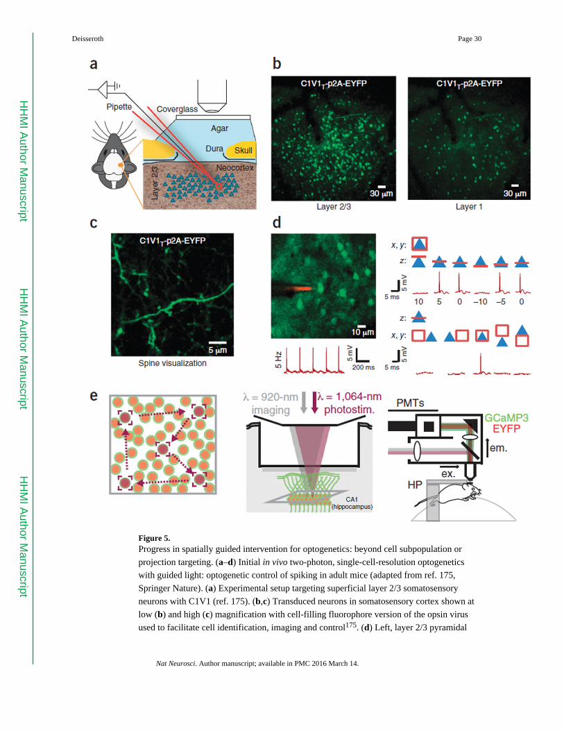
Figure 5. Progress in spatially guided intervention for optogenetics: beyond cell subpopulation or
projection targeting. (a–d) Initial in vivo two-photon, single-cell-resolution optogenetics
with guided light: optogenetic control of spiking in adult mice (adapted from ref. 175,
Springer Nature). (a) Experimental setup targeting superficial layer 2/3 somatosensory
neurons with C1V1 (ref. 175). (b,c) Transduced neurons in somatosensory cortex shown at
low (b) and high (c) magnification with cell-filling fluorophore version of the opsin virus
used to facilitate cell identification, imaging and control175. (d) Left, layer 2/3 pyramidal
Deisseroth Page 30
Nat Neurosci. Author manuscript; available in PMC 2016 March 14.
HH
MI A
uthor Manuscript
HH
MI A
uthor Manuscript
HH
MI A
uthor Manuscript
cells transduced with C1V1 under loose patch conditions (note red dye-filled patch
electrode). Lower left, trace showing 5-Hz control of spiking with 1,040-nm raster-scanning
illumination. Right, axial (upper) and lateral (lower) single-cell resolution of two-photon
optogenetic spiking control in vivo. Blue triangles indicate pyramidal neurons and red boxes
illustrate region-of-interest raster-scan positioning; traces show spiking occurring only while
scanning within the cell. (e) An example of single-cell targeting with optogenetics using
temporally focused two-photon control of C1V1, with single-cell resolution optical
feedback, in a virtual reality environment in behaving mice (adapted from ref. 177, Springer
Nature); for spatial light modulator use together with C1V1, see refs. 175,176,178. Target
neurons are illuminated in turn via temporally focused soma-sized spots (<15 µm). Left,
light path; center, lasers used; right, behavioral preparation177. PMTs, photomultiplier tubes;
ex., excitation; em., emission; H P, headplate.
Deisseroth Page 31
Nat Neurosci. Author manuscript; available in PMC 2016 March 14.
HH
MI A
uthor Manuscript
HH
MI A
uthor Manuscript
HH
MI A
uthor Manuscript
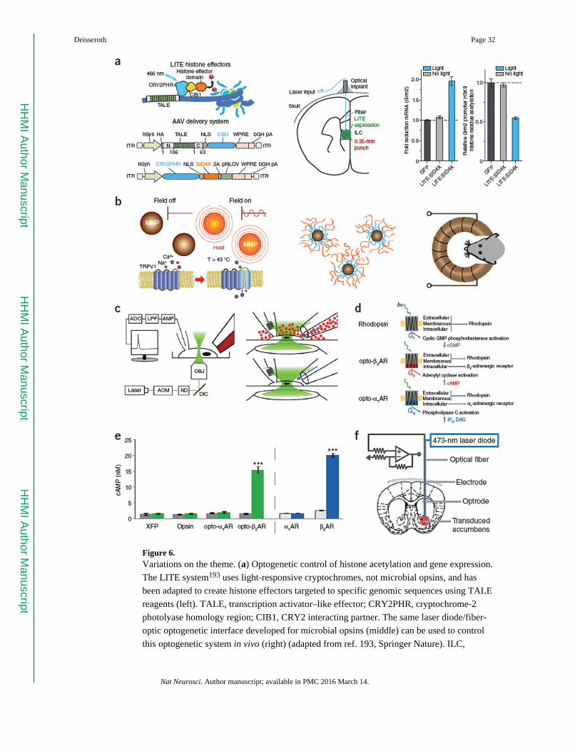
Figure 6. Variations on the theme. (a) Optogenetic control of histone acetylation and gene expression.
The LITE system193 uses light-responsive cryptochromes, not microbial opsins, and has
been adapted to create histone effectors targeted to specific genomic sequences using TALE
reagents (left). TALE, transcription activator–like effector; CRY2PHR, cryptochrome-2
photolyase homology region; CIB1, CRY2 interacting partner. The same laser diode/fiber-
optic optogenetic interface developed for microbial opsins (middle) can be used to control
this optogenetic system in vivo (right) (adapted from ref. 193, Springer Nature). ILC,
Deisseroth Page 32
Nat Neurosci. Author manuscript; available in PMC 2016 March 14.
HH
MI A
uthor Manuscript
HH
MI A
uthor Manuscript
HH
MI A
uthor Manuscript
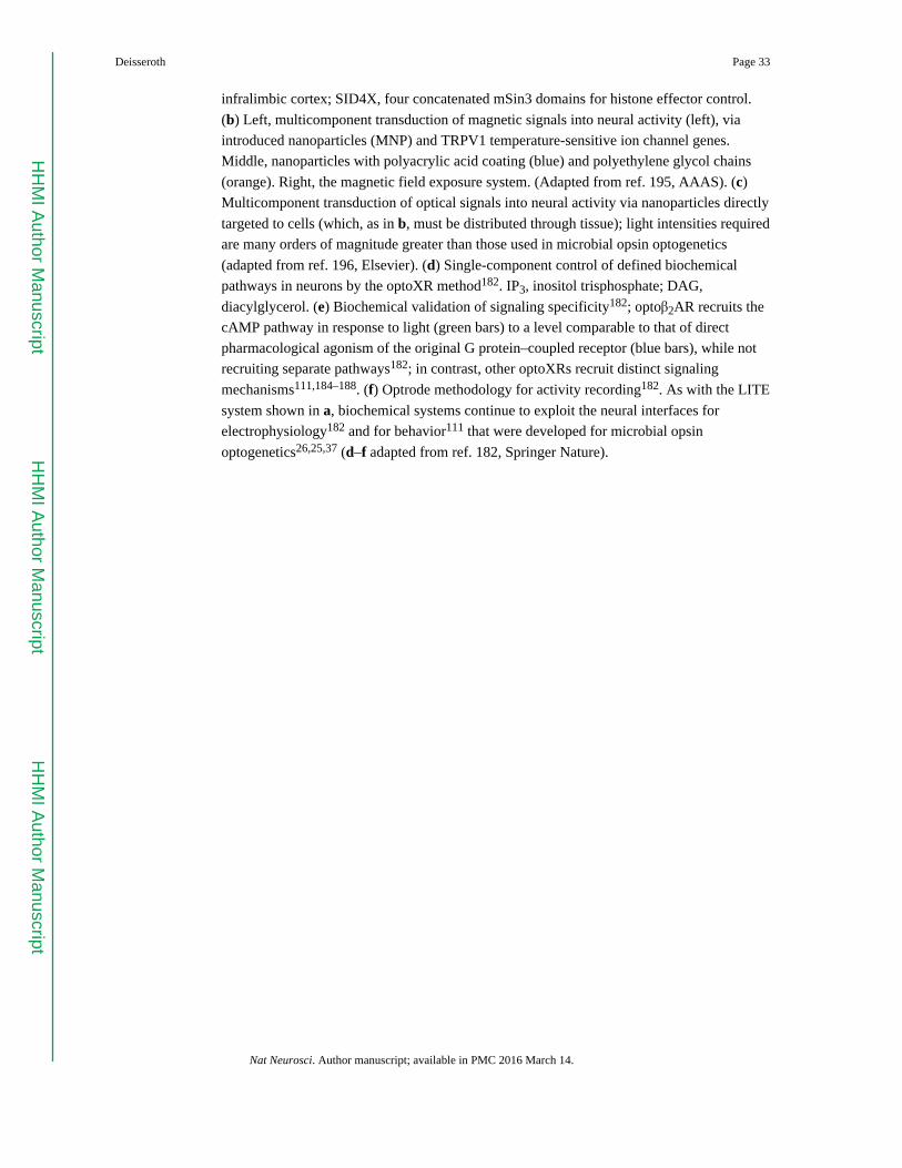
infralimbic cortex; SID4X, four concatenated mSin3 domains for histone effector control.
(b) Left, multicomponent transduction of magnetic signals into neural activity (left), via
introduced nanoparticles (MNP) and TRPV1 temperature-sensitive ion channel genes.
Middle, nanoparticles with polyacrylic acid coating (blue) and polyethylene glycol chains
(orange). Right, the magnetic field exposure system. (Adapted from ref. 195, AAAS). (c)
Multicomponent transduction of optical signals into neural activity via nanoparticles directly
targeted to cells (which, as in b, must be distributed through tissue); light intensities required
are many orders of magnitude greater than those used in microbial opsin optogenetics
(adapted from ref. 196, Elsevier). (d) Single-component control of defined biochemical
pathways in neurons by the optoXR method182. IP3, inositol trisphosphate; DAG,
diacylglycerol. (e) Biochemical validation of signaling specificity182; optoβ2AR recruits the
cAMP pathway in response to light (green bars) to a level comparable to that of direct
pharmacological agonism of the original G protein–coupled receptor (blue bars), while not
recruiting separate pathways182; in contrast, other optoXRs recruit distinct signaling
mechanisms111,184–188. (f) Optrode methodology for activity recording182. As with the LITE
system shown in a, biochemical systems continue to exploit the neural interfaces for
electrophysiology182 and for behavior111 that were developed for microbial opsin
optogenetics26,25,37 (d–f adapted from ref. 182, Springer Nature).
Deisseroth Page 33
Nat Neurosci. Author manuscript; available in PMC 2016 March 14.
HH
MI A
uthor Manuscript
HH
MI A
uthor Manuscript
HH
MI A
uthor Manuscript
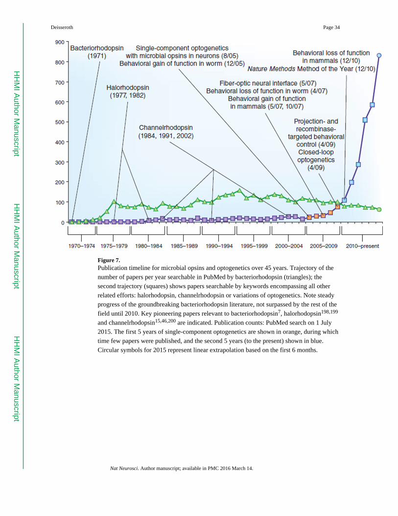
Figure 7. Publication timeline for microbial opsins and optogenetics over 45 years. Trajectory of the
number of papers per year searchable in PubMed by bacteriorhodopsin (triangles); the
second trajectory (squares) shows papers searchable by keywords encompassing all other
related efforts: halorhodopsin, channelrhodopsin or variations of optogenetics. Note steady
progress of the groundbreaking bacteriorhodopsin literature, not surpassed by the rest of the
field until 2010. Key pioneering papers relevant to bacteriorhodopsin7, halorhodopsin198,199
and channelrhodopsin15,46,200 are indicated. Publication counts: PubMed search on 1 July
2015. The first 5 years of single-component optogenetics are shown in orange, during which
time few papers were published, and the second 5 years (to the present) shown in blue.
Circular symbols for 2015 represent linear extrapolation based on the first 6 months.
Deisseroth Page 34
Nat Neurosci. Author manuscript; available in PMC 2016 March 14.
HH
MI A
uthor Manuscript
HH
MI A
uthor Manuscript
HH
MI A
uthor Manuscript