Embed Size (px)
Citation preview
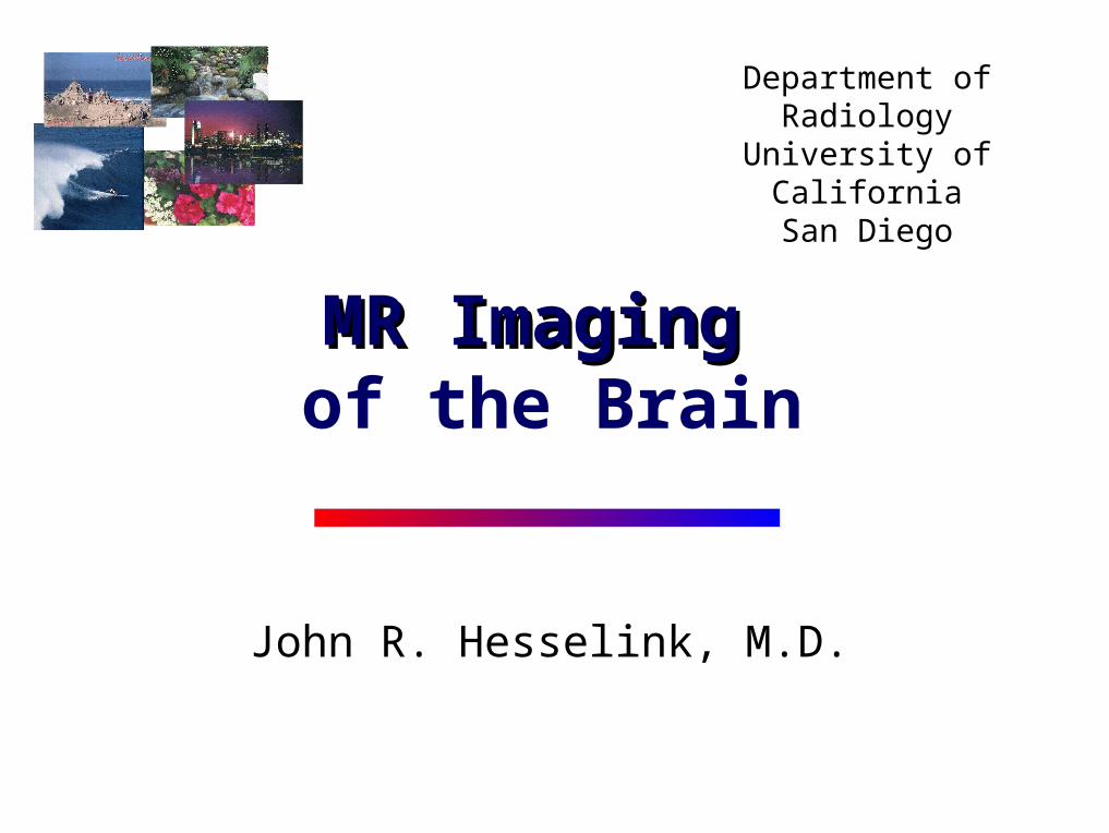
Department of RadiologyUniversity of California
San Diego
John R. Hesselink, M.D.
MR ImagingMR Imaging of the Brain
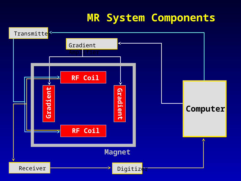
MR System Components
Computer
Gradient Amplifier
Receiver
Transmitter
Digitizer
RF Coil
Gra
die
nt
RF Coil
Grad
ient
Magnet

MR Imaging
Proton density
T1 relaxation time
T2 relaxation time
Flow effects
Source of Signal and Contrast

Spin-echo Pulse SequenceSingle Echo T1-weighted
RF
Signal
TR
TE
1st
echo2nd
echo
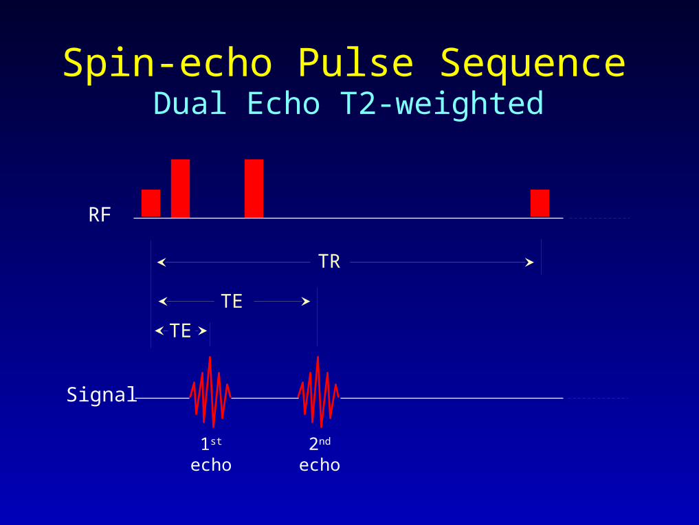
Spin-echo Pulse SequenceDual Echo T2-weighted
1st
echo2nd
echo
RF
Signal
TR
TE
TE
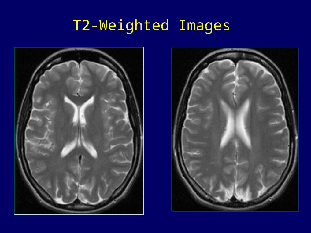
T2-Weighted Images

Proton Density Weighted Images
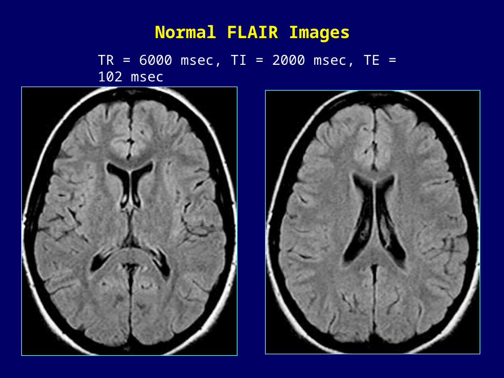
Normal FLAIR Images
TR = 6000 msec, TI = 2000 msec, TE = 102 msec
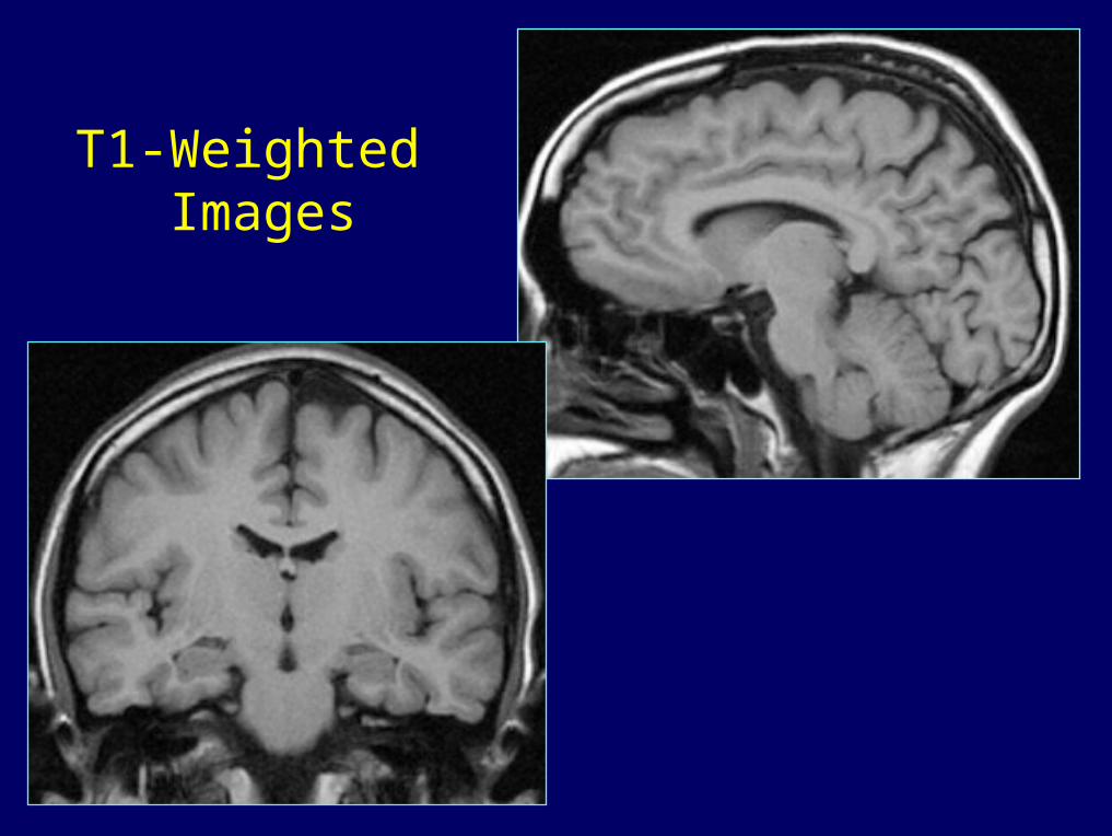
T1-Weighted Images

MR Signal ofMR Signal ofBrain LesionsBrain Lesions

SolidBrainLesion PDW
T2W T1W

CysticLesion
T2W
T1W
FLAIR
236

SubacuteHemorrhage
PDW
T1W
T2W

AcuteHemorrhage
T2W
FLAIR
T1WGRE

History: 9 y/o boy with prior head trauma
466

Hemorrhage SequenceGradient-echo

PDW
Fator
Lipoma
T2W
T1W
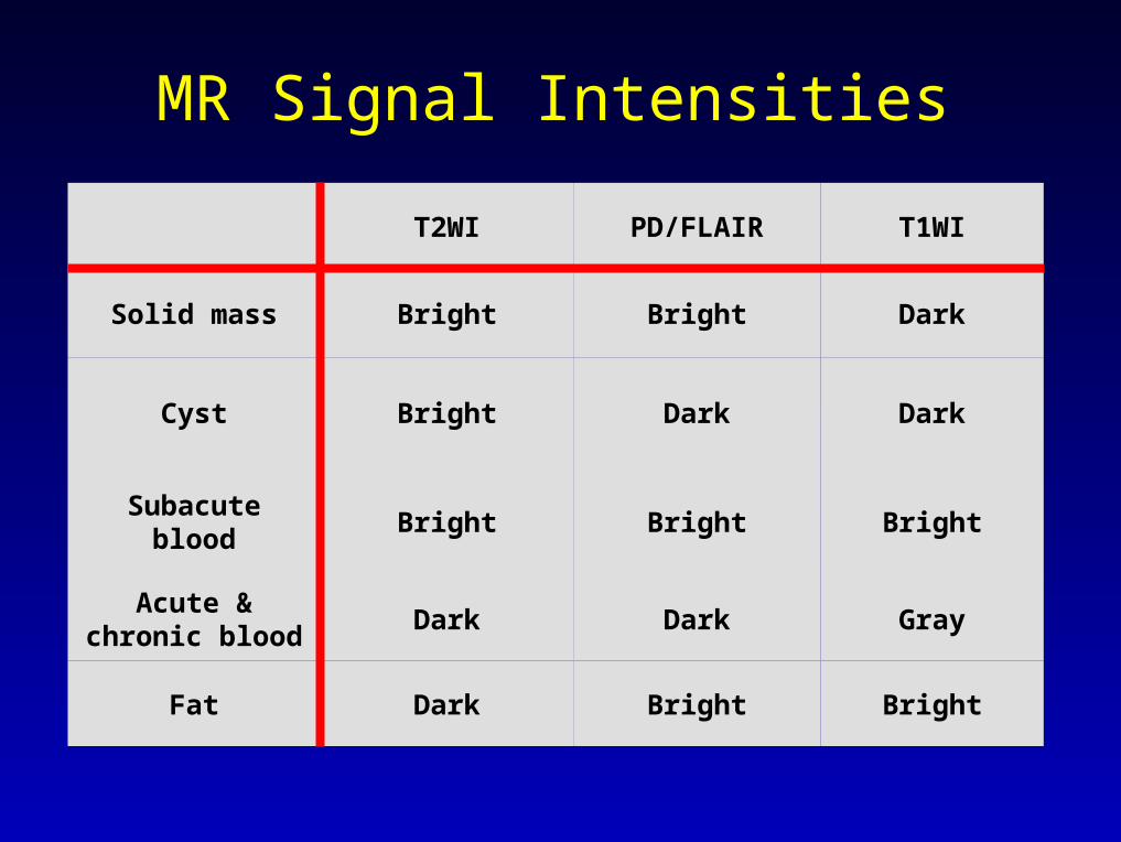
MR Signal Intensities
T2WI PD/FLAIR T1WI
Solid mass Bright Bright Dark
Cyst Bright Dark Dark
Subacute blood Bright Bright Bright
Acute & chronic blood
Dark Dark Gray
Fat Dark Bright Bright
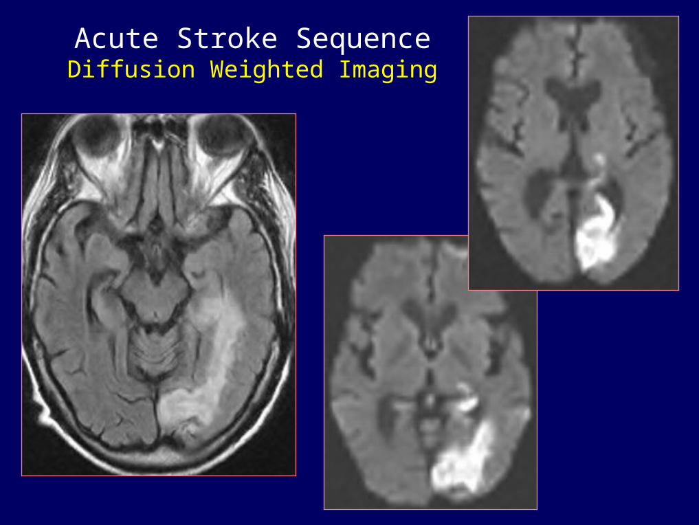
Acute Stroke SequenceDiffusion Weighted Imaging

MR Angiography

Dx: Anaplastic astrocytoma > GBM
MR Spectroscopy

Brain Screening Protocol
Sagittal T1-weighted images Axial T2-weighted images Axial FLAIR images Diffusion-weighted images Gradient-echo images Axial T1-weighted images Gadolinium: Axial & Coronal T1

History: 46 y.o. manwith headaches & increasing confusion
21
T2W PDW
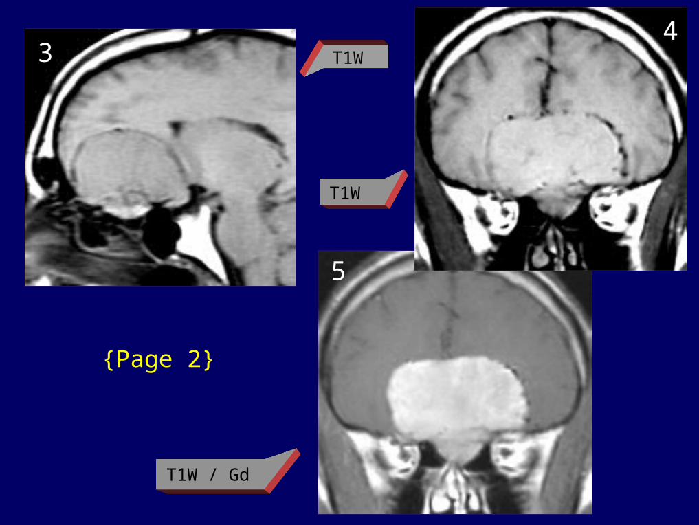
{Page 2}
5
34
T1W
T1W / Gd
T1W

Dx: Meningioma & right MCA infarct
Increasing confusion {Page 3}
76

Student Cases
"May the FORCE be with you!"

History: 69 y/o man with nausea& memory deficits for several weeks
413
Case 1

Dx: Glioblastoma
{Page 2}
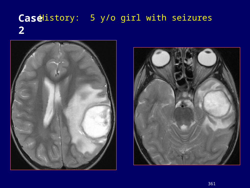
History: 5 y/o girl with seizures
361
Case 2

{Page 2}

Dx: Amoebic abscesses
{Page 3}

History: 62 y.o. man with mumbling speech
26
21
Case 3

{Page 2}
3 4

Mumbling speech {Page 3}
5 6

Dx: Enhancing subacute infarct
{Page 4}
87

21
History: 48 y.o. male with a sensorineural hearing loss
Case 4

Dx: Acoustic neuroma
{Page 2}
5
4
3

History: 48 y.o. man with headaches & confusion
32
12
Case 5
{Page 2}
43

{Page 3}
7
6
5

Dx: Brain metastases - Lung carcinoma
{Page 4}
10
98

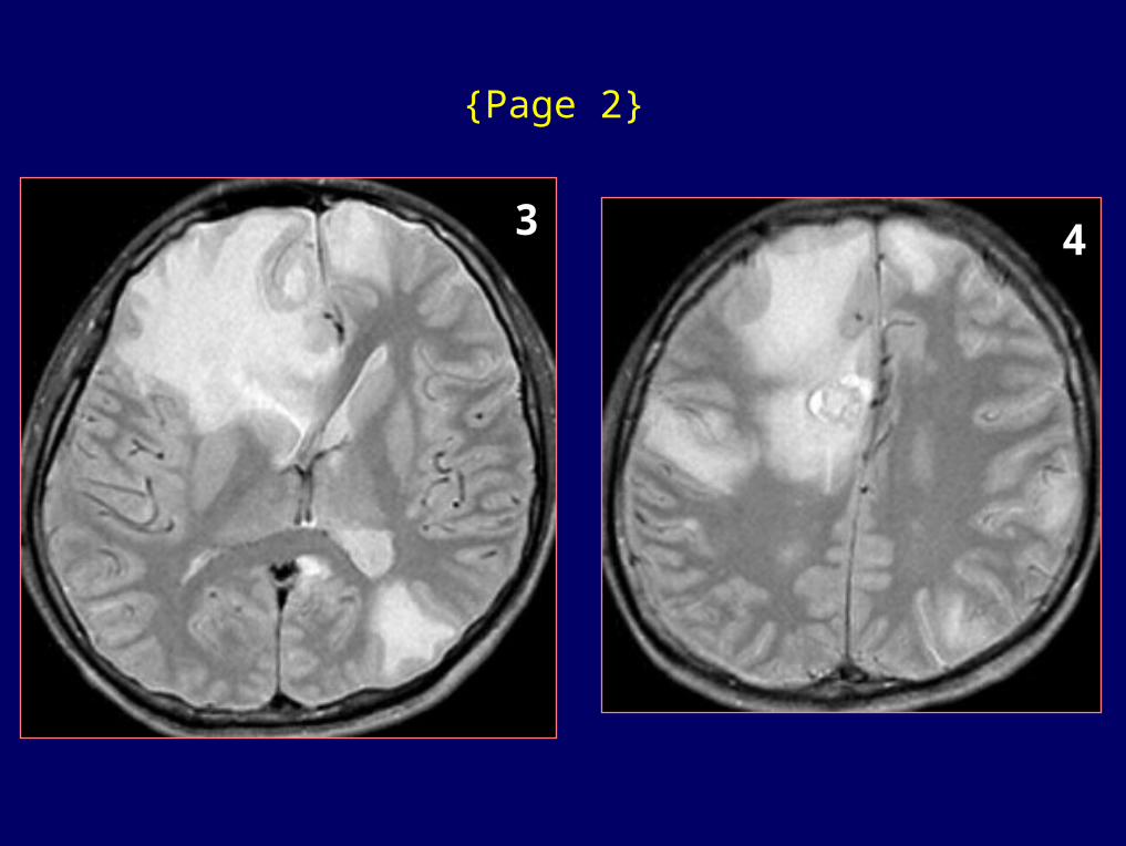
History: 36 y.o. man with seizures
21
Case 6
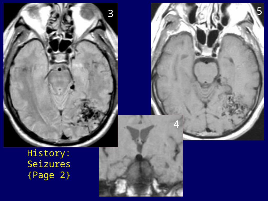
History: Seizures{Page 2}
5
4
3

Dx: AVM & basilar tip aneurysm
{Page 3}
8
7
6

History: 62 y/o woman with headachesCase 7

Dx: Arachnoid cyst
{Page 2}

History: 27 y.o. man with increasing confusion & lethargy
5
21
Case 8
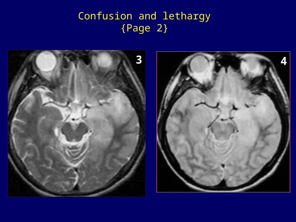
Confusion and lethargy {Page 2}
3 4

Dx: Herpes simplex encephalitis
{Page 3}
6
5

History: 72 y.o. woman with headaches
49
3
21
Case 9
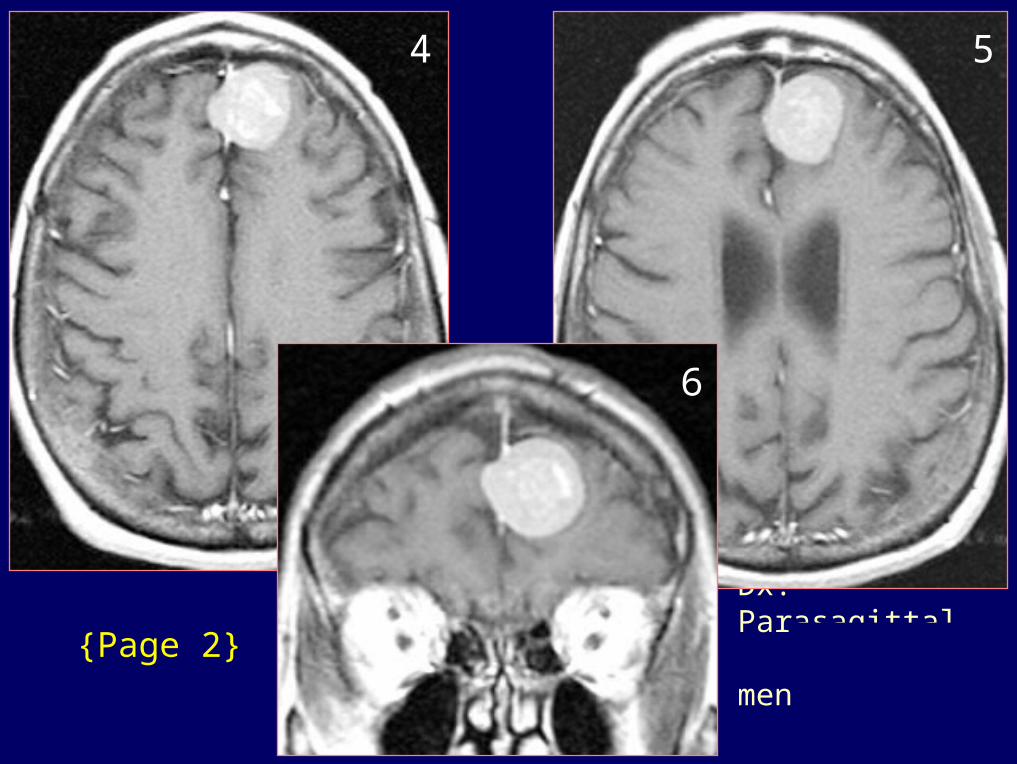
Dx: Parasagittal meningioma
{Page 2}
54
6

History: 55 y/o female with motor weakness, double vision, & emotional lability
272
Case 10

{Page 2}

Dx: Chronic multiple sclerosis; Incidental sebaceous cyst left posterior scalp
{Page 3}
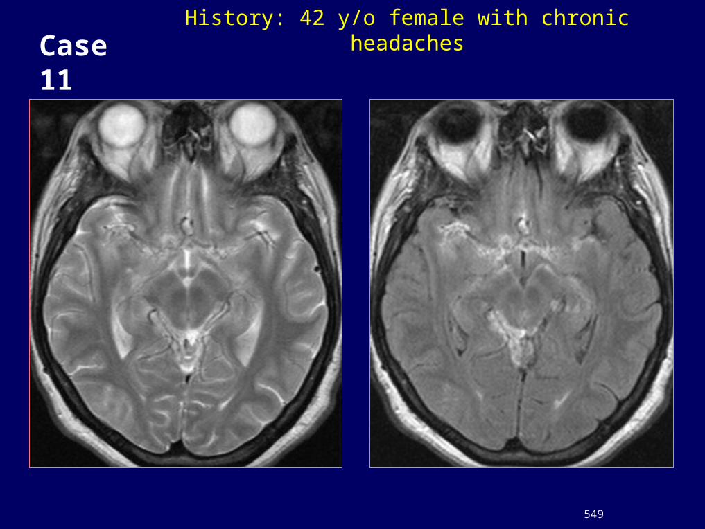
History: 42 y/o female with chronic headaches
549
Case 11
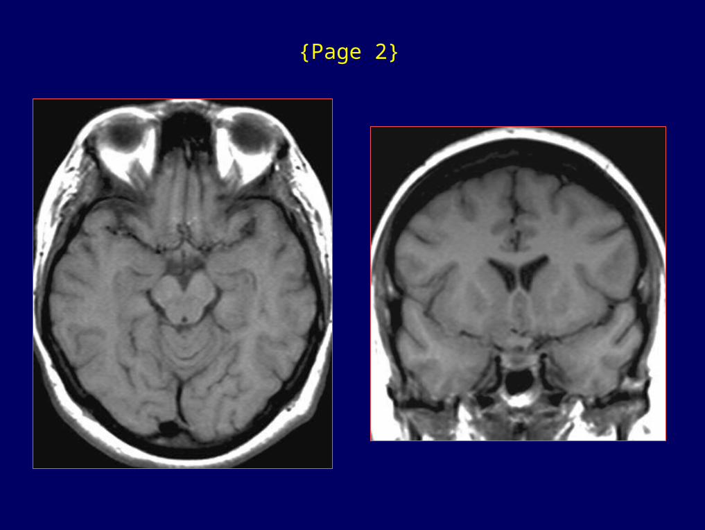
{Page 2}
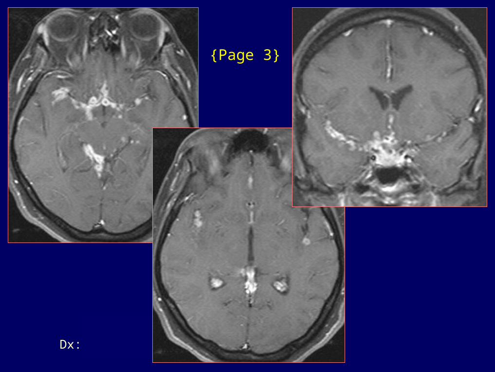
Dx: TB meningitis - HIV+
{Page 3}

History: 75 y.o. malewith dysmetria in upper extremities
1 2
Case 12

{Page 2}
3
5
4

Dx: Lipoma
{Page 3}
8
6
7

History: Young male with increasing somnolence and irritability
11/20 -
2
1
3

Dx: Planum Meningioma in a dog
4
{Page 2}
6
5