Embed Size (px)
Citation preview

on June 30, 2018http://rsnr.royalsocietypublishing.org/Downloaded from
Notes Rec. R. Soc.
doi:10.1098/rsnr.2011.0023
*a
Published online
AMBIGUOUS CELLS: THE EMERGENCE OF THE STEM CELL CONCEPT IN
THE NINETEENTH AND TWENTIETH CENTURIES
by
ANDREAS-HOLGER MAEHLE*
Department of Philosophy, Durham University, 50 Old Elvet, Durham DH1 3HN, UK
This paper elucidates the origins of scientific work on stem cells. From the late nineteenth
century onwards, the notion of stem cells became customary in scientific communities of
Imperial Germany. Adopting the term Stammzelle from Ernst Haeckel, Theodor Boveri
was influential in introducing the concept in embryological studies and early genetics
around 1900, describing a capacity of stem cells for self-renewal as well as
differentiation. At the same time, blood stem cells were conceptualized by histologists
such as Ernst Neumann and Artur Pappenheim in studies of physiological haematopoiesis
and various forms of leukaemia. Furthermore, building on Julius Cohnheim’s theory that
tumours arise from ‘embryonic remnants’ in the adult body, pathologists aimed at
identifying the cells of origin, particularly in the embryo-like teratomas. Embryonic stem
cells thus assumed an ambiguous status, partly representing common heritage and normal
development, and partly being seen as potential causes of cancer if they had been left
behind or displaced during ontogeny. In the 1950s and 1960s experimental research on
teratocarcinomas by Leroy Stevens and Barry Pierce in the USA brought together the
strands of embryological and pathological work. Alongside the work of Ernest McCulloch
and James Till at the Ontario Cancer Institute from the early 1960s on stem cells in
haematopoiesis, this led into the beginnings of modern stem cell research.
.h.m
Keywords: stem cells; embryology; haematopoiesis; tumours; teratoma
INTRODUCTION
In an article in 2009, Canadian historian and philosopher of medicine Lawrence Burns has
argued that much of the current public discourse on embryonic stem cells follows the
metaphor of the ‘superhero’.1 Because of their pluripotency, stem cells are expected to be
capable of virtually anything in terms of providing future regenerative therapies. For
sufferers of conditions such as spinal cord injuries or severe Parkinson’s disease they may be
the last source of hope. However, summoning the ‘superhero’ also involves a difficult moral
choice: the destruction of early human embryos (in their blastocyst stage) in harvesting
embryonic stem cells from the inner cell mass. In view of the pervasiveness of metaphors in
1 This journal is q 2011 The Royal Society

A.-H. Maehle2
on June 30, 2018http://rsnr.royalsocietypublishing.org/Downloaded from
the present debates on stem cells, a phenomenon that has similarly been emphasized by the
linguist Andreas Musolff,2 it seems worthwhile to enquire into the meanings and
connotations of those cells when they were first conceptualized—more than a century ago.
This paper discusses the specific scientific contexts in which the notion of ‘stem cells’
was established in the late nineteenth century. Moreover, it shows how, at the start of the
twentieth century, embryonic cells became a concern for pathologists who attempted to
explain the genesis of tumours. It concludes by indicating how this early work on
embryonic cells and tumours led to modern stem cell research after 1950. In this way it
complements recent scholarship on this topic that has focused on developments since
World War II, especially the study of blood-forming (haematopoietic) stem cells.3 I argue
that in the early twentieth century embryonic stem cells assumed an ambiguous status by
carrying positive connotations of common heritage and normal development as well as
negative meanings as potential sources of cancer.
ERNST HAECKEL AND THE STAMMZELLE
The origins of the term ‘stem cell’ can be traced back to the late nineteenth century.4 Ernst
Haeckel (1834–1919), the controversial Darwinist and professor of zoology at Jena,5
referred in his published lectures on Naturliche Schopfungsgeschichte (1868) to
unicellular organisms or protozoa, which he believed to be the phylogenetic ancestors of
multicellular organisms, as Stammzellen (stem cells). The genealogical and evolutionary
concept of the Stammbaum (family tree or phylogenetic tree) and of the biological Stamm
(phylum) formed the linguistic context of his coinage of this new term. According to
Haeckel, the stem cells themselves had originated from the most primitive forms of life,
the so-called Moneren, which he thought of as tiny lumps of mucus or protein. The ‘fact’
that the stem cells formed the evolutionary basis of all plants and animals was in his view
evident from the analogy of individual embryological development from a single egg
cell.6 Obviously, this assertion derived from Haeckel’s famous ‘biogenetic law’ that
ontogeny is a rapid and shortened recapitulation of phylogeny.7 In 1877 he applied the
notion of stem cells to ontogeny against this background and used the name Stammzelle
or Cytula to describe the fertilized egg cell as the cell of origin of all other cells of an
animal or human organism. Addressing a general educated audience in another series of
lectures, on Anthropogenie, he explained:
The name ‘stem cell’ seems to me the most simple and appropriate one, because all other
cells stem from it and because it is in its most literal sense the stem father as well as the
stem mother of all the countless generations of cells of which the multicellular organism
is later composed.8
The new term was necessary, according to Haeckel, to make clear that the fertilized egg cell
was quite different—morphologically, chemically and physiologically—from the original
egg cell. In fact, in 1875 his student Oscar Hertwig (1849–1922) had demonstrated in sea
urchins that fertilization could be understood as the fusion of the nucleus of an egg cell
with that of a spermatozoon.9 The stem cell, Haeckel stressed, ‘is partly of fatherly and
partly of motherly origin; and we will now no longer find it astonishing if the child who
develops from this stem cell inherits individual characteristics from both parents’.10 Thus,
for Haeckel, the stem cell represented the whole future child.

Emergence of the stem cell concept 3
on June 30, 2018http://rsnr.royalsocietypublishing.org/Downloaded from
Haeckel’s neologism was rooted in the metaphorical language that was then common in
talking about cells and the body. Particularly through the influence of Rudolf Virchow
(1821–1902), who since the 1850s had compared the body’s cells with citizens cooperatively
forming a state (Zellenstaat, ‘cell state’), it had become customary to speak metaphorically of
cells as human individuals.11 As Virchow had argued in his influential Cellularpathologie:
The character and the unity of life cannot be found in one particular single point of higher
organization, such as the human brain, but only in particular, constantly recurring
arrangements, which every single element owns. From this it follows that the
composition of a larger body, the so-called individual, always results in a kind of
social arrangement, [and] represents an organism of a social kind, in which a mass of
single existences is dependent on each other, but in such a manner that each element
(cell or, as Brucke says very well, elementary organism) has a particular activity for
itself, and that each, although it may receive the stimulus for its activity from other
parts, still is itself the origin of its actual work.12
Haeckel, who had studied medicine with Virchow, similarly described the human body as a
social arrangement of cells, but turned Virchow’s liberal, relatively egalitarian conception of
a cell state into a more hierarchical and centralized version.13 Significantly, not only were
such metaphors employed when addressing lay audiences, but they were also used in
scientific texts. Metaphors had a heuristic value for scientists in shaping new research
questions.14 Haeckel’s notion of stem cells soon found its way into research papers of
other zoologists and anatomists.
STEM CELLS IN EMBRYOLOGY AROUND 1900
In the early 1890s the term ‘stem cell’ was adopted in the context of embryological studies in the
wake of August Weismann’s (1834–1914) theory of the continuity of the ‘germ plasm’.15
Segregated into primordial germ cells during the earliest phases of embryonic development,
this ‘germ plasm’ (Keimplasma), which was identified with the organized substance of the
cell’s nucleus, was thought to carry hereditary characteristics through the egg and sperm cells
from one generation to the next.16 Valentin Haecker (1864–1927), then an assistant to
Weismann at the Zoological Institute of the University of Freiburg (Breisgau), published a
paper in 1892 on the embryonic development of the crustacean Cyclops, in which he referred
to the ‘stem cell’ (Stammzelle) as the common precursor cell of the primordial germ cells
and of the primordial somatic (mesoderm) cells.17 In a similar sense ‘stem cells’ were
introduced later in the same year by Theodor Boveri (1862–1915). At this time Boveri
worked at the Zoological Institute of the University of Munich under Richard Hertwig
(1850–1937), who like his brother Oscar had been a student of Haeckel’s. In a lecture to the
Munich Society for Morphology and Physiology on the embryo of the roundworm of the
horse (Ascaris megalocephala), then a common model organism for cytological research,
Boveri described as Stammzellen those cells that derived from the fertilized egg cell and led
to the primordial germ cell (Urgeschlechtszelle), and from which the various primordial
somatic cells (Ursomazellen) branched off. In other words, in each of the earliest cell
generations, from the two-cell stage of the embryo onwards, one ‘stem cell’ divided into two
daughter cells, of which only one maintained the character of a stem cell whereas the other
divided into the precursors of somatic cells. The various primordial somatic cells

A.-H. Maehle4
on June 30, 2018http://rsnr.royalsocietypublishing.org/Downloaded from
subsequently formed the basis of the different layers—ectoderm, endoderm and mesoderm—of
the embryo. After five divisions, in the 32-cell stage of the Ascaris embryo, the stem cell (now
called the primordial germ cell) began to differentiate into germ cells, leading ultimately to the
formation of eggs or of spermatozoa. Boveri explicitly mentioned that he had adopted the term
‘stem cell’ from Ernst Haeckel.18
However, neither Theodor Boveri nor Valentin Haecker turned stem cells as such into
central objects of investigation. Of interest to them was, rather, the distribution of
‘chromatin’, namely the stainable nuclear substance suspected to carry hereditary
characteristics, to the germ cells on the one hand and to somatic cells on the other. In line
with Weismann’s theory of a continuity of the ‘germ plasm’, the stem cells were thought to
maintain and pass on the full chromatin of the fertilized egg cell, whereas it was believed
to be only partly distributed to the somatic cells (‘chromatin diminution’), thus leading to
cell differentiation. In this early work Haecker and Boveri were already describing the
doubling and distribution of ‘chromatin loops’ or ‘chromosomes’ during cell divisions.
Boveri, who was appointed to the chair of zoology and comparative anatomy at Wurzburg
University in 1893, became a founder of the chromosome theory of heredity in the early
1900s.19 Haecker, who was made director of the Zoological Institute at the Technical
University of Stuttgart in 1900, and subsequently at the University of Halle from 1909,
likewise developed his main research interests in the field of genetics. He established the
approach of ‘phenogenetics’, which worked ‘backwards’ from the outer traits of an
organism to their suspected causes in the germ cells.20 In 1914 he propagated the notion of
‘pluripotency’ as the potential for several different developmental options, which he
ascribed generally to the ‘germ plasm’ of an organism (not specifically to its stem cells).
The germ plasm was now thought to be a complex ‘biomolecule’, in which even small
changes in a few ‘atom groups’ might produce new qualities.21
Boveri’s description of the cell lineage in the Ascaris embryo, which he had illustrated
with drawings and diagrams (figure 1) that reappeared with small modifications in several
of his publications,22 found its way into textbook and handbook accounts of early
embryonic development. For example, the American biologist Edmund Beecher Wilson
(1856–1939), a lifelong friend of Boveri’s since their first collaboration at Richard
Hertwig’s Zoological Institute in Munich in the early 1890s, included versions of
Boveri’s cell lineage diagrams and drawings in his influential handbook The cell in
development and heredity.23 Robert William Hegner (1880–1942), then assistant professor
of zoology at the University of Michigan and later an international research leader in
protozoology and parasitology at Johns Hopkins University, did the same in his textbook
The germ-cell cycle in animals.24 Similarly, the early development of Ascaris
megalocephala as well as that of stem cells featured in a biological contribution by the
Berlin anatomist Richard Weissenberg (1882–1974) to the Handbuch der
Sexualwissenschaften of the sexologist Albert Moll (1862–1939).25 Weissenberg was a
former assistant to Oscar Hertwig, who had been appointed director of the Anatomical–
Biological Institute of Berlin University in 1888.26 In Weissenberg’s account, the term
‘stem cells’ (Stammzellen) referred specifically to early precursor cells of egg cells and
sperm cells, the Oogonien and Spermiogonien.27 In this way the concept of stem cells
gradually became established in the fields of embryology and cytology, although with
some variations in its precise meaning. However, the essential characteristic of a stem
cell, as described by Boveri, was clear: a capacity for self-renewal as well as for
differentiation into specific types of somatic cells or germ cells.

Figure 1. Theodor Boveri’s representations of the early embryonic development of the intestinal worm Ascarismegalocephala, showing self-renewal as well as differentiation of stem cells.96 The blackened circles along thehorizontal line in Fig. 1 represent the ‘stem cells’ (Stammzellen) with full chromatin content, starting with thefertilized egg cell and leading after five divisions to the ‘primordial germ cell’ (Urgeschlechtszelle). The emptycircles with surrounding black dots symbolize the ‘primordial somatic cells’ (Ursomazellen), in which ‘chromatindiminution’ occurs as the initial step towards cell differentiation. The empty circles represent early somatic cellswith reduced chromatin. Figs 2 and 3 illustrate, respectively, a ventral view and an optical section of the Ascarisembryo at a stage of about 120 cells; two primordial germ cells that have just divided are shown in the centre ofFig. 2. (Bayerische Staatsbibliothek Munchen, Signatur: Bavar. 2469 dz-7/8.)
Emergence of the stem cell concept 5
on June 30, 2018http://rsnr.royalsocietypublishing.org/Downloaded from
STEM CELLS IN HAEMATOLOGICAL RESEARCH BEFORE WORLD WAR I
The notion of stem cells also resonated with researchers outside the specific field of
embryology. For example, in 1896 Artur Pappenheim (1870–1916), working at Virchow’s
Pathological Institute in Berlin on the formation of red blood cells, called the common
precursor cell of the red and white blood cell lineages the ‘stem cell’ (Stammzelle).28
Assuming that he was justified in approaching a matter of clinical relevance (such as for
the diagnosis and prognosis of anaemia) through comparative and embryological studies
on phylogenetically lower animals, he examined the blood of amphibians (for example
frogs and salamanders) at different developmental stages and ages. Amphibians were at
that time commonly used for haematological studies because of the relatively large size
of their blood cells. Moreover, Pappenheim believed that his findings on blood formation
during the ontogeny of different species of amphibians illustrated Haeckel’s biogenetic
law.29 Although he cited neither Valentin Haecker nor Theodor Boveri, Pappenheim was
aware that ‘stem cells’, or ‘mother cells’ (Mutterzellen) as he also called them, with
different developmental options had been described more widely in embryology. He
mentioned the differentiation of stem cells into egg cells and follicle cells; into
spermatoblasts and spermatogonia; into the sensory cells and supporting cells of sense

A.-H. Maehle6
on June 30, 2018http://rsnr.royalsocietypublishing.org/Downloaded from
organs; into ganglion cells and neuroglia cells; and into the different cell types of connective
tissues.30 Thus, his conception of the stem cell was that of an embryonic cell that had the
potential to differentiate into diverse cell lines and in this way to form the basis of
different types of blood cells, body tissues and germ cells.
By the early twentieth century the stem cell concept seems to have been fairly well
established in haematological research.31 Pappenheim continued to apply the notion of
stem cells in his later clinical–pathological work on different forms of leukaemia, arguing
that myelocytes and lymphocytes originated from the same ‘lymphomyeloblast
multipotent stem cell’.32 To illustrate his hypotheses about the genealogical relationships
of different types of blood cells he drew increasingly complex ‘stem trees’ (Stammbaume)
(figure 2). Wera Dantschakoff (born in 1879), who then worked at the Institute for
Histology and Embryology of the University of Moscow on blood formation in the
chicken embryo, concluded that the ‘lymphocyte’ was the ‘common, indifferent stem cell’
for erythrocytes as well as granulated leucocytes.33 And the St Petersburg histologist
Alexander Maximow (1874–1928), speaking in 1909 to the Berlin Haematological
Society, suggested that the ‘lymphocyte’ was ‘the common stem cell’ of all types of
blood cells, both during embryonic development and in the adult life of mammals.
Significantly, the term ‘stem cell’ (Stammzelle) featured in the title of Maximow’s
paper.34 All three authors were committed to the ‘monophyletic’ or ‘unitarian’ view that
the various types of blood cells ultimately derived from a common, haematopoietic stem cell.
A powerful supporter of this view was the Konigsberg professor of pathology, Ernst
Neumann (1834–1918), who had demonstrated in 1868 that bone marrow was a site of
blood formation in humans and other mammals.35 On the basis of his later studies in frogs,
he suggested that ‘lymphocytes’ in the bone marrow were the earliest, common precursor
cells of the erythrocytes, of the polymorphonuclear leucocytes (granulocytes) and of the
lymphocytes of the circulating blood. This ‘unitarian’ view contrasted with the ‘dualist
doctrine’ of Paul Ehrlich (1854–1915), who assumed that the lymphocytes and leucocytes
(granulocytes) originated from morphologically different precursor cells in different organs:
the lymphocytes developed in lymph nodes and the spleen, and the leucocytes in the bone
marrow.36 In 1912 Neumann speculated that the haematological controversy between
‘unitarians’ and ‘dualists’ might one day be decided if it became possible to grow isolated
blood cells in pure cultures, as Robert Koch (1843–1910) had done with bacteria.37
BEGINNINGS OF TISSUE CULTURE
By this time, embryonic cells that gave rise to nervous tissue, the so-called neuroblasts, had
for the first time been studied in isolation, outside the embryo.38 During the years 1907–09
the American anatomist and biologist Ross Granville Harrison (1870–1959) investigated the
growth of nerve fibres from these cells. The context for his investigation was contemporary
controversy about the processes involved in the formation of nervous connections during
embryonic development. In experiments on frog embryos, started at Johns Hopkins
University and continued after his move to Yale in 1907, Harrison explanted small pieces
of prospective nervous tissue and placed them in drops of frog lymph on coverslips. After
the lymph had clotted, the slips were inverted over hollow slides (thus encasing the drops
and tissues), and the preparations were sealed with paraffin (the so-called ‘hanging-drop
technique’, which was then common in bacteriology for studying the growth of

Figure 2. A haematopoietic stem tree by Artur Pappenheim.97 The multipotent stem cell (no. 1) in the middle of thescheme is emphasized by the bold circle. It is the origin of the ‘myeloleukoplastic branch’ (to the left), the‘erythroplastic branch’ (to the right), ‘the lymphoplastic branch’ (in the middle, upwards), and the ‘splenoplasticbranch’ (in the middle, downwards). (Staatsbibliothek zu Berlin, Signatur: 400 Kv 1955.)
Emergence of the stem cell concept 7
on June 30, 2018http://rsnr.royalsocietypublishing.org/Downloaded from
microorganisms). The embryonic tissue stayed alive for a week or more under these
conditions if strict asepsis had been observed during the procedure, and Harrison was able
to observe under the microscope how the neuroblasts grew nerve fibres into their
environment. In control experiments with tissues from various other parts of the embryo
he described how other embryonic cells differentiated in a characteristic manner under the
same conditions, for example into epidermis cells or muscle fibres. In epidermis cells,
cilia were formed that moved; and muscle fibres contracted if they had a connection with
nervous tissue.39

A.-H. Maehle8
on June 30, 2018http://rsnr.royalsocietypublishing.org/Downloaded from
The differentiation of the embryonic cell, specifically that of the neuroblast into a nerve
cell with an axon, was in Harrison’s view due to ‘forces immanent in the neuroblast
itself’ or ‘self-differentiation’, although he conceded that secondary factors in the
environment of the cell might influence the path of the growing nerve fibre.40 His
experiments marked the beginning of the method of tissue culture, which was
developed by other researchers, especially the Franco-American experimental surgeon
Alexis Carrel (1873–1944) at the Rockefeller Institute for Medical Research, over
the subsequent decades.41 From the beginning, Harrison himself was well aware of the
great potential of his method, speaking of its ‘many possibilities in the study of the
growth and differentiation of tissues’.42 Ernst Neumann pointed to the first successes of
Carrel in tissue culturing as a reason for his hope that blood cell cultures might in
future be produced as well.43
EARLY CONNOTATIONS OF STEM CELLS
Collectively, those research developments concerning various aspects of stem cells in
embryology, haematology and histology carried positive connotations of development,
differentiation and reproduction, which entered the public discourse on the ‘cell state’ as
notions of common heritage, equality, and division of labour. Oscar Hertwig, reflecting
on the state as an organism in the early years of the Weimar Republic, made such
associations very explicit, treating descent from a common ‘mother cell’ as a scientific
argument for the principle of human equality:
The doctrine of the equality of human beings, correctly understood, rests neither on an
empty delusion of out-of-touch founders of a religion nor on a misleading, passionate
philosophy of Rousseau, who wanted to realize the gospel of Christianity also in the
institutions of the earthly world. It can also be explained, as I have attempted,
scientifically. For, in full recognition of the countless inequalities that exist between
human beings, they are still equal in the core of their nature; they are equal in exactly
the same way as cells in a cell state, which—although they are in their higher
development, as in the body of a vertebrate animal, differentiated into sometimes very
different forms of tissue—are still equal among themselves as cells of one and the
same organism and as descendents of a common mother cell. Inequality and equality
are therefore in both cases only apparent contradictions; they are quite well compatible,
like many contradictions that philosophy introduces to us, if thought about more
deeply, because they can be dissolved.44
Moreover, a popular edition, in 1926, of Haeckel’s Naturliche Schopfungsgeschichte explained
that the ‘stem cell’ or fertilized egg cell was a ‘totally new being’ due to the mixture of the
‘nuclear masses’ of the sperm and egg cell and that this fertilization process, not birth,
marked the true beginning of ‘the living existence of the [human] individual’.45 At about
the same time, popular science writer and medical doctor Fritz Kahn (1888–1968)
disseminated knowledge about human ontogeny and cell differentiation in his work Das
Leben des Menschen by arranging the various kinds of cells in a Stammbaum (family tree),
starting with the egg cell.46 The ‘cell state’ of the human body was for him a ‘republic’
that was led by a ‘hereditary aristocracy of intelligence’ (erbliche Geistesaristokratie), the
brain cells.47 He also explained the formation of primordial germ cells and body cells,
using the example of Boveri’s Ascaris studies. Echoing Weismann’s theory of the

Emergence of the stem cell concept 9
on June 30, 2018http://rsnr.royalsocietypublishing.org/Downloaded from
continuity of the germ plasm, Kahn compared the germ cells with ‘state property’ or ‘national
assets’ that had to be passed on from one generation to the next.48 However, by this time the
notion of pluripotent stem cells and germ cells had also acquired a ‘darker’ and dangerous side:
they could be the sources of tumours and cancer.
EMBRYONIC CELLS AND TUMOUR FORMATION BEFORE WORLD WAR I
Considerable and long-lasting influence derived from the theory of the Breslau professor of
pathology, Julius Cohnheim (1839–94), a former student of Virchow’s, which stated that
tumours arose from residual, displaced embryonic cells or rudiments (Anlagen) in the
extra-uterine body. Provided that they received a sufficient supply of blood, these cells
could start to grow, because of their ‘embryonic nature’, in an uncontrolled manner to
form tumours and cancers. Tumours were thus, in Cohnheim’s definition, ‘atypical
neoplasms of tissue based on an embryonic rudiment’ and were thus related to embryonic
malformations. Both tumours and malformations resulted from some ‘mistake’ during
embryonic development.49 The malignancy of a tumour depended in his view on the lack
of ‘physiological resistances’ in the body.50
First fully formulated in 1877 as part of his lectures on general pathology, Cohnheim’s
theory (as it came to be known) was widely discussed in late nineteenth-century and early
twentieth-century medicine, being seen as an alternative to then current ‘parasitic’
(bacteriological), mechanical, and chemical theories on the causes of cancer.51 To test
Cohnheim’s theory, numerous animal experiments were performed that involved the
implantation or injection of embryonic tissues with the aim of producing tumours
artificially. Many of these experiments were only partly ‘successful’: the implanted
embryonic cells initially multiplied and differentiated, but growth stopped after some
weeks or months, and the new tissue was resorbed.52 However, if embryonic tissues were
injected into the abdominal cavity of rats, there was more frequent development of ‘real’,
lasting tumours. Max Askanazy (1865–1940), professor of general pathology in Geneva
and formerly assistant in Neumann’s Pathological Institute in Konigsberg, was able to
produce teratoma-like tumours (‘teratoids’) in this way.53
In fact, the analogy between embryonic development and tumour growth was regarded as
particularly obvious in the case of teratomas, because these tumours typically included a
mixture of undifferentiated and differentiated tissues (such as skin, hair, bone, cartilage,
teeth, gut, muscle, glands and nervous tissue). According to Askanazy, the development
of teratomas from ‘embryonic germs’, was the most ‘beautiful’ illustration of Cohnheim’s
theory.54 In 1907 Askanazy gave an overview lecture on teratomas at the conference of
the German Pathological Society, in which he emphasized the ‘organism-like’
(organismoide) structure of these tumours and their presumed origin from displaced
embryonic cells that were almost equivalent to eggs (eiwertige Keime). He also referred
to these ‘egg-equivalent’ cells as ‘stem cells’ (Stammzellen) and assumed that they were
residual embryonic cells that had been segregated during an early embryonic phase, up to
the blastocyst stage, and been delayed or arrested in their development.55
This was the so-called ‘blastomere theory’ (Blastomerentheorie) of teratoma formation,
which had been proposed around the turn of the century by Felix Marchand (1846–1928),
professor of pathology in Marburg, and the Greifswald anatomist Robert Bonnet (1851–
1921).56 Another hypothesis, first suggested by Marchand, was that teratomas might
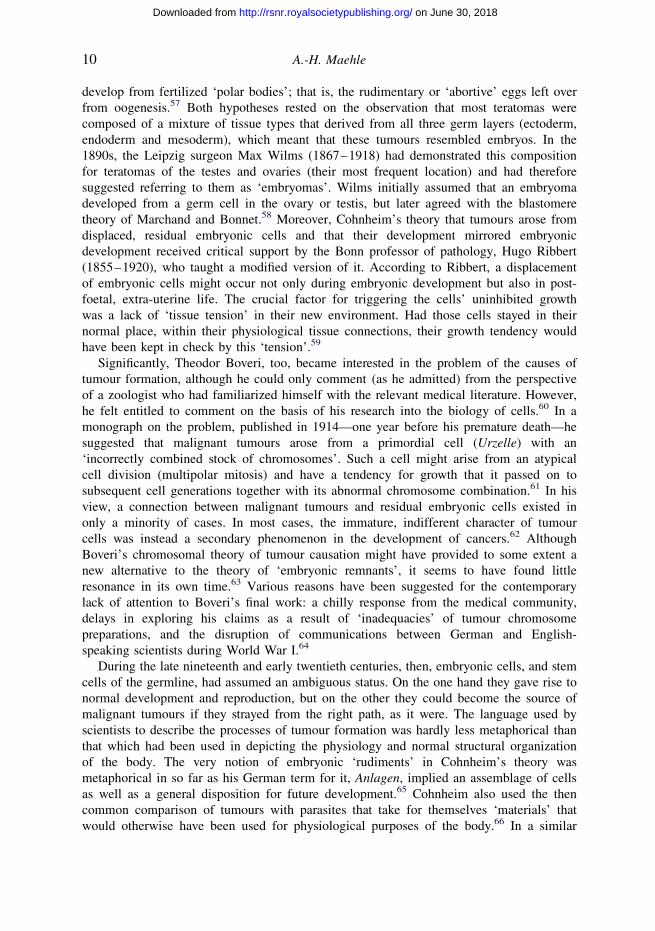
A.-H. Maehle10
on June 30, 2018http://rsnr.royalsocietypublishing.org/Downloaded from
develop from fertilized ‘polar bodies’; that is, the rudimentary or ‘abortive’ eggs left over
from oogenesis.57 Both hypotheses rested on the observation that most teratomas were
composed of a mixture of tissue types that derived from all three germ layers (ectoderm,
endoderm and mesoderm), which meant that these tumours resembled embryos. In the
1890s, the Leipzig surgeon Max Wilms (1867–1918) had demonstrated this composition
for teratomas of the testes and ovaries (their most frequent location) and had therefore
suggested referring to them as ‘embryomas’. Wilms initially assumed that an embryoma
developed from a germ cell in the ovary or testis, but later agreed with the blastomere
theory of Marchand and Bonnet.58 Moreover, Cohnheim’s theory that tumours arose from
displaced, residual embryonic cells and that their development mirrored embryonic
development received critical support by the Bonn professor of pathology, Hugo Ribbert
(1855–1920), who taught a modified version of it. According to Ribbert, a displacement
of embryonic cells might occur not only during embryonic development but also in post-
foetal, extra-uterine life. The crucial factor for triggering the cells’ uninhibited growth
was a lack of ‘tissue tension’ in their new environment. Had those cells stayed in their
normal place, within their physiological tissue connections, their growth tendency would
have been kept in check by this ‘tension’.59
Significantly, Theodor Boveri, too, became interested in the problem of the causes of
tumour formation, although he could only comment (as he admitted) from the perspective
of a zoologist who had familiarized himself with the relevant medical literature. However,
he felt entitled to comment on the basis of his research into the biology of cells.60 In a
monograph on the problem, published in 1914—one year before his premature death—he
suggested that malignant tumours arose from a primordial cell (Urzelle) with an
‘incorrectly combined stock of chromosomes’. Such a cell might arise from an atypical
cell division (multipolar mitosis) and have a tendency for growth that it passed on to
subsequent cell generations together with its abnormal chromosome combination.61 In his
view, a connection between malignant tumours and residual embryonic cells existed in
only a minority of cases. In most cases, the immature, indifferent character of tumour
cells was instead a secondary phenomenon in the development of cancers.62 Although
Boveri’s chromosomal theory of tumour causation might have provided to some extent a
new alternative to the theory of ‘embryonic remnants’, it seems to have found little
resonance in its own time.63 Various reasons have been suggested for the contemporary
lack of attention to Boveri’s final work: a chilly response from the medical community,
delays in exploring his claims as a result of ‘inadequacies’ of tumour chromosome
preparations, and the disruption of communications between German and English-
speaking scientists during World War I.64
During the late nineteenth and early twentieth centuries, then, embryonic cells, and stem
cells of the germline, had assumed an ambiguous status. On the one hand they gave rise to
normal development and reproduction, but on the other they could become the source of
malignant tumours if they strayed from the right path, as it were. The language used by
scientists to describe the processes of tumour formation was hardly less metaphorical than
that which had been used in depicting the physiology and normal structural organization
of the body. The very notion of embryonic ‘rudiments’ in Cohnheim’s theory was
metaphorical in so far as his German term for it, Anlagen, implied an assemblage of cells
as well as a general disposition for future development.65 Cohnheim also used the then
common comparison of tumours with parasites that take for themselves ‘materials’ that
would otherwise have been used for physiological purposes of the body.66 In a similar
Emergence of the stem cell concept 11
on June 30, 2018http://rsnr.royalsocietypublishing.org/Downloaded from
sense, Boveri wrote that the cell of a malignant tumour had lost certain characteristics, so that
it fell back from an ‘altruistic state’ (serving the body as a whole) to an ‘egoistic state’,
manifested in ‘unlimited reproduction’. According to Boveri, the uncontrolled
multiplication of tumour cells might also result from a lack of ‘inhibitory mechanisms’
(Hemmungsvorrichtungen) that were normally present in cells.67 Ribbert’s idea that cells
that had escaped the control of their physiological tissue environment would become
dangerous through their unchecked growth was mirrored in the statement by the Berlin
pathologist Otto Lubarsch (1860–1933) that disease originated from the ‘uprising of the
small people, the proletariat’ in the cell state.68 In Lubarsch’s mind, cells were likened to
proletarians who might become dangerous to the state if they were not integrated into
society.
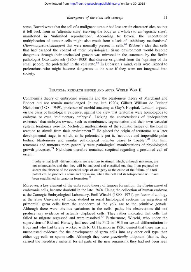
TERATOMA RESEARCH BEFORE AND AFTER WORLD WAR II
Cohnheim’s theory of embryonic remnants and the blastomere theory of Marchand and
Bonnet did not remain unchallenged. In the late 1920s, Gilbert William de Poulton
Nicholson (1878–1949), professor of morbid anatomy at Guy’s Hospital, London, argued,
on the basis of histological evidence, against the view that teratomas were homologous to
embryos or even ‘rudimentary embryos’. Lacking the characteristics of ‘independent
existence’ that embryos owned, such as membranes, segmentation and their own vascular
system, teratomas were for Nicholson malformations of the somatic tissues of the host in
reaction to stimuli from their environment.69 He placed the origin of teratomas at a later
developmental stage, in which, as he polemically put it, ‘nebulous and impossible polar
bodies, blastomeres and similar pathological monstra cease to trouble’.70 For him,
teratomas and tumours more generally were pathological manifestations of physiological
growth processes.71 Nicholson therefore remained sceptical regarding a presumed cell of
origin:
I believe that [cell] differentiations are reactions to stimuli which, although unknown, are
not unknowable, and that they will be analysed and classified one day. I am prepared to
accept the absence of the essential steps of ontogeny as the cause of the failure of a toti-
potent cell to produce a soma and organism, when the cell and its toti-potence will have
been established in teratoma formation.72
Moreover, a key element of the embryonic theory of tumour formation, the displacement of
embryonic cells, became doubtful in the late 1940s. Using the collection of human embryos
at the Carnegie Embryological Laboratory, Emil Witschi (1890–1971), professor of zoology
at the State University of Iowa, studied in serial histological sections the migration of
primordial germ cells from the endoderm of the yolk sac to the primitive gonads.
Although there were obvious variations in the cells’ paths, his observations did not
produce any evidence of actually displaced cells. They rather indicated that cells that
failed to migrate regressed and were resorbed.73 Furthermore, Witschi, who under the
supervision of Richard Hertwig had received his PhD in 1913 on sexual differentiation in
frogs and who had briefly worked with R. G. Harrison in 1926, denied that there was any
uncontested evidence for the development of germ cells into any other cell type than
either egg cells or sperm cells. Although they were genetically totipotent (that is, they
carried the hereditary material for all parts of the new organism), they had not been seen

A.-H. Maehle12
on June 30, 2018http://rsnr.royalsocietypublishing.org/Downloaded from
to transform into somatic cells during ontogeny. For Witschi, they were therefore as
specialized as neurons or muscle cells.74
Despite such findings and expert criticisms, the theory that teratomas and other tumours
might arise from residual embryonic, pluripotent cells, particularly those of the germ line,
continued to have currency in the period after World War II. Since the nineteenth century,
a large number of cases of human teratoma, mostly in the ovaries and testicles but also in
various other locations, had been described.75 In 1953 the pathologists Frank J. Dixon
(1920–2008) of the University of Pittsburgh and Robert A. Moore of Washington
University in St Louis reported on their histological re-examination of about 1000 cases
of testicular tumours recorded in the files of the Armed Forces Institute of Pathology
in Washington DC.76 The vast majority (96.5%) of these tumours were classed by
them as germinal tumours (that is, seminomas, embryonal carcinomas, teratomas,
choriocarcinomas, or combinations of these types). Dixon and Moore postulated that these
tumours originated from germ cells because their tissues displayed ‘a multipotentiality
that approaches that of the germ cell itself’—an argument that was remarkably similar to
Askanazy’s hypothesis of ‘egg-equivalent’ stem cells formulated almost half a century
earlier.77 The fact that teratoid tumours had generally been found most frequently in the
gonads (in men as well as women) further supported Dixon and Moore’s view; in
addition, regarding the relatively uncommon cases of such tumours occurring in other
sites, the idea of germ cells that had been somehow ‘misplaced’ during early embryonic
development still was for them ‘an attractive hypothesis’. They had to concede, however,
that the precise type of cell that gave rise to germinal (teratoid) tumours, whether
‘undifferentiated germ cell, embryonic germ cell, spermatogonia, or spermatocyte’,
remained an open question.78
EMBRYONAL CARCINOMA CELLS AND THE BEGINNINGS OF MODERN STEM CELL RESEARCH
This was the state of knowledge about the cellular origins of teratomas when, in 1953,
Leroy Stevens, then a postdoctoral researcher at the Jackson Laboratory in Bar Harbor,
Maine, made a chance finding in a particular strain of mice—a finding that set in
motion an extensive series of experiments that ultimately led to the isolation of
embryonic stem cell lines. The Jackson Laboratory had been founded in 1929 by the
Harvard-trained geneticist and former president of the University of Michigan,
Clarence Cook Little (1888–1971), as a facility for the breeding of standardized,
genetically defined strains of mice, particularly for the purpose of cancer research.79
Stevens, who had earned his PhD in developmental biology and had become Little’s
assistant in 1952, was given the task of systematically studying potential mutagenic
and carcinogenic factors, including the influence of tobacco and cigarette paper, in
particular strains of mice.80 Routinely examining a large number of mice, he
encountered within a period of eight months three cases of teratoma in the testes of
inbred ‘strain 129’ mice, a finding that attracted attention in the laboratory because
this type of tumour had only very rarely been observed previously in mice.81
By September 1954 Stevens and Little were able to report that 30 out of 3557 male
strain 129 mice examined—that is, nearly 1%—had a spontaneous testicular teratoma.
One of these tumours could be maintained through serial transplantation; that is, the
injection of tumour material under the skin or into the abdominal cavity of other

Emergence of the stem cell concept 13
on June 30, 2018http://rsnr.royalsocietypublishing.org/Downloaded from
mice, over 16 generations. Significantly, this transplantable tumour was composed of
‘undifferentiated, rapidly dividing embryonic-type cells’, and Stevens and Little
therefore agreed with Dixon and Moore’s view that testicular teratomas probably arose
from totipotent, undifferentiated cells of the germ line. Moreover, these findings
indicated for Stevens and Little not only a particular genetic disposition for this type
of tumour in this specific strain of laboratory mice but provided, as they pointed out,
an ‘important tool’ for studying the biology of teratomas extensively and in depth.82
Stevens grasped this opportunity ‘with great excitement’ and made teratomas the focus
of his research over the following decades.83
In his subsequent research Stevens demonstrated that teratoma cells produced embryo-like
(‘embryoid’) bodies when transplanted into the abdomen of mice84 and that, conversely,
certain embryonic tissues and fertilized egg cells gave rise to teratomas and
teratocarcinomas if implanted into the testes of mice.85 The notions of pluripotent
embryonic stem cells and of embryonal carcinoma cells became increasingly
exchangeable.86 Lewis Kleinsmith and Barry Pierce of the Pathology Department of the
University of Michigan found in the early 1960s that embryonal carcinoma cells
transplanted into mice gave rise to differentiated somatic tissues as well as embryonal
carcinoma, and interpreted this result as supporting the stem cell theory of cancer.87
Remarkably, they pointed out that Askanazy, in 1907, had been the first to propose that
the somatic tissues of teratomas might derive from undifferentiated stem cells.88 Working
with teratocarcinoma cells from one of Stevens’s mouse cell lines, Beatrice Mintz and
Karl Illmensee at the Institute for Cancer Research in Philadelphia reported in 1975 that
these tumour cells supported normal development if injected into early mouse embryos at
the blastocyst stage. The malignancy of those cells thus seemed to be reversible.89 Six
years later, Martin Evans and Matthew H. Kaufman at the University of Cambridge and
Gail Martin at the University of California in San Francisco isolated and cultured mouse
embryonic stem cells, which resembled teratocarcinoma cells and were pluripotent.90
Retrospectively, this constituted a decisive step in the history of modern embryonic stem
cell research, because these cells could now be cultured directly from blastocysts without
taking the detour of producing teratocarcinoma cell lines.91 In 1998 the groups of John
Gearhart at Johns Hopkins University and of James Thomson at the University of
Wisconsin each reported that they had been successful in isolating and culturing human
embryonic stem cells.92 The sources of these cell lines were aborted embryos (Gearhart)
and supernumerary in vitro fertilization embryos (Thomson), respectively.
Another important contribution to modern stem cell research was developed in the early
1960s by Ernest McCulloch (1926–2011) and James Till of the Ontario Cancer Institute in
Toronto when studying the effects of radiation on haematopoiesis in the bone marrow. In
irradiated mice, which had subsequently been injected with bone marrow cells, nodules
were found in the spleens on post-mortem examination. As Till and McCulloch showed,
the number of these nodules was proportional to the dose of marrow cells that the mice
had received, each nodule representing a cell colony that derived from one
haematopoietic stem cell or ‘colony-forming unit’ (CFU), as the two researchers
cautiously called it.93 The ‘spleen colony assay’ was the first quantitative assay for
blood stem cells and still retains its relevance for our understanding of haematopoiesis.
It helped in the eventual vindication of the ‘monophyletic’ or ‘unitarian’ view of
Pappenheim and Neumann that all types of blood cells originate from one multipotent
stem cell in the bone marrow.
A.-H. Maehle14
on June 30, 2018http://rsnr.royalsocietypublishing.org/Downloaded from
CONCLUSION
The concept of stem cells was first used in the writings of zoologists and medical scientists in
Imperial Germany. Adopting the term Stammzellen from Ernst Haeckel, Theodor Boveri was
particularly important in making these cells known as carriers of the so-called ‘germ plasm’
and as the starting points in embryological development of differentiated body cells as well
as germ cells. Boveri’s concept of a stem cell included both a capacity for self-renewal and a
capacity for differentiation; these are the basic defining characteristics of stem cells that are
still accepted today. The notion of stem cells was also readily applied at the start of the
twentieth century in haematological work by histologists such as Artur Pappenheim and
Ernst Neumann who, contrary to Paul Ehrlich’s ‘dualist’ view, assumed the existence of a
multipotent stem cell in the bone marrow that was able to differentiate into any type of
blood cell. In the haematological context the focus of discussion of stem cells gradually
began to move from the genealogical perspective of Haeckel and Boveri to the cells’
developmental possibilities or pluripotency. Although the exact meaning of ‘stem cells’
fluctuated between different authors, and names such as ‘mother cells’ or ‘primordial
germ cells’ were sometimes used synonymously, they carried broader, positive
connotations of common heritage, renewal, physiological development and differentiation.
In the same period, however, embryonic cells began to be suspected as causes of cancer in
the extra-uterine body. This hypothesis, originating with Julius Cohnheim, turned out to be
very fruitful for tumour research, in particular experimental and pathological–anatomical
work on teratomas. The multiple types of tissues found in teratomas led Max Askanazy
and others to the hypothesis of pluripotent ‘germs’ or ‘stem cells’ as their points of
origin. Embryonic cells were also seen as potentially dangerous, if they had lost their
integration in a physiological tissue environment. The 1950s research of Leroy Stevens
into teratomas and teratocarcinomas, which is commonly regarded as a foundation of
modern work on embryonic stem cells, should be seen against this historical ambiguity of
positive and negative sides of the ‘stem cell’. Transplanted embryonal carcinoma cells, as
Stevens, Barry Pierce and others showed in animal experiments, could produce various
differentiated tissues as well as cancer. Moreover, as Alison Kraft has recently argued, in
the history of leukaemia research in the second half of the twentieth century stem cells
also assumed an ambiguous status, being seen as a ‘biological force for good’ in the
context of bone marrow transplantation as well as having a ‘dark side’ through the
concept of the cancer stem cell.94 Indeed, as novel stem cell therapies, for example for
neurological conditions such as Parkinson’s disease, have been tested in the last few
years, concern about the risk of tumour formation has become a feature of both clinical
and ethical debate.95 In this situation it may well be appropriate to remember the
historical understandings of stem cells as delineated in this article.
ACKNOWLEDGEMENTS
I am grateful to the Wellcome Trust, London, for the support of my research as part of a
Strategic Award in the History of Medicine (grant 082800), and to Durham University for
the award of a Sir Derman Christopherson/Sir James Knott Foundation Fellowship in
Michaelmas Term 2010. I would also like to thank Dr Sebastian Pranghofer for his
research assistance and the anonymous reviewers of this paper for their helpful comments.

Emergence of the stem cell concept 15
on June 30, 2018http://rsnr.royalsocietypublishing.org/Downloaded from
NOTES
1 L. Burns, ‘“You are our only hope”: trading metaphorical “magic bullets” for stem cell
“superheroes”’, Theor. Med. Bioethics 30, 427–442 (2009).
2 A. Musolff, ‘“Progressive” evolution and “totipotent” stem cells: metaphors in British and
German debates about the “life sciences”’, IBERICA 17, 45–59 (2009).
3 M. B. Fagan, ‘The search for the hematopoietic stem cell: social interaction and epistemic
success in immunology’, Stud. Hist. Phil. Biol. Biomed. Sci. 38, 217–237 (2007);
M. B. Fagan, ‘Stems and standards: social interaction in the search for blood stem cells’,
J. Hist. Biol. 43, 67–109 (2010); A. Kraft, ‘Manhattan transfer: lethal radiation, bone marrow
transplantation, and the birth of stem cell biology, ca. 1942–1961’, Hist. Stud. Nat. Sci. 39,
171–218 (2009); A. Kraft, ‘Converging histories, reconsidered potentialities: the stem cell
and cancer’, BioSocieties 6, 195–216 (2011). For historical accounts of embryonic stem cell
research see note 91 below.
4 M. Ramalho-Santos and H. Willenbring, ‘On the origin of the term “stem cell”’, Cell Stem Cell
1, 35–38 (2007).
5 R. J. Richards, The tragic sense of life: Ernst Haeckel and the struggle over evolutionary thought
(University of Chicago Press, 2008).
6 E. Haeckel, Naturliche Schopfungsgeschichte (Georg Reimer, Berlin, 1868), 15th lecture.
7 Richards, op. cit. (note 5), pp. 148–156; N. Hopwood, ‘Embryology’, in The Cambridge history
of science, vol. 6 (The modern biological and earth sciences) (ed. P. J. Bowler and
J. V. Pickstone), pp. 285–315 (Cambridge University Press, 2009).
8 E. Haeckel, Anthropogenie oder Entwickelungsgeschichte des Menschen, 3rd edn (Wilhelm
Engelmann, Leipzig, 1877), p. 144 [my translation].
9 P. J. Weindling, Darwinism and Social Darwinism in Imperial Germany: the contribution of the
cell biologist Oscar Hertwig (1849–1922) (Gustav Fischer, Stuttgart, 1991), pp. 63–72.
10 Haeckel, op. cit. (note 8), p. 149 [my translation].
11 E. Johach, Krebszelle und Zellenstaat. Zur medizinischen und politischen Metaphorik in Rudolf
Virchows Zellularpathologie (Rombach Verlag, Freiburg i. Br., 2008); A. Reynolds, ‘The theory
of the cell state and the question of cell autonomy in nineteenth and early twentieth-century
biology’, Sci. Context 20, 71–95 (2007); D. J. Nicholson, ‘Biological atomism and cell
theory’, Stud. Hist. Phil. Biol. Biomed. Sci. 41, 202–211 (2010).
12 R. Virchow, Die Cellularpathologie in ihrer Begrundung auf physiologische und pathologische
Gewebelehre, 4th edn (August Hirschwald, Berlin, 1871), p. 17 [my translation; emphasis in the
original].
13 E. Haeckel, ‘Zellseelen und Seelenzellen’ (1878), in Vortrage und Abhandlungen
(ed. H. Schmidt-Jena) (Alfred Kroner, Leipzig, 1924), pp. 162–195; A. Reynolds, ‘Ernst
Haeckel and the theory of the cell state: remarks on the history of a bio-political metaphor’,
Hist. Sci. 46, 123–152 (2008); Weindling, op. cit. (note 9), pp. 258–261.
14 E. B. Wilson, The cell in development and heredity, 3rd edn (Macmillan, New York, 1925),
pp. 101–102; Johach, op. cit. (note 11), pp. 22–25.
15 See also Ramalho-Santos and Willenbring, op. cit. (note 4), p. 35.
16 A. Weismann, Die Continuitat des Keimplasma’s als Grundlage einer Theorie der Vererbung
(Gustav Fischer, Jena, 1885), pp. 5–21.
17 V. Haecker, ‘Die Kerntheilungsvorgange bei der Mesoderm- und Entodermbildung von
Cyclops’, Arch. Mikrosk. Anat. 39, 556–581 (1892).
18 T. Boveri, ‘Ueber die Entstehung des Gegensatzes zwischen den Geschlechtszellen und den
somatischen Zellen bei Ascaris megalocephala, nebst Bemerkungen zur Entwicklungsgeschichte
der Nematoden’, Sitzungsber. Ges. Morphol. Physiol. Munchen 8, 114–125 (1892).
19 H. Satzinger, Differenz und Vererbung. Geschlechterordnungen in der Genetik und
Hormonforschung 1890–1950 (Bohlau Verlag, Cologne, 2009), pp. 85–97; F. Baltzer,
A.-H. Maehle16
on June 30, 2018http://rsnr.royalsocietypublishing.org/Downloaded from
Theodor Boveri: life and work of a great biologist 1862–1915 (University of California Press,
Berkeley, 1967); K. B. Moritz, Theodor Boveri (1862–1915), Pionier der modernen Zell- und
Entwicklungsbiologie (Gustav Fischer, Jena, 1993); H. A. Neumann, Vom Ascaris zum Tumor.
Leben und Werk des Biologen Theodor Boveri (1862–1915) (Blackwell, Berlin, 1998).
20 J. Harwood, Styles of scientific thought. The German genetics community 1900–1933
(University of Chicago Press, 1993), pp. 52–55; R. Haecker, ‘Das Leben von Valentin
Haecker’, Zool. Anz. 174, 1–22 (1965).
21 V. Haecker, Uber Gedachtnis, Vererbung und Pluripotenz (Gustav Fischer, Jena, 1914), pp. 63
and 85.
22 For example, T. Boveri, ‘Die Entwickelung von Ascaris megalocephala mit besonderer
Rucksicht auf die Kernverhaltnisse’, in Festschrift zum siebenzigsten Geburtstag von Carl von
Kupffer, pp. 383–430 (Gustav Fischer, Jena, 1899); T. Boveri, ‘Die Potenzen der Ascaris-
Blastomeren bei abgeanderter Furchung. Zugleich ein Beitrag zur Frage qualitativ-ungleicher
Chromosomen-Teilung’, in Festschrift zum sechzigsten Geburtstag Richard Hertwigs
(Munchen), pp. 133–214 (Gustav Fischer, Jena, 1910).
23 Wilson, op. cit. (note 14), pp. 322–325. On Wilson’s book, see J. Maienschein, ‘From
presentation to representation in E. B. Wilson’s The Cell’, Biol. Phil. 6, 227–254 (1991).
24 R. W. Hegner, The germ-cell cycle in animals (Macmillan, New York, 1914), pp. 174–179.
25 R. Weissenberg, ‘Zeugung und Sexualitat in ihren anatomischen und biologischen Grundlagen’,
in Handbuch der Sexualwissenschaften, 3rd edn (2 volumes) (ed. A. Moll), vol. 1, pp. 1–232
(F. C. W. Vogel, Leipzig, 1926).
26 Weindling, op. cit. (note 9), pp. 205–213, 218–220 and 342. Boveri’s work on Ascaris and stem
cells was also reviewed in Wilhelm Waldeyer’s contribution on ‘Die Geschlechtszellen’ in
Handbuch der vergleichenden und experimentellen Entwickelungslehre der Wirbeltiere (ed.
Oskar Hertwig), vol. 1, part 1, pp. 86–476 (Gustav Fischer, Jena, 1906), at pp. 222–223.
27 Weissenberg (note 25), pp. 19–20.
28 A. Pappenheim, ‘Ueber Entwickelung und Ausbildung der Erythroblasten’, Virchows Arch.
Pathol. Anat. 145, 587–643 (1896).
29 Ibid., pp. 600–601 and 635–636.
30 Ibid., p. 640.
31 See also Ramalho-Santos and Willenbring, op. cit. (note 4), p. 37. On the further development of
research on haematopoietic stem cells, see L. G. Lajtha, ‘The common ancestral cell’, in Blood,
pure and eloquent. A story of discovery, of people, and of ideas (ed. M. M. Wintrobe), pp. 81–95
(McGraw-Hill, New York, 1980); E. A. McCulloch, The Ontario Cancer Institute: successes and
reverses at Sherbourne Street (McGill-Queen’s University Press, Montreal, 2003), pp. 46–60;
N. Brown, A. Kraft and P. Martin, ‘The promissory pasts of blood stem cells’, BioSocieties 1,
329–348 (2006); A. Kraft, ‘Atomic medicine: the cold war origins of biological research’,
Hist. Today 59 (11), 26–33 (2009); A. Kraft, ‘Manhattan transfer’, op. cit. (note 3); Kraft,
‘Converging histories’, op. cit. (note 3); Fagan, ‘The search’, op. cit. (note 3); Fagan,
‘Stems’, op. cit. (note 3).
32 A. Pappenheim, ‘Zwei Falle akuter grosslymphozytarer Leukamie’, Fol. Haematol. 4, 301–308
(1907). On Pappenheim and his haematological work, see Ricarda Dinser, ‘Der Beitrag Artur
Pappenheims zur Hamatologie um die Jahrhundertwende’, MD thesis, Ruhr-Universitat
Bochum (2001).
33 W. Dantschakoff, ‘Untersuchungen uber die Entwickelung des Blutes und Bindegewebes bei den
Vogeln’, Anat. Hefte 37, 471–589 (1908). On Dantschakoff’s international career, see Satzinger,
op. cit. (note 19), pp. 231–232 and 394–396.
34 A. Maximow, ‘Der Lymphozyt als gemeinsame Stammzelle der verschiedenen Blutelemente in
der embryonalen Entwicklung und im postfetalen Leben der Saugetiere’, Fol. Haematol. 8,
125–134 (1909); I. E. Konstantinov, ‘In search of Alexander A. Maximow: the man behind
the unitarian theory of hematopoiesis’, Perspect. Biol. Med. 43, 269–276 (2000).

Emergence of the stem cell concept 17
on June 30, 2018http://rsnr.royalsocietypublishing.org/Downloaded from
35 E. Neumann, ‘Ueber die Bedeutung des Knochenmarkes fur die Blutbildung, Vorlaufige
Mittheilung’, CentBlatt Med. Wiss. 6, no. 44 (1868); E. Neumann-Redlin v. Meding, Der
Pathologe Ernst Neumann (1834–1918) und sein Beitrag zur Begrundung der Hamatologie
im 19. Jahrhundert (Demeter Verlag, Munich, 1987), pp. 30–33.
36 E. Neumann, ‘Hamatologische Studien I.–III.’, Virchows Arch. Pathol. Anat. 143, 225–277
(1896); 174, 41–78 (1903); 207, 379–412 (1912); P. Ehrlich and A. Lazarus, Histology of
the blood: normal and pathological (Cambridge University Press, 1900), pp. 81–120;
Neumann-Redlin v. Meding, op. cit. (note 35), pp. 33–35, 43 and 46–54.
37 E. Neumann, ‘Hamatologische Studien. III. Leukocyten ynd Leukamie’, Virchows Arch. Pathol.
Anat. 207, 379–412 (1912), at p. 382.
38 J. Maienschein, ‘Regenerative medicine in historical context’, Med. Stud. 1, 33–40 (2009).
39 R. G. Harrison, ‘Observations on the living developing nerve fiber’, Anat. Rec. 1, 116–118
(1907); R. G. Harrison, ‘The outgrowth of the nerve fiber as a mode of protoplasmic
movement’, J. Exp. Zool. 9, 787–846 (1910).
40 Harrison, ‘The outgrowth’, op. cit. (note 39), pp. 833–841.
41 H. Landecker, Culturing life. How cells became technologies (Harvard University Press,
Cambridge, MA, 2007); D. H. Stapleton, ‘Tissue culture and tissue culture technologies at
the Rockefeller Institute for Medical Research: roots of regenerative medicine, 1910–1950’,
Med. Stud. 1, 77–81 (2009).
42 Harrison, ‘The outgrowth’, op. cit. (note 39), p. 791.
43 Neumann, op. cit. (note 37), p. 382.
44 O. Hertwig, Der Staat als Organismus. Gedanken zur Entwicklung der Menschheit (Gustav
Fischer, Jena, 1922), p. 65 [my translation]. Hertwig here quoted himself from the second
edition of his work Zur Abwehr des ethischen, des sozialen und politischen Darwinismus
(Gustav Fischer, Jena, 1921). For a detailed discussion of O. Hertwig’s ideas on the state as
an organism, see Weindling (note 9), pp. 262–303.
45 E. Haeckel, Naturliche Schopfungs-Geschichte. Gemeinverstandliche wissenschaftliche
Vortrage uber die Entwickelungslehre, Volksausgabe (Walter de Gruyter, Berlin, 1926),
p. 226 [my translation].
46 F. Kahn, Das Leben des Menschen. Eine volkstumliche Anatomie, Biologie, Physiologie und
Entwicklungsgeschichte des Menschen (5 volumes) (Kosmos, Stuttgart, 1922–31), vol. 1,
plate XVII opp. p. 240. On Kahn and his use of visual representations in the popularization
of biological and medical information, see C. Borck, ‘Der industrialisierte Mensch: Fritz
Kahns Visualisierungen des Korpers als Interferenzzone von Medizin, Technik und Kultur’,
WerkstattGeschichte 47, 7–22 (2008).
47 Kahn, op. cit. (note 46), vol. 1, p. 193.
48 Ibid., vol. 5, pp. 144–146.
49 J. Cohnheim, Vorlesungen uber allgemeine Pathologie. Ein Handbuch fur Aerzte und Studirende
(2 volumes) (August Hirschwald, Berlin, 1877–80), vol. 1, pp. 634–650 and 655–656.
50 Ibid., pp. 661–666, 674, 678. On Cohnheim’s theory, see also L. J. Rather, The genesis of
cancer. A study in the history of ideas (Johns Hopkins University Press, Baltimore, MD,
1978), pp. 169–174; Johach, op. cit. (note 11), pp. 245–246; M. Cooper, ‘Regenerative
pathologies: stem cells, teratomas and theories of cancer’, Med. Stud. 1, 55–66 (2009).
51 H. Ribbert, Ueber Ruckbildung an Zellen und Geweben und uber die Entstehung der
Geschwulste (Erwin Nagele, Stuttgart, 1897), pp. 41–42; H. Ribbert, Beitrage zur Entstehung
der Geschwulste (Friedrich Cohen, Bonn, 1906), pp. 6–13; M. Borst, Die Lehre von den
Geschwulsten (2 volumes) (J. F. Bergmann, Wiesbaden, 1902), vol. 1, p. 39a; vol. 2,
pp. 817–818; E. Schwalbe, Allgemeine Missbildungslehre (Teratologie). Eine Einfuhrung in
das Studium der Abnormen Entwicklung (Gustav Fischer, Jena, 1906), pp. 155–158;
L. Aschoff, E. Kuster and W. J. Schmidt, Hundert Jahre Zellforschung (Gebruder
Borntraeger, Berlin, 1938), pp. 250–258.
A.-H. Maehle18
on June 30, 2018http://rsnr.royalsocietypublishing.org/Downloaded from
52 A. Birch-Hirschfeld and S. Garten, ‘Ueber das Verhalten implantirter embryonaler Zellen im
erwachsenen Thierkorper’, Beitr. Pathol. Anat. Allg. Pathol. 26, 132–172 (1899); C. Fere and
A. Lutier, ‘Nouvelles observations sur les teratomes experimentaux’, Arch. Anat. Microsc. 3,
337–368 (1900); R. Traina, ‘Ueber Transplantationen von Embryonalgeweben ins Ovarium
und die Bildung von Ovarialcysten’, Centralblatt Allg. Pathol. Pathol. Anat. 13, 49–56
(1902); M. Wilms, ‘Wachstum embryonaler Implantation und Geschwulstbildung’, Verh. Dt.
Pathol. Ges., year 1904, 79–80 (1905); R. Rossle, ‘Ueber die Einverleibung von
Embryonalzellen’, Munch. Med. WochSchr. 53, 143–144 (1906); M. Borst, ‘Die Teratome
und ihre Stellung zu anderen Geschwulsten’, Verh. Dt. Pathol. Ges., year 1907, 83–104
(1908); Eugen Korschelt, Regeneration und Transplantation (2 volumes) (Gebruder
Borntraeger, Berlin, 1927–31), vol. 2, pp. 1205–1208.
53 M. Askanazy, ‘Die Teratome nach ihrem Bau, ihrem Verlauf, ihrer Genese und im Vergleich
zum experimentellen Teratoid’, Verh. Dt. Pathol. Ges., year 1907, 39–82 (1908).
Significantly, numerous experiments on the transplantation of tumours in mice were
performed in the institutes of Paul Ehrlich in Frankfurt am Main and Oscar Hertwig in Berlin
during the same period. See P. Ehrlich, ‘Experimentelle Studien an Mausetumoren’,
Z. Krebsforsch. 5, 59–81 (1907); O. Hertwig and H. Poll, Zur Biologie der Mausetumoren.
Experimentelle Untersuchungen (Konigliche Akademie der Wissenschaften, Berlin, 1907).
For a discussion of Ehrlich’s cancer research, see A. C. Huntelmann, Paul Ehrlich: Leben,
Forschung, Okonomien, Netzwerke (Wallstein Verlag, Gottingen, 2011), pp. 174–176.
54 Askanazy, op. cit. (note 53), p. 47.
55 Ibid., pp. 71–73.
56 F. Marchand, Die Missbildungen, 3rd edn (Gistel, Vienna, 1898), pp. 75–79; R. Bonnet, ‘Zur
Aetiologie der Embryome’, Monatsschr. Geburtshulfe Gynaekol. 13, 149–176 (1901). See
also M. Askanazy, Die Dermoidcysten des Eierstocks: ihre Geschichte, ihr Bau und ihre
Entstehung sowie ihre Beziehung zu verwandten pathologischen Bildungen (Erwin Nagele,
Stuttgart, 1905), pp. 16–18; Borst, op. cit. (note 52), pp. 91–93.
57 Marchand, op. cit. (note 56), pp. 76–78; Marchand, ‘Mißbildungen’, in Real-Encyclopadie der
gesamten Heilkunde, 4th edn (ed. A. Eulenburg), vol. 9, pp. 722–853 (Urban & Schwarzenberg,
Berlin, 1910), at pp. 778–779.
58 M. Wilms, Die Mischgeschwulste, III. (Schluss-) Heft (Arthur Georgi, Berlin, 1902), pp. 227–252.
59 Ribbert, Ruckbildung, op. cit. (note 51), pp. 42–43; Ribbert, Beitrage, op. cit. (note 51), pp. 8–13.
See also Johach, op. cit. (note 11), pp. 246–247.
60 T. Boveri, Zur Frage der Entstehung maligner Tumoren (Gustav Fischer, Jena, 1914), p. 2.
61 Ibid., pp. 1 and 22.
62 Ibid., pp. 4 and 42.
63 Neumann, op. cit. (note 19), p. 210.
64 T. Boveri, Concerning the origin of malignant tumours (transl. and annot. H. Harris) (Company
of Biologists Limited and Cold Spring Harbor Laboratory Press, Woodbury, NY, 2008), preface
(by Harris), p. v.
65 Cf. Johach, op. cit. (note 11), p. 246.
66 Cohnheim, op. cit. (note 49), p. 686. On the concept of the parasite in biological and socio-
political thought of the late nineteenth and early twentieth centuries, see Johach, op. cit. (note
11), pp. 302–331; A. Musolff, Metaphor, nation and the Holocaust: the concept of the body
politic (Routledge, New York, 2010), pp. 1–8.
67 Boveri (note 60), pp. 4–5.
68 O. Lubarsch, Ein bewegtes Gelehrtenleben (Springer, Berlin, 1931), pp. 591–592; cited in
Johach (note 11), p. 267. On Lubarsch’s nationalist views and his directorship of the
Pathological Institute in Berlin from 1917 to 1928, see C.-R. Prull, Medizin am Toten oder
am Lebenden? Pathologie in Berlin und in London, 1900–1945 (Schwabe, Basel, 2003),
pp. 85–88 and 358–367.
Emergence of the stem cell concept 19
on June 30, 2018http://rsnr.royalsocietypublishing.org/Downloaded from
69 G. W. de P. Nicholson, ‘The histogeny of teratomata’, J. Pathol. Bacteriol. 32, 365–386 (1929).
Similar points on the ‘non-foetiformity of teratomas’ were later made by R. A. Willis, Pathology
of tumours (Butterworth, London, 1953), pp. 976–978.
70 Nicholson, op. cit. (note 69), p. 371.
71 Ibid., pp. 366 and 384. See also Prull, op. cit. (note 68), p. 197.
72 Nicholson, op. cit. (note 69), p. 384.
73 E. Witschi, ‘Migration of the germ cells of human embryos from the yolk sac to the primitive
gonadal folds’, Contrib. Embryol. 32, 67–80 (1948). See also J. E. Wheeler, ‘History of
teratomas’, in The human teratomas: experimental and clinical biology (ed. I. Damjanov,
B. B. Knowles and D. Solter), pp. 1–22 (Humana, Clifton, NJ, 1983).
74 Witschi, op. cit. (note 73), pp. 78–79.
75 See Wheeler, op. cit. (note 73).
76 F. J. Dixon and R. A. Moore, ‘Testicular tumors: a clinicopathological study’, Cancer 6, 427–
454 (1953). See also R. H. Young, ‘A brief history of the pathology of the gonads’, Mod. Pathol.
18, S3–S17 (2005).
77 Dixon and Moore, op. cit. (note 76), p. 429; Askanazy, op. cit. (note 53), pp. 51 and 70–71.
78 Dixon and Moore, op. cit. (note 76), p. 429–430.
79 K. Rader, Making mice: standardizing animals for biomedical research 1900–1955 (Princeton
University Press, 2004).
80 Leroy C. Stevens, ‘Spontaneous and experimentally induced testicular teratomas in mice’, Cell
Differ. 15, 69–74 (1984); R. A. Lewis, Discovery: windows on the life sciences (Blackwell,
Malden, MA, 2001), p. 132.
81 Stevens, op. cit. (note 80), p. 69.
82 L. C. Stevens and C. C. Little, ‘Spontaneous testicular teratomas in an inbred strain of mice’,
Proc. Natl Acad. Sci. USA 40, 1080–1087 (1954).
83 Lewis, op. cit. (note 80), p. 133.
84 L. C. Stevens, ‘Embryology of testicular teratomas in strain 129 mice’, J. Natl Cancer Inst. 23,
1249–1295 (1959); L. C. Stevens, ‘Embryonic potency of embryoid bodies derived from
transplantable testicular teratoma of the mouse’, Dev. Biol. 2, 285–297 (1960).
85 L. C. Stevens, ‘Experimental production of testicular teratomas in mice’, Proc. Natl Acad. Sci.
USA 52, 654–661 (1964); L. C. Stevens, ‘The development of teratomas from intratesticular
grafts of tubal mouse eggs’, J. Embryol. Exp. Morphol. 20, 329–341 (1968); L. C. Stevens,
‘The development of transplantable teratocarcinomas from intratesticular grafts of pre- and
postimplantation mouse embryos’, Dev. Biol. 21, 364–382 (1970).
86 See also Lewis, op. cit. (note 80), p. 136.
87 L. J. Kleinsmith and G. B. Pierce Jr, ‘Multipotentiality of single embryonal carcinoma cells’,
Cancer Res. 24, 1544–1551 (1964).
88 Ibid., p. 1544, making reference to Askanazy, op. cit. (note 53). Askanazy’s paper was also cited,
in the same context, in G. B. Pierce, ‘Teratocarcinoma: model developmental concept of cancer’,
Curr. Top. Dev. Biol. 2, 223–246 (1967), at p. 224.
89 B. Mintz and K. Illmensee, ‘Normal genetically mosaic mice produced from malignant
teratocarcinoma cells’, Proc. Natl Acad. Sci. USA 72, 3585–3589 (1975). See also the similar
results by R. L. Brinster, ‘The effect of cells transferred into the mouse blastocyst on
subsequent development’, J. Exp. Med. 140, 1049–1056 (1974).
90 M. J. Evans and M. H. Kaufman, ‘Establishment in culture of pluripotential cells from mouse
embryos’, Nature 292, 154–156 (1981); G. R. Martin, ‘Isolation of a pluripotent cell line
from early mouse embryos cultured in medium conditioned by teratocarcinoma stem cells’,
Proc. Natl Acad. Sci. USA 78, 7634–7638 (1981).
91 P. W. Andrews, ‘From teratocarcinomas to embryonic stem cells’, Phil. Trans. R. Soc. Lond. B
357, 405–417 (2002); D. Solter, ‘From teratocarcinomas to embryonic stem cells and beyond: a
history of embryonic stem cell research’, Nature Rev. Genet. 7, 319–327 (2006); M. Morange,

A.-H. Maehle20
on June 30, 2018http://rsnr.royalsocietypublishing.org/Downloaded from
‘What history tells us. VII. Twenty-five years ago: the production of mouse embryonic stem
cells’, J. Biosci. 31, 537–541 (2006); Cooper, op. cit. (note 50).
92 M. J. Shamblott, J. Axelman, S. Wang, E. M. Bugg, J. W. Littlefield, P. J. Donovan,
P. D. Blumenthal, G. R. Huggins and J. D. Gearhart, ‘Derivation of pluripotent stem cells
from cultured human primordial germ cells’, Proc. Natl Acad. Sci. USA 95, 13726–13731
(1998); J. A. Thomson, J. Itskovitz-Eldor, S. S. Shapiro, M. A. Waknitz, J. J. Swiergiel,
V. S. Marshall and J. M. Jones, ‘Embryonic stem cell lines derived from human blastocysts’,
Science 282, 1145–1147 (1998).
93 J. E. Till and E. A. McCulloch, ‘A direct measurement of the radiation sensitivity of normal
mouse bone marrow cells’, Radiat. Res. 14, 213–222 (1961); A. J. Becker, E. A. McCulloch
and J. E. Till, ‘Cytological demonstration of the clonal nature of spleen colonies derived from
transplanted mouse marrow cells’, Nature 197, 452–454 (1963); McCulloch, op. cit. (note
31), pp. 46–50.
94 Kraft, ‘Converging histories’, op. cit. (note 3), p. 1.
95 Z. Master, M. McLeod and I. Mendez, ‘Benefits, risks and ethical considerations in translation of
stem cell research to clinical applications in Parkinson’s disease’, J. Med. Ethics 33, 169–173
(2007); N. Amariglio, A. Hirschberg, B. W. Scheithauer, Y. Cohen, R. Loewenthal,
L. Trakhtenbrot, N. Paz, M. Koren-Michowitz, D. Waldman, L. Leider-Trejo, A. Toren,
S. Constantini and G. Rechavi, ‘Donor-derived brain tumor following neural stem cell
transplantation in an ataxia teleangiectasia patient’, PLoS Med. 6 (2), e1000029 (doi:10.1371/journal.pmed.1000029).
96 From Boveri, op. cit. (note 18), at p. 118.
97 From A. Pappenheim, Atlas der menschlichen Blutzellen (Gustav Fischer, Jena, 1905–12),
vol. 1, opp. p. 347.