Embed Size (px)
Citation preview

The dental pulp
Dr. Gábor Varga
Department of Oral Biology
2016

The dental pulp (introduction)
• Structure of the pulp
• Extracellular matrix and cells in the pulp
• Blood and lymph supply of the pulp
• Innervation of the pulp
• Role of pathological changes in circulation in
the development of tissue inflammation
• Regressive changes in the pulp

What is the dental pulp?
• Connective tissue infilling of the pulp
chamber and upper root
• Remnant of the dental papilla
• It maintains health of dentine,
• repairs dentine,
• provides sensory pathways from dentine.
Radiograph of teeth – pulp chambers are visible well

Section of the tooth – pulp chamber is encapsulated
Pulp chamber
with pulp

Incisor
longitudinal
section
observe the central
location of pulp
cavity
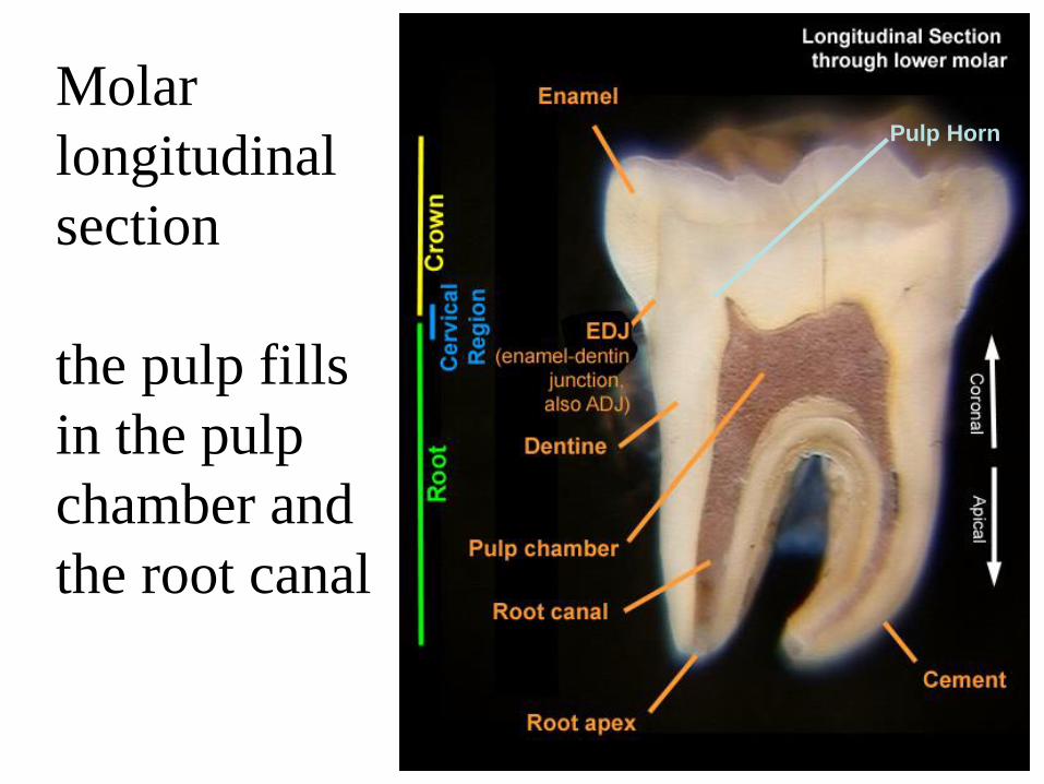
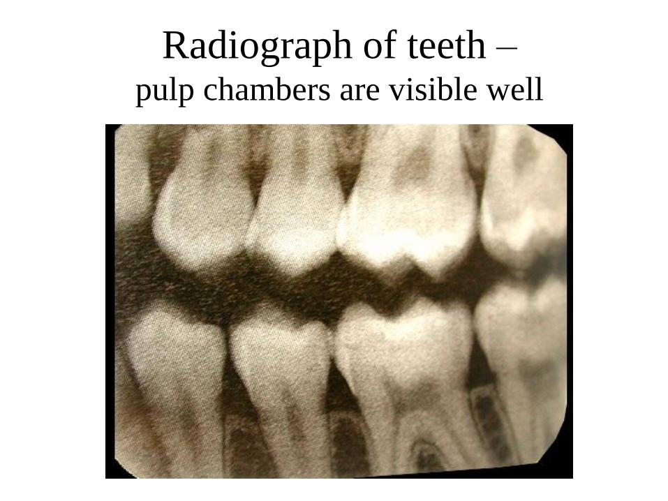
Molar
longitudinal
section
the pulp fills
in the pulp
chamber and
the root canal
Pulp Horn

LAMINA BUD STAGE CAP STAGE BELL STAGE ERUPTION
Tooth development

Gene activation during tooth
development Epithelium
Mesenchyme

Tooth development – details 1
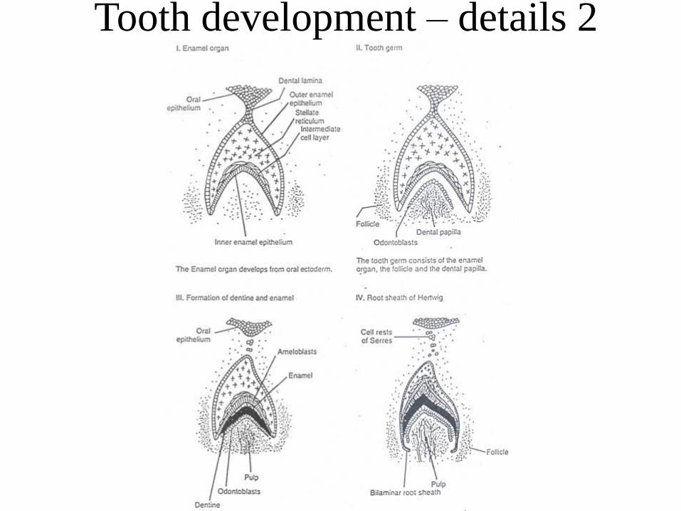
Tooth development – details 2

Section of tooth – pulp is inside

Fine structure of the pulp

Figure :
A) D: dentine; O: odontoblast; S.sz.: cell free zone; S.g: cell reach zone;
C: central zone
B) O: odontoblast; IR: nerve fibres
Fine structure of the pulp -
histochemistry

Histochemical picture of pulp margin
dentine
predentine
odontoblasts
capillaries
and nerves
arterioles,
venules, and
nerve bundles

Odontoblast layer between predentine
and pulp
dentine
Predentine
Odontoblasts
Mineralization
front
Mesenchyme

Constituents of the pulp
75 % water
and
25 % organic and
water soluble
inorganic material
Pulp Matrix
• Fibers are collagen type III, type I, and type V
(Type III confers elasticity, Type I gives tensile
strength, Type V also typical of mesenchymal
tissue)
• Ground substance is made up of proteoglycans
(which retain water to form gel and keep Ca2+ in
solution)

Structure of collagen

Structure of proteoglycans

Cell Types in Pulp
1. Odontoblasts
2. Fibroblasts (maintain pulp matrix)
3. Undifferentiated Mesenchyme Cells
4. Macrophages
5. Accessory Cells (T-cells,dendritic cells, etc.)
Plus
• Blood vessels
• Nerve axons

Blood supply of the pulp
• It comes from branches of inferior and superior
alveolar artery and vein
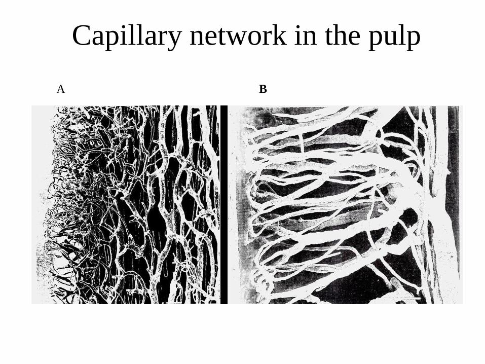
• It is organized into larger central vessels (large
venule and 1-2 arterioles) with a rich superficial
plexus of capillaries around periphery in the crown
• It is important for maintaining living cells (especi-
ally for odontoblasts) and for regulating fluid
• Excess matrix fluid is removed by lymphatics

Blood supply

Blood supply of the pulp

Capillary loops
Pre-capillary
Terminal
arteriole
Arteriole Venule
Lymphatic
vessel
Collecting
venule
Odontoblasts
AVA
Blood supply of the pulp

Arterioles and venules
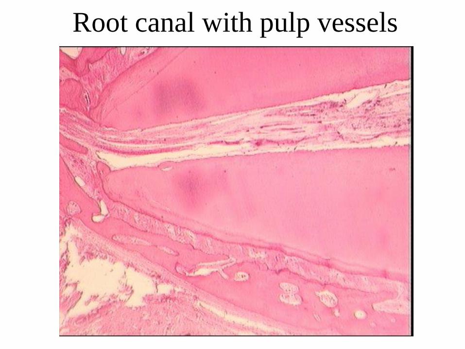
Root canal with pulp vessels
A B
Capillary network in the pulp

Nerve supply of the pulp • The pulp is supplied by sensory fibers of the trigeminal nerve (V)
and by sympathetic fibers of the gl cervicalis superior
• It contains both myelinated and unmyelinated axons
• Contains both sensory (CGRP, SP, NKA release) and sympathetic
fibers (NA, NPY) (the latter regulates blood flow)
• A single tooth may contain 2000 A-delta myelinated axons (to
conduct sharp, piercing pain) and 500 C type unmyelinated ones (to
conduct dull ache in response to thermal, mechanical, and chemical
stimuli)
• Fibers are concentrated in plexus beneath the odontoblast layer
(subodontic or Rashkov’s plexus)
• Nerve fibers may extend into dentine tubules, most concentrated at
pulp horns or in areas undergoing repair

Network of nerve fibres in the pulp

Hemodynamics in pulp vessels – Starling forces

Blood pressure in pulp

Effect of grinding on pulpal blood flow

Significance of arterio-venous
anastomoses (AVA)

Pulpal blood flow and sensory nerve activity
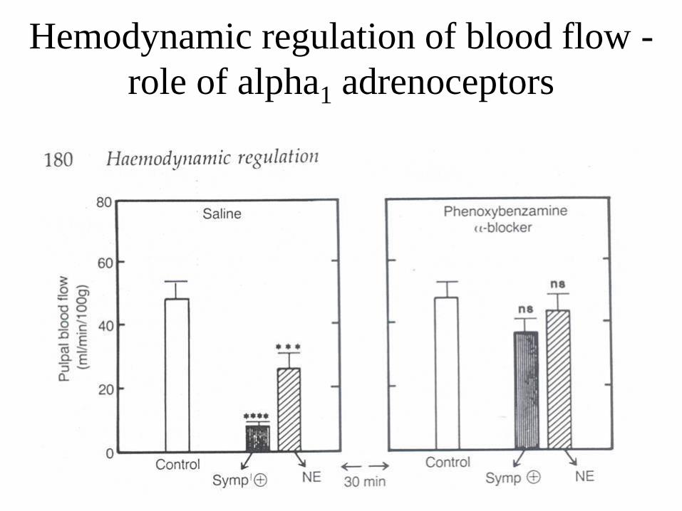
Hemodynamic regulation of blood flow -
role of alpha1 adrenoceptors


Current concepts on the generation of dentinal pain

Hydrodynamic
stimuli
Cortex pain
Brain stem
Trigeminal ganglion
Sensory nerves
CGRP SP
Vessel
CGRP
SP Vessel
Sensation from tooth to brain

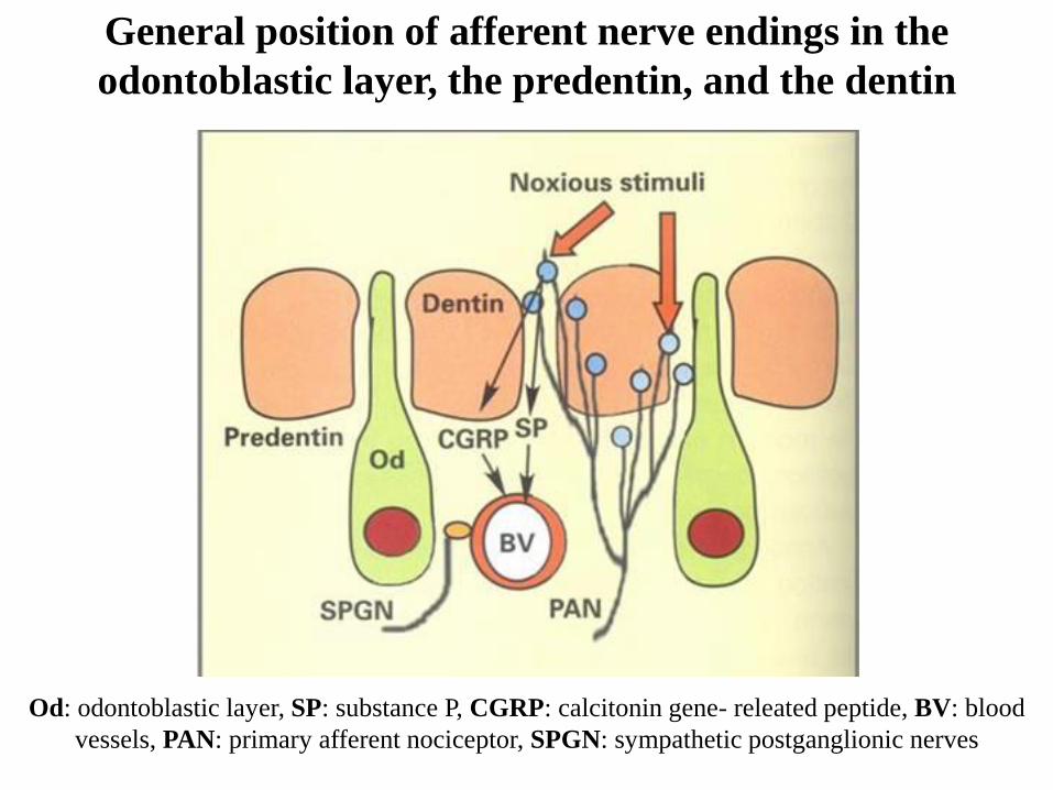
General position of afferent nerve endings in the
odontoblastic layer, the predentin, and the dentin
Od: odontoblastic layer, SP: substance P, CGRP: calcitonin gene- releated peptide, BV: blood
vessels, PAN: primary afferent nociceptor, SPGN: sympathetic postganglionic nerves

Release of neuropeptides from sensory nerve fibers
Nociceptive
stimulation
to center
C-type polimodal
afferent
Mast cell
histamine
capillary
network arteriole
to center
C-type polimodal
afferent
nociceptive stimulation
Mast cell
histamine
capillary
network arteriole
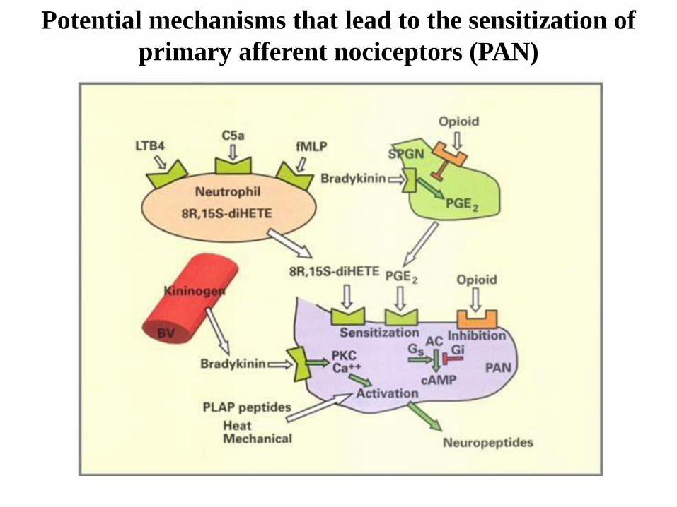
Potential mechanisms that lead to the sensitization of
primary afferent nociceptors (PAN)

Sensitization of primary afferent nociceptors (PAN)
by arachidonic acid (AA) cascade and by the
phospholipase A-activating protein (PALP)

Nociceptor discharge
Vasodilation
Plasma extravasation
Mast cell degranulation
Arachidonic acid cascade
Lymphocyte + neutrophil
invasion
Nociceptor sensitization
Epithelial proliferation
Collagen synthesis
Regulation of gene expression
Phenotype changes
Responses to Tissue Injury
Julius & Basbaum, Nature, 413:203-210, 2001

Important definitions:
Hyperesthesia
Hyperalgesia
Allodynia
Anesthesia
Analgesia

Analgetic system transmitting endogenous
pain suppression
Periaq.
nuclei
Spinal chord
opioid inhibitory
neuron
GABAergic inh.
neuron
monoaminergic inh.
neuron
Morphine
Inhibitory
mechanism
Opioid inh.
interneuron
(lower) morphine
Pre- & postsynaptic inhibition

Pain development: gate control theory
A: stimulator effect, SG: spinal ganglion, B: interneuron, T: transmitting neurons

Pain
“An unpleasant sensory
and emotional experience
arising from actual or
potential tissue damage or
described in terms of such
damage.”
International Association
for the Study of Pain

Thermoreceptors in dental pulp are similar to
skin
cold receptors
warm receptors

The rate of pain depends on the level of heat
exposure

Development of increased intrapulpal pressure
- misbalance in inflamed tissue

Increased capillary
pressure
Increased venous
blood pressure
Compression of
venules
Vasodilation
Vicious circle
Tissue
pressure
Adsorption in
noninflamed area
Increased fluid volume
Increased capillary
filtration
Increased vessel permeability
Blood
vessel
Lymph
vessel
Vicious circle of pulp inflammation

Regressive changes in pulp
• It progresses gradually and continuously with age
- diameter of pulp chamber decreases
• Sclerotic alterations (blood vessel wall calcification)
• Denticuli (pulp stones)
- denticuli (produced by odontoblasts)
- false denticuli (spontaneous calcification)

The dental pulp (summary)
• Structure of the pulp
• Extracellular matrix and cells in the pulp
• Blood and lymph supply of the pulp
• Innervation of the pulp
• Role of pathological changes in circulation in
the development of tissue inflammation
• Regressive changes in the pulp