Embed Size (px)
Citation preview
DEPARTMENT OF OPHTHALMOLOGY
FACULTY OF MEDICINE PADJADJARAN UNIVERSITY
CICENDO EYE HOSPITAL NATIONAL EYE CENTER BANDUNG
Case Series : Different Visual Field Defects in Cerebral Infarction
Patients
Presenter : Sindi Dwijayanti
Supervisor : Antonia Kartika, MD
Has been reviewed and approved
by Supervisor of Neuro-Ophthalmology Unit
Antonia Kartika, MD
Monday, November 5th 2018
08.15 AM
1
DIFFERENT VISUAL FIELD DEFECTS IN CEREBRAL INFARCTION
PATIENTS
ABSTRACT
Objective
Vascular occlusion on the visual pathway can cause a variety of symptoms, includes
transient monocular vision loss, visual field impairment, or ocular movement
abnormalities. Homonymous hemianopia is the most frequent visual field defect in patients
with posterior cerebral artery stroke, affecting up to 75%. This case series showed different
visual field defects in a cerebral infarction patients.
Methods
A Case series
Results
Case one, a 53 year old man with a chief complaint of right side vision loss at the right eye
especially when looking to the right, after being admitted to the hospital for lost of
consciousness caused by severe hypotension. Ophthalmic examination showed visual
acuity of 0.4 ph 0.8 right eye and 0.4 ph 1.0 left eye, humphrey 30.2 visual field test
revealed right macular-sparing homonymous hemianopia, MRI showed an old infarct at
the cortical and subcortical left parietooccipital lobe. Patient was diagnosed with right
macular-sparing homonymous hemianopia caused by cortical and subcortical left
parietoocipital infarct.
Case two, a 43 year old man with a chief complaint of vision loss at the left side of both
eyes, with a history of stroke. Ophthalmic examination showed visual acuity of 0.8f2 ph(-)
right eye and 0.8f2 ph (-) left eye, slight restriction of lateral rectus muscle (-1) to right and
left gaze with right central cranial nerve VII palsy, and left cranial nerve XII palsy.
Humphrey 30.2 visual field test revealed left homonymous hemianopia, CT scan results
showed an infarct at the cortical and subcortical right temporoparietooccipital lobe and left
parietal lobe. Patient was diagnosed with left homonymous hemianopia caused by cortical
and subcortical right temporoparietooccipital infarct, multiple cranial nerve palsy (bilateral
cranial nerve VI, right central cranial nerve VII, left cranial nerve XII)
Conclusion
A sudden onset homonymous visual field defects is the hallmark of vascular lesion in the
occipital lobe supplied by the posterior cerebral artery. Neuroimaging should be performed
on all patients with homonymous visual field defects. Searching for etiology and secondary
prevention is also important to prevent reccurance of stroke.
Keywords
Visual field defect, cerebral infraction, homonymous hemianopia, posterior cerebral artery
stroke
I. Introduction
Cerebral artery or arteriole occlusion leads to blood flow reduction causing
cerebral ischemia that can precede to cerebral infarction in the particular vessels
territory. Stroke is a neurologic dysfunction of the brain caused by circulatory
disorder lasting more than 24 hours, according to World Health Organization. Sign
and symptoms depends on type of vessels affected (arteries or veins), size of arteries
2
(large or small), and type of stroke (ischemic or hemorrhagic). Vascular occlusion
on the visual pathway cause a variety of symptoms, includes transient monocular
vision loss, visual field impairment, or ocular movement abnormalities. 1–4
About 5 to 10% of all ischemic strokes is posterior cerebral artery ischemia,
and over 90% of patients have visual field defects. Homonymous hemianopia is the
most frequent type of visual field defects, up to 75% of patients. The types of visual
field defects can be hemianopia, macular sparing, quadrantanopia, isolated central
hemianopia, either congruous or incongruous. Eye movement palsies after stroke
can include cranial nerve palsy, horizontal and/or vertical gaze palsy. This case
series shows different visual field defects in a cerebral infarction patients.1,3–5
II. Case Report
Case one, a 53 year old man came to the outpatient clinic at Cicendo National
Eye Hospital on May 22nd 2018 with a chief complaint of right side vision loss at
the right eye especially when looking to the right since 5 months ago, after being
admitted to the hospital for of lost of consciousness caused by severe hypotension.
There are no dizziness, extremity weakness, or double vision. No history of trauma,
hypertension, diabetes mellitus (DM), or other systemic diseases. Patient has been
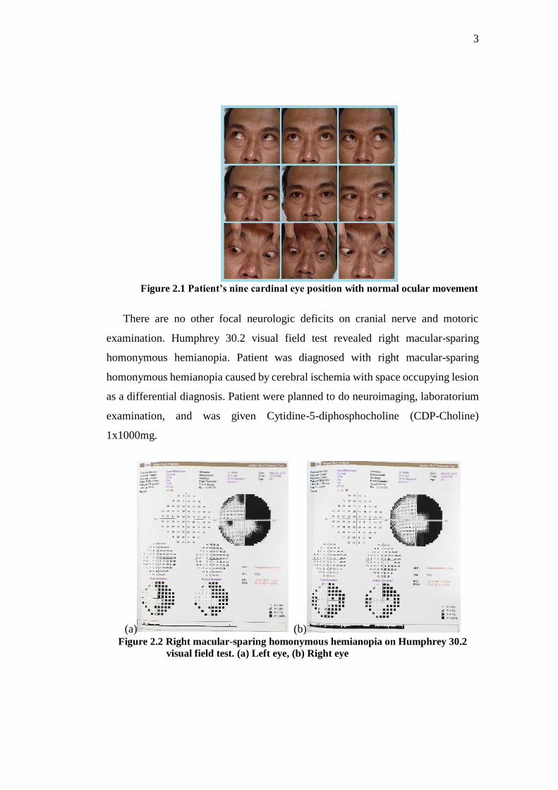
smoking for 30 years, 6 cigarettes a day. Ophthalmic examination showed visual
acuity of 0.4 ph 0.8 right eye and 0.4 ph 1.0 left eye with snellen chart. Intraocular
pressure of both eyes within normal limits. Primary eye position was orthotropia
with normal ocular movement. Direct and consensual light reflex were within
normal limits at both eyes with no relative afferent pupillary defect (RAPD).
Posterior segment examination showed round optic disc, defined margin, and
normal cup-disk ratio at both eyes. Amsler grid, colour vision and contrast
sensitivity was within normal limits.
3
Figure 2.1 Patient’s nine cardinal eye position with normal ocular movement
There are no other focal neurologic deficits on cranial nerve and motoric
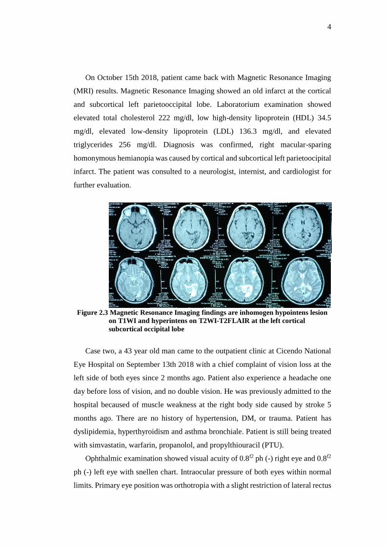
examination. Humphrey 30.2 visual field test revealed right macular-sparing
homonymous hemianopia. Patient was diagnosed with right macular-sparing
homonymous hemianopia caused by cerebral ischemia with space occupying lesion
as a differential diagnosis. Patient were planned to do neuroimaging, laboratorium
examination, and was given Cytidine-5-diphosphocholine (CDP-Choline)
1x1000mg.
(a) (b) Figure 2.2 Right macular-sparing homonymous hemianopia on Humphrey 30.2
visual field test. (a) Left eye, (b) Right eye
4
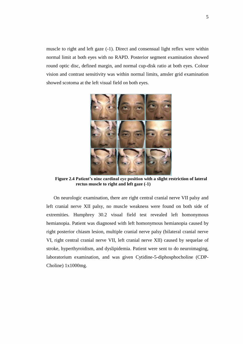
On October 15th 2018, patient came back with Magnetic Resonance Imaging
(MRI) results. Magnetic Resonance Imaging showed an old infarct at the cortical
and subcortical left parietooccipital lobe. Laboratorium examination showed
elevated total cholesterol 222 mg/dl, low high-density lipoprotein (HDL) 34.5
mg/dl, elevated low-density lipoprotein (LDL) 136.3 mg/dl, and elevated
triglycerides 256 mg/dl. Diagnosis was confirmed, right macular-sparing
homonymous hemianopia was caused by cortical and subcortical left parietoocipital
infarct. The patient was consulted to a neurologist, internist, and cardiologist for
further evaluation.
Figure 2.3 Magnetic Resonance Imaging findings are inhomogen hypointens lesion
on T1WI and hyperintens on T2WI-T2FLAIR at the left cortical
subcortical occipital lobe
Case two, a 43 year old man came to the outpatient clinic at Cicendo National
Eye Hospital on September 13th 2018 with a chief complaint of vision loss at the
left side of both eyes since 2 months ago. Patient also experience a headache one
day before loss of vision, and no double vision. He was previously admitted to the
hospital becaused of muscle weakness at the right body side caused by stroke 5
months ago. There are no history of hypertension, DM, or trauma. Patient has
dyslipidemia, hyperthyroidism and asthma bronchiale. Patient is still being treated
with simvastatin, warfarin, propanolol, and propylthiouracil (PTU).
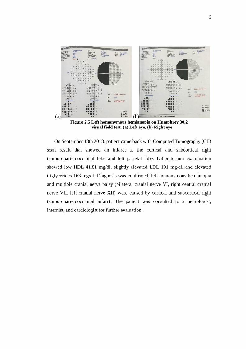
Ophthalmic examination showed visual acuity of 0.8f2 ph (-) right eye and 0.8f2
ph (-) left eye with snellen chart. Intraocular pressure of both eyes within normal
limits. Primary eye position was orthotropia with a slight restriction of lateral rectus
5
muscle to right and left gaze (-1). Direct and consensual light reflex were within
normal limit at both eyes with no RAPD. Posterior segment examination showed
round optic disc, defined margin, and normal cup-disk ratio at both eyes. Colour
vision and contrast sensitivity was within normal limits, amsler grid examination
showed scotoma at the left visual field on both eyes.
Figure 2.4 Patient’s nine cardinal eye position with a slight restriction of lateral
rectus muscle to right and left gaze (-1)
On neurologic examination, there are right central cranial nerve VII palsy and
left cranial nerve XII palsy, no muscle weakness were found on both side of
extremities. Humphrey 30.2 visual field test revealed left homonymous
hemianopia. Patient was diagnosed with left homonymous hemianopia caused by
right posterior chiasm lesion, multiple cranial nerve palsy (bilateral cranial nerve
VI, right central cranial nerve VII, left cranial nerve XII) caused by sequelae of
stroke, hyperthyroidism, and dyslipidemia. Patient were sent to do neuroimaging,
laboratorium examination, and was given Cytidine-5-diphosphocholine (CDP-
Choline) 1x1000mg.
6
(a) (b) Figure 2.5 Left homonymous hemianopia on Humphrey 30.2
visual field test. (a) Left eye, (b) Right eye
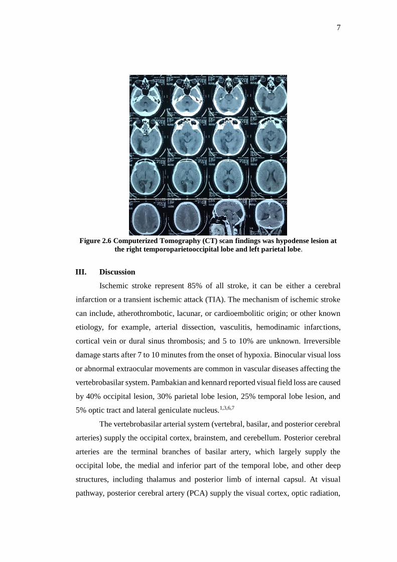
On September 18th 2018, patient came back with Computed Tomography (CT)
scan result that showed an infarct at the cortical and subcortical right
temporoparietooccipital lobe and left parietal lobe. Laboratorium examination
showed low HDL 41.81 mg/dl, slightly elevated LDL 101 mg/dl, and elevated
triglycerides 163 mg/dl. Diagnosis was confirmed, left homonymous hemianopia
and multiple cranial nerve palsy (bilateral cranial nerve VI, right central cranial
nerve VII, left cranial nerve XII) were caused by cortical and subcortical right
temporoparietooccipital infarct. The patient was consulted to a neurologist,
internist, and cardiologist for further evaluation.
7
Figure 2.6 Computerized Tomography (CT) scan findings was hypodense lesion at
the right temporoparietooccipital lobe and left parietal lobe.
III. Discussion
Ischemic stroke represent 85% of all stroke, it can be either a cerebral
infarction or a transient ischemic attack (TIA). The mechanism of ischemic stroke
can include, atherothrombotic, lacunar, or cardioembolitic origin; or other known
etiology, for example, arterial dissection, vasculitis, hemodinamic infarctions,
cortical vein or dural sinus thrombosis; and 5 to 10% are unknown. Irreversible
damage starts after 7 to 10 minutes from the onset of hypoxia. Binocular visual loss
or abnormal extraocular movements are common in vascular diseases affecting the
vertebrobasilar system. Pambakian and kennard reported visual field loss are caused
by 40% occipital lesion, 30% parietal lobe lesion, 25% temporal lobe lesion, and
5% optic tract and lateral geniculate nucleus.1,3,6,7
The vertebrobasilar arterial system (vertebral, basilar, and posterior cerebral
arteries) supply the occipital cortex, brainstem, and cerebellum. Posterior cerebral
arteries are the terminal branches of basilar artery, which largely supply the
occipital lobe, the medial and inferior part of the temporal lobe, and other deep
structures, including thalamus and posterior limb of internal capsul. At visual
pathway, posterior cerebral artery (PCA) supply the visual cortex, optic radiation,
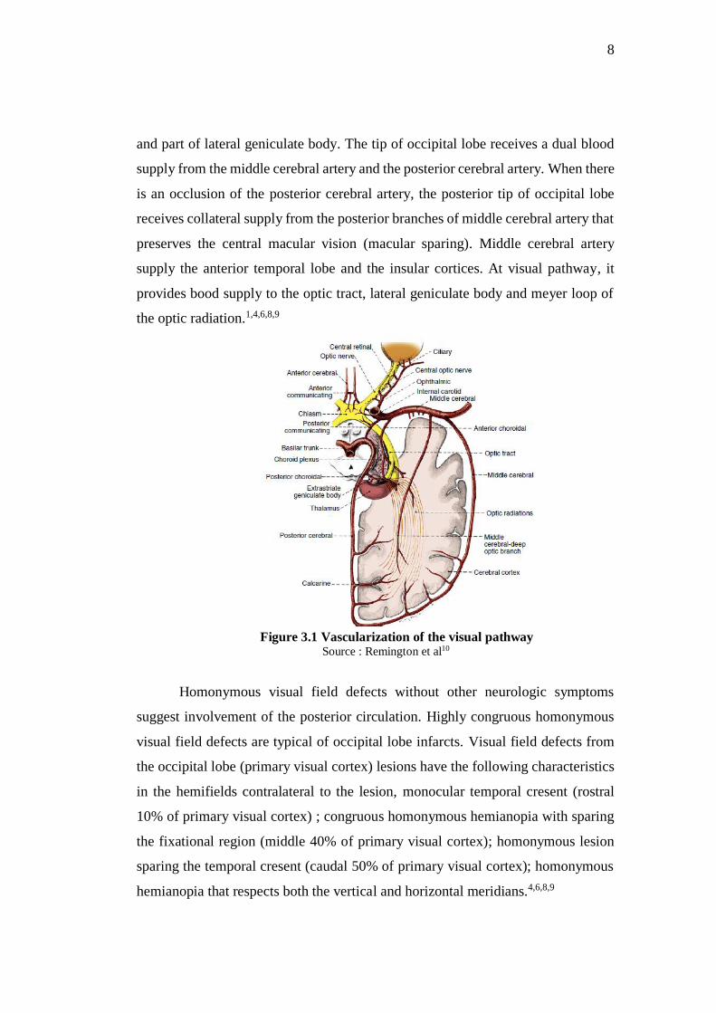
8
and part of lateral geniculate body. The tip of occipital lobe receives a dual blood
supply from the middle cerebral artery and the posterior cerebral artery. When there
is an occlusion of the posterior cerebral artery, the posterior tip of occipital lobe
receives collateral supply from the posterior branches of middle cerebral artery that
preserves the central macular vision (macular sparing). Middle cerebral artery
supply the anterior temporal lobe and the insular cortices. At visual pathway, it
provides bood supply to the optic tract, lateral geniculate body and meyer loop of
the optic radiation.1,4,6,8,9
Figure 3.1 Vascularization of the visual pathway
Source : Remington et al10
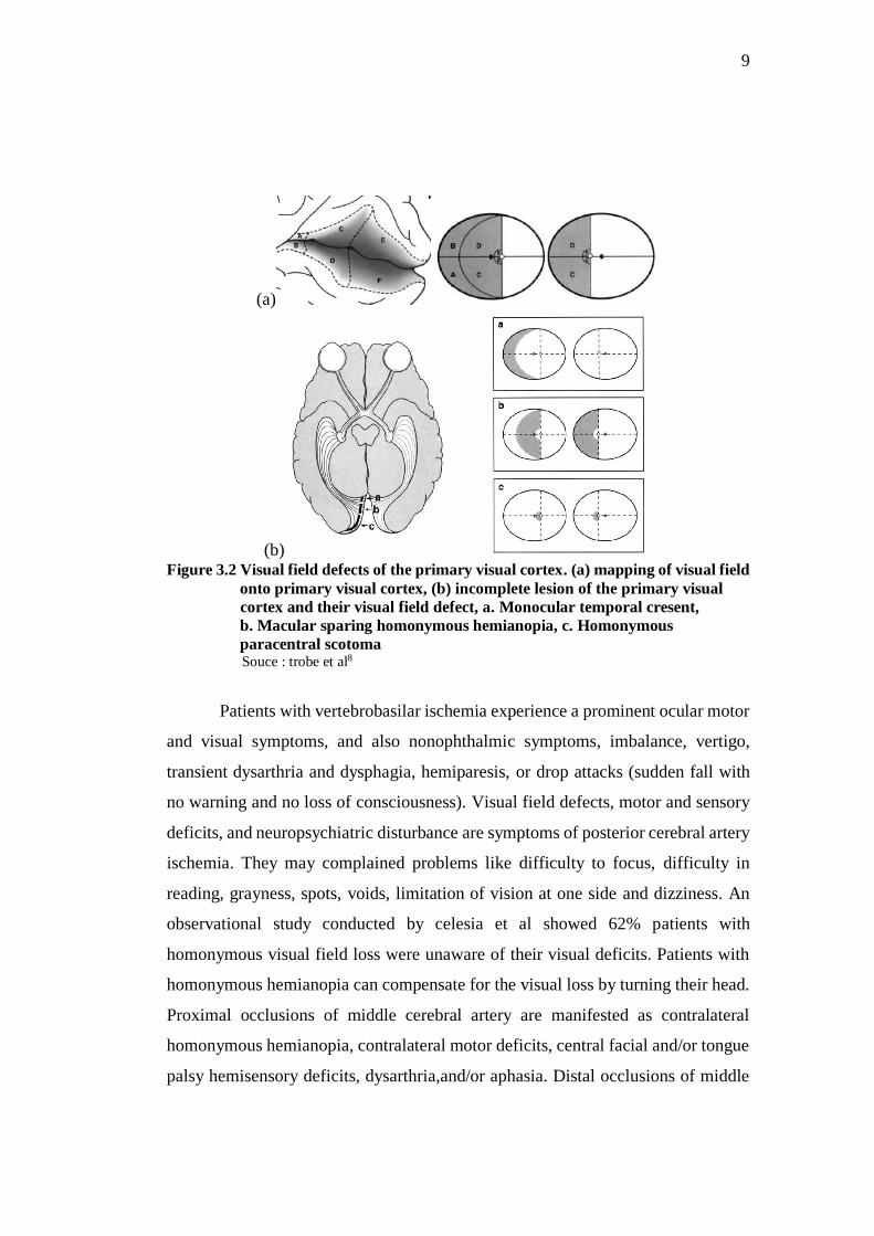
Homonymous visual field defects without other neurologic symptoms
suggest involvement of the posterior circulation. Highly congruous homonymous
visual field defects are typical of occipital lobe infarcts. Visual field defects from
the occipital lobe (primary visual cortex) lesions have the following characteristics
in the hemifields contralateral to the lesion, monocular temporal cresent (rostral
10% of primary visual cortex) ; congruous homonymous hemianopia with sparing
the fixational region (middle 40% of primary visual cortex); homonymous lesion
sparing the temporal cresent (caudal 50% of primary visual cortex); homonymous
hemianopia that respects both the vertical and horizontal meridians.4,6,8,9
9
(a)
(b) Figure 3.2 Visual field defects of the primary visual cortex. (a) mapping of visual field
onto primary visual cortex, (b) incomplete lesion of the primary visual
cortex and their visual field defect, a. Monocular temporal cresent,
b. Macular sparing homonymous hemianopia, c. Homonymous
paracentral scotoma Souce : trobe et al8
Patients with vertebrobasilar ischemia experience a prominent ocular motor
and visual symptoms, and also nonophthalmic symptoms, imbalance, vertigo,
transient dysarthria and dysphagia, hemiparesis, or drop attacks (sudden fall with
no warning and no loss of consciousness). Visual field defects, motor and sensory
deficits, and neuropsychiatric disturbance are symptoms of posterior cerebral artery
ischemia. They may complained problems like difficulty to focus, difficulty in
reading, grayness, spots, voids, limitation of vision at one side and dizziness. An
observational study conducted by celesia et al showed 62% patients with
homonymous visual field loss were unaware of their visual deficits. Patients with
homonymous hemianopia can compensate for the visual loss by turning their head.
Proximal occlusions of middle cerebral artery are manifested as contralateral
homonymous hemianopia, contralateral motor deficits, central facial and/or tongue
palsy hemisensory deficits, dysarthria,and/or aphasia. Distal occlusions of middle
10
cerebral artery usually manifest with less severe neurologic deficits than proximal
occlusions, depens on the affected distal middle cerebral artery territory.1,5,6,11
Diplopia is the most common complaint for ocular motor disturbances
caused by vertebrobasilar insufficiency. Cranial nerve palsies post stroke can
affects the third, fourth, and sixth nerves. Diplopia can be a consequence of
horizontal or vertical ocular misalignment, either from third, fourth, or sixth cranial
nerve palsy. Patient can also experience perceptual problems, the most recognised
is visual inattention, where the individual does not respond to visual stimuli on the
affected side. Other perceptual problems are agnosia, visual hallucinations, and
image movement problems.1,4–6
Case one, patient complained right side vision loss at the right eye especially
when looking to the right. Visual acuity 0.4 ph 0.8 right eye and 0.4 ph 1.0 left eye,
no RAPD or posterior segment abnormalities. After visual field examination
revealed right macular-sparing homonymous hemianopia. This explained why the
patient only complained vision loss at right eye, because macular function is still
intact. This type of visual field defect can be caused by an occipital lobe lesion.
Case two, patient complained vision loss at the left side of both eyes, headache,
history of stroke and dyslipidemia with visual acuity 0.8f2 ph (-) right eye and 0.8f2
ph (-) left eye, bilateral cranial nerve VI palsy, no RAPD, no posterior segment
abnormalities, amsler grid examination showed scotoma at the left visual field on
both eyes, right central cranial nerve VII and left cranial nerve XII palsy. These
symptomps suggests visual field defect caused by a posterior chiasm lesion.
Homonymous visual field defect can be caused by stroke, tumor, trauma,
arteriovenous malformation, neurosurgery, demyelination, infection, and migrane.
Both patients are older, with rapid onset of visual field loss, this suggest a vascular
etiology.1,3
Most common location for homonymous visual field defects are occipital
lobe (45%), optic radiation (32%), lateral geniculate body (1.3%), and multiple
visual pathway segments (11%). Neuroimaing should be performed on all patients
with homonymous visual field defects. Magnetic resonance imaging or CT scan can
detect cerebral infarction or bleeding, but arterial neuroimaging is usually needed
11
to evaluate the posterior circulation. Magnetic resonance angiography (MRA) and
computed tomography angiography (CTA) are the best noninvasive methods and
should be performed immediately. Underlying systemic disorders, including
hypercholesterolemia, hypertension, diabetes mellitus, and postural hypotension
should be evaluated. Echocardiography should be performed to detect
cardioembolic origin for ischemic stroke.1,6
Case one has MRI showed an old infarct at the cortical and subcortical left
parietooccipital lobe and laboratorium examination showed dyslipidemia. Case two
has CT scan showed an infarct at the cortical and subcortical right
temporoparietooccipital lobe and left parietal lobe, laboratorium examination also
showed dyslipidemia. Case two had more extensive area of infarction and multiple
location, this explains the different clinical manifestation where case two patient
had worst sign and symptoms. Both patients have vascular etiology that causes
cerebral infarction, one of the risk factor that exist in both patients is dyslipidemia.
Case one also had a history of severe hypotension and smoking, and case two had
a history of stroke, hyperthyroidsm, and asthma. Case one is consulted to a
neurologist and internist for further evaluation and secondary prevention, case two
has received treatment from previous stroke and then consulted to the cardiologist
to do an echocardiography.
Treatment of acute ischemic stroke is reopening the occluded artery using
recombinant tissue plasminogen activator and endovascular treatment.
Homonymous hemianopia caused by occlusion of the posterior cerebral artery is an
indication for intravenous thrombolysis within 4.5 hours time window from
symptom onset. If ischemia stroke of posterior cerebral artery is present, searching
for etiology and secondary prevention must be initiated to prevent bilateral
hemianopia. Secondary prevention is to reduced the risk of early stroke and late
stroke recurrence by including antiplatelet therapy or anticoagulantion, statin use,
blood pressure control, and periodic monitoring. Persistent visual field defect
require rehabilitation. Homonymous hemianopia leads to spatial dysorientation,
reading problem and reduced mobility. Spontaneous adaptive mechanism may not
12
be sufficient enough, therefore rehabilitation is required to enlarge the functional
field view.1,5,6,12
Prognosis for case one is ad bonam for quo ad vitam and dubia ad bonam
for quo ad functionam, for case two is ad bonam for quo ad vitam and dubia ad
malam for quo ad functionam. Infarcts in the territory of the posterior cerebral artery
are typically embolic in origin affecting the entire supply area of posterior cerebral
artery. Both patient had infarction of the occipital lobe on neuroimaging, but case
two had more extensive area of infarction and multiple location. Macular function
is still intact for case one. A study by Hepworth et all shows the percentage of
patients having complete recovery from visual field loss ranges from 0 to 44%, and
for partial recovery up to 72.2%. Visual field defect seriously impact patient’s
functional ability and quality of life following stroke.5,7,12,13
IV. Conclusion
A sudden onset homonymous visual field defects is the hallmark of vascular
lesion in the ooccipital lobe supplied by the posterior cerebral artery. Types of
occipital lobe visual field defect depends on the affected area supplied by the
posterior and middle cerebral artery. Homonymous hemianopia is the most frequent
visual field defect, affecting up to 75% of patients with posterior cerebral artery
stroke. Neuroimaging should be performed on all patients with homonymous visual
field defects. Magnetic resonance angiography (MRA) and CTA should be
performed immediately. Thrombolysis should not be delay to reopen the occluded
artery if no contraindication is found. Searching for etiology and secondary
prevention is also important to prevent reccurance of stroke.
13
References
1. Volný O, Haršány M, Mikulík R. Ischemic Stroke and Homonymous Visual
Field Defects. In: Homonymous Hemianopia Visual Field Defects. Springer
International Publishing AG; 2017. 31–45.
2. Simone Vidale, Giuseppe D’Aliberti, Luca Valvassori. Brief Description of
Recent Developments in Diagnosis and Treatment. In: Ischemic Stroke.
Switzerland: Springer International Publishing; 2017. 1–2.
3. V. Biousse, N.J. Newman. Stroke and Eye Findings. In: Primer on
Cerebrovascular Diseases. 2nd ed. Elsevier Inc; 2017. 427–33.
4. Pula JH, Yuen CA. Eyes and stroke: the visual aspects of cerebrovascular
disease. Stroke and vascular neurology. 2017 Jul 6:svn-2017.
5. Hepworth L, Rowe F, Walker M, Rockliffe J, Noonan C, Howard C, Currie J.
Post-stroke visual impairment: a systematic literature review of types and
recovery of visual conditions. Ophthalmology Research: An International
Journal. 2015 Nov 16;5(1):1-43.
6. American Academy of Ophthalmology. Selected Systemic Conditions With
Neuro-Ophthalmic Signs. In: Basic and Clinical Science Course : Neuro-
Ophthalmology. American Academy of Ophthalmology; 2016.
7. Rowe FJ, Wright D, Brand D, Jackson C, Harrison S, Maan T, Scott C,
Vogwell L, Peel S, Akerman N, Dodridge C. A prospective profile of visual
field loss following stroke: prevalence, type, rehabilitation, and outcome.
BioMed research international. 2013;2013.
8. Jonathan D. Trobe, M.D. The Neurology of Vision. Oxford University Press,
Inc.; 2001. 23–28.
9. Brad Bowling. Kanski’s Clinical Ophthalmology : A systemic approach. 8th
ed. Australian: Elsevier Limited; 2016. 821.
10. Lee Ann Remington. Clinical Anatomy and Physiology of the Visual System.
3rd ed. United States of America: Elsevier Inc; 2012. 240.
11. Caplan, Louis R. Caplan’s Stroke: A Clinical Approach. Elsevier Inc; 2009.
630–631, 278–282.
12. Crotty M, van den Berg M, Hayes A, Chen C, Lange K, George S. Hemianopia
after stroke: A randomised controlled trial of the effectivenessof a
standardised versus an individualised rehabilitation program, on scanning
ability whilst walking. NeuroRehabilitation. 2018 Jan 1(Preprint):1-9.
13. Seitz RJ, Donnan GA. Recovery potential after acute stroke. Frontiers in
neurology. 2015 Nov 11;6:238.