Embed Size (px)
Citation preview
DENTAL PLAQUE

• Most important chapter
• Understanding required
• Health maintenance
• Disease progression
OBJECTIVES
• DEFINITION
• Difference between plaque, calculus , materia alba
• Types of plaque
• Composition of plaque
• Formation of plaque
Lecture 2
• Microscopic structure of plaque
• Concept of biofilm
• Plaque Hypothesis
• Microbial complexes
• Microbial tests for plaque sample

Definition
• Dental Plaque
“is a specific but highly variable structural entity, resulting from sequential colonization of microorganisms on tooth surfaces, restorations & other parts of oral cavity, composed of salivary components like mucin, desquamated epithelial cells, debris & microorganisms, all embedded in extracellular gelatinous matrix.”
W.H.O ,1961

• Dental Plaque can be defined as:
“ soft deposits that form the biofilm adhering to the tooth surface or other hard surfaces in the oral cavity, including removable & fixed restorations”
Bowen , 1976

DIFFERENTIATION:

• Materia Alba
“soft accumulations of bacteria and tissue cells; lack organised structure of dental plaque; easily displaced by water spray”
• Dental Calculus
‘mineralised dental plaque”

DETECTION OF DENTAL PLAQUE
DETECTION
• Visual
• Periodontal Probe or Explorer
• Disclosing Agents

Timeline of plaque development:
• At birth: sterile
• Hours: facultative aerobic bacteria
• Second day: anaerobic bacteria
• 2 weeks: mature microbiota
• Weaning (> 2 years): 400 different types of bacteria
• Body contains 10 times more bacteria than human cells

• Open growth system: communication with the pharynx
• > 500 species in mouth
• In any individual: 150 or more species at any given time.
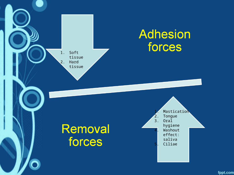
1. Mastication2. Tongue3. Oral hygiene4. Washout
effect: saliva5. Ciliae
1. Soft tissue2. Hard tissue

Niches of plaque accumulation:
• Supragingival and hard surgfaces: teeth, implant, restorations.
• Periodontal pocket (hard: root cementum & soft: pocket epithelium)
• Buccal, palatal and floor of the mouth epithelium
• Dorsum of the tongue
• Tonsils

Natural cleansing mechanism:
• Gingival crevicular fluid & salivary flow
• Cleansing effect of mastication and tongue movement
• Rapid turnover rate of intraoral epithelial cells
• Host defence mechanisms like langerhans cells.
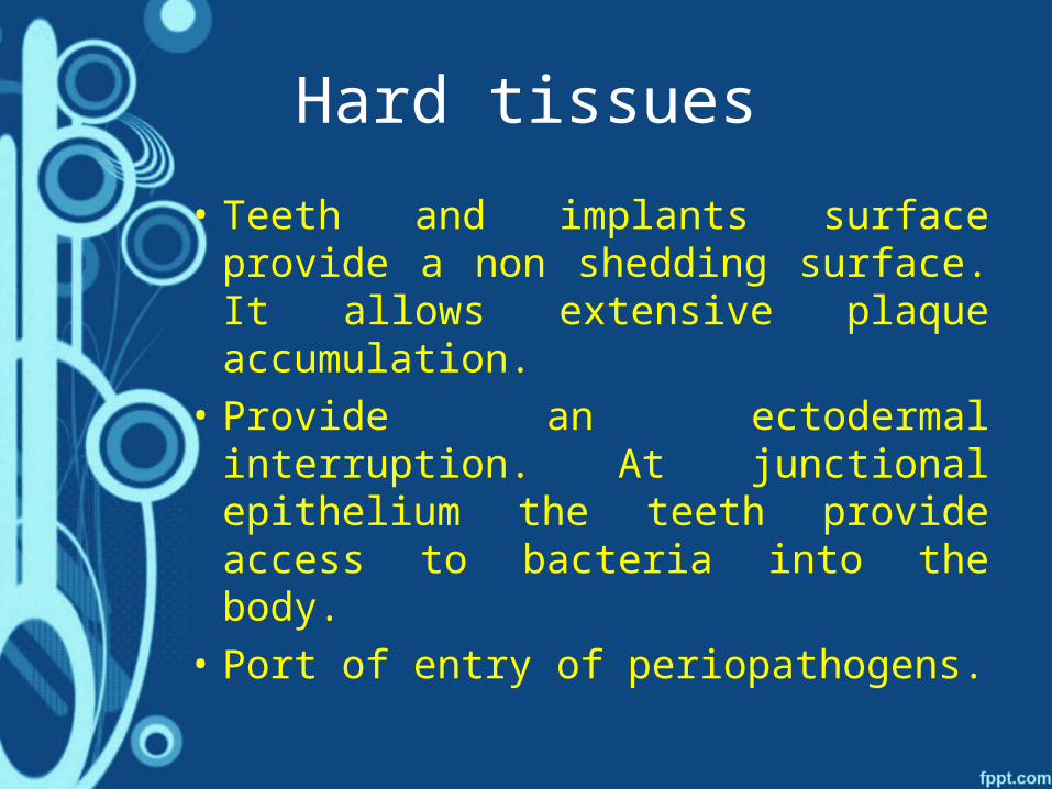
Hard tissues
• Teeth and implants surface provide a non shedding surface. It allows extensive plaque accumulation.
• Provide an ectodermal interruption. At junctional epithelium the teeth provide access to bacteria into the body.
• Port of entry of periopathogens.

Macroscopic Structure
• Supragingival Plaque
• Subgingival Plaque
• Marginal Plaque

Composition of Plaque
• Microorganisms
• Intercellular Matrix
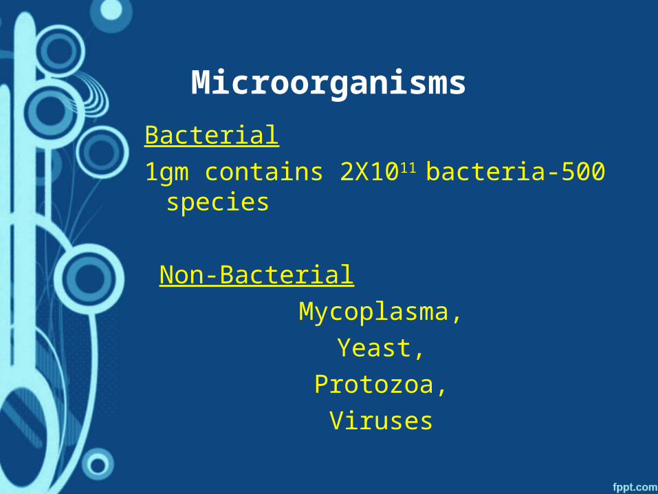
Microorganisms
Bacterial
1gm contains 2X10¹¹ bacteria-500 species
Non-Bacterial
Mycoplasma,
Yeast,
Protozoa,
Viruses
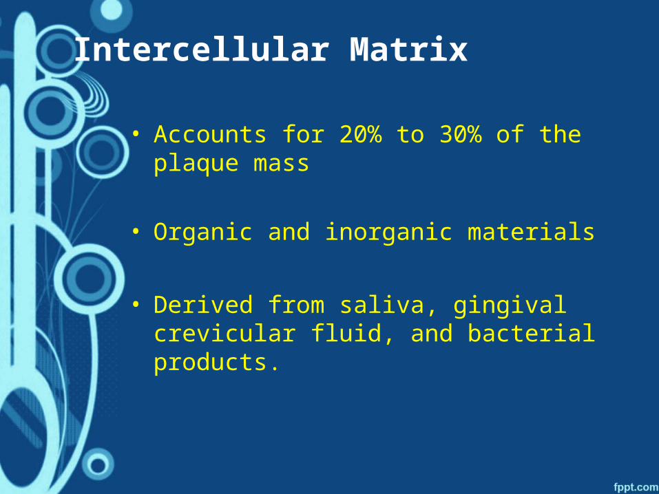
Intercellular Matrix
• Accounts for 20% to 30% of the plaque mass
• Organic and inorganic materials
• Derived from saliva, gingival crevicular fluid, and bacterial products.
Organic Component
• Organic constituents : polysaccharides, proteins, glycoproteins, and lipid material
• Polysaccharides produced by bacteria-Dextran: predominant form
• Albumin: originating from crevicular fluid
• Lipid material: debris from the membranes of disrupted bacterial and host cells and possibly food debris.
Inorganic Component
• Calcium and phosphorus (Most)• Trace amounts: sodium, potassium
and fluoride.• Source -supragingival plaque
(saliva) & subgingival plaque (GCF & Blood)
• Calculus frequently found in areas of the dentition adjacent to salivary ducts

PHASES OF
PLAQUE FORMATION

1. Formation of dental Pellicle
2. Initial Colonization
3. Secondary Colonization & Plaque Maturation
Formation of dental Pellicle
• Acquired enamel pellicle forms rapidly - Early pellicle
• Characterized by an absence of bacteria and their products.
• Composed of proteins and glycoproteins.
• Demonstrate a higher content of threonine, serine, and alanine & less proline than saliva- selective adsorption
• Electron microscopic -reveals a thin, amorphous, electron-dense layer immediately adjacent to the hard surface- thickness from 1 to 2 nm.
• involves a combination of physical forces (ionic, hydrophobic, hydrogen bonding, and van der Waals)
• May involve the interaction of phosphate groups with calcium ions in saliva to form "bridges"
• Protective functions of early enamel pellicle: protection , lubrication by decreasing frictional forces, may selectively concentrate antimicrobial substances such as immunoglobulins, lysozyme, and cystatins at different oral surfaces.
• Formation of later pellicle most likely involves protein-protein or protein-carbohydrate interactions,- stereospecific in nature .
• For example, A. viscosus and Streptococcus mitis produce a neuraminidase that cleaves terminal sialic acid residues on the glycoproteins in saliva or early pellicles to expose galactose residues (Costello et aI., 1979)
• Collectively, these mechanisms may be important for the initial colonization
Initial adhesion
Phase I : Transport to the surface
Phase II : Initial adhesion
Phase III : Attachment
Phase IV : Colonisation of the surface and biofilm formation

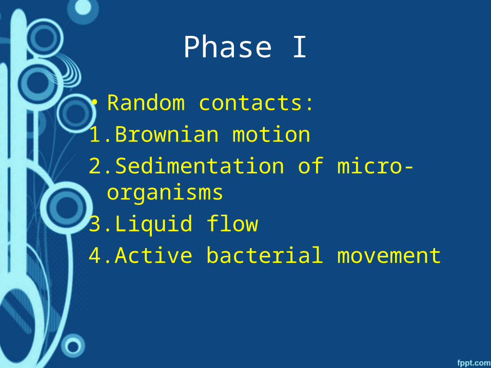
Phase I
• Random contacts:
1.Brownian motion
2.Sedimentation of micro- organisms
3.Liquid flow
4.Active bacterial movement

Phase II
• Initial reversible adhesion
• Long range and short range forces:
1.van der Walls attractive forces
2.Electrostatic repulsive forces

Phase III
• Firm anchorage
• Specific interactions:
1.Covalent
2.Ionic
3.Hydrogen bonding


• Adhesions: specific extracellular proteinacious components on microorganisms.
• Complimentary receptors: proteins, glycoproteins or polysaccharides on the pellicle surface.

• Example:
• S. Sanguis- binds to acidic proline rich proteins, alpha amylase & sialic acid
• A. viscosus- fimbriae that contain adhesins- bind to proline rich proteins of dental pellicle.

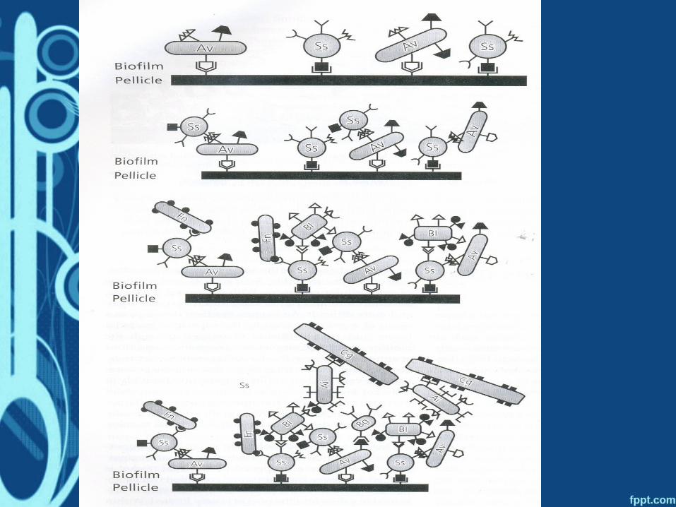
Colonisation & Plaque maturation
• Initial colonisers attach to the tooth surface- provide substrate for secondary colonisers to attach.
• They create a favourable environment for secondary colonisers to survive.
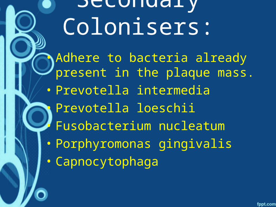
Secondary Colonisers:
• Adhere to bacteria already present in the plaque mass.
• Prevotella intermedia
• Prevotella loeschii
• Fusobacterium nucleatum
• Porphyromonas gingivalis
• Capnocytophaga
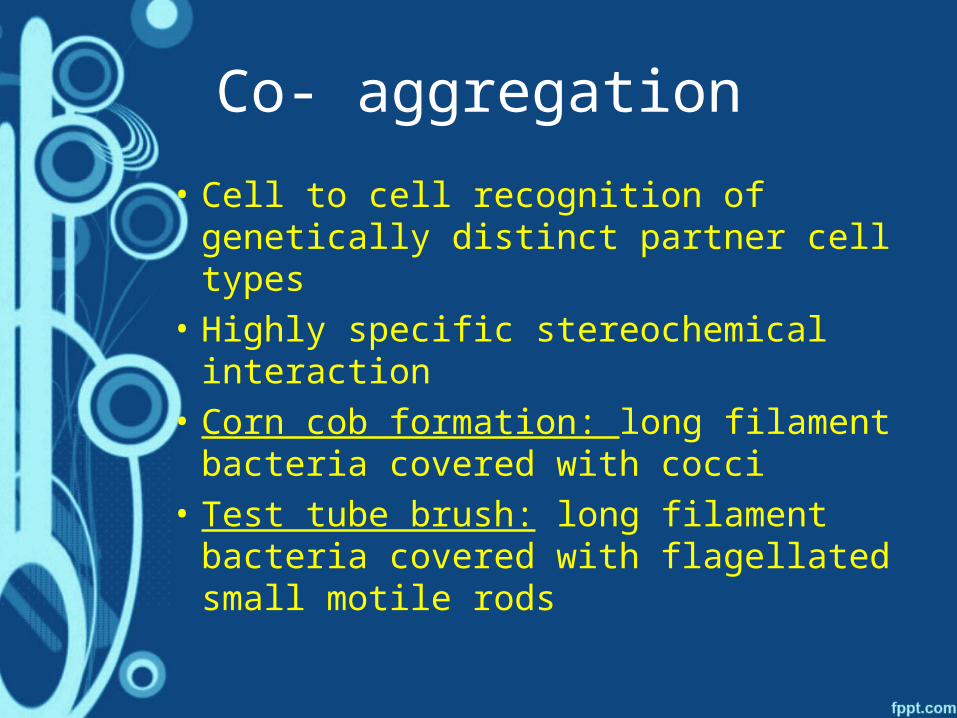
Co- aggregation
• Cell to cell recognition of genetically distinct partner cell types
• Highly specific stereochemical interaction
• Corn cob formation: long filament bacteria covered with cocci
• Test tube brush: long filament bacteria covered with flagellated small motile rods

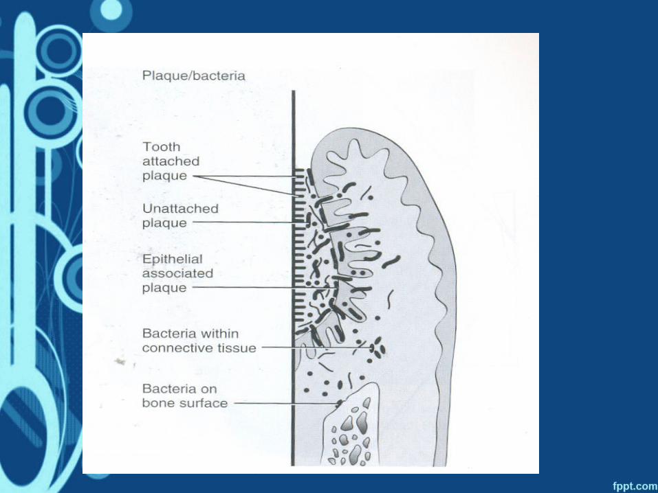
MICROSCOPIC STRUCTURE

MICROSCOPIC STRUCTURE
SUPRAGINGIVAL PLAQUE
• typically demonstrates a stratified organization of the bacterial morphotypes.
• Gram-positive cocci and short rods predominate at the tooth surface
• gram-negative rods and filaments ,spirochetes predominate in the outer surface of the mature plaque mass.
• Highly specific cell-to-cell interactions are also evident from the "corncob"

CORN-COB STRUCTURECORN COB STRUCTURE

Thin section of supragingival plaque
GRAM POSITIVE BACTERIA IN
PALISADING ARRANGEMENT
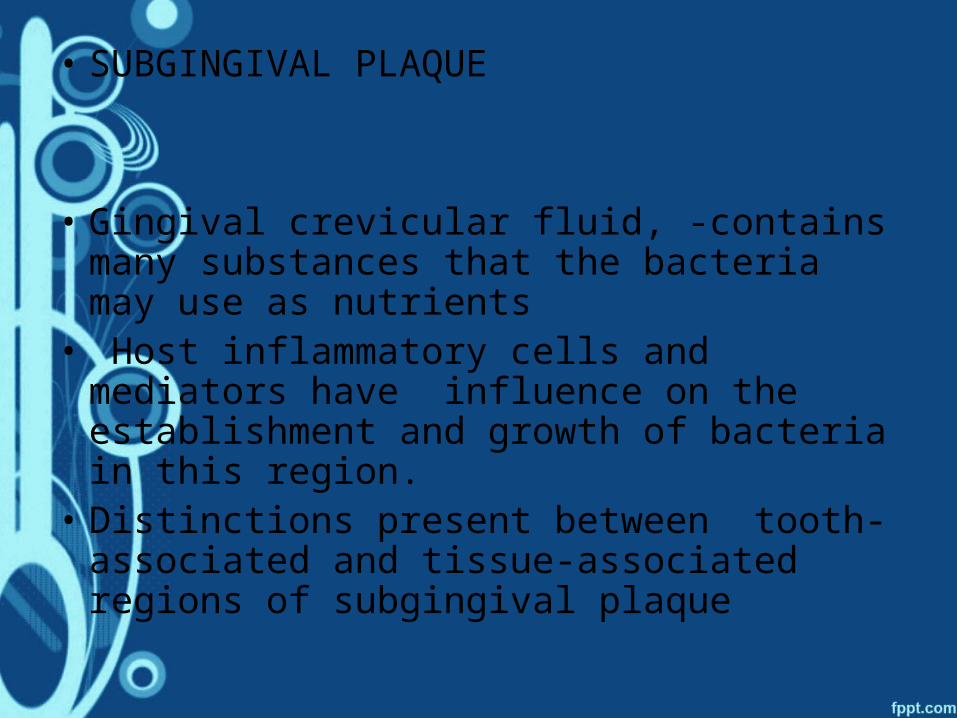
• SUBGINGIVAL PLAQUE
• Gingival crevicular fluid, -contains many substances that the bacteria may use as nutrients
• Host inflammatory cells and mediators have influence on the establishment and growth of bacteria in this region.
• Distinctions present between tooth-associated and tissue-associated regions of subgingival plaque

Thin section of plaque in a deep pocket
FILAMENTS
RODS
COCCI

DENTAL PLAQUE AS BIOFILM
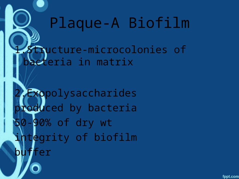
Plaque-A Biofilm
1.Structure-microcolonies of bacteria in matrix
2.Exopolysaccharides
produced by bacteria
50-90% of dry wt
integrity of biofilm
buffer
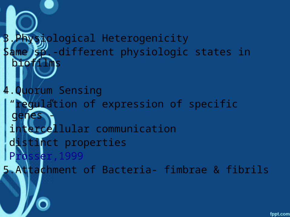
3.Physiological HeterogenicitySame sp.-different physiologic states in biofilms
4.Quorum Sensing “regulation of expression of specific genes”- intercellular communication distinct properties Prosser,19995.Attachment of Bacteria- fimbrae & fibrils
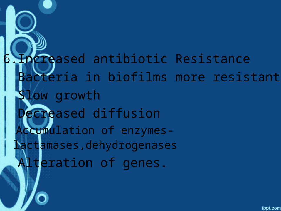
6.Increased antibiotic Resistance
Bacteria in biofilms more resistant
Slow growth
Decreased diffusion
Accumulation of enzymes- lactamases,dehydrogenases
Alteration of genes.
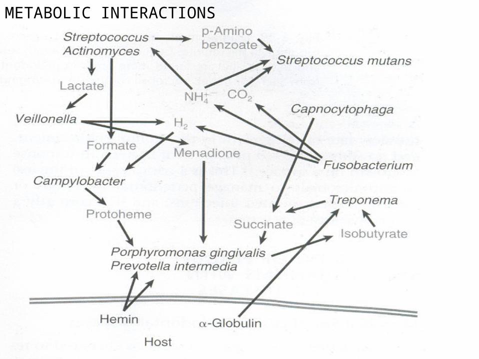
METABOLIC INTERACTIONS
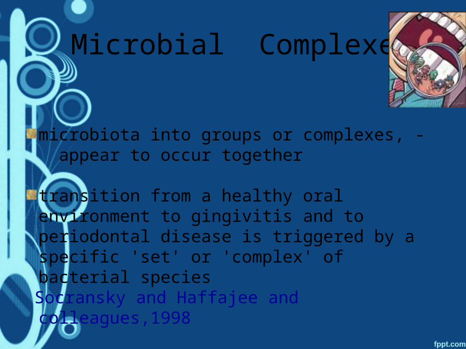
Microbial Complexes
microbiota into groups or complexes, - appear to occur together
transition from a healthy oral environment to gingivitis and to periodontal disease is triggered by a specific 'set' or 'complex' of bacterial species
Socransky and Haffajee and colleagues,1998
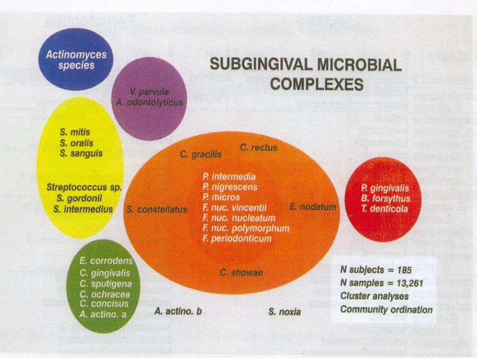
THE VARIOUS COMPLEXES
Porphyromonas gingivalis, Treponema denticola, and Tannerella forsythia: the 'red
complex‘Several characteristics make them prime candidates
as pathogens in the clinical destruction of periodontal tissues:
1. occur concomitantly with the clinical signs of periodontal destruction;
2.appear closely 'linked' topologically in the
developing biofilm;
3. in vitro studies demonstrate their ability to produce a number of outer membrane-associated proteinases
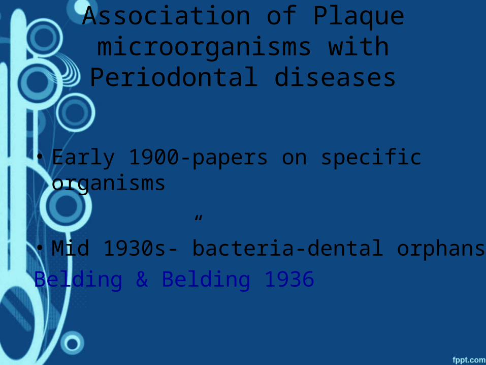
Association of Plaque microorganisms with Periodontal
diseases
• Early 1900-papers on specific organisms
• Mid 1930s-”bacteria-dental orphans”
Belding & Belding 1936
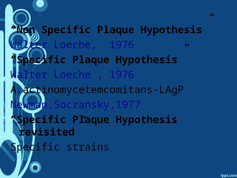
“Non Specific Plaque Hypothesis”
Walter Loeche, 1976
“Specific Plaque Hypothesis”
Walter Loeche , 1976
A.actinomycetemcomitans-LAgP
Newman,Socransky,1977
“Specific Plaque Hypothesis revisited”
Specific strains
MICROORGANISMS IN SPECIFIC PERIODONTAL DISEASES
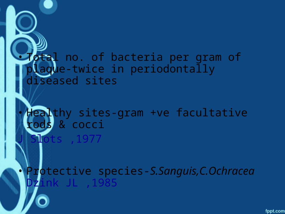
• Total no. of bacteria per gram of plaque-twice in periodontally diseased sites
• Healthy sites-gram +ve facultative rods & cocci
J Slots ,1977
• Protective species-S.Sanguis,C.Ochracea Dzink JL ,1985
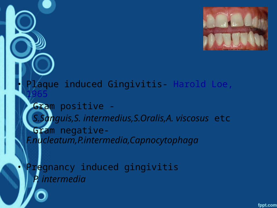
• Plaque induced Gingivitis- Harold Loe, 1965 Gram positive - S.Sanguis,S. intermedius,S.Oralis,A. viscosus etc Gram negative-
F.nucleatum,P.intermedia,Capnocytophaga
• Pregnancy induced gingivitis P. intermedia

• Chronic periodontitis
Spirochetes,
gingivalis,
B.forsythus,
A.actinomycetemcomitans
P. micros,
Treponema
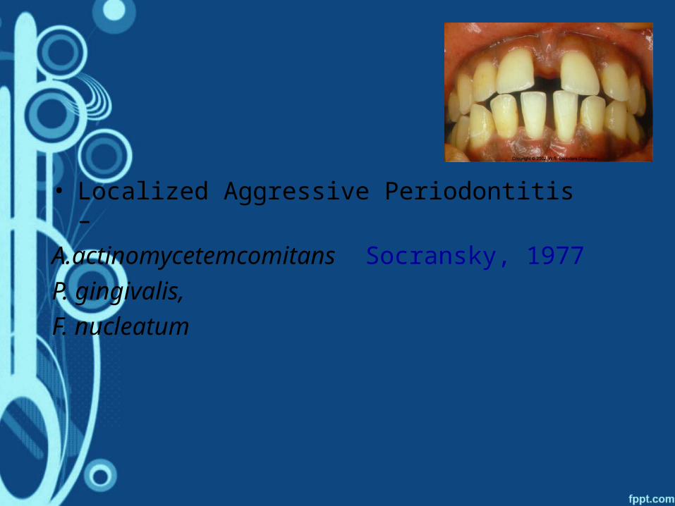
• Localized Aggressive Periodontitis –
A.actinomycetemcomitans Socransky, 1977
P. gingivalis,
F. nucleatum
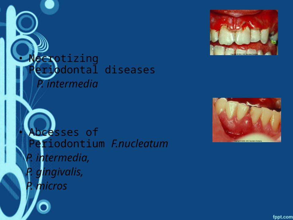
• Necrotizing Periodontal diseases
P. intermedia
• Abcesses of Periodontium F.nucleatum
P. intermedia, P. gingivalis, P. micros

CRITERIA for ASSOCIATION
of
PERIODONTAL PATHOGENS

KOCH’S POSTULATES
In I870s, Robert Koch’s criteria for causative agent in human infections.
1. Be routinely isolated from diseased individuals
2. Be grown in pure culture in the laboratory
3. Produce a similar disease when inoculated into susceptible laboratory animals
4. Be recovered from lesions in a diseased laboratory animal
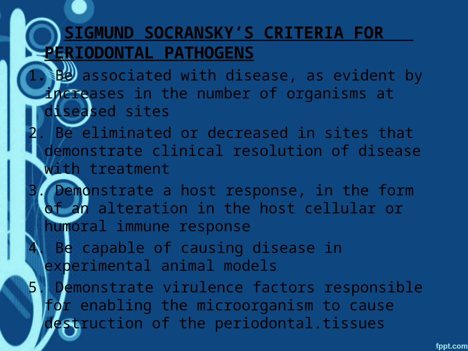
SIGMUND SOCRANSKY’S CRITERIA FOR PERIODONTAL PATHOGENS
1. Be associated with disease, as evident by increases in the number of organisms at diseased sites
2. Be eliminated or decreased in sites that demonstrate clinical resolution of disease with treatment
3. Demonstrate a host response, in the form of an alteration in the host cellular or humoral immune response
4. Be capable of causing disease in experimental animal models
5. Demonstrate virulence factors responsible for enabling the microorganism to cause destruction of the periodontal.tissues
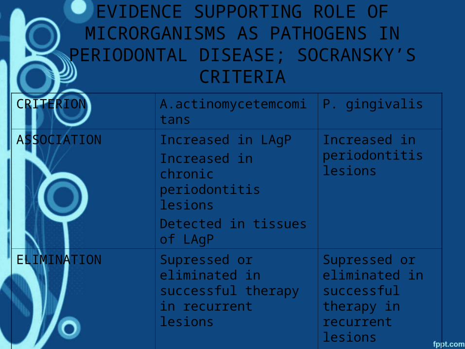
EVIDENCE SUPPORTING ROLE OF MICRORGANISMS AS PATHOGENS IN
PERIODONTAL DISEASE; SOCRANSKY’S CRITERIA
CRITERION A.actinomycetemcomitans P. gingivalis
ASSOCIATION Increased in LAgP
Increased in chronic periodontitis lesions
Detected in tissues of LAgP
Increased in periodontitis lesions
ELIMINATION Supressed or eliminated in successful therapy in recurrent lesions
Supressed or eliminated in successful therapy in recurrent lesions
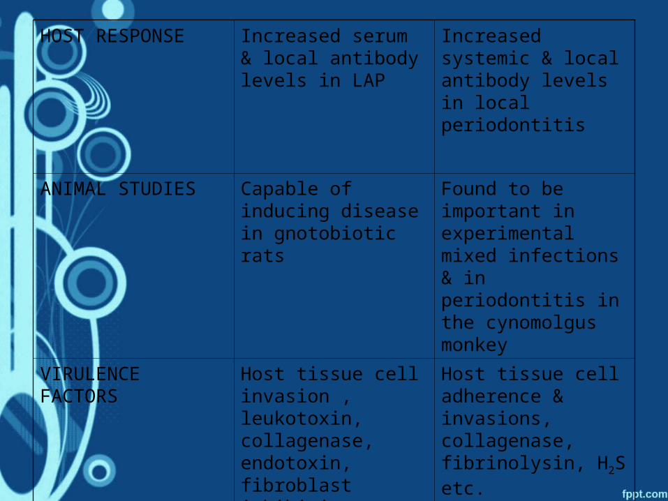
HOST RESPONSE Increased serum & local antibody levels in LAP
Increased systemic & local antibody levels in local periodontitis
ANIMAL STUDIES Capable of inducing disease in gnotobiotic rats
Found to be important in experimental mixed infections & in periodontitis in the cynomolgus monkey
VIRULENCE FACTORS
Host tissue cell invasion , leukotoxin, collagenase, endotoxin, fibroblast inhibiting factor etc.
Host tissue cell adherence & invasions, collagenase, fibrinolysin, H2S etc.

MICROBIOLOGICAL TESTS FOR PLAQUE SAMPLES
• Microscopic identification• Microbiological culturing• Enzymatic assays• Immunoassays• Nucleic acid probes• Polymerase chain reaction assays
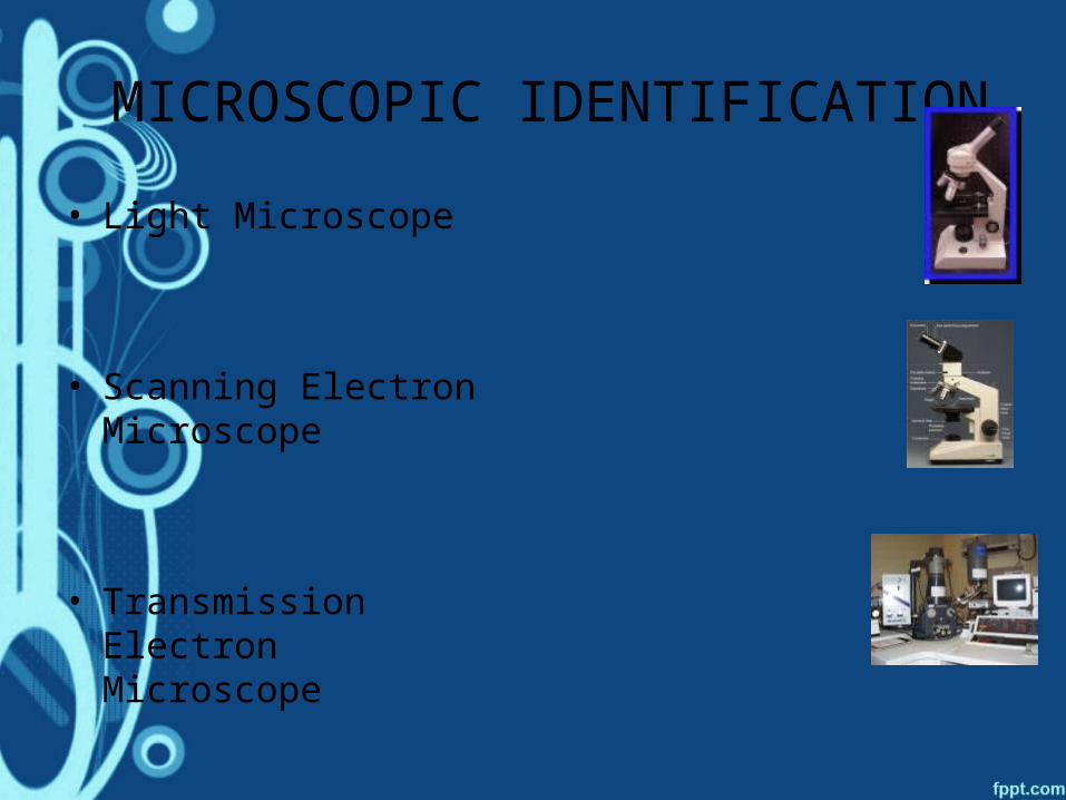
MICROSCOPIC IDENTIFICATION
• Light Microscope
• Scanning Electron Microscope
• Transmission Electron Microscope
MICROBIOLOGICAL CULTURING• Culture methods -the gold standard
• Available for the positive identification of many putative periodontopathogenic microorganisms
• Use of selective and non-selective media
• One unique advantage - permits the assessment of antibiotic sensitivity.
• Disadvantages- inability to detect low levels of microorganisms, high cost, labor intensiveness, prolonged time ,difficulty in growing several bacterial species
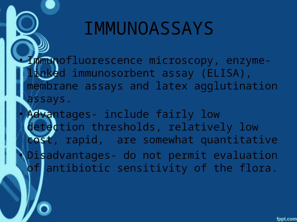
IMMUNOASSAYS
• Immunofluorescence microscopy, enzyme-linked immunosorbent assay (ELISA), membrane assays and latex agglutination assays.
• Advantages- include fairly low detection thresholds, relatively low cost, rapid, are somewhat quantitative
• Disadvantages- do not permit evaluation of antibiotic sensitivity of the flora.
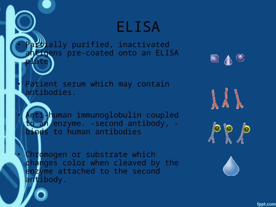
ELISA• Partially purified, inactivated antigens
pre-coated onto an ELISA plate
• Patient serum which may contain antibodies.
• Anti-human immunoglobulin coupled to an enzyme. -second antibody, -binds to human antibodies
• Chromogen or substrate which changes color when cleaved by the enzyme attached to the second antibody.
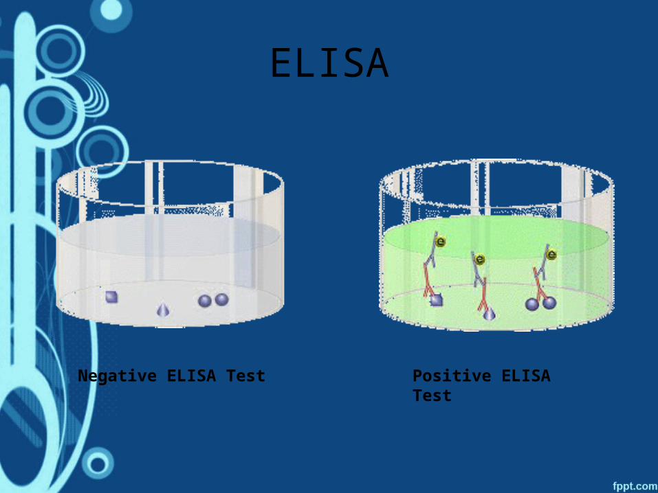
ELISA
Negative ELISA Test Positive ELISA Test

NUCLEIC ACID PROBES• Consist of nucleic acid sequences labeled with a
radioactive or enzymatic-colorimetric marker • Bind to complementary nucleic acid sequences on
corresponding microorganisms. • Newer-synthetic oligonucleotides (also known as
16s rRNA probes) • Oligonucleotide probes have greatest specificity
and lowest cross-reactivity as they target genes specific to a bacterial species.
• Advantages-greater sensitivity than culture methods,viability of microorganisms is not a requirement

POLYMERASE CHAIN REACTION
• A molecular biological technique for high-yield replication of DNA.
• Allows to synthesize vast numbers of copies of minute samples of DNA
• Modification of the original PCR technology, "real-time" PCR, permits detection of specific microorganisms & also its quantification
• PCR assays, used in combination with synthesized 16S rRNA probes -enable the detection of virtually any microorganism in a plaque sample

ENZYMATIC ASSAYS
• Enzymatic assay - detects bacteria that possess trypsin-like enzymes such as T. Forsythensis, treponema denticola and P. Gingivalis.
• Plaque sample containing any combination of these three bacteria- placed on a paper strip impregnated with a colorless substrate n-benzoyl-dl-arginine-2-naphthylamide (BANA)
• BANA substrate breakdown produces a blue-black color
• Disavantages-test unable to distinguish between relative proportions of the three bacteria,cannot identify the presence of other oral microorganisms,.