Embed Size (px)
Citation preview
779
Management of myofascial paindysfunction syndrome
Maj Priyanka Prakash1, Col Rath SK 2, Lt Col Mukherjee M3
Division of Periodontology,Department of Dental Surgery,AFMC, Pune.
Email for correspondence:[email protected]
Introduction
Myofascial pain dysfunction syndrome (MPDS)
is a painful condition in which distinct trigger point
areas, generally within muscles or fasciae, become
abnormally active and produce local and referred
pain. The traditional and narrow definition of
myofascial pain is that it is a pain that arises from
trigger points (TRPs) in a muscle.1
One of the most characteristic features of MPDS
is the presence of trigger points which are small and
sensitive areas in a muscle that spontaneously or
upon compression cause pain to a distant region,
known as the referred pain zone. Taut bands (TBs) are
groups of muscle fibres that are hard and painful on
palpation and constitute an objective and consistent
palpatory finding in muscles with myofascial pain.
The muscles are in spasm, with increased tension and
decreased flexibility [2].
Precipitating and perpetuating factors such as
macrotrauma including contusions, sprains and
strains may give rise to MPDS acutely but in case of
microtrauma the onset is more subtle. Chronic
repetitive overloading or overuse of muscles may
lead to fatigue, nutritional deficiencies, vitamin or
mineral insufficiencies and chronic viral and parasitic
infections may perpetuate MPDS.
Article Info
Received: January 17, 2012
Review Completed: February, 18, 2012
Accepted: March 20, 2012
Available Online: April, 2012
© NAD, 2012 - All rights reserved
CASE REPORT
ABSTRACT:
Myofascial pain dysfunction syndrome is a painful condition
arising from trigger points in a muscle that occur due to the facial
muscles going into spasm. There are numerous precipitating
factors that could lead to the causation of myofascial pain
dysfunction syndrome and if undiagnosed or left untreated could
lead to chronic pain and loss of function. The aim of this case report
is to highlight the management of the symptoms of myofascial
pain dysfunction syndrome and to regain and maintain normal
function with as much independence as possible. The treatment
plan included the construction of a relaxation splint with an aim
of disoccluding the posterior teeth. A flat plane appliance for arch
stabilization was constructed over the maxillary anterior teeth. The
relaxation appliance fabricated for the patient in this case helped
in reducing the activity of masticatory muscles and reduce
parafunctional habits.
Key words: Myofascial pain dysfunction syndrome, trigger points,relaxation splint.
INDIAN JOURNAL OF DENTAL ADVANCEMENTS
Jour nal homepage: www. nacd. in
PG Resident1
Associate Professor2 & 3
doi: 10.5866/4.1. 779
Quick Response Code
Indian J Dent Adv 2012; 4(1): 779-782
780
The aim of this case report is to highlight the
management of the symptoms of MPDS and to
regain and maintain normal function with as much
independence as possible
Cases of MPDS if left untreated, may become an
irritative focus and send persistent pain impulses via
a sensory neuron into the spinal cord. The spinal loop
that is constantly bombarded with noxious stimuli
and irritated may develop the facilitated release of
nociceptive neurotransmitters.
Case Report
A twenty nine year old female patient reported
to the Dept of Dental Surgery with a chief complaint
of pain with respect to both sides of face and neck
for past one year. She is a house wife and first
experienced pain with relation to sides of face a year
ago. The pain radiated to the sides of the temples,
pre and post auricular area, jaws, neck and shoulders.
The pain aggravated on chewing food and was
associated with frequent headache. Her medical
history revealed that she had visited many hospitals
& undergone many investigations for the relief of the
pain which she suffered. She had been treated for
migraine and trigeminal neuralgia after consultation
with a neurologist. Her investigations included
magnetic resonance imaging (MRI) brain (Normal
unenhanced MR Scan of brain) and 2-D Echo scan
(Normal study report) and the patient was put on Tab
Pregalin (gamma aminobutryic acid analogue for
neuropathic pain) 75mg bid, Tab Ketoral (Ketorolac)
10 mg bid each for 5 days, Tab Dolo (Paracetamol)
650 mg bid and Tab Nuloc (Esmoprazol) 20 mg od
for 5 days. She had also undergone physiotherapy
and Transcutaneous Electric Nerve Stimulation
(TENS) therapy without any significant relief. The
patient was advised by an ENT specialist to take
steam inhalation & antihistamines. She also gave a
history of no deleterious oral habits.
On intraoral examination it was observed she
had a normal class I occlusion with a full complement
of teeth. She had no restorations and no decay in any
of her teeth. The patient complains of clicking on
opening the temporomandibular joint (TMJ) and on
palpation revealed tenderness pre-auricularly. She
had a normal mouth opening. She also had
tenderness on palpation of the body of the mandible,
side of the head, anterior cervical aspect and
posterior lateral aspect of neck. Her routine blood
investigations were within normal limits and the
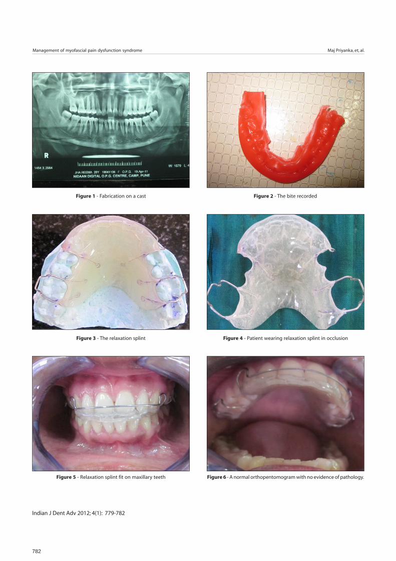
orthopantomogram revealed no anomaly of TMJ
[Fig.1]. There was no erosion of the head of the
condyle characteristic of rheumatoid arthritis and no
osteophytes were visible.
A RF factor test was done to rule out rheumatoid
arthritis and a diagnosis of Myofascial pain
dysfunction syndrome was arrived at.
A treatment plan was formulated which included
the impression of the maxillary and mandibular
arches. A bite was recorded using a wax template by
asking the patient to bring the mandible forward and
allowing the posterior teeth to disocclude [Fig.2]. A
flat plane appliance for arch stabilization was
constructed over the maxillary anterior teeth with the
aim of disoccluding the teeth and relaxing the
musculature [Fig.3&4]. The patient was advised to
wear the appliance 24 hours a day and remove it only
while eating food [Fig.5&6]. She was asked to take
soft diet perform certain muscle relaxation exercises.
The patient reported four months later with
complete relief of muscle tenderness and pain in the
region of TMJ.
Discussion
Musculoskeletal pain is a major cause of
morbidity.2 It is more prevalent in women and
increases with age. A growing number of individuals
Management of myofascial pain dysfunction syndrome Maj Priyanka, et, al.
Indian J Dent Adv 2012; 4(1): 779-782
781
have musculoskeletal pain that affects their daily
activities and function and has a significant impact
on their quality of life causing a financial burden on
our healthcare system.3 Muscles in general, and
myofascial pain in particular, have received less
attention as a major source of pain and dysfunction.
Precipitating factors of MPDS may cause the
facilitated release of acetylcholine at motor end
plates, sustained muscle fibre contractions, local
ischaemia with release of vascular and neuroactive
substances, and muscle pain. More acetylcholine may
then be released, thus perpetuating the muscle pain
and spasm. Electrodiagnostic studies have shown
increased electromyographic activities at trigger
points and tender spots.4,5
The relaxation appliance fabricated for the
patient in this case helped in reducing the activity of
masticatory muscles and helped to reduce
parafunctional habits.
The main differential diagnosis of MPDS includes
neuropathy, bursitis, tendonitis, psychiatric disorders
including depression, fibromyalgia and referred
visceral pain to name a few.
Myofascial pain, which is a common treatable
cause of morbidity, is often under-diagnosed and
under-treated. If left undiagnosed and untreated, it
may develop into chronic pain with overlying
psychosocial and functional problems. This may lead
to further distress, anxiety and even depression. The
vicious cycle may give rise to further somatic
preoccupation. This major source of musculoskeletal
dysfunction requires more focused attention. Its early
diagnosis and treatment may help to reduce
overlying psychosocial complications and the
attending financial burden of chronic pain syndrome.
A large number of patients can be left suffering
in pain for years. Once diagnosed MPDS can be
completely cured with limited rate of recurrence.6,7
Conclusion
Diagnosing a case of myofascial pain
dysfunction syndrome is challenging yet once
diagnosed it can be completely cured. The pain arises
from trigger points present in the muscles that are
in spasm. Construction of a relaxation splint restores
the normal functioning of the muscle and provides
relief from pain due to relief of the spasm. On a four
month follow up the patient had relief from pain
however long term follow up is required in order to
evaluate the recurrence of symptoms.
Acknowledgements and conflicts of interest - Nil
References
1. Travel JG, Simons DG. Myofascial pain and dysfunction: The
trigger point manual vol. 1 and Baltimore: Williams &
Wilkins, 1999.
2. Wolfe F, Smythe HA, Yunus MB, Bennett RM, Bombardier C,
Goldenberg DL, et al. The American College of
Rheumatology 1990 Criteria for Classification of
Fibromyalgia. Report of the Multicenter Criteria Committee.
Arthritis Rheum 1990;33: 160-172.
3. World Health Organization: The burden of musculoskeletal
conditions at the start of the new millennium: Report of a
WHO scientific group. Geneva, Switzerland: WHO, 2003.
4. Association of American Medical Colleges, Report VII,
Contemporary Issues in Medicine: Musculoskeletal
Medicine Education, Medical School Objectives Project.
Washington DC, 2005.
5. Borg-Stein J, Simons DG. Focused review: myofascial pain.
Arch Phys Med Rehabil 2002;83 (3 Suppl 1):40-47.
6. Hsueh TC, Yu S, Kuan TS, Hong CZ. Association of active
myofascial trigger points and cervical disc lesions. J Formos
Med Assoc 1998;97: 174-180.
7. Fernandez-de-Las-Penas C, Arendt-Nielsen L. Sympathetic
facilitation of hyperalgesia evoked from myofascial tender
and trigger points in patients with unilateral shoulder pain.
Clin Neurophysiol 2006;117:1545-1550.
Management of myofascial pain dysfunction syndrome Maj Priyanka, et, al.
Indian J Dent Adv 2012; 4(1): 779-782
782
Figure 1 - Fabrication on a cast Figure 2 - The bite recorded
Figure 3 - The relaxation splint Figure 4 - Patient wearing relaxation splint in occlusion
Figure 5 - Relaxation splint fit on maxillary teeth Figure 6 - A normal orthopentomogram with no evidence of pathology.
Management of myofascial pain dysfunction syndrome Maj Priyanka, et, al.
Indian J Dent Adv 2012; 4(1): 779-782