Embed Size (px)
DESCRIPTION
DENTAL GROSS ANATOMY CASE 2.2. HISTORY A 62-year old woman complained to her dentist a bout sudden bouts of excruciating pain on the left side of her face. The bouts had started ~ 2 months previously and had been - PowerPoint PPT Presentation
Citation preview
DENTAL GROSS ANATOMY
CASE 2.2
HISTORY
A 62-year old woman complained to her dentist about sudden bouts of excruciating pain on the left side of her face.The bouts had started ~ 2 months previously and had been increasing in severity. The stabbing pains lasted 15-20 seconds, occurred several times a day and were so severe that she had once contemplated suicide.After examination the dentist told her there was no dental cause for the pain and he referred her to a physician.

PHYSICAL EXAMINATION
The woman told the physician that the onset of the pain was sometimes triggered by chewing or a cold wind blowing on her upper lip.When asked to point out the area where the pain occurred she pointed to her left upper lip and cheek. She indicated that the pain also radiated to her lower eyelid, lateral side of the nose and the inside of the mouth.The physician applied firm pressure over the patient’s left cheek and over her infraorbital area, but detected no tenderness indicative of maxillary sinusitis.The physician did detect acute sensitivity to touch on the left upper lip and to pin-pricking over the entire left maxillary region. No abnormality of sensation was found in the forehead or mandibular regions.

1. What is the diagnosis?

TRIGEMINAL NEURALGIA (“TIC DOULOUREUX”)
2. Which branch of what major nerve supplies the area of skin and mucous membrane where the paroxysms (sudden recurring attacks) of stabbing pain were felt?

Maxillary n. (V2)
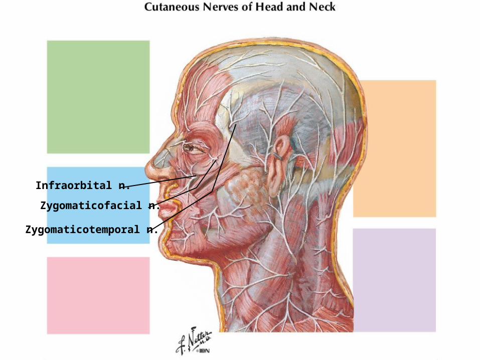
Infraorbital n.
Zygomaticofacial n.
Zygomaticotemporal n.
3. Through what foramen and what bone does this nerve leave the skull?
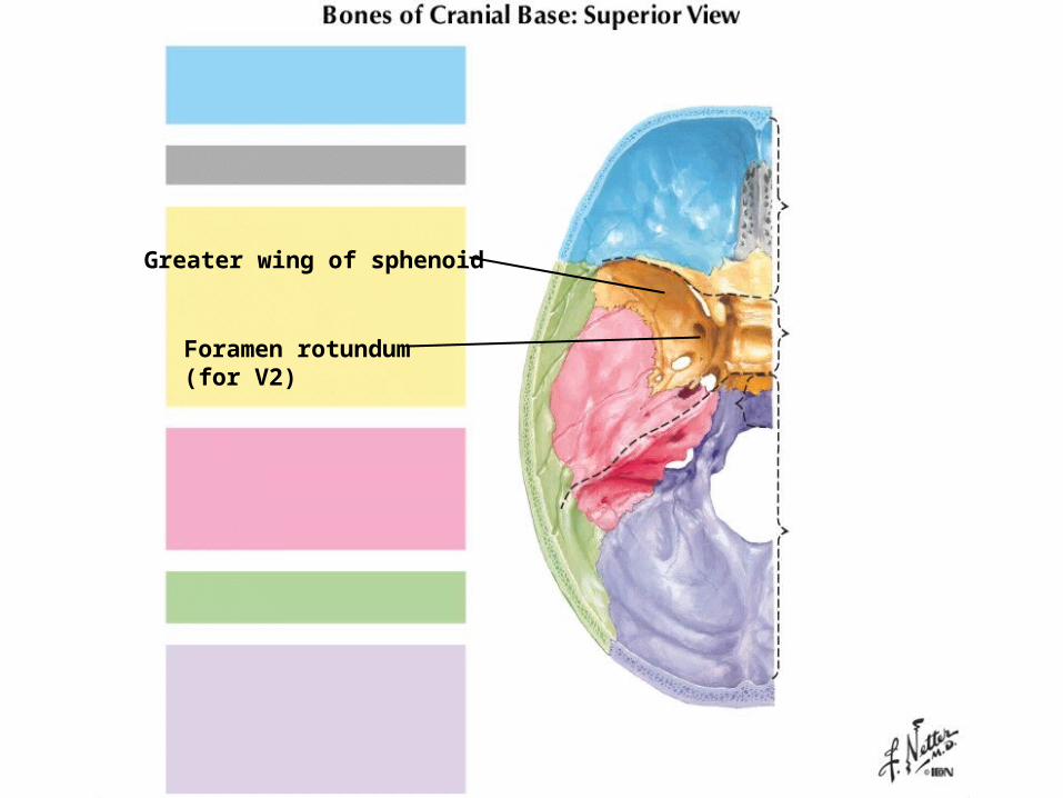
Foramen rotundum(for V2)
Greater wing of sphenoid
4. Why was no abnormality of sensation found in the forehead region? In the mandibular region?
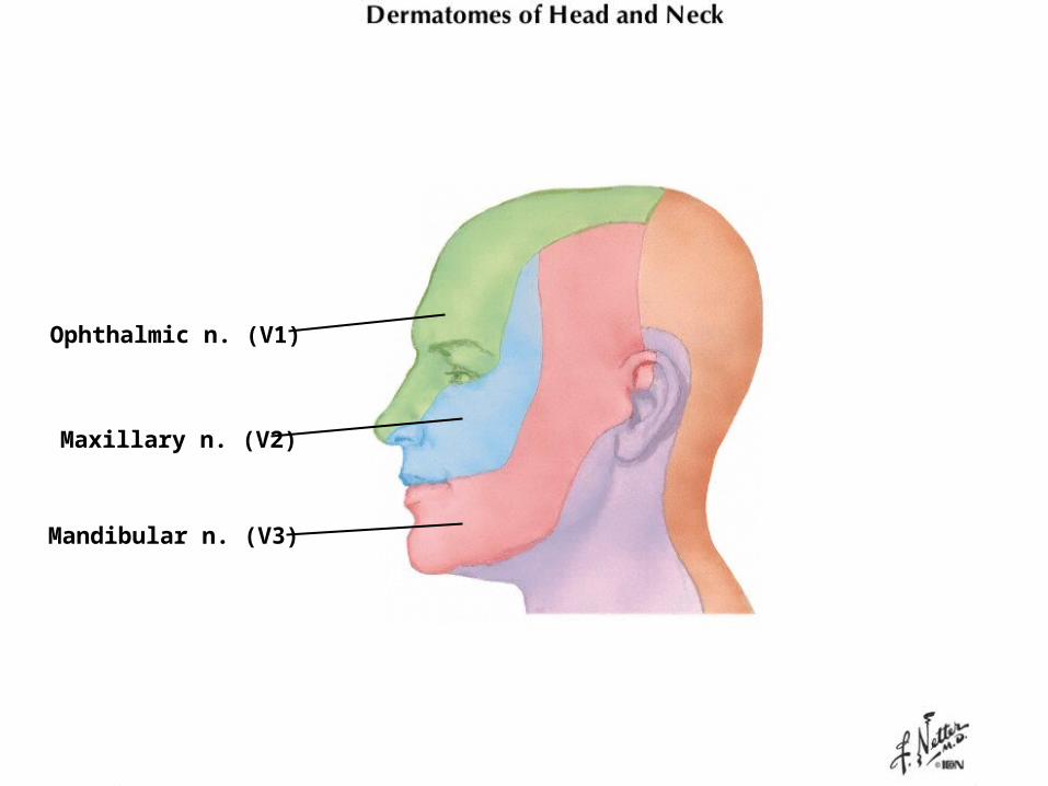
Ophthalmic n. (V1)
Mandibular n. (V3)
Maxillary n. (V2)
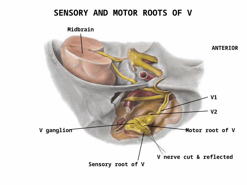
5. Where are the cell bodies of the affected nerve located?
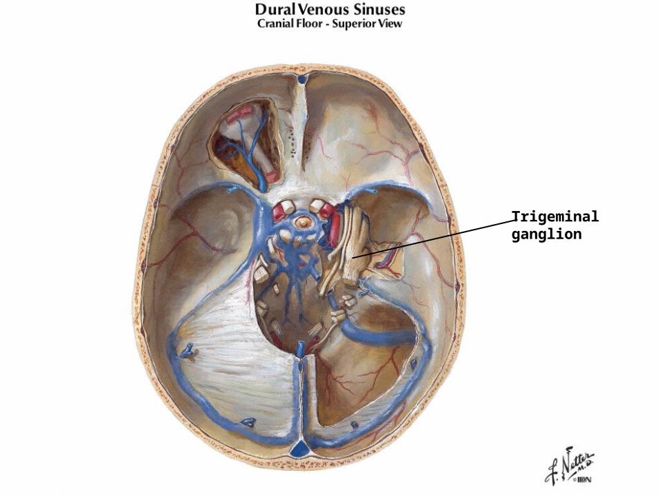
Trigeminalganglion
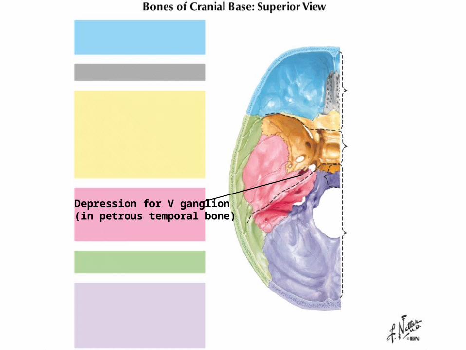
Depression for V ganglion(in petrous temporal bone)
6. Why were no motor deficits observed in this patient?
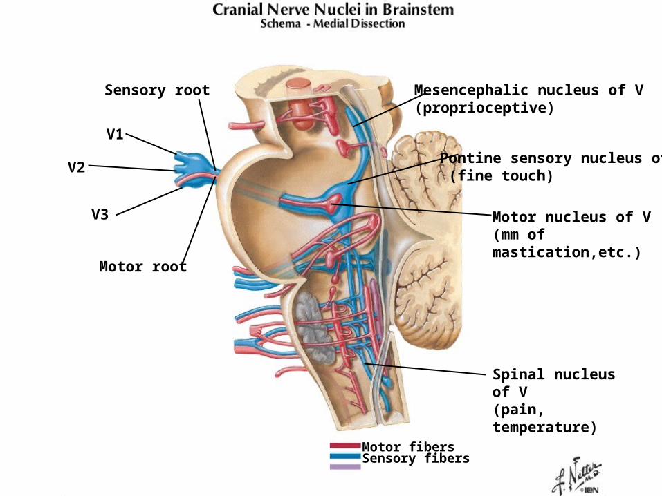
Mesencephalic nucleus of V(proprioceptive)
Pontine sensory nucleus of V (fine touch)
Spinal nucleus of V(pain, temperature)
Motor nucleus of V(mm of mastication,etc.)
Motor fibersSensory fibers
V2
V1
V3
Sensory root
Motor root
Motor root of V
Sensory root of V
V ganglion
V nerve cut & reflected
V1
V2
Midbrain
ANTERIOR
SENSORY AND MOTOR ROOTS OF V
7. What may be the cause(s) of this condition?

Usually the cause of trigeminal neuralgia is unknown.
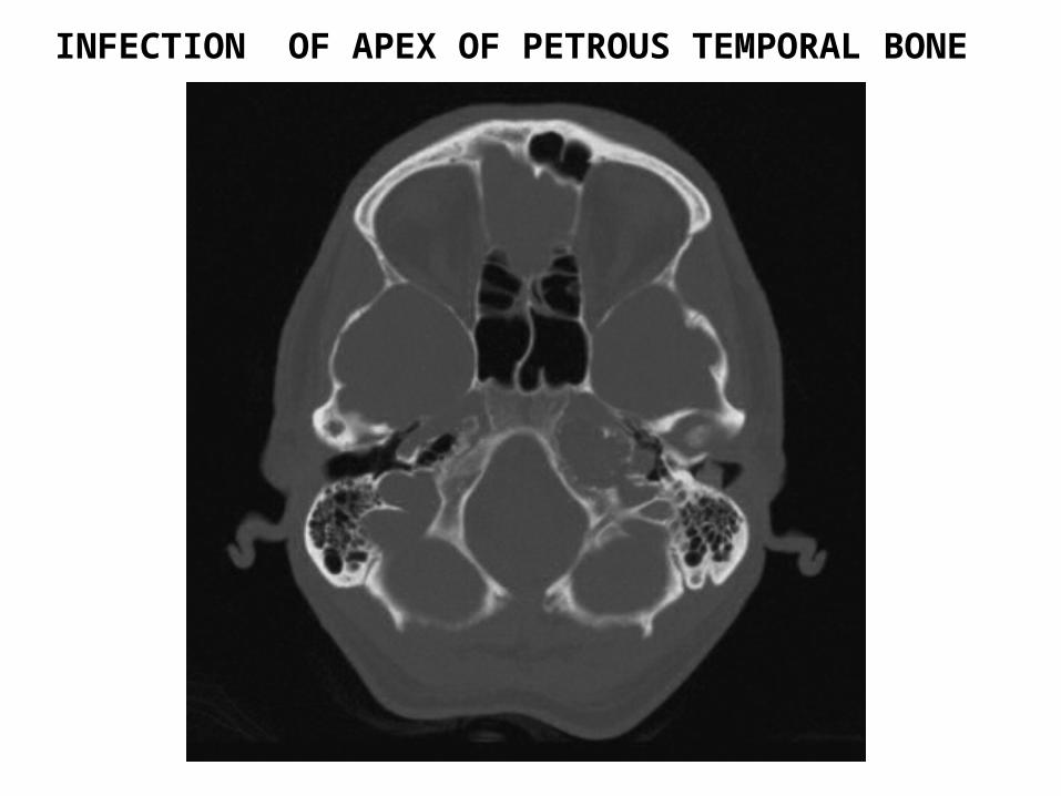
INFECTION OF APEX OF PETROUS TEMPORAL BONE
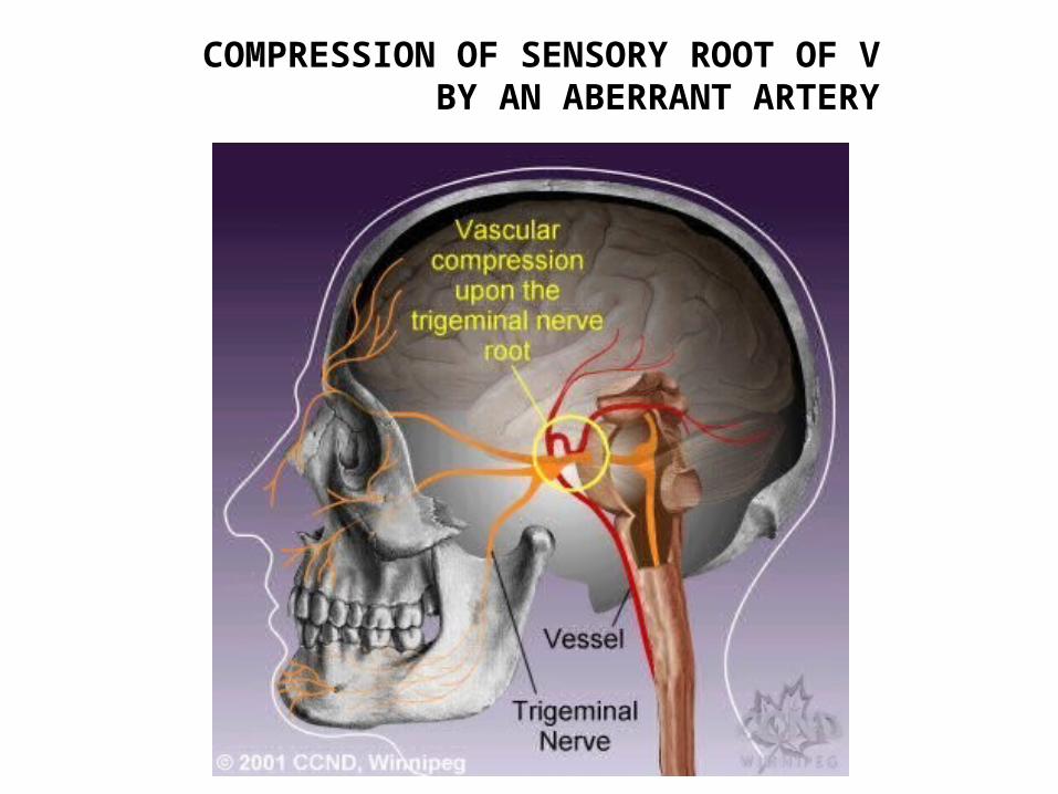
COMPRESSION OF SENSORY ROOT OF V BY AN ABERRANT ARTERY
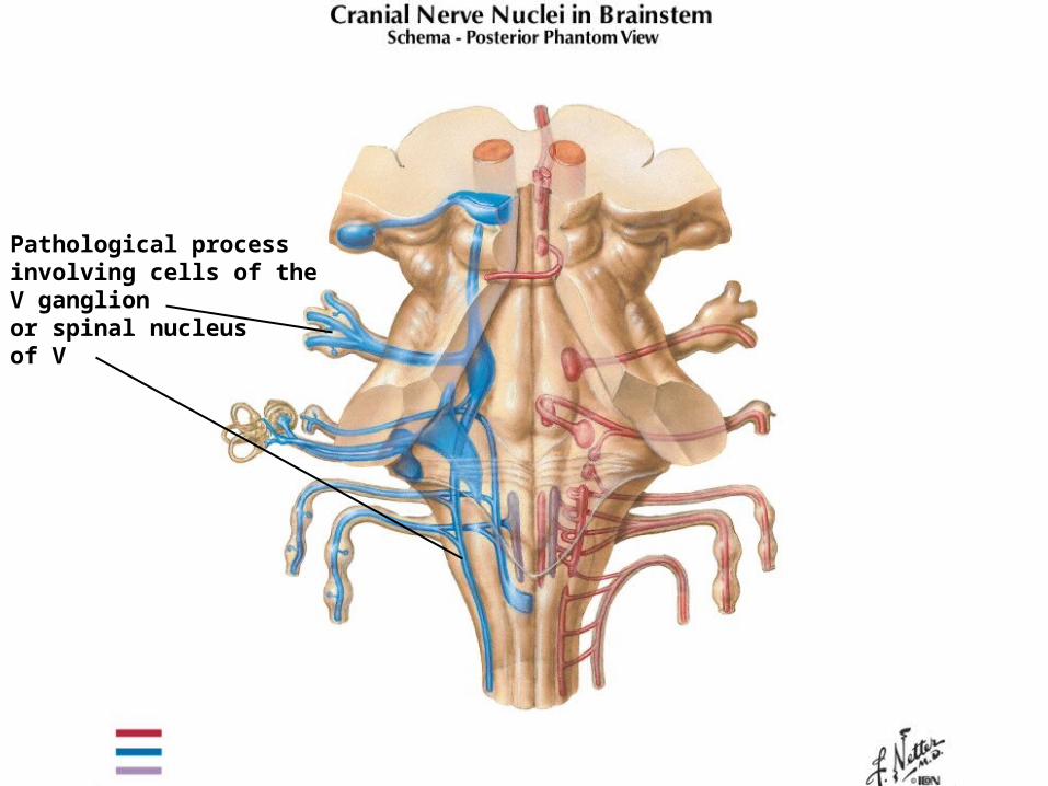
Pathological processinvolving cells of theV ganglionor spinal nucleusof V
8. How might this condition be treated?
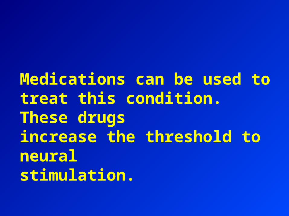
Medications can be used to treat this condition. These drugs increase the threshold to neuralstimulation.
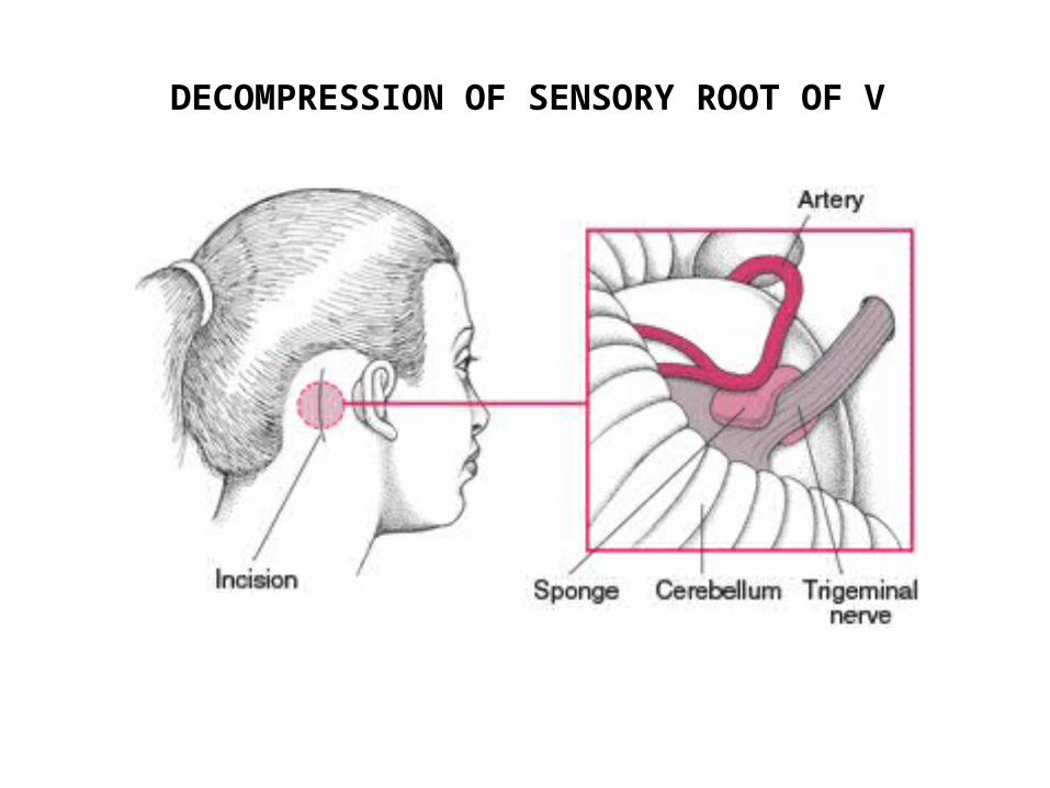
DECOMPRESSION OF SENSORY ROOT OF V
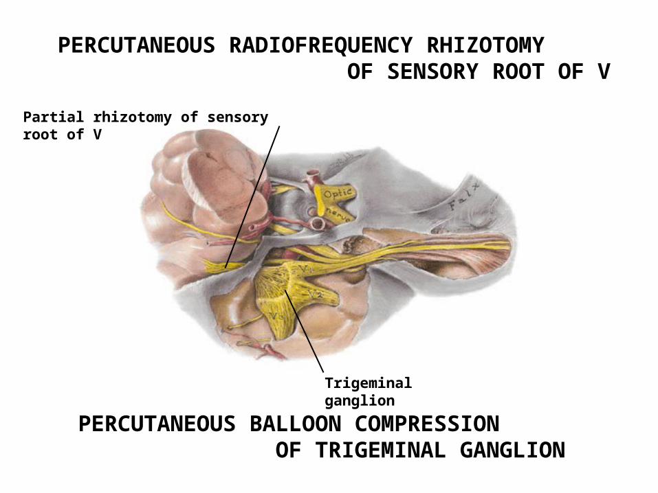
PERCUTANEOUS RADIOFREQUENCY RHIZOTOMY OF SENSORY ROOT OF V
Partial rhizotomy of sensory root of V
PERCUTANEOUS BALLOON COMPRESSION OF TRIGEMINAL GANGLION
Trigeminal ganglion

ADDITIONAL NOTE
It is noteworthy that the patient first consulted her dentist about her problem and that her physician examined her for possible maxillary sinusitis. Under the mistaken belief that the pain of trigeminal neuralgia is due to dental disease or sinusitis, patients have had upper teeth extracted and their maxillary sinuses drained, but with no relief.
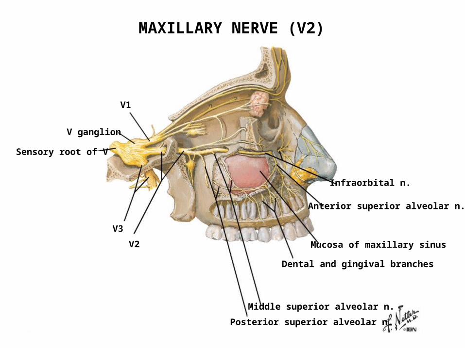
V ganglion
V1
V2
V3
Sensory root of V
Infraorbital n.
Anterior superior alveolar n.
Mucosa of maxillary sinus
Dental and gingival branches
Middle superior alveolar n.
Posterior superior alveolar n.
MAXILLARY NERVE (V2)
END OF CASE 2.2

TIME