Embed Size (px)
Citation preview
79:1450-1460, 1998. J NeurophysiolDenis Paré, Eric Shink, Hélène Gaudreau, Alain Destexhe and Eric J. Lang
You might find this additional information useful...
26 articles, 13 of which you can access free at: This article cites http://jn.physiology.org/cgi/content/full/79/3/1450#BIBL
98 other HighWire hosted articles, the first 5 are: This article has been cited by
[PDF] [Full Text] [Abstract]
, December 2, 2009; 29 (48): 15341-15350. J. Neurosci.I. Pavlov, L. P. Savtchenko, D. M. Kullmann, A. Semyanov and M. C. Walker
Neuronal Offset, Not GainOutwardly Rectifying Tonically Active GABAA Receptors in Pyramidal Cells Modulate
[PDF] [Full Text] [Abstract], February 10, 2010; 30 (6): 2150-2159. J. Neurosci.
S. Otte, A. Hasenstaub and E. M. Callaway Inhibition
Cell Type-Specific Control of Neuronal Responsiveness by Gamma-Band Oscillatory
[PDF] [Full Text], February 17, 2010; 30 (7): 2407-2413. J. Neurosci.
M. N. Economo, F. R. Fernandez and J. A. White Neurophysiology
Dynamic Clamp: Alteration of Response Properties and Creation of Virtual Realities in
[PDF] [Full Text] [Abstract], July 1, 2010; 90 (3): 1195-1268. Physiol Rev
X.-J. Wang Neurophysiological and Computational Principles of Cortical Rhythms in Cognition
[PDF] [Full Text] [Abstract]
, July 1, 2010; 104 (1): 280-290. J NeurophysiolL. C. Faria and D. A. Prince
Posttraumatic EpilepsyPresynaptic Inhibitory Terminals Are Functionally Abnormal in a Rat Model of
on the following topics: http://highwire.stanford.edu/lists/artbytopic.dtlcan be found at Medline items on this article's topics
Physiology .. Cats Medicine .. Tetrodotoxins Physiology .. Pyramidal Cells Physiology .. Cortical Neurons Physiology .. Synaptic Transmission
including high-resolution figures, can be found at: Updated information and services http://jn.physiology.org/cgi/content/full/79/3/1450
can be found at: Journal of Neurophysiologyabout Additional material and information http://www.the-aps.org/publications/jn
This information is current as of August 18, 2010 .
http://www.the-aps.org/.Physiological Society. ISSN: 0022-3077, ESSN: 1522-1598. Visit our website at by the American Physiological Society, 9650 Rockville Pike, Bethesda MD 20814-3991. Copyright © 1998 by the American
publishes original articles on the function of the nervous system. It is published 12 times a year (monthly)Journal of Neurophysiology
on August 18, 2010
jn.physiology.orgD
ownloaded from
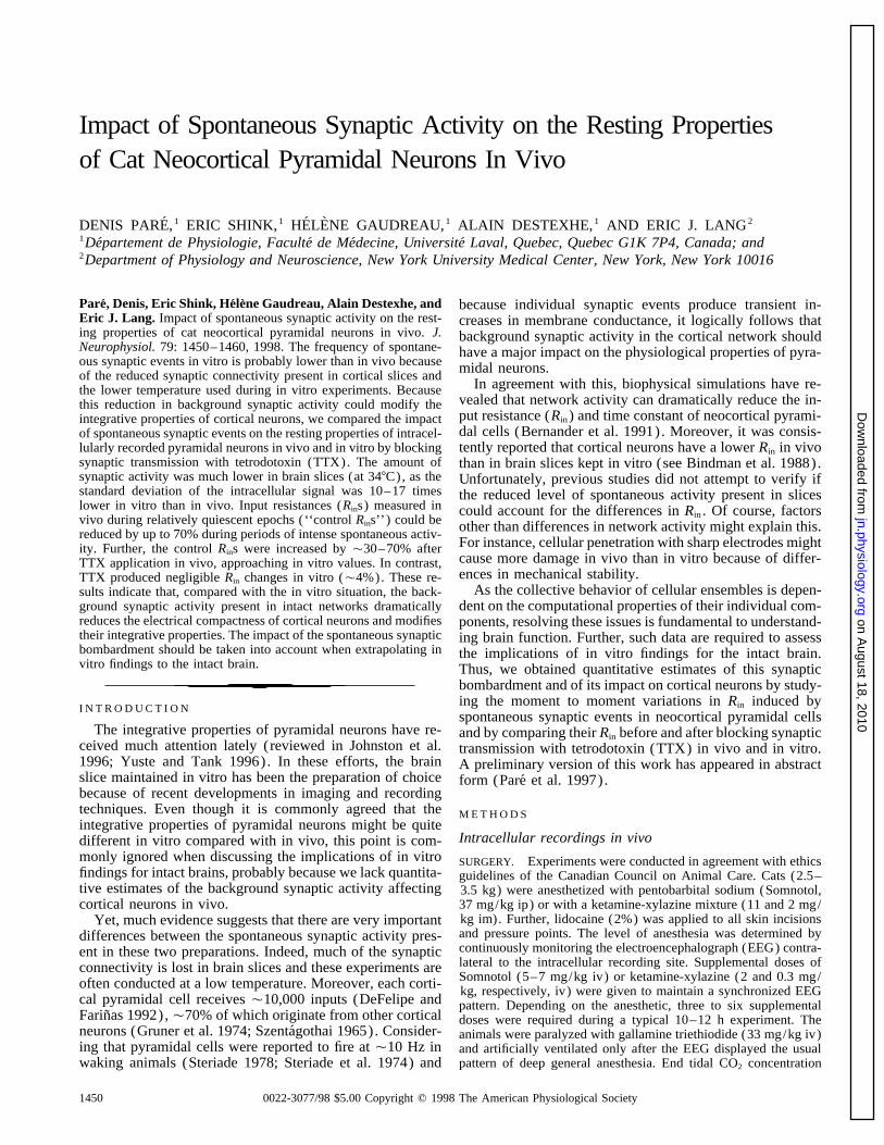
Impact of Spontaneous Synaptic Activity on the Resting Propertiesof Cat Neocortical Pyramidal Neurons In Vivo
DENIS PARE,1 ERIC SHINK,1 HELENE GAUDREAU,1 ALAIN DESTEXHE,1 AND ERIC J. LANG2
1Departement de Physiologie, Faculte de Medecine, Universite Laval, Quebec, Quebec G1K 7P4, Canada; and2Department of Physiology and Neuroscience, New York University Medical Center, New York, New York 10016
Pare, Denis, Eric Shink, Helene Gaudreau, Alain Destexhe, and because individual synaptic events produce transient in-Eric J. Lang. Impact of spontaneous synaptic activity on the rest- creases in membrane conductance, it logically follows thating properties of cat neocortical pyramidal neurons in vivo. J. background synaptic activity in the cortical network shouldNeurophysiol. 79: 1450–1460, 1998. The frequency of spontane- have a major impact on the physiological properties of pyra-ous synaptic events in vitro is probably lower than in vivo because midal neurons.of the reduced synaptic connectivity present in cortical slices and
In agreement with this, biophysical simulations have re-the lower temperature used during in vitro experiments. Becausevealed that network activity can dramatically reduce the in-this reduction in background synaptic activity could modify theput resistance (Rin ) and time constant of neocortical pyrami-integrative properties of cortical neurons, we compared the impactdal cells (Bernander et al. 1991). Moreover, it was consis-of spontaneous synaptic events on the resting properties of intracel-
lularly recorded pyramidal neurons in vivo and in vitro by blocking tently reported that cortical neurons have a lower Rin in vivosynaptic transmission with tetrodotoxin (TTX). The amount of than in brain slices kept in vitro (see Bindman et al. 1988).synaptic activity was much lower in brain slices (at 347C), as the Unfortunately, previous studies did not attempt to verify ifstandard deviation of the intracellular signal was 10–17 times the reduced level of spontaneous activity present in sliceslower in vitro than in vivo. Input resistances (Rins) measured in could account for the differences in Rin . Of course, factorsvivo during relatively quiescent epochs (‘‘control Rins’’) could be other than differences in network activity might explain this.reduced by up to 70% during periods of intense spontaneous activ-
For instance, cellular penetration with sharp electrodes mightity. Further, the control Rins were increased by Ç30–70% aftercause more damage in vivo than in vitro because of differ-TTX application in vivo, approaching in vitro values. In contrast,ences in mechanical stability.TTX produced negligible Rin changes in vitro (Ç4%). These re-
As the collective behavior of cellular ensembles is depen-sults indicate that, compared with the in vitro situation, the back-ground synaptic activity present in intact networks dramatically dent on the computational properties of their individual com-reduces the electrical compactness of cortical neurons and modifies ponents, resolving these issues is fundamental to understand-their integrative properties. The impact of the spontaneous synaptic ing brain function. Further, such data are required to assessbombardment should be taken into account when extrapolating in the implications of in vitro findings for the intact brain.vitro findings to the intact brain. Thus, we obtained quantitative estimates of this synaptic
bombardment and of its impact on cortical neurons by study-ing the moment to moment variations in Rin induced by
I N T R O D U C T I O Nspontaneous synaptic events in neocortical pyramidal cells
The integrative properties of pyramidal neurons have re- and by comparing their Rin before and after blocking synapticceived much attention lately (reviewed in Johnston et al. transmission with tetrodotoxin (TTX) in vivo and in vitro.1996; Yuste and Tank 1996). In these efforts, the brain A preliminary version of this work has appeared in abstractslice maintained in vitro has been the preparation of choice form (Pare et al. 1997).because of recent developments in imaging and recordingtechniques. Even though it is commonly agreed that the M E T H O D Sintegrative properties of pyramidal neurons might be quite
Intracellular recordings in vivodifferent in vitro compared with in vivo, this point is com-monly ignored when discussing the implications of in vitro SURGERY. Experiments were conducted in agreement with ethicsfindings for intact brains, probably because we lack quantita- guidelines of the Canadian Council on Animal Care. Cats (2.5–tive estimates of the background synaptic activity affecting 3.5 kg) were anesthetized with pentobarbital sodium (Somnotol,
37 mg/kg ip) or with a ketamine-xylazine mixture (11 and 2 mg/cortical neurons in vivo.kg im). Further, lidocaine (2%) was applied to all skin incisionsYet, much evidence suggests that there are very importantand pressure points. The level of anesthesia was determined bydifferences between the spontaneous synaptic activity pres-continuously monitoring the electroencephalograph (EEG) contra-ent in these two preparations. Indeed, much of the synapticlateral to the intracellular recording site. Supplemental doses ofconnectivity is lost in brain slices and these experiments areSomnotol (5–7 mg/kg iv) or ketamine-xylazine (2 and 0.3 mg/often conducted at a low temperature. Moreover, each corti- kg, respectively, iv) were given to maintain a synchronized EEG
cal pyramidal cell receives Ç10,000 inputs (DeFelipe and pattern. Depending on the anesthetic, three to six supplementalFarinas 1992),Ç70% of which originate from other cortical doses were required during a typical 10–12 h experiment. Theneurons (Gruner et al. 1974; Szentagothai 1965). Consider- animals were paralyzed with gallamine triethiodide (33 mg/kg iv)ing that pyramidal cells were reported to fire at Ç10 Hz in and artificially ventilated only after the EEG displayed the usual
pattern of deep general anesthesia. End tidal CO2 concentrationwaking animals (Steriade 1978; Steriade et al. 1974) and
1450 0022-3077/98 $5.00 Copyright q 1998 The American Physiological Society
J591-7/ 9k26$$mr16 02-10-98 19:59:50 neupal LP-Neurophys
on August 18, 2010
jn.physiology.orgD
ownloaded from
IMPACT OF NETWORK ACTIVITY ON PYRAMIDAL CELLS 1451
When testing the effect of TTX on the Rin of cortical neurons,intracellular current injection was used to maintain their Vm atapproximately 070 to 075 mV (‘‘manual clamp’’) , below theactivation threshold of the persistent Na/ current. The amplitudeof this steady current was adjusted so that the Vm returned to aconstant value during epochs that were relatively free of synapticevents. As TTX always produced a gradual hyperpolarization invivo, this maneuver aimed at dissociating the effects of the synapticblockade on the Rin from the voltage-dependent activation or inacti-vation of intrinsic currents that could have been produced by thehyperpolarization. In addition to the steady current injection, a brief(200–400 ms) hyperpolarizing current pulse of constant amplitude(0.1–0.4 nA) and an intracortical shock were applied at regularintervals (every 3–6 s) to monitor the Rin and the effect of TTXon spike-dependent synaptic transmission, respectively.
Unless otherwise stated, the standard deviation of the intracellu-lar signal was measured from Ç1 min epochs of spontaneous dataobtained at a Vm of approximately 070 mV as determined by intra-
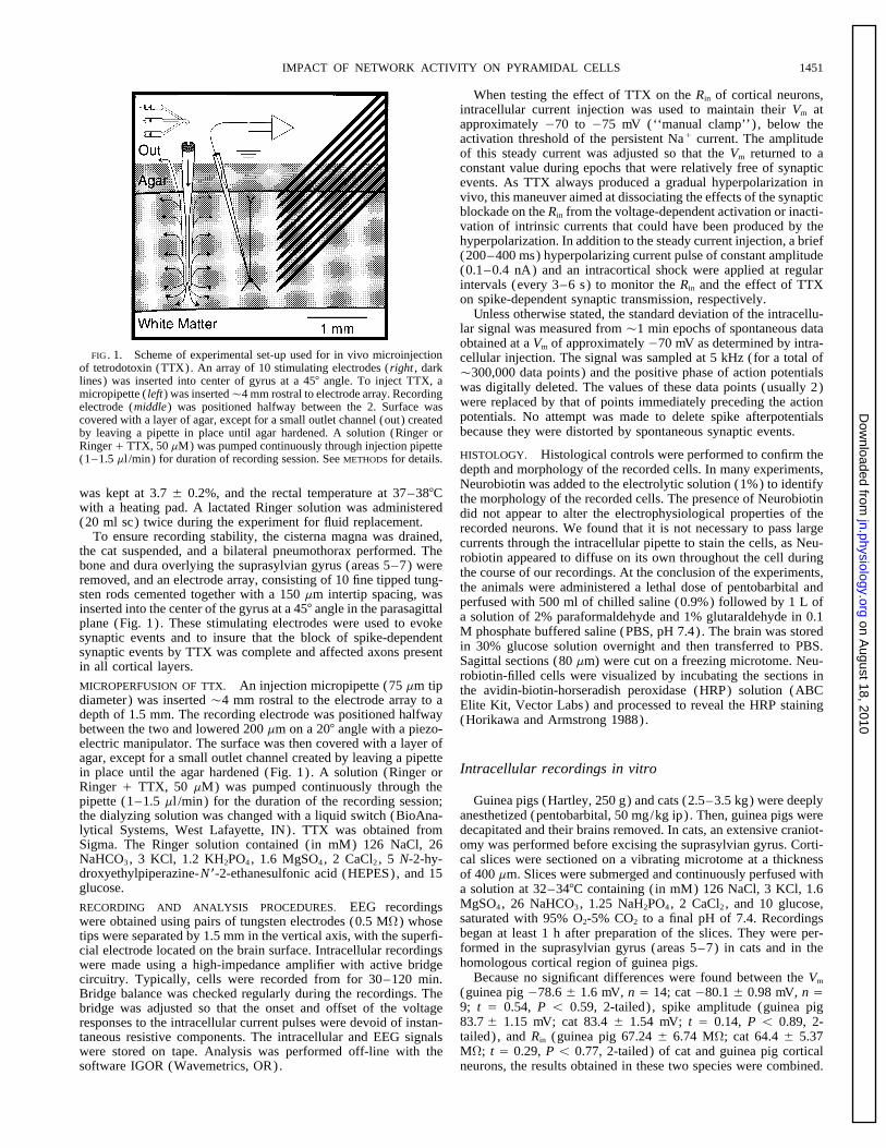
FIG. 1. Scheme of experimental set-up used for in vivo microinjection cellular injection. The signal was sampled at 5 kHz (for a total ofof tetrodotoxin (TTX). An array of 10 stimulating electrodes (right , dark Ç300,000 data points) and the positive phase of action potentialslines) was inserted into center of gyrus at a 457 angle. To inject TTX, a was digitally deleted. The values of these data points (usually 2)micropipette ( left) was insertedÇ4 mm rostral to electrode array. Recording
were replaced by that of points immediately preceding the actionelectrode (middle) was positioned halfway between the 2. Surface waspotentials. No attempt was made to delete spike afterpotentialscovered with a layer of agar, except for a small outlet channel (out) createdbecause they were distorted by spontaneous synaptic events.by leaving a pipette in place until agar hardened. A solution (Ringer or
Ringer / TTX, 50 mM) was pumped continuously through injection pipetteHISTOLOGY. Histological controls were performed to confirm the(1–1.5 ml /min) for duration of recording session. See METHODS for details.depth and morphology of the recorded cells. In many experiments,Neurobiotin was added to the electrolytic solution (1%) to identify
was kept at 3.7 { 0.2%, and the rectal temperature at 37–387C the morphology of the recorded cells. The presence of Neurobiotinwith a heating pad. A lactated Ringer solution was administered did not appear to alter the electrophysiological properties of the(20 ml sc) twice during the experiment for fluid replacement. recorded neurons. We found that it is not necessary to pass large
To ensure recording stability, the cisterna magna was drained, currents through the intracellular pipette to stain the cells, as Neu-the cat suspended, and a bilateral pneumothorax performed. The robiotin appeared to diffuse on its own throughout the cell duringbone and dura overlying the suprasylvian gyrus (areas 5–7) were the course of our recordings. At the conclusion of the experiments,removed, and an electrode array, consisting of 10 fine tipped tung- the animals were administered a lethal dose of pentobarbital andsten rods cemented together with a 150 mm intertip spacing, was
perfused with 500 ml of chilled saline (0.9%) followed by 1 L ofinserted into the center of the gyrus at a 457 angle in the parasagittala solution of 2% paraformaldehyde and 1% glutaraldehyde in 0.1plane (Fig. 1) . These stimulating electrodes were used to evokeM phosphate buffered saline (PBS, pH 7.4). The brain was storedsynaptic events and to insure that the block of spike-dependentin 30% glucose solution overnight and then transferred to PBS.synaptic events by TTX was complete and affected axons presentSagittal sections (80 mm) were cut on a freezing microtome. Neu-in all cortical layers.robiotin-filled cells were visualized by incubating the sections in
MICROPERFUSION OF TTX. An injection micropipette (75 mm tip the avidin-biotin-horseradish peroxidase (HRP) solution (ABCdiameter) was inserted Ç4 mm rostral to the electrode array to a Elite Kit, Vector Labs) and processed to reveal the HRP stainingdepth of 1.5 mm. The recording electrode was positioned halfway (Horikawa and Armstrong 1988).between the two and lowered 200 mm on a 207 angle with a piezo-electric manipulator. The surface was then covered with a layer ofagar, except for a small outlet channel created by leaving a pipette
Intracellular recordings in vitroin place until the agar hardened (Fig. 1) . A solution (Ringer orRinger / TTX, 50 mM) was pumped continuously through the
Guinea pigs (Hartley, 250 g) and cats (2.5–3.5 kg) were deeplypipette (1–1.5 ml /min) for the duration of the recording session;anesthetized (pentobarbital, 50 mg/kg ip) . Then, guinea pigs werethe dialyzing solution was changed with a liquid switch (BioAna-decapitated and their brains removed. In cats, an extensive craniot-lytical Systems, West Lafayette, IN). TTX was obtained from
Sigma. The Ringer solution contained (in mM) 126 NaCl, 26 omy was performed before excising the suprasylvian gyrus. Corti-NaHCO3, 3 KCl, 1.2 KH2PO4, 1.6 MgSO4, 2 CaCl2 , 5 N-2-hy- cal slices were sectioned on a vibrating microtome at a thicknessdroxyethylpiperazine-N*-2-ethanesulfonic acid (HEPES), and 15 of 400 mm. Slices were submerged and continuously perfused withglucose. a solution at 32–347C containing (in mM) 126 NaCl, 3 KCl, 1.6
MgSO4, 26 NaHCO3, 1.25 NaH2PO4, 2 CaCl2 , and 10 glucose,RECORDING AND ANALYSIS PROCEDURES. EEG recordingssaturated with 95% O2-5% CO2 to a final pH of 7.4. Recordingswere obtained using pairs of tungsten electrodes (0.5 MV) whosebegan at least 1 h after preparation of the slices. They were per-tips were separated by 1.5 mm in the vertical axis, with the superfi-formed in the suprasylvian gyrus (areas 5–7) in cats and in thecial electrode located on the brain surface. Intracellular recordingshomologous cortical region of guinea pigs.were made using a high-impedance amplifier with active bridge
Because no significant differences were found between the Vmcircuitry. Typically, cells were recorded from for 30–120 min.(guinea pig 078.6 { 1.6 mV, n Å 14; cat 080.1 { 0.98 mV, n ÅBridge balance was checked regularly during the recordings. The9; t Å 0.54, P õ 0.59, 2-tailed) , spike amplitude (guinea pigbridge was adjusted so that the onset and offset of the voltage83.7 { 1.15 mV; cat 83.4 { 1.54 mV; t Å 0.14, P õ 0.89, 2-responses to the intracellular current pulses were devoid of instan-tailed) , and Rin (guinea pig 67.24 { 6.74 MV; cat 64.4 { 5.37taneous resistive components. The intracellular and EEG signalsMV; t Å 0.29, P õ 0.77, 2-tailed) of cat and guinea pig corticalwere stored on tape. Analysis was performed off-line with the
software IGOR (Wavemetrics, OR). neurons, the results obtained in these two species were combined.
J591-7/ 9k26$$mr16 02-10-98 19:59:50 neupal LP-Neurophys
on August 18, 2010
jn.physiology.orgD
ownloaded from
D. PARE, E. SHINK, H. GAUDREAU, A. DESTEXHE, AND E. J. LANG1452
R E S U L T S and in vitro (at 347C; Fig. 3B1) is shown with the samegain and time base. Whereas the in vivo recording displayed
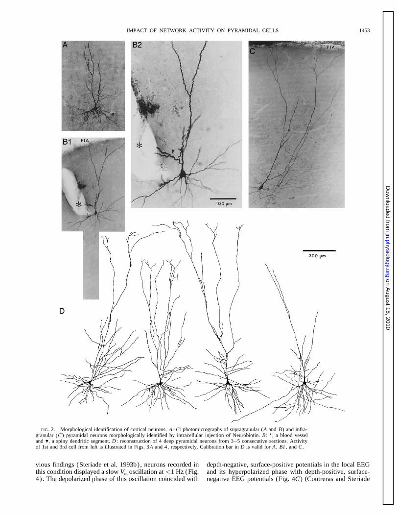
Intracellular recordings of regular spiking (Connors et al. a continuous barrage of compound postsynaptic potentials1982; McCormick et al. 1985) neocortical neurons that had (PSPs) that often summated into large (2–15 mV) events,membrane potentials (Vm) ú 060 mV and overshooting ac- the in vitro recording was characterized by long quiescenttion potentials were obtained in anesthetized cats and from periods interrupted by brief bursts of small amplitude PSPs.cat or guinea pig cortical slices kept in vitro. Fast-spiking Furthermore, spontaneous synaptic potentials often gave riseneurons (Connors et al. 1982; McCormick et al. 1985) were to action potentials in vivo (Fig. 3A1) , but rarely did so innot considered in this study. All intracellular recordings were vitro (Fig. 3B1) . An example of a high-frequency (Ç150obtained from the suprasylvian gyrus (areas 5–7) in cats Hz) spike train triggered by spontaneous synaptic eventsand from the homologous region in guinea pigs. A total of that coincided with a depth negative EEG potential is shown110 neurons were recorded in vivo and 39 in vitro (9 in cats with an expanded time base in Fig. 3A3 . These differencesand 30 in guinea pigs) . All cells were recorded with KCl- were observed despite the fact that neurons recorded in vitrofilled pipettes (2.5 M; Ç25 MV; tip diameter õ 0.5 mm) had much higher Rins than those recorded in vivo (see below)pulled from the same batch of glass capillaries with identical and displayed normal responses to a graded series of currentpuller settings. Moreover, to insure uniform electrode char- pulses (Fig. 3B3) . To facilitate comparison between in vivoacteristics, no attempt was made to bevel or otherwise alter and in vitro data, epochs of spontaneous activity are depictedthe shape of the pipette tips. at a higher gain and faster time base in Fig. 3, A4 and B2 ,
Ideally, the in vivo experiments should have been carried respectively.out in unanesthetized animals. However, because it is impos- To quantify the difference in spontaneous synaptic activitysible to obtain stable intracellular recordings of long duration between in vivo and in vitro recordings, we measured thein unanesthetized animals, these experiments were per- standard deviation of the intracellular signal in neurons keptformed under two different anesthetic conditions to insure at around 070 mV with intracellular current injection. Sub-that our conclusions were not critically dependent on the type sets of 10 neurons were analyzed in each condition andof anesthetic. These anesthetics were pentobarbital, which action potentials were digitally deleted from the in vivo data.potentiates g-aminobutyric acid (GABAA) synaptic events As shown in Fig. 3C , in vivo neurons recorded under keta-(Barker and McBurney 1979) and ketamine-xylazine, which mine-xylazine displayed the highest standard deviation, fol-blocks N-methyl-D-aspartate (NMDA)(Anis et al. 1983) lowed by those recorded under barbiturate. In neurons re-and activates a2 noradrenergic receptors (Nicoll et al. corded in vitro, the standard deviation of the intracellular1990), respectively. Of the 110 neurons recorded in vivo, signal was 10 to 17 times lower, close to that of the equip-63 were recorded under barbiturate anesthesia, and 47 under ment noise (0.18 { 0.01 mV; n Å 3) measured in the extra-ketamine-xylazine. No attempt was made to control for dif- cellular space after withdrawing the recording pipette fromferences in temperature between the in vivo and in vitro the cells. The differences between the standard deviation ofpreparations, because we wanted to compare them as they the intracellular signal of neurons recorded in vitro and inare commonly used. vivo were statistically significant ( in vitro vs. barbiturate,
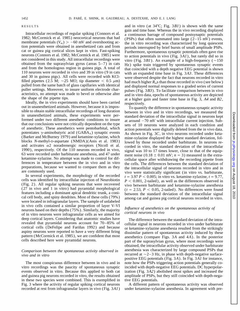
In several experiments, the morphology of the recorded t Å 3.97 Põ 0.005; in vitro vs. ketamine-xylaxine, t Å 9.77,cells was identified by intracellular injection of Neurobiotin P õ 0.001, 2-tailed) , as well as the differences observed in(Fig. 2) . All regular spiking neurons that were recovered vivo between barbiturate and ketamine-xylazine anesthesia(27 in vivo and 1 in vitro) had pyramidal morphological ( t Å 2.53, P õ 0.05, 2-tailed) . No differences were foundfeatures including a dominant apical dendritic trunk, a coni- between the standard deviation of the intracellular signalcal cell body, and spiny dendrites. Most of these cells (79%) among cat and guinea pig cortical neurons recorded in vitro.were located in infragranular layers. The sample of unlabeledin vivo cells contained a similar proportion of layer V-VI
Influence of anesthetics on the spontaneous activity ofneurons based on their depths (75%). Similarly, the majoritycortical neurons in vivoof in vitro neurons were infragranular cells as we aimed for
deep cortical layers. Considering that anatomic studies have The difference between the standard deviation of the intra-revealed that pyramidal neurons account for 70–85% of cellular signal in neurons recorded in vivo under barbituratecortical cells (DeFelipe and Farinas 1992) and because or ketamine-xylazine anesthesia resulted from the strikinglyaspiny neurons were reported to have a very different firing dissimilar pattern of spontaneous activity induced by thesepattern (McCormick et al. 1985), we are confident that most anesthetics (compare Figs. 3A and 4A) . In the posteriorcells described here were pyramidal neurons. part of the suprasylvian gyrus, where most recordings were
obtained, the intracellular activity observed under barbiturateanesthesia was characterized by large compound PSPs thatComparison between the spontaneous activity observed inrecurred at Ç2–3 Hz, in phase with depth-negative surface-vivo and in vitropositive EEG potentials (Fig. 3A) . In Fig. 3A1 for instance,note how the PSPs triggering action potentials generally co-The most conspicuous difference between in vivo and in
vitro recordings was the paucity of spontaneous synaptic incided with depth-negative EEG potentials. DC hyperpolar-ization (Fig. 3A2) abolished most spikes and increased theevents observed in vitro. Because this applied to both cat
and guinea pig neurons recorded in vitro, the results obtained amplitude of PSPs, but they still coincided with depth-nega-tive EEG potentials.in these two species were combined. This is exemplified in
Fig. 3 where the activity of regular spiking cortical neurons A different pattern of spontaneous activity was observedunder ketamine-xylazine anesthesia. In agreement with pre-recorded at rest from infragranular layers in vivo (Fig. 3A1)
J591-7/ 9k26$$mr16 02-10-98 19:59:50 neupal LP-Neurophys
on August 18, 2010
jn.physiology.orgD
ownloaded from
IMPACT OF NETWORK ACTIVITY ON PYRAMIDAL CELLS 1453
FIG. 2. Morphological identification of cortical neurons. A–C: photomicrographs of supragranular (A and B) and infra-granular (C) pyramidal neurons morphologically identified by intracellular injection of Neurobiotin. B : ∗, a blood vesseland ., a spiny dendritic segment. D : reconstruction of 4 deep pyramidal neurons from 3–5 consecutive sections. Activityof 1st and 3rd cell from left is illustrated in Figs. 3A and 4, respectively. Calibration bar in D is valid for A , B1 , and C .
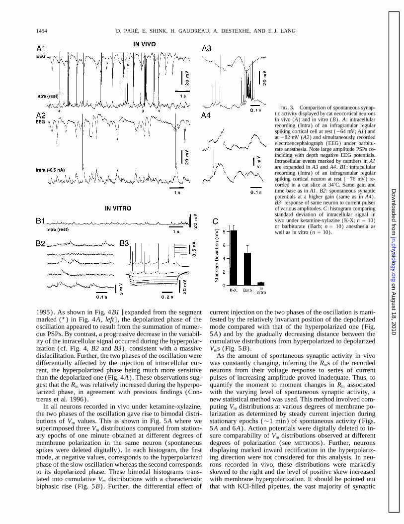
vious findings (Steriade et al. 1993b), neurons recorded in depth-negative, surface-positive potentials in the local EEGand its hyperpolarized phase with depth-positive, surface-this condition displayed a slow Vm oscillation atõ1 Hz (Fig.
4) . The depolarized phase of this oscillation coincided with negative EEG potentials (Fig. 4C) (Contreras and Steriade
J591-7/ 9k26$$mr16 02-10-98 19:59:50 neupal LP-Neurophys
on August 18, 2010
jn.physiology.orgD
ownloaded from
D. PARE, E. SHINK, H. GAUDREAU, A. DESTEXHE, AND E. J. LANG1454
FIG. 3. Comparison of spontaneous synap-tic activity displayed by cat neocortical neuronsin vivo (A) and in vitro (B). A : intracellularrecording (Intra) of an infragranular regularspiking cortical cell at rest (064 mV; A1) andat 082 mV (A2) and simultaneously recordedelectroencephalograph (EEG) under barbitu-rate anesthesia. Note large amplitude PSPs co-inciding with depth negative EEG potentials.Intracellular events marked by numbers in A1are expanded in A3 and A4 . B1 : intracellularrecording (Intra) of an infragranular regularspiking cortical neuron at rest (076 mV) re-corded in a cat slice at 347C. Same gain andtime base as in A1 . B2 : spontaneous synapticpotentials at a higher gain (same as in A4).B3 : response of same neuron to current pulsesof various amplitudes. C : histogram comparingstandard deviation of intracellular signal invivo under ketamine-xylazine (K-X; n Å 10)or barbiturate (Barb; n Å 10) anesthesia aswell as in vitro (n Å 10).
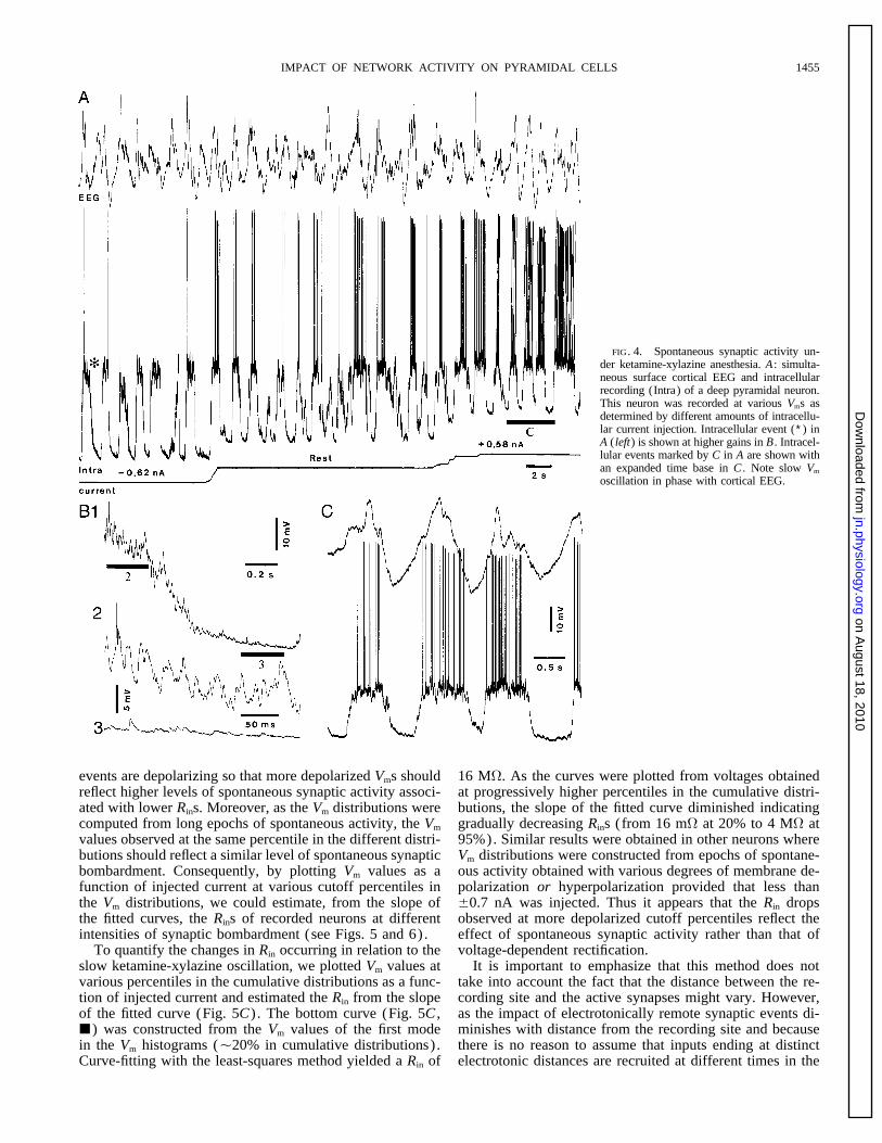
1995). As shown in Fig. 4B1 [expanded from the segment current injection on the two phases of the oscillation is mani-fested by the relatively invariant position of the depolarizedmarked (∗) in Fig. 4A , left] , the depolarized phase of the
oscillation appeared to result from the summation of numer- mode compared with that of the hyperpolarized one (Fig.5A) and by the gradually decreasing distance between theous PSPs. By contrast, a progressive decrease in the variabil-
ity of the intracellular signal occurred during the hyperpolar- cumulative distributions from hyperpolarized to depolarizedVms (Fig. 5B) .ization (cf. Fig. 4, B2 and B3) , consistent with a massive
disfacilitation. Further, the two phases of the oscillation were As the amount of spontaneous synaptic activity in vivowas constantly changing, inferring the Rins of the recordeddifferentially affected by the injection of intracellular cur-
rent, the hyperpolarized phase being much more sensitive neurons from their voltage response to series of currentpulses of increasing amplitude proved inadequate. Thus, tothan the depolarized one (Fig. 4A) . These observations sug-
gest that the Rin was relatively increased during the hyperpo- quantify the moment to moment changes in Rin associatedwith the varying level of spontaneous synaptic activity, alarized phase, in agreement with previous findings (Con-
treras et al. 1996). new statistical method was used. This method involved com-puting Vm distributions at various degrees of membrane po-In all neurons recorded in vivo under ketamine-xylazine,
the two phases of the oscillation gave rise to bimodal distri- larization as determined by steady current injection duringstationary epochs (Ç1 min) of spontaneous activity (Figs.butions of Vm values. This is shown in Fig. 5A where we
superimposed three Vm distributions computed from station- 5A and 6A) . Action potentials were digitally deleted to in-sure comparability of Vm distributions observed at differentary epochs of one minute obtained at different degrees of
membrane polarization in the same neuron (spontaneous degrees of polarization (see METHODS). Further, neuronsdisplaying marked inward rectification in the hyperpolariz-spikes were deleted digitally) . In each histogram, the first
mode, at negative values, corresponds to the hyperpolarized ing direction were not considered for this analysis. In neu-rons recorded in vivo, these distributions were markedlyphase of the slow oscillation whereas the second corresponds
to its depolarized phase. These bimodal histograms trans- skewed to the right and the level of positive skew increasedwith membrane hyperpolarization. It should be pointed outlated into cumulative Vm distributions with a characteristic
biphasic rise (Fig. 5B) . Further, the differential effect of that with KCl-filled pipettes, the vast majority of synaptic
J591-7/ 9k26$$mr16 02-10-98 19:59:50 neupal LP-Neurophys
on August 18, 2010
jn.physiology.orgD
ownloaded from
IMPACT OF NETWORK ACTIVITY ON PYRAMIDAL CELLS 1455
FIG. 4. Spontaneous synaptic activity un-der ketamine-xylazine anesthesia. A : simulta-neous surface cortical EEG and intracellularrecording (Intra) of a deep pyramidal neuron.This neuron was recorded at various Vms asdetermined by different amounts of intracellu-lar current injection. Intracellular event (∗) inA ( left) is shown at higher gains in B . Intracel-lular events marked by C in A are shown withan expanded time base in C . Note slow Vm
oscillation in phase with cortical EEG.
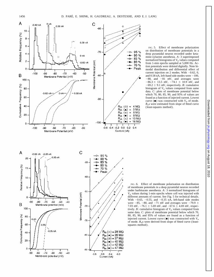
events are depolarizing so that more depolarized Vms should 16 MV. As the curves were plotted from voltages obtainedat progressively higher percentiles in the cumulative distri-reflect higher levels of spontaneous synaptic activity associ-
ated with lower Rins. Moreover, as the Vm distributions were butions, the slope of the fitted curve diminished indicatinggradually decreasing Rins (from 16 mV at 20% to 4 MV atcomputed from long epochs of spontaneous activity, the Vm
values observed at the same percentile in the different distri- 95%). Similar results were obtained in other neurons whereVm distributions were constructed from epochs of spontane-butions should reflect a similar level of spontaneous synaptic
bombardment. Consequently, by plotting Vm values as a ous activity obtained with various degrees of membrane de-polarization or hyperpolarization provided that less thanfunction of injected current at various cutoff percentiles in
the Vm distributions, we could estimate, from the slope of {0.7 nA was injected. Thus it appears that the Rin dropsobserved at more depolarized cutoff percentiles reflect thethe fitted curves, the Rins of recorded neurons at different
intensities of synaptic bombardment (see Figs. 5 and 6). effect of spontaneous synaptic activity rather than that ofvoltage-dependent rectification.To quantify the changes in Rin occurring in relation to the
slow ketamine-xylazine oscillation, we plotted Vm values at It is important to emphasize that this method does nottake into account the fact that the distance between the re-various percentiles in the cumulative distributions as a func-
tion of injected current and estimated the Rin from the slope cording site and the active synapses might vary. However,as the impact of electrotonically remote synaptic events di-of the fitted curve (Fig. 5C) . The bottom curve (Fig. 5C ,
j) was constructed from the Vm values of the first mode minishes with distance from the recording site and becausethere is no reason to assume that inputs ending at distinctin the Vm histograms (Ç20% in cumulative distributions) .
Curve-fitting with the least-squares method yielded a Rin of electrotonic distances are recruited at different times in the
J591-7/ 9k26$$mr16 02-10-98 19:59:50 neupal LP-Neurophys
on August 18, 2010
jn.physiology.orgD
ownloaded from
D. PARE, E. SHINK, H. GAUDREAU, A. DESTEXHE, AND E. J. LANG1456
FIG. 5. Effect of membrane polarizationon distribution of membrane potentials in adeep pyramidal neuron recorded under keta-mine-xylazine anesthesia. A : 3 superimposednormalized histograms of Vm values computedfrom 1-min epochs sampled at 5,000 Hz. Ac-tion potentials were deleted digitally. Note bi-modal distribution and differential effect ofcurrent injection on 2 modes. With 00.62, 0,and 0.58 nA, left-hand side modes were0100,088, and 081 mV, and averages were086.3 { 13.5 mV, 074.1 { 10.9 mV, and069.2 { 9.1 mV, respectively. B : cumulativehistogram of Vm values computed from samedata. C : plots of membrane potential belowwhich 70, 80, 85, 90, and 95% of values arefound as a function of injected current. Lowestcurve (j) was constructed with Vm of mode.Rins were estimated from slope of fitted curve(least-squares method).
FIG. 6. Effect of membrane polarization on distributionof membrane potentials in a deep pyramidal neuron recordedunder barbiturate anesthesia. A : 3 normalized histograms ofVm values during 1-min epochs where cell was injected withdifferent amounts of current. See Fig. 5 for technical details.With 00.65, 00.55, and 00.35 nA, left-hand side modeswere 085, 080, and 071 mV and averages were 079.9 {7.03 mV, 076.1 { 5.69 mV, and 067.6 { 4.69 mV, respec-tively. B : cumulative histogram of Vm values computed fromsame data. C : plots of membrane potential below which 70,80, 85, 90, and 95% of values are found as a function ofinjected current. Lowest curve (j) was constructed with Vm
of mode. Rins were derived from slope of fitted curve (least-squares method).
J591-7/ 9k26$$mr16 02-10-98 19:59:50 neupal LP-Neurophys
on August 18, 2010
jn.physiology.orgD
ownloaded from
IMPACT OF NETWORK ACTIVITY ON PYRAMIDAL CELLS 1457
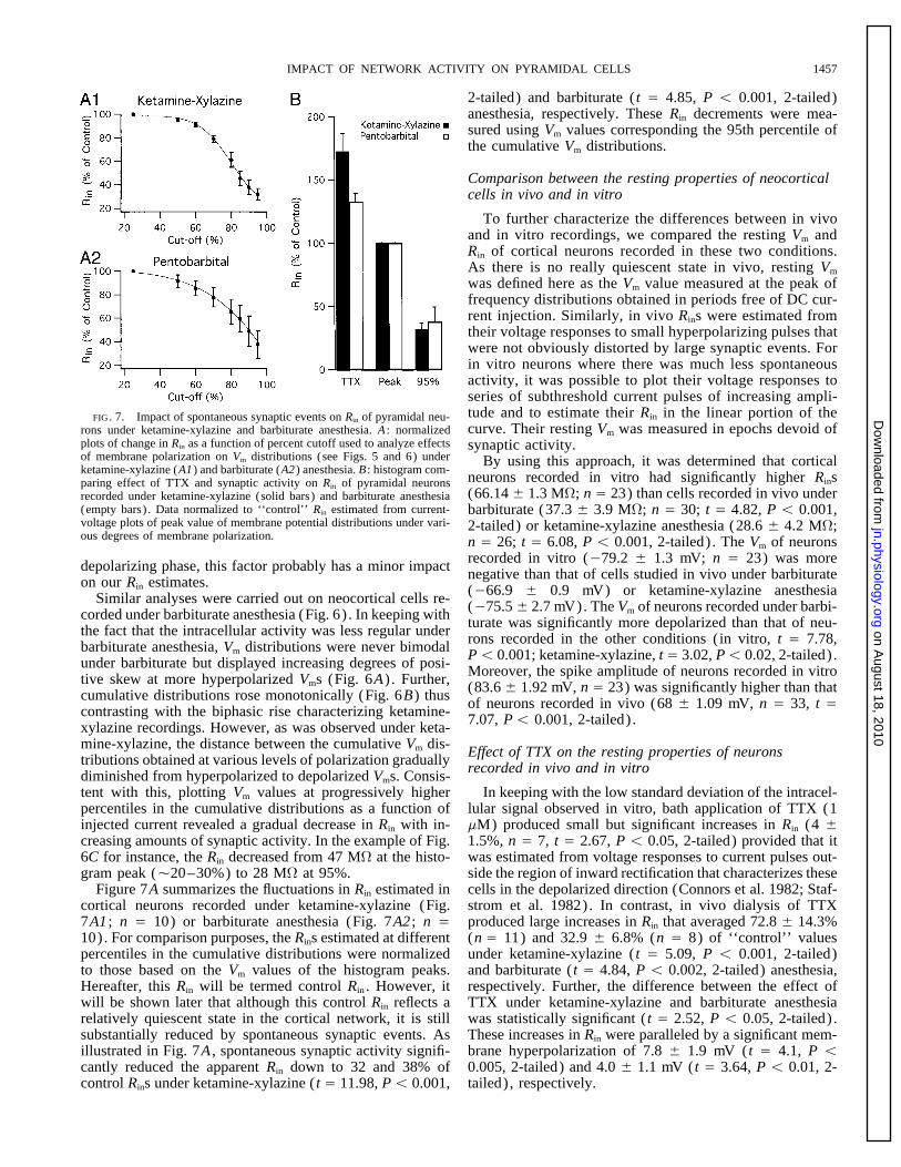
2-tailed) and barbiturate ( t Å 4.85, P õ 0.001, 2-tailed)anesthesia, respectively. These Rin decrements were mea-sured using Vm values corresponding the 95th percentile ofthe cumulative Vm distributions.
Comparison between the resting properties of neocorticalcells in vivo and in vitro
To further characterize the differences between in vivoand in vitro recordings, we compared the resting Vm andRin of cortical neurons recorded in these two conditions.As there is no really quiescent state in vivo, resting Vm
was defined here as the Vm value measured at the peak offrequency distributions obtained in periods free of DC cur-rent injection. Similarly, in vivo Rins were estimated fromtheir voltage responses to small hyperpolarizing pulses thatwere not obviously distorted by large synaptic events. Forin vitro neurons where there was much less spontaneousactivity, it was possible to plot their voltage responses toseries of subthreshold current pulses of increasing ampli-tude and to estimate their Rin in the linear portion of theFIG. 7. Impact of spontaneous synaptic events on Rin of pyramidal neu-curve. Their resting Vm was measured in epochs devoid ofrons under ketamine-xylazine and barbiturate anesthesia. A : normalized
plots of change in Rin as a function of percent cutoff used to analyze effects synaptic activity.of membrane polarization on Vm distributions (see Figs. 5 and 6) under By using this approach, it was determined that corticalketamine-xylazine (A1) and barbiturate (A2) anesthesia. B : histogram com-
neurons recorded in vitro had significantly higher Rinsparing effect of TTX and synaptic activity on Rin of pyramidal neurons(66.14 { 1.3 MV; n Å 23) than cells recorded in vivo underrecorded under ketamine-xylazine (solid bars) and barbiturate anesthesia
(empty bars) . Data normalized to ‘‘control’’ Rin estimated from current- barbiturate (37.3 { 3.9 MV; n Å 30; t Å 4.82, P õ 0.001,voltage plots of peak value of membrane potential distributions under vari- 2-tailed) or ketamine-xylazine anesthesia (28.6 { 4.2 MV;ous degrees of membrane polarization. n Å 26; t Å 6.08, P õ 0.001, 2-tailed) . The Vm of neurons
recorded in vitro (079.2 { 1.3 mV; n Å 23) was moredepolarizing phase, this factor probably has a minor impact negative than that of cells studied in vivo under barbiturateon our Rin estimates. (066.9 { 0.9 mV) or ketamine-xylazine anesthesiaSimilar analyses were carried out on neocortical cells re- (075.5 { 2.7 mV). The Vm of neurons recorded under barbi-corded under barbiturate anesthesia (Fig. 6) . In keeping with turate was significantly more depolarized than that of neu-the fact that the intracellular activity was less regular under rons recorded in the other conditions (in vitro, t Å 7.78,barbiturate anesthesia, Vm distributions were never bimodal Põ 0.001; ketamine-xylazine, t Å 3.02, Põ 0.02, 2-tailed) .under barbiturate but displayed increasing degrees of posi- Moreover, the spike amplitude of neurons recorded in vitrotive skew at more hyperpolarized Vms (Fig. 6A) . Further, (83.6 { 1.92 mV, n Å 23) was significantly higher than thatcumulative distributions rose monotonically (Fig. 6B) thus of neurons recorded in vivo (68 { 1.09 mV, n Å 33, t Åcontrasting with the biphasic rise characterizing ketamine- 7.07, Põ 0.001, 2-tailed) .xylazine recordings. However, as was observed under keta-mine-xylazine, the distance between the cumulative Vm dis- Effect of TTX on the resting properties of neuronstributions obtained at various levels of polarization gradually recorded in vivo and in vitrodiminished from hyperpolarized to depolarized Vms. Consis-tent with this, plotting Vm values at progressively higher In keeping with the low standard deviation of the intracel-
lular signal observed in vitro, bath application of TTX (1percentiles in the cumulative distributions as a function ofinjected current revealed a gradual decrease in Rin with in- mM) produced small but significant increases in Rin (4 {
1.5%, n Å 7, t Å 2.67, P õ 0.05, 2-tailed) provided that itcreasing amounts of synaptic activity. In the example of Fig.6C for instance, the Rin decreased from 47 MV at the histo- was estimated from voltage responses to current pulses out-
side the region of inward rectification that characterizes thesegram peak (Ç20–30%) to 28 MV at 95%.Figure 7A summarizes the fluctuations in Rin estimated in cells in the depolarized direction (Connors et al. 1982; Staf-
strom et al. 1982). In contrast, in vivo dialysis of TTXcortical neurons recorded under ketamine-xylazine (Fig.7A1 ; n Å 10) or barbiturate anesthesia (Fig. 7A2 ; n Å produced large increases in Rin that averaged 72.8 { 14.3%
(n Å 11) and 32.9 { 6.8% (n Å 8) of ‘‘control’’ values10). For comparison purposes, the Rins estimated at differentpercentiles in the cumulative distributions were normalized under ketamine-xylazine ( t Å 5.09, P õ 0.001, 2-tailed)
and barbiturate ( t Å 4.84, P õ 0.002, 2-tailed) anesthesia,to those based on the Vm values of the histogram peaks.Hereafter, this Rin will be termed control Rin . However, it respectively. Further, the difference between the effect of
TTX under ketamine-xylazine and barbiturate anesthesiawill be shown later that although this control Rin reflects arelatively quiescent state in the cortical network, it is still was statistically significant ( t Å 2.52, P õ 0.05, 2-tailed) .
These increases in Rin were paralleled by a significant mem-substantially reduced by spontaneous synaptic events. Asillustrated in Fig. 7A , spontaneous synaptic activity signifi- brane hyperpolarization of 7.8 { 1.9 mV ( t Å 4.1, P õ
0.005, 2-tailed) and 4.0 { 1.1 mV ( t Å 3.64, P õ 0.01, 2-cantly reduced the apparent Rin down to 32 and 38% ofcontrol Rins under ketamine-xylazine ( t Å 11.98, P õ 0.001, tailed) , respectively.
J591-7/ 9k26$$mr16 02-10-98 19:59:50 neupal LP-Neurophys
on August 18, 2010
jn.physiology.orgD
ownloaded from
D. PARE, E. SHINK, H. GAUDREAU, A. DESTEXHE, AND E. J. LANG1458
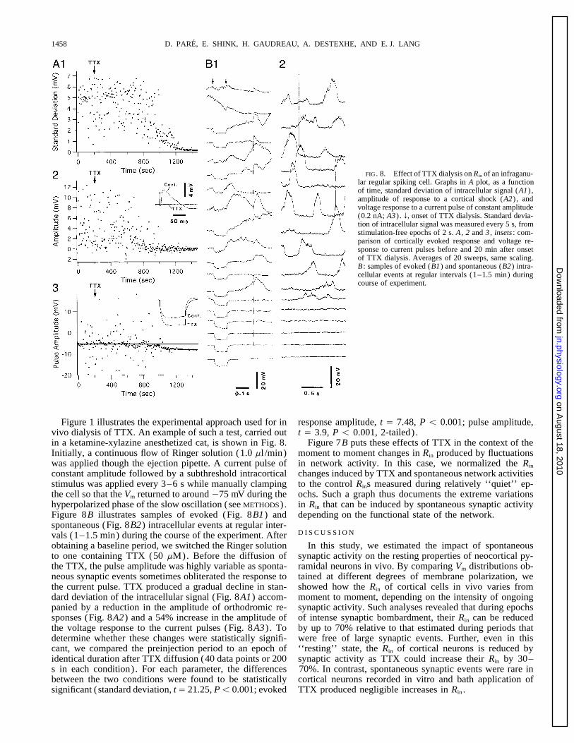
FIG. 8. Effect of TTX dialysis on Rin of an infraganu-lar regular spiking cell. Graphs in A plot, as a functionof time, standard deviation of intracellular signal (A1) ,amplitude of response to a cortical shock (A2) , andvoltage response to a current pulse of constant amplitude(0.2 nA; A3) . f, onset of TTX dialysis. Standard devia-tion of intracellular signal was measured every 5 s, fromstimulation-free epochs of 2 s. A , 2 and 3 , insets : com-parison of cortically evoked response and voltage re-sponse to current pulses before and 20 min after onsetof TTX dialysis. Averages of 20 sweeps, same scaling.B : samples of evoked (B1) and spontaneous (B2) intra-cellular events at regular intervals (1–1.5 min) duringcourse of experiment.
Figure 1 illustrates the experimental approach used for in response amplitude, t Å 7.48, P õ 0.001; pulse amplitude,t Å 3.9, P õ 0.001, 2-tailed) .vivo dialysis of TTX. An example of such a test, carried out
in a ketamine-xylazine anesthetized cat, is shown in Fig. 8. Figure 7B puts these effects of TTX in the context of themoment to moment changes in Rin produced by fluctuationsInitially, a continuous flow of Ringer solution (1.0 ml /min)
was applied though the ejection pipette. A current pulse of in network activity. In this case, we normalized the Rin
changes induced by TTX and spontaneous network activitiesconstant amplitude followed by a subthreshold intracorticalstimulus was applied every 3–6 s while manually clamping to the control Rins measured during relatively ‘‘quiet’’ ep-
ochs. Such a graph thus documents the extreme variationsthe cell so that the Vm returned to around 075 mV during thehyperpolarized phase of the slow oscillation (see METHODS). in Rin that can be induced by spontaneous synaptic activity
depending on the functional state of the network.Figure 8B illustrates samples of evoked (Fig. 8B1) andspontaneous (Fig. 8B2) intracellular events at regular inter-
D I S C U S S I O Nvals (1–1.5 min) during the course of the experiment. Afterobtaining a baseline period, we switched the Ringer solution In this study, we estimated the impact of spontaneous
synaptic activity on the resting properties of neocortical py-to one containing TTX (50 mM). Before the diffusion ofthe TTX, the pulse amplitude was highly variable as sponta- ramidal neurons in vivo. By comparing Vm distributions ob-
tained at different degrees of membrane polarization, weneous synaptic events sometimes obliterated the response tothe current pulse. TTX produced a gradual decline in stan- showed how the Rin of cortical cells in vivo varies from
moment to moment, depending on the intensity of ongoingdard deviation of the intracellular signal (Fig. 8A1) accom-panied by a reduction in the amplitude of orthodromic re- synaptic activity. Such analyses revealed that during epochs
of intense synaptic bombardment, their Rin can be reducedsponses (Fig. 8A2) and a 54% increase in the amplitude ofthe voltage response to the current pulses (Fig. 8A3) . To by up to 70% relative to that estimated during periods that
were free of large synaptic events. Further, even in thisdetermine whether these changes were statistically signifi-cant, we compared the preinjection period to an epoch of ‘‘resting’’ state, the Rin of cortical neurons is reduced by
synaptic activity as TTX could increase their Rin by 30–identical duration after TTX diffusion (40 data points or 200s in each condition). For each parameter, the differences 70%. In contrast, spontaneous synaptic events were rare in
cortical neurons recorded in vitro and bath application ofbetween the two conditions were found to be statisticallysignificant (standard deviation, tÅ 21.25, Põ 0.001; evoked TTX produced negligible increases in Rin .
J591-7/ 9k26$$mr16 02-10-98 19:59:50 neupal LP-Neurophys
on August 18, 2010
jn.physiology.orgD
ownloaded from
IMPACT OF NETWORK ACTIVITY ON PYRAMIDAL CELLS 1459
It should be noted that the methods used here to estimate in this model of the waking state, cortical cells are kept atdepolarized levels where their Rin is lowest.the changes in Rin do not distinguish the direct effects of
These considerations thus suggest that the cortical net-synaptic inputs from their indirect effects on intrinsic mem-work is constantly humming in conscious animals and thatbrane conductances. Indeed, the depolarization caused bythis produces dramatic reductions in the electrical compact-the synaptic bombardment probably activated various inwardness of cortical neurons. As this is likely to modify theand outward voltage-dependent conductances that reducedintegrative properties of pyramidal neurons, future modelingthe Rin . However, even if these changes in membrane con-studies should aim at characterizing how the synaptic bom-ductance are not caused directly by synaptic events, theybardment affects the interaction between synaptic inputs andremain an integral part of the impact of spontaneous synapticactive membrane properties in the dendrites of neocorticalactivity on cortical cells.pyramidal neurons. Preliminary simulations (Destexhe andAnother factor that should be considered is that becausePare 1997) suggest that the intense synaptic bombardmentwe used sharp electrodes, the impact of synaptic events waspresent in vivo tends to subdivide the dendritic tree of pyra-probably underestimated. Indeed, the mechanical damagemidal neurons into localized regions that, to some extent,produced by the impalement with sharp electrodes has beenprocess synaptic inputs independently.shown to introduce a somatic shunt (Spruston and Johnston
1992). By reducing the Rin , this shunt probably decreasedWe thank M. Steriade for comments on an earlier version of this manu-the influence of electrotonically remote synaptic events on
script, as well as P. Giguere, E. Lebel, D. Drolet, and G. Oakson forour measurements thus leading us to underestimate the mag-technical and programming support.
nitude of Rin decrements produced by spontaneous network This work was supported by grants from the National Sciences andactivities. Engineering Research Council, the Medical Research Council, and the Na-
tional Institute of Neurological Disorders and Stroke.Similarly, it should be kept in mind that TTX applicationAddress for reprint requests: D. Pare, Dept. de Physiologie, Faculte dein vivo did not only abolish fast GABAergic and gluta-
Medecine, Universite Laval, Quebec City, Quebec G1K 7P4, Canada.matergic synaptic events but also suppressed modulatory
Received 15 July 1997; accepted in final form 14 November 1997.inputs. Many modulatory actions (McCormick 1992), suchas those of acetylcholine through muscarinic receptors
REFERENCES(McCormick and Prince 1986) or those of glutamate throughmetabotropic receptors (Charpak et al. 1990), increase the ANIS, N. A., BERRY, S. C., BURTON, N. R., AND LODGE, D. The dissociativeRin of cortical neurons. Thus, suppression of these effects anaesthetics, ketamine and phencyclidine, selectively reduce excitation
of central mammalian neurones by N-methyl-D-aspartate. Br. J. Pharma-by TTX probably led us to further underestimate the impactcol. 79: 565–575, 1983.of fast synaptic events on the Rin of cortical neurons.
BARKER, J. L. AND MCBURNEY, R. M. Pentobarbitone modulation of post-Nevertheless, the results of the present study suggest that synaptic GABA receptor function on cultured mammalian neurones.
differences in background synaptic activity account for a Proc. R. Soc. Lond. B Biol. Sci. 206: 319–327, 1979.BERNANDER, O., DOUGLAS, R. J., MARTIN, K. C., AND KOCH, C. Synapticlarge portion of Rin differences between in vivo and in vitro
background activity influences spatiotemporal integration in single pyra-recordings. Indeed, correcting control Rins for the impact ofmidal cells. Proc. Natl. Acad. Sci. USA 88: 11569–11573, 1991.
the synaptic noise documented by in vivo applications of BINDMAN, L. J., MEYER, T., AND PRINCE, C. A. Comparison of the electricalTTX revealed that background synaptic activities account properties of neocortical neurons in slices in vitro and in the anaesthetized
rat. Exp. Brain Res. 69: 489–496, 1988.for Ç40–53% of Rin differences between in vivo and inCHARPAK, S., GAHWILER, B. H., DO, K., AND KNOPFEL, T. Potassium con-vitro recordings. It would be premature to conclude that the
ductances in hippocampal neurons blocked by excitatory amino-acidremaining differences in Rin are due to the fact that sharp transmitters. Nature 347: 765–767, 1990.electrodes produce more damage in vivo than in vitro. First, CONNORS, B. W., GUTNICK, M. J., AND PRINCE, D. A. Electrophysiological
properties of neocortical neurons in vitro. J. Neurophysiol. 48: 1302–pentobarbital directly activates GABAA receptors (Rho et1320, 1982.al. 1996), whereas xylazine hyperpolarizes mammalian neu-
CONTRERAS, D. AND STERIADE, M. Cellular basis of EEG slow rhythms: arons by the activation of a potassium conductance through study of dynamic corticothalamic relationships. J. Neurosci. 15: 604–a2 noradrenergic receptors (Nicoll et al. 1990), thus reduc- 622, 1995.
CONTRERAS, D., TIMOFEEV, I., AND STERIADE, M. Mechanisms of longing the Rin of in vivo neurons. Second, spike-independentlasting hyperpolarizations underlying slow sleep oscillations in cat corti-synaptic events are more frequent in vivo than in vitro andcothalamic networks. J. Physiol. (Lond.) 494: 251–264, 1996.produce significant Rin reductions in neocortical pyramidal DEFELIPE, J. AND FARINAS, I. The pyramidal neuron of the cerebral cortex:
cells (Pare et al. 1997). morphological and chemical characteristics of the synaptic inputs. Prog.Neurobiol. 39: 563–607, 1992.It could be argued that the large impact of synaptic events
DESTEXHE, A. AND PARE, D. Spontaneous synaptic activity modulate actiondocumented in this study is an artifact induced by the anes-potential generation in neocortical pyramidal cells in vivo. Soc. Neurosci.thetics and that the background synaptic activity is much Abstr. 23: 448, 1997.
lower in conscious animals. However, this is inconsistent GRUNER, J. E., HIRSCH, J. C., AND SOTELO, C. Ultrastructural features ofthe isolated suprasylvian gyrus. J. Comp. Neurol. 154: 1–27, 1974.with the high rate of spontaneous discharge reported for
HORIKAWA, K. AND ARMSTRONG, W. E. A versatile means of intracellularcorticofugal pyramidal neurons in unanesthetized animalslabeling: injection of biocytin and its detection with avidin conjugates.(Steriade 1978; Steriade et al. 1974) and with the depressant J. Neurosci. Methods 25: 1–11, 1988.
actions of anesthetics on mammalian neurons. Moreover, JOHNSTON, D., MAGEE, J. C., COLBERT, C. M., AND CHRISTIE, B. R. Activeproperties of neuronal dendrites. Annu. Rev. Neurosci. 19: 165–186,under ketamine-xylazine anesthesia, electrical stimulation of1996.brain stem activating systems that are believed to maintain
MCCORMICK, D. A. Neurotransmitter actions in the thalamus and cerebralthe awake state in normal circumstances abolishes the hyper- cortex and their role in neuromodulation of thalamocortical activity. Prog.polarized phase of the slow oscillation and maintains cortical Neurobiol. 39: 337–388, 1992.
MCCORMICK, D. A. AND PRINCE, D. A. Mechanisms of action of acetylcho-cells in the depolarized phase (Steriade et al. 1993a). Thus,
J591-7/ 9k26$$mr16 02-10-98 19:59:50 neupal LP-Neurophys
on August 18, 2010
jn.physiology.orgD
ownloaded from
D. PARE, E. SHINK, H. GAUDREAU, A. DESTEXHE, AND E. J. LANG1460
line in the guinea-pig cerebral cortex in vitro. J. Physiol. (Lond.) 375: STAFSTROM, C. E., SCHWINDT, P. C., AND CRILL, W. E. Negative slope con-ductance due to a persistent sodium current in cat neocortical neurons in169–194, 1986.
MCCORMICK, D. A., CONNORS, B. W., LIGHTALL, J. W., AND PRINCE, D. A. vitro. Brain Res. 236: 221–226, 1982.STERIADE, M. Cortical long-axoned cells and putative interneurons duringComparative electrophysiology of pyramidal and sparsely spiny stellate
neurons of the neocortex. J. Neurophysiol. 54: 782–806, 1985. the sleep-waking cycle. Behav. Brain Sci. 3: 465–514, 1978.STERIADE, M., AMZICA, F., AND NUNEZ, A. Cholinergic and noradrenergicNICOLL, R. A., MALENKA, R. C., AND KAUER, J. Functional comparison of
neurotransmitter receptor subtypes in mammalian central nervous system. modulation of the slow (É0.3 Hz) oscillation in neocortical cells. J.Neurophysiol. 70: 1385–1400, 1993a.Physiol. Rev. 70: 513–565, 1990.
PARE, D., LEBEL, E., AND LANG, E. J. Differential impact of miniature STERIADE, M., DESCHENES, M., AND OAKSON, G. Inhibitory processes andinterneuronal apparatus in motor cortex during sleep and waking. I. Back-synaptic potentials on the soma and dendrites of pyramidal neurons in
vivo. J. Neurophysiol. 78: 1735–1739, 1997. ground firing and responsiveness of pyramidal tract neurons and interneu-rons. J. Neurophysiol. 37: 1065–1092, 1974.PARE, D., SHINK, E., GAUDREAU, H., DESTEXHE, A., AND LANG, E. J. Impact
of spontaneous synaptic activity on the resting properties of neocortical STERIADE, M., NUNEZ, A., AND AMZICA, F. Intracellular analysis of relationsbetween the slow (õ1 Hz) neocortical oscillation and other sleep rhythmspyramidal neurons in vivo. Soc. Neurosci. Abstr. 23: 448, 1997.
RHO, J. M., DONEVAN, S. D., ROGAWSKI, M. A. Direct activation of GABAA of the electroencephalogram. J. Neurosci. 13: 3266–3283, 1993b.SZENTAGOTHAI, J. The use of degeneration in the investigation of shortreceptors by barbiturates in cultured rat hippocampal neurons. J. Physiol.
(Lond.) 497: 509–522, 1996. neuronal connections. In: Progress in Brain Research, edited by M.Singer and J. P. Shade. Amsterdam: Elsevier, 1965, vol. 14, p. 1–32.SPRUSTON, N. AND JOHNSTON, D. Perforated patch-clamp analysis of the
passive membrane properties of three classes of hippocampal neurons. YUSTE, R. AND TANK, D. W. Dendritic integration in mammalian neurons,a century after Cajal. Neuron 16: 701–716, 1996.J. Neurophysiol. 67: 508–529, 1992.
J591-7/ 9k26$$mr16 02-10-98 19:59:50 neupal LP-Neurophys
on August 18, 2010
jn.physiology.orgD
ownloaded from