Embed Size (px)
Citation preview

Submitted 22 September 2016Accepted 25 October 2016Published 13 December 2016
Corresponding authorJing-Shan Shi, [email protected]
Academic editorJie Liu
Additional Information andDeclarations can be found onpage 13
DOI 10.7717/peerj.2739
Copyright2016 Nie et al.
Distributed underCreative Commons CC-BY 4.0
OPEN ACCESS
Dendrobium alkaloids prevent Aβ25–35-induced neuronal and synaptic lossvia promoting neurotrophic factorsexpression in miceJing Nie1,2, Yong Tian2, Yu Zhang2, Yan-Liu Lu2, Li-Sheng Li2 andJing-Shan Shi1,2
1 Shanghai University of Traditional Chinese Medicine, Shanghai, China2Department of Pharmacology and the Key Laboratory of Basic Pharmacology of Guizhou Province, ZunyiMedical College, Zunyi, Guizhou Province, China
ABSTRACTBackground. Neuronal and synaptic loss is the most important risk factor for cognitiveimpairment. Inhibiting neuronal apoptosis and preventing synaptic loss are promisingtherapeutic approaches for Alzheimer’s disease (AD). In this study, we investigate theprotective effects of Dendrobium alkaloids (DNLA), a Chinese medicinal herb extract,on β-amyloid peptide segment 25–35 (Aβ25-35)-induced neuron and synaptic loss inmice.Method. Aβ25–35(10 µg) was injected into the bilateral ventricles of male mice followedby an oral administration of DNLA (40 mg/kg) for 19 days. The Morris water maze wasused for evaluating the ability of spatial learning and memory function of mice. Themorphological changes were examined via H&E staining and Nissl staining. TUNELstaining was used to check the neuronal apoptosis. The ultrastructure changes ofneurons were observed under electron microscope. Western blot was used to evaluatethe protein expression levels of ciliary neurotrophic factor (CNTF), glial cell line-derived neurotrophic factor (GDNF), and brain-derived neurotrophic factor (BDNF)in the hippocampus and cortex.Results. DNLA significantly attenuated Aβ25–35-induced spatial learning and memoryimpairments in mice. DNLA prevented Aβ25–35-induced neuronal loss in the hip-pocampus and cortex, increased the number of Nissl bodies, improved the ultrastruc-tural injury of neurons and increased the number of synapses in neurons. Furthermore,DNLA increased the protein expression of neurotrophic factors BDNF, CNTF andGDNF in the hippocampus and cortex.Conclusions. DNLA can prevent neuronal apoptosis and synaptic loss. This effect ismediated at least in part via increasing the expression of BDNF, GDNF and CNTFin the hippocampus and cortex; improving Aβ-induced spatial learning and memoryimpairment in mice.
Subjects Neuroscience, Drugs and Devices, PharmacologyKeywords Alzheimer’s disease, Neuron, Synaptic loss, Dendrobium nobile Lindl. alkaloids,Neurotrophic factor, Apoptosis
How to cite this article Nie et al. (2016), Dendrobium alkaloids prevent Aβ25–35-induced neuronal and synaptic loss via promoting neu-rotrophic factors expression in mice. PeerJ 4:e2739; DOI 10.7717/peerj.2739
INTRODUCTIONAlzheimer’s disease (AD) is the most common type of dementia, and is characterizedby progressive memory impairment and cognitive decline (Roberson & Mucke, 2006).Mounting evidence has indicated that dementia attributed to synaptic dysfunction andneuronal loss in the hippocampus and its associated cortex (Youssef et al., 2008; Selkoe, 2002;Niikura et al., 2002; Scheff et al., 2007), which are caused by the accumulation of solubleAβ oligomers (Rowan et al., 2007; Lacor et al., 2004; Haass & Selkoe, 2007). Aβ is themajor constituent in senile plaques and cerebral amyloid angiopathy, which are two mostdistinctive histopathologies inAD.The accumulation ofAβ in the brain initiates a cascade ofevents, such as activating astrocytes andmicroglia, initiating inflammatory responses, whichlead to oxidative injury, altering neuronal ionic homeostasis, kinases/phosphatase activities,and so on (Klafki et al., 2006). These cascades result in a wide range of neuronal/synapticdysfunction and loss, as well as loss of neurotrophin retrograde transport, thereforecausing patients to present with the symptoms of dementia. Adjustment of the pathologicalprogress of amyloid peptide is a key strategy to slow down the AD progression. In additionto reduction of the Aβ levels in the brain (Klafki et al., 2006), the concomitant application ofneuroprotective agents may be the alternative therapeutics for AD, and a strategy to preventprogressive synaptic and cognitive degeneration (Klafki et al., 2006). Neurotrophins (NT)are synthesized and secreted by the target tissue, and after binding to its receptors andtransported in a retrograde manner to the cell body. They exert a wide range of actions,including neuronal survival and differentiation, modulation of neuronal excitability,development and maintenance of synapses and modification of synaptic structure andfunction (Poo, 2001). The application of neurotrophic factors such as BDNF and CNTFcould enable the modulation of neuronal survival and synaptic connectivity (Lu, Christian& Lu, 2008; Garcia et al., 2010)
Dendrobium nobile is a traditional Chinese herbal medicine. In our previous studies,alkaloids extract fromDendrobium nobile Lindl. (DNLA) showed neuro-protective activity.For example, DNLA can prevent neuronal damages induced by LPS (Li et al., 2011; Zhanget al., 2011), and oxygen-glucose deprivation and reperfusion (Wang et al., 2010), decreaseneuronal apoptosis, hyperphosphorylation of tau protein (Yang et al., 2014) and Aβdeposition in rat brain (Chen et al., 2008). The present study aimed to explore the effects ofDNLA in protecting neurons from Aβ25–35-induced neurotoxicity in mice, and analyzedthe mechanism from the aspects of promoting the secretion of neurotrophic factors.
MATERIALS AND METHODSDrugs and reagentsDendrobium was collected from Dendrobium planting regions of Xintian TraditionalChinese Medicine Industry Development co., LTD of Guizhou Province in 2014. The driedstems of the herb (10 kg) were extracted by 95% ethanol solution. DNLA was isolated fromthe extracts, and analyzed by LC-MS/MS. Alkaloids accounted for 79.8% of DNLA, andmainly contained Dendrobine (C16H25O2N, 92.6%), Dendrobine-N-oxide (C16H25O3N,
Nie et al. (2016), PeerJ, DOI 10.7717/peerj.2739 2/16

Figure 1 Chemical structures and chromatograms of Dendrobium alkaloids. Chemical structures and chromatograms of Dendrobium alkaloids(A) Dendrobine, (B) Dendrobine-N-oxide, (C) Nobilonine, (D) Dendroxine, (E) 6-Hydroxy-nobilonine, and (F) 13-Hydroxy-14-oxodendrobine.(G) Chromatogams of Dendrobium alkaloids. Differences between the accurately tested and calculated m/z values were <2 ppm for [M+H]+ions:(1) Dendrobine, (2) Dendrobine-N-oxide, (3) Nobilonine, (4) Dendroxine, (5) 6-Hydroxy-nobilonine, and (6) 13-Hydroxy-14-oxodendrobine.
3.3%), Nobilonine (C17H27O3N, 2.0%), Dendroxine (C17H25O3N, 0.9%), 6-Hydroxy-nobilonine (C17H27O4N, 0.32%), and 13-Hydroxy-14-oxodendrobine (C16H23O4N,0.07%). The chemical structures of these ingredients were shown in the Fig. 1A, and thechromatograms of the sample solutions were shown in the Fig. 1B.
Aβ25–35 (Sigma, St Louis, MO, USA) was dissolved in physiological saline to a finalconcentration of 2.5 g/L, and incubated for one week at 37 ◦C to reach a state of aggregation.CNTF antibodies (ab190985) and GDNF antibodies (ab18956) were obtained from Abcam(Cambridge, England). BDNF (sc-546) was purchased from Santa Cruz Biotechnologies
Nie et al. (2016), PeerJ, DOI 10.7717/peerj.2739 3/16

(Santa Cruz, CA, USA). TUNEL kits (In Situ Cell Death Detection Kit and POD) werepurchased from Roche (Switzerland).
AnimalsMale Kunming mice (20–25 g) were purchased from the Laboratory Animal Center,Chongqing, China (Grade: specific pathogen-free [SPF], Certificate no.: SCXK 2012-0005).Mice were housed in SPF-grade animal facilities (Certificate no.: SYXK 2014-003), with22–23 ◦C, and a 12-hour light/dark cycle. Mice were given food and water freely. All animalprocedures were approved by the animal experimental ethical committee of Zunyi MedicalCollege.
Experimental designsThe animals were randomly divided into three groups (n= 6): sham surgery, model, andDNLA (40 mg/kg) treatment group. Mice were anesthetized with 7% chloral hydrate(35–45 mg/kg, i.p.) and fixed on a stereotaxic instrument (RWD Life Science, Guangdong,China). Then Aβ25–35 (2.5 µg/µL, 2 µL) was injected into both lateral cerebral ventricle inthe model and DNLA groups via a 5-µl microinjector. The injection site was posterior fromthe bregma (AP) = −0.4, mediolateral from the midline (MR) = 1.2, and dorsoventralfrom the skull (DV) = 2.7. Mice in the sham group were treated the same as the modelgroup except for the injection of Aβ25–35 instead of sterile normal saline (Nie et al., 2010).Mice in the DNLA group were administered intragastrically with DNLA (40 mg/kg) onthe first day after the injection for 19 consecutive days, while mice in the sham and modelgroups were administered with distilled water.
Morris water maze testThe ability of spatial learning and memory of the mice was evaluated through the MorrisWater Maze (MWM) test (Edwards et al., 2014;Milner et al., 2014). The test was performedon the 14th day after drug administration. The pool ofMWMwas 120 cm in diameter, filledwith water (22–25 ◦C) and divided into four quadrants. A hidden platform was placedin the center of the target quadrant 1.5–2.0 cm under the surface of the water. MWMincluded the place navigation test and the spatial probe test. In the place navigation test, allmice were trained three times per day for four days. Mice were initially placed in the threequadrants which did not have a platform. Each trail lasted for 60 s or ended as soon as themouse climbed on the platform. The time was recorded as the escape latency, if the mouseclimbed on the platform within 60 s. If the mouse failed to find the platform within 60 s,its escape latency was recorded as 60 s. The spatial probe test was performed following theend of the place navigation test. In this test, the platform was removed, and each mousewas allowed to swim for 60 s. The searching distance in the target area and total area weremeasured. Data was recorded and analyzed by TopViewAnimal Behavior Analyzing System(Version 3.00).
Morphometric analysisAfter the MWM test, three mice were selected randomly from each group and anesthetizedwith 7% chloral hydrate. Then these mice were perfused with phosphate-buffered saline
Nie et al. (2016), PeerJ, DOI 10.7717/peerj.2739 4/16

(0.1 M, 4 ◦C) via the ascending aorta, followed by 4% paraformaldehyde until the tailand limbs were rigid. Thereafter, the brains were removed and cut in half. Half of thebrain was fixed with 4% paraformaldehyde for seven days and cut into coronal sections(4-µm thick) for hematoxylin-eosin (H&E), Nissl and TUNEL staining (Yang et al., 2014;Liu et al., 2015). The other half was fixed with 2.5% glutaraldehyde for electron microscopedetection.
Western blot analysisWestern blot analysis was performed as previously reported (Liu et al., 2015; Li et al., 2015).Three mice from each group were sacrificed after WMW test, and the hippocampal andcortical tissues were collected and homogenized in RIPA lysis buffer (1:5, w/v). Proteinconcentrations were determined by BCA protein assay kit. An aliquot of 45 µg of proteinwas applied for electrophoresis followed by the transferring of protein to the polyvinylidenedifluoride (PVDF) membranes. The membranes were blocked with 5% nonfat dry milkin TBST buffer for one hour at room temperature, and incubated with the primaryantibody: anti-CNTF (1:1,000), BDNF (1:1,000), and GDNF (1:1,000) at 4 ◦C overnight.Next, HRP-labeled goat anti-rabbit IgG (Beyotime Biotechnology, Jiangsu, China; A0208,1:2,000) was incubated with the membrane at room temperature for one hour. The blotswere visualized using the enhanced ECLWestern blot detection kit (7Sea Biotech, Shanghai,China) and scanned to Gel Imaging. The band intensity was quantified using QuantityOne 1-D analysis software v4.52 (BioRad, Hercules, California, USA).
Statistical analysisAll data were presented asmean± standard error. Data were analyzed by SPSS 13.0 statisticssoftware by one-way ANOVA or student’s t -test. P < 0.05 was considered as statisticallysignificant.
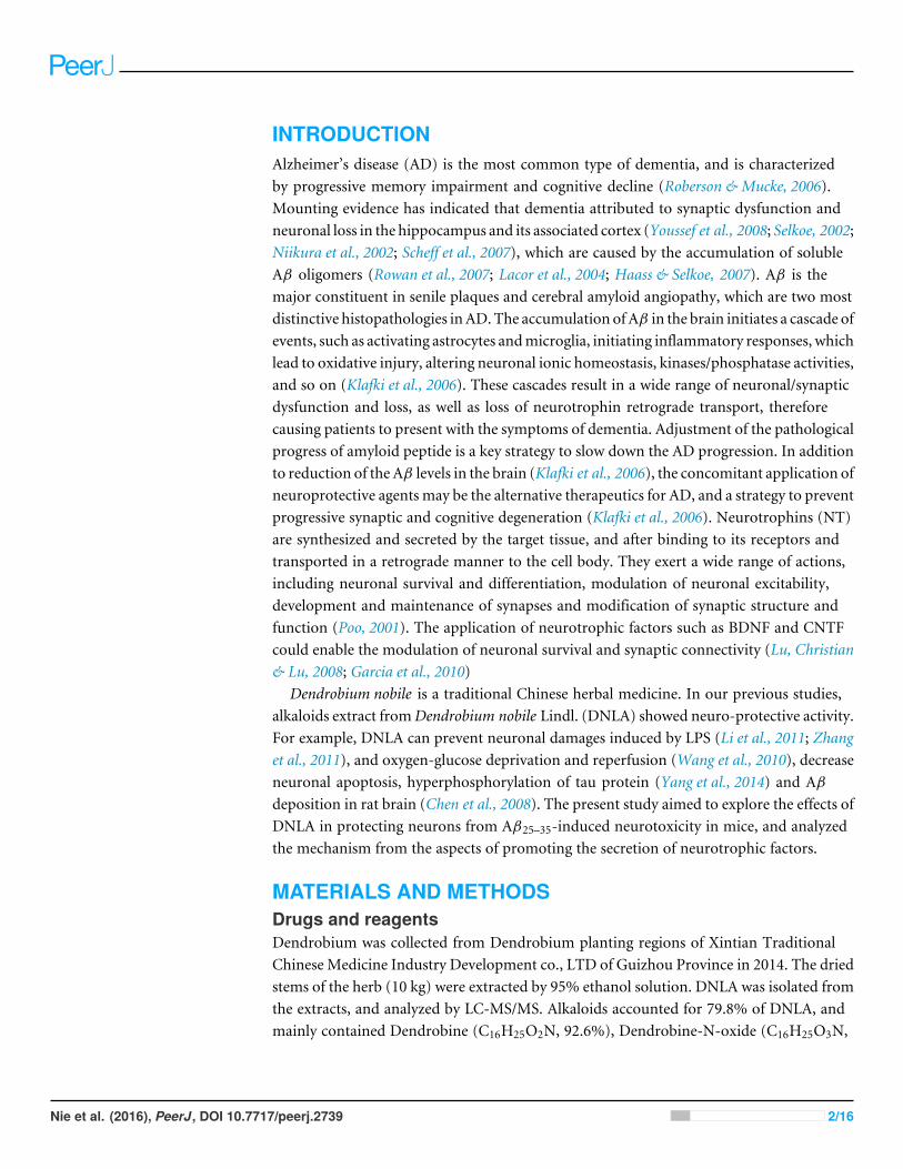
RESULTSProtective effects of DNLA on learning and memory deficits inducedby Aβ25–35Compared with the sham group, the escape latency of the model group was significantlyprolonged in the navigation test, and the explored distance in the target area was decreasedin the spatial probe test. After treatment for 14 days, the escape latency of the DNLA groupwas decreased and the explored distance in the target area was increased as compared withthe model group. The results indicated that DNLA could protect the mice against learningand memory deficits which were induced by Aβ25–35 injection (Fig. 2).
Morphological changes of cortex and hippocampus tissuesH&E staining was performed to observe the morphological changes of cortex andhippocampus tissues in mice brain. The results showed that the morphology of braintissues in the sham group was normal, and the cell density was higher. However, loosestructures in hippocampal and cortical neurons with disordered hippocampal pyramidalcell layers occurred in the model group. Furthermore, the images showed that not onlythe number of neurons was reduced, but also that neurons staining were abnormal with
Nie et al. (2016), PeerJ, DOI 10.7717/peerj.2739 5/16
Figure 2 Results of the Morris water maze test. Effect of DNLA on Aβ25−35-induced learning andmemory impairment. (A) The graph represented the escape latency of the different groups. (B) Graphsdescribed the adjusted searching distance in the space probe test. Data were expressed as mean± SEM(n= 6). ∗P < 0.05 vs. the sham group; #P < 0.05 vs. model.
nuclear condensation in the model group. DNLA treatment significantly decreased thenumber of abnormally stained neurons. Also, themorphology of brain tissues was generallynormal in the DNLA group (Fig. 3).
Effect of DNLA on apoptosis in the cortex and hippocampusThe effect of DNLA on neuronal apoptosis induced by Aβ25–35 was observed using theTUNELmethod. The results revealed that the number of apoptotic cells in the hippocampusand cortex were not affected in the sham group, but were increased in the model group.DNLA treatment decreased the number of apoptotic cells (Fig. 4).
Nie et al. (2016), PeerJ, DOI 10.7717/peerj.2739 6/16

Figure 3 Images of H&E staining. Effects of DNLA on morphological alterations in the hippocampusand cortex induced by Aβ25−35. Sections of the hippocampus CA3 region and cortex were obtained andstained with H&E (magnification, 200×). (A–C) showed the CA3 hippocampal cyto-architecture of micein sham, model and DNLA groups, respectively; (D–F) showed the cortical cyto-architecture of mice inthe sham group, model group and DNLA groups, respectively.
Figure 4 Images of TUNEL staining. Effects of DNLA on Aβ25-35-induced neuronal apoptosis in hip-pocampus and cortex. Sections of the hippocampus CA3 region and cortex were obtained and stainedwith TUNEL 184 staining (magnification, 200×). (A–C) showed the CA3 hippocampal cyto-architectureof mice in sham, model and DNLA groups, respectively; (D–F) showed the cortical cyto-architecture ofmice in the sham group, model group and DNLA groups, respectively. The cells dyed brown are apoptoticcells. (G) The number of the apoptotic neurons in hippocampus and cortex (x̄± SEM ,n= 3). **P < 0.01vs. the sham group; ##P < 0.01 vs. the model group.
Changes in Nissl bodies in cortex and hippocampus tissuesAs a neural characteristic structure, the number of Nissl bodies reflects the state ofneurons. In physiological conditions, the Nissl bodies were big and abundant, showing thatthe function of neuronal protein synthesis was strong; on the other hand, when nerve cellswere damaged, the number of Nissl bodies will be reduced or even disappear. IntraneuralNissl bodies of the cortex and hippocampus were lightly stained and appeared to be sparsely
Nie et al. (2016), PeerJ, DOI 10.7717/peerj.2739 7/16

Figure 5 Images of Nissl staining. Effects of DNLA on Aβ25−35-induced neuronal Nissl bodies in thehippocampus and cortex. Sections of the hippocampus CA3 region and cortex were obtained and stainedwith Nissl staining (400×). (A–C) showed the CA3 hippocampal cyto-architecture of mice in sham, modeland DNLA groups, respectively; (D–F) showed the cortical cyto-architecture of mice in the sham group,model group and DNLA groups, respectively.
arranged in the model group. However, deeper stained Nissl bodies with higher density incortex and hippocampus neurons were found in the sham and DNLA groups (Fig. 5).
Effects of DNLA on the ultrastructures of neurons in Aβ25–35-injectedmiceThe ultrastructures of neurons were observed under electron microscope. The resultsrevealed that the nuclear morphology of mice in the sham group was normal, and thechromatin was normally distributed. In the cytoplasm, mitochondrial morphology wasnormal with clear and regularly-arranged cristae. Furthermore, a number of ribosomescould be observed in these regularly-arranged rough endoplasmic reticulum. However, inthe model group, the ultrastructures of neurons were abnormal. It could be found thatnuclear membrane had folded or had obscure boundaries, mitochondria were swellingeven without the cristae, and the endoplasmic reticulum was swelling. DNLA treatmentcould reduce the injury. Compared with the model group, nuclear membrane was moresmooth, swelling ofmitochondrial was reduced, and partial rupture of cristae were observedin the DNLA group (Fig. 6). The neuronal synapses were also observed under electronmicroscope. Injection of Aβ25–35 would reduce the number of synapses and synapticvesicles, and cause the shape and size of synaptic vesicles uneven. The structure of synapseswas obscured, and the structure of the post-synaptic lattice became thin. DNLA inhibitedthe loss of synapses significantly (P < 0.05). Also, in the DNLA group, the generally normalsynaptic structures were maintained (Fig. 7).
Nie et al. (2016), PeerJ, DOI 10.7717/peerj.2739 8/16

Figure 6 Images of the ultrastructures of neurons. Effects of DNLA on the ultrastructures of neuronsin Aβ25−35-injected mice. The ultrastructures were observed under electron microscope (12kx×). (A–C)showed the ultrastructures of hippocampal neurons in sham, model and DNLA groups, respectively; (D–F) showed the ultrastructures of cortical neurons in sham, model and DNLA groups, respectively.
Effects of DNLA on the protein expressions of BDNF, CNTF and GDNFin the cortex and hippocampusThe protein expressions were analyzed by Western blot. Compared with the sham group,the expressions of BDNF, CNTF and GDNF significantly decreased in the model group.DNLA group had significantly increased expressions of BDNF, CNTF and GDNF in thecortex and hippocamous (Fig. 8).
DISCUSSIONNumerous literatures have reported that intra-hippocampal or intra-cerebroventricularinjections of Aβ1–42 or Aβ25–35 fragments into rats or mice could induce neuronal death,and alter spatial learning and memory performance. Aβ could induce a variety of injuries,such as oxidative injury and disturbed neuronal ionic homeostasis, which could eventuallyresult in neuronal dysfunction and selective neuronal loss (Lue et al., 1999; Niikura et al.,2002). Excessive neuronal loss can damage brain functions, and the cognitive ability isdestroyed firstly. In this study, we established the mice model which was induced by theintra-cerebroventricular injection of Aβ25–35, and found that there were a lot of apoptoticneurons in the hippocampus CA3 region and cortex. The results of MWM test indicatedthat neuronal apoptosis was accompanied with an obvious failure in spatial learning andmemory performance. On the other hand, DNLA treatment could significantly improvethe ability of spatial learning and memory, and decreased apoptosis in TUNEL and H&Estaining. This result demonstrated that DNLA could improve learning andmemory deficitsin AD model mice, and the protection may be due to the decreased apoptosis induced byAβ25–35.
Nie et al. (2016), PeerJ, DOI 10.7717/peerj.2739 9/16

Figure 7 Effects of DNLA on neuronal synapses in Aβ25–35-injected mice. The neuronal synapses wereobserved under electron microscope (50kx×). (A–C) showed the synapses of hippocampal neuronsin sham, model and DNLA groups, respectively; (D–F) showed the synapses of cortical neurons in thesham, model and DNLA groups, respectively. (G) showed the effects of DNLA on the number of neuronalsynapses in Aβ25−35-induced mice. Data were expressed as mean± SEM (n = 3). ∗P < 0.05 vs. the shamgroup; #P < 0.05 vs. the model group.
As it is known, when neurons are damaged, Nissl bodies are most sensitive. Themain changes of Nissl bodies associated with neuronal injury include the dissolutionand disappearance of Nissl bodies. In the model group, the number of Nissl bodies wasdecreased in cells. After the administration of DNLA, the number was increased. Theresults indicated DNLA could alleviate neuronal damage.
It has reported that memory disorders in AD patients begin with subtle changes inhippocampal synaptic efficacy earlier than extensive neuronal degeneration. Davies et al.(1987) performed a quantitativemorphometric analysis tomeasure the densities of neuronsand synapses in cerebral cortical biopsy tissues collected from AD patients. They foundthat the numerical density of synapses decreased 25%–35%, and the number of synapsesper cortical neuron decreased 15%–35% in AD patients (Davies et al., 1987). Evidence alsosuggested that synaptic dysfunction is caused by oligomeric assemblies of Aβ (Lacor et al.,2004; Rowan et al., 2007). Even in very mildly impaired patients, the degree of synapticloss in the cortex showed a significant association with soluble Aβ levels. Our results
Nie et al. (2016), PeerJ, DOI 10.7717/peerj.2739 10/16

Figure 8 Results of western blot. Effects of DNLA on the protein expressions of BDNF, CNTF andGDNF in brain tissues. (A) Protein contents of hippocampal tissues were plotted for sham, model andDNLA groups, respectively. The corresponding quantitation of BDNF, CNTF, GDNF protein wereshowed in (B–D) respectively. (E) Protein contents of cortex tissues were plotted for sham, model andDNLA groups, respectively. The corresponding quantitation of BDNF, CNTF, GDNF protein wereshowed in (F–H) respectively. The relative optical density was normalized to β-actin. Data were expressedas mean± SEM (n = 3). Significance ∗P < 0.05, ∗∗P < 0.01 compared to the sham group, #P < 0.05,##P < 0.01 compared to the model group.
Nie et al. (2016), PeerJ, DOI 10.7717/peerj.2739 11/16
revealed that Aβ25–35 induced synapse loss in the hippocampus and cortex, and DLNAcould dramatically inhibit the loss of synapses and improve synaptic structures, therebyimproving the learning and memory abilities of mice.
The application of neurotrophic factors could enable the modulation of neuronalsurvival and synaptic connectivity (Poo, 2001). Neurotrophic factors are synthesized andsecreted by the target tissue. After binding with receptors (tyrosine receptor kinase [Trk]and pan-neurotrophin receptot [p75]), these factors are internalized and transportedto the cell body in a retrograde manner, where they would affect neuronal survival anddifferentiation (Poo, 2001; Barker & Shooter, 1994). The loss of neurons in the AD processis at least partly due to the lack of one or more neurotrophic factors. It has been found thatthe mRNA expression level of BDNF is low in the hippocampus and cortex of AD patients.Christensen et al. (2008) reported that the injection of Aβ1–42 in the hippocampus of ratwould lead to learning and memory impairment and reduce cortical BDNF levels. Theseresults indicate that memory deficit induced by Aβ1–42 is associated with the disorder ofthe expression of BDNF, which is reflected in lower cortical BDNF levels. Another studyhas shown that secretory vesicles containing BDNF exist within axon terminals, dendritesof pyramidal, and granule cells. BDNF can be secreted into the synaptic gap and binds withTrkB. After binding, BDNF serves as a key regulator in synaptic plasticity and memory,and plays a role in long-term and short-term memory (Lu, Christian & Lu, 2008). Thepresent study proved that intracerebroventricular injection of Aβ25–35 could decrease theexpression of BDNF in the hippocampus and cortex. However, DNLA have significantlyincreased BDNF expression, suggesting that the abilities of DNLA to reduce neuronalapoptosis and synaptic loss in mice may be partly associated with increased BDNF proteinlevel.
Themaintenance of the neuronal survival and function needs simultaneous support froma variety of neural survival factors, which form an information network through signaltransduction pathways in nerve cells, and generate an amplified effect via intracellulargeneralization (Poo, 2001). Therefore, we examined two other neurotrophic factors, CNTFand GDNF. CNTF is a member of a cytokine family (Duff & Baile, 2003), and is knownfor its neuroprotective effects, being as a survival factor for sympathetic, sensory andhippocampal neurons. Garcia et al. reported that CNTF reduced the impairments ofsynaptic and cognitive functions in the Tg2576 AD mouse model, and rescues neuronsfrom degeneration induced by Aβ in vitro and in vivo (Pasquin, Sharma & Gauchat, 2015;Garcia et al., 2010). GDNF family and its different receptor systems are now recognized asone of the major neurotrophic networks in the nervous system, which are important forthe development, maintenance and function of neurons and glial cells. GDNF signalingthrough NCAM can result in the activation of kinases MAP and Src-like kinases, andcontributes to the regulation of several different processes, including synapse formationand neurite outgrowth (Allen et al., 2013). In hippocampal neuronal cultures, GDNF hasbeen found to increase the number of synapses. Although most studies on the functionof GDNF and its receptors has been concentrated on midbrain dopaminergic neurons,the receptors of GDNF are observed in many other brain regions. A study analyzed250 blood plasma samples of AD patients, and identified GDNF as one of 18 signaling
Nie et al. (2016), PeerJ, DOI 10.7717/peerj.2739 12/16

Figure 9 Overview diagram illustrating the neuronal damage induced by Aβ25−35 and DNLA interven-tion. DNLA could prevent neuronal apoptosis and synaptic loss via increasing the expression of BDNF,GDNF and CNTF in the hippocampus and cortex, improving Aβ-induced spatial learning and memoryimpairment in mice.
proteins. GDNF level was lower in AD patients than the controls. It is not difficult toenvision how a signaling molecule such as GDNF, which has potent effects on neuronalmaturation, cell survival and synapse formation, can effect cognitive functions; and ifaberrantly or insufficiently expressed or secreted, contribute to cognitive decline (Ibáñez& Andressoo, 2016; Pertusa et al., 2008). Our study also found DNLA can significantlyincrease the expression of CNTF and GDNF. The results indicated that the ability of DNLAto prevent neuronal and synaptic loss may be attributed to increase the expression ofBDNF, CNTF and GDNF.
In summary, the present study demonstrates that DNLA could improve Aβ-inducedlearning and memory impairment in mice. This may be due to the prevention of neuronalapoptosis and synaptic loss via the increased expression of BDNF, GDNF and CNTF in thehippocampus and cortex (Fig. 9).
ADDITIONAL INFORMATION AND DECLARATIONS
FundingThis work has been supported by the National Natural Science Foundation of China(Grant No. 81473201, 81660685) and Science Research Project of Education Departmentin Guizhou Province (NO. KY[2015]373). The funders had no role in study design, datacollection and analysis, decision to publish, or preparation of the manuscript.
Grant DisclosuresThe following grant information was disclosed by the authors:National Natural Science Foundation of China: 81473201, 81660685.Science Research Project of Education Department in Guizhou Province: KY[2015]373.
Competing InterestsThe authors declare there are no competing interests.
Nie et al. (2016), PeerJ, DOI 10.7717/peerj.2739 13/16

Author Contributions• Jing Nie conceived and designed the experiments, performed the experiments, analyzedthe data, contributed reagents/materials/analysis tools, wrote the paper, prepared figuresand/or tables.• Yong Tian and Yu Zhang performed the experiments.• Yan-Liu Lu analyzed the data, contributed reagents/materials/analysis tools.• Li-Sheng Li contributed reagents/materials/analysis tools.• Jing-Shan Shi conceived and designed the experiments, reviewed drafts of the paper.
Animal EthicsThe following information was supplied relating to ethical approvals (i.e., approving bodyand any reference numbers):
Animal Ethics Committee of Zunyi Medical College.
Field Study PermissionsThe following information was supplied relating to field study approvals (i.e., approvingbody and any reference numbers):
Dendrobium was collected from Dendrobium planting regions of Xintian TraditionalChinese Medicine Industry Development Co., Ltd of Guizhou Province.
Data AvailabilityThe following information was supplied regarding data availability:
The raw data has been supplied as a Supplementary File.
Supplemental InformationSupplemental information for this article can be found online at http://dx.doi.org/10.7717/peerj.2739#supplemental-information.
REFERENCESAllen SJ, Watson JJ, Shoemark DK, Barua NU, Patel NK. 2013. GDNF, NGF and
BDNF as therapeutic options for neurodegeneration. Pharmacology & Therapeutics138:155–175 DOI 10.1016/j.pharmthera.2013.01.004.
Barker PA, Shooter EM. 1994. Disruption of NGF binding to the low affinity neu-rotrophin receptor p75LNTR reduces NGF binding to TrkA on PC12 cells. Neuron13:203–215.
Chen JW,MaH, Huang XN, Gong QH, QinW, Shi JS. 2008. Improvement of Dendro-bium nobile Lindl. alkaloids on cognitive deficit in rats induced by lipopolysaccha-rides. Chinese Journal of Pharmacology and Toxicology 22:406–411.
Christensen R, Marcussen AB,Wörtwein G, Knudsen GM, Aznar S. 2008. Abeta(1–42) injection causes memory impairment, lowered cortical and serum BDNF levels,and decreased hippocampal 5-HT(2A) levels. Experimental Neurology 210:164–171DOI 10.1016/j.expneurol.2007.10.009.
Nie et al. (2016), PeerJ, DOI 10.7717/peerj.2739 14/16
Davies CA, Mann DM, Sumpter PQ, Yates PO. 1987. A quantitative morphometricanalysis of the neuronal and synaptic content of the frontal and temporal cortex inpatients with Alzheimer’s disease. Journal of the Neurological Sciences 78:151–164.
Duff E, Baile CA. 2003. Ciliary neurotrophic factor: a role in obesity? Nutrition Reviews61:423–426.
Edwards SR, Hamlin AS, Marks N, Coulson EJ, SmithMT. 2014. Comparative studiesusing the Morris water maze to assess spatial memory deficits in two transgenicmouse models of Alzheimer’s disease. Clinical and Experimental Pharmacology andPhysiology 41:798–806 DOI 10.1111/1440-1681.12277.
Garcia P, Youssef I, Utvik JK, Florent-Béchard S, Barthélémy V, Malaplate-Armand C,Kriem B, Stenger C, Koziel V, Olivier JL, EscanyeMC, Hanse M, Allouche A, Des-bène C, Yen FT, Bjerkvig R, Oster T, Niclou SP, Pillot T. 2010. Ciliary neurotrophicfactor cell-based delivery prevents synaptic impairment and improves memoryin mouse models of Alzheimer’s disease. Journal of Neuroscience 30:7516–7527DOI 10.1523/JNEUROSCI.4182-09.2010.
Haass C, Selkoe DJ. 2007. Soluble protein oligomers in neurodegeneration: lessonsfrom the Alzheimer’s amyloid beta-peptide. Nature Reviews Molecular Cell Biology8:101–112 DOI 10.1038/nrm2101.
Ibáñez CF, Andressoo JO. 2016. Biology of GDNF and its receptors—relevance fordisorders of the central nervous system. Neurobiology of Disease Epub ahead of printJan 29 2016 DOI 10.1016/j.nbd.2016.01.021.
Klafki H-W, Kornhuber J, Staufenbiel M,Wiltfang J. 2006. Therapeutic approaches toAlzheimer’s disease. Brain 129:2840–2855 DOI 10.1093/brain/awl280.
Lacor PN, Buniel MC, Chang L, Fernandez SJ, Gong Y, Viola KL, Lambert MP,Velasco PT, Bigio EH, Finch CE, Krafft GA, KleinWL. 2004. Synaptic targeting byAlzheimer’s-related amyloid β oligomers. Journal of Neuroscience 24:10191–10200DOI 10.1523/JNEUROSCI.3432-04.2004.
LiWX, Deng YY, Li F, Liu B, Liu HY, Shi JS, Gong QH. 2015. Icariin, a major con-stituent of flavonoids from Epimedium brevicornum, protects against cognitivedeficits induced by chronic brain hypoperfusion via its anti-amyloidogenic effect inrats. Pharmacology Biochemistry and Behavior 138:40–48DOI 10.1016/j.pbb.2015.09.001.
Li Y, Li F, Gong Q,WuQ, Shi J. 2011. Inhibitory effects of dendrobium alkaloidson memory impairment induced by lipopolysaccharide in rats. Planta Medica77:117–121 DOI 10.1055/s-0030-1250235.
Liu H, Deng Y, Gao J, Liu Y, LiW, Shi J, Gong Q. 2015. Sodium Hydrosulfide attenuatesbeta-amyloid-induced cognitive deficits and neuroinflammation via modulation ofMAPK/NF-κB pathway in rats. Current Alzheimer Research 12:673–683.
Lu Y, Christian K, Lu B. 2008. BDNF: a key regulator for protein synthesis-dependenLTP and long-term memory? Neurobiology of Learning and Memory 89:312–323.
Lue LF, Kuo YM, Roher AE, Brachova L, Shen Y, Sue L, Beach T, Kurth JH, Rydel RE,Rogers J. 1999. Soluble amyloid β peptide concentration as a predictor of synapticchange in Alzheimer’s disease. The American Journal of Pathology 155:853–862.
Nie et al. (2016), PeerJ, DOI 10.7717/peerj.2739 15/16

Milner E, Holtzman JC, Friess S, Hartman RE, Brody DL, Han BH, Zipfel GJ. 2014.Endovascular perforation subarachnoid hemorrhage fails to cause Morris watermaze deficits in the mouse. Journal of Cerebral Blood Flow and Metabolism 34:e1–e9DOI 10.1038/jcbfm.2014.108.
Nie J, Luo Y, Huang XN, Gong QH,WuQ, Shi JS. 2010. Icariin inhibits beta-amyloidpeptide segment 25–35 induced expression of β-secretase in rat hippocampus.European Journal of Pharmacology 626:213–218 DOI 10.1016/j.ejphar.2009.09.039.
Niikura T, Hashimoto Y, Tajima H, Nishimoto I. 2002. Death and survival of neuronalcells exposed to Alzheimer’s insults. Journal of Neuroscience Research 70:380–391DOI 10.1002/jnr.10354.
Pasquin S, SharmaM, Gauchat JF. 2015. Ciliary neurotrophic factor (CNTF) Newfacets of an old molecule for treating neurodegenerative and metabolic syndromepathologies. Cytokine & Growth Factor Reviews 26:507–515DOI 10.1016/j.cytogfr.2015.07.007.
Pertusa M, García-Matas S, Mammeri H, Adell A, Rodrigo T, Mallet J, CristòfolR, Sarkis C, Sanfeliu C. 2008. Expression of GDNF transgene in astrocytesimproves cognitive deficits in aged rats. Neurobiology of Aging 29:1366–1379DOI 10.1016/j.neurobiolaging.2007.02.026.
PooMM. 2001. Neurotrophins as synaptic modulators. Nature Reviews Neuroscience2:24–32.
Roberson ED, Mucke L. 2006. 100 years and counting prospects for defeating alzheimersdisease. Science 314:781–784 DOI 10.1126/science.1132813.
RowanMJ, Klyubin I, Wang Q, Hu NW, Anwyl R. 2007. Synaptic memory mechanisms:alzheimer’s disease amyloid β-peptide-induced dysfunction. Biochemical SocietyTransactions 35:1219–1223 DOI 10.1042/BST0351219.
Scheff SW, Price DA, Schmitt FA, DeKosky ST, Mufson EJ. 2007. Synaptic alterationsin CA1 in mild Alzheimer disease and mild cognitive impairment. Neurology68:1501–1508 DOI 10.1212/01.wnl.0000260698.46517.8f.
Selkoe DJ. 2002. Alzheimer’s disease is a synaptic failure. Science 298(5594):789–791.Wang Q, Gong Q,WuQ, Shi J. 2010. Neuroprotective effects of Dendrobium alkaloids
on rat cortical neurons injured by oxygen-glucose deprivation and reperfusion.Phytomedicine 17:108–115 DOI 10.1016/j.phymed.2009.05.010.
Yang S, Gong Q,WuQ, Li F, Lu Y, Shi J. 2014. Alkaloids enriched extract fromDendrobium nobile Lindl. attenuatestau protein hyperphosphorylation andapoptosis induced bylipopolysaccharide in rat brain. Phytomedicine 21:712–716DOI 10.1016/j.phymed.2013.10.026.
Youssef I, Florent-Béchard S, Malaplate-Armand C, Koziel V, Bihain B, OlivierJL, Leininger-Muller B, Kriem B, Oster T, Pillot T. 2008. N-truncated amyloidoligomers induce learning impairment and neuronal apoptosis. Neurobiology ofAging 29:1319–1333 DOI 10.1016/j.neurobiolaging.2007.03.005.
Zhang JQ,WuQ, Gong QH, Zhang F, Jin F, Nie J, Shi JS. 2011. Effects of Dendrobiumnobile total alkaloids on lipopolysaccharide-induced astrocyte activation and pro-inflammatory factors production. Chinese Pharmacological Bulletin 27:824–827.
Nie et al. (2016), PeerJ, DOI 10.7717/peerj.2739 16/16


![Identification of Vietnamese Native Dendrobium · 2018. 1. 4. · Identification of Vietnamese native Dendrobium species … 3 bium [4] , Cymbidium [5] Vanda [6] Paphiopedilum [7]](https://img.pdfslide.us/doc/110x75/6039bfe187aa56292402f27d/identiication-of-vietnamese-native-dendrobium-2018-1-4-identiication-of.jpg)


![ReviewArticle Dendrobium officinale Kimura et Migo: A Review ...downloads.hindawi.com/journals/ecam/2017/7436259.pdfSince 1994, polysaccharides of Dendrobium officinale havebeenextractedandanalyzed[5],andpolysaccharides](https://img.pdfslide.us/doc/110x75/5fedcac136c40f2819328ddd/reviewarticle-dendrobium-officinale-kimura-et-migo-a-review-since-1994-polysaccharides.jpg)