Embed Size (px)
Citation preview
4BIOLOGICAL PROPERTIESOF PHOSPHORUS DENDRIMERS
Anne-Marie Caminade and Jean-Pierre Majoral
4.1. INTRODUCTION
The aim of this chapter is to emphasize the unique role played by phosphorus-
containing dendrimers in the field of biology, with highlight on their ability for drug
delivery or as drugs by themselves. Phosphorus is a key element for all known forms of
life. In particular, various crucial roles are played by phosphates PO43�, which
constitutes the structural framework of DNA. They are also implied in nearly all
energetic cellular processes (as ATP, adenosine triphosphate), and are a crucial
component for stiffening the structure of bones. Furthermore, phosphorylation is a
key regulatory event in cells, and phospholipids are the main cellular components of
all cellular membranes. In view of all these key biological properties, it is not
surprising that many phosphorus chemicals are able to interfere with biological
systems, for theworse and for the best, from lethal nerve gases, eco-toxic insecticides,
detergents, and fertilizers, to various drugs, for instance against osteoporosis.
Thus, phosphorus-containing dendrimers, that are dendrimers having one phos-
phorus atom at each branching point, should play particular roles when interacting
with biological systems [1]. In this chapter, we will describe the main method of
synthesis of phosphorus-containing dendrimers, somemethods of functionalization
to render them water-soluble and biocompatible, their use for diverse biological
purposes, such as biological imaging, drug delivery (including transfection experi-
ments), and how can some of these compounds be considered as drugs by
themselves.
Dendrimer-BasedDrugDelivery Systems: FromTheory to Practice, First Edition. Edited byYiyun Cheng.� 2012 John Wiley & Sons, Inc. Published 2012 by John Wiley & Sons, Inc.
139
4.2. SYNTHESIS AND FUNCTIONALIZATION
OF PHOSPHOROUS-CONTAINING DENDRIMERS
FOR BIOLOGICAL PURPOSES
There exist several methods of synthesis of phosphorus-containing dendrimers,
including some examples based on phosphates at each branching points [2], but
none of these compounds were used up to now for biological purposes. The most
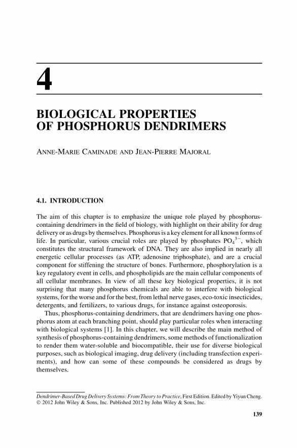
widely used method of synthesis of phosphorus dendrimers necessitates two steps
to build one generation (one layer). The core is most generally trifunctional (from
P(S)Cl3) [3] or hexafunctional (from (N3P3)Cl6) [4]. The first step is the reaction of the
core with 4-hydroxybenzaldehyde in basic conditions (generally the sodium salt of
the phenol), and the second step is the condensation reaction of the aldehydes with the
phosphorhydrazide H2NNMeP(S)Cl2. Both reactions are quantitative and produce
only NaCl and H2O as by-products. The repetition of both steps allows the growing of
the dendrimers, andwas carried out up to generation 12 (PS)-G12 [5] from the P(S)Cl3core and up to generation 8 (N3P3)-G8 from the (N3P3)Cl6 core [4] (Fig. 4.1). It is well
known that the characterization of dendrimers is never trivial [6], but the presence of
phosphorus in these compounds allows their easy characterization by 31P-NMR,
which is an invaluable tool for assessing the completion of reactions at each step of the
synthesis, as well as the integrity of the whole structure [7].
These dendrimers have either P(S)Cl2 or aldehyde terminal functions, depending
on the step considered. These functions are among the most reactive and versatile in
phosphorus chemistry and organic chemistry, respectively. Numerous types of
reactions have already been carried out with such terminal groups, but we will focus
FIGURE 4.1 Chemical structure of the first and second generations built from P(S)Cl3((PS)-G1 and (PS)-G2, respectively), method of synthesis used, and chemical structure of the
first generation built from the (N3P3)Cl6 core ((N3P3)-G1).
140 BIOLOGICAL PROPERTIES OF PHOSPHORUS DENDRIMERS
here on those that have led to dendrimers suitable for biological purposes. The main
point is that these compoundsmust be soluble inwater [8]. In contrast to other types of
dendrimers that are “naturally” soluble in water, thanks to their rather hydrophilic
interior, and water-solubilizing terminal groups, these phosphorus dendrimers have
both a hydrophobic interior [9] and hydrophobic terminal groups in their “native”
form (aldehydes or P(S)Cl2). The only way to render them soluble in water [10] is to
modify their terminal functions, in particular by grafting functions bearing a charge
(positive or negative) [11]. Positively charged phosphorus dendrimers were first
obtained by reacting N,N-diethylethylene diamine directly with the terminal P(S)Cl2functions [12]. This reaction is shown in (Fig. 4.2) with the fourth generation
dendrimer 1-G4, but was carried out up to generation 8 [13].
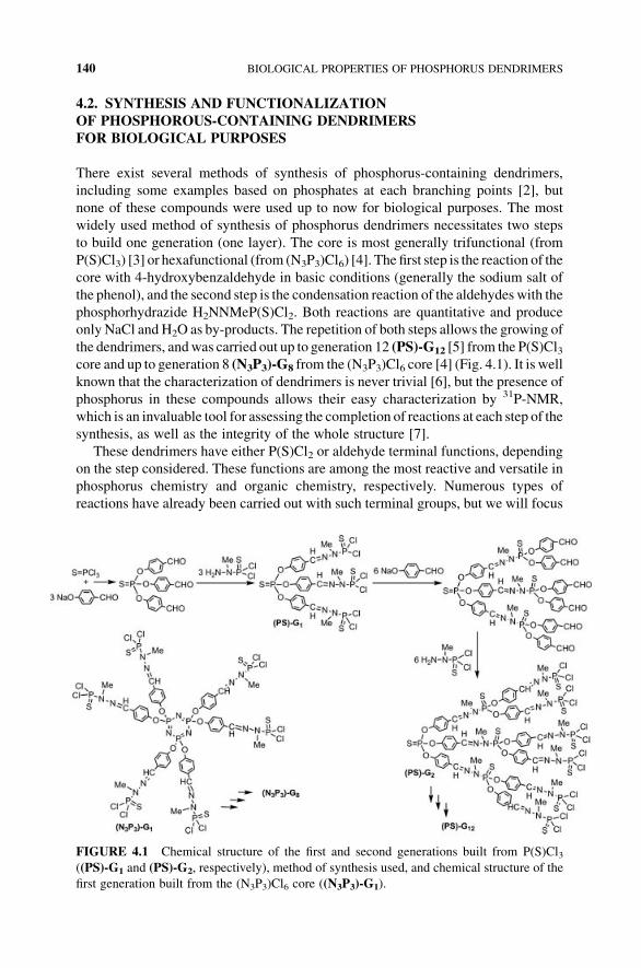
Negatively charged terminal groups are also usable for inducing solubility in
water of phosphorus dendrimers. In a first attempt, carboxylic acids were grafted
starting from the aldehyde terminal groups, using a Doebner-like reaction with
malonic acid in pyridine and piperidine. This synthesis was carried out first with
dendrimers built from the P(S) core [14], then from (N3P3) core [13]. The second
generation 2-G2 was obtained as shown in (Fig. 4.3). The neutral form is generally
not soluble in water, contrarily to the carboxylate form, obtained for instance by
reaction with NaOH.
Phosphonic acid salts are also very hydrophilic. Theywere obtained in two steps as
terminal functions of dendrimers, starting from the P(S)Cl2 terminal groups. The first
step is the grafting of tyramine functionalized by two phosphonic esters; the second
step is the deprotection of the esters, to obtain azabisphosphonic acid salts, as shown
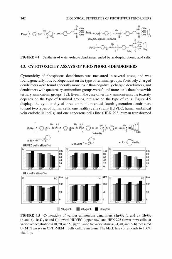
in (Fig. 4.4) for the first generation 3-G1 [15].
FIGURE 4.2 Synthesis of water-soluble dendrimers ended by ammonium groups.
FIGURE 4.3 Synthesis of dendrimers ended by carboxylic acids.
SYNTHESIS AND FUNCTIONALIZATION OF PHOSPHOROUS 141
4.3. CYTOTOXICITY ASSAYS OF PHOSPHORUS DENDRIMERS
Cytotoxicity of phosphorus dendrimers was measured in several cases, and was
found generally low, but dependent on the type of terminal groups. Positively charged
dendrimers were found generally more toxic than negatively charged dendrimers, and
dendrimerswith quaternary ammoniumgroupswere foundmore toxic than thosewith
tertiary ammonium groups [12]. Even in the case of tertiary ammoniums, the toxicity
depends on the type of terminal groups, but also on the type of cells. Figure 4.5
displays the cytotoxicity of three ammonium-ended fourth generation dendrimers
toward two types of human cells: one healthy cells strain (HUVEC, human umbilical
vein endothelial cells) and one cancerous cells line (HEK 293, human transformed
FIGURE 4.4 Synthesis of water-soluble dendrimers ended by azabisphosphonic acid salts.
FIGURE 4.5 Cytotoxicity of various ammonium dendrimers (1a-G4 (a and d), 1b-G4
(b and e), 1c-G4 (c and f)) toward HUVEC (upper row) and HEK 293 (lower row) cells, at
various concentrations (10, 20, and 50mg/mL) and for various times (24, 48, and 72 h)measured
by MTT assays in OPTI-MEM 1 cells culture medium. The black line corresponds to 100%
viability.
142 BIOLOGICAL PROPERTIES OF PHOSPHORUS DENDRIMERS
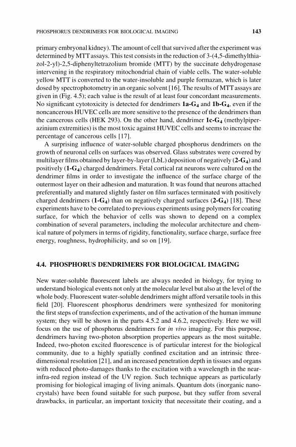
primary embryonal kidney). The amount of cell that survived after the experimentwas
determined byMTTassays. This test consists in the reduction of 3-(4,5-dimethylthia-
zol-2-yl)-2,5-diphenyltetrazolium bromide (MTT) by the succinate dehydrogenase
intervening in the respiratory mitochondrial chain of viable cells. The water-soluble
yellow MTT is converted to the water-insoluble and purple formazan, which is later
dosed by spectrophotometry in an organic solvent [16]. The results ofMTTassays are
given in (Fig. 4.5); each value is the result of at least four concordant measurements.
No significant cytotoxicity is detected for dendrimers 1a-G4 and 1b-G4, even if the
noncancerous HUVEC cells are more sensitive to the presence of the dendrimers than
the cancerous cells (HEK 293). On the other hand, dendrimer 1c-G4 (methylpiper-
azinium extremities) is the most toxic against HUVEC cells and seems to increase the
percentage of cancerous cells [17].
A surprising influence of water-soluble charged phosphorus dendrimers on the
growth of neuronal cells on surfaces was observed. Glass substrates were covered by
multilayer films obtained by layer-by-layer (LbL) deposition of negatively (2-G4) and
positively (1-G4) charged dendrimers. Fetal cortical rat neurons were cultured on the
dendrimer films in order to investigate the influence of the surface charge of the
outermost layer on their adhesion and maturation. It was found that neurons attached
preferentially and matured slightly faster on film surfaces terminated with positively
charged dendrimers (1-G4) than on negatively charged surfaces (2-G4) [18]. These
experiments have to be correlated to previous experiments using polymers for coating
surface, for which the behavior of cells was shown to depend on a complex
combination of several parameters, including the molecular architecture and chem-
ical nature of polymers in terms of rigidity, functionality, surface charge, surface free
energy, roughness, hydrophilicity, and so on [19].
4.4. PHOSPHORUS DENDRIMERS FOR BIOLOGICAL IMAGING
New water-soluble fluorescent labels are always needed in biology, for trying to
understand biological events not only at the molecular level but also at the level of the
whole body. Fluorescent water-soluble dendrimers might afford versatile tools in this
field [20]. Fluorescent phosphorus dendrimers were synthesized for monitoring
the first steps of transfection experiments, and of the activation of the human immune
system; they will be shown in the parts 4.5.2 and 4.6.2, respectively. Here we will
focus on the use of phosphorus dendrimers for in vivo imaging. For this purpose,
dendrimers having two-photon absorption properties appears as the most suitable.
Indeed, two-photon excited fluorescence is of particular interest for the biological
community, due to a highly spatially confined excitation and an intrinsic three-
dimensional resolution [21], and an increased penetration depth in tissues and organs
with reduced photo-damages thanks to the excitation with a wavelength in the near-
infra-red region instead of the UV region. Such technique appears as particularly
promising for biological imaging of living animals. Quantum dots (inorganic nano-
crystals) have been found suitable for such purpose, but they suffer from several
drawbacks, in particular, an important toxicity that necessitate their coating, and a
PHOSPHORUS DENDRIMERS FOR BIOLOGICAL IMAGING 143
blinking phenomenon that diminishes their fluorescence properties. Thus, the syn-
thesis of a fully organic alternative to these quantum dots, which will not have such
defects, is of major importance.
In a first attempt, specially engineered fluorophores possessing two-photon
absorption (TPA) properties were grafted as terminal groups of phosphorus dendri-
mers. An additive behavior was observed for the fluorescence intensity, depending on
the generation of the dendrimer, hence on the number of fluorophores; generation four
has the same two-photon absorption efficiency than the best quantum dots [22].
Furthermore, TPAcooperative enhancement between thefluorophore terminal groups
was observed [23]. The high modularity of these systems allowed the synthesis of
other series of TPA dendrimers in which a blue fluorophore is used as core and the
terminal groups are ammonium derivatives to ensure the solubility in water. Such
architecture should prevent the quenching offluorescence often induced bywater. The
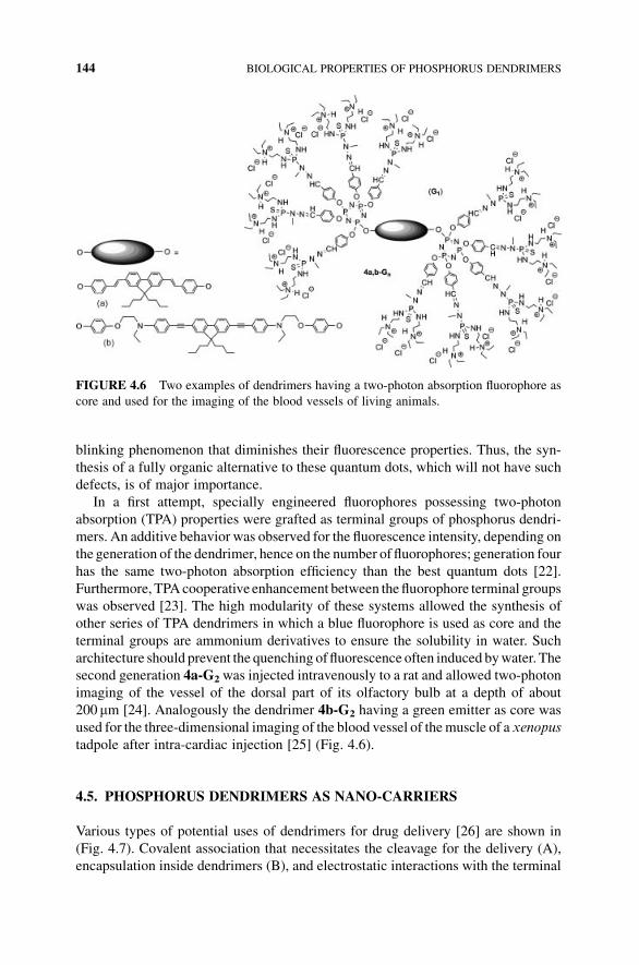
second generation 4a-G2was injected intravenously to a rat and allowed two-photon
imaging of the vessel of the dorsal part of its olfactory bulb at a depth of about
200 mm [24]. Analogously the dendrimer 4b-G2 having a green emitter as core was
used for the three-dimensional imaging of the blood vessel of themuscle of a xenopus
tadpole after intra-cardiac injection [25] (Fig. 4.6).
4.5. PHOSPHORUS DENDRIMERS AS NANO-CARRIERS
Various types of potential uses of dendrimers for drug delivery [26] are shown in
(Fig. 4.7). Covalent association that necessitates the cleavage for the delivery (A),
encapsulation inside dendrimers (B), and electrostatic interactions with the terminal
FIGURE 4.6 Two examples of dendrimers having a two-photon absorption fluorophore as
core and used for the imaging of the blood vessels of living animals.
144 BIOLOGICAL PROPERTIES OF PHOSPHORUS DENDRIMERS
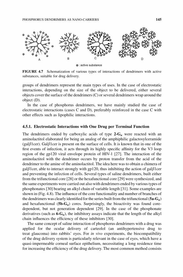
groups of dendrimers represent the main types of uses. In the case of electrostatic
interactions, depending on the size of the object to be delivered, either several
objects cover the surface of the dendrimers (C) or several dendrimers wrap around the
object (D).
In the case of phosphorus dendrimers, we have mainly studied the case of
electrostatic interactions (cases C and D), preferably reinforced in the case C with
other effects such as lipophilic interactions.
4.5.1. Electrostatic Interactions with One Drug per Terminal Function
The dendrimers ended by carboxylic acids of type 2-Gn were reacted with an
aminolactitol elaborated for being an analog of the amphiphilic galactosylceramide
(galb1cer). Galb1cer is present on the surface of cells. It is known that in one of the
first events of infection, it acts through its highly specific affinity for the V3 loop
region of the gp120 viral envelope protein of HIV-1 [27]. The interaction of the
aminolactitol with the dendrimer occurs by proton transfer from the acid of the
dendrimer to the amine of the aminolactitol. The idea here was to obtain a chimera of
galb1cer, able to interact strongly with gp120, thus inhibiting the action of galb1cerand preventing the infection of cells. Several types of saline dendrimers, built either
from the trifunctional core [28] or the hexafunctional core [29] were synthesized, and
the same experiments were carried out alsowith dendrimers ended by various types of
phosphonates [30] bearing an alkyl chain of variable length [31]. Some examples are
shown in (Fig. 4.8). The influence of the core functionality and number of branches of
the dendrimerswas clearly identified for the series built from the trifunctional (5a-Gn)
and hexafunctional (5b-Gn) cores. Surprisingly, the bioactivity was found core-
dependent, but not generation dependent [29]. In the case of the phosphonate
derivatives (such as 6-Gn), the inhibitory assays indicate that the length of the alkyl
chain influences the efficiency of these inhibitors [30].
The same concept of saline interaction of phosphorus dendrimers with a drug was
applied for the ocular delivery of carteolol (an antihypertensive drug to
treat glaucoma) into rabbits’ eyes. For in vivo experiments, the biocompatibility
of the drug delivery system is particularly relevant in the case of eyes, which have a
quasi-impermeable corneal surface epithelium, necessitating a long residence time
for increasing the efficiency of the drug delivery. The most common method consists
FIGURE 4.7 Schematization of various types of interactions of dendrimers with active
substances, suitable for drug delivery.
PHOSPHORUS DENDRIMERS AS NANO-CARRIERS 145
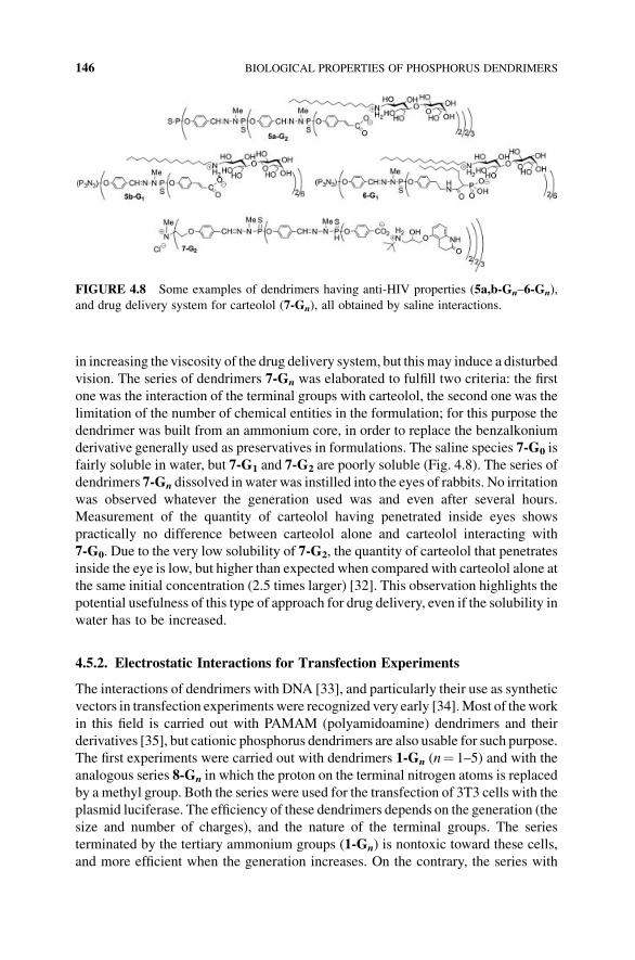
in increasing the viscosity of the drug delivery system, but this may induce a disturbed
vision. The series of dendrimers 7-Gn was elaborated to fulfill two criteria: the first
one was the interaction of the terminal groups with carteolol, the second one was the
limitation of the number of chemical entities in the formulation; for this purpose the
dendrimer was built from an ammonium core, in order to replace the benzalkonium
derivative generally used as preservatives in formulations. The saline species 7-G0 is
fairly soluble in water, but 7-G1 and 7-G2 are poorly soluble (Fig. 4.8). The series of
dendrimers 7-Gn dissolved in water was instilled into the eyes of rabbits. No irritation
was observed whatever the generation used was and even after several hours.
Measurement of the quantity of carteolol having penetrated inside eyes shows
practically no difference between carteolol alone and carteolol interacting with
7-G0. Due to the very low solubility of 7-G2, the quantity of carteolol that penetrates
inside the eye is low, but higher than expected when compared with carteolol alone at
the same initial concentration (2.5 times larger) [32]. This observation highlights the
potential usefulness of this type of approach for drug delivery, even if the solubility in
water has to be increased.
4.5.2. Electrostatic Interactions for Transfection Experiments
The interactions of dendrimers with DNA [33], and particularly their use as synthetic
vectors in transfection experiments were recognized very early [34].Most of thework
in this field is carried out with PAMAM (polyamidoamine) dendrimers and their
derivatives [35], but cationic phosphorus dendrimers are also usable for such purpose.
The first experiments were carried out with dendrimers 1-Gn (n¼ 1–5) and with the
analogous series 8-Gn in which the proton on the terminal nitrogen atoms is replaced
by a methyl group. Both the series were used for the transfection of 3T3 cells with the
plasmid luciferase. The efficiency of these dendrimers depends on the generation (the
size and number of charges), and the nature of the terminal groups. The series
terminated by the tertiary ammonium groups (1-Gn) is nontoxic toward these cells,
and more efficient when the generation increases. On the contrary, the series with
FIGURE 4.8 Some examples of dendrimers having anti-HIV properties (5a,b-Gn–6-Gn),
and drug delivery system for carteolol (7-Gn), all obtained by saline interactions.
146 BIOLOGICAL PROPERTIES OF PHOSPHORUS DENDRIMERS
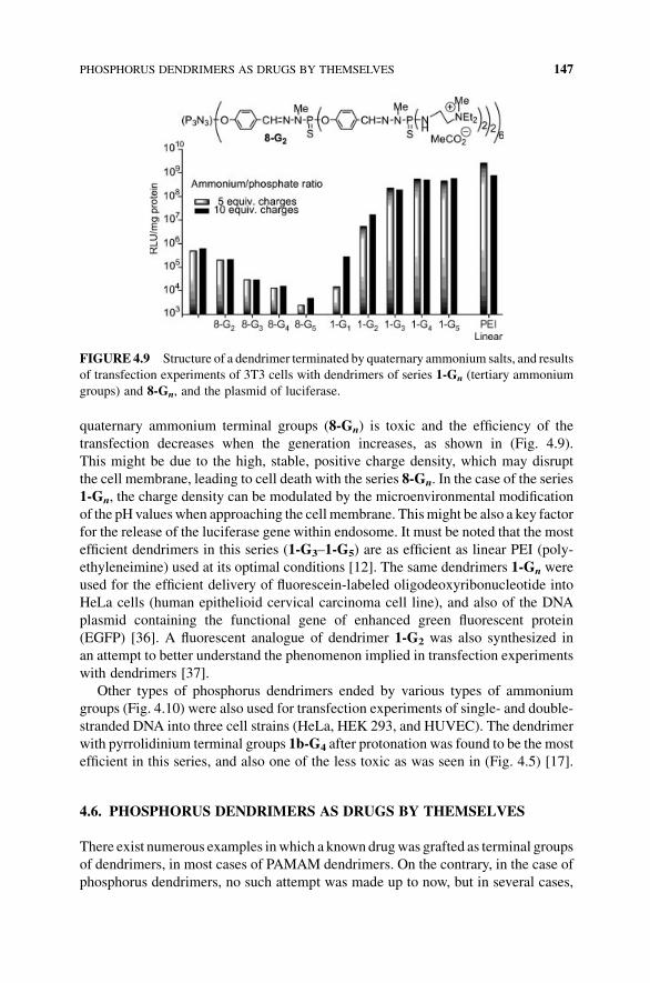
quaternary ammonium terminal groups (8-Gn) is toxic and the efficiency of the
transfection decreases when the generation increases, as shown in (Fig. 4.9).
This might be due to the high, stable, positive charge density, which may disrupt
the cell membrane, leading to cell death with the series 8-Gn. In the case of the series
1-Gn, the charge density can be modulated by the microenvironmental modification
of the pH valueswhen approaching the cell membrane. Thismight be also a key factor
for the release of the luciferase gene within endosome. It must be noted that the most
efficient dendrimers in this series (1-G3–1-G5) are as efficient as linear PEI (poly-
ethyleneimine) used at its optimal conditions [12]. The same dendrimers 1-Gn were
used for the efficient delivery of fluorescein-labeled oligodeoxyribonucleotide into
HeLa cells (human epithelioid cervical carcinoma cell line), and also of the DNA
plasmid containing the functional gene of enhanced green fluorescent protein
(EGFP) [36]. A fluorescent analogue of dendrimer 1-G2 was also synthesized in
an attempt to better understand the phenomenon implied in transfection experiments
with dendrimers [37].
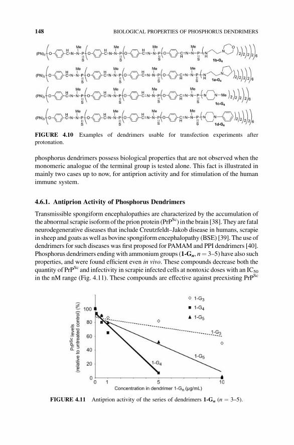
Other types of phosphorus dendrimers ended by various types of ammonium
groups (Fig. 4.10) were also used for transfection experiments of single- and double-
stranded DNA into three cell strains (HeLa, HEK 293, and HUVEC). The dendrimer
with pyrrolidinium terminal groups 1b-G4 after protonation was found to be the most
efficient in this series, and also one of the less toxic as was seen in (Fig. 4.5) [17].
4.6. PHOSPHORUS DENDRIMERS AS DRUGS BY THEMSELVES
There exist numerous examples inwhich a known drugwas grafted as terminal groups
of dendrimers, in most cases of PAMAM dendrimers. On the contrary, in the case of
phosphorus dendrimers, no such attempt was made up to now, but in several cases,
FIGURE4.9 Structure of a dendrimer terminated by quaternary ammonium salts, and results
of transfection experiments of 3T3 cells with dendrimers of series 1-Gn (tertiary ammonium
groups) and 8-Gn, and the plasmid of luciferase.
PHOSPHORUS DENDRIMERS AS DRUGS BY THEMSELVES 147
phosphorus dendrimers possess biological properties that are not observed when the
monomeric analogue of the terminal group is tested alone. This fact is illustrated in
mainly two cases up to now, for antiprion activity and for stimulation of the human
immune system.
4.6.1. Antiprion Activity of Phosphorus Dendrimers
Transmissible spongiform encephalopathies are characterized by the accumulation of
the abnormal scrapie isoformof the prion protein (PrPSc) in the brain [38]. They are fatal
neurodegenerative diseases that include Creutzfeldt–Jakob disease in humans, scrapie
in sheep and goats aswell as bovine spongiform encephalopathy (BSE) [39]. The use of
dendrimers for such diseases was first proposed for PAMAM and PPI dendrimers [40].
Phosphorus dendrimers endingwith ammoniumgroups (1-Gn, n¼ 3–5) have also such
properties, and were found efficient even in vivo. These compounds decrease both the
quantity of PrPSc and infectivity in scrapie infected cells at nontoxic doses with an IC50
in the nM range (Fig. 4.11). These compounds are effective against preexisting PrPSc
FIGURE 4.10 Examples of dendrimers usable for transfection experiments after
protonation.
FIGURE 4.11 Antiprion activity of the series of dendrimers 1-Gn (n ¼ 3–5).
148 BIOLOGICAL PROPERTIES OF PHOSPHORUS DENDRIMERS
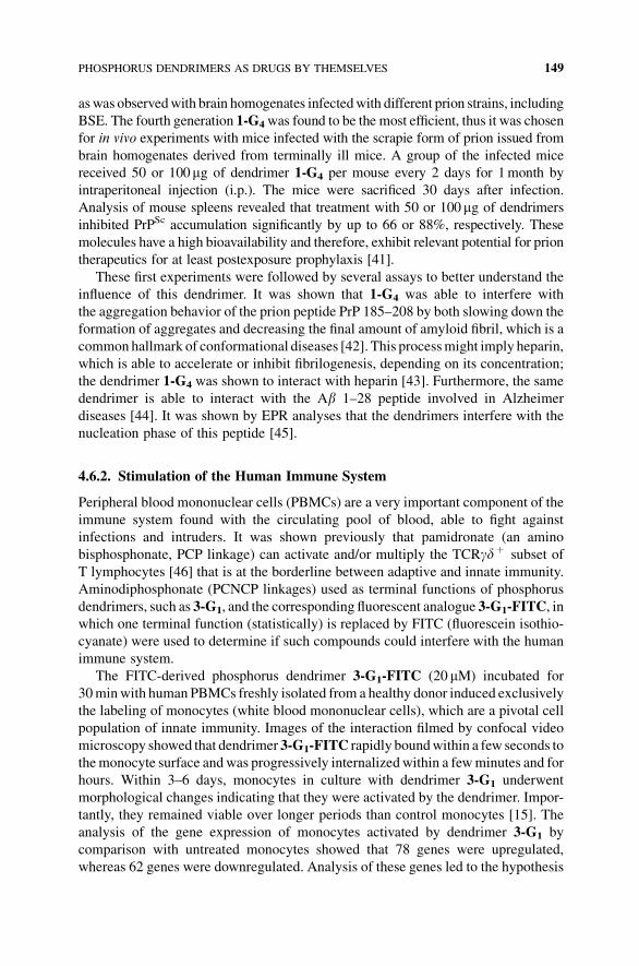
aswas observedwith brain homogenates infectedwith different prion strains, including
BSE. The fourth generation 1-G4was found to be the most efficient, thus it was chosen
for in vivo experiments with mice infected with the scrapie form of prion issued from
brain homogenates derived from terminally ill mice. A group of the infected mice
received 50 or 100mg of dendrimer 1-G4 per mouse every 2 days for 1month by
intraperitoneal injection (i.p.). The mice were sacrificed 30 days after infection.
Analysis of mouse spleens revealed that treatment with 50 or 100mg of dendrimers
inhibited PrPSc accumulation significantly by up to 66 or 88%, respectively. These
molecules have a high bioavailability and therefore, exhibit relevant potential for prion
therapeutics for at least postexposure prophylaxis [41].
These first experiments were followed by several assays to better understand the
influence of this dendrimer. It was shown that 1-G4 was able to interfere with
the aggregation behavior of the prion peptide PrP 185–208 by both slowing down the
formation of aggregates and decreasing the final amount of amyloid fibril, which is a
common hallmark of conformational diseases [42]. This processmight imply heparin,
which is able to accelerate or inhibit fibrilogenesis, depending on its concentration;
the dendrimer 1-G4 was shown to interact with heparin [43]. Furthermore, the same
dendrimer is able to interact with the Ab 1–28 peptide involved in Alzheimer
diseases [44]. It was shown by EPR analyses that the dendrimers interfere with the
nucleation phase of this peptide [45].
4.6.2. Stimulation of the Human Immune System
Peripheral blood mononuclear cells (PBMCs) are a very important component of the
immune system found with the circulating pool of blood, able to fight against
infections and intruders. It was shown previously that pamidronate (an amino
bisphosphonate, PCP linkage) can activate and/or multiply the TCRgdþ subset of
T lymphocytes [46] that is at the borderline between adaptive and innate immunity.
Aminodiphosphonate (PCNCP linkages) used as terminal functions of phosphorus
dendrimers, such as 3-G1, and the corresponding fluorescent analogue 3-G1-FITC, inwhich one terminal function (statistically) is replaced by FITC (fluorescein isothio-
cyanate) were used to determine if such compounds could interfere with the human
immune system.
The FITC-derived phosphorus dendrimer 3-G1-FITC (20 mM) incubated for
30minwith human PBMCs freshly isolated from a healthy donor induced exclusively
the labeling of monocytes (white blood mononuclear cells), which are a pivotal cell
population of innate immunity. Images of the interaction filmed by confocal video
microscopy showed that dendrimer 3-G1-FITC rapidly boundwithin a few seconds to
themonocyte surface andwas progressively internalizedwithin a fewminutes and for
hours. Within 3–6 days, monocytes in culture with dendrimer 3-G1 underwent
morphological changes indicating that they were activated by the dendrimer. Impor-
tantly, they remained viable over longer periods than control monocytes [15]. The
analysis of the gene expression of monocytes activated by dendrimer 3-G1 by
comparison with untreated monocytes showed that 78 genes were upregulated,
whereas 62 genes were downregulated. Analysis of these genes led to the hypothesis
PHOSPHORUS DENDRIMERS AS DRUGS BY THEMSELVES 149
of an alternative-like, anti-inflammatory activation of human monocytes [47]. Begin-
ning the study of the structure/activity relationship, it was shown that the correspond-
ingmonomeric azadiphosphonic derivative, analogous to the terminal groups of 3-G1,
displayed no activity. Furthermore, varying the number of terminal phosphonate
functions by using a core-controlled strategy for the selective functionalization of
1–6 Cl of (N3P3)Cl6 core shows that the activation of human monocytes depends on
the number of phosphonic groups, with a neat decrease of the efficiency for
compounds with less than six aminodiphosphonate groups per dendrimer [48].
The activation of monocytes occurs in short time cultures of PBMCs (maximum
6 days, and generally 1 day). Very surprisingly, a totally different behavior was
observed for longer times of culture. First, an important increase in the number of
PBMCs was observed (proliferation index 5.5 in 2week old cultures). Second,
phenotyping of the cells multiplied in cultures with 3-G1 revealed the prominence
of natural killer (NK) cells, which are part of the innate immunity. Experiments with
PBMCs obtained from six healthy donors revealed in all cases an important increase
in both the percentage and the number of NK cells. A mean multiplication of the
number of NK cells by a factor of 105 was achieved in medium supplemented with
10-G1 versus a mean multiplication only by a factor of 7.5 without it, after 4 weeks in
culture. These large-scale prototype cultures of PBMCs comprised 1million NK cells
on average at the beginning; multiplications over 500-fold were obtained with some
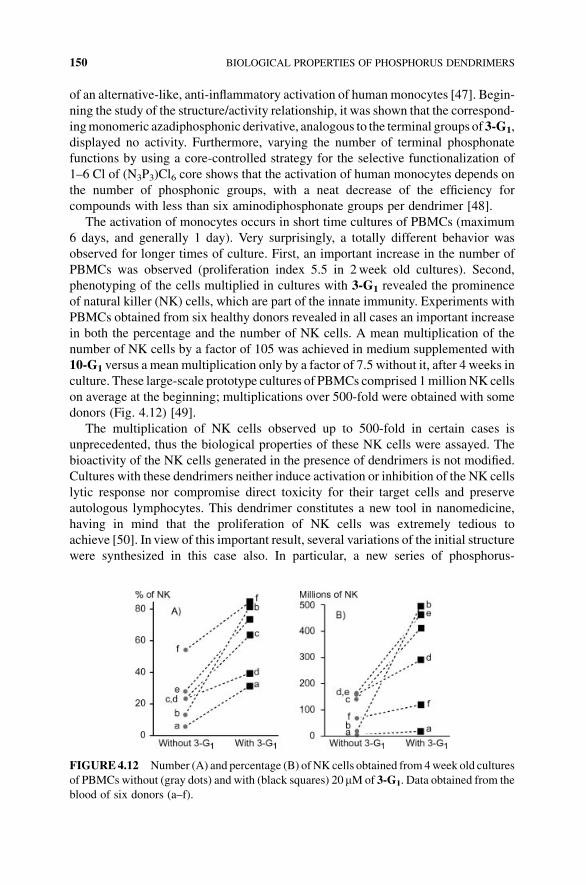
donors (Fig. 4.12) [49].
The multiplication of NK cells observed up to 500-fold in certain cases is
unprecedented, thus the biological properties of these NK cells were assayed. The
bioactivity of the NK cells generated in the presence of dendrimers is not modified.
Cultures with these dendrimers neither induce activation or inhibition of the NK cells
lytic response nor compromise direct toxicity for their target cells and preserve
autologous lymphocytes. This dendrimer constitutes a new tool in nanomedicine,
having in mind that the proliferation of NK cells was extremely tedious to
achieve [50]. In view of this important result, several variations of the initial structure
were synthesized in this case also. In particular, a new series of phosphorus-
FIGURE4.12 Number (A) and percentage (B) ofNK cells obtained from4week old cultures
of PBMCs without (gray dots) and with (black squares) 20mMof 3-G1. Data obtained from the
blood of six donors (a–f).
150 BIOLOGICAL PROPERTIES OF PHOSPHORUS DENDRIMERS
containing dendrimers capped with nonsymmetrical azadiphosphonic acids was
synthesized. Their ability to activate human monocytes of healthy individuals was
assessed. All of them were found active, but none of them displayed a higher activity
than 3-G1 [51]. Themechanism of action of this dendrimer is very complex; only part
of it is elucidated to date. As shown in the previous paragraph, the first step is the
activation of monocytes. It was also found that phosphonate-capped dendrimers
inhibit the activation, and therefore the proliferation of CD4þ T lymphocytes,
without affecting their viability. This allows a rapid enrichment of NK cells and
further expansion [52].
4.7. CONCLUSIONS
Dendrimers in general and phosphorus-containing dendrimers in particular constitute
a versatile platform whose intrinsic parameters can be controlled and modified on
demand. Such properties open the way to nano-medicine, with great promises as
nano-vehicles for drug delivery, and also as drugs by themselves, generally correlated
to their multivalency effects [53]. We are still working in all the fields evoked in this
paper, and exciting new results have been published recently [54], opening new
perspectives for biological uses of phosphorus dendrimers. The real challenge now is
to bridge the gap between fundamental researches and market applications. This
milestone has been reached recently for a topic related to those displayed in this
chapter, which concerns the elaboration of biosensors using phosphorus dendrimers.
They were found particularly useful for improving the sensitivity. Indeed, the
detection sensitivity of these devices was found 10- to 100-fold higher than arrays
made with most other functionalized glass slides [55]. Furthermore, the stability and
the reusability of these devices elaborated with phosphorus dendrimers were found
excellent, as well as the possibility to detect mutations [56]. This work has led to the
creation of a start-up last year (DendrisTD).
REFERENCES
1. Caminade, A.M., Turrin, C.O., Majoral, J.P. (2010). Biological properties of phosphorus
dendrimers. New J. Chem., 34, 1512–1524.
2. Hudson, R.H.E., Damha, M.J. (1993). Nucleic-acid dendrimers—novel biopolymer
structures. J. Am. Chem. Soc., 115, 2119–2124; (b) Salamonczyk, G.M., Kuznikowski,
M., Poniatowska, E. (2001). Synthesis and oxygenation of selenophosphate dendrimers.
Chem. Commun. 2202–2203.
3. Launay,N., Caminade,A.M., Lahana, R.,Majoral, J.P. (1994).A general synthetic strategy
for neutral phosphorus-containing dendrimers. Angew. Chem. Int. Ed., 33, 1589–1592.
4. Launay, N., Caminade, A.M., Majoral, J.P. (1997). Synthesis of bowl-shaped dendrimers
from generation 1 to generation 8. J. Organomet. Chem., 529, 51–58.
5. Lartigue, M.L., Donnadieu, B., Galliot, C., Caminade, A.M., Majoral, J.P., Fayet, J.P.
(1997). Large dipole moments of phosphorus-containing dendrimers. Macromolecules,
30, 7335–7337.
REFERENCES 151
6. Caminade, A.M., Laurent, R., Majoral, J.P. (2005). Characterization of dendrimers. Adv.
Drug Deliv. Rev., 57, 2130–2146.
7. Caminade, A.M., Laurent, R., Turrin, C.O., Rebout, C., Delavaux-Nicot, B., Ouali, A.,
Zablocka, M., Majoral, J.P. (2010). Phosphorus dendrimers as viewed by 31P NMR
spectroscopy; synthesis and characterization. C.R. Chim., 13, 1006–1027.
8. Caminade, A.M., Majoral, J.P. (2005). Water-soluble phosphorus-containing dendrimers.
Prog. Polym. Sci., 30, 491–505.
9. Leclaire, J., Coppel, Y., Caminade, A.M., Majoral, J.P. (2004). Nanometric sponges made
of water-soluble hydrophobic dendrimers. J. Am. Chem. Soc., 126, 2304–2305.
10. Camina, A.M., Majoral, J.P. (2005). Water-soluble phosphorus-containing dendrimers.
Prog. Polym. Sci., 30, 491–505.
11. Caminade, A.M., Turrin, C.O., Laurent, R., Rebout, C., Majoral, J.P. (2006). Phosphorus
dendritic architectures: polyanionic and polycationic derivatives. Polym. Int., 55,
1155–1160.
12. Loup, C., Zanta, M.A., Caminade, A.M., Majoral, J.P., Meunier, B. (1999). Preparation of
water-soluble cationic phosphorus-containing dendrimers as DNA transfecting agents.
Chem. Eur. J., 5, 3644–3650.
13. Reinert, P., Chane-Ching, J.Y., Bull, L., Dagiral, R., Batail, P., Laurent, R., Caminade,
A.M.,Majoral, J.P. (2007). Influence of cationic phosphorus dendrimers on the surfactant-
induced synthesis of mesostructured nanoporous silica. New J. Chem., 31, 1259–1263.
14. Soler-Illia, G.J.D.A., Rozes, L., Boggiano, M.K., Sanchez, C., Turrin, C.O., Caminade,
A.M., Majoral, J.P. (2000). New mesotextured hybrid materials made from assemblies of
dendrimers and titanium(IV)-oxo-organo clusters.Angew. Chem. Int. Ed., 39, 4250–4254.
15. Poupot, M., Griffe, L., Marchand, P., Maraval, A., Rolland, O., Martinet, L., L’Faqihi-
Olive, F.E., Turrin, C.O., Caminade, A.M., Fournie, J.J., Majoral, J.P., Poupot, R. (2006).
Design of phosphorylated dendritic architectures to promote human monocyte activation.
FASEB J., 20, 2339–2351.
16. Mosmann, T. (1983). Rapid colorimetric assay for cellular growth and survival:
application to proliferation and cytotoxicity assays. J. Immunol. Methods, 65, 55–63.
17. Padie, C., Maszewska, M., Majchrzak, K., Nawrot, B., Caminade, A.M., Majoral, J.P.
(2009). Polycationic phosphorus dendrimers: synthesis, characterization, study of
cytotoxicity, complexation of DNA, and transfection experiments. New J. Chem., 33,
318–326.
18. Hernandez-Lopez, J.L., Khor, H.L., Caminade, A.M., Majoral, J.P., Mittler, S., Knoll, W.,
Kim, D.H. (2008). Bioactive multilayer thin films of chargedN,N-disubstituted hydrazine
phosphorus dendrimers fabricated by layer-by-layer self-assembly. Thin Solid Films, 516,
1256–1264.
19. Picart, C., Elkaim, R., Richert, L., Audoin, T., Arntz, Y., Cardoso,M.D., Schaaf, P., Voegel,
J.C., Frisch, B. (2005). Primary cell adhesion on RGD-functionalized and covalently
crosslinked thin polyelectrolyte multilayer films. Adv. Funct. Mater., 15, 83–94.
20. Caminade, A.M., Hameau, A., Majoral, J.P. (2009). Multicharged and/or water-soluble
fluorescent dendrimers: properties and uses. Chem. Eur. J., 15, 9270–9285.
21. Denk, W., Strickler, J.H., Webb, W.W. (1990). 2-Photon laser scanning fluorescence
microscopy. Science, 248, 73–76.
22. Mongin, O., Krishna, T.R., Werts, M.H.V., Caminade, A.M., Majoral, J.P., Blanchard-
Desce, M. (2006). A modular approach to two-photon absorbing organic nanodots:
152 BIOLOGICAL PROPERTIES OF PHOSPHORUS DENDRIMERS
brilliant dendrimers as an alternative to semiconductor quantum dots? Chem. Commun.
915–917.
23. Terenziani, F., Parthasarathy, V., Pla-Quintana, A., Maishal, T., Caminade, A.M., Majoral,
J.P., Blanchard-Desce, M. (2009). Cooperative two-photon absorption enhancement by
through-space interactions in multichromophoric compounds. Angew. Chem. Int. Ed., 48,
8691–8694.
24. Krishna, T.R., Parent,M.,Werts,M.H.V.,Moreaux, L.,Gmouh, S., Charpak, S., Caminade,
A.M., Majoral, J.P., Blanchard-Desce, M. (2006). Water-soluble dendrimeric two-photon
tracers for in vivo imaging. Angew. Chem. Int. Ed., 45, 4645–4648.
25. Mongin, O., Rouxel, C., Robin, A.C., Pla-Quintana, A., Krishna, T.R., Recher, G., Tiaho,
F., Caminade, A.M., Majoral, J.P., Blanchard-Desce, M. (2008). Brilliant organic
nanodots: novel nano-objects for bionanophotonics—art. no. 704006. Nanobiosystems:
Processing, Characterization, and Applications, 7040, 4006–4006.
26. Goller, R., Vors, J.P., Caminade,A.M.,Majoral, J.P. (2001). Phosphorus dendrimers as new
tools to deliver active substances. Tetrahedron Lett., 42, 3587–3590.
27. Harouse, J.M., Bhat, S., Spitalnik, S.L., Laughlin, M., Stefano, K., Silberberg, D.H.,
Gonzalezscarano, F. (1991). Inhibition of entry of HIV-1 in neural cell-lines by antibodies
against galactosyl ceramide. Science, 253, 320–323.
28. Blanzat, M., Turrin, C.O., Perez, E., Rico-Lattes, I., Caminade, A.M.,Majoral, J.P. (2002).
Phosphorus-containing dendrimers bearing galactosylceramide analogs: self-assembly
properties. Chem. Commun., 17, 1864–1865.
29. Blanzat, M., Turrin, C.O., Aubertin, A.M., Couturier-Vidal, C., Caminade, A.M.,Majoral,
J.P., Rico-Lattes, I., Lattes A. (2005). Dendritic catanionic assemblies: in vitro anti-HIV
activity of phosphorus-containing dendrimers bearing Gal beta(1)cer analogues.
ChemBioChem, 6, 2207–2213.
30. Perez-Anes, A., Stefaniu, C.,Moog, C.,Majoral, J.P., Blanzat,M., Turrin, C.O., Caminade,
A.M., Rico-Lattes, I. (2010). Multivalent catanionic GalCer analogs derived from first
generation dendrimeric phosphonic acids. Bioorg. Med. Chem., 18, 242–248.
31. Perez-Anes, A., Spataro, G., Coppel, Y., Moog, C., Blanzat, M., Turrin, C.O., Caminade,
A.M., Rico-Lattes, I., Majoral, J.P. (2009). Phosphonate terminated PPH dendrimers:
influence of pendant alkyl chains on the in vitro anti-HIV-1 properties. Org. Biomol.
Chem., 7, 3491–3498.
32. Spataro, G., Malecaze, F., Turrin, C.O., Soler, V., Duhayon, C., Elena, P.P., Majoral, J.P.,
Caminade, A.M. (2010). Designing dendrimers for ocular drug delivery. Eur. J. Med.
Chem., 45, 326–334.
33. Caminade, A.M., Turrin, C.O., Majoral, J.P. (2008). Dendrimers and DNA:
combinations of two special topologies for nanomaterials and biology. Chem. Eur.
J., 14, 7422–7432.
34. Haensler, J., Szoka, F.C. (1993). Polyamidoamine cascade polymers mediate efficient
transfection of cells in culture. Bioconjugate Chem., 4, 372–379.
35. Svenson, S., Tomalia,D.A. (2005).Commentary—dendrimers in biomedical applications—
reflections on the field. Adv. Drug Deliv. Rev., 57, 2106–2129.
36. Maszewska, M., Leclaire, J., Cieslak, M., Nawrot, B., Okruszek, A., Caminade, A.M.,
Majoral, J.P. (2003).Water-soluble polycationic dendrimers with a phosphoramidothioate
backbone: preliminary studies of cytotoxicity and oligonucleotide/plasmid delivery in
human cell culture. Oligonucleotides, 13, 193–205.
REFERENCES 153
37. Kazmierczak-Baranska, J., Pietkiewicz, A., Janicka, M.,Wei, Y.Q., Turrin, C.O., Majoral,
J.P., Nawrot, B., Caminade, A.M. (2010). Synthesis of a fluorescent cationic phosphorus
dendrimer and preliminary biological studies of its interactionwithDNA.Nucleo.Nucleot.
Nucl., 29, 155–167.
38. Prusiner, S.B. (1982).Novel proteinaceous infectious particles cause scrapie. Science, 216,
136–144.
39. Collinge, J. (2001). Prion diseases of humans and animals: their causes and molecular
basis. Annu. Rev. Neurosci., 24, 519–550.
40. Supattapone, S., Wille, H., Uyechi, L., Safar, J., Tremblay, P., Szoka, F.C., Cohen, F.E.,
Prusiner, S.B., Scott, M.R. (2001). Branched polyamines cure prion-infected
neuroblastoma cells. J. Virol., 75, 3453–3461.
41. Solassol, J., Crozet, C., Perrier, V., Leclaire, J., Beranger, F., Caminade,A.M.,Meunier, B.,
Dormont, D., Majoral, J.P., Lehmann, S. (2004). Cationic phosphorus-containing
dendrimers reduce prion replication both in cell culture and in mice infected with
scrapie. J. Gen. Virol., 85, 1791–1799.
42. Klajnert, B., Cortijo-Arellano, M., Cladera, J., Majoral, J.P., Caminade, A.M.,
Bryszewska, M. (2007). Influence of phosphorus dendrimers on the aggregation of the
prion peptide PrP 185–208. Biochem. Biophys. Res. Co., 364, 20–25.
43. Klajnert, B., Cangiotti, M., Calici, S., Ionov, M.,Majoral, J.P., Caminade, A.M., Cladera, J.,
Bryszewska, M., Ottaviani, M.F. (2009). Interactions between dendrimers and heparin and
their implications for the anti-prion activity of dendrimers. New J. Chem., 33, 1087–1093.
44. Klajnert, B., Cangiotti, M., Calici, S., Majoral, J.P., Caminade, A.M., Cladera, J.,
Bryszewska, M., Ottaviani, M.F. (2007). EPR study of the interactions between
dendrimers and peptides involved in Alzheimer’s and prion diseases. Macromol.
Biosci., 7, 1065–1074.
45. Ottaviani, M.F., Mazzeo, R., Cangiotti, M., Fiorani, L., Majoral, J.P., Caminade, A.M.,
Pedziwiatr, E., Bryszewska, M., Klajnert, B. Time evolution of the aggregation process of
peptides involved in neurodegenerative diseases and preventing aggregation effect of
phosphorus dendrimers studied by EPR. Biomacromolecules, 11, 3015.
46. Kunzmann, V., Bauer, E., Feurle, J., Weissinger, F., Tony, H.P., Wilhelm, M. (2000).
Stimulation, of gdT cells by aminobisphosphonates and induction of antiplasma cell
activity in multiple myeloma. Blood, 96, 384–392.
47. Fruchon, S., Poupot, M., Martinet, L., Turrin, C.O., Majoral, J.P., Fournie, J.J., Caminade,
A.M., Poupot, R. (2009). Anti-inflammatory and immunosuppressive activation of human
monocytes by a bioactive dendrimer. J. Leukocyte Biol., 85, 553–562.
48. Rolland, O., Griffe, L., Poupot, M., Maraval, A., Ouali, A., Coppel, Y., Fournie, J.J.,
Bacquet, G., Turrin, C.O., Caminade, A.M., Majoral, J.P., Poupot, R. (2008). Tailored
control and optimisation of the number of phosphonic acid termini on phosphorus-
containing dendrimers for the ex-vivo activation of human monocytes. Chem. Eur. J.,
14, 4836–4850.
49. Griffe, L., Poupot, M., Marchand, P., Maraval, A., Turrin, C.O., Rolland, O., Metivier, P.,
Bacquet, G., Fournie, J.J., Caminade, A.M., Poupot, R., Majoral, J.P. (2007).
Multiplication of human natural killer cells by nanosized phosphonate-capped
dendrimers. Angew. Chem. Int. Ed., 46, 2523–2526.
50. Klingemann, H.G. (2005). Natural killer cell-based immunotherapeutic strategies.
Cytotherapy, 7, 16–22.
154 BIOLOGICAL PROPERTIES OF PHOSPHORUS DENDRIMERS
51. Marchand, P., Griffe, L., Poupot,M., Turrin, C.O., Bacquet, G., Fournie, J.J., Majoral, J.P.,
Poupot, R., Caminade, A.M. (2009). Dendrimers ended by non-symmetrical
azadiphosphonate groups: synthesis and immunological properties. Bioorg. Med.
Chem. Lett., 19, 3963–3966.
52. Portevin, D., Poupot, M., Rolland, O., Turrin, C.O., Fournie, J.J., Majoral, J.P., Caminade,
A.M., Poupot, R. (2009). Regulatory activity of azabisphosphonate-capped dendrimers on
humanCD4(þ ) T cell proliferation enhances ex-vivo expansion of NK cells fromPBMCs
for immunotherapy. J. Transl. Med. 7, 13.
53. Rolland, O., Turrin, C.O., Caminade, A.M., Majoral, J.P. (2009). Dendrimers and
nanomedicine: multivalency in action. New J. Chem., 33, 1809–1824.
54. Hayder, M., Poupot, M., Baron, M., Nigon, D., Turrin, C.O., Caminade, A.M., Majoral,
J.P., Eisenberg, R.A., Fournie, J.J., Cantagrel, A., Poupot, R., Davignon, J.L. (2011).
A phosphorus-based dendrimer targets inflammation and osteoclastogenesis in
experimental arthritis. Science Transl. Med., 3, 81ra35.
55. Le Berre, V., Trevisiol, E., Dagkessamanskaia, A., Sokol, S., Caminade, A.M., Majoral,
J.P., Meunier, B., Francois, J. (2003). Dendrimeric coating of glass slides for sensitive
DNA microarrays analysis. Nucleic Acids Res., 31, e88.
56. Trevisiol, E., Le Berre-Anton, V., Leclaire, J., Pratviel, G., Caminade, A.M., Majoral, J.P.,
Francois, J.M., Meunier, B. (2003). Dendrislides, dendrichips: a simple chemical
functionalization of glass slides with phosphorus dendrimers as an effective means for
the preparation of biochips. New J. Chem., 27, 1713–1719.
REFERENCES 155


![A dual targeting dendrimer-mediated siRNA delivery system …nanosized volume.[16-20] We have recently developed a series of cationic amphiphilic dendrimers[21-24] which couple the](https://img.pdfslide.us/doc/110x75/5feda2b5c8964e53213a54f4/a-dual-targeting-dendrimer-mediated-sirna-delivery-system-nanosized-volume16-20.jpg)