Embed Size (px)
Citation preview
19 International Journal of Scientific Study | November 2015 | Vol 3 | Issue 8
Demographic, Clinical Profile of Oral Lichen Planus and its Possible Correlation with Thyroid Disorders: A Case-Control StudyMohsin Muzaffar Tak1, Altaf Hussain Chalkoo2
1Assistant Professor, Department of Oral Medicine & Radiology, Government Dental College, Srinagar, Jammu and Kashmir, India, 2Professor & Head, Department of Oral Medicine & Radiology, Government Dental College, Srinagar, Jammu and Kashmir, India
The clinical course of OLP lesions normally last for years with alternating periods of exacerbation and quiescence. There is an increase in pain and erythematous or ulcerated areas and during the exacerbation phase.5 This phase is also associated with periods of anxiety, psychological stress, and mechanical trauma (Koebner phenomenon). Chronic low-intensity irritation due to the presence of plaque or dental calculus may also increase the severity of gingival LP and is considered Koebner phenomenon, other factors such as the mechanical trauma of odontological procedures, friction of sharp points, rough dental restorations, heat and cigarette irritants, and oral habits like chewing gum.6,7
The clinical presentation of OLP ranges from mild, painless white keratotic lesions to painful erosions and ulcerations.8 The most common affected site is buccal mucosa, usually bilateral. Clinically OLP may occur in six clinical variants as reticular, papular, plaque-like, erosive, atrophic, and bullous as classified by Andreasen.9
The reticular variant of OLP is the most recognized form, encompass white lesions, which clinically appear as a
INTRODUCTION
Oral lichen planus (OLP) is a mucocutaneous disease which can affect the skin, oral mucosa, and other mucous membranes. The etiology of LP which is an inflammatory mucocutaneous disease is unknown and thought to arise as a result of an immune response – mainly by CD8+ lymphocytes – to antigens on lesional keratinocytes.1,2 English physician Erasmus Wilson in 1866 presented the designation and description of the pathology. He also suggested “nervous tension” could be the cause of its etiology.3 It was Louis-Frédéric Wickham who provided an addition to the description of the lesion stories et punctuations grisatre (grayish striae and dots), named Wickham Striae in 1895.4
Original Article
AbstractObjectives: Oral lichen planus (OLP) is one of the common chronic conditions involving the oral mucosa. It is more common and persistent than the cutaneous form causing significant discomfort to the patient. The aim is to study the demographic pattern, clinical profile, and to find out any possible correlation with the thyroid disorders.
Materials and Methods: A total of 50 patients comprising 32 females and 18 males of OLP, and an equal number of age and sex-matched controls were evaluated, for demographic trends, clinical profiling, and relevance to thyroid disorders.
Results: Out of 50 OLP patients the thyroid function tests (TFTs) were deranged in 9 (18%); 5 females and 4 males while in the control group, TFTs were deranged in 1 (2%) female. The results obtained were statistically significant (P < 0.05).
Conclusions: The results of our study show that a significant percentage of OLP patients have deranged thyroid function, especially hypothyroidism. As the sample size in our study was small, this calls for further studies involving a larger number of patients.
Key words: Demographic, Diagnosis, Etiology, Lichen planus, Oral mucosa, Thyroid
Access this article online
www.ijss-sn.com
Month of Submission : 10-2015 Month of Peer Review : 10-2015 Month of Acceptance : 10-2015 Month of Publishing : 11-2015
Corresponding Author: Dr. Mohsin Muzaffar Tak, Habak Zakura Crossing, Hazratbal, Srinagar - - 190 006, Jammu and Kashmir, India. Phone: +91-9018830030. E-mail: [email protected]
DOI: 10.17354/ijss/2015/500
Tak and Chalkoo: OLP-Demographic, Clinical profile and its possible correlation with thyroid disorders
20International Journal of Scientific Study | November 2015 | Vol 3 | Issue 8
network of connecting and overlapping lines, papules or plaques. Although clinical presentation in certain patients may be an impressive array of diffuse and widespread reticulated lesions, they are usually asymptomatic and often, are unaware of the presence of these lesions. A significant degree of discomfort is associated with the erythematous and erosive OLP lesions. The site, size and a number of ulcerations is variable; rarely, bulla may be observed in the erosive form as they rupture easily.10 In the case of erosive lesions, the remission is not spontaneous and as such due to the similarity in clinical features it may lead to confusion with other autoimmune mucosal, vesiculo-erosive diseases. The most frequent intraoral site of involvement is the posterior part of buccal mucosa followed by the tongue, gingiva, labial mucosa, and vermilion of the lower lip.7,11,12 It has been observed that OLP affects from 0.1 to about 4% of individuals, occurring mostly in middle-aged adults, with a female predominance at a ratio of approximately 2:1.13,14 It has been found that, approximately, 15% of the patients with OLP develop cutaneous lesions and genital lesions have been found in exist in 20% of the patients diagnosed with OLP.15,16 One of the most important complications concerning the progression and prognosis of OLP is the development of oral squamous cell carcinoma with a frequency of malignant transformation 0.4-5.3%17 which led the World Health Organization (WHO) to classify OLP as a potentially malignant disorder.18
MATERIALS AND METHODS
In the present study 50 patients diagnosed with OLP who visited the Department of Oral Medicine and Radiology, were selected and an equal number of age and sex-matched controls were enrolled in the study. The diagnosis of OLP was made as per the criteria’s specified by the WHO as follows:
WHO Clinical Definition of OLP19
Clinical criteria:1. Presence of bilateral lesions2. Presence of a network of a slightly raised grayish white
striae (reticular form)3. Erosive, atrophic, bullous or plaque-like lesions
(accepted as subtypes only in the presence of reticular lesions in some part of oral mucosa).
A detailed medical history was recorded for all the patients followed by a thorough clinical examination. Thyroid function was assessed using triiodothyronine (T3), and thyroxine (T4), thyroid-stimulating hormone (TSH) levels recorded by radioimmunoassays for both the groups. The age and sex distribution pattern of these patients were recorded, and they were divided into five groups based on
the age. Intraoral distribution of the lesions according to clinical variants-(reticular, erosive and atrophic) and based on the site involved (buccal mucosa, gingiva, labial mucosa, tongue, palate, and floor of mouth) was recorded. Patients selected for the present study were in the age group of 20-70 years.
Statistical MethodsStatistical software Statistical Package for the Social Sciences (Version 20.0) by IBM was used to carry out the statistical analysis of data. Data were analyzed using descriptive statistics viz., percentages, means, and standard deviations. Student’s independent t-test was employed for parametric data. Chi-square test or Fisher’s exact test whichever appropriate was applied for non-parametric data. A P < 0.05 was considered statistically significant.
RESULTS
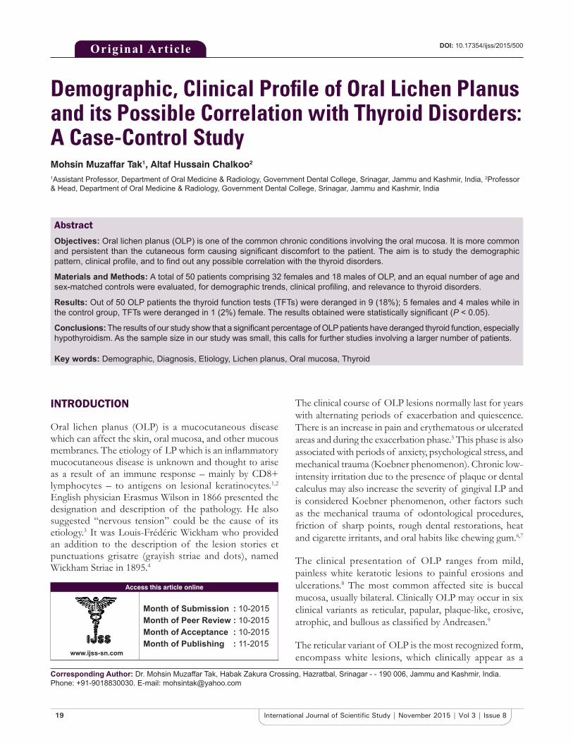
In the present study 50 patients diagnosed with OLP who visited the Department of Oral Medicine and Radiology, were selected along with 50 age and sex matched controls. These patients comprised 18 male patients in the OLP group and 32 female patients and an equal number of patients for each gender and age were selected for the control group (Table 1) & (Graph 1).
These lesions comprised typical reticular form, erosive as well as atrophic form. The reticular type was the most common form present in 37 patients, followed by erosive in 11 patients and atrophic in 2 patients in the OLP group. The reticular form was present in 14 out of 18 male patients, and 23 out of 32 female patients, whereas erosive form was present in 04 male patients and 07 female patients, and atrophic form was only present in 02 female patient (Table 2) & (Graph 2).
Table 1: Age and gender distribution of patientsAge (years)
n (%) P valueMale (n=18) Female (n=32) Total
20-30 4 (22.2) 5 (15.6) 9 (18) 0.115#
31-40 6 (33.3) 6 (18.8) 12 (24)41-50 5 (27.8) 9 (28.1) 14 (28)51-60 2 (11.1) 7 (21.9) 9 (18)61-70 1 (5.6) 5 (15.6) 6 (12)Mean±SD 39.1±11.86 45.2±13.19 43.0±12.94#Statistically non‑significant difference (P<0.05), SD: Standard deviation
Table 2: Distribution of OLP lesions according to clinical variantClinical variant Male Female Total (n (%))Reticular 14 23 37 (74)Erosive 4 7 11 (22)Atrophic 0 2 2 (4)OLP: Oral lichen planus
Tak and Chalkoo: OLP-Demographic, Clinical profile and its possible correlation with thyroid disorders
21 International Journal of Scientific Study | November 2015 | Vol 3 | Issue 8
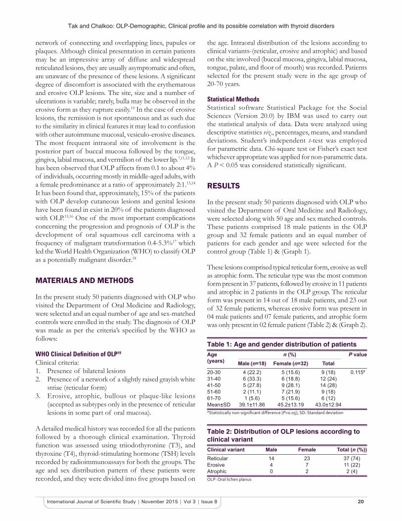
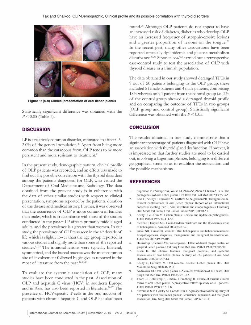
Multiple oral lesions were also present at different sites in the oral cavity (Figure 1), with buccal mucosa being predominantly in the picture in most of the cases followed by gingiva and tongue. Isolated lesions involving palate, the floor of mouth were also present (Table 3) & (Graph 3).
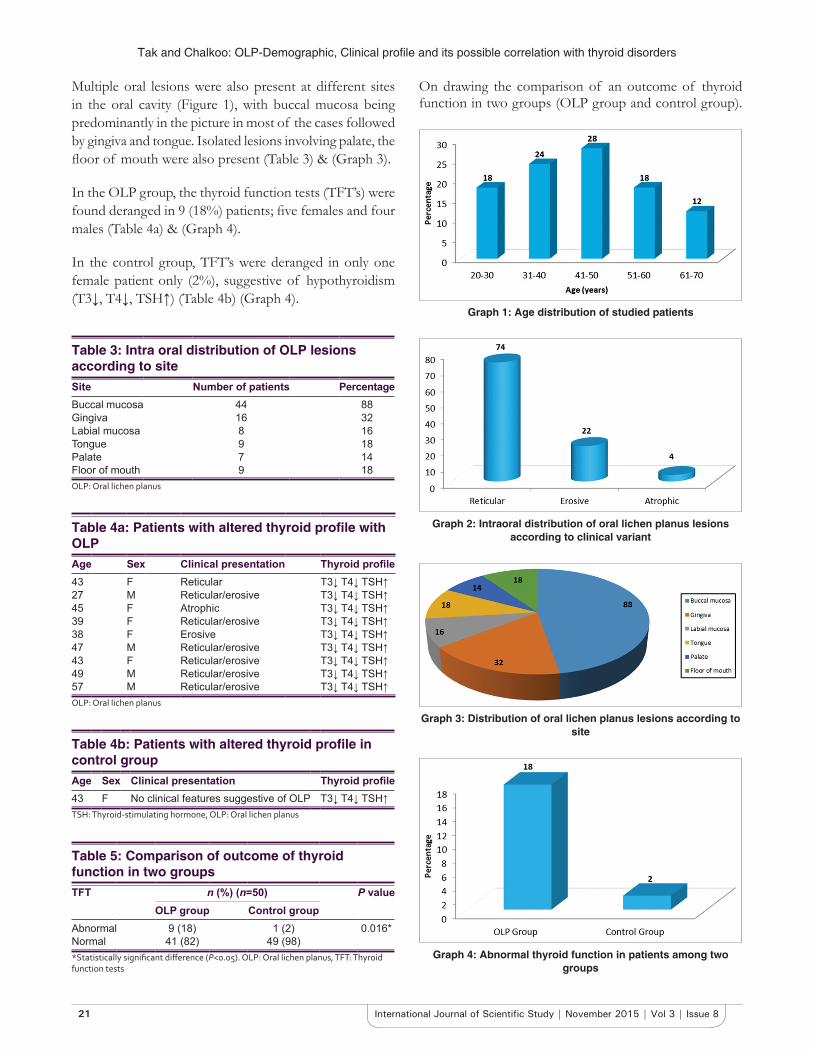
In the OLP group, the thyroid function tests (TFT’s) were found deranged in 9 (18%) patients; five females and four males (Table 4a) & (Graph 4).
In the control group, TFT’s were deranged in only one female patient only (2%), suggestive of hypothyroidism (T3↓, T4↓, TSH↑) (Table 4b) (Graph 4).
Table 3: Intra oral distribution of OLP lesions according to siteSite Number of patients PercentageBuccal mucosa 44 88Gingiva 16 32Labial mucosa 8 16Tongue 9 18Palate 7 14Floor of mouth 9 18OLP: Oral lichen planus
Table 4a: Patients with altered thyroid profile with OLPAge Sex Clinical presentation Thyroid profile43 F Reticular T3↓ T4↓ TSH↑27 M Reticular/erosive T3↓ T4↓ TSH↑45 F Atrophic T3↓ T4↓ TSH↑39 F Reticular/erosive T3↓ T4↓ TSH↑38 F Erosive T3↓ T4↓ TSH↑47 M Reticular/erosive T3↓ T4↓ TSH↑43 F Reticular/erosive T3↓ T4↓ TSH↑49 M Reticular/erosive T3↓ T4↓ TSH↑57 M Reticular/erosive T3↓ T4↓ TSH↑OLP: Oral lichen planus
Table 4b: Patients with altered thyroid profile in control groupAge Sex Clinical presentation Thyroid profile43 F No clinical features suggestive of OLP T3↓ T4↓ TSH↑TSH: Thyroid‑stimulating hormone, OLP: Oral lichen planus
Graph 1: Age distribution of studied patients
Graph 2: Intraoral distribution of oral lichen planus lesions according to clinical variant
Graph 3: Distribution of oral lichen planus lesions according to site
Graph 4: Abnormal thyroid function in patients among two groups
On drawing the comparison of an outcome of thyroid function in two groups (OLP group and control group).
Table 5: Comparison of outcome of thyroid function in two groupsTFT n (%) (n=50) P value
OLP group Control groupAbnormal 9 (18) 1 (2) 0.016*Normal 41 (82) 49 (98)*Statistically significant difference (P<0.05). OLP: Oral lichen planus, TFT: Thyroid function tests
Tak and Chalkoo: OLP-Demographic, Clinical profile and its possible correlation with thyroid disorders
22International Journal of Scientific Study | November 2015 | Vol 3 | Issue 8
Statistically significant difference was obtained with the P < 0.05 (Table 5).
DISCUSSION
LP is a relatively common disorder, estimated to affect 0.5-2.0% of the general population.20 Apart from being more common than the cutaneous form, OLP tends to be more persistent and more resistant to treatment.21
In the present study, demographic pattern, clinical profile of OLP patients was recorded, and an effort was made to find out any possible correlation with the thyroid disorders among the patients diagnosed for OLP, who visited the Department of Oral Medicine and Radiology. The data obtained from the present study is in coherence with the data of other similar studies with respect to clinical presentation, symptoms reported by the patients, duration of the disease and medical history. Further, it was observed that the occurrence of OLP is more common in females than males, which is in accordance with most of the studies conducted in the past.22 LP affects primarily middle-aged adults, and the prevalence is a greater than women. In our study, the prevalence of OLP was seen in the 4th decade of life which is slightly lower than the age group reported in various studies and slightly more than some of the reported studies.12,23 The intraoral lesions were typically bilateral, symmetrical, and the buccal mucosa was the most common site of involvement followed by gingiva as reported in the most of literature from the past.12,23,24
To evaluate the systemic association of OLP, many studies have been conducted in the past. Association of OLP and hepatitis C virus (HCV) in southern Europe and in Asia, has also been reported in literature.24-27 The presence of HCV-specific T-cells in the oral mucosa of patients with chronic hepatitis C and OLP has also been
found.28 Although OLP patients do not appear to have an increased risk of diabetes, diabetics who develop OLP have an increased frequency of atrophic-erosive lesions and a greater proportion of lesions on the tongue.29 In the recent past, many other associations have been reported especially dyslipidemia and glucose metabolism disturbance.30,31 Siponen et al.32 carried out a retrospective case-control study to test the association of OLP with thyroid disease in a Finnish population.
The data obtained in our study showed deranged TFTs in 9 out of 50 patients belonging to the OLP group, these included 5 female patients and 4 male patients, comprising 18% whereas only 1 patient from the control group i.e., 2% of the control group showed a deranged thyroid profile and on comparing the outcome of TFTs in two groups (OLP group and control group). Statistically significant difference was obtained with the P < 0.05.
CONCLUSION
The results obtained in our study demonstrate that a significant percentage of patients diagnosed with OLP have an association with thyroid gland dysfunction. However, it is impressed on that further studies are need to be carried out, involving a larger sample size, belonging to a different geographical strata so as to establish the association and the possible mechanisms.
REFERENCES
1. Sugerman PB, Savage NW, Walsh LJ, Zhao ZZ, Zhou XJ, Khan A, et al. The pathogenesis of oral lichen planus. Crit Rev Oral Biol Med 2002;13:350-65.
2. LodiG,ScullyC,CarrozzoM,GriffithsM,SugermanPB,ThongprasomK.Current controversies in oral lichen planus: Report of an international consensus meeting. Part 1. Viral infections and etiopathogenesis. Oral Surg Oral Med Oral Pathol Oral Radiol Endod 2005;100:40-51.
3. Scully C, el-Kom M. Lichen planus: Review and update on pathogenesis. J Oral Pathol 1985;14:431-58.
4. Steffen C, Dupree ML. Louis-Frédéric Wickham and the Wickham’s striae of lichen planus. Skinmed 2004;3:287-9.
5. Ismail SB, Kumar SK, Zain RB. Oral lichen planus and lichenoid reactions: Etiopathogenesis, diagnosis, management and malignant transformation. J Oral Sci 2007;49:89-106.
6. Holmstrup P, Schiøtz AW, Westergaard J. Effect of dental plaque control on gingival lichen planus. Oral Surg Oral Med Oral Pathol 1990;69:585-90.
7. Eisen D. The clinical features, malignant potential, and systemic associations of oral lichen planus: A study of 723 patients. J Am Acad Dermatol 2002;46:207-14.
8. Scully C, Carrozzo M. Oral mucosal disease: Lichen planus. Br J Oral Maxillofac Surg 2008;46:15-21.
9. Andreasen JO. Oral lichen planus 1. A clinical evaluation of 115 cases. Oral Surg Oral Med Oral Pathol 1968;25:31-42.
10. Thorn JJ, Holmstrup P, Rindum J, Pindborg JJ. Course of various clinical forms of oral lichen planus. A prospective follow-up study of 611 patients. J Oral Pathol 1988;17:213-8.
11. Silverman S Jr, Gorsky M, Lozada-Nur F. A prospective follow-up study of 570 patients with oral lichen planus: Persistence, remission, and malignant association. Oral Surg Oral Med Oral Pathol 1985;60:30-4.
Figure 1: (a-d) Clinical presentation of oral lichen planus
dc
ba
Tak and Chalkoo: OLP-Demographic, Clinical profile and its possible correlation with thyroid disorders
23 International Journal of Scientific Study | November 2015 | Vol 3 | Issue 8
How to cite this article: Tak MM, Chalkoo AH. Demographic, Clinical Profile of Oral Lichen Planus and its Possible Correlation with Thyroid Disorders: A Case-Control Study. Int J Sci Stud 2015;3(8):19-23.
Source of Support: Nil, Conflict of Interest: None declared.
12. Bagán-Sebastián JV, Milián-Masanet MA, Peñarrocha-Diago M, Jiménez Y. A clinical study of 205 patients with oral lichen planus. J Oral Maxillofac Surg 1992;50:116-8.
13. ScullyC,BeyliM,FerreiroMC,FicarraG,GillY,GriffithsM,et al. Update on oral lichen planus: Etiopathogenesis and management. Crit Rev Oral Biol Med 1998;9:86-122.
14. Kragelund C, Thomsen CE, Bardow A, Pedersen AM, Nauntofte B, Reibel J, et al. Oral lichen planus and intake of drugs metabolized by polymorphic cytochrome P450 enzymes. Oral Dis 2003;9:177-87.
15. Farhi D, Dupin N. Pathophysiology, etiologic factors, and clinical management of oral lichen planus, part I: Facts and controversies. Clin Dermatol 2010;28:100-8.
16. Edwards PC, Kelsch R. Oral lichen planus: Clinical presentation and management. J Can Dent Assoc 2002;68:494-9.
17. Eisen D. The evaluation of cutaneous, genital, scalp, nail, esophageal, and ocular involvement in patients with oral lichen planus. Oral Surg Oral Med Oral Pathol Oral Radiol Endod 1999;88:431-6.
18. Shi P, Liu W, Zhou ZT, He QB, Jiang WW. Podoplanin and ABCG2: Malignant transformation risk markers for oral lichen planus. Cancer Epidemiol Biomarkers Prev 2010;19:844-9.
19. González-García A, Diniz-Freitas M, Gándara-Vila P, Blanco-Carrión A, García-García A, Gándara-Rey J. Triamcinolone acetonide mouth rinses for treatment of erosive oral lichen planus: Efficacyandriskof fungalover-infection. Oral Dis 2006;12:559-65.
20. McCreary CE, McCartan BE. Clinical management of oral lichen planus. Br J Oral Maxillofac Surg 1999;37:338-43.
21. Mollaoglu N. Oral lichen planus: A review. Br J Oral Maxillofac Surg 2000;38:370-7.
22. Brown RS, Bottomley WK, Puente E, Lavigne GJ. A retrospective evaluation of 193 patients with oral lichen planus. J Oral Pathol Med 1993;22:69-72.
23. Carrozzo M, Gandolfo S. The management of oral lichen planus. Oral Dis 1999;5:196-205.
24. Bagán JV, Aguirre JM, del Olmo JA, Milián A, Peñarrocha M, Rodrigo JM, et al. Oral lichen planus and chronic liver disease: A clinical and morphometric study of the oral lesions in relation to transaminase elevation. Oral Surg Oral Med Oral Pathol 1994;78:337-42.
25. Nagao Y, Sata M, Tanikawa K, Itoh K, Kameyama T. Lichen planus and hepatitis C virus in the northern Kyushu region of Japan. Eur J Clin Invest 1995;25:910-4.
26. Carrozzo M, Gandolfo S, Carbone M, Colombatto P, Broccoletti R, Garzino-Demo P, et al. Hepatitis C virus infection in Italian patients with oral lichen planus: A prospective case-control study. J Oral Pathol Med 1996;25:527-33.
27. Chuang TY, Stitle L, Brashear R, Lewis C. Hepatitis C virus and lichen planus: A case-control study of 340 patients. J Am Acad Dermatol 1999;41:787-9.
28. Pilli M, Penna A, Zerbini A, Vescovi P, Manfredi M, Negro F, et al. Oral lichenplanuspathogenesis:A role for theHCV-specificcellular immuneresponse. Hepatology 2002;36:1446-52.
29. Bagan JV, Donat JS, Penarrocha M, Milian MA, Sanchis JM. Oral lichen planus and diabetes mellitus. A clinico-pathological study. Bull Group Int Rech Sci Stomatol Odontol 1993;36:3-6.
30. Dreiher J, Shapiro J, Cohen AD. Lichen planus and dyslipidaemia: A case-control study. Br J Dermatol 2009;161:626-9.
31. Seyhan M, Ozcan H, Sahin I, Bayram N, Karincaoglu Y. High prevalence of glucose metabolism disturbance in patients with lichen planus. Diabetes Res Clin Pract 2007;77:198-202.
32. Siponen M, Huuskonen L, Läärä E, Salo T. Association of oral lichen planus with thyroid disease in a Finnish population: A retrospective case-control study. Oral Surg Oral Med Oral Pathol Oral Radiol Endodontol 2010;110:319-24.