Embed Size (px)
Citation preview
Best Practice & Research Clinical Anaesthesiology 26 (2012) 355–366
Contents lists available at SciVerse ScienceDirect
Best Practice & Research ClinicalAnaesthesiology
journal homepage: www.elsevier .com/locate/bean
7
Delirium: Is sleep important?
Paula L. Watson, MD, Assistant Professor a,*, Piero Ceriana, MD, Director b,d,Francesco Fanfulla, MD, Head of Sleep Medicine Unit c,d
aDepartment of Medicine, Division of Allergy, Pulmonary, Critical Care and Sleep Medicine, Vanderbilt University Medical Center,Nashville, TN, USAbRespiratory Rehabilitation and Intensive Care Unit, Fondazione Maugeri, Pavia, Italyc Sleep Medicine Unit, S. Maugeri Foundation, Scientific Institute of Pavia, Pavia, Italy
Keywords:sleepdeliriumsedationmechanical ventilation
* Corresponding author. Vanderbilt University S37232-5735, USA.
E-mail addresses: [email protected](F. Fanfulla).
d Istituto Scientifico di Pavia, Fondazione S.Maug
1521-6896/$ – see front matter � 2012 Elsevier Lthttp://dx.doi.org/10.1016/j.bpa.2012.08.005
Delirium and poor sleep quality are common and often co-exist inhospitalised patients. A link between these disorders has beenhypothesised but whether this link is a cause-and-effect relation-ship or simply an association resulting from shared mechanisms isyet to be determined. Potential shared mechanisms include:abnormalities of neurotransmitters, tissue ischaemia, inflamma-tion and sedative exposure. Sedatives, while decreasing sleeplatency, often cause a decrease in slow wave sleep and stage rapideye movement (REM) sleep and therefore may not provide thesame restorative properties as natural sleep. Mechanical ventila-tion, an important cause of sleep disruption in intensive care unit(ICU) patients, may lead to sleep disruption not only from thediscomfort of the endotracheal tube but also as a result of inef-fective respiratory efforts and by inducing central apnoea events ifnot properly adjusted for the patient’s physiologic needs. Whenpossible, efforts should be made to optimise the patient–ventilatorinteraction to minimise sleep disruptions.
� 2012 Elsevier Ltd. All rights reserved.
Sleep is an essential biologic function that is important for both physiologic rest and emotionalwell-being. Hospitalised patients often have poor-quality sleep experiencing difficulty with sleepinitiation, sleep fragmentation, earlymorning awakenings and decreased total sleep time. The patient’s
chool of Medicine, B-817 TVC, 1301 Medical Center Drive, Nashville, TN
u (P.L. Watson), [email protected] (P. Ceriana), [email protected]
eri, Via Maugeri 10, 27100 Pavia (PV), Italy.
d. All rights reserved.
P.L. Watson et al. / Best Practice & Research Clinical Anaesthesiology 26 (2012) 355–366356
underlying illness, pain, environmental noise and artificial light, patient care activities and medicationeffects all are potential contributors to their sleep disruption. Even the sedative and analgesic medi-cations administered in an attempt to promote sleep and comfort can lead to abnormal sleep patternsthat may not provide the same restorative function as natural sleep.
Normal sleep architecture consists of two major sleep stage categories – non-rapid eye movement(NREM) and rapid eye movement (REM) sleep. NREM is further subdivided into three stages ofprogressively deeper sleep, which are N1, N2 and N3 (also known as slow wave sleep (SWS)). DuringNREM sleep, brain activity is decreased and there is a corresponding decrease in metabolic rate. Incontrast, during REM sleep the brain is active and there is skeletal muscle atonia. Although thefunctions of the different sleep stages remain largely unknown, both NREM and REM sleep are thoughtto be involved in memory consolidation.1
Delirium is common in hospitalised patients and occurs in 11% of elderly emergency departmentpatients,2 in 37% of postoperative patients3 and in as many as 80% of critically ill patients receivingmechanical ventilation.4 Older patients are more susceptible, with up to 50% of geriatric patientsdeveloping delirium at some point in their hospital course.5,6 The pathogenesis of delirium is poorlyunderstood. Hypotheses regarding contributing aetiologies have included neurotransmitter imbal-ances, pro-inflammatory cytokines, tissue hypoxia and sleep deprivation.7–9
Delirium is characterised by inattention, fluctuating mental status, disorganised thinking and analtered level of consciousness – findings also characteristics of sleep deprivation. Sleep disturbancesare common in delirious patients. Further, while sleep deprivation is regarded to be a potentiallymodifiable risk factor for the development of delirium, it is also likely that delirium itself contributes tosleep disturbances.
This article reviews (1) the potential relationship between sleep deprivation and delirium, (2) themodification of sleep with sedative administration and (3) the effects of mechanical ventilation onsleep quality.
Sleep–delirium interactions
Hospital-related sleep disturbances
Poor sleep quality is a problem that affects many hospitalised adult patients and that is yetunderreported and not properly considered by the majority of health professionals. Baseline acutedisease, change of sleep habits, use of sedatives and other medications, environmental noise andartificial light can be listed among the main causes of this problem. An association has been shownbetween sleep disturbances and the number of chronic diseases, the presence of pain, use of tricyclicantidepressants and the length of hospital stay.10
Almost 50% of patients admitted in general hospital medical wards experience insomnia, excessivedaytime sleepiness or both.11 Few, if any, of these sleep problems are ever reported in the medicalrecords. The use of hypnotic medications both prior to hospital admission and also during the hospitalstay is common; therefore, physicians should be aware of not only the direct adverse effects of thesemedications but also the potential effects of sedative–hypnotic withdrawal in hospitalised patients.
Intensive care units (ICUs) are the hospital setting where the problem of sleep disturbances likelyreaches its highest level and represents the source of most relevant data available in the literature.During their ICU stay, patients often experience prolonged sleep onset, fragmented sleep, poor sleepefficiency and decreased amounts of REM sleep.12 The overall sleep time during a 24-h period can bewithin normal limits (7–9 h), but almost 50% of this time is spent in short bouts during the day ratherthan a consolidated nocturnal sleep, so that patients have difficulty achieving the REM and N3 (SWS)sleep stages.13 ICU patients complain of long periods of wakefulness; the sleep architecture in thesepatients is remarkable for increased stage N1 (light) sleep, decreased amounts of N2 andN3 and notablyamarked reduction in REM-stage sleep. In contrast to healthy individuals who experience an average of20–25% REM sleep, ICU patients may experience little or no REM-stage sleep during their ICU stay.14,15
Sleep deprivation can cause both physiological and behavioural consequences. The most relevantphysiologic consequences are an impaired immune response,16 an alteration in metabolic and endo-crine systems,17 an increase in pain sensitivity18 and changes in cardiac modulations from sympathetic
P.L. Watson et al. / Best Practice & Research Clinical Anaesthesiology 26 (2012) 355–366 357
and parasympathetic systems.19 The behavioural changes include impaired attention, mood andpsychomotor performance, increased daytime sleepiness, sensation of fatigue and irritability.20
Delirium and possible links to sleep disturbances
Delirium is a form of brain dysfunction whose main features are acute onset of a fluctuatingawareness, inattention, disorganised thinking and an altered level of consciousness. These samefeatures can occur to varying degrees as a result of sleep deprivation. Delirium can be either hypoactive,hyperactive or mixed in character. The hypoactive form occurs frequently and though known to beassociated with poor outcomes, it too often goes unrecognised by the physicians and nurses caring forthese patients.
Both sleep disturbances and delirium are very common in hospitalised patients, especially criticallyill patients in the ICU environment. A possible link between these two disorders has been hypothesisedin terms of a common pathophysiologic pathway, shared mechanisms and a potential cause–effectrelationship.21
Studies conducted mainly in cardiac surgical patients indicate that sleep deprivation can eithercause delirium,22 be a result of it23 or may simply lower the clinical threshold for delirium. DecreasedSWS and decreased REM-stage sleep have been hypothesised as contributing factors for the devel-opment of delirium. While a recent study of ICU patients demonstrated an association betweendelirium and severe REM sleep reduction (<6% of total sleep time), a cause-and-effect relationship wasnot established.15 Although the association between sleep disturbance and delirium has been studiedfor many years, at present it is difficult to define the true relationship between them. Perhaps, though,it is possible to view their relationship as part of a shared pathophysiologic pathway.
Although themechanism of delirium is not completely understood, a neurotransmitter imbalance isone of the leading hypotheses, with dopamine and acetylcholine felt to be the most importantneurotransmitters involved. An imbalance of these same neurotransmitters also occurs in associationwith sleep deprivation.24 During delirium the levels of acetylcholine are generally thought to be lowand those of dopamine high, though a few reports tend to hypothesise the opposite imbalance (highacetylcholine and low dopamine).25 The importance of dopamine on the development of delirium, inparticular, seems to be supported by the therapeutic effect of haloperidol, a powerful dopamineblocker.
In addition to acetylcholine and dopamine, there is evidence that other neurotransmitters such astryptophan can play a significant role in delirium. Tryptophan, a serotonin precursor, was reduced ina population of cardiac surgery patients with delirium.26 Importantly, it appears that an abnormaltryptophan metabolism can modulate the type of delirium: hyperactive or hypoactive.27 Tryptophan,moreover, is tightly connected tomelatonin, a hormone involved in the regulation of circadian rhythm,which has also been linked to delirium. Tryptophan is a direct precursor of melatonin; melatonin issecreted by the pineal gland and metabolised by the liver to 6-hydroxymelatonin and then conjugatedwith sulphuric acid to 6-sulphatoxymelatonin (excreted in the urine and sometimes used in clinicaltrials to assess melatonin secretion).
There is increasing interest in melatonin and its role in postoperative and critical illness. Critically illpatients undergoing mechanical ventilation exhibit a derangement of circadian rhythm and decreasedmelatonin production.28,29 Moreover, the involvement of melatonin in surgical stress, sepsis anddelirium is becoming more and more evident. In particular, postoperative patients in whommelatoninplasma levels were measured for 2 days after surgery showed a correspondence between abnormalmelatonin levels and delirium. Patients without postoperative delirium had normal plasma levels,while those who developed delirium had plasma melatonin levels lower than preoperative values iftheir operative course was uncomplicated or increased melatonin levels if they had further compli-cations such as pneumonia or sepsis.30 A possible derangement, not only in melatonin secretion, butalso in melatonin metabolism can also be suspected from the observation of Balan, who showed thatpatients with hypoactive or hyperactive delirium had, respectively, increased or decreased urinary 6-sulphatoxymelatonin levels.31 The role of melatonin in the regulation of the sleep–wake cycle, reset-ting of circadian rhythm disturbances and its extensive antioxidant activity have potential applicationsin critical care patients.
P.L. Watson et al. / Best Practice & Research Clinical Anaesthesiology 26 (2012) 355–366358
Other possible mechanisms believed to contribute to the occurrence of delirium are acuteinflammation and ischaemia. The anatomical pathway thought to be involved in delirium includesbrain areas such as the thalamus, the prefrontal, posterior parietal and fusiform cortex and the basalganglia.25 Hence, in theory, ischaemia in one of these areas can lead to delirium. Systemic inflammationhas also been implicated in the pathogenesis of delirium. It has been observed that delirious patientsafter hip replacement surgery had higher serum levels of C-reactive protein.32 Other inflammatorycytokines, such as interleukin (IL)-6 and IL-8 are associated with and can possibly induce delirium,either directly or through a neurotransmitter imbalance.33–36
Another link between sleep disturbances and delirium comes from the observation that delirium, ina population of elderly patients undergoing knee replacement surgery, occurred more frequentlyamong those with obstructive sleep apnoea (OSA).37 The mechanism whereby sleep apnoea increasesthe risk of delirium is not clear but we do know that oxygen desaturations occur during apnoeic phasesand that hypoxia is one of the possible causes of delirium. Moreover, the cyclic oxygen desaturationsand restorations during apnoeic episodes can cause an increase of inflammatory cytokines; and, sleepapnoea, per se, can be viewed as an inflammatory process, with increased levels of ILs and tumournecrosis factor.38 As stated above, the occurrence of delirium may be facilitated by ischaemia andinflammation. Moreover, a connection has been found between sleep-disordered breathing anddementia-like cognitive impairment, and patients affected by dementia are more prone to developdelirium,39 thus representing the rationale for a further link between sleep disruption and delirium.
Further evidence of a connection between sleep disorders and delirium comes from the currentpreventive strategies for delirium. The Hospital Elder Life Program (HELP) tested by the Yale DeliriumPrevention trial40 assessed six risk factors (dehydration, sleep deprivation, immobility, visual impair-ment, cognitive impairment and hearing impairment) at the time of hospital admission. When one ofthese risk factors was determined to be present, it was treated with a targeted intervention bya dedicated team. The non-pharmacologic sleep protocol included sips of warm milk, back andshoulder rubs and relaxing music when going to sleep. This protocol not only reduced the need forsedative and hypnotic drugs, but also reduced the incidence of delirium.41 As a further reinforcement ofthis evidence, the guidelines for prevention of delirium issued by the National Institute for Health andClinical Excellence (NICE) recommend efforts to improve sleep quality (i.e., avoiding unnecessarynursing or staff procedures during nighttime and reducing environmental noise as much as possible) asa mainstay measure to reduce the incidence of delirium in hospitalised patients.42
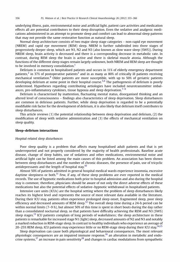
The existence of a link between delirium and sleep disruption is very likely though it is not yetevident whether the former can be the cause of the latter and vice versa or, whether more likely theysimply share a common pathophysiologic pathway. This last mechanism appears to be the most likelyexplanation, involving the above-mentioned complex pathways between ischaemia/inflammation,hypoxia, neurotransmitter imbalance and abnormalities of tryptophan/melatonin metabolism (seeFig. 1). The potential role of sedative medications will be discussed in the following section.
Sedation, sleep and delirium
Critically ill patients are almost universally administered sedative–hypnotics and often at highdoses. These medications are given to aid with the comfort and anxiety of patients as well as tofacilitate procedures andmechanical ventilation. In addition, these medications are often administeredin an attempt to improve the sleep of ICU patients. The relationship between sedation and sleep ishowever complicated. While sedatives produce a state that can physically resemble sleep, there are infact significant differences between pharmacologically induced sleep and natural sleep that may beimportant clinically.
For years, sleep has been used as a metaphor for sedation; and indeed, sleep and sedation do haveseveral similarities. Both lead to decreased responsiveness to external stimuli, decreased muscle toneand respiratory depression. There are, however, many important differences. While the functions ofsleep are not fully known, it is an essential biologic function necessary for life. Sleep occurs sponta-neously and is reversible by external stimuli whereas sedation is not. Moreover, natural sleep ischaracterised by a circadian rhythm and a cyclical progression through the sleep stages with classicelectroencephalographic (EEG) patterns for each sleep stage. Sedation effects are medication specific
Fig. 1. Possible common pathophysiological pathways between delirium and sleep disruption.
P.L. Watson et al. / Best Practice & Research Clinical Anaesthesiology 26 (2012) 355–366 359
and dose dependent rather than cyclical and will frequently lead to atypical EEG patterns not observedin natural sleep.
Most sedatives and analgesics are known to cause a decrease in SWS and REM sleep, the stages thatare considered the most restorative. Whether sedatives provide the same restorative properties asnatural sleep is largely unknown and is often debated, as few studies have addressed this question.Studies conducted on rodents indicate that sedation with propofol for 12 h is not associated with anincrease in sleep need compared to baseline and also that sedation with propofol appears to allow forrecovery from sleep deprivation.43,44 In human study, day-surgery patients undergoing sedation withpropofol for 1 h were found to have a longer nocturnal sleep latency but a shorter latency to stage N2sleep and no difference in total sleep time (TST) or sleep efficiency.45 However, this lack of difference inTST following sedation may not have reached significance due to the relatively short sedation period.
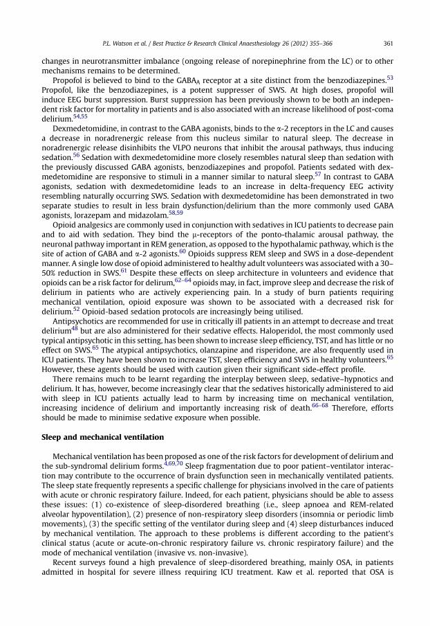
There is evidence that sedatives exert their effect at least in part by acting on the neuronal pathwaysgoverning natural sleep and therefore it may be helpful to review some of the basics of these neuronalsleep pathways. The sleep–wake state is regulated by a complex interaction of neurotransmittersproduced by clusters of neurons located in the reticular formation of the brain stem, midbrain, thal-amus and hypothalamus with connections to the brain cortex. The neurotransmitters norepinephrine,serotonin, acetylcholine and histamine all play important roles in maintenance of wakefulness.Important nuclei of the sleep pathway include: (1) the locus coeruleus (LC) located in the pons and theprincipal site of norepinephrine production in the brain, (2) the ventrolateral preoptic nucleus (VLPO)of the hypothalamus that produces the sleep active neurotransmitters, g-aminobutyric acid (GABA)and galanin and (3) the tuberomammillary nucleus (TMN), the site of histamine production.
The natural sleep–wake state is controlled in a flip–flop manner where each state inhibits theactivity of the other (i.e., the wake active neurons inhibit the activity of neurons producing sleepneurotransmitters and vice versa).46 At sleep onset, there is an inhibition of norepinephrine releasefrom the LC, which leads to an increase in GABA and galanin release from the ventrolateral preopticnucleus (VLPO) (Fig. 2A). GABA then inhibits the release of histamine, a wake-promoting neuro-transmitter, from the TMN. Serotonin-producing neurons located in the medial and dorsal raphe alsocontribute to the maintenance of a quiet awake state. And also, acetylcholine is important in main-tainingwakefulness and attention. Other neurotransmitters including dopamine, orexin and adenosinealso play a significant role in the regulation of the sleep–wake state.
Sedatives cause their effects by intersecting the neuronal sleep pathway at various locations. Themost commonly used sedatives, benzodiazepines and propofol, exert their effect by activating GABAAreceptors in the VLPO,47 thereby suppressing histamine release from the TMN (Fig. 2B). However,because these sedatives act downstream from the LC, norepinephrine release from the LC is not
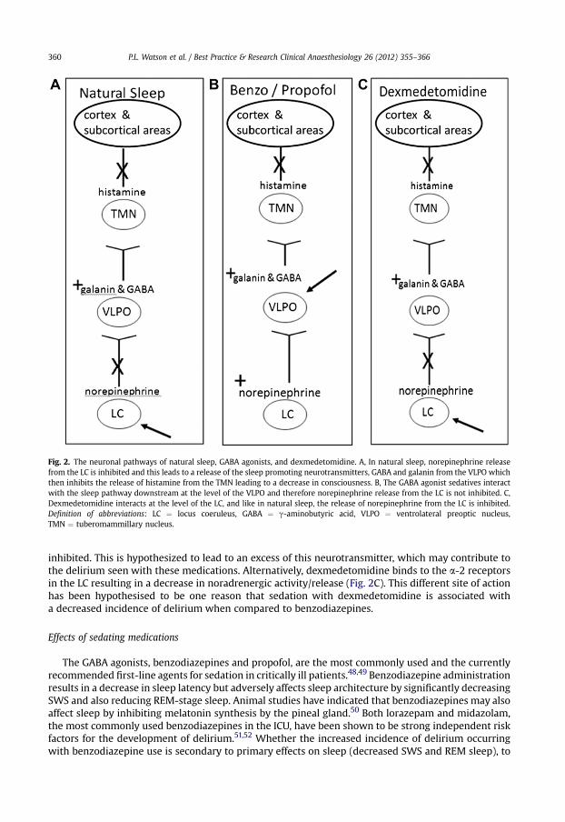
Fig. 2. The neuronal pathways of natural sleep, GABA agonists, and dexmedetomidine. A, In natural sleep, norepinephrine releasefrom the LC is inhibited and this leads to a release of the sleep promoting neurotransmitters, GABA and galanin from the VLPO whichthen inhibits the release of histamine from the TMN leading to a decrease in consciousness. B, The GABA agonist sedatives interactwith the sleep pathway downstream at the level of the VLPO and therefore norepinephrine release from the LC is not inhibited. C,Dexmedetomidine interacts at the level of the LC, and like in natural sleep, the release of norepinephrine from the LC is inhibited.Definition of abbreviations: LC ¼ locus coeruleus, GABA ¼ g-aminobutyric acid, VLPO ¼ ventrolateral preoptic nucleus,TMN ¼ tuberomammillary nucleus.
P.L. Watson et al. / Best Practice & Research Clinical Anaesthesiology 26 (2012) 355–366360
inhibited. This is hypothesized to lead to an excess of this neurotransmitter, which may contribute tothe delirium seen with these medications. Alternatively, dexmedetomidine binds to the a-2 receptorsin the LC resulting in a decrease in noradrenergic activity/release (Fig. 2C). This different site of actionhas been hypothesised to be one reason that sedation with dexmedetomidine is associated witha decreased incidence of delirium when compared to benzodiazepines.
Effects of sedating medications
The GABA agonists, benzodiazepines and propofol, are the most commonly used and the currentlyrecommended first-line agents for sedation in critically ill patients.48,49 Benzodiazepine administrationresults in a decrease in sleep latency but adversely affects sleep architecture by significantly decreasingSWS and also reducing REM-stage sleep. Animal studies have indicated that benzodiazepines may alsoaffect sleep by inhibiting melatonin synthesis by the pineal gland.50 Both lorazepam and midazolam,the most commonly used benzodiazepines in the ICU, have been shown to be strong independent riskfactors for the development of delirium.51,52 Whether the increased incidence of delirium occurringwith benzodiazepine use is secondary to primary effects on sleep (decreased SWS and REM sleep), to
P.L. Watson et al. / Best Practice & Research Clinical Anaesthesiology 26 (2012) 355–366 361
changes in neurotransmitter imbalance (ongoing release of norepinephrine from the LC) or to othermechanisms remains to be determined.
Propofol is believed to bind to the GABAA receptor at a site distinct from the benzodiazepines.53
Propofol, like the benzodiazepines, is a potent suppresser of SWS. At high doses, propofol willinduce EEG burst suppression. Burst suppression has been previously shown to be both an indepen-dent risk factor for mortality in patients and is also associated with an increase likelihood of post-comadelirium.54,55
Dexmedetomidine, in contrast to the GABA agonists, binds to the a-2 receptors in the LC and causesa decrease in noradrenergic release from this nucleus similar to natural sleep. The decrease innoradrenergic release disinhibits the VLPO neurons that inhibit the arousal pathways, thus inducingsedation.56 Sedation with dexmedetomidine more closely resembles natural sleep than sedation withthe previously discussed GABA agonists, benzodiazepines and propofol. Patients sedated with dex-medetomidine are responsive to stimuli in a manner similar to natural sleep.57 In contrast to GABAagonists, sedation with dexmedetomidine leads to an increase in delta-frequency EEG activityresembling naturally occurring SWS. Sedation with dexmedetomidine has been demonstrated in twoseparate studies to result in less brain dysfunction/delirium than the more commonly used GABAagonists, lorazepam and midazolam.58,59
Opioid analgesics are commonly used in conjunctionwith sedatives in ICU patients to decrease painand to aid with sedation. They bind the m-receptors of the ponto-thalamic arousal pathway, theneuronal pathway important in REM generation, as opposed to the hypothalamic pathway, which is thesite of action of GABA and a-2 agonists.60 Opioids suppress REM sleep and SWS in a dose-dependentmanner. A single low dose of opioid administered to healthy adult volunteers was associatedwith a 30–50% reduction in SWS.61 Despite these effects on sleep architecture in volunteers and evidence thatopioids can be a risk factor for delirum,62–64 opioids may, in fact, improve sleep and decrease the risk ofdelirium in patients who are actively experiencing pain. In a study of burn patients requiringmechanical ventilation, opioid exposure was shown to be associated with a decreased risk fordelirium.52 Opioid-based sedation protocols are increasingly being utilised.
Antipsychotics are recommended for use in critically ill patients in an attempt to decrease and treatdelirium48 but are also administered for their sedative effects. Haloperidol, the most commonly usedtypical antipsychotic in this setting, has been shown to increase sleep efficiency, TST, and has little or noeffect on SWS.65 The atypical antipsychotics, olanzapine and risperidone, are also frequently used inICU patients. They have been shown to increase TST, sleep efficiency and SWS in healthy volunteers.65
However, these agents should be used with caution given their significant side-effect profile.There remains much to be learnt regarding the interplay between sleep, sedative–hypnotics and
delirium. It has, however, become increasingly clear that the sedatives historically administered to aidwith sleep in ICU patients actually lead to harm by increasing time on mechanical ventilation,increasing incidence of delirium and importantly increasing risk of death.66–68 Therefore, effortsshould be made to minimise sedative exposure when possible.
Sleep and mechanical ventilation
Mechanical ventilation has been proposed as one of the risk factors for development of delirium andthe sub-syndromal delirium forms.4,69,70 Sleep fragmentation due to poor patient–ventilator interac-tion may contribute to the occurrence of brain dysfunction seen in mechanically ventilated patients.The sleep state frequently represents a specific challenge for physicians involved in the care of patientswith acute or chronic respiratory failure. Indeed, for each patient, physicians should be able to assessthese issues: (1) co-existence of sleep-disordered breathing (i.e., sleep apnoea and REM-relatedalveolar hypoventilation), (2) presence of non-respiratory sleep disorders (insomnia or periodic limbmovements), (3) the specific setting of the ventilator during sleep and (4) sleep disturbances inducedby mechanical ventilation. The approach to these problems is different according to the patient’sclinical status (acute or acute-on-chronic respiratory failure vs. chronic respiratory failure) and themode of mechanical ventilation (invasive vs. non-invasive).
Recent surveys found a high prevalence of sleep-disordered breathing, mainly OSA, in patientsadmitted in hospital for severe illness requiring ICU treatment. Kaw et al. reported that OSA is
P.L. Watson et al. / Best Practice & Research Clinical Anaesthesiology 26 (2012) 355–366362
associated with an increasing risk of ICU admission in the postoperative period.71 Further, Diaz-Abadet al. found a high prevalence of unrecognised sleep-disordered breathing in patients who arecandidates for decannulation after weaning from prolonged mechanical ventilation.72 Similarly,insomnia and periodic leg movements as well as other sleep disorders are very common in elderlypatients and in those with chronic disease such as chronic obstructive pulmonary disease (COPD),making it difficult to discriminate the effect of mechanical ventilation on sleep quality.73
While mechanical ventilation certainly affects sleep quality, sleep quality has also been shown toaffect the success of ventilator therapies. Rocheet al. reported that late non-invasive ventilation failurein elderly patients with acute hypercapnic respiratory failure was associated with early sleep distur-bances. They found that patients failing non-invasive ventilation had poorer sleep quality with greatercircadian sleep-cycle disruption and less nocturnal REM sleep compared with patients successfullytreated with non-invasive ventilation.74
Ventilatory parameters are mainly determined empirically, based on diurnal arterial blood-gasvariations or the patient’s tolerance when awake and conscious. However, during sleep, patientsmay have a profound modification in the recruitment of the respiratory muscles leading to worseningof alveolar hypoventilation.75 This is particularly the case in patients with severe respiratory muscleweakness. Sustained arterial oxygen saturation (SpO2) desaturations (>10 min) can be seen in thesepatients as the result of this residual hypoventilation. On the other hand, excessive inspiratory supportmay induce the development of periodic breathing or central apnoeas, especially with pressuresupport ventilation (PSV).76 Indeed, setting the inspiratory support too high compared to the patient’sdemand during sleep may result in passive hyperventilation. In this way, carbon dioxide partialpressure (PCO2) may diminish during sleep, triggering central apnoea, which in turn induces arousalsand awakenings.
The presence of ineffective respiratory efforts, mouth leaks in patients receiving non-invasiveventilation (NIV) and auto-triggering also reduce the effectiveness of mechanical ventilation. Aphenomenon of missing or ineffective efforts has been shown to occur both in ventilator-dependentCOPD patients and in others with difficult weaning from the ventilator. This was attributed toseveral factors including: a reduction in respiratory drive, reduction in respiratory muscle strength,increased inspiratory load due to augmented upper airway resistance and mouth leaks. These factorsall contribute to the inability to trigger the ventilator adequately.77
Fanfulla et al. found that the patients with recurrent ineffective breathing during sleep were thosewith the highest level of inspiratory assistance. High levels of inspiratory assistance that produce largertidal volumes, may induce dynamic hyperinflation, which has also been implicated in the genesis ofineffective efforts.78 Furthermore, in PSV and assisted volume-cycled ventilation, the end of theventilator’s inflation cycle is not always synchronised with the end of the patient’s inspiratory effort. Asa result, inflation may continue into the phase of neural expiration. When the ventilator cycle extendsinto neural expiration, the time available for expiratory flow, before the next inspiratory effort, isreduced. If passive functional residual capacity is not reached during the shortened expiratory phase,dynamic hyperinflation can occur or worsen. Dynamic hyperinflation increases the work of breathingand makes it difficult for the patient to trigger the ventilator, making the occurrence of ineffectivebreathing more likely.
Patients with the co-existence of acute respiratory failure and OSA require specific assessmentwhen NIV is identified as the treatment of choice. Two different goals should be reached: the first is tostabilise the upper airway and the second is to provide adequate inspiratory support to maintainoptimal alveolar ventilation. Intermittent obstruction of the upper airway is frequent in patients duringNIV.79 Moreover, upper airway obstruction may also occur in patients without co-existence of OSA andis generally related to episodes of intermittent obstruction at the glottic level reflecting cyclic glotticclosure induced by hyperventilation.80
The exact role of mechanical ventilation in sleep fragmentation in ICU patients remains poorlyunderstood. Mechanical ventilation has been shown to disrupt sleep in patients with acute or chronicrespiratory failure through several different mechanisms: level of inspiratory support, development ofcentral apnoeas and/or Cheyne–Stokes breathing, patient–ventilator asynchronies (particularly withineffective inspiratory efforts) and the presence of an endotracheal tube. Parthasarathy and Tobinfound that critically ill patients experience greater fragmentation of sleep during PSV than during
P.L. Watson et al. / Best Practice & Research Clinical Anaesthesiology 26 (2012) 355–366 363
assist-control ventilation (ACV) because of the development of central apnoeas.76 Bosma et al. foundthat patient–ventilator discordance causes sleep disruption and that proportional-assist ventilationseems more efficacious than PSV in matching the patient’s ventilatory requirements with ventilatorassistance, therefore resulting in fewer patient–ventilator asynchronies and a better quality of sleep.81
In contrast, Cabello et al., in a study designed to compare the influence of three common ventilationmodes, observed that the ventilatory mode did not influence the sleep pattern.82 The three modes ofventilation were ACV, clinically adjusted PSV and automatically adjusted PSV, which offers a contin-uous adaptation of the pressure support (PS) level. A possible explanation of these results, clearlydifferent from those previously published, is that the authors take care to avoid excess of inspiratorysupport as demonstrated by similar minute ventilation and tidal volume observed with the threedifferent ventilators.
In a study designed to compare two non-invasive PSV settings, one based on clinical parameters andthe other based on physiological requirements as estimated from an assessment of the patient’srespiratory effort and mechanics, Fanfulla et al. showed that the physiology-based setting was asso-ciated with improvements in sleep quantity and quality.83 They found a statistically significant asso-ciation between the reduction of ineffective respiratory efforts and an improvement in sleep quality.However, when evaluating the effects of mechanical ventilation on sleep in the clinical arena, othersources of variability should be taken into account: the physicians’ approach may vary greatly; clinicalseverity of the patients (increased respiratory drive during acute respiratory failure and during acuteinflammatory states); aetiology of acute respiratory failure; different mechanical respiratory proper-ties; and methods used for monitoring the patient–ventilator interaction.
Studies of the newest modes of ventilation, such as adaptive support ventilation and proportional-assist ventilation with load adjustable gain factors and neutrally adjusted ventilatory assist, havedemonstrated an improvement of patient–ventilator interaction when compared to the more tradi-tional ventilator modes. However, data on sleep quality during these modes of ventilation are stilllimited and further investigation is needed.81,83–86
Practice points
� Sleep complaints are common in hospitalised patients.� There is an association between poor sleep quality and delirium but a cause-and-effectrelationship has not been established.
� Sedating medications cause alterations in sleep architecture with most causing a decreaseSWS and REM-stage sleep.
� Patient–ventilator interactions can differ depending on whether the patient is awake orasleep and should be monitored during sleep in patients requiring long-term mechanicalventilation.
� Upper airway obstructive events during sleep occur frequently in patients treated with non-invasive mechanical ventilation, especially those with signs or symptoms of OSA or with theobesity–hypoventilation syndrome.
Research agenda
� Are sleep alterations, which are common in hospitalised patients, a cause of delirium?� Does sedation provide the same restorative benefits as natural sleep?� How and when should sleep quality be assessed in patients admitted to ICU?� Is polysomnography or cardio-respiratorymonitoring the gold standard for the assessment ofpatient–ventilator interaction?
P.L. Watson et al. / Best Practice & Research Clinical Anaesthesiology 26 (2012) 355–366364
Summary
Poor sleep quality is common in hospitalised patients and leads to emotional distress and fatigueand can potentially affect the patient’s recovery from illness. Sleep disturbances and deliriumfrequently co-exist and while a cause-and-effect relationship has not been established, it is likely thatboth can cause and/or worsen the other. Commonly used sedatives cause abnormalities of sleeparchitecture (decreases in SWS and REM-stage sleep) and are also known risk factors for delirium,representing a potential for a shared mechanism. Therefore, whenever possible exposure to thesemedications should be minimised. As a prominent cause of sleep disruptions in ICU patients,mechanical ventilation settings should be adjusted to optimise patient–ventilator synchrony bearing inmind that the optimal settings during sleep may not be the same as when the patient is awake.
The extent that sleep disruptions contribute to hospital and ICU delirium continues to be debated.Regardless, the comfort of our patients is reason enough to make efforts to promote better sleep in thisenvironment.
Funding
No funding.
Conflict of interest
No conflict of interest for this paper.
References
1. Stickgold R & Walker MP. Sleep-dependent memory consolidation and reconsolidation. Sleep Med 2007; 8: 331–343.2. Han JH, Morandi A, Zhou C et al. Delirium in the nursing home patients seen in the emergency department. J Am Geriatr
Soc 2009; 57: 889–894.3. Dyer CB, Ashton CM & Teasdale TA. Postoperative delirium: a review of 80 primary data collection studies. Arch Intern Med
1995; 155: 461–465.4. Ely EW, Shintani A, Truman B et al. Delirium as a predictor of mortality in mechanically ventilated patients in the intensive
care unit. J Am Med Assoc 2004; 291: 1753–1762.5. Inouye SK, Rushing JT, Foreman MD et al. Does delirium contribute to poor hospital outcomes? A three-site epidemiologic
study. J Gen Intern Med 1998; 13: 234–242.6. Francis J & Kapoor WN. Delirium in hospitalized elderly. J Gen Intern Med 1990; 5: 65–79.*7. Inouye SK. Delirium in older persons. N Engl J Med 2006; 354: 1157–1165.8. Inouye SK, Bogardus ST, Williams CS et al. The role of adherence on the effectiveness of nonpharmacologic interventions:
evidence from the delirium prevention trial. Arch Intern Med 2003; 163: 958–964.9. Weinhouse GL, Schwab RJ, Watson PL et al. Bench-to-bedside review: delirium in ICU patients – importance of sleep
deprivation. Crit Care 2009; 13: 234.10. Frighetto L, Marra C, Bandali S et al. An assessment of quality of sleep and the use of drugs with sedating properties in
hospitalized adult patients. Health Qual Life Outcomes 2004; 2: 17.11. Meissner HH, Riemer A, Santiago SM et al. Failure of physician documentation of sleep complaints in hospitalized patients.
West J Med 1998; 169: 146–149.*12. Weinhouse GL & Schwab RJ. Sleep in the critically ill patient. Sleep 2006; 29: 707–716.*13. Cooper AB, Thornley KS, Young GB et al. Sleep in critically ill patients requiring mechanical ventilation. Chest 2000; 117:
809–818.*14. Freedman NS, Gazendam J, Levan L et al. Abnormal sleep/wake cycles and the effect of environmental noise on sleep
disruption in the intensive care unit. Am J Respir Crit Care Med 2001; 163: 451–457.15. Trompeo AC, Vidi Y, Locane MD et al. Sleep disturbances in the critically ill patients: role of delirium and sedative agents.
Minerva Anestesiol 2011; 77: 604–612.16. Benca RM & Quintans J. Sleep and host defenses: a review. Sleep 1997; 20: 1027–1037.17. Spiegel K, Leproult R & Van CE. Impact of sleep debt on metabolic and endocrine function. Lancet 1999; 354: 1435–1439.18. Lautenbacher S, Kundermann B & Krieg JC. Sleep deprivation and pain perception. Sleep Med Rev 2006; 10: 357–369.19. Zhong X, Hilton HJ, Gates GJ et al. Increased sympathetic and decreased parasympathetic cardiovascular modulation in
normal humans with acute sleep deprivation. J Appl Physiol 2005; 98: 2024–2032.20. Bonnet MH & Arand DL. Clinical effects of sleep fragmentation versus sleep deprivation. Sleep Med Rev 2003; 7: 297–310.21. Figueroa-Ramos MI, Arroyo-Novoa CM, Lee KA et al. Sleep and delirium in ICU patients: a review of mechanisms and
manifestations. Intensive Care Med 2009; 35: 781–795.22. Sveinsson IS. Postoperative psychosis after heart surgery. J Thorac Cardiovasc Surg 1975; 70: 717–726.23. Harrell RG & Othmer E. Postcardiotomy confusion and sleep loss. J Clin Psychiatry 1987; 48: 445–446.24. Gabor JY, Cooper AB & Hanly PJ. Sleep disruption in the intensive care unit. Curr Opin Crit Care 2001; 7: 21–27.
P.L. Watson et al. / Best Practice & Research Clinical Anaesthesiology 26 (2012) 355–366 365
25. Trzepacz PT. Is there a final common neural pathway in delirium? Focus on acetylcholine and dopamine. Semin ClinNeuropsychiatry 2000; 5: 132–148.
26. Van Der Mast RC, van den Broek WW, Fekkes D et al. Is delirium after cardiac surgery related to plasma amino acids andphysical condition? J Neuropsychiatry Clin Neurosci 2000; 12: 57–63.
27. Lewis MC & Barnett SR. Postoperative delirium: the tryptophan dysregulation model. Med Hypotheses 2004; 63: 402–406.28. Mundigler G, Delle-Karth G, Koreny M et al. Impaired circadian rhythm of melatonin secretion in sedated critically ill
patients with severe sepsis. Crit Care Med 2002; 30: 536–540.29. Gehlbach BK, Chapotot F, Leproult R et al. Temporal disorganization of circadian rhythmicity and sleep–wake regulation in
mechanically ventilated patients receiving continuous intravenous sedation. Sleep 2012; 35(8): 1105–1114.30. Shigeta H, Yasui A, Nimura Y et al. Postoperative delirium and melatonin levels in elderly patients. Am J Surg 2001; 182:
449–454.31. Balan S, Leibovitz A, Zila SO et al. The relation between the clinical subtypes of delirium and the urinary level of 6-SMT. J
Neuropsychiatry Clin Neurosci 2003; 15: 363–366.32. Beloosesky Y, Grinblat J, Pirotsky A et al. Different C-reactive protein kinetics in post-operative hip-fractured geriatric
patients with and without complications. Gerontology 2004; 50: 216–222.33. Flacker JM & Wei JY. Endogenous anticholinergic substances may exist during acute illness in elderly medical patients. J
Gerontol A Biol Sci Med Sci 2001; 56: M353–M355.34. van Munster BC, Bisschop PH et al. Cortisol, interleukins and S100B in delirium in the elderly. Brain Cogn 2010; 74: 18–23.35. Plaschke K, Fichtenkamm P, Schramm C et al. Early postoperative delirium after open-heart cardiac surgery is associated
with decreased bispectral EEG and increased cortisol and interleukin-6. Intensive Care Med 2010; 36(12): 2081–2089.36. van Gool WA, van de Beek D & Eikelenboom P. Systemic infection and delirium: when cytokines and acetylcholine collide.
Lancet 2010; 375: 773–775.37. Bateman BT & Eikermann M. Obstructive sleep apnea predicts adverse perioperative outcome: evidence for an association
between obstructive sleep apnea and delirium. Anesthesiology 2012; 116: 753–755.38. Dyugovskaya L, Lavie P & Lavie L. Increased adhesion molecules expression and production of reactive oxygen species in
leukocytes of sleep apnea patients. Am J Respir Crit Care Med 2002; 165: 934–939.39. Yaffe K, Laffan AM, Harrison SL et al. Sleep-disordered breathing, hypoxia, and risk of mild cognitive impairment and
dementia in older women. J Am Med Assoc 2011; 306: 613–619.40. McDowell JA, Mion LC, Lydon TJ et al. A nonpharmacologic sleep protocol for hospitalized older patients. J Am Geriatr Soc
1998; 46: 700–705.41. Inouye SK, Bogardus Jr. ST, Charpentier PA et al. A multicomponent intervention to prevent delirium in hospitalized older
patients. N Engl J Med 1999; 340: 669–676.42. O’Mahony R, Murthy L, Akunne A et al. Synopsis of the National Institute for Health and Clinical Excellence guideline for
prevention of delirium. Ann Intern Med 2011; 154: 746–751.43. Tung A, Lynch JP & Mendelson WB. Prolonged sedation with propofol in the rat does not result in sleep deprivation. Anesth
Analg 2001; 92: 1232–1236.44. Tung A, Takase L, Fornal C et al. Effects of sleep deprivation and recovery sleep upon cell proliferation in adult rat dentate
gyrus. Neuroscience 2005; 134: 721–723.45. Ozone M, Itoh H, Yamadera W et al. Changes in subjective sleepiness, subjective fatigue and nocturnal sleep after
anaesthesia with propofol. Psychiatry Clin Neurosci 2000; 54: 317–318.46. Saper CB, Scammell TE & Lu J. Hypothalamic regulation of sleep and circadian rhythms. Nature 2005; 437: 1257–1263.47. Tobler I, Kopp C, Deboer T et al. Diazepam-induced changes in sleep: role of the alpha 1 GABA(A) receptor subtype. Proc
Natl Acad Sci U S A 2001; 98: 6464–6469.48. Jacobi J, Fraser GL, Coursin DB et al. Clinical practice guidelines for the sustained use of sedatives and analgesics in the
critically ill adult. Crit Care Med 2002; 30: 119–141.49. Patel RP, Gambrell M, Speroff T et al. Delirium and sedation in the intensive care unit: survey of behaviors and attitudes of
1384 healthcare professionals. Crit Care Med 2009; 37: 825–832.50. Djeridane Y & Touitou Y. Chronic diazepam administration differentially affects melatonin synthesis in rat pineal and
harderian glands. Psychopharmacology (Berl) 2001; 154: 403–407.*51. Pandharipande P, Shintani A, Peterson J et al. Lorazepam is an independent risk factor for transitioning to delirium in
intensive care unit patients. Anesthesiology 2006; 104: 21–26.52. Pandharipande P, Cotton BA, Shintani A et al. Prevalence and risk factors for development of delirium in surgical and
trauma intensive care unit patients. J Trauma 2008; 65: 34–41.53. Hales TG & Lambert JJ. The actions of propofol on inhibitory amino acid receptors of bovine adrenomedullary chromaffin
cells and rodent central neurons. Br J Pharmacol 1991; 104: 619–628.54. Watson PL, Shintani AK, Tyson R et al. Presence of electroencephalogram burst suppression in sedated, critically ill
patients is associated with increased mortality. Crit Care Med 2008; 36: 3171–3177.55. Andresen J, King MS, Davidson MA et al. Deeper sedation during coma as measured by bispectral index monitoring is
associated with delirium upon emergence from coma. Intensive Care Med 2010. Ref Type: Abstract.56. Nelson LE, Lu J, Guo T et al. The a2-adrenoceptor agonist dexmedetomidine converges on an endogenous sleep-promoting
pathway to exert its sedative effects. Anesthesiology 2003; 98: 428–436.57. Hall JE, Uhrich TD, Barney JA et al. Sedative, amnestic, and analgesic properties of small-dose dexmedetomidine infusions.
Anesth Analg 2000; 90: 699–705.58. Pandharipande PP, Pun BT, Herr DL et al. Effect of sedation with dexmedetomidine vs lorazepam on acute brain
dysfunction in mechanically ventilated patients: the MENDS randomized controlled trial. J Am Med Assoc 2007; 298:2644–2653.
*59. Riker RR, Shehabi Y, Bokesch PM et al. Dexmedetomidine vs midazolam for sedation of critically ill patients: a randomizedtrial. J Am Med Assoc 2009; 301: 489–499.
60. Keifer JC, Baghdoyan HA & Lydic R. Sleep disruption and increased apneas after pontine microinjection of morphine.Anesthesiology 1992; 77: 973–982.
P.L. Watson et al. / Best Practice & Research Clinical Anaesthesiology 26 (2012) 355–366366
61. Dimsdale JE, Norman D, DeJardin D et al. The effect of opioids on sleep architecture. J Clin Sleep Med 2007; 3: 33–36.62. Marcantonio ER, Juarez G, Goldman L et al. The relationship of postoperative deliriumwith psychoactive medications. J Am
Med Assoc 1994; 272: 1518–1522.63. Dubois MJ, Bergeron N, Dumont M et al. Delirium in an intensive care unit: a study of risk factors. Intensive Care Med 2001;
27: 1297–1304.64. Pisani MA, Murphy TE, Araujo KL et al. Factors associated with persistent delirium after intensive care unit admission in an
older medical patient population. J Crit Care 2010; 25(3): 540.e1–540.e7.65. Gimenez S, Clos S, Romero S et al. Effects of olanzapine, risperidone and haloperidol on sleep after a single oral morning
dose in healthy volunteers. Psychopharmacology (Berl) 2007; 190: 507–516.66. Kollef MH, Levy NT, Ahrens TS et al. The use of continuous i.v. sedation is associated with prolongation of mechanical
ventilation. Chest 1998; 114: 541–548.67. Kress JP, Pohlman AS, O’Connor MF et al. Daily interruption of sedative infusions in critically ill patients undergoing
mechanical ventilation. N Engl J Med 2000; 342: 1471–1477.*68. Girard TD, Kress JP, Fuchs BD et al. Efficacy and safety of a paired sedation and ventilator weaning protocol for
mechanically ventilated patients in intensive care (awakening and breathing controlled trial): a randomised controlledtrial. Lancet 2008; 371: 126–134.
69. Lat I, McMillian W, Taylor S et al. The impact of delirium on clinical outcomes in mechanically ventilated surgical andtrauma patients. Crit Care Med 2009; 37: 1898–1905.
70. Harlan R, Oberman A, Grimm R et al. Chronic congestive heart failure in coronary artery disease: clinical criteria. Ann InternMed 1977; 86: 133–138.
71. Kaw R, Michota F, Jaffer A et al. Unrecognized sleep apnea in the surgical patient: implications for the perioperative setting.Chest 2006; 129: 198–205.
72. Diaz-Abad M, Verceles AC, Brown JE et al. Sleep-disordered breathing may be under-recognized in patients who weanfrom prolonged mechanical ventilation. Respir Care 2012; 57: 229–237.
73. Valipour A, Lavie P, Lothaller H et al. Sleep profile and symptoms of sleep disorders in patients with stable mild tomoderate chronic obstructive pulmonary disease. Sleep Med 2011; 12: 367–372.
*74. Roche CF, Drouot X, Thille AW et al. Poor sleep quality is associated with late noninvasive ventilation failure in patientswith acute hypercapnic respiratory failure. Crit Care Med 2010; 38: 477–485.
75. Becker HF, Piper AJ, Flynn WE et al. Breathing during sleep in patients with nocturnal desaturation. Am J Respir Crit CareMed 1999; 159: 112–118.
76. Parthasarathy S & Tobin MJ. Effect of ventilator mode on sleep quality in critically ill patients. Am J Respir Crit Care Med2002; 166: 1423–1429.
77. Georgopoulos D, Prinianakis G & Kondili E. Bedside waveforms interpretation as a tool to identify patient–ventilatorasynchronies. Intensive Care Med 2006; 32: 34–47.
78. Fanfulla F, Taurino AE, Lupo ND et al. Effect of sleep on patient/ventilator asynchrony in patients undergoing chronic non-invasive mechanical ventilation. Respir Med 2007; 101: 1702–1707.
79. Rabec C, Rodenstein D, Leger P et al. Ventilator modes and settings during non-invasive ventilation: effects on respiratoryevents and implications for their identification. Thorax 2011; 66: 170–178.
80. Delguste P, Aubert-Tulkens G & Rodenstein DO. Upper airway obstruction during nasal intermittent positive-pressurehyperventilation in sleep. Lancet 1991; 338: 1295–1297.
81. Bosma K, Ferreyra G, Ambrogio C et al. Patient–ventilator interaction and sleep in mechanically ventilated patients:pressure support versus proportional assist ventilation. Crit Care Med 2007; 35: 1048–1054.
*82. Cabello B, Thille AW, Drouot X et al. Sleep quality in mechanically ventilated patients: comparison of three ventilatorymodes. Crit Care Med 2008; 36: 1749–1755.
83. Fanfulla F, Delmastro M, Berardinelli A et al. Effects of different ventilator settings on sleep and inspiratory effort inpatients with neuromuscular disease. Am J Respir Crit Care Med 2005; 172: 619–624.
*84. Ambrogio C, Koebnick J, Quan SF et al. Assessment of sleep in ventilator-supported critically III patients. Sleep 2008; 31:1559–1568.
85. Delisle S, Ouellet P, Bellemare P et al. Sleep quality in mechanically ventilated patients: comparison between NAVA andPSV modes. Ann Intensive Care 2011; 1: 42.
86. Carlucci A, Fanfulla F, Mancini M et al. Volume assured pressure support ventilation – induced arousals. Sleep Med 2012;13(6): 767–768.