Embed Size (px)
Citation preview
JOURNAL OF VIROLOGY,0022-538X/98/$04.0010
Apr. 1998, p. 2881–2889 Vol. 72, No. 4
Copyright © 1998, American Society for Microbiology
Deletion of a CD2-Like Gene, 8-DR, from African Swine FeverVirus Affects Viral Infection in Domestic Swine
M. V. BORCA, C. CARRILLO, L. ZSAK, W. W. LAEGREID, G. F. KUTISH, J. G. NEILAN,T. G. BURRAGE, AND D. L. ROCK*
Plum Island Animal Disease Center, Agricultural Research Service, U.S. Department of Agriculture,Greenport, New York 11944-0848
Received 17 October 1997/Accepted 31 December 1997
An African swine fever virus (ASFV) gene with similarity to the T-lymphocyte surface antigen CD2 has beenfound in the pathogenic African isolate Malawi Lil-20/1 (open reading frame [ORF] 8-DR) and a cellculture-adapted European virus, BA71V (ORF EP402R) and has been shown to be responsible for thehemadsorption phenomenon observed for ASFV-infected cells. The structural and functional similarities of theASFV gene product to CD2, a cellular protein involved in cell-cell adhesion and T-cell-mediated immuneresponses, suggested a possible role for this gene in tissue tropism and/or immune evasion in the swine host.In this study, we constructed an ASFV 8-DR gene deletion mutant (D8-DR) and its revertant (8-DR.R) fromthe Malawi Lil-20/1 isolate to examine gene function in vivo. In vitro, D8-DR, 8-DR.R, and the parental virusexhibited indistinguishable growth characteristics on primary porcine macrophage cell cultures. In vivo, 8-DRhad no obvious effect on viral virulence in domestic pigs; disease onset, disease course, and mortality weresimilar for the mutant D8-DR, its revertant 8-DR.R, and the parental virus. Altered viral infection was,however, observed for pigs infected with D8-DR. A delay in spread to and/or replication of D8-DR in thedraining lymph node, a delay in generalization of infection, and a 100- to 1,000-fold reduction in virus titersin lymphoid tissue and bone marrow were observed. Onset of viremia for D8-DR-infected animals wassignificantly delayed (by 2 to 5 days), and mean viremia titers were reduced approximately 10,000-fold at 5 dayspostinfection and 30- to 100-fold at later times; moreover, unlike in 8-DR.R-infected animals, the viremia wasno longer predominantly erythrocyte associated but rather was equally distributed among erythrocyte, leuko-cyte, and plasma fractions. Mitogen-dependent lymphocyte proliferation of swine peripheral blood mononu-clear cells in vitro was reduced by 90 to 95% following infection with 8-DR.R but remained unaltered followinginfection with D8-DR, suggesting that 8-DR has immunosuppressive activity in vitro. Together, these resultssuggest an immunosuppressive role for 8-DR in the swine host which facilitates early events in viral infection.This may be of most significance for ASFV infection of its highly adapted natural host, the warthog.
African swine fever (ASF) is a highly lethal and economi-cally significant disease of domestic pigs for which there is novaccine or disease control strategy other than animal quaran-tine and slaughter. The causative agent of ASF, a large envel-oped double-stranded DNA virus (ASFV), is the sole memberof an unnamed family of animal viruses (4, 12, 15). ASFVgenomic organization and its cytoplasmic replication strategysuggest some relationship to the Poxviridae (17, 37, 54).
ASFV is the only known DNA arbovirus (4, 12, 15). Innature, the perpetuation and transmission of this virus involvethe cycling of virus between two highly adapted hosts, Orni-thodoros ticks and wild pig populations (warthogs and bush-pigs) in sub-Saharan Africa (41, 42, 57, 63). In the warthoghost, ASFV infection is subclinical, characterized by low-titerviremias (44, 56). An important aspect of this natural virus-host interaction is persistent infection, where virus persists inboth ticks and wild pigs following infection (7, 13, 14, 51, 57).
In domestic pigs, ASF occurs in several disease forms, rang-ing from highly lethal to subclinical infections, depending oncontributing viral and host factors which remain poorly under-stood (11, 27). ASFV infects cells of the mononuclear-phago-cytic system, including highly differentiated fixed-tissue macro-phages and reticular cells; affected tissues show extensive
damage after infection with highly virulent viral strains (11, 24,25, 27, 31). ASFV strains of lesser virulence also appear toinfect these cell types, but the degree of tissue involvement andresulting tissue damage are much less severe (20, 27, 28). Theabilities of ASFV to replicate and induce marked cytopathol-ogy in these cell types in vivo appear to be critical factors inASFV virulence. Two ASFV genes, NL-S and UK, with func-tions involving virulence and host range have been identified inthe European pathogenic isolate E70 (65, 66). While thesegenes are necessary for ASFV virulence, they alone are notsufficient, indicating that other viral determinants must playsignificant roles in viral virulence (65, 66).
An ASFV gene encoding a protein with similarity to theT-lymphocyte surface antigen CD2 has been found in thepathogenic African isolate Malawi Lil-20/1 (open readingframe [ORF] 8DR) and a cell culture-adapted European virus,BA71V (ORF EP402R) (3, 48, 64). The CD2 protein, which isexpressed late in infection, has been shown to be both neces-sary (3, 48) and sufficient (3, 50) for mediating hemadsorptionof swine erythrocytes to ASFV-infected cells. Deletion of thegene from the BA71V virus did not affect viral growth on Verocell cultures (48).
CD2, a nonpolymorphic surface glycoprotein present on thesurface of T lymphocytes and natural killer cells, plays animportant role in augmenting both antigen-dependent and an-tigen-independent T-cell activation and in natural killer cellactivity (1, 2, 26, 35, 59). The natural ligands for CD2 are CD58(LFA-3), a surface glycoprotein present on most cell types
* Corresponding author. Mailing address: Plum Island Animal Dis-ease Center, P.O. Box 848, Greenport, NY 11944-0848. Phone: (516)323-2500, ext. 330. Fax: (516) 323-2507.
2881
on July 11, 2018 by guesthttp://jvi.asm
.org/D
ownloaded from
which is responsible for the rosetting of sheep erythrocytes onthe surface of T cells, and CD59, a membrane protein whichinhibits cell lysis by human complement (47, 62). The CD2protein has been shown to play an important physiological rolein facilitating adhesion between T cells and antigen-presentingcells (APC) by specifically interacting with LFA-3, thus pro-moting T-cell recognition of foreign antigens presented by themajor histocompatibility complex on APC (30). Blocking ofthis CD2-LFA3 interaction by free ligand, anti-CD2 antibod-ies, or soluble CD2 resulted in inhibition of a variety of T-cellfunctions (19, 26, 29, 35, 38, 46, 47, 52, 59).
The structural and functional similarities of the ASFV 8-DRgene product to CD2, a cellular protein involved in cell-celladhesion and T-cell-mediated immune responses, suggested apossible role for this gene in tissue tropism and/or immuneevasion in the swine host.
MATERIALS AND METHODS
Viruses and cell cultures. The pathogenic African ASFV isolate Malawi Lil-20/1 was obtained from L. Dixon (Institute of Animal Health, Pirbright Labo-ratory, Woking, Surrey, United Kingdom). Primary porcine macrophage cellcultures were prepared from heparinized swine blood as previously described(16, 33).
Construction of ASFV recombinant virus D8-DR and its revertant 8-DR.R.ASFV recombinant viruses were generated by homologous recombination be-tween ASFV genomes and engineered recombination transfer vectors followinginfection/transfection of primary swine macrophages (33, 65). Flanking genomicregions mapping to the left (1,013 bp) and right (1,034 bp) of 8-DR wereamplified by PCR using Malawi Lil-20/1 genomic DNA as a template. The leftflanking region was amplified by using a primer pair (forward primer, 59-ATTATTGCATGCTTGGTGCTATTACTC-39; reverse primer, 59-TTATTATCTCGAGATGCACATATGTTTT-39) that introduced SphI and XhoI restriction sites(underlined) at the 59 and 39 ends, respectively, of the fragment. The rightflanking region was amplified by using a primer pair (forward primer, 59-CATTACTCGAGCTTTCAAGTCGGT-39; reverse primer, 59-TAGCGGGCTGAATTCTAGGCC-39) that introduced XhoI and EcoRI restriction sites at the 59 and39 ends, respectively, of the fragment. Amplified fragments were digested withappropriate restriction enzymes, cloned into pUC19 to give pUC.1, and se-quenced to verify ASFV sequences. The nucleotide sequences of cloned ASFVflanking regions were identical over their entire lengths to that of the templateDNA used, purified Malawi Lil-20/1 genomic DNA. A reporter gene cassettecontaining the b-glucuronidase (GUS) gene with the ASFV p72 late gene pro-moter, p72GUS (33), was inserted into XhoI-digested pUC.1 to yield the transfervector p72GUS D8-DR. This construction removed the complete 8-DR ORFwith the exception of 9 bp at the amino terminus. Macrophage cell cultures wereinfected with Malawi Lil-20/1 and transfected with p72GUS D8-DR as describedpreviously (33, 65). GUS-expressing recombinant viruses were detected in aplaque assay by overlaying cultures with 0.5% agarose containing 100 mg ofX-Gluc (5-bromo-4-chloro-3-indolyl-b-D-glucuronic acid) per ml. Recombinantviruses were purified by six to eight rounds of plaque assay on macrophages andverified as products of a double-crossover recombination event using PCR andSouthern blot analysis. A recombinant, D8-DR, was selected for further study. A2,217-bp region of the D8-DR genome containing the genomic regions flankingthe 8-DR gene deletion was amplified by PCR using purified viral genomic DNA,cloned into the TA cloning vector pCR II (Invitrogen, San Diego, Calif.), andsequenced to confirm the integrity of sequences surrounding the gene deletion.The left flanking region of 1,115 bp was amplified by using the forward primer59-TAGGCGCGGCAACATGTACTACTC-39 (position 228 bp from SphI site)and reverse primer 59-TGACACGCTCTTGCTAGCAGA-39 (position 174 bpfrom XhoI site). The right flanking region of 1,102 bp was amplified by using theforward primer 59-TATCGCGCGCGGTGTCATCTATGT-39 (position 236 bpfrom XhoI site) and reverse primer 59-GTGCAATGGCTGCGTTGTAGCGAG-39 (position 132 bp from EcoRI site). Three independent clones of eachflanking region were sequenced in their entirety with an Applied Biosystems Inc.model 370A automated DNA sequencer as previously described (65).
A D8-DR revertant virus, 8-DR.R, was constructed in a similar fashion. Mac-rophages were infected with the deletion mutant D8-DR and then transfectedwith a recombinant plasmid containing a 3.07-kbp Malawi genomic fragment thatincluded the intact 8-DR gene and adjacent flanking regions. Hemadsorption-positive revertant viruses were selected, purified in macrophage cell cultures byeight rounds of limited dilution, and characterized as described above.
Animal infections. Yorkshire pigs (30 to 35 kg) were inoculated intramuscu-larly in the left rear leg with 102 50% tissue culture infective doses (TCID50) ofthe deletion mutant D8-DR, the revertant virus 8-DR.R, or the parental virusMalawi Lil-20/1. Three to five animals from each group were monitored through-out the disease course. Clinical signs of ASF infection, i.e., fever (a rectaltemperature of greater than 40°C), anorexia, lethargy, shivering, cyanosis, and
recumbency, were monitored daily. Blood samples were collected every otherday for the course of the experiment. Virus isolation and titration of ASFV inblood or tissue samples were performed as previously described (36). Hepa-rinized blood samples were fractionated into plasma, leukocyte, and erythrocytecomponents on Ficoll-Hypaque gradients and adjusted to the original volumeprior to titration. Other randomly selected animals were euthanized at varioustimes postinfection (p.i.) (three animals/time point/group), and tissue sampleswere collected for virus titration and in some cases in situ hybridization andhistopathology. Tissues removed at necropsy were weighed and immediatelyfrozen at 270°C. Tissue homogenates (10% suspensions in Dulbecco modifiedEagle medium containing 10% fetal bovine serum) were clarified by low-speedcentrifugation and then titrated on porcine macrophage cell cultures. Tissuesamples for histopathology were fixed in 10% neutral buffered formalin, pro-cessed by standard paraffin procedures, and stained with hematoxylin and eosin.
In situ hybridization. In situ hybridization was performed essentially as de-scribed previously (55). Tissue samples were fixed in periodate-lysine-parafor-maldehyde fixative for 24 h at 4°C and embedded in paraffin. Tissue sections weredeparaffinized with xylene, rehydrated in graded ethanols, pretreated with 0.2 NHCl for 20 min, and treated with proteinase K (10 mg/ml) in 10 mM Tris (pH7.4)–2 mM CaCl2 for 20 min at 37°C. After incubation in 0.2 M dithiothreitol for20 min, the sections were treated with 0.1 M iodoacetamide in acetic anhydride-triethanolamine buffer (pH 8.2) for 30 min at 37°C. Prior to hybridization, DNAwas denatured by heating to 65°C in deionized formamide in 0.13 SSC (13 SSCis 0.15 M NaCl plus 0.015 M sodium citrate) for 15 min and then quenched inice-cold 0.13 SSC. Hybridization was performed at 42°C for 48 h by using35S-labeled ASFV cosmid DNA probes (5 3 106 cpm) in a hybridization solutioncontaining 23 SSC, 45% formamide, 10% dextran sulfate, 10 mM EDTA, 13Denhardt solution (0.02% bovine serum albumin, 0.02% polyvinylpyrrolidone,0.02% Ficoll), and 0.5 mg of calf thymus DNA per ml. After hybridization,sections were washed in 23 SSC–45% formamide–10 mM Tris (pH 7.4)–1 mMEDTA for 1 day at room temperature, coated with Kodak NTB-2 emulsion,exposed for 5 days at 4°C, developed, and stained with hematoxylin and eosin byusing standard procedures. The number of positive cells in a tissue section wasestimated by counting those found in 20 random fields at a magnification of3250.
Lymphocyte proliferation assay. Peripheral blood mononuclear cells (PBMC)from naive pigs (n 5 4 to 10) were obtained by Ficoll-Hypaque gradient cen-trifugation, washed twice in Dulbecco modified Eagle medium, and seeded in96-well plates at a concentration of 105 cells/well. Cells were then immediatelyinfected with D8-DR or 8-DR.R (multiplicity of infection [MOI] 5 10), andtreated with one of the following mitogens: phytohemagglutinin (PHA; 0.25mg/ml), concanavalin A (ConA; 2.5 mg/ml), pokeweed mitogen (PWM; 0.1 mg/ml), and O-tetradecanoylphorbol-13-acetate (TPA; 0.01 mg/ml). At 72 h, cultureswere pulse-labeled overnight with 1 mCi of [3H]thymidine per well and automat-ically harvested. Radioactivity incorporated was expressed as 103 cpm per well.
Ultrastructural analysis of D8-DR- and 8-DR.R-infected peripheral PBMCcultures. PHA-treated PBMC cultures were infected with D8-DR or 8.DR.R(MOI 5 10). At various times p.i., infected cells were gently scraped from culturedishes and resuspended in 2.5% glutaraldehyde in 0.1 M sodium cacodylatebuffer (pH 7.4) for 1 h at 4°C. Cells were then washed twice with 0.1 M sodiumcacodylate buffer (pH 7.4) containing 10% sucrose, postfixed (1% osmium tet-roxide followed by 1% tannic acid), and stained in block overnight at 4°C with2% aqueous uranyl acetate. The fixed cell pellet was embedded in 2% agarose,dehydrated in ethanol, and embedded in EM 812 epoxy resin (Electron Micros-copy Sciences, Fort Washington, Pa.). Thin sections were collected on Formvar-coated, carbon-stabilized slot grids or uncoated 200 mesh grids and examinedand photographed with a Philips 410 electron microscope operating at 80 kV.
RESULTS
Construction and analysis of D8-DR and 8-DR.R. An ASFV8-DR gene deletion mutant, D8-DR, was constructed from thepathogenic African isolate Malawi Lil-20/1 by homologous re-combination between the parental genome and an engineeredrecombination transfer vector following infection/transfectionof primary swine macrophage cell cultures as described inMaterials and Methods. Sequence analysis of D8-DR indicatedthat the deletion introduced into the Malawi Lil-20/1 genomeremoved 1,146 bp which included the complete 8-DR ORFwith the exception of 9 bp at the amino terminus and 30 bp of39 flanking sequence and inserted in its place a 2.4-kb p72GUSreporter gene cassette. No other nucleotide changes werefound in flanking genomic regions of D8-DR. A revertant ofD8-DR, 8-DR.R, was constructed as described in Materialsand Methods. Genomic DNA from D8-DR and 8-DR.R wasanalyzed by Southern and PCR analysis. Viral DNA purifiedfrom infected macrophage cell cultures was digested with
2882 BORCA ET AL. J. VIROL.
on July 11, 2018 by guesthttp://jvi.asm
.org/D
ownloaded from
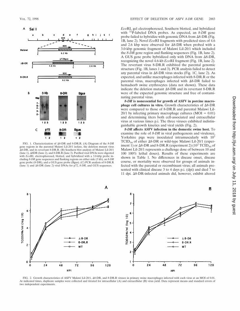
EcoRI, gel electrophoresed, Southern blotted, and hybridizedwith 32P-labeled DNA probes. As expected, an 8-DR geneprobe failed to hybridize with genomic DNA from D8-DR (Fig.1B, lane 2). Novel EcoRI fragments with predicted sizes of 4.6and 2.6 kbp were observed for D8-DR when probed with a3.0-kbp genomic fragment of Malawi Lil-20/1 which includedthe 8-DR gene region and flanking sequences (Fig. 1B, lane 2).A GUS gene probe hybridized only with DNA from D8-DR,recognizing the novel 4.6-kb EcoRI fragment (Fig. 1B, lane 2).The revertant virus 8-DR.R exhibited the parental genomicstructure (Fig. 1B, lanes 1 and 3). PCR analysis failed to detectany parental virus in D8-DR virus stocks (Fig. 1C, lane 2). Asexpected, and unlike macrophages infected with 8-DR.R or theparental virus, macrophages infected with D8-DR failed tohemadsorb swine erythrocytes (data not shown). These dataindicate the deletion mutant D8-DR and its revertant 8-DR.Rwere of the expected genomic structure and free of contami-nating parental virus.
8-DR is nonessential for growth of ASFV in porcine macro-phage cell cultures in vitro. Growth characteristics of D8-DRwere compared to those of 8-DR.R and parental Malawi Lil-20/1 by infecting primary macrophage cultures (MOI 5 0.01)and determining titers both cell-associated and extracellularvirus at various times p.i. The three viruses exhibited indistin-guishable growth kinetics and viral yields (Fig. 2).
8-DR affects ASFV infection in the domestic swine host. Toexamine the role of 8-DR in viral pathogenesis and virulence,Yorkshire pigs were inoculated intramuscularly with 102
TCID50 of either D8-DR or wild-type Malawi Lil-20/1 (exper-iment 1) or D8-DR and 8-DR.R (experiment 2) (102 TCID50 ofMalawi Lil-20/1 represents a challenge dose of between 10 and100 100% lethal doses). Results of these experiments areshown in Table 1. No differences in disease onset, diseasecourse, or mortality were observed for groups of animals in-fected with the parental or recombinant virus; all animals pre-sented with clinical disease 3 to 4 days p.i. (dpi) and died 7 to11 dpi. D8-DR-infected animals did, however, exhibit altered
FIG. 1. Characterization of D8-DR and 8-DR.R. (A) Diagram of the 8-DRgene regions in the parental Malawi Lil-20/1 isolate, the deletion mutant virusD8-DR, and its revertant 8-DR.R. (B) Southern blot analysis of Malawi Lil-20/1(lane 1), D8DR (lane 2), and 8-DR.R (lane 3). Purified viral DNAs were digestedwith EcoRI, electrophoresed, blotted, and hybridized with a 3.0-kbp probe in-cluding 8-DR gene sequences and flanking regions on either side (3 kb), an 8-DRgene probe (8-DR), and a GUS gene probe (Bgus). (C) PCR analysis of 8-DR.R(lane 1) and D8-DR (lane 2) viral DNAs for p72, 8-DR, and GUS sequences.
FIG. 2. Growth characteristics of ASFV Malawi Lil-20/1, D8-DR, and 8-DR.R viruses in primary swine macrophages infected with each virus at an MOI of 0.01.At indicated times, duplicate samples were collected and titrated for intracellular (A) and extracellular (B) virus yield. Data represent means and standard errors oftwo independent experiments.
VOL. 72, 1998 EFFECT OF DELETION OF ASFV 8-DR GENE 2883
on July 11, 2018 by guesthttp://jvi.asm
.org/D
ownloaded from
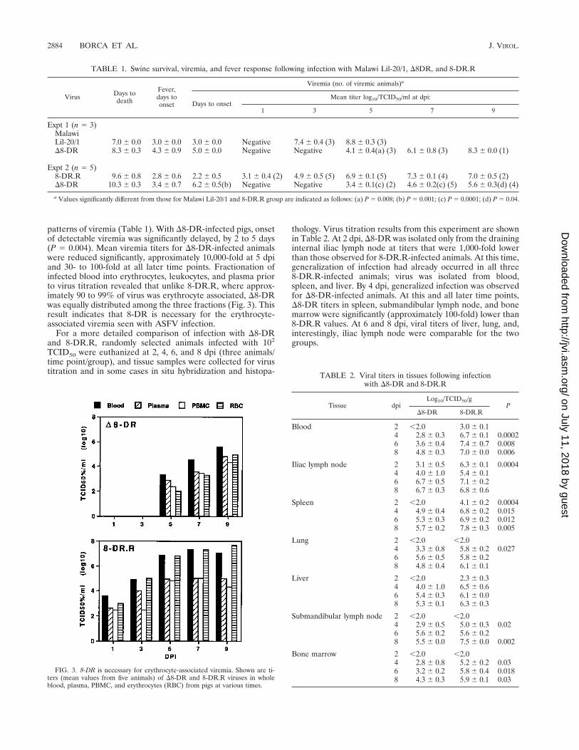
patterns of viremia (Table 1). With D8-DR-infected pigs, onsetof detectable viremia was significantly delayed, by 2 to 5 days(P 5 0.004). Mean viremia titers for D8-DR-infected animalswere reduced significantly, approximately 10,000-fold at 5 dpiand 30- to 100-fold at all later time points. Fractionation ofinfected blood into erythrocytes, leukocytes, and plasma priorto virus titration revealed that unlike 8-DR.R, where approx-imately 90 to 99% of virus was erythrocyte associated, D8-DRwas equally distributed among the three fractions (Fig. 3). Thisresult indicates that 8-DR is necessary for the erythrocyte-associated viremia seen with ASFV infection.
For a more detailed comparison of infection with D8-DRand 8-DR.R, randomly selected animals infected with 102
TCID50 were euthanized at 2, 4, 6, and 8 dpi (three animals/time point/group), and tissue samples were collected for virustitration and in some cases in situ hybridization and histopa-
thology. Virus titration results from this experiment are shownin Table 2. At 2 dpi, D8-DR was isolated only from the draininginternal iliac lymph node at titers that were 1,000-fold lowerthan those observed for 8-DR.R-infected animals. At this time,generalization of infection had already occurred in all three8-DR.R-infected animals; virus was isolated from blood,spleen, and liver. By 4 dpi, generalized infection was observedfor D8-DR-infected animals. At this and all later time points,D8-DR titers in spleen, submandibular lymph node, and bonemarrow were significantly (approximately 100-fold) lower than8-DR.R values. At 6 and 8 dpi, viral titers of liver, lung, and,interestingly, iliac lymph node were comparable for the twogroups.
FIG. 3. 8-DR is necessary for erythrocyte-associated viremia. Shown are ti-ters (mean values from five animals) of D8-DR and 8-DR.R viruses in wholeblood, plasma, PBMC, and erythrocytes (RBC) from pigs at various times.
TABLE 1. Swine survival, viremia, and fever response following infection with Malawi Lil-20/1, D8DR, and 8-DR.R
Virus Days todeath
Fever,days toonset
Viremia (no. of viremic animals)a
Days to onsetMean titer log10/TCID50/ml at dpi:
1 3 5 7 9
Expt 1 (n 5 3)MalawiLil-20/1 7.0 6 0.0 3.0 6 0.0 3.0 6 0.0 Negative 7.4 6 0.4 (3) 8.8 6 0.3 (3)D8-DR 8.3 6 0.3 4.3 6 0.9 5.0 6 0.0 Negative Negative 4.1 6 0.4(a) (3) 6.1 6 0.8 (3) 8.3 6 0.0 (1)
Expt 2 (n 5 5)8-DR.R 9.6 6 0.8 2.8 6 0.6 2.2 6 0.5 3.1 6 0.4 (2) 4.9 6 0.5 (5) 6.9 6 0.1 (5) 7.3 6 0.1 (4) 7.0 6 0.5 (2)D8-DR 10.3 6 0.3 3.4 6 0.7 6.2 6 0.5(b) Negative Negative 3.4 6 0.1(c) (2) 4.6 6 0.2(c) (5) 5.6 6 0.3(d) (4)
a Values significantly different from those for Malawi Lil-20/1 and 8-DR.R group are indicated as follows: (a) P 5 0.008; (b) P 5 0.001; (c) P 5 0.0001; (d) P 5 0.04.
TABLE 2. Viral titers in tissues following infectionwith D8-DR and 8-DR.R
Tissue dpiLog10/TCID50/g
PD8-DR 8-DR.R
Blood 2 ,2.0 3.0 6 0.14 2.8 6 0.3 6.7 6 0.1 0.00026 3.6 6 0.4 7.4 6 0.7 0.0088 4.8 6 0.3 7.0 6 0.0 0.006
Iliac lymph node 2 3.1 6 0.5 6.3 6 0.1 0.00044 4.0 6 1.0 5.4 6 0.16 6.7 6 0.5 7.1 6 0.28 6.7 6 0.3 6.8 6 0.6
Spleen 2 ,2.0 4.1 6 0.2 0.00044 4.9 6 0.4 6.8 6 0.2 0.0156 5.3 6 0.3 6.9 6 0.2 0.0128 5.7 6 0.2 7.8 6 0.3 0.005
Lung 2 ,2.0 ,2.04 3.3 6 0.8 5.8 6 0.2 0.0276 5.6 6 0.5 5.8 6 0.28 4.8 6 0.4 6.1 6 0.1
Liver 2 ,2.0 2.3 6 0.34 4.0 6 1.0 6.5 6 0.66 5.4 6 0.3 6.1 6 0.08 5.3 6 0.1 6.3 6 0.3
Submandibular lymph node 2 ,2.0 ,2.04 2.9 6 0.5 5.0 6 0.3 0.026 5.6 6 0.2 5.6 6 0.28 5.5 6 0.0 7.5 6 0.0 0.002
Bone marrow 2 ,2.0 ,2.04 2.8 6 0.8 5.2 6 0.2 0.036 3.2 6 0.2 5.8 6 0.4 0.0188 4.3 6 0.3 5.9 6 0.1 0.03
2884 BORCA ET AL. J. VIROL.
on July 11, 2018 by guesthttp://jvi.asm
.org/D
ownloaded from
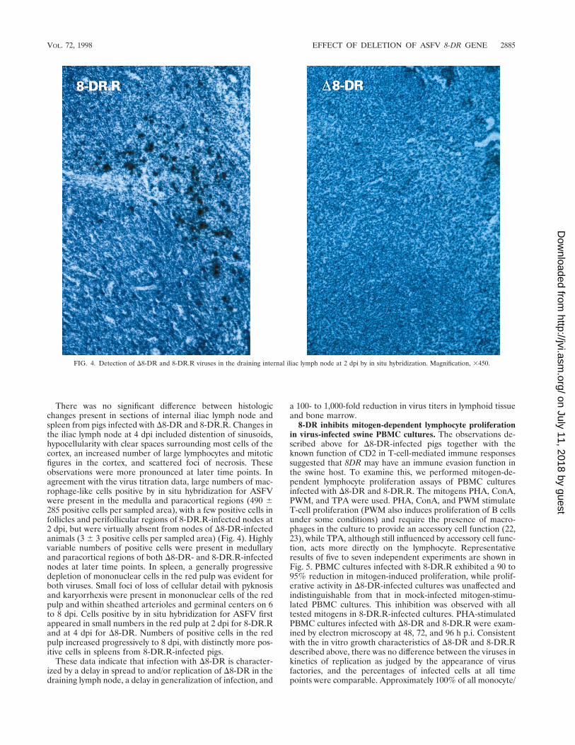
There was no significant difference between histologicchanges present in sections of internal iliac lymph node andspleen from pigs infected with D8-DR and 8-DR.R. Changes inthe iliac lymph node at 4 dpi included distention of sinusoids,hypocellularity with clear spaces surrounding most cells of thecortex, an increased number of large lymphocytes and mitoticfigures in the cortex, and scattered foci of necrosis. Theseobservations were more pronounced at later time points. Inagreement with the virus titration data, large numbers of mac-rophage-like cells positive by in situ hybridization for ASFVwere present in the medulla and paracortical regions (490 6285 positive cells per sampled area), with a few positive cells infollicles and perifollicular regions of 8-DR.R-infected nodes at2 dpi, but were virtually absent from nodes of D8-DR-infectedanimals (3 6 3 positive cells per sampled area) (Fig. 4). Highlyvariable numbers of positive cells were present in medullaryand paracortical regions of both D8-DR- and 8-DR.R-infectednodes at later time points. In spleen, a generally progressivedepletion of mononuclear cells in the red pulp was evident forboth viruses. Small foci of loss of cellular detail with pyknosisand karyorrhexis were present in mononuclear cells of the redpulp and within sheathed arterioles and germinal centers on 6to 8 dpi. Cells positive by in situ hybridization for ASFV firstappeared in small numbers in the red pulp at 2 dpi for 8-DR.Rand at 4 dpi for D8-DR. Numbers of positive cells in the redpulp increased progressively to 8 dpi, with distinctly more pos-itive cells in spleens from 8-DR.R-infected pigs.
These data indicate that infection with D8-DR is character-ized by a delay in spread to and/or replication of D8-DR in thedraining lymph node, a delay in generalization of infection, and
a 100- to 1,000-fold reduction in virus titers in lymphoid tissueand bone marrow.
8-DR inhibits mitogen-dependent lymphocyte proliferationin virus-infected swine PBMC cultures. The observations de-scribed above for D8-DR-infected pigs together with theknown function of CD2 in T-cell-mediated immune responsessuggested that 8DR may have an immune evasion function inthe swine host. To examine this, we performed mitogen-de-pendent lymphocyte proliferation assays of PBMC culturesinfected with D8-DR and 8-DR.R. The mitogens PHA, ConA,PWM, and TPA were used. PHA, ConA, and PWM stimulateT-cell proliferation (PWM also induces proliferation of B cellsunder some conditions) and require the presence of macro-phages in the culture to provide an accessory cell function (22,23), while TPA, although still influenced by accessory cell func-tion, acts more directly on the lymphocyte. Representativeresults of five to seven independent experiments are shown inFig. 5. PBMC cultures infected with 8-DR.R exhibited a 90 to95% reduction in mitogen-induced proliferation, while prolif-erative activity in D8-DR-infected cultures was unaffected andindistinguishable from that in mock-infected mitogen-stimu-lated PBMC cultures. This inhibition was observed with alltested mitogens in 8-DR.R-infected cultures. PHA-stimulatedPBMC cultures infected with D8-DR and 8-DR.R were exam-ined by electron microscopy at 48, 72, and 96 h p.i. Consistentwith the in vitro growth characteristics of D8-DR and 8-DR.Rdescribed above, there was no difference between the viruses inkinetics of replication as judged by the appearance of virusfactories, and the percentages of infected cells at all timepoints were comparable. Approximately 100% of all monocyte/
FIG. 4. Detection of D8-DR and 8-DR.R viruses in the draining internal iliac lymph node at 2 dpi by in situ hybridization. Magnification, 3450.
VOL. 72, 1998 EFFECT OF DELETION OF ASFV 8-DR GENE 2885
on July 11, 2018 by guesthttp://jvi.asm
.org/D
ownloaded from

macrophage cells showed evidence of virus infection at 24 hp.i., with extensive cytopathology evident at 72 h p.i. (Fig. 6).Ultrastructural evidence of virus infection and/or replication inlymphocytes was not observed in cultures infected with eithervirus.
Thus, 8-DR is necessary for inhibition of mitogen-dependentlymphocyte proliferation observed for ASFV-infected PBMCcultures, and this effect does not appear to involve altered viralreplication and/or cell tropism.
DISCUSSION
Here, we have shown that deletion of 8-DR from the ASFVMalawi Lil-20/1 genome did not affect disease onset, diseasecourse, or mortality (Table 1). This finding indicates that thegene is nonessential for acute disease and viral virulence indomestic swine. Consistent with this observation are prior dataindicating a lack of correlation between hemadsorption and pigvirulence; relatively avirulent hemadsorbing isolates and non-hemadsorbing pathogenic ASFV isolates have been describedelsewhere (9, 10, 39, 60).
An altered pattern of viral infection was, however, observedfor D8-DR-infected animals. A delay in spread to and/or rep-lication in the draining iliac lymph node, a delay in generali-zation of infection with viremia no longer erythrocyte-associ-
ated, and a 100- to 1,000-fold reduction in virus titers inlymphoid tissue and bone marrow were observed for pigs in-fected with the 8-DR deletion mutant D8-DR. It is unlikely thatthe decreased level of D8-DR replication in tissues is due toalteration of cell tropism. D8-DR exhibited normal growthcharacteristics in macrophages in vitro (Fig. 2), and there wasa similar pattern, although at reduced levels, of tissue involve-ment as evidenced by histopathology and localization ofASFV-infected cells by in situ hybridization. The most plausi-ble explanation for this defect is a block which prevents effi-cient virus replication in these tissues. The observed delay ingeneralization of D8-DR infection is most likely a direct con-sequence of this early replication defect. What role, if any, anerythrocyte-associated viremia may play in increasing virushalf-life in blood or virus infectivity in tissues is unknown.While viremia with many pathogenic ASFV isolates is largelyerythrocyte associated (90 to 99% of viremia), there are exam-ples of highly pathogenic hemadsorbing viruses where rela-tively high titer viremias are maintained without this high de-gree of erythrocyte association (40), as well as examples ofpathogenic nonhemadsorbing ASFV isolates (10, 39).
Interestingly, and unlike infection with 8-DR.R, infection ofPBMC cultures with D8-DR had no inhibitory effect on mito-gen-dependent lymphocyte proliferation (Fig. 5). This lack of
FIG. 5. Mitogen-induced proliferation in PBMC cultures infected with D8-DR and 8-DR.R. PBMC (105/ml) from 4 to 10 naive swine were either infected (MOI 510) with D8-DR or 8-DR.R or mock infected in the presence of the mitogen PHA, ConA, PWM, or TPA for 3 days and then pulsed with 1 mCi of [3H]thymidine perml for 16 h. Bars indicate mean values of five replicates.
2886 BORCA ET AL. J. VIROL.
on July 11, 2018 by guesthttp://jvi.asm
.org/D
ownloaded from
FIG. 6. Electron micrographs of PHA-treated PBMC cultures infected with 8-DR.R (A) and D8-DR (B) at 72 h p.i. Note the extensive macrophage cytopathology(arrows) and the normal appearance of lymphocytes in cultures infected with both viruses. The bar represents 5 mm.
2887
on July 11, 2018 by guesthttp://jvi.asm
.org/D
ownloaded from
effect was not the result of altered virus replication in macro-phages or altered lymphocyte tropism. 8-DR.R and D8-DRdemonstrated similar patterns of virus replication in macro-phages (Fig. 2 and 6), and neither virus appeared to infectand/or replicate in lymphocytes present in PBMC cultures(Fig. 6). The lack of lymphocyte susceptibility to ASFV infec-tion has been described previously: in vitro, resting or mitogen-stimulated B and T cells were not susceptible to infection (8),and no evidence of lymphocyte infection was observed inlymph nodes of ASFV-infected swine (32). These observationssuggest that 8-DR mediates this inhibition via an immunosup-pressive mechanism.
Significant inhibition of lectin-dependent lymphocyte prolif-eration in PBMC cultures infected with ASFV or those incu-bated in the presence of virus-free infected cell extracts hasbeen previously described (5, 6, 18, 61). While the nature of theresponsible inhibitory factor(s) is unknown, it appears to be asoluble protein with a molecular mass estimated to be between40 and 80 kDa (6, 18). Notably, a soluble hemagglutinin, madeup of 51-kDa monomers, has been identified in the medium ofcultures infected with some ASFV isolates (49). 8-DR, with apredicted molecular mass of 42 kDa, may be both the solubleinhibitory factor and the hemagglutinin identified previously inASFV-infected PBMC cultures (18, 49).
Although CD2 functions only as an adhesion molecule on Tcells, interacting with its natural ligand LFA-3, blocking thisinteraction with soluble ligand, anti-CD2 antibodies, or solubleCD2 resulted in inhibition of a variety of T-cell functions,including antigen-specific cytotoxicity, mitogen-induced andantigen-specific lymphocyte proliferation, interleukin-2 secre-tion, and interleukin-2 receptor expression (19, 26, 29, 35, 38,46, 47, 52, 59). Soluble CD2 effectively inhibited T-cell prolif-erative responses to several bacterial and viral antigens as wellas inhibiting reactivity to alloantigens in mixed lymphocytecultures (46). A secreted or released form of ASFV 8-DR frominfected cells could conceivably mimic or compete with hostCD2, resulting in inhibition of T-cell function. Additionally,8-DR could conceivably have an immunomodulatory role thateffectively involves sequestration of LFA-3 molecules withinthe infected macrophage that prevents membrane presenta-tion, thus interfering with macrophage (or other APC)-T cellinteractions.
In nature, perpetuation of ASFV involves the cycling ofvirus between two highly adapted hosts, Ornithodoros ticks andwarthogs or bushpigs, in sub-Saharan Africa (41, 57, 63). In thewarthog host, acute ASFV infection is subclinical and charac-terized by low-titer viremias (44, 56). This high degree ofvirus-host adaptation may necessitate viral immune evasionstrategies that will ensure that sufficient levels of viral replica-tion occur in the warthog and that resulting viremias are highenough to infect new populations of feeding ticks. Given that8-DR gene sequences have been selected for under these nat-ural conditions and that the gene is conserved among fieldisolates, it is possible that the protein performs a host rangefunction involving immune evasion in the warthog host. Addi-tionally, persistent infection of warthogs with ASFV has beenreported. In ASFV enzootic areas adult warthogs are typicallynonviremic, although most are seropositive and virus can usu-ally be isolated only from lymph nodes (21, 41, 43, 53, 58).Conceivably, 8-DR could have an immune evasion functionhere that permits continued virus replication and persistence.
Interestingly, the ASFV genome contains a second gene thatalso may be involved in immune modulation and evasion in thewarthog. A highly conserved gene, 5-EL, with similarity to thegene for cellular inhibitor of NF-kB, has been described andshown to be capable of downregulating NF-kB-regulated gene
expression in vitro (45). We have recently demonstrated, usinga 5-EL gene deletion mutant of Malawi Lil-20/1, that the genedoes not affect acute disease or viral virulence in domesticswine (34).
In summary, the results reported here suggest an immuno-suppressive role for 8-DR in the swine host which facilitatesearly events in viral replication and generalization of infection.This effect may be of most significance for ASFV infection ofits highly adapted natural host, the warthog.
ACKNOWLEDGMENTS
We thank R. Mireles, A. Zsak, J. R. Emmanuelli, and the PIADCanimal care staff for excellent technical assistance.
REFERENCES
1. Beyers, A. D., A. N. Barclay, D. A. Law, Q. He, and A. F. Williams. 1989.Activation of T lymphocytes via monoclonal antibodies against rat cell sur-face antigens with particular reference to CD2 antigen. Immunol. Rev.111:59–77.
2. Bierer, B. E., and S. J. Burakoff. 1989. T-lymphocyte activation: the biologyand function of CD2 and CD4. Immunol. Rev. 111:267–294.
3. Borca, M. V., G. F. Kutish, C. L. Afonso, P. Irusta, C. Carrillo, A. Brun, M.Sussman, and D. L. Rock. 1994. An African swine fever virus gene withsimilarity to the T-lymphocyte surface antigen CD2 mediates hemadsorp-tion. Virology 199:463–468.
4. Brown, F. 1986. The classification and nomenclature of viruses: summary ofresults of meetings of the International Committee on Taxonomy of Virusesin Sendai, September 1984. Intervirology 25:141–143.
5. Canals, A., F. Alonso, J. Tomillo, and J. Domınguez. 1992. Analysis of Tlymphocyte subsets proliferating in response to infective and UV-inactivatedAfrican swine fever virus. Vet. Microbiol. 33:117–127.
6. Canals, A., J. Domınguez, J. Tomillo, M. Babın, and F. Alonso. 1995. Inhi-bition of IL-2R and SLA class II expression on stimulated lymphocytes by asuppressor activity found in homogenates of African swine fever virus in-fected cultures. Arch. Virol. 140:1075–1085.
7. Carrillo, C., M. V. Borca, C. L. Afonso, D. V. Onisk, and D. L. Rock. 1994.Long-term persistent infection of swine monocytes/macrophages with Afri-can swine fever virus. J. Virol. 68:580–583.
8. Casal, I., L. Enjuanes, and E. Vinuela. 1984. Porcine leukocyte cellularsubsets sensitive to African swine fever virus in vitro. J. Virol. 52:37–46.
9. Coggins, L. 1968. Segregation of a nonhemadsorbing African swine fevervirus in tissue culture. Cornell Vet. 58:12–20.
10. Coggins, L., J. E. Moulton, and G. S. Colgrove. 1968. Studies with hindeattenuated African swine fever virus. Cornell Vet. 58:525–540.
11. Colgrove, G. S., E. O. Haelterman, and L. Coggins. 1969. Pathogenesis ofAfrican swine fever in young pigs. Am. J. Vet. Res. 30:1343–1359.
12. Costa, J. V. 1990. African swine fever virus, p. 247–270. In G. Darai (ed.),Molecular biology of iridoviruses. Kluwer Academic Publishers, Norwell,Mass.
13. DeKock, G., E. M. Robinson, and J. J. G. Keppel. 1994. Swine fever in SouthAfrica. Onderstepoort J. Vet. Sci. Anim. Ind. 14:31–93.
14. DeTray, D. E. 1957. Persistence of viremia and immunity in African swinefever. Am. J. Vet. Res. 18:811–816.
15. Dixon, L. K., D. L. Rock, and E. Vinuela. 1995. African swine fever-likeviruses. Arch. Virol. 10(Suppl.):92–94.
16. Genovesi, E. V., F. Villinger, D. J. Gerstner, T. C. Whyard, and R. C.Knudsen. 1990. Effect of macrophage-specific colony-stimulating factor(CSF-1) on swine monocyte/macrophage susceptibility to in vitro infection byAfrican swine fever virus. Vet. Microbiol. 25:153–176.
17. Gonzalez, A., A. Talavera, J. M. Almendral, and E. Vinuela. 1986. Hairpinloop structure of African swine fever virus DNA. Nucleic Acids Res. 14:6835–6844.
18. Gonzalez, S., C. Mendoza, J. M. Sanchez-Vizcaino, and F. Alonso. 1990.Inhibitory effect of African swine fever virus on lectin-dependent swinelymphocyte proliferation. Vet. Immunol. Immunopathol. 26:71–80.
19. Guckel, B., C. Berek, M. Lutz, P. Altevogt, V. Schirrmacher, and B. A.Kyewski. 1991. Anti-CD2 antibodies induce T cell unresponsiveness in vivo.J. Exp. Med. 174:957–967.
20. Hess, W. R. 1982. African swine fever: a reassessment. Adv. Vet. Sci. Comp.Med. 25:39–69.
21. Heuschele, W. P., and L. Coggins. 1969. Epizootiology of African swine feverin warthogs. Bull. of Epizoot. Dis. Afr. 17:179–183.
22. Kern, J. A., R. P. Daniele, and P. C. Nowell. 1985. Accessory cells providemore than one signal for lectin mitogen-stimulated proliferation of humanlymphocytes. J. Leukocyte Biol. 38:495–507.
23. Kern, J. A., J. C. Reed, R. P. Daniele, and P. C. Nowell. 1986. The role of theaccessory cell in mitogen-stimulated human T cell gene expression. J. Im-munol. 137:764–769.
2888 BORCA ET AL. J. VIROL.
on July 11, 2018 by guesthttp://jvi.asm
.org/D
ownloaded from
24. Konno, S., W. D. Taylor, and A. H. Dardiri. 1971. Acute African swine fever.Proliferative phase in lymphoreticular tissue and the reticuloendothelialsystem. Cornell Vet. 61:71–84.
25. Konno, S., W. D. Taylor, W. R. Hess, and W. P. Heuschele. 1971. Liverpathology in African swine fever. Cornell Vet. 61:125–150.
26. Krensky, A. M., F. Sanchez-Madrid, E. Robbins, J. A. Nagy, T. A. Springer,and S. J. Burakoff. 1983. The functional significance, distribution, and struc-ture of LFA-1, LFA-2, and LFA-3: cell surface antigens associated withCTL-target interactions. J. Immunol. 131:611–616.
27. Mebus, C. A. 1988. African swine fever. Adv. Virus Res. 35:251–269.28. Mebus, C. A., J. W. McVicar, and A. H. Dardiri. 1981. Comparison of the
pathology of high and low virulence African swine fever virus infections, p.183–194. In P. J. Wilkinson (ed.), Proceedings of CEC/FAO expert consul-tation in African swine fever research, Sardinia, Italy, September 1981.Commission of the European Communities, Luxemburg, Belgium.
29. Miller, G. T., P. S. Hochman, W. Meier, R. Tizard, S. A. Bixler, M. D. Rosa,and B. P. Wallner. 1993. Specific interaction of lymphocyte function-associ-ated antigen 3 with CD2 can inhibit T cell responses. J. Exp. Med. 178:211–222.
30. Moingeon, P., H.-C. Chang, B. P. Wallner, C. Stebbins, A. Z. Frey, and E. L.Reinherz. 1989. CD2-mediated adhesion facilitates T lymphocyte antigenrecognition function. Nature (London) 339:312–314.
31. Moulton, J., and L. Coggins. 1968. Comparison of lesions in acute andchronic African swine fever. Cornell Vet. 58:364–388.
32. Moura Nunes, J. F., and J. L. Nunes-Petisca. 1983. Replication of Africanswine fever virus in lymph nodes of experimentally infected swine, p. 132–142. In P. J. Wilkinson (ed.), ASF, EUR 8466 EN, proceedings of CEC/FAOResearch Seminar, Sardinia, Italy, September 1981. Commission of the Eu-ropean Communities, Luxemburg, Belgium.
33. Neilan, J. G., Z. Lu, G. F. Kutish, L. Zsak, T. G. Burrage, M. V. Borca, C.Carrillo, and D. L. Rock. 1997. A BIR motif containing gene of Africanswine fever virus, 4CL, is nonessential for growth in vitro and viral virulence.Virology 230:252–264.
34. Neilan, J. G., Z. Lu, G. F. Kutish, L. Zsak, T. L. Lewis, and D. L. Rock. 1997.A conserved African swine fever virus IkB homolog, 5EL, is nonessential forgrowth in vitro and virulence in domestic pigs. Virology 235:377–385.
35. O’Flynn, K., M. Russul-Saib, I. Ando, D. L. Wallace, P. C. L. Beverley, A. W.Boylston, and D. C. Linch. 1986. Different pathways of human T-cell acti-vation revealed by PHA-P and PHA-M. Immunology 57:55–60.
36. Onisk, D. V., M. V. Borca, G. Kutish, E. Kramer, P. Irusta, and D. L. Rock.1994. Passively transferred African swine fever virus antibodies protect swineagainst lethal infection. Virology 198:350–354.
37. Ortin, J., L. Enjuanes, and E. Vinuela. 1979. Cross-links in African swinefever virus DNA. J. Virol. 31:579–583.
38. Palacios, R., and O. Martinez-Maza. 1982. Is the E receptor on human Tlymphocytes a “negative signal receptor”? J. Immunol. 129:2479–2485.
39. Pini, A., and G. Wagenaar. 1974. Isolation of a non-haemadsorbing strain ofAfrican swine fever (ASF) virus from a natural outbreak of the disease. Vet.Rec. 94:2.
40. Plowright, W., J. Parker, and R. F. Staple. 1968. The growth of a virulentstrain of African swine fever virus in domestic pigs. J. Hyg. (Cambridge)66:117–134.
41. Plowright, W., J. Parker, and M. A. Pierce. 1969. The epizootiology ofAfrican swine fever in Africa. Vet. Rec. 85:668–674.
42. Plowright, W., J. Parker, and M. A. Pierce. 1969. African swine fever virus inticks (Ornithodoros moubata, Murray) collected from animal burrows inTanzania. Nature (London) 221:1071–1073.
43. Plowright, W. 1981. African swine fever, p. 178–190. In J. W. Davis, L. H.Karstad, and D. O. Trainer (ed.), Infectious diseases of wild mammals, 2nded. Iowa State University Press, Ames, Iowa.
44. Plowright, W., G. R. Thomson, and J. A. Neser. 1994. African swine fever, p.568–599. In J. A. W. Coetzer, G. R. Thomson, and R. C. Tustin (ed.),Infectious diseases in livestock with special reference to South Africa. vol. 1.Oxford University Press, Capetown, South Africa.
45. Powell, P. P., L. K. Dixon, and R. M. Parkhouse. 1996. An IkB homologencoded by African swine fever virus provides a novel mechanism for down-regulation of proinflammatory cytokine responses in host macrophages.J. Virol. 70:8527–8533.
46. Rabin, E. M., K. Gordon, M. H. Knoppers, M. A. Luther, E. A. Neidhardt,J. F. Flynn, C. A. Sardonini, T. M. Sampo, M. F. Concino, M. A. Recny, E. L.Reinherz, and D. S. Dwyer. 1993. Inhibition of T cell activation and adhesionfunctions by soluble CD2 protein. Cell. Immunol. 149:24–38.
47. Reed, J. C., W. Tadmori, M. Kamoun, G. Koretzky, and P. C. Nowell. 1985.Suppression of interleukin 2 receptor acquisition by monoclonal antibodiesrecognizing the 50 KD protein associated with the sheep erythrocyte recep-tor on human T lymphocytes. J. Immunol. 134:1631–1639.
48. Rodrıguez, J. M., R. J. Yanez, F. Almazan, E. Vinuela, and J. F. Rodriguez.1993. African swine fever virus encodes a CD2 homolog responsible for theadhesion of erythrocytes to infected cells. J. Virol. 67:5312–5320.
49. Ruız-Gonzalvo, F., and J. M. Coll. 1993. Characterization of a soluble hem-agglutinin induced in African swine fever virus-infected cells. Virology 196:769–777.
50. Ruız-Gonzalvo, F., F. Rodrıguez, and J. M. Escribano. 1996. Functional andimmunological properties of the baculovirus-expressed hemagglutinin of Af-rican swine fever virus. Virology 218:285–289.
51. Sanchez-Botija, C. 1963. Reservorios del virus de la peste porcina africana.Investigacion del virus de la P.P.A. en los artropodos mediante la prueba dela hemoadsorcion. Bull. Off. Int. Epizoot. 60:895–899.
52. Sanchez-Madrid, F., A. M. Krensky, C. F. Ware, E. Robbins, J. L.Strominger, S. J. Burakoff, and T. A. Springer. 1982. Three distinct antigensassociated with human T-lymphocyte-mediated cytolysis: LFA-1, LFA-2, andLFA-3. Proc. Natl. Acad. Sci. USA 79:7489–7493.
53. Simpson, V. R., and N. Drager. 1979. African swine fever antibody detectionin warthogs. Vet. Rec. 105:61.
54. Sogo, J. M., J. M. Almendral, A. Talavera, and E. Vinuela. 1984. Terminaland internal inverted repetitions in African swine fever virus DNA. Virology133:271–275.
55. Stroop, W. G., D. L. Rock, and N. W. Fraser. 1984. Localization of herpessimplex virus in the trigeminal and olfactory systems of the mouse centralnervous system during acute and latent infections by in situ hybridization.Lab. Invest. 51:27–38.
56. Thomson, G. R., M. D. Gainaru, and A. F. Van Dellen. 1980. Experimentalinfection of warthog (Phacochoerus aethiopicus) with African swine fevervirus. Onderstepoort J. Vet. Res. 47:19–22.
57. Thomson, G. R., M. Gainaru, A. Lewis, H. Biggs, E. Nevill, M. Van DerPypekamp, L. Gerbes, J. Esterhuysen, R. Bengis, D. Bezuidenhout, and J.Condy. 1983. The relationship between ASFV, the warthog and Ornithodo-ros species in southern Africa, p. 85–100. In P. J. Wilkinson (ed.), ASF, EUR8466 EN, proceedings of CEC/FAO Research Seminar, Sardinia, Italy, Sep-tember 1981. Commission of the European Communities, Luxemburg, Bel-gium.
58. Thomson, G. R. 1985. The epidemiology of African swine fever: the role offree-living hosts in Africa. Onderstepoort J. Vet. Res. 52:201–209.
59. Van Wauwe, J., J. Goossens, W. Decock, P. Kung, and G. Goldstein. 1981.Suppression of human T-cell mitogenesis and E-rosette formation by themonoclonal antibody OKT11A. Immunology 44:865–871.
60. Vigario, J. D., A. M. Terrinha, and J. F. Moura Nunes. 1974. Antigenicrelationships among strains of African swine fever virus. Arch. GesamteVirusforsch. 45:272–277.
61. Wardley, R. C. 1982. Effect of African swine fever on lymphocyte mitogen-esis. Immunology 46:215–220.
62. Whitlow, M. B., K. Iida, I. Stefanova, A. Bernard, and V. Nussenzweig. 1990.H19, a surface membrane molecule involved in T-cell activation, inhibitschannel formation by human complement. Cell. Immunol. 126:176–184.
63. Wilkinson, P. J. 1989. African swine fever virus, p. 17–35. In M. B. Pensaert(ed.), Virus infections of porcines. Elsevier Science Publishers, Amsterdam,The Netherlands.
64. Yanez, R. J., J. M. Rodri“guez, M. L. Nogal, L. Yuste, C. Enrıquez, J. F.Rodriguez, and E. Vinuela. 1995. Analysis of the complete nucleotide se-quence of African swine fever virus. Virology 208:249–278.
65. Zsak, L., Z. Lu, G. F. Kutish, J. G. Neilan, and D. L. Rock. 1996. An Africanswine fever virus virulence-associated gene NL-S with similarity to the herpessimplex virus ICP34.5 gene. J. Virol. 70:8865–8871.
66. Zsak, L., E. Caler, Z. Lu, G. F. Kutish, J. G. Neilan, and D. L. Rock. 1998.A nonessential African swine fever virus gene UK is a significant virulencedeterminant in domestic swine. J. Virol. 72:1028–1035.
VOL. 72, 1998 EFFECT OF DELETION OF ASFV 8-DR GENE 2889
on July 11, 2018 by guesthttp://jvi.asm
.org/D
ownloaded from


![swine flu kbk-1.ppt [Read-Only]ocw.usu.ac.id/.../1110000141-tropical-medicine/tmd175_slide_swine_… · MAP of H1 N1 Swine Flu. Swine Influenza (Flu) Swine Influenza (swine flu) is](https://img.pdfslide.us/doc/110x75/5f5a2f7aee204b1010391ac9/swine-flu-kbk-1ppt-read-onlyocwusuacid1110000141-tropical-medicinetmd175slideswine.jpg)