Embed Size (px)
Citation preview

Vol. 126, No. 2, 1985
January 3 1, 1985
BIOCHEMICAL AND BIOPHYSICAL RESEARCH COMMUNICATIONS
Pages 678-684
DELAYED MRTHYLATION AND THE MATRIX BOUND DNA MRTHYLASE
Terence Davis, David Kirk, Angela Rinaldi, Roy II. Burdon, and Roger L.P. Adams
Department of Biochemistry, University of Glasgow, Glasgow G12 SQQ U.K.
Received November 20, 1984
It is shown that the methylation of DNA that occurs in isolated nuclei is "delayed methylation". This methylation is not reduced in nuclei which have been pretreated with 0.2M NaCl to extract the soluble methylase suggesting that this methylation is the product of a firmly bound matrix associated DNA methylase. Evidence is provided that, like the methylase, the DNA substrate is associated with the nuclear matrix. 0 1985 Academic Press, Inc.
DNA methylation in eukaryotes involves the transfer of a methyl group
from S-adenosyl methionine to certain cytosine bases in DNA (1,2). This
reaction is catalysed by a specific DNA methylase, a number of examples of
which have been characterised (see 3). The bulk of DNA methylation occurs
rapidly after DNA synthesis (4-6) but a significant number of methyl groups
continue to be added to DNA for several hours after its synthesis (7-9).
This latter methylation is termed "delayed methylation". There is now
evidence for the presence of DNA methylase activity in two fractions in cell
homogenates; a low salt (<0.4M) extractable DNA methylase and a firmly
bound DNA methylase thought to be associated with the nuclear matrix
(10,111. it is not clear at present which of these activities is
responsible for either the delayed or the rapid methylation of DNA. This
manuscript presents evidence that the delayed methylation is a product of
the firmly bound form of the DNA methylase.
MATERIALS AND METHODS
Determination of delayed methylation: L929 cells in log phase were cultured in the presence of 5OhCi of
[6-3H]uridine (Amersham) for either 48 hours or 50 minutes and harvested while still in log phase. Another batch were labelled and harvested later when they had attained stationary phase. Nuclei were prepared by homogenisation of cells in 1% (v/v) Tween 80 (Sigma). DNA, purified as
0006-291X/85 $1.50 Copyright 0 1985 by Academic Press, inc. All rights qf reproduction in any form reserved. 678
Vol. 126, No. 2, 1985 BIOCHEMICAL AND BIOPHYSICAL RESEARCH COMMUNICATIONS
described previously (121, was dissolved in 98% formic acid and hydrolysed at 170°C for l-l/2 hours. The excess formic acid was evaporated and the residue dissolved in 2OmM ammonium carbonate (pH1O.O) and the bases separated on an Aminex A6 HPLC column as described previously (12). The base8 cytosine and methylcytosine (mC) were collected and the proportion of cytosines methylated was determined. Nuclear Methylation
L929 cells were prelabelled with deoxy-[U-14C]cytidine (2nCi per ml> for three days prior to harvesting. In some cases 3mM hydroxyurea was present for the final hour. Nuclei were prepared and extracted separately with buffer M(tris-HCl pH7.8, 50mM; EDTA, 1mM; DTT, 1mM; glycerol, 10%) containing NaCl at concentrations varying from 0 to 0.35 Molar. The nuclei were then washed twice in buffer M and incubated with 5/.lCi of S-adenosyl-L-[methyl-3H]methionine (1.5Ci/mmole, Amersham) for 2 hour8 at 37oc. The DNA was purified and the 3H/14C ratio determined. Isolation of matrix DNA:
Following incubation nuclei were resuspended in buffer M + 2.OM NaCl and centrifuged at 165,000xg for 48 hours. The pelleted material was resuspended in buffer M + 5mM CaC12 and digested using micrococcal nuclease (Boehringer) at 1000 units per ml for 1 hour at 37OC. Sample8 were taken at each stage of the procedure and the 3H/14C ratio of the DNA was determined. Use of 5-azadeoxycytidine:
L929 cells were released from stationary nhase and cultured for 10 hours at 37OC. Then 5-azadeoxycytidine was-added to a final concentration of l.Oc(M and the cells grown for a further 10 hours at 37OC (11). By this means cells were produced containing hemimethylated DNA and only low levels of 'soluble' DNA methylase (11,13). The cells were harvested and nuclei prepared. These nuclei and control nuclei were methylated as before except that in some instances 40 units of a partially purified ascites DNA methylase (14) was added to the incubation. The DNA was purified and the amount of methyl groups incorporated was determined. DNA was assayed by the method of Burton (15).
RESULTS
Delayed methylation in vivo
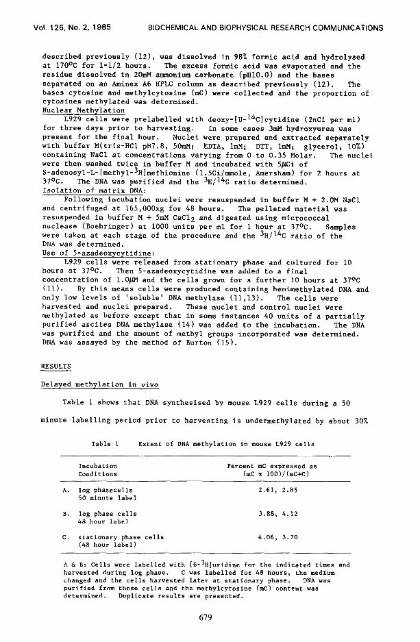
Table 1 shows that DNA synthesised by mouse L929 cells during a 50
minute labelling period prior to harvesting is undermethylated by about 30%
Table 1 Extent of DNA methylation in mouse L929 cells
Incubation Percent mC expressed as Conditions bnc x 100)/(mC+c)
A. log phasecells 2.61, 2.85 50 minute label
B. log phase cells 3.88, 4.12 48 hour label
C. stationary phase cells 4.06, 3.70 (48 hour label)
A h B: Cells were labelled with [6-3H] uridine for the indicated times and harvested during log phase. C was labelled for 48 hours, the medium changed and the cells harvested later at stationary phase. DNA was purified from these cells and the methylcytosine (mC) content was determined. Duplicate results are presented.
679

Vol. 126, No. 2, 1985 BIOCHEMICAL AND BIOPHYSICAL RESEARCH COMMUNICATIONS
compared with DNA labelled for 48 hours. The conclusion is that a
considerable amount of methylation occurs more than 50 minutes after DNA
synthesis. This finding confirms previous results demonstrating the
occurrence of delayed methylation (7-9). For 1 to 2 minutes after
synthesis DNA is not methylated at all but this DNA represents less than 4%
of the DNA synthesised during a 50 minute labelling period and hence its
presence is insufficient to explain the lower level of methylation seen in
the present experiment. In contract to the findings of several authors
(16,171 we find no difference between the level of DNA methylation in log
phase cells and stationary phase cells when both are labelled for 48 hours
(table 1).
Delayed methylation in isolated nuclei
It has been shown previously that methylation in isolated nuclei occurs
predominantly on DNA more than 10 minutes old (50% is on DNA more than 4.25
hours old) i.e. it is delayed methylation (18). This was confirmed by
studying the effect of treatment of the cells with hydroxyurea for one hour
prior to nuclear isolation. Despite the fact that in this case DNA
synthesis has been almost completely inhibited, the methylation occurring on
DNA in nuclei from both hydroxyurea treated cells and control cells is the
same (figure 1). There is thus no distinction between total and delayed
methylation in this system (i.e. we are not looking at the rapid methylation
of nascent DNA).
Mouse cells contain two DNA methylase activities: one extractable at
low salt concentrations, and the other is thought to be a matrix bound form
of the readily extractable enzyme (10,ll). It is not known which activity
is responsible for DNA methylation in vivo, but the present communciation
reports experiments designed to discover which form of the enzyme is
responsible for the methylation which occurs in isolated nuclei. Nuclei
were prepared and extracted with increasing concentrations of NaCl in order
to remove the readily extractable DNA methylase. Figure 1 shows that
extracting nuclei with 0.35M NaCl prior to methylation results in, at most,
680

Vol. 126, No. 2, 1985 BIOCHEMICAL AND BIOPHYSICAL RESEARCH COMMUNICATIONS
0.4
0.3 0 \
G
!
#- .
= 2 0.2
7 NT
0.1
OI 0.1 0.2 0.3 0.4
LNaCll (Molar)
Figure 1 The effect of salt extraction on DNA methylation in isolated nuclei
Nuclei isolated from [U-L4C]deoxycytidine prelabelled L929 cells were extracted with buffer M containing NaCl at the indicated concentrations. These nuclei were then incubated with 13H] AdoMet (methods) and the 3H/14C ratio of the DNA determined. Open circles: cells treated with hydroxyurea for lh before nuclear isolation. Closed circles: control cells.
a 20% decrease in methylation compared with unextracted nuclei. As most of
the DNA methylase is extracted by 0.2M NaCl (11,14) the majority (>80%) of
the methylation in these nuclei is a product of the so-called matrix bound
DNA methylase.
Further evidence that the bound methylase is responsible for DNA
methylation comes from the use of 5-azadeoxycytidine (table 2). When cells
are treated with this drug, in vivo DNA methylation is inhibited by more
than 80% (19,201. Associated with this inhibition is a small increase in
the amount of bound DNA methylase and a dramatic decrease in the amount of
soluble DNA methylase (11,131.
Table 2 Effect of Pretreatment with Azadeoxycytidine
Sample p mole methyl groups incorporatedlpg DNA
endogenous methylase
with added methylase
control nuclei
nuclei from cells pretreated with 5-azadeoxycytidine
0.17, 0.18 0.21, 0.23
0.20, 0.25 1.00, 1.05
Methylation of DNA in nuclei from control L929 cells or from cells pretreated with 5-azadeoxycytidine for 10 hours. Duplicate results are presented (see methods for details).
681
Vol. 126, No. 2, 1985 BIOCHEMICAL AND BIOPHYSICAL RESEARCH COMMUNICATIONS
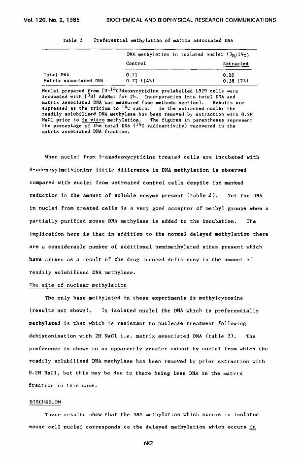
Table 3 Preferential methylation of matrix associated DNA
DNA methylation in isolated nuclei (3R/14C)
Control Extracted
Total DNA 0.11 0.10 Matrix associated DNA 0.22 (14%) 0.38 (7%)
Nuclei prepared from [U-14C]deoxycytidine prelabelled L929 cells were incubated with [3H1 AdoMet for 2h. Incorporation into total DNA and matrix associated DNA was measured (see methods section). Results are expressed as the tritium to 14C ratio. In the extracted nuclei the readily solubilised DNA methylase has been removed by extraction with 0.2M NaCl prior to in vitro methylation. The figures in parentheses represent the percentage of the total DNA ( 14C radioactivity) recovered in the matrix associated DNA fraction.
When nuclei from 5-azadeoxycytidine treated cells are incubated with
S-adenosylmethionine little difference in DNA methylation is observed
compared with nuclei from untreated control cells despite the marked
reduction in the amount of soluble enzyme present (table 2). Yet the DNA
in nuclei from treated cells is a very good acceptor of methyl groups when a
partially purified mouse DNA methylase is added to the incubation. The
implication here is that in addition to the normal delayed methylation there
are a considerable number of additional hemimethylated sites present which
have arisen as a result of the drug induced deficiency in the amount of
readily solubilised DNA methylase.
The site of nuclear methylation
The only base methylated in these experiments is methylcytosine
(results not shown). In isolated nuclei the DNA which is preferentially
methylated is that which is resistant to nuclease treatment following
dehistonisation with 2M NaCl i.e. matrix associated DNA (table 3). The
preference is shown to an apparently greater extent by nuclei from which the
readily solubilised DNA methylase has been removed by prior extraction with
0.2M NaCl, but this may be due to there being less DNA in the matrix
fraction in this case.
DISCUSSION
These results show that the DNA methylation which occurs in Isolated
mouse cell nuclei corresponds to the delayed methylation which occurs in -
682

Vol. 126, No. 2, 1985 BIOCHEMICAL AND BIOPHYSICAL RESEARCH COMMUNICATIONS
vivo on DNA up to several hours following replication. The enzyme
responsible for this delayed methylation is the tightly bound or matrix
associated DNA methylase which is the only DNA methylase remaining following
extraction of nuclei with 0.2-0.35M NaCl. It is a reasonable extrapolation
to suggest that the matrix bound enzyme is also responsible for the delayed
methylation observed in intact cells.
Moreover, the DNA methylated in isolated nuclei is also preferentially
associated with tight binding proteins in the so-called nuclear-matrix
fraction. The very presence of DNA methylase tightly bound to a region of
DNA may well render that DNA resistant to nuclease action while at the same
time allowing the sedimentation of the enzyme along with the DNA following
treatment with 2M NaCl. Such are the characteristics of the nuclear matrix
though it is by no means clear that the DNA:protein complex studied in the
present and related experiments is the same as that involved in replication
or transcription. Thus the term nuclear matrix is used purely to describe
the fraction resulting from a certain experimental procedure rather than to
imply a functional nuclear entity.
Some of the proteins associated in vivo with the DNA:methylase complex
may interfere with the action of the enzyme thereby allowing hemfmethylated
sites to persist for several hours after replication of the DNA. It is
these sites which are filled when isolated nuclei are incubated with
S-adenosylmethionine and this reaction does not require the 'soluble' DNA
methylase. In contrast, the hemimethylated sites arising following
treatment of cells with azadeoxycytidine are only filled when additional
soluble DNA methylase is added to the isolated nuclei. We would suggest
that normally the 'soluble' DNA methylase becomes firmly associated with the
DNA and is released again only following methylation of the DNA. Most
methylation takes place very shortly after replication but some is delayed
causing a proportion of the enzyme to be recovered in the matrix bound
fraction. The presence of azacytosine in the DNA leads to permanent
inactivation of enzyme when it interacts with the foreign base (21). There
683

Vol. 126, No. 2, 1985 BIOCHEMICAL AND BIOPHYSICAL RESEARCH COMMUNICATIONS
is, however, little effect on the nuclear DNA methylation perhaps because
the matrix bound enzyme equilibrates only slowly with the 'soluble' enzyme.
REFERENCES
1.
2. 3.
4.
5. 6.
7. 8.
9.
10.
11.
12.
13.
14. 15. 16. 17. 18.
19. 20. 21.
Adams, R.L.P. and Burdon, R.H. (1982) CRC Crit. Rev. Biochem. 13, 347-384. Doerfler, W. (1983) Ann. Rev. Biochem. 52, 93-124. Adams, R.L.P. and Burdon, R.H. (1983) in 'Enzymes of Nucleic Acid Synthesis and Modification' vol.1 p.119-144 (Jacob. S.T. ed) CRC Press Inc., Boca Ratan. Burdon, R.H. and Adams, R.L.P. (1969) Biochim. Biophys. Acta 174, 322-329. Kappler, J. (1970) J. Cell Physiol. 15, 21-32. Gruenbaum, Y., Szyf, H., Cedar, H. and Razin, A. (1983) Proc. Natl. Acad. Sci. USA E, 4919-4921. Adams, R.L.P. (1981) Biochim. Biophys. Acta 254, 205-212. Woodcock, D.M., Adams, J.K. and Cooper, I.A. (1982) Biochim. Biophys. Acta 696, 15-22. GeraccD., Eremenko, T., Cocciara, A., Scarano, E. & Volpe, P. (1974) Biochem. Biophys. Res. Comm. 57, 353-358. Qureshi, M.A., Adams, R.L.P. and Burdon, R.H. (1982) Biochem. SOC.
Trans. lo, 455-456. Burdon, R.H., Qureshi, M., Adams, R.L.P. and Brooks, W. (1985) (paper submitted). Adams, R.L.P., McKay, E.L., Craig, L.M. and Burdon, R.H. (1979) Biochim. Biophys. Acta 563, 72-81. Tanaka, M., Hibasami, H., Nagai, J. and Ikeda, T. (1980) Aus. J. Exp. Biol. Med. Sci. 2, 391-396. Turnbull, J.F. and Adams, R.L.P. (1976) Nucleic Acids Res. 3, 677-695. Burton, K. (19561 Biochem. J. 62, 315-323. Rubery, E.D. and Newton, A.A. (1973) Biochim. Biophys. Acta 324, 24-36. Kunnath, L. and Locker, J. (1982) Biochim. Biophys. Acta 699, 264-271. Adams, R.L.P. and Hogarth, C. (1973) Biochim. Biophys. Acta 331, 214-220. Jones, P.A. and Taylor, S.M. (1980) Cell 20, 85-90. Jones, P.A. and Taylor, S.M. (1981) Nucleic Acids Res. 2, 2933-2947. Santi, D.V., Garrett, C.E. and Barr, P.J. (1983) Cell 2, 9-30.
684