Embed Size (px)
Citation preview
CLINICAL ARTICLEJ Neurosurg 129:299–307, 2018
Hemifacial spasm (HFS) was originally described by Gowers as a condition characterized by the vol-untary contraction of muscles in the region inner-
vated by the ipsilateral facial nerve.34 The prevalence of HFS appears to be much higher in some Asian races than in Western races, although precise epidemiological data are not available. Most cases occur unilaterally, and onset begins in the orbicularis oculi muscles. The most common cause of HFS is the compression of the facial nerve by cerebral arteries.2,5,9,25,28 Botulinum neurotoxin (Botox) is
commonly used to treat medication-refractory HFS. An-other treatment modality that is widely used to treat HFS is microvascular decompression (MVD), which has been shown to completely cure HFS.4 However, some compli-cations have been reported in association with the surgi-cal approach, particularly for surgeries performed near the facial and vestibulocochlear nerve complexes.6 One such complication is delayed facial palsy (DFP), which is a transient or self-limited and unpredictable condition. Al-though most cases of postoperative facial palsy have am-
ABBREVIATIONS CI = confidence interval; CISS = constructive interference in steady state; DFP = delayed facial palsy; fEMG = facial electromyography; HB = House-Brackmann; HFS = hemifacial spasm; LSR = lateral spread response; MVD = microvascular decompression; OR = odds ratio; PTA = pure tone audiometry; REZ = root entry zone; SDA = speech discrimination audiometry; SNUH = Seoul National University Hospital; VA = vertebral artery.SUBMITTED November 16, 2016. ACCEPTED March 6, 2017.INCLUDE WHEN CITING Published online September 1, 2017; DOI: 10.3171/2017.3.JNS162869.
Delayed facial palsy after microvascular decompression for hemifacial spasm: friend or foe?Jae Meen Lee, MD,1 Hye Ran Park, MD,2 Young Doo Choi, BS,1 Sung Min Kim, MD, PhD,3,4 Beomseok Jeon, MD, PhD,3,4 Han-Joon Kim, MD, PhD,3,4 Dong Gyu Kim, MD, PhD,1,5 and Sun Ha Paek, MD, PhD1,6
Departments of 1Neurosurgery and 3Neurology, Seoul National University Hospital; 2Department of Neurosurgery, Soonchunhyang University Seoul Hospital; Departments of 4Neurology and 5Neurosurgery, Seoul National University College of Medicine; and 6Department of Neurosurgery, Cancer Research Institute, Ischemic/Hypoxic Disease Institute, Seoul National University College of Medicine, Seoul, Republic of Korea
OBJECTIVE The authors investigated the incidence, clinical course, and predisposing factors associated with delayed facial palsy (DFP) following microvascular decompression (MVD).METHODS The authors reviewed the records of 310 patients (311 cases) who were followed after MVD for hemifacial spasm (HFS). Of these patients, 45 (14.5%) developed DFP after MVD. The clinical characteristics and predisposing fac-tors of the patients with HFS were investigated to identify prognostic factors that predicted the development of DFP after MVD. Log-rank tests were used to compare times to symptom disappearance, and a logistic regression analysis was performed to compare clinical characteristics between patients who developed DFP and those who did not.RESULTS HFS was completely resolved immediately after MVD in 158 cases (50.8%), and HFS eventually disappeared in 289 (92.9%) of the cases. Of the 45 patients with DFP, 17 were men and 28 were women. DFP occurred between postoperative Days 1 and 44 (mean 9.67 days). Finally, 44 patients (97.8%) completely recovered. The average time to recovery was 3.9 months (range 1–24 months). Patients who had experienced an immediate disappearance of HFS experienced a significantly higher occurrence of DFP than those who did not (odds ratio 0.383, 95% confidence interval 0.183–0.802; p = 0.011). In addition, preoperative botulinum neurotoxin injections negatively influenced the occurrence of DFP (p = 0.016).CONCLUSIONS In this study, the incidence rate of DFP was slightly higher than previously reported values. Moreover, DFP can occur even when spasms disappear immediately after MVD, but the patients with DFP can fully recover within weeks.https://thejns.org/doi/abs/10.3171/2017.3.JNS162869KEY WORDS delayed facial palsy; hemifacial spasm; microvascular decompression; functional neurosurgery
J Neurosurg Volume 129 • August 2018 299©AANS 2018, except where prohibited by US copyright law
Unauthenticated | Downloaded 01/28/22 05:57 PM UTC

J. M. Lee et al.
J Neurosurg Volume 129 • August 2018300
biguous causes, intraoperative damage can cause perma-nent facial palsy. The definition of DFP is a sudden onset of symptoms that usually occurs more than 24 hours after the operation.3,12,23,30 DFP is not a rare complication after MVD for HFS. As shown in previous reports, the rate of DFP following MVD is 2.8%–10.4%.12,18,19,21,23,30 Nonethe-less, few studies have explored DFP after MVD.
The aim of the present study was to investigate the clinical course and incidence of DFP following MVD and to use a literature review to identify predisposing factors associated with this condition.
MethodsStudy Population
Three hundred and twelve patients who were followed up after MVD surgery at the Seoul National University Hospital (SNUH) between September 2006 and June 2015 were identified as candidates for this study. All in-cluded patients suffered from medically refractory HFS. The exclusion criteria included undergoing a revision sur-gery. Of the 312 evaluated patients, 310 fulfilled the in-clusion criteria. One patient underwent bilateral MVD for bilateral HFS.
To determine whether DFP frequently occurs in a par-ticular age group, we divided the patient group based on generation (twenties, thirties, forties, fifties, sixties, and >70) and compared the DFP frequency among the divided groups. The following clinical characteristics of patients with HFS were investigated to identify prognostic indica-tors for the development of DFP after MVD: age, sex, side of spasm, preoperative symptom duration, follow-up du-ration after MVD, and offending vessels. We categorized patients into 4 groups according to the offending vessel. Cases that presented with the involvement of both the vertebral artery (VA) and small perforating arteries were considered “VA.” “Complex” was defined as the presenta-tion of 2 or more large, dominant, offending vessels with clear proximity to the facial nerve. No vein was observed to compress the root entry zone (REZ) in any case.
Weakness associated with facial palsy was assessed using the House-Brackmann (HB) scale, which assigns patients to 1 of 6 classes. Our definition of postoperative DFP was facial weakness that was scored up to Grade II on the HB scale more than 24 hours after undergoing MVD involving the facial nerve. We evaluated the follow-ing clinical characteristics in all patients: age, sex, side of MVD, preoperative symptom duration, follow-up dura-tion after MVD, offending vessel, obesity, postoperative hearing loss, postoperative hearing reduction (including hearing loss), and preoperative Botox injection. Obesity is considered when patients have a body mass index greater than 25 kg/m2. We investigated the following prognostic factors in the patients with DFP: age, sex, side of spasm, offending vessels, smoking, alcohol, hypertension, diabe-tes mellitus, and obesity. Patient characteristics and demo-graphics are described in Table 1.
We divided and analyzed the patients according to 3 additional factors (Teflon felts, lateral spread response [LSR], and Botox injections) to investigate the possible influence of other variables on the incidence of DFP. Tef-
lon felts were counted for all patients who underwent 3D constructive interference in steady state (3D-CISS) at 1 year after MVD. The obtained images were analyzed and recorded using electronic medical records. We inserted at least 3 Teflon felts in every patient, so the number was de-fined as “3” when the felts were not observed in 3D-CISS except in vessels. Patients with complete LSR recordings were investigated. LSR was subdivided into zygomatic and mandibular branches based on its status to identify correlations between the LSR recordings and the inci-dence of DFP. Finally, the influence of Botox injections was analyzed to identify possible relations with the devel-opment of DFP. The IRB of SNUH approved this study.
Pre- and Postoperative Evaluation ProtocolPreoperative and postoperative evaluations were per-
formed according to a previously described SNUH pro-tocol. All patients were admitted to the SNUH for pre-operative evaluations, which included temporal bone CT, internal auditory canal MRI, pure tone audiometry (PTA), and speech discrimination audiometry (SDA). Clinical outcomes were assessed immediately after MVD; at 1, 2, 3, and 6 months postoperatively; and at 1, 2, 3, 5, and 7 years by the surgeon and a specially trained nurse. PTA-SDA was assessed preoperatively and after 6 months, and at 1, 3, 5, 7, and 10 years.
Immediately after MVD, we determined the status of both facial palsy and HFS and included all new cases of facial palsy that occurred within 24 hours after surgery. If DFP occurred during the follow-up period, we rechecked symptom improvement at 2 weeks after treatment for DPF was started. Patients were administered oral prednisolone (1 mg/kg/day), which was gradually tapered off over 2 weeks.31 In severe cases of DFP (HB Grade IV), an intra-venous infusion of methylprednisolone was administered for 5 days. None of the patients exhibited signs of infec-tion, such as blisters around the lips, when DFP occurred. Serological studies for specific herpes simplex virus anti-bodies and antiviral drugs were not performed in the pa-tients with DFP who underwent MVD.
Patients were observed postoperatively until spasms and facial palsies completely disappeared. Additionally, the time from HFS re-occurrence after MVD to its complete disappearance was observed and recorded for the analysis.
Surgical ProcedureAll surgical procedures were performed via a previ-
ously described lateral retrosigmoid suboccipital ap-proach.1,14,29,32 First, intraoperative brainstem auditory-evoked potentials, facial electromyography (fEMG), and LSR were recorded prior to surgery. A lazy-S skin inci-sion was made medially behind the ear along the hairline. A bone flap approximately 3 cm in diameter was removed using a high-speed drill. An incision was made in the dura mater along the inferoposterior margin of the sigmoid si-nus, and the CSF was then drained enough to relax the cerebellum. Using an operating microscope, we gently dissected the arachnoid membrane to expose the REZ, re-laxed the brain, and minimized traction on cranial nerve VIII. We designed Teflon felts (Impra Inc., a subsidiary
Unauthenticated | Downloaded 01/28/22 05:57 PM UTC

Delayed facial palsy after microvascular decompression
J Neurosurg Volume 129 • August 2018 301
of C. R. Bard, Inc.) with a narrow and long shape and in-serted them between the REZ and the offending artery to subsequently identify the offending vessel loop.17 After thorough irrigation, the dura mater was closed with a wa-tertight seal.
Statistical AnalysisAll analyses were performed using SPSS (version 21.0,
IBM Corp.). The results were expressed as a percentage and the mean ± standard deviation. The Student t-test, Fisher’s exact probability test, and chi-square test were
used for the statistical analyses. Moreover, a logistic re-gression analysis was used to identify prognostic factors for DFP in patients who underwent MVD using the factors shown in Tables 1 and 2. A correlation analysis was per-formed to test for associations with HB grades using the Spearman rank correlation coefficient. Kaplan-Meier sur-vival curves are presented for the time to symptom disap-pearance after MVD. The log-rank test was used to com-pare the time to symptom disappearance between the 2 study groups. A statistical threshold of p < 0.05 (2-tailed) was used to determine significance.
TABLE 1. Demographic and clinical characteristics of the patient groups
Variable TotalDFP
p ValueYes (%) No (%)
Sex (M/F) 102/208 17/28 85/181 0.442Side of MVD (rt/lt/both) 173/136/1 18/27/0 118/146/1 0.554Age at op (yrs) 0.448 Mean ± SD 55.62 ± 10.86 56.76 ± 11.61 55.42 ± 10.74 Range 24–80 32–80 24–79Preop symptom duration (mos) 0.563 Mean ± SD 61.10 ± 52.51 65.29 ± 56.80 60.39 ± 51.84 Range 3–360 3–252 3–360Spasm immediately after MVD 0.007 Disappearance 158 33 (20.9) 125 (79.1) Improvement 143 12 (8.4) 131 (91.6) No improvement 9 0 9 (100.0) Aggravation 1 0 1 (100)Spasm at final follow-up 0.765 Disappearance 289 44 (15.2) 245 (84.8) Improvement 17 1 (5.9) 16 (94.1) No improvement 2 0 2 (100.0) Aggravation 3 0 3 (100.0)Offending arteries 0.161 AICA 206 27 (13.1) 179 (86.9) PICA 31 7 (22.6) 24 (77.4) VA 38 3 (7.9) 35 (92.1) Complex 36 8 (22.2) 28 (77.8)Obesity 0.954 Yes 137 20 (14.6) 117 (85.4) No 174 25 (14.4) 149 (85.6)Postop hearing loss 0.203 Yes 11 3 (27.3) 8 (72.7) No 300 42 (14.0) 258 (86.0)Postop hearing reduction* 0.027 Yes 31 9 (29.0) 22 (71.0) No 280 36 (12.9) 244 (87.1)Preop Botox injection 0.065 Yes 99 9 (9.1) 90 (90.9) No 212 36 (17.0) 176 (83.0)
AICA = anterior inferior cerebellar artery; PICA = posterior inferior cerebellar artery.Bold type indicates statistical significance.* Included postoperative hearing loss.
Unauthenticated | Downloaded 01/28/22 05:57 PM UTC

J. M. Lee et al.
J Neurosurg Volume 129 • August 2018302
ResultsOverall Clinical Outcomes
HFS completely resolved immediately after MVD in 158 cases (50.8%). HFS had completely disappeared at the last follow-up after MVD in 289 cases (92.9%). No im-mediate postoperative facial palsy was observed, but 45 patients (14.5%) developed DFP after MVD.
The age distribution for patients with DFP was: 0% of the patients were in their twenties (0/7), 22.2% were in their thirties (4/18), 14.3% were in their forties (8/56), 14.7% were in their fifties (16/109), 10.5% were in their sixties (10/95), and 26.9% were older than 70 years (7/26). There was no significant difference in prevalence accord-ing to age (Fig. 1).
The average preoperative symptom duration was 65 months, and the median was 60 months (range 3–252 months). Of the 45 patients affected, 17 were men, and 28 were women. Twenty-seven patients had spasms on the left side, and 18 patients had spasms on the right side. The mean duration of the follow-up period was 27 months (range 2–84 months).
As shown in Table 1, the spasm state immediately after MVD and postoperative hearing reduction (p < 0.05) were the only variables with a statistically significant associa-tion with the incidence of DFP between patients who did and did not develop DFP after MVD. Of the 9 DFP cases with postoperative hearing reduction, 6 (66.7%) displayed DFP the day after surgery. Finally, we observed that only one-third of the patients had received a Botox injection prior to MVD, and no statistical correlation was observed between the occurrence of DFP and Botox injections.
The onset of DFP occurred on average on postoperative Day 9.67 ± 10.05 (range 1–44 days; Fig. 2A). The time to complete remission of palsy averaged 3.90 ± 4.63 months (range 1–24 months), when the value was calculated af-ter patients with residual facial palsy at the last follow-up were excluded (Fig. 2B). Twenty-six patients (57.8%) were HB Grade II, 12 (26.7%) were Grade III, and 7 (15.6%) were Grade IV (Fig. 2C). Final outcomes were measured in terms of the complete recovery of DFP (HB Grade I). DFP was resolved completely in 40 patients (88.9%). All patients who were HB Grade IV recovered completely, while 23 (88.5%) and 9 (75%) patients who were Grade II and Grade III, respectively, had completely recovered at the final examination. A total of 44 patients (97.8%) com-pletely recovered. Unfortunately, of the cases with DFP, 4 had residual weakness, and 1 displayed no improvement in symptoms at the end of follow-up. We confirmed that 4 patients with residual weakness exhibited a complete re-covery, whereas when evaluated through telephone inter-views, 1 patient reported continued symptoms. Therefore, we excluded these patients from the calculation of time to complete remission of palsy because we could not deter-mine their exact recovery time points.
Figure 3 shows that there was a significant difference in HFS disappearance between patients who underwent MVD with and without DFP: patients with DFP had a higher probability of symptom disappearance after MVD (p = 0.005, log-rank test).
A logistic regression analysis revealed that the disap-
pearance of spasms immediately after MVD was the only prognostic factor that predicted the occurrence of DFP. The odds ratio (OR) for improvement in spasms was 0.383 (95% confidence interval [CI] 0.183–0.802). The risk of DFP occurring in the group that showed improvement in spasms was 0.383-fold lower than the risk of DFP in the group in which spasms disappeared (Table 2). The onset of DFP after MVD was significantly correlated (p < 0.05) with the HB grade (r = 0.321), but the recovery time from the onset of DFP was not (p > 0.05; Table 3).
As shown in Table 4, 156 patients underwent 3D-CISS
TABLE 2. Logistic regression results for DFP
Variable Cases OR 95% CIp
Value
Age 1.011 0.976–1.048 0.532Sex M 102 1 F 209 0.760 0.318–1.819 0.538Side of MVD Rt 137 1 Lt 174 1.121 0.547–0.296 0.755Offending vessel AICA 206 1 PICA 31 1.953 0.714–5.337 0.192 VA 38 0.498 0.133–1.870 0.302 Complex 36 1.692 0.632–4.532 0.295Spasm immediately after MVD Disappearance 158 1 Improvement 143 0.383 0.183–0.802 0.011 No improvement 9 0.000 0.999 Aggravation 1 0.000 1.000Postop hearing state Normal 280 1 Decreased 20 2.585 0.783–8.535 0.119 Lost 11 1.658 0.371–7.415 0.508Preop symptom period 1.002 0.996–1.009 0.475Smoking No 278 1 Yes 33 0.516 0.130–2.055 0.348Alcohol No 222 1 Yes 89 1.188 0.517–2.730 0.684Hypertension No 235 1 Yes 76 0.820 0.341–1.970 0.657Diabetes mellitus No 299 1 Yes 12 0.420 0.039–4.534 0.475Obesity No 174 1 Yes 137 0.874 0.426–1.791 0.713
Bold type indicates statistical significance.
Unauthenticated | Downloaded 01/28/22 05:57 PM UTC

Delayed facial palsy after microvascular decompression
J Neurosurg Volume 129 • August 2018 303
sequences 1 year after MVD. Of these patients, 29 expe-rienced DFP and 127 did not. Although a significant dif-ference in the number of Teflon felts was not observed be-tween the two groups, the patients with DFP had a higher median number of Teflon felts than the other group. In addition, complete LSR recordings were obtained for 110 patients who were included in this study. As shown in the table, 17 experienced DFP, and 93 did not. The compari-sons of each outcome with the results of LSR monitor-ing indicated that there were no significant differences between the groups. The patients who had received more than 2 Botox injections prior to MVD experienced signifi-cantly less DFP than those who had not received a Botox injection or who received 1 Botox injection prior to sur-gery (p = 0.016).
DiscussionThe incidences of DFP after MVD have been described
by several authors.12,18,19,21,23,30 Hongo et al.15 first noted this phenomenon in 1985 and reported that all affected pa-tients exhibited a complete recovery within several weeks.
Table 5 shows the results of comparisons between the clinical results from our study and those from previous reports. The incidence of DFP following MVD was some-what higher in our study (14.5%) than in previous stud-ies (2.8%–10.4%).12,18,19,21,23,30 Other results described in these studies were similar to our findings. For example, the median range of the onset time after MVD was 7–12 days, and all patients experienced nearly complete recov-ery from DFP. Furthermore, consistent with a previous study,23 we found no correlations between DFP and pre-dictive variables, such as age, sex, and offending vessels. The current study also demonstrates that the immediate disappearance of HFS after MVD is one of the most im-portant prognostic indicators of the onset of DFP.
The continuous outflow of CSF can cause a thin and long Teflon felt to temporarily move within a neurovas-
FIG. 1. Graph of the occurrence of DFP in each generation, which corresponded to 14.5% of all cases. No significant difference was observed in the prevalence of DFP according to age. The values within the black bars indicate the percentage of patients with DFP in each generation.
FIG. 2. Graphs showing the onset time for DFP (A; mean 9.67 days, range 1–44 days); the time to complete recovery from DFP (B; mean 3.9 months, range 1–24 months); and the HB scale grade for DFP (C).
Unauthenticated | Downloaded 01/28/22 05:57 PM UTC

J. M. Lee et al.
J Neurosurg Volume 129 • August 2018304
cular region, and this could result in the unwanted stim-ulation of the nerve. The higher rate of DFP observed in our institution might result from such events. Additionally, although many different shapes and sizes of Teflon felts are preferred by surgeons, we cannot completely rule out the possibility that the number of felts contributes to the occurrence of DFP.
The term “lateral spread response (LSR)” is used to define the hyperexcitability of the facial motor nucleus induced by the compression of blood vessels or ephaptic transmission between facial nerve fibers, which is caused by various factors such as demyelination. Thus, LSR is widely used to predict adequate nerve decompression in addition to outcomes in MVD for HFS.13 However, in this study, the usefulness of LSR monitoring in predicting the occurrence of DFP was limited because there was no significant correlation between the incidence of DFP and LSR status (Table 4). Kim et al. also reported that the role of fEMG as a predictive factor for DFP is uncertain.19
Facial nerves are known to be more sensitive to external stimuli, they are influenced by electrical changes caused by decompression, which results in a higher frequency of DFP. DFP is speculated to occur less frequently once fa-cial nerves have become insensitive to electrical changes because of various kinds of immune responses induced by Botox injections, such as antiinflammatory reactions. Moreover, multiple injections, rather than a single injec-tion, have been reported to induce greater changes in the immune system,20,26 supporting our observation that the number of cases of DFP is lower in patients who received multiple Botox injections than in patients who only re-ceived a single injection.
According to our study, only one-third of the patients had previously received Botox injections; however, an-other study by Wang et al.33 reported that two-thirds of
the patients had received Botox injections prior to MVD to treat HFS. Although Botox injections are regarded as a first line of treatment for medically refractory HFS in Western countries, they are not generally favored in South Korea, where acupuncture is traditionally highly preferred as a treatment for HFS. Thus, patients who had previously received acupuncture tended to choose MVD over Botox injections, which were commonly perceived as a similar treatment option to acupuncture because they both involve needle injection. For this reason, the patients probably preferred MVD because they experienced little or no im-provements from acupuncture treatment.
In our study, a high HB grade was correlated with a late onset of DFP, but the recovery time from the onset of DFP was not related to the HB grade. Although almost all patients with DFP in our cohort recovered over time, 5 patients were left with residual facial paresis for a certain period of time after MVD. However, all but 1 patient re-ported that they were free of facial palsy at the last follow-up, which was performed via telephone interview. These cases were evaluated only to determine short-term recov-ery patterns: if the follow-up period had been extended to cover 1 year, more detailed information could have been obtained regarding their recovery status.
Treatment for DFP is aimed at facilitating a functional recovery. Some authors have reported that a combined treatment consisting of a corticosteroid and an antiviral agent is more effective for treating severe facial palsy than a corticosteroid alone.22 In our study, we did not perform antiviral marker tests or administer antiviral agents; nev-ertheless, most patients completely recovered from DFP. These results are similar to the results from a previous study showing that there is no significant difference in the recovery period between treatments.10 Figure 3 shows that the patients with DFP experienced significantly better clinical courses of recovery. Based on this result, it can be assumed that the occurrence of DFP is correlated with good outcomes in MVD.
Although DFP has been reported in various studies, its specific mechanisms remain elusive. Furthermore, DFP can develop even after a successful surgery. Many factors have been proposed as probable causes of DFP, including REZ injury to cranial nerve VII caused by a Teflon felt, delayed facial nerve edema, and a microcirculation disor-der resulting from vasospasm;16,23,30 viral infection has also been implicated.8,27
We propose the following mechanisms to explain the occurrence of DFP. First, a cranial nerve, such as cranial nerve VII, does not histologically have an epineurium. The absence of an epineurium means that there is no neural support for the facial nerve by a firm perineurium, and the facial nerve is therefore enveloped only by a single or double layer of flattened sheath cells without a continu-
FIG. 3. Kaplan-Meier survival curves depicting the probability of symp-tom disappearance within months after MVD for HFS in patients with or without DFP. Patients with DFP had a higher probability of symptom disappearance after MVD than those without DFP (p = 0.005).
TABLE 3. Correlations between HB grade and time factors
Time Factor HB Grade correlation
Postop onset of DFP (days) 0.321*HB grade 1Recovery time from onset of DFP (mos) 0.148
* Correlation significant at p < 0.05.
Unauthenticated | Downloaded 01/28/22 05:57 PM UTC

Delayed facial palsy after microvascular decompression
J Neurosurg Volume 129 • August 2018 305
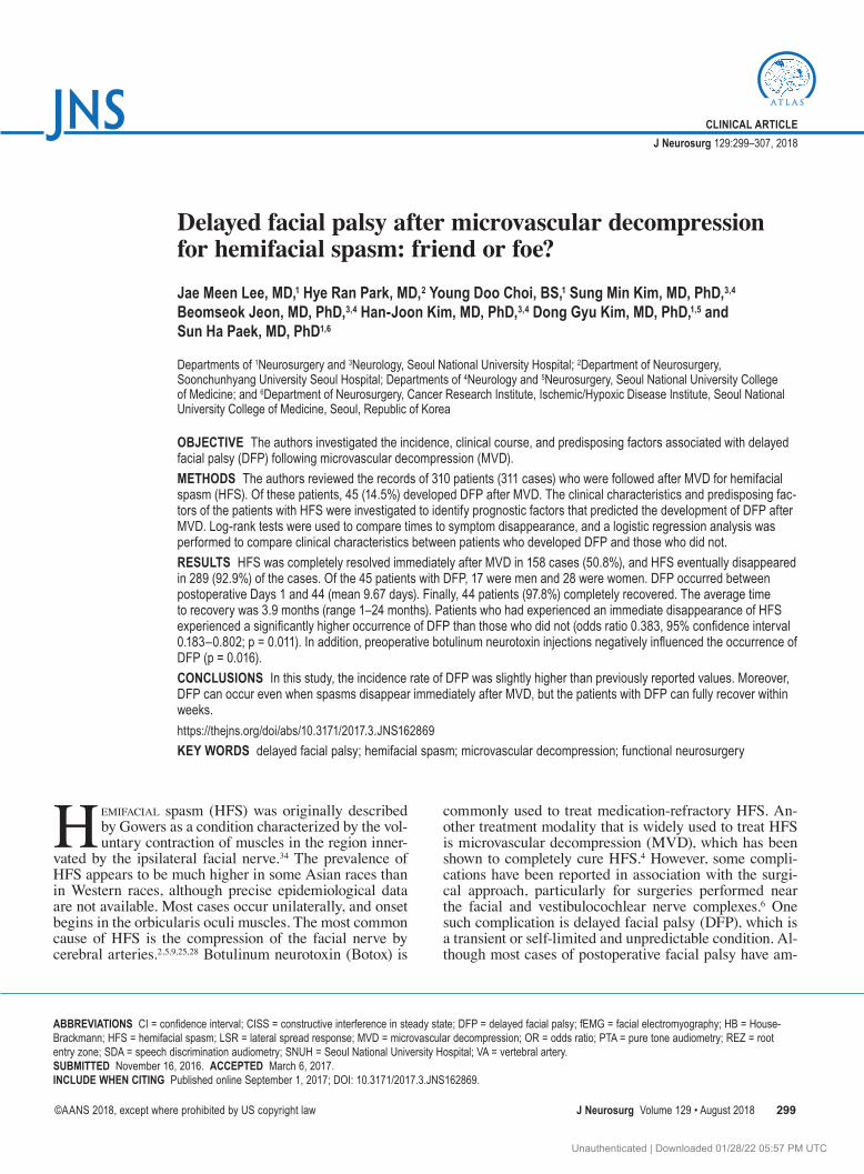
ous basal lamina. Manipulations or stimulations of cranial nerve VII are therefore not mechanically buffered by the surrounding supportive tissues.24 Second, the transition zone, a dome-shaped region of the REZ that differentiates between central myelination (by oligodendrocytes) and peripheral myelination (by Schwann cells), is known as the weakest point during manipulation (Fig. 4).11,24 The REZ is generally known to consist of central myelin and the tran-sition zone to the cranial nerve. The transition zone from the REZ to cranial nerve VII is located in the immediate vicinity of the brainstem, and thus DFP may be triggered by the manipulation of the nerve during surgery. While the transition zone from the REZ to cranial nerve VIII is locat-ed at the internal acoustic meatus, it does not experience as much manipulation as cranial nerve VII during MVD.7 It is likely, however, based on our results, that such a manipu-lation triggers not only DFP but also hearing impairment because of the intimate contact between cranial nerves VII and VIII in the cerebellopontine angle (Table 1). Finally, the gradual development of postoperative edema that pro-gresses into the operative field has been reported to cause DFP at the location where the cranial nerves overrun the body canals.24 While the symptoms of DFP are not present
immediately after MVD, they manifest within 1–7 days. Steroid therapy has been shown to be effective in resolving edema in addition to treating cases of DFP that may have been caused by edema. Accordingly, most of the patients in our study completely recovered from facial weakness after steroid therapy.
Furukawa et al.8 described DFP after MVD for HFS that was caused by the reactivation of a virus, which may also underlie Bell’s palsy. Because serological tests to detect viral antibodies were not performed in this study, we cannot exclude the potential involvement of a reacti-vated virus in the development of DFP. Likewise, the ex-act pathophysiology of DFP still remains controversial, although many studies have been and are being performed to fully elucidate its mechanism. Further studies are cer-tainly needed to fully clarify the cause of DFP.
The occurrence of DFP after MVD has been reported in many studies. However, there have been no reports that demonstrated the possibility that DFP could be used as a prognostic indicator for patients with HFS. Therefore, through this study, we provide a new understanding on the occurrence of DFP after MVD in patients with HFS.
This study has several limitations. The first limita-
TABLE 4. Number of Teflon felts and probability values for LSR monitoring and Botox injections in the two groups with and without DFP
Variable TotalDFP
p ValueYes No
Teflon feltsNo. of patients 156 29 127Mean no. of Teflon felts ± SD (median) 4.59 ± 1.028 (4.5) 4.76 ± 1.023 (5) 4.55 ± 1.029 (4) 0.328LSR (%) 0.571 Remained 39 5 (12.8) 34 (87.2) Disappeared 71 12 (16.9) 59 (83.1)LSR remained (%) >0.999 Zygomatic branch 10 1 (10.0) 9 (90.0) Mandibular branch 8 1 (12.5) 7 (87.5) Both branches 21 3 (14.3) 18 (85.7) None 71 12 (16.9) 59 (83.1)Botox injection (%) 0.016 None or 1 249 42 (16.9) 207 (83.1) ≥2 62 3 (4.8) 59 (95.2)
Bold type indicates statistical significance.
TABLE 5. Summary of previously reported results for DFP after MVD for HFS
Authors & Year Cases (%) Mean Yrs of Age (range) Days of DFP Onset After MVD (range)Time to Complete Recovery
(range)
Kuroki et al., 1991 6 (7.5) 53.5 (41–77) 8 (7–10) 2.8 mos (1.5–6 mos)Lovely et al., 1998 28 (2.8) 51.8 (28–79) 12 (7–16) 6.5 wks (1–28 wks)Kim et al., 1999 12 (8.3) 53 (45–60) 8.2 (6–11) 6.3 mos (1–12 mos)Furukawa et al., 2003 1 56 7 9 mosRhee et al., 2006 21 (5.4) 46.7 (33–62) 12.1 (7–23) 5.7 wks (25 days–17 wks)Han et al., 2012 100 (7.4) 48 (25–65) 11.2 (2–23) 9.2 wks (16–270 days)Kim et al., 2012 9 (10.4) 49.7 (26–63) 9.2 (3–14) 5.4 wks (22–57 days)Present study 45 (14.5) 56.8 (32–80) 9.67 (1–44) 3.9 mos (1–24 mos)
Unauthenticated | Downloaded 01/28/22 05:57 PM UTC

J. M. Lee et al.
J Neurosurg Volume 129 • August 2018306
tion is the variation in follow-up periods across patients. There were also some cases in which clinical evaluations were incomplete because of the retrospective nature of our study. The second limitation is that our study did not in-clude a separate group of patients with DFP who received a treatment other than steroids. Finally, the number of Tef-lon felts was not counted in every patient, and LSR moni-toring results were not obtained for every case. The influ-ence of these limitations on the principal conclusions of this report can be significant and should not be minimized. However, to our knowledge, a possible role of DFP after MVD in regards to clinical outcome of patients with HFS has not been examined. Although further work is needed to investigate the potential impact of the above-mentioned limitations on the occurrence of DFP, our study provides clinically meaningful insights into the possible role of DFP after MVD as a prognostic indicator for outcome in patients with HFS. Finally, our study did not investigate the possible effects and correlations between Asian medi-cations and/or treatment modalities (such as acupuncture, which is widely used to treat HFS in South Korea) and the incidence of DFP. This topic is worthy of further investiga-tion to illuminate possible influences of Oriental medica-tions on patients with DFP.
ConclusionsIn this study, the incidence of DFP was slightly higher
than previously reported values. Moreover, DFP may oc-cur even when spasms disappear immediately after MVD, but the patients in this study exhibited a complete recovery within weeks. Surgeons should be aware that DFP may oc-
cur even when a hemifacial spasm disappears immediately after MVD. Moreover, significantly better results were observed in the patients with DFP than in those without DFP in terms of the overall disappearance of hemifacial spasms.
AcknowledgmentsWe thank Mr. Yona Kim for his contributions to the paper. This
study was supported by the Korea Institute of Planning & Evalu-ation for Technology in Food, Agriculture, Forestry, and Fisher-ies, Republic of Korea (grant no. 311011-05-3-SB020); the Korea Healthcare Technology R&D Project (grant no. HI11C21100200), funded by the Ministry of Health & Welfare, Republic of Korea; the Technology Innovation Program (grant no. 10050154, Business Model Development for Personalized Medicine Based on Inte-grated Genome and Clinical Information) funded by the Ministry of Trade, Industry & Energy (MI, Korea); and the Bio & Medical Technology Development Program of the NRF funded by the Korean government, MSIP (grant no. 2015M3C7A1028926).
References 1. Acevedo JC, Sindou M, Fischer C, Vial C: Microvascular
decompression for the treatment of hemifacial spasm. Ret-rospective study of a consecutive series of 75 operated pa-tients—electrophysiologic and anatomical surgical analysis. Stereotact Funct Neurosurg 68:260–265, 1997
2. Adams CB: Microvascular compression: an alternative view and hypothesis. J Neurosurg 70:1–12, 1989
3. Auger RG, Whisnant JP: Hemifacial spasm in Rochester and Olmsted County, Minnesota, 1960 to 1984. Arch Neurol 47:1233–1234, 1990
4. Barker FG II, Jannetta PJ, Bissonette DJ, Shields PT, Larkins MV, Jho HD: Microvascular decompression for hemifacial spasm. J Neurosurg 82:201–210, 1995
FIG. 4. Drawing showing the components of cranial nerve VII root: 1) the central myelin portion of the root, 2) the transition zone (green lines), and 3) the peripheral myelinated portion of the root. The REZ of cranial nerve VIII is located at the internal acoustic meatus (IAM). Illustration by Jae Meen Lee. Copyright Sun Ha Paek. Published with permission.
Unauthenticated | Downloaded 01/28/22 05:57 PM UTC

Delayed facial palsy after microvascular decompression
J Neurosurg Volume 129 • August 2018 307
5. Chang JW, Chang JH, Choi JY, Kim DI, Park YG, Chung SS: Role of postoperative magnetic resonance imaging after microvascular decompression of the facial nerve for the treat-ment of hemifacial spasm. Neurosurgery 50:720–726, 2002
6. Dallan I, De Vito A, Fattori B, Casani AP, Panicucci E, Ber-rettini S, et al: Intratympanic methylprednisolone in refracto-ry sudden hearing loss: a 27-patient case series with univari-ate and multivariate analysis. Otol Neurotol 31:25–30, 2010
7. De Ridder D, Møller A, Verlooy J, Cornelissen M, De Rid-der L: Is the root entry/exit zone important in microvascular compression syndromes? Neurosurgery 51:427–434, 2002
8. Furukawa K, Sakoh M, Kumon Y, Teraoka M, Ohta S, Ohue S, et al: [Delayed facial palsy after microvascular decompres-sion for hemifacial spasm due to reactivation of varicella-zoster virus.] No Shinkei Geka 31:899–902, 2003 (Jpn)
9. Gardner WJ: Concerning the mechanism of trigeminal neu-ralgia and hemifacial spasm. J Neurosurg 19:947–958, 1962
10. Gök U, Alpay HC, Akpolat N, Yoldaş T, Kilic A, Yilmaz B, et al: Comparisons of steroid, acyclovir, lipoprostoglandin E1 and steroid + acyclovir treatments in facial paralysis: a rat study. Int J Pediatr Otorhinolaryngol 69:1199–1204, 2005
11. Guclu B, Sindou M, Meyronet D, Streichenberger N, Simon E, Mertens P: Cranial nerve vascular compression syndromes of the trigeminal, facial and vago-glossopharyngeal nerves: comparative anatomical study of the central myelin portion and transitional zone; correlations with incidences of corre-sponding hyperactive dysfunctional syndromes. Acta Neuro-chir (Wien) 153:2365–2375, 2011
12. Han JS, Lee JA, Kong DS, Park K: Delayed cranial nerve palsy after microvascular decompression for hemifacial spasm. J Korean Neurosurg Soc 52:288–292, 2012
13. Harper CM: Intraoperative cranial nerve monitoring. Muscle Nerve 29:339–351, 2004
14. Hitotsumatsu T, Matsushima T, Inoue T: Microvascular decompression for treatment of trigeminal neuralgia, hemi-facial spasm, and glossopharyngeal neuralgia: three surgical approach variations: technical note. Neurosurgery 53:1436–1443, 2003
15. Hongo K, Kobayashi S, Takemae T, Sugita K: [Posterior fossa microvascular decompression for hemifacial spasm and trigeminal neuralgia—some improvements on operative de-vices and technique.] No Shinkei Geka 13:1291–1296, 1985 (Jpn)
16. Huh R, Han IB, Moon JY, Chang JW, Chung SS: Microvas-cular decompression for hemifacial spasm: analyses of opera-tive complications in 1582 consecutive patients. Surg Neurol 69:153–157, 2008
17. Jannetta PJ, Abbasy M, Maroon JC, Ramos FM, Albin MS: Etiology and definitive microsurgical treatment of hemifacial spasm. Operative techniques and results in 47 patients. J Neurosurg 47:321–328, 1977
18. Kim BT, Hwang SC, Chang JC, Shin WH, Choi SK, Byun BJ: Delayed facial palsy following microvascular decompres-sion in hemifacial spasm patients. J Korean Neurosurg Soc 28:1332–1336, 1999
19. Kim HJ, Park YS, Ryu JS, Huh R, Han I, Shin DA, et al: In-traoperative facial electromyography and brainstem auditory evoked potential findings in microvascular decompression for hemifacial spasm: correlation with postoperative delayed facial palsy. Stereotact Funct Neurosurg 90:260–265, 2012
20. Kuo HC: Repeated onabotulinumtoxin-A injections provide better results than single injection in treatment of painful bladder syndrome. Pain Physician 16:E15–E23, 2013
21. Kuroki A, Itagaki S, Nagai O: Delayed facial weakness after microvascular decompression for hemifacial spasm. Facial Nerve Res 11:147–150, 1991
22. Lee HY, Byun JY, Park MS, Yeo SG: Steroid-antiviral treat-ment improves the recovery rate in patients with severe Bell’s palsy. Am J Med 126:336–341, 2013
23. Lovely TJ, Getch CC, Jannetta PJ: Delayed facial weakness after microvascular decompression of cranial nerve VII. Surg Neurol 50:449–452, 1998
24. Menovsky T, van Overbeeke JJ: On the mechanism of tran-sient postoperative deficit of cranial nerves. Surg Neurol 51:223–226, 1999
25. Møller AR: The cranial nerve vascular compression syn-drome: II. A review of pathophysiology. Acta Neurochir (Wien) 113:24–30, 1991
26. Nesbitt-Hawes EM, Won H, Jarvis SK, Lyons SD, Vancaillie TG, Abbott JA: Improvement in pelvic pain with botulinum toxin type A – single vs. repeat injections. Toxicon 63:83–87, 2013
27. Nguyen DQ, Franco-Vidal V, Guérin J, Darrouzet V: [De-layed facial palsy after vestibular schwannoma resection: the role of viral reactivation. Our experience in 8 cases.] Rev Laryngol Otol Rhinol (Bord) 125:23–29, 2004 (Fr)
28. Nielsen VK: Pathophysiology of hemifacial spasm: I. Ephap-tic transmission and ectopic excitation. Neurology 34:418–426, 1984
29. Radtke RA, Erwin CW, Wilkins RH: Intraoperative brain-stem auditory evoked potentials: significant decrease in post-operative morbidity. Neurology 39:187–191, 1989
30. Rhee DJ, Kong DS, Park K, Lee JA: Frequency and prognosis of delayed facial palsy after microvascular decompression for hemifacial spasm. Acta Neurochir (Wien) 148:839–843, 2006
31. Santos-Lasaosa S, Pascual-Millán LF, Tejero-Juste C, Mo-rales-Asín F: [Peripheral facial paralysis: etiology, diagnosis and treatment.] Rev Neurol 30:1048–1053, 2000 (Span)
32. Sindou MP: Microvascular decompression for primary hemifacial spasm. Importance of intraoperative neurophysi-ological monitoring. Acta Neurochir (Wien) 147:1019–1026, 2005
33. Wang X, Thirumala PD, Shah A, Gardner P, Habeych M, Crammond DJ, et al: Effect of previous botulinum neurotoxin treatment on microvascular decompression for hemifacial spasm. Neurosurg Focus 34(3):E3, 2013
34. Wilkins RH: Hemifacial spasm: a review. Surg Neurol 36:251–277, 1991
DisclosuresThe authors report no conflict of interest concerning the materi-als or methods used in this study or the findings specified in this paper.
Author ContributionsConception and design: Paek, Lee, SM Kim, Jeon, HJ Kim, DG Kim. Acquisition of data: Lee, Choi. Analysis and interpretation of data: all authors. Drafting the article: Paek, Lee. Critically revising the article: Lee, SM Kim. Reviewed submitted version of manuscript: Lee, Jeon, HJ Kim, DG Kim. Statistical analysis: Lee. Administrative/technical/material support: Choi. Study supervision: Park, SM Kim, HJ Kim, DG Kim.
Supplemental Information Current AffiliationsDr. Lee: Department of Neurosurgery, Pusan National University Hospital, Busan, Republic of Korea.
CorrespondenceSun Ha Paek, Department of Neurosurgery, Seoul National Uni-versity Hospital, 101 Daehak-Ro, Jongno-gu, Seoul, 110-744, Republic of Korea. email: [email protected].
Unauthenticated | Downloaded 01/28/22 05:57 PM UTC