Embed Size (px)
Citation preview
doi:10.1152/ajpheart.00503.2010 299:H1505-H1514, 2010. First published 20 August 2010;Am J Physiol Heart Circ Physiol
and Thomas J. HundRoseanne M. Wolf, Colleen C. Mitchell, Matthew D. Christensen, Peter J. Mohlercomputational model of ankyrin-B syndromeDefining new insight into atypical arrhythmia: a
You might find this additional info useful...
42 articles, 30 of which can be accessed free at:This article cites http://ajpheart.physiology.org/content/299/5/H1505.full.html#ref-list-1
including high resolution figures, can be found at:Updated information and services http://ajpheart.physiology.org/content/299/5/H1505.full.html
can be found at:AJP - Heart and Circulatory Physiologyabout Additional material and information http://www.the-aps.org/publications/ajpheart
This infomation is current as of September 2, 2011.
ISSN: 0363-6135, ESSN: 1522-1539. Visit our website at http://www.the-aps.org/.Physiological Society, 9650 Rockville Pike, Bethesda MD 20814-3991. Copyright © 2010 by the American Physiological Society. intact animal to the cellular, subcellular, and molecular levels. It is published 12 times a year (monthly) by the Americanlymphatics, including experimental and theoretical studies of cardiovascular function at all levels of organization ranging from the
publishes original investigations on the physiology of the heart, blood vessels, andAJP - Heart and Circulatory Physiology
on Septem
ber 2, 2011ajpheart.physiology.org
Dow
nloaded from
Defining new insight into atypical arrhythmia: a computational modelof ankyrin-B syndrome
Roseanne M. Wolf,1,2 Colleen C. Mitchell,2 Matthew D. Christensen,1 Peter J. Mohler,1,3
and Thomas J. Hund1
1Department of Internal Medicine, Division of Cardiovascular Medicine, 2Department of Mathematics,and 3Department of Molecular Physiology and Biophysics, University of Iowa Carver College of Medicine, Iowa City, Iowa
Submitted 26 May 2010; accepted in final form 16 August 2010
Wolf RM, Mitchell CC, Christensen MD, Mohler PJ, Hund TJ.Defining new insight into atypical arrhythmia: a computational model ofankyrin-B syndrome. Am J Physiol Heart Circ Physiol 299: H1505–H1514,2010. First published August 20, 2010; doi:10.1152/ajpheart.00503.2010.—Normal cardiac excitability depends on the coordinated activity ofspecific ion channels and transporters within specialized domains atthe plasma membrane and sarcoplasmic reticulum. Ion channel dys-function due to congenital or acquired defects has been linked tohuman cardiac arrhythmia. More recently, defects in ion channel-associated proteins have been associated with arrhythmia. Ankyrin-Bis a multifunctional adapter protein responsible for targeting select ionchannels, transporters, cytoskeletal proteins, and signaling moleculesin excitable cells, including neurons, pancreatic �-cells, and cardio-myocytes. Ankyrin-B dysfunction has been linked to cardiac arrhyth-mia in human patients and ankyrin-B heterozygous (ankyrin-B�/�)mice with a phenotype characterized by sinus node dysfunction,susceptibility to ventricular arrhythmias, and sudden death(“ankyrin-B syndrome”). At the cellular level, ankyrin-B�/� cellshave defects in the expression and membrane localization of theNa�/Ca2� exchanger and Na�-K�-ATPase, Ca2� overload, and fre-quent afterdepolarizations, which likely serve as triggers for lethalcardiac arrhythmias. Despite knowledge gathered from mouse modelsand human patients, the molecular mechanism responsible for cardiacarrhythmias in the setting of ankyrin-B dysfunction remains unclear.Here, we use mathematical modeling to provide new insights into thecellular pathways responsible for Ca2� overload and afterdepolariza-tions in ankyrin-B�/� cells. We show that the Na�/Ca2� exchangerand Na�-K�-ATPase play related, yet distinct, roles in intracellularCa2� accumulation, sarcoplasmic reticulum Ca2� overload, and af-terdepolarization generation in ankyrin-B�/� cells. These findingsprovide important insights into the molecular mechanisms underlyinga human disease and are relevant for acquired human arrhythmia,where ankyrin-B dysfunction has recently been identified.
Na�/Ca2� exchanger; Na�-K�-ATPase; mathematical model; traf-ficking; calcium
NORMAL CARDIAC EXCITABILITY depends on the coordinated bio-physical activity of specific ion channels and transporters onthe plasma membrane. Over the past decade, gene mutationsthat affect ion channel biophysical activity have been linkedwith fatal human arrhythmias (19). Recently, a new class ofgene mutations has been identified that alters the local couplingof ion channels with cytoskeletal and regulatory proteins (5,20, 22, 27, 28, 36, 41, 42). Based in part on these importantfindings, it is now clear that ion channel and transporterfunction depend on normal biophysical properties and properlocal membrane localization/organization.
Ankyrin polypeptides are responsible for the targeting andstabilization of ion channels, transporters, and signaling mol-ecules at cell membranes of diverse cell types, includingcardiomyocytes, neurons, pancreatic �-cells, and erythrocytes(12). Over the past decade, members of the ankyrin family(ankyrin-R, ankyrin-B, and ankyrin-G) have been linked to anumber of human diseases, including hereditary spherocytosis,cardiac arrhythmia, diabetes, and muscular dystrophy (1, 8, 16,27, 28). Ankyrin-B dysfunction has been linked to both con-genital and acquired arrhythmias (15, 17, 23, 26, 28, 35). Infact, nine loss-of-function variants in ANK2 (the gene encodingankyrin-B) have been identified in human patients with acomplex cardiac arrhythmia syndrome (“long QT 4” or“ankyrin-B syndrome”) characterized by sinus node dysfunc-tion, increased susceptibility to ventricular arrhythmias, andsudden death under stress (17, 26, 28, 29, 34). Ankyrin-B-deficient (ankyrin-B�/�) mice display an increased suscepti-bility to stress-induced arrhythmias and sudden death, similarto human patients (23, 26, 28). Importantly, ankyrin-B�/� miceshow defects in ion channel and transporter targeting, abnormalCa2� homeostasis, and an increased likelihood of potentiallylife-threatening afterdepolarizations (28). In ventricular cardiomy-ocytes, ankyrin-B is responsible for the proper membrane local-ization and function of Na�-K�-ATPase (NKA), Na�/Ca2� ex-changer (NCX), inositol 1,4,5-trisphosphate (InsP3) receptor, andprotein phosphatase 2A (PP2A) (2, 7, 29). Despite the wealth ofinformation gathered from human patients and the mouse modelof ankyrin-B syndrome, the direct link between specific mem-brane protein changes and cardiac arrhythmia has yet to beestablished.
Mathematical modeling of excitable cells has been used togenerate important insights into the cellular mechanisms un-derlying a wide variety of human diseases, including cardiacarrhythmia, epilepsy, and diabetes (6, 14, 16, 32). In this study,we used mathematical modeling to identify the cellular path-way responsible for abnormal Ca2� handling and cardiacarrhythmia in ankyrin-B�/� cells. Our computer simulationsshowed that loss of NCX and NKA membrane targeting inankyrin-B�/� cells resulted in accumulation of intracellularNa� and Ca2� under basal conditions. Furthermore, ankyrin-Breduction predisposed the cell to Ca2� overload, frequentspontaneous Ca2� release, and action potential (AP) afterde-polarizations during rapid pacing in the presence of isoproter-enol. While loss of NKA function contributed to the increasedCa2� transients in ankyrin-B�/� cardiomyocytes, abnormalNCX targeting was the dominant mechanism for Ca2� over-load in the sarcoplasmic reticulum (SR), spontaneous Ca2�
release, and afterdepolarizations. These findings provide im-portant insights into the molecular mechanism underlying a
Address for reprint requests and other correspondence: T. J. Hund, Dept. ofInternal Medicine, Univ. of Iowa Carver College of Medicine, 285 NewtonRd., CBRB 2283, Iowa City, IA 52242 (e-mail: [email protected]).
Am J Physiol Heart Circ Physiol 299: H1505–H1514, 2010.First published August 20, 2010; doi:10.1152/ajpheart.00503.2010.
0363-6135/10 Copyright © 2010 the American Physiological Societyhttp://www.ajpheart.org H1505
on Septem
ber 2, 2011ajpheart.physiology.org
Dow
nloaded from

human disease and highlight the related, yet distinct, roles ofNCX and NKA in ventricular cardiomyocytes. Considering theassociation between ankyrin-B dysfunction and acquired ar-rhythmia as well as arrhythmia susceptibility in the generalpopulation (15, 35), we expect the insights generated by thisstudy to have broader relevance for human disease.
METHODS
Mathematical model of the ankyrin-B�/� cardiomyocyte. Themathematical model used in this study was based on a model of themurine ventricular AP (3, 4) since much of the experimental datacame from the mouse. Importantly, the formulations for NKA and
NCX current have been well validated against experimental datafrom mammalian ventricular myocytes (21) (Supplemental Mate-rial, Supplemental Fig. S1).1 Simulations were also performed usinga well-validated model of the human ventricular myocyte (38). Mod-ifications to the equations were made to account for experimentallymeasured changes in NCX and NKA membrane expression inankyrin-B�/� cardiomyocytes (Fig. 1, A–E) (23, 25, 28, 29). Specif-ically, NCX was scaled to produce a 40% reduction in current at a testpotential of �10 mV compared with control (wild type), consistent
1Supplemental Material for this article is available online at the AmericanJournal of Physiology-Heart and Circulatory Physiology website.
Fig. 1. Mathematical model of the ankyrin-B-deficient (ankyrin-B�/�) cell. A: schematic of the mouse ventricular cell model. Na�/Ca2� exchanger (NCX) current(INaCa) and Na�-K�-ATPase (NKA) current (INaK) were altered in the model of the ankyrin-B�/� cell (shaded boxes). INa, Na� current; INa,b, background Na�
current; ICaL, L-type Ca2� current; Ip(Ca), sarcolemmal Ca2� pump; ICa,b, background Ca2� current; Ins(Ca), nonspecific Ca2� current; IKto,f, fast component ofthe transient outward K� current; IKto,s, slow component of the transient outward K� current; IKr, fast component of the delayed recifier K� current; IKs, slowcomponent of the delayed recifier K� current; IKss, steady-state K� current; IKur, ultrarapid K� current; IK1, inward rectifier K� current; Irel, Ca2� release flux;Iup, Ca2� uptake flux; SER, sarco(endo)plasmic reticulum Ca2�-ATPase; JSR, junctional sarcoplasmic reticulum (SR); NSR, network SR; Itr, Ca2� transfer flux;Ileak, Ca2� leak flux. B–E: simulated action potentials (APs) in control (B and D) and ankyrin-B�/� (C and E) cardiomyocytes from the mouse (B and C) andhuman (D and E) ventricular cell models [10th action potential (AP) shown at a cycle length (CL) of 1,000 ms]. F: simulated INaCa at a test potential of �10mV from wild-type cell and an ankyrin-B�/� cell compared with experimental measurements (n � 12, *P � 0.05) (17). In both the simulation and experiment,intracellular Na� concentration � 20 mM, extracellular Na� concentration � 145 mM, intracellular Ca2� concentration ([Ca2�]i) � 1 �M, and extracellular Ca2�
concentration � 2 mM. G: simulated and experimentally measured (n � 3, *P � 0.01) (25) NKA membrane expression in wild-type and ankyrin-B�/� mouseventricular cells. H: simulated and experimentally measured (n � 17, P � not significant) (28) AP duration (APD) at 90% repolarization. I: simulated andexperimentally measured (n � 18, *P � 0.001) peak Ca2� transients. In both the simulation and experiment, the cell was pulsed four times to test potential of�0 mV from a holding potential of �40 mV (28).
H1506 COMPUTATIONAL MODEL OF ANKYRIN-B SYNDROME
AJP-Heart Circ Physiol • VOL 299 • NOVEMBER 2010 • www.ajpheart.org
on Septem
ber 2, 2011ajpheart.physiology.org
Dow
nloaded from
with experimental measurements from adult ankyrin-B�/� myocytes(17) (Fig. 1F). Also, the maximum NKA current was decreased 25%based on experimental measurements showing reduced NKA surfaceexpression in ankyrin-B�/� myocytes (as assessed by [3H]ouabainbinding) (25) (Fig. 1G). The peak L-type Ca2� current (ICaL)-voltagerelationship was no different between the control and ankyrin-B�/�
cell models (not shown), consistent with experimental measurementsin ventricular myocytes (28). Importantly, simulated APs and Ca2�
transients showed good agreement with experimental measurements(28) (Fig. 1, H and I). The complete equations and parameters for themodels used in this study may be found in the Supplemental Material.
Mathematical model of isoproterenol effects. The effects of the�-adrenergic receptor agonist isoproterenol (saturating concentration� 0.1 �M) on the AP and Ca2� transient were simulated according topreviously published formulations (9, 40). Specifically, isoproterenoleffects on ICaL, the slow component of the delayed rectifier K�
current (IKs), and sarco(endo)plasmic reticulum Ca2�-ATPase current(Iup) were incorporated into the models to simulate �-adrenergic
stimulation (Supplemental Material) (9, 40). The modified equationsand parameters may be found in the Supplemental Material.
Pacing protocol. Models were paced from rest to steady state overa range of pacing cycle lengths (CLs; from 2,000 to 200 ms, stimulusamplitude � �60 �A/�F for the mouse and �52 �A/�F for thehuman; stimulus duration � 0.5 ms for the mouse and 1 ms for thehuman).
RESULTS
Ankyrin-B deficiency promotes Ca2� overload at baseline.To determine the electrophysiological consequences of abnor-mal NCX and NKA membrane expression in ankyrin-B�/�
cardiomyocytes, we paced the control and ankyrin-B�/� cellmodels (CL � 1,000 ms; Fig. 2). Consistent with experimentaldata (28), the simulated AP from the ankyrin-B�/� mousemyocyte was slightly longer than the control AP (Figs. 1H and
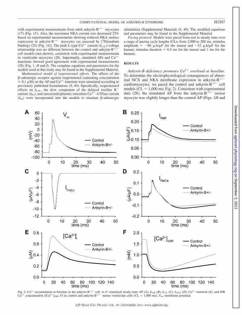
Fig. 2. Ca2� accumulation at baseline in the ankyrin-B�/� cell. A–F: simulated steady-state AP (A), INaK (B), ICaL (C), INaCa (D), Ca2� transient (E), and JSRCa2� concentration ([Ca2�]JSR; F) in control and ankyrin-B�/� mouse ventricular cells (CL � 1,000 ms). Vm, membrane potential.
H1507COMPUTATIONAL MODEL OF ANKYRIN-B SYNDROME
AJP-Heart Circ Physiol • VOL 299 • NOVEMBER 2010 • www.ajpheart.org
on Septem
ber 2, 2011ajpheart.physiology.org
Dow
nloaded from
2A). Whereas the AP morphology and AP duration (APD)were similar between the ankyrin-B�/� and control model, lossof NCX and NKA targeting led to dramatic Ca2� and Na�
accumulation in the cytosol and SR of the ankyrin-B�/� cell(Fig. 2, E and F, and Table 1). Specifically, ankyrin-B-deficientcells displayed an increase in intracellular Na� concentration(Table 1), Ca2� concentration in the junctional SR ([Ca2�]JSR),and Ca2� transient amplitude (Fig. 2, E and F), consistent withexperimental measurements from ankyrin-B�/� mice (28).Ca2� bound to SR calsequestrin ([Ca2�-CSQN]) was alsoincreased in ankyrin-B�/� cells but remained below the thresh-old for eliciting spontaneous Ca2� release (not shown).
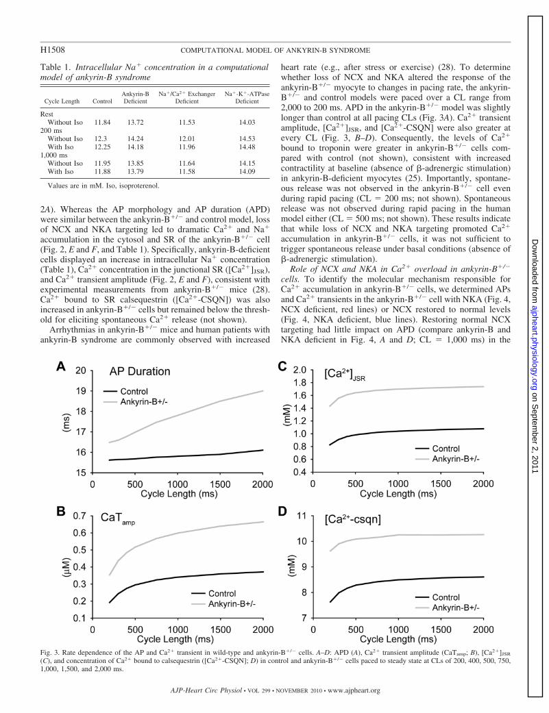
Arrhythmias in ankyrin-B�/� mice and human patients withankyrin-B syndrome are commonly observed with increased
heart rate (e.g., after stress or exercise) (28). To determinewhether loss of NCX and NKA altered the response of theankyrin-B�/� myocyte to changes in pacing rate, the ankyrin-B�/� and control models were paced over a CL range from2,000 to 200 ms. APD in the ankyrin-B�/� model was slightlylonger than control at all pacing CLs (Fig. 3A). Ca2� transientamplitude, [Ca2�]JSR, and [Ca2�-CSQN] were also greater atevery CL (Fig. 3, B–D). Consequently, the levels of Ca2�
bound to troponin were greater in ankyrin-B�/� cells com-pared with control (not shown), consistent with increasedcontractility at baseline (absence of �-adrenergic stimulation)in ankyrin-B-deficient myocytes (25). Importantly, spontane-ous release was not observed in the ankyrin-B�/� cell evenduring rapid pacing (CL � 200 ms; not shown). Spontaneousrelease was not observed during rapid pacing in the humanmodel either (CL � 500 ms; not shown). These results indicatethat while loss of NCX and NKA targeting promoted Ca2�
accumulation in ankyrin-B�/� cells, it was not sufficient totrigger spontaneous release under basal conditions (absence of�-adrenergic stimulation).
Role of NCX and NKA in Ca2� overload in ankyrin-B�/�
cells. To identify the molecular mechanism responsible forCa2� accumulation in ankyrin-B�/� cells, we determined APsand Ca2� transients in the ankyrin-B�/� cell with NKA (Fig. 4,NCX deficient, red lines) or NCX restored to normal levels(Fig. 4, NKA deficient, blue lines). Restoring normal NCXtargeting had little impact on APD (compare ankyrin-B andNKA deficient in Fig. 4, A and D; CL � 1,000 ms) in the
Table 1. Intracellular Na� concentration in a computationalmodel of ankyrin-B syndrome
Cycle Length ControlAnkyrin-BDeficient
Na�/Ca2� ExchangerDeficient
Na�-K�-ATPaseDeficient
RestWithout Iso 11.84 13.72 11.53 14.03
200 msWithout Iso 12.3 14.24 12.01 14.53With Iso 12.25 14.18 11.96 14.48
1,000 msWithout Iso 11.95 13.85 11.64 14.15With Iso 11.88 13.79 11.58 14.09
Values are in mM. Iso, isoproterenol.
Fig. 3. Rate dependence of the AP and Ca2� transient in wild-type and ankyrin-B�/� cells. A–D: APD (A), Ca2� transient amplitude (CaTamp; B), [Ca2�]JSR
(C), and concentration of Ca2� bound to calsequestrin ([Ca2�-CSQN]; D) in control and ankyrin-B�/� cells paced to steady state at CLs of 200, 400, 500, 750,1,000, 1,500, and 2,000 ms.
H1508 COMPUTATIONAL MODEL OF ANKYRIN-B SYNDROME
AJP-Heart Circ Physiol • VOL 299 • NOVEMBER 2010 • www.ajpheart.org
on Septem
ber 2, 2011ajpheart.physiology.org
Dow
nloaded from
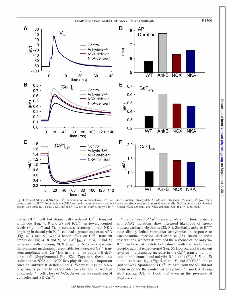
ankyrin-B�/� cell but dramatically reduced Ca2� transientamplitude (Fig. 4, B and E) and [Ca2�]JSR toward controllevels (Fig. 4, C and F). In contrast, restoring normal NKAtargeting in the ankyrin-B�/� cell had a greater impact on APD(Fig. 4, A and D), with a lesser effect on Ca2� transientamplitude (Fig. 4, B and E) or [Ca2�]JSR (Fig. 4, C and F)compared with restoring NCX targeting. NCX loss was alsothe dominant mechanism responsible for increased Ca2� tran-sient amplitude and [Ca2�]JSR in the human ankyrin-B-defi-cient cell (Supplemental Fig. S2). Together, these dataindicate that NKA and NCX loss play distinct but importantroles in ankyrin-B deficient cells. Whereas loss of NKAtargeting is primarily responsible for changes in APD inankyrin-B�/� cells, loss of NCX drives the accumulation ofcytosolic and SR Ca2�.
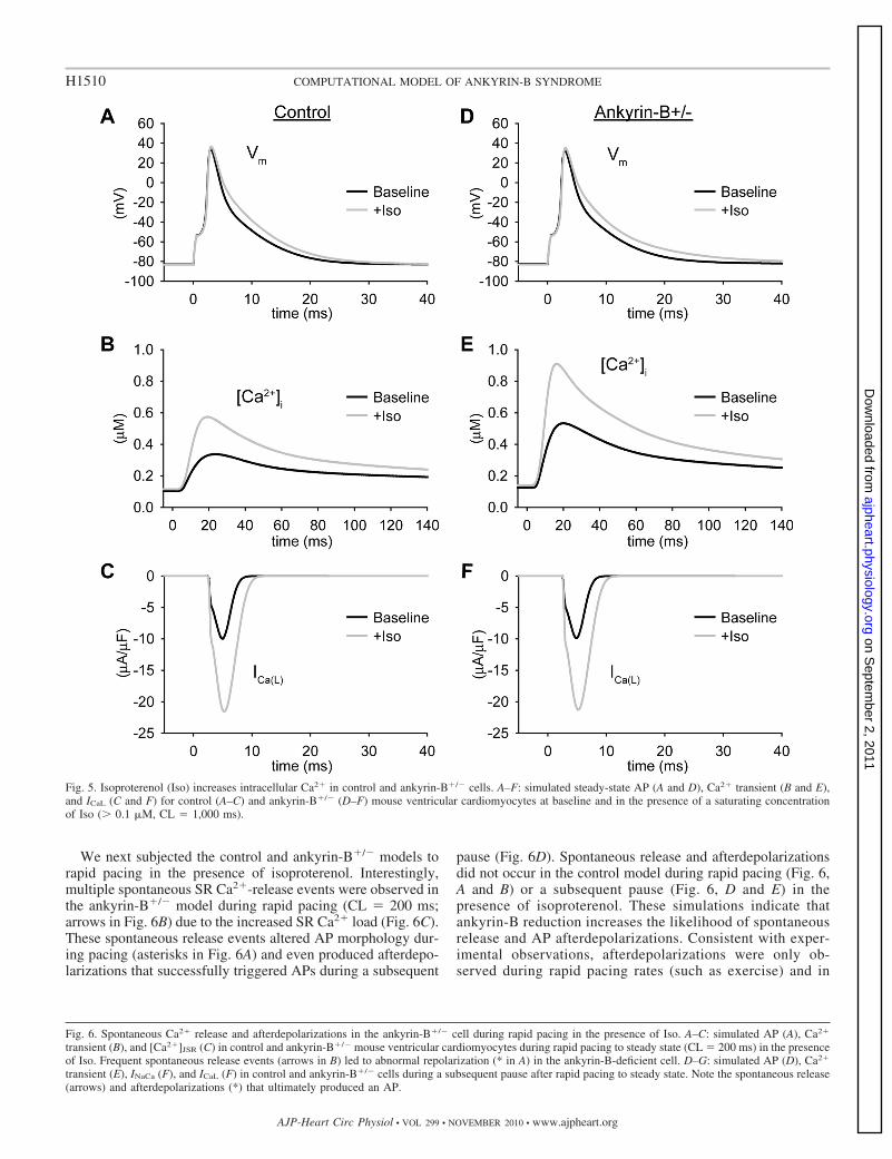
Increased levels of Ca2� with isoproterenol. Human patientswith ANK2 mutations show increased likelihood of stress-induced cardiac arrhythmias (28, 34). Similarly, ankyrin-B�/�
mice display lethal ventricular arrhythmias in response tocatecholamine injection after exercise (28). Based on theseobservations, we next determined the response of the ankyrin-B�/� and control models to treatment with the �-adrenergicreceptor agonist isoproterenol (Fig. 5). Isoproterenol treatmentresulted in a dramatic increase in the Ca2� transient ampli-tude in both control and ankyrin-B�/� cells (Fig. 5, B and E)due to increased ICaL (Fig. 5, C and F) and SR Ca2� uptake(not shown). Spontaneous Ca2� release from the SR did notoccur in either the control or ankyrin-B�/� models duringslow pacing (CL � 1,000 ms) even in the presence ofisoproterenol.
Fig. 4. Role of NCX and NKA in Ca2� accumulation in the ankyrin-B�/� cell. A–C: simulated steady-state AP (A), Ca2� transient (B), and [Ca2�]JSR (C) incontrol, ankyrin-B�/�, NCX-deficient (NKA restored to normal levels), and NKA-deficient (NCX restored to normal levels) cells. D–F: summary data showingsteady-state APD (D), CaTamp (E), and [Ca2�]JSR (F) in control, ankyrin-B�/� (AnkB), NCX-deficient, and NKA-deficient cells (CL � 1,000 ms).
H1509COMPUTATIONAL MODEL OF ANKYRIN-B SYNDROME
AJP-Heart Circ Physiol • VOL 299 • NOVEMBER 2010 • www.ajpheart.org
on Septem
ber 2, 2011ajpheart.physiology.org
Dow
nloaded from
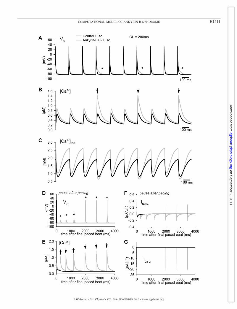
We next subjected the control and ankyrin-B�/� models torapid pacing in the presence of isoproterenol. Interestingly,multiple spontaneous SR Ca2�-release events were observed inthe ankyrin-B�/� model during rapid pacing (CL � 200 ms;arrows in Fig. 6B) due to the increased SR Ca2� load (Fig. 6C).These spontaneous release events altered AP morphology dur-ing pacing (asterisks in Fig. 6A) and even produced afterdepo-larizations that successfully triggered APs during a subsequent
pause (Fig. 6D). Spontaneous release and afterdepolarizationsdid not occur in the control model during rapid pacing (Fig. 6,A and B) or a subsequent pause (Fig. 6, D and E) in thepresence of isoproterenol. These simulations indicate thatankyrin-B reduction increases the likelihood of spontaneousrelease and AP afterdepolarizations. Consistent with exper-imental observations, afterdepolarizations were only ob-served during rapid pacing rates (such as exercise) and in
Fig. 6. Spontaneous Ca2� release and afterdepolarizations in the ankyrin-B�/� cell during rapid pacing in the presence of Iso. A–C: simulated AP (A), Ca2�
transient (B), and [Ca2�]JSR (C) in control and ankyrin-B�/� mouse ventricular cardiomyocytes during rapid pacing to steady state (CL � 200 ms) in the presenceof Iso. Frequent spontaneous release events (arrows in B) led to abnormal repolarization (* in A) in the ankyrin-B-deficient cell. D–G: simulated AP (D), Ca2�
transient (E), INaCa (F), and ICaL (F) in control and ankyrin-B�/� cells during a subsequent pause after rapid pacing to steady state. Note the spontaneous release(arrows) and afterdepolarizations (*) that ultimately produced an AP.
Fig. 5. Isoproterenol (Iso) increases intracellular Ca2� in control and ankyrin-B�/� cells. A–F: simulated steady-state AP (A and D), Ca2� transient (B and E),and ICaL (C and F) for control (A–C) and ankyrin-B�/� (D–F) mouse ventricular cardiomyocytes at baseline and in the presence of a saturating concentrationof Iso (� 0.1 �M, CL � 1,000 ms).
H1510 COMPUTATIONAL MODEL OF ANKYRIN-B SYNDROME
AJP-Heart Circ Physiol • VOL 299 • NOVEMBER 2010 • www.ajpheart.org
on Septem
ber 2, 2011ajpheart.physiology.org
Dow
nloaded from
H1511COMPUTATIONAL MODEL OF ANKYRIN-B SYNDROME
AJP-Heart Circ Physiol • VOL 299 • NOVEMBER 2010 • www.ajpheart.org
on Septem
ber 2, 2011ajpheart.physiology.org
Dow
nloaded from
the presence of isoproterenol (�-adrenergic stimulation)(28). We observed similar behavior in simulations with thehuman model (Supplemental Fig. S3). Specifically, sponta-neous release was observed during rapid pacing in thepresence of isoproterenol in the ankyrin-B�/� cell but not inthe control cell (Supplemental Fig. S3A). In fact, spontane-ous release produced afterdepolarizations that generatedAPs during pacing in the human model (asterisk in Supple-mental Fig. S3A).
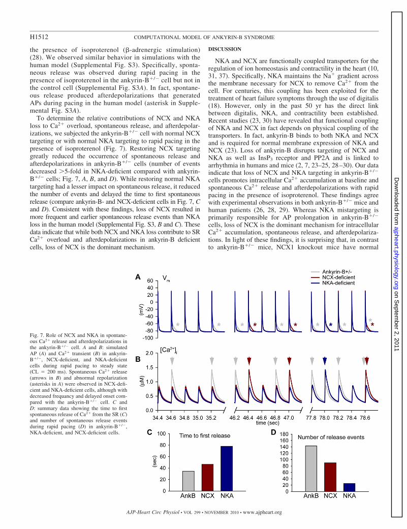
To determine the relative contributions of NCX and NKAloss to Ca2� overload, spontaneous release, and afterdepolar-izations, we subjected the ankyrin-B�/� cell with normal NCXtargeting or with normal NKA targeting to rapid pacing in thepresence of isoproterenol (Fig. 7). Restoring NCX targetinggreatly reduced the occurrence of spontaneous release andafterdepolarizations in ankyrin-B�/� cells (number of eventsdecreased �5-fold in NKA-deficient compared with ankyrin-B�/� cells; Fig. 7, A, B, and D). While restoring normal NKAtargeting had a lesser impact on spontaneous release, it reducedthe number of events and delayed the time to first spontaneousrelease (compare ankyrin-B- and NCX-deficient cells in Fig. 7, Cand D). Consistent with these findings, loss of NCX resulted inmore frequent and earlier spontaneous release events than NKAloss in the human model (Supplemental Fig. S3, B and C). Thesedata indicate that while both NCX and NKA loss contribute to SRCa2� overload and afterdepolarizations in ankyrin-B deficientcells, loss of NCX is the dominant mechanism.
DISCUSSION
NKA and NCX are functionally coupled transporters for theregulation of ion homeostasis and contractility in the heart (10,31, 37). Specifically, NKA maintains the Na� gradient acrossthe membrane necessary for NCX to remove Ca2� from thecell. For centuries, this coupling has been exploited for thetreatment of heart failure symptoms through the use of digitalis(18). However, only in the past 50 yr has the direct linkbetween digitalis, NKA, and contractility been established.Recent studies (23, 30) have revealed that functional couplingof NKA and NCX in fact depends on physical coupling of thetransporters. In fact, ankyrin-B binds to both NKA and NCXand is required for normal membrane expression of NKA andNCX (23). Loss of ankyrin-B disrupts targeting of NCX andNKA as well as InsP3 receptor and PP2A and is linked toarrhythmia in humans and mice (2, 7, 23–25, 28–30). Our dataindicate that loss of NCX and NKA targeting in ankyrin-B�/�
cells promotes intracellular Ca2� accumulation at baseline andspontaneous Ca2� release and afterdepolarizations with rapidpacing in the presence of isoproterenol. These findings agreewith experimental observations in both ankyrin-B�/� mice andhuman patients (26, 28, 29). Whereas NKA mistargeting isprimarily responsible for AP prolongation in ankyrin-B�/�
cells, loss of NCX is the dominant mechanism for intracellularCa2� accumulation, spontaneous release, and afterdepolariza-tions. In light of these findings, it is surprising that, in contrastto ankyrin-B�/� mice, NCX1 knockout mice have normal
Fig. 7. Role of NCX and NKA in spontane-ous Ca2� release and afterdepolarizations inthe ankyrin-B�/� cell. A and B: simulatedAP (A) and Ca2� transient (B) in ankyrin-B�/�, NCX-deficient, and NKA-deficientcells during rapid pacing to steady state(CL � 200 ms). Spontaneous Ca2� release(arrows in B) and abnormal repolarization(asterisks in A) were observed in NCX-defi-cient and NKA-deficient cells, although withdecreased frequancy and delayed onset com-pared with the ankyrin-B�/� cell. C andD: summary data showing the time to firstspontaneous release of Ca2� from the SR (C)and number of spontaneous release eventsduring rapid pacing (D) in ankyrin-B�/�,NKA-deficient, and NCX-deficient cells.
H1512 COMPUTATIONAL MODEL OF ANKYRIN-B SYNDROME
AJP-Heart Circ Physiol • VOL 299 • NOVEMBER 2010 • www.ajpheart.org
on Septem
ber 2, 2011ajpheart.physiology.org
Dow
nloaded from
Ca2� transients with a modest decrease in contractility andnormal lifespan (13). However, unlike ankyrin-B-deficientmice, NCX1 knockout mice show a compensatory decrease inICaL, which limits Ca2� influx and likely mitigates the detri-mental effects of NCX loss (13). Future studies are required todetermine the mechanism for ICaL downregulation in NCX1mice and the reason for differential regulation in NCX1 knock-out and ankyrin-B-deficient mice.
It is important to note that additional factors not accountedfor in the model may regulate triggered activity in ankyrin-Bdeficiency. One possibility includes an altered phosphorylationstate of SR and/or membrane target proteins due to loss ofPP2A activity (2). In fact, reduced expression of the B56�regulatory subunit of PP2A has been shown to cause Ca2�/calmodulin-dependent kinase II-dependent hyperphosphoryla-tion of ryanodine receptor 2 and afterdepolarizations (39).Moreover, Ca2� overload itself due to loss of NKA and NCXmay result in abnormal kinase activity (33). Thus, perturbationof kinase/phosphatase regulation in ankyrin-B deficiency hasthe potential to exacerbate dysfunction due to loss of NKA andNCX targeting.
While not a consistent clinical finding, a subpopulation ofpatients with ankyrin-B syndrome present with prolongation ofthe QT interval on the electrocardiogram (26, 28, 34). Themechanism responsible for this phenotype is unclear. Oursimulation results indicate that loss of NKA is responsible forthe slight prolongation of APD in ankyrin-B�/� cells. None-theless, it is unclear whether QT prolongation is due to APprolongation or another mechanism [e.g., abnormal conduc-tion (28)]. Future studies are required to address QT pro-longation in the subpopulation of ankyrin-B syndrome pa-tients with long QT.
A recent study (11) from our group has identified Eps15homology domain (EHD) proteins as a new class of cardiacproteins involved in ankyrin-B-based targeting of NCX. EHDknockdown in wild-type neonatal cardiomyocytes reducesNCX membrane expression and function. Conversely, EHDoverexpression increases cell surface NCX and current. Theresults from the present study support the idea that restoringNCX function by targeting ankyrin and/or EHD may be aneffective strategy for preventing cardiac arrhythmia and suddendeath in patients with congenital or acquired ankyrin-B defi-ciency.
Limitations. Our model of the ankyrin-B�/� cardiomyocyteis based closely on cellular data from an animal model ofankyrin-B�/� that phenocopies human patients with ankyrin-Bsyndrome. While this effort is an important first step, it isimportant to note that our model is limited by the availableexperimental data. While loss of current and surface expressionof NCX and NKA are well documented in ankyrin-B�/� mice,data on NCX and NKA biophysical properties are limited.However, it has been shown that while there is a loss ofouabain-binding sites (parallels loss of NKA) in ankyrin-B-deficient cardiomyocytes, the affinity for ouabain is unchangedfor residual sites (25). These data, while far from conclusive,suggest that the population of NKA that targets successfully inthe absence of ankyrin-B functions normally. Furthermore,while the model addresses loss of NCX and NKA targeting inankyrin-B-deficient cells, it does not account for defects in thelocalization of the InsP3 receptor (or PP2A, as discussedabove) in ventricular cardiomyocytes. In fact, the role of the
InsP3 receptor in cardiomyocytes is unclear, particularly in theventricle, where Ca2� release is controlled by ryanodine re-ceptor Ca2�-release channels. We expect our model to serve asa quantitative framework into which new data on InsP3 func-tion (and PP2A) in ventricular cardiomyocytes may be incor-porated to determine the consequences of InsP3 mislocalizationin ankyrin-B�/� cells.
GRANTS
This work was supported by National Heart, Lung, and Blood InstituteGrants HL-096805 (to T. J. Hund) and HL-084583 and HL-083422 (to P. J.Mohler), the Pew Scholars Trust (to P. J. Mohler), and a Fondation LeducqAward to the Alliance for Calmodulin Kinase Signaling in Heart Disease.
DISCLOSURES
No conflicts of interest, financial or otherwise, are declared by the author(s).
REFERENCES
1. Ayalon G, Davis JQ, Scotland PB, Bennett V. An ankyrin-basedmechanism for functional organization of dystrophin and dystroglycan.Cell 135: 1189–1200, 2008.
2. Bhasin N, Cunha SR, Mduannayake M, Gigena MS, Rogers TB,Mohler PJ. Molecular basis for PP2A regulatory subunit B56� targetingin cardiomyocytes. Am J Physiol Heart Circ Physiol 293: H109–H119,2007.
3. Bondarenko VE, Rasmusson RL. Transmural heterogeneity of repolar-ization and Ca2� handling in a model of mouse ventricular tissue. Am JPhysiol Heart Circ Physiol 299: H454–H469, 2010.
4. Bondarenko VE, Szigeti GP, Bett GC, Kim SJ, Rasmusson RL.Computer model of action potential of mouse ventricular myocytes. Am JPhysiol Heart Circ Physiol 287: H1378–H1403, 2004.
5. Chen L, Marquardt ML, Tester DJ, Sampson KJ, Ackerman MJ,Kass RS. Mutation of an A-kinase-anchoring protein causes long-QTsyndrome. Proc Natl Acad Sci USA 104: 20990–20995, 2007.
6. Christensen MD, Dun W, Boyden PA, Anderson ME, Mohler PJ,Hund TJ. Oxidized calmodulin kinase II regulates conduction followingmyocardial infarction: A computational analysis. PLoS Comput Biol 5:e1000583, 2009.
7. Cunha SR, Bhasin N, Mohler PJ. Targeting and stability of Na/Caexchanger 1 in cardiomyocytes requires direct interaction with the mem-brane adaptor ankyrin-B. J Biol Chem 282: 4875–4883, 2007.
8. Eber SW, Gonzalez JM, Lux ML, Scarpa AL, Tse WT, Dornwell M,Herbers J, Kugler W, Ozcan R, Pekrun A, Gallagher PG, Schroter W,Forget BG, Lux SE. Ankyrin-1 mutations are a major cause of dominantand recessive hereditary spherocytosis. Nat Genet 13: 214–218, 1996.
9. Faber GM, Rudy Y. Calsequestrin mutation and catecholaminergicpolymorphic ventricular tachycardia: a simulation study of cellular mech-anism. Cardiovasc Res 75: 79–88, 2007.
10. Fujioka Y, Matsuoka S, Ban T, Noma A. Interaction of the Na�-K�
pump and Na�-Ca2� exchange via [Na�]i in a restricted space of guinea-pig ventricular cells. J Physiol 509: 457–470, 1998.
11. Gudmundsson H, Hund TJ, Wright PJ, Kline CF, Snyder JS, Qian L,Koval OM, Cunha SR, George M, Rainey MA, Kashef FE, Dun W,Boyden PA, Anderson ME, Band H, Mohler PJ. EH domain proteinsregulate cardiac membrane targeting. Circ Res 107: 84–95, 2010.
12. Hashemi SM, Hund TJ, Mohler PJ. Cardiac ankyrins in health anddisease. J Mol Cell Cardiol 47: 203–209, 2009.
13. Henderson SA, Goldhaber JI, So JM, Han T, Motter C, Ngo A,Chantawansri C, Ritter MR, Friedlander M, Nicoll DA, Frank JS,Jordan MC, Roos KP, Ross RS, Philipson KD. Functional adult myo-cardium in the absence of Na�-Ca2� exchange: cardiac-specific knockoutof NCX1. Circ Res 95: 604–611, 2004.
14. Hund TJ, Decker KF, Kanter E, Mohler PJ, Boyden PA, SchuesslerRB, Yamada KA, Rudy Y. Role of activated CaMKII in abnormalcalcium homeostasis and INa remodeling after myocardial infarction:Insights from mathematical modeling. J Mol Cell Cardiol 45: 420–428,2008.
15. Hund TJ, Wright PJ, Dun W, Snyder JS, Boyden PA, Mohler PJ.Regulation of the ankyrin-B-based targeting pathway following myocar-dial infarction. Cardiovasc Res 81: 742–749, 2009.
H1513COMPUTATIONAL MODEL OF ANKYRIN-B SYNDROME
AJP-Heart Circ Physiol • VOL 299 • NOVEMBER 2010 • www.ajpheart.org
on Septem
ber 2, 2011ajpheart.physiology.org
Dow
nloaded from
16. Kline CF, Kurata HT, Hund TJ, Cunha SR, Koval OM, Wright PJ,Christensen M, Anderson ME, Nichols CG, Mohler PJ. Dual role ofKATP channel C-terminal motif in membrane targeting and metabolicregulation. Proc Natl Acad Sci USA 106: 16669–16674, 2009.
17. Le Scouarnec S, Bhasin N, Vieyres C, Hund TJ, Cunha SR, Koval O,Marionneau C, Chen B, Wu Y, Demolombe S, Song LS, Le Marec H,Probst V, Schott JJ, Anderson ME, Mohler PJ. Dysfunction in ankyrin-B-dependent ion channel and transporter targeting causes human sinusnode disease. Proc Natl Acad Sci USA 105: 15617–15622, 2008.
18. Lee CO. 200 years of digitalis: the emerging central role of the sodium ionin the control of cardiac force. Am J Physiol Cell Physiol 249: C367–C378, 1985.
19. Lehnart SE, Ackerman MJ, Benson DW Jr, Brugada R, Clancy CE,Donahue JK, George AL Jr, Grant AO, Groft SC, January CT,Lathrop DA, Lederer WJ, Makielski JC, Mohler PJ, Moss A, Ner-bonne JM, Olson TM, Przywara DA, Towbin JA, Wang LH, MarksAR. Inherited arrhythmias: a National Heart, Lung, and Blood Instituteand Office of Rare Diseases workshop consensus report about the diag-nosis, phenotyping, molecular mechanisms, and therapeutic approachesfor primary cardiomyopathies of gene mutations affecting ion channelfunction. Circulation 116: 2325–2345, 2007.
20. London B, Michalec M, Mehdi H, Zhu X, Kerchner L, Sanyal S,Viswanathan PC, Pfahnl AE, Shang LL, Madhusudanan M, Baty CJ,Lagana S, Aleong R, Gutmann R, Ackerman MJ, McNamara DM,Weiss R, Dudley SC Jr. Mutation in glycerol-3-phosphate dehydrogenase1 like gene (GPD1-L) decreases cardiac Na� current and causes inheritedarrhythmias. Circulation 116: 2260–2268, 2007.
21. Luo CH, Rudy Y. A dynamic model of the cardiac ventricular actionpotential. I. Simulations of ionic currents and concentration changes. CircRes 74: 1071–1096, 1994.
22. Medeiros-Domingo A, Kaku T, Tester DJ, Iturralde-Torres P, Itty A,Ye B, Valdivia C, Ueda K, Canizales-Quinteros S, Tusie-Luna MT,Makielski JC, Ackerman MJ. SCN4B-encoded sodium channel �4
subunit in congenital long-QT syndrome. Circulation 116: 134–142,2007.
23. Mohler PJ, Davis JQ, Bennett V. Ankyrin-B coordinates the Na/KATPase, Na/Ca exchanger, and InsP3 receptor in a cardiac T-tubule/SRmicrodomain. PLoS Biol 3: e423, 2005.
24. Mohler PJ, Davis JQ, Davis LH, Hoffman JA, Michaely P, Bennett V.Inositol 1,4,5-trisphosphate receptor localization and stability in neonatalcardiomyocytes requires interaction with ankyrin-B. J Biol Chem 279:12980–12987, 2004.
25. Mohler PJ, Healy JA, Xue H, Puca AA, Kline CF, Allingham RR,Kranias EG, Rockman HA, Bennett V. Ankyrin-B syndrome: enhancedcardiac function balanced by risk of cardiac death and premature senes-cence. PLoS One 2: e1051, 2007.
26. Mohler PJ, Le Scouarnec S, Denjoy I, Lowe JS, Guicheney P, CaronL, Driskell IM, Schott JJ, Norris K, Leenhardt A, Kim RB, EscandeD, Roden DM. Defining the cellular phenotype of “ankyrin-B syndrome”variants: human ANK2 variants associated with clinical phenotypes dis-play a spectrum of activities in cardiomyocytes. Circulation 115: 432–441,2007.
27. Mohler PJ, Rivolta I, Napolitano C, LeMaillet G, Lambert S, PrioriSG, Bennett Nav1 V.5. E1053K mutation causing Brugada syndromeblocks binding to ankyrin-G and expression of Nav1.5 on the surface ofcardiomyocytes. Proc Natl Acad Sci USA 101: 17533–17538, 2004.
28. Mohler PJ, Schott JJ, Gramolini AO, Dilly KW, Guatimosim S,duBell WH, Song LS, Haurogne K, Kyndt F, Ali ME, Rogers TB,
Lederer WJ, Escande D, Le Marec H, Bennett V. Ankyrin-B mutationcauses type 4 long-QT cardiac arrhythmia and sudden cardiac death.Nature 421: 634–639, 2003.
29. Mohler PJ, Splawski I, Napolitano C, Bottelli G, Sharpe L, TimothyK, Priori SG, Keating MT, Bennett V. A cardiac arrhythmia syndromecaused by loss of ankyrin-B function. Proc Natl Acad Sci USA 101:9137–9142, 2004.
30. Moore ED, Etter EF, Philipson KD, Carrington WA, Fogarty KE,Lifshitz LM, Fay FS. Coupling of the Na�/Ca2� exchanger, Na�/K�
pump and sarcoplasmic reticulum in smooth muscle. Nature 365: 657–660, 1993.
31. Reuter H, Henderson SA, Han T, Ross RS, Goldhaber JI, PhilipsonKD. The Na�-Ca2� exchanger is essential for the action of cardiacglycosides. Circ Res 90: 305–308, 2002.
32. Rudy Y, Silva JR. Computational biology in the study of cardiac ionchannels and cell electrophysiology. Q Rev Biophys 39: 57–116, 2006.
33. Sapia L, Palomeque J, Mattiazzi A, Petroff MV. Na�/K�-ATPaseinhibition by ouabain induces CaMKII-dependent apoptosis in adult ratcardiac myocytes. J Mol Cell Cardiol 49: 459–468, 2010.
34. Schott JJ, Charpentier F, Peltier S, Foley P, Drouin E, Bouhour JB,Donnelly P, Vergnaud G, Bachner L, Moisan JP, Le Marec H, PascalO. Mapping of a gene for long QT syndrome to chromosome 4q25–27. AmJ Hum Genet 57: 1114–1122, 1995.
35. Sedlacek K, Stark K, Cunha SR, Pfeufer A, Weber S, Berger I, PerzS, Kaab S, Wichmann HE, Mohler PJ, Hengstenberg C, Jeron A.Common genetic variants in ANK2 modulate QT interval: results from theKORA study. Circ Cardiovasc Genet 1: 93–99, 2008.
36. Stagg MA, Carter E, Sohrabi N, Siedlecka U, Soppa GK, Mead F,Mohandas N, Taylor-Harris P, Baines A, Bennett P, Yacoub MH,Pinder JC, Terracciano CM. Cytoskeletal protein 4.1R affects repolar-ization and regulates calcium handling in the heart. Circ Res 103: 855–863, 2008.
37. Su Z, Zou A, Nonaka A, Zubair I, Sanguinetti MC, Barry WH.Influence of prior Na� pump activity on pump and Na�/Ca2� exchangecurrents in mouse ventricular myocytes. Am J Physiol Heart Circ Physiol275: H1808–H1817, 1998.
38. Ten Tusscher KH, Noble D, Noble PJ, Panfilov AV. A model for humanventricular tissue. Am J Physiol Heart Circ Physiol 286: H1573–H1589,2004.
39. Terentyev D, Belevych AE, Terentyeva R, Martin MM, Malana GE,Kuhn DE, Abdellatif M, Feldman DS, Elton TS, Gyorke S. miR-1overexpression enhances Ca2� release and promotes cardiac arrhythmo-genesis by targeting PP2A regulatory subunit B56� and causing CaMKII-dependent hyperphosphorylation of RyR2. Circ Res 104: 514–521, 2009.
40. Terrenoire C, Clancy CE, Cormier JW, Sampson KJ, Kass RS.Autonomic control of cardiac action potentials: role of potassium channelkinetics in response to sympathetic stimulation. Circ Res 96: e25–e34,2005.
41. Ueda K, Valdivia C, Medeiros-Domingo A, Tester DJ, Vatta M,Farrugia G, Ackerman MJ, Makielski JC. Syntrophin mutation asso-ciated with long QT syndrome through activation of the nNOS-SCN5Amacromolecular complex. Proc Natl Acad Sci USA 105: 9355–9360, 2008.
42. Vatta M, Ackerman MJ, Ye B, Makielski JC, Ughanze EE, TaylorEW, Tester DJ, Balijepalli RC, Foell JD, Li Z, Kamp TJ, Towbin JA.Mutant caveolin-3 induces persistent late sodium current and is associatedwith long-QT syndrome. Circulation 114: 2104–2112, 2006.
H1514 COMPUTATIONAL MODEL OF ANKYRIN-B SYNDROME
AJP-Heart Circ Physiol • VOL 299 • NOVEMBER 2010 • www.ajpheart.org
on Septem
ber 2, 2011ajpheart.physiology.org
Dow
nloaded from