Embed Size (px)
Citation preview
Pl. Path. 111 (Cr. Hrs. 3+1)
P.N. Sharma
Department of Plant Pathology,
CSK HPKV, Palampur (H.P.)
Defense Mechanism in Plants
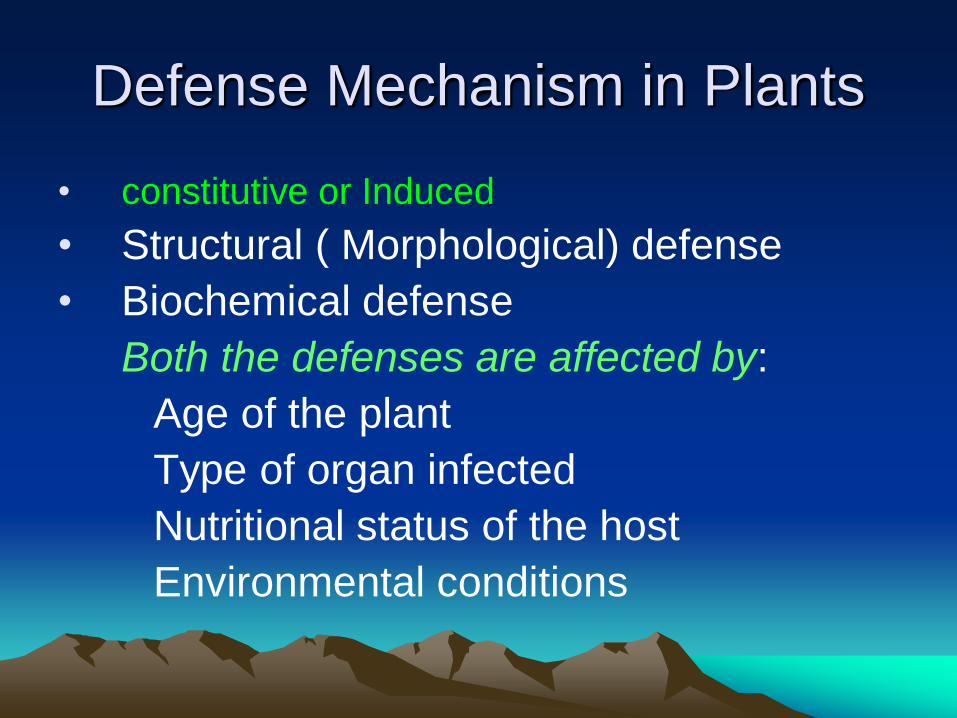
Defense Mechanism in Plants
• constitutive or Induced
• Structural ( Morphological) defense
• Biochemical defense
Both the defenses are affected by:
Age of the plant
Type of organ infected
Nutritional status of the host
Environmental conditions
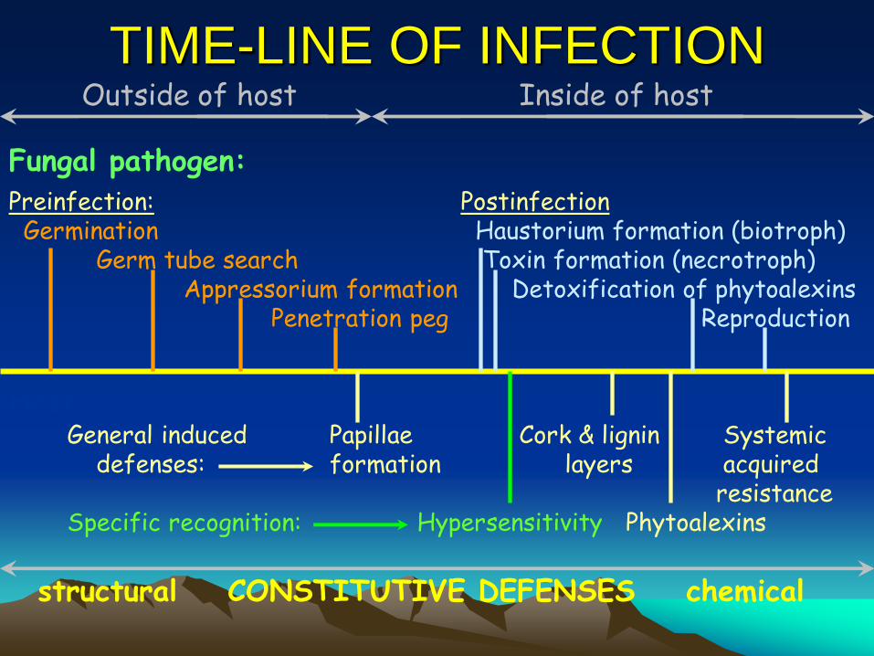
TIME-LINE OF INFECTION
Fungal pathogen:
Host:
Preinfection: Germination Germ tube search Appressorium formation Penetration peg
Postinfection Haustorium formation (biotroph) Toxin formation (necrotroph) Detoxification of phytoalexins Reproduction
General induced Papillae Cork & lignin Systemic defenses: formation layers acquired resistance Specific recognition: Hypersensitivity Phytoalexins
structural CONSTITUTIVE DEFENSES chemical
Outside of host Inside of host
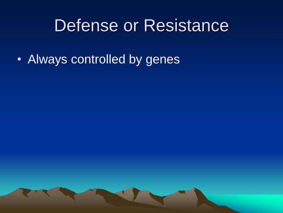
Defense or Resistance
• Always controlled by genes
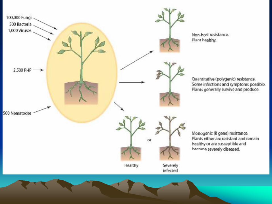
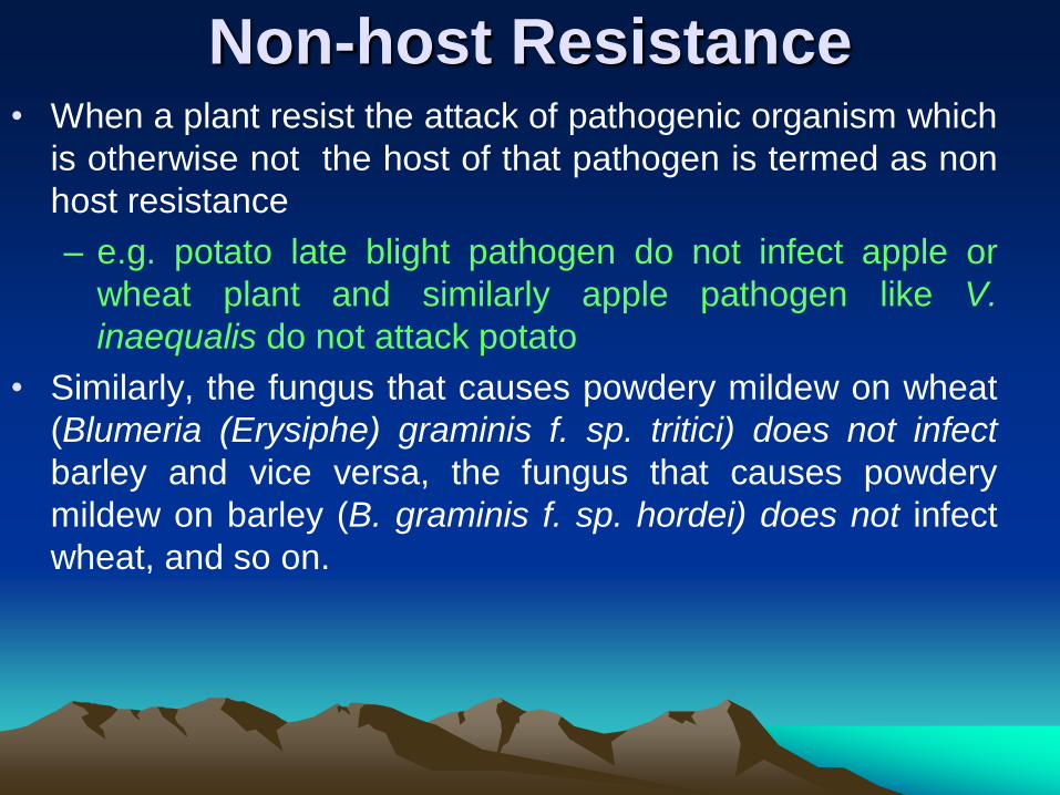
Non-host Resistance • When a plant resist the attack of pathogenic organism which
is otherwise not the host of that pathogen is termed as non
host resistance
– e.g. potato late blight pathogen do not infect apple or
wheat plant and similarly apple pathogen like V.
inaequalis do not attack potato
• Similarly, the fungus that causes powdery mildew on wheat
(Blumeria (Erysiphe) graminis f. sp. tritici) does not infect
barley and vice versa, the fungus that causes powdery
mildew on barley (B. graminis f. sp. hordei) does not infect
wheat, and so on.
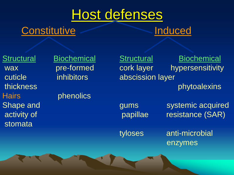
Host defenses Constitutive Induced
Structural Biochemical Structural Biochemical
wax pre-formed cork layer hypersensitivity
cuticle inhibitors abscission layer
thickness phytoalexins
Hairs phenolics
Shape and gums systemic acquired
activity of papillae resistance (SAR)
stomata
tyloses anti-microbial
enzymes
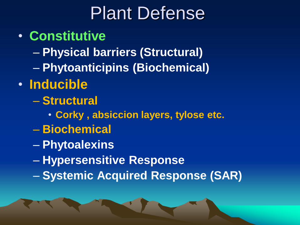
Plant Defense • Constitutive
– Physical barriers (Structural)
– Phytoanticipins (Biochemical)
• Inducible
– Structural • Corky , absiccion layers, tylose etc.
– Biochemical
– Phytoalexins
– Hypersensitive Response
– Systemic Acquired Response (SAR)
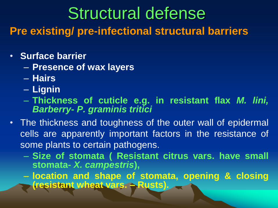
Structural defense Pre existing/ pre-infectional structural barriers
• Surface barrier
– Presence of wax layers
– Hairs
– Lignin
– Thickness of cuticle e.g. in resistant flax M. lini, Barberry- P. graminis tritici
• The thickness and toughness of the outer wall of epidermal
cells are apparently important factors in the resistance of
some plants to certain pathogens.
– Size of stomata ( Resistant citrus vars. have small stomata- X. campestris),
– location and shape of stomata, opening & closing (resistant wheat vars. – Rusts).
Post infectional defense
structures
• Histological defense
• Tylose formation e.g. wilts
• Corky layers e.g. in potato- R. solani
– Cellular defense – altered walls of cells
(changes in cell wall- thickening,
deposition of callose papillae)
– Cytoplasmic defense
– Hypersensitive responseR
Hypersensitive response
• Result in sudden death of the host cells in
the vicinity of the pathogen
– Highest degree of resistance
• Both structural & biochemical in nature
• Common in obligate pathogens like funji,
viruses and nematodes, also found in other
fungal & bacterial infection
• Due to HR
– The necrotic tissues isolate the obligate pathogen
from living cells.
– Devoid the pathogen of nutrition, thus starved
and die.
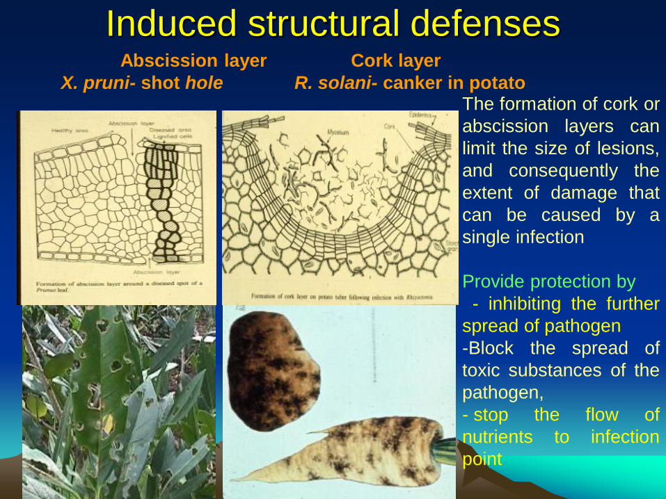
Induced structural defenses Abscission layer Cork layer
X. pruni- shot hole R. solani- canker in potato The formation of cork or
abscission layers can
limit the size of lesions,
and consequently the
extent of damage that
can be caused by a
single infection
Provide protection by
- inhibiting the further
spread of pathogen
-Block the spread of
toxic substances of the
pathogen,
- stop the flow of
nutrients to infection
point
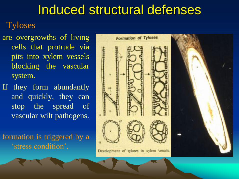
Induced structural defenses Tyloses
are overgrowths of living
cells that protrude via
pits into xylem vessels
blocking the vascular
system.
If they form abundantly
and quickly, they can
stop the spread of
vascular wilt pathogens.
Their
formation is triggered by a
„stress condition‟.
Biochemical defense
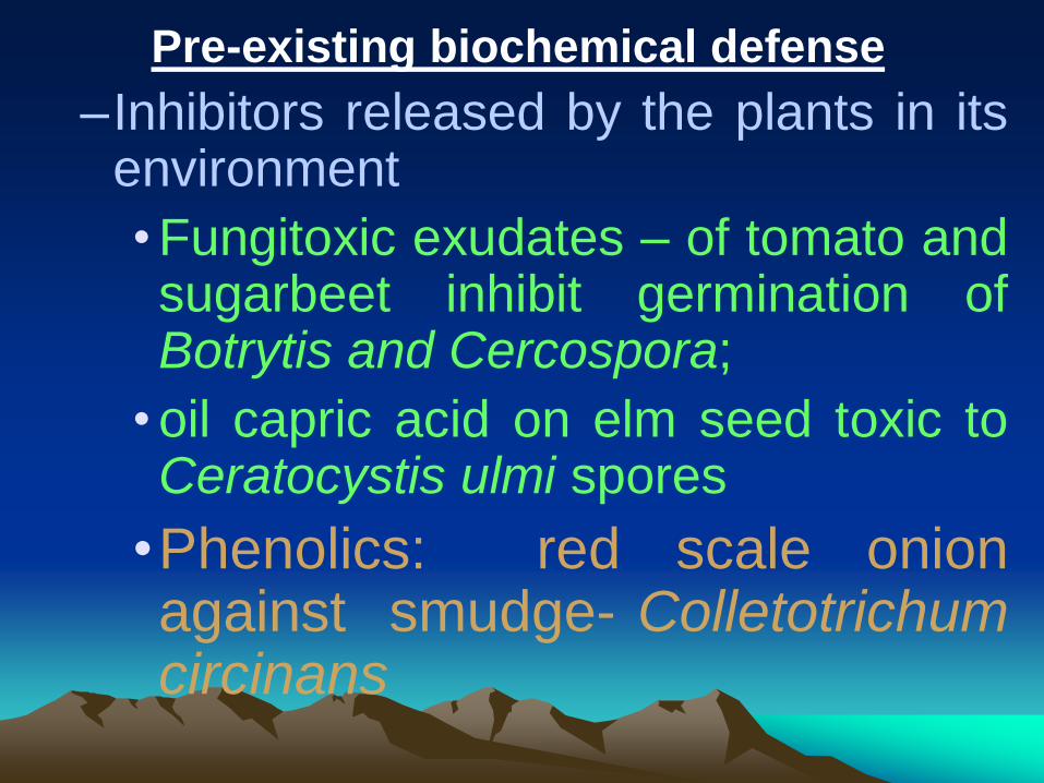
Pre-existing biochemical defense
–Inhibitors released by the plants in its environment
•Fungitoxic exudates – of tomato and sugarbeet inhibit germination of Botrytis and Cercospora;
•oil capric acid on elm seed toxic to Ceratocystis ulmi spores
•Phenolics: red scale onion against smudge- Colletotrichum circinans
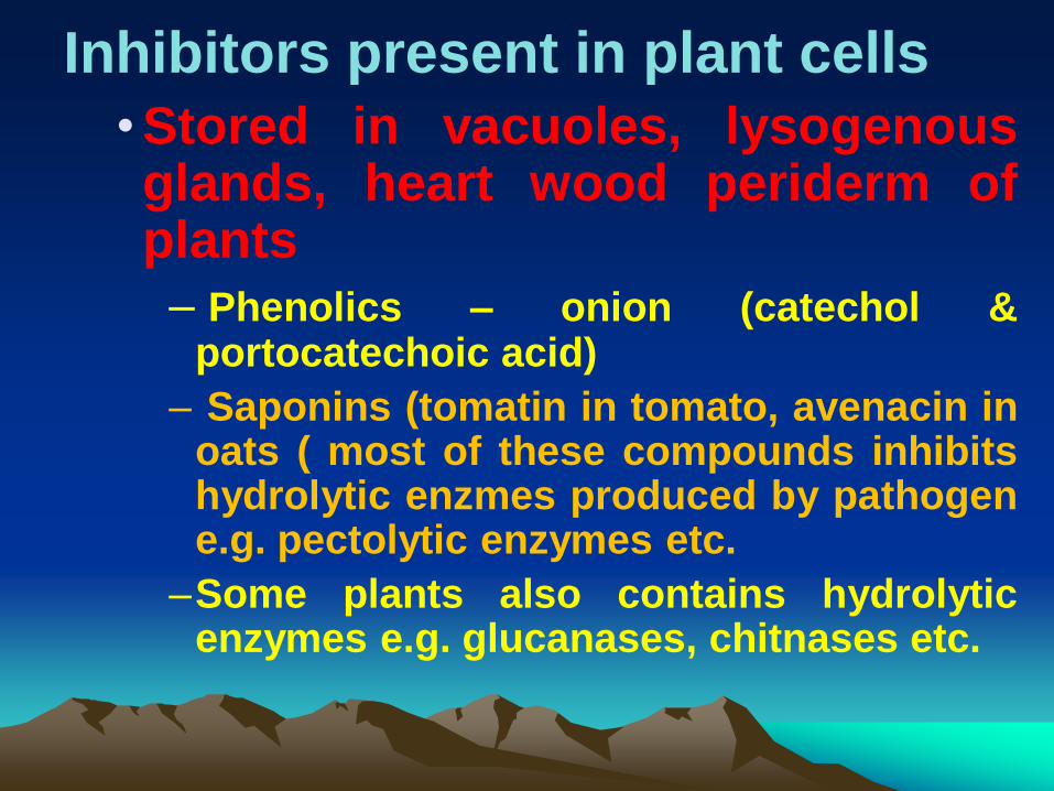
Inhibitors present in plant cells
•Stored in vacuoles, lysogenous glands, heart wood periderm of plants
– Phenolics – onion (catechol & portocatechoic acid)
– Saponins (tomatin in tomato, avenacin in oats ( most of these compounds inhibits hydrolytic enzmes produced by pathogen e.g. pectolytic enzymes etc.
–Some plants also contains hydrolytic enzymes e.g. glucanases, chitnases etc.

Constitutive biochemical
defense Onion smudge
In contrast to red and yellow
onions, white onions do not
contain significant quantities
of certain phenolic chemicals
(one is catechol). If present,
these phenolics confer
resistance to onion smudge
disease.
susceptible
resistant
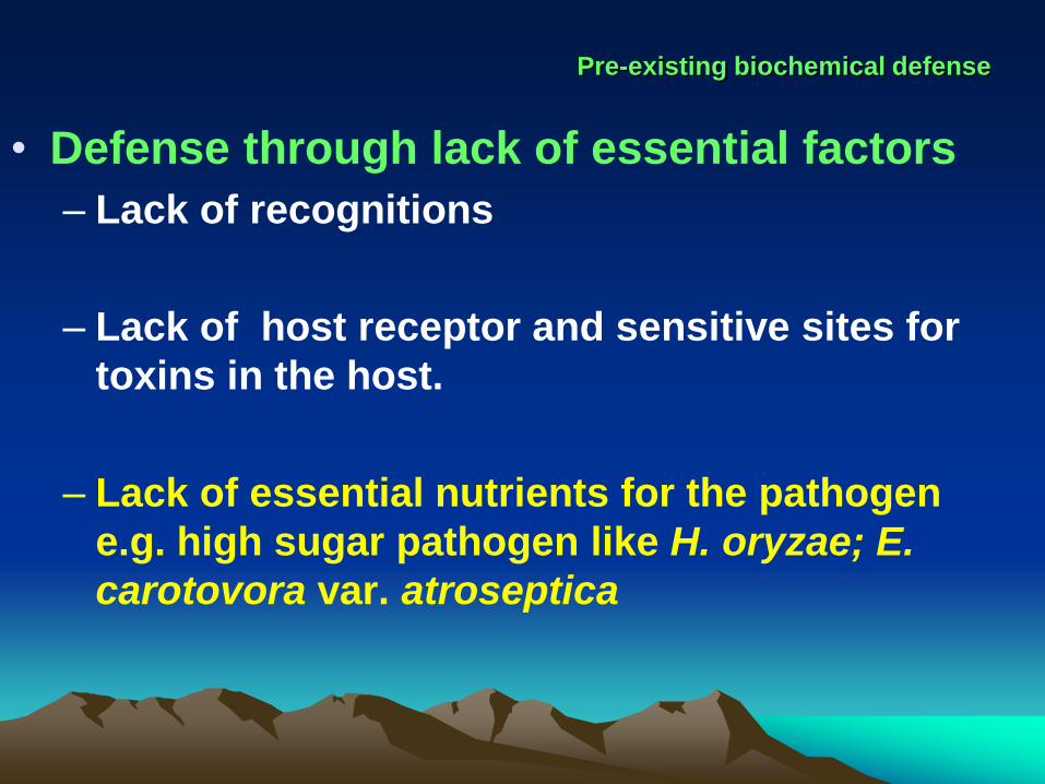
Pre-existing biochemical defense
• Defense through lack of essential factors
– Lack of recognitions
– Lack of host receptor and sensitive sites for
toxins in the host.
– Lack of essential nutrients for the pathogen
e.g. high sugar pathogen like H. oryzae; E.
carotovora var. atroseptica
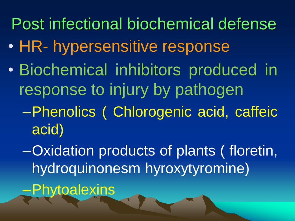
Post infectional biochemical defense
• HR- hypersensitive response
• Biochemical inhibitors produced in
response to injury by pathogen
–Phenolics ( Chlorogenic acid, caffeic
acid)
–Oxidation products of plants ( floretin,
hydroquinonesm hyroxytyromine)
–Phytoalexins

Induced biochemical defenses
hypersensitive response The hypersensitive response (HR) is a localized
death of host cells at the site of infection. It is the result of a specific recognition of a pathogen attack by the host.
The HR is considered to be a type of programmed
cell death.
The HR act by:
-Isolation of the pathogen
-Stop flow of nutrients- thus
starvation
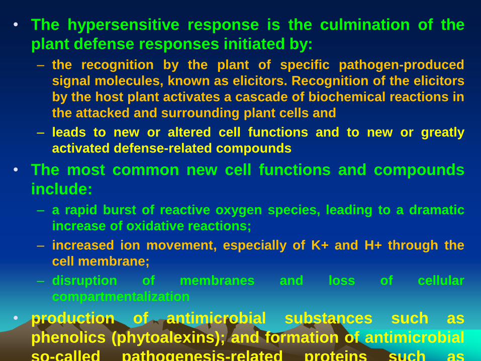
• The hypersensitive response is the culmination of the
plant defense responses initiated by:
– the recognition by the plant of specific pathogen-produced
signal molecules, known as elicitors. Recognition of the elicitors
by the host plant activates a cascade of biochemical reactions in
the attacked and surrounding plant cells and
– leads to new or altered cell functions and to new or greatly
activated defense-related compounds
• The most common new cell functions and compounds
include:
– a rapid burst of reactive oxygen species, leading to a dramatic
increase of oxidative reactions;
– increased ion movement, especially of K+ and H+ through the
cell membrane;
– disruption of membranes and loss of cellular
compartmentalization
• production of antimicrobial substances such as
phenolics (phytoalexins); and formation of antimicrobial
so-called pathogenesis-related proteins such as
chitinases.
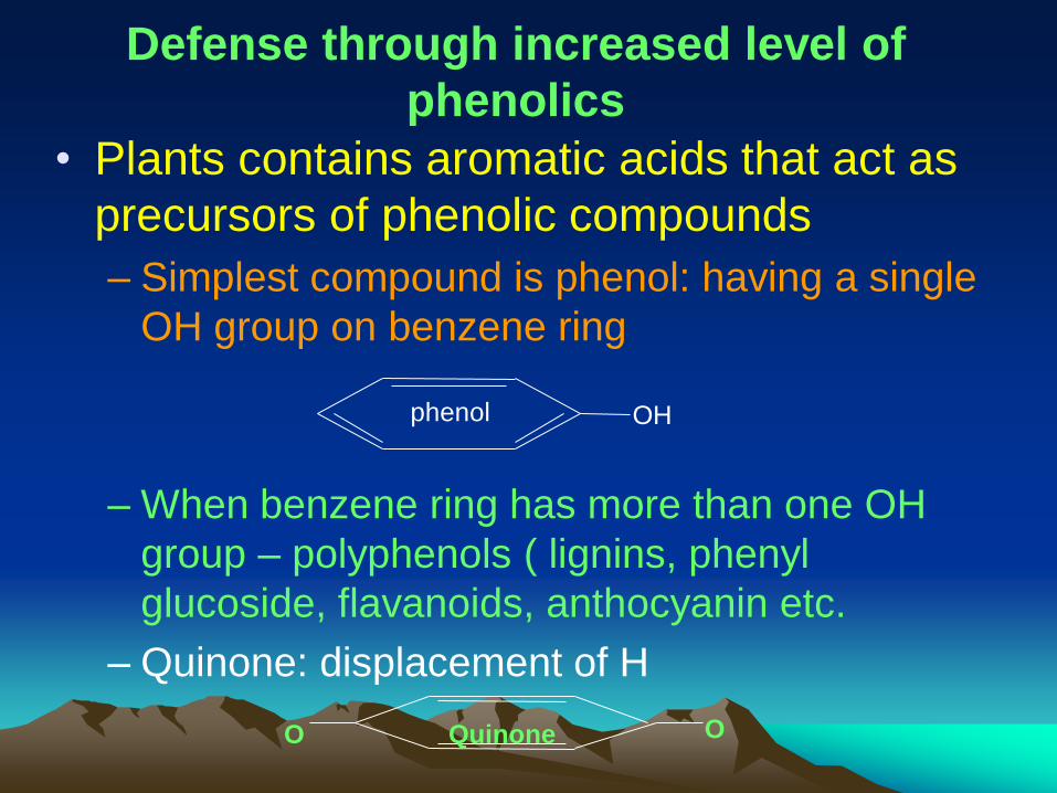
Defense through increased level of
phenolics
• Plants contains aromatic acids that act as
precursors of phenolic compounds
– Simplest compound is phenol: having a single
OH group on benzene ring
– When benzene ring has more than one OH
group – polyphenols ( lignins, phenyl
glucoside, flavanoids, anthocyanin etc.
– Quinone: displacement of H
phenol OH
O O Quinone
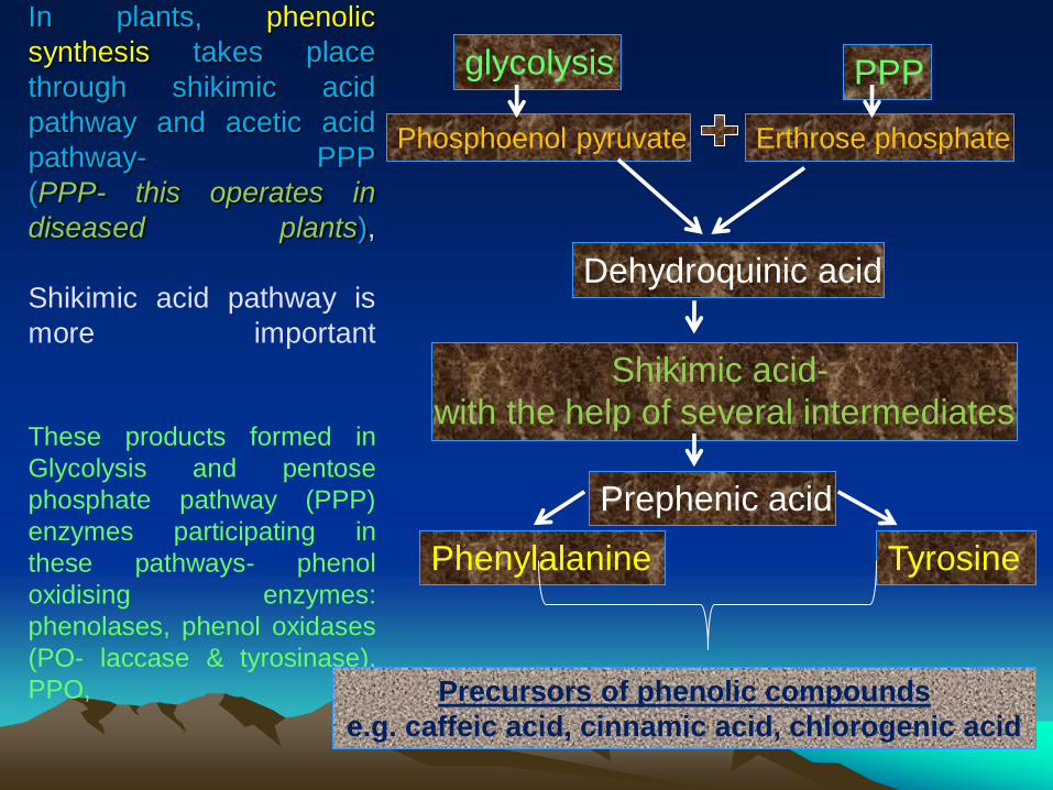
In plants, phenolic
synthesis takes place
through shikimic acid
pathway and acetic acid
pathway- PPP
(PPP- this operates in
diseased plants),
Shikimic acid pathway is
more important
These products formed in
Glycolysis and pentose
phosphate pathway (PPP)
enzymes participating in
these pathways- phenol
oxidising enzymes:
phenolases, phenol oxidases
(PO- laccase & tyrosinase),
PPO, Precursors of phenolic compounds
e.g. caffeic acid, cinnamic acid, chlorogenic acid
glycolysis PPP
Phosphoenol pyruvate Erthrose phosphate
Dehydroquinic acid
Shikimic acid-
with the help of several intermediates
Prephenic acid
Phenylalanine Tyrosine
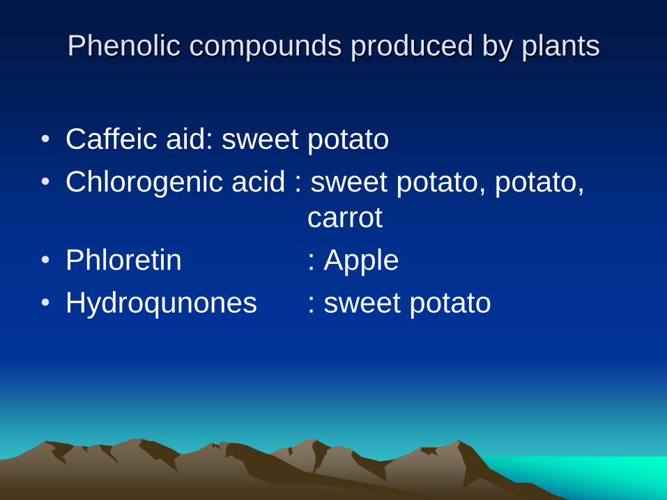
Phenolic compounds produced by plants
• Caffeic aid: sweet potato
• Chlorogenic acid : sweet potato, potato,
carrot
• Phloretin : Apple
• Hydroqunones : sweet potato
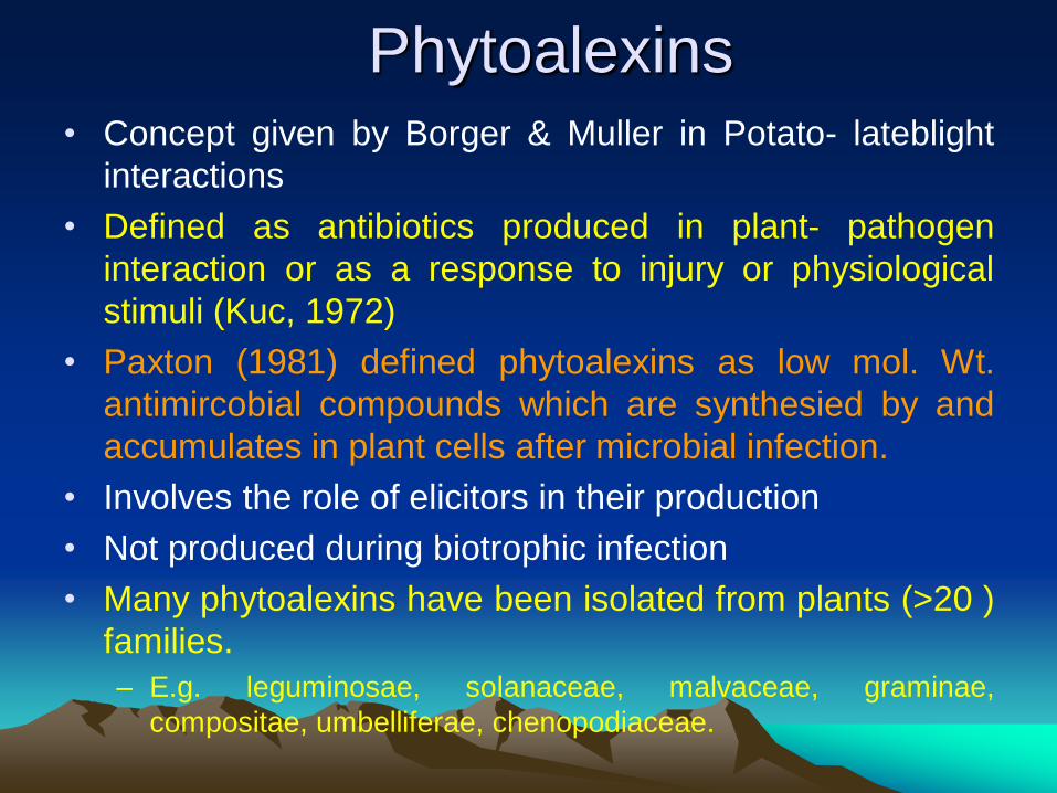
Phytoalexins • Concept given by Borger & Muller in Potato- lateblight
interactions
• Defined as antibiotics produced in plant- pathogen
interaction or as a response to injury or physiological
stimuli (Kuc, 1972)
• Paxton (1981) defined phytoalexins as low mol. Wt.
antimircobial compounds which are synthesied by and
accumulates in plant cells after microbial infection.
• Involves the role of elicitors in their production
• Not produced during biotrophic infection
• Many phytoalexins have been isolated from plants (>20 )
families.
– E.g. leguminosae, solanaceae, malvaceae, graminae,
compositae, umbelliferae, chenopodiaceae.
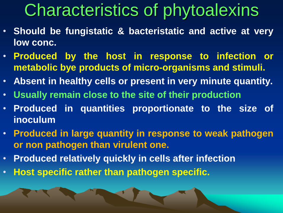
Characteristics of phytoalexins • Should be fungistatic & bacteristatic and active at very
low conc.
• Produced by the host in response to infection or
metabolic bye products of micro-organisms and stimuli.
• Absent in healthy cells or present in very minute quantity.
• Usually remain close to the site of their production
• Produced in quantities proportionate to the size of
inoculum
• Produced in large quantity in response to weak pathogen
or non pathogen than virulent one.
• Produced relatively quickly in cells after infection
• Host specific rather than pathogen specific.
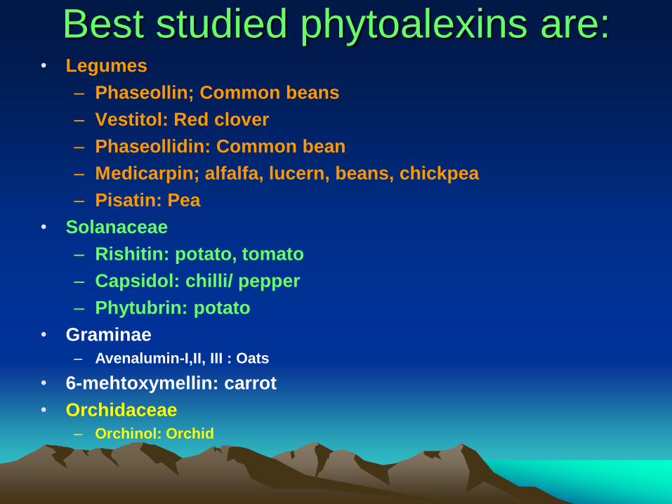
Best studied phytoalexins are: • Legumes
– Phaseollin; Common beans
– Vestitol: Red clover
– Phaseollidin: Common bean
– Medicarpin; alfalfa, lucern, beans, chickpea
– Pisatin: Pea
• Solanaceae
– Rishitin: potato, tomato
– Capsidol: chilli/ pepper
– Phytubrin: potato
• Graminae
– Avenalumin-I,II, III : Oats
• 6-mehtoxymellin: carrot
• Orchidaceae
– Orchinol: Orchid
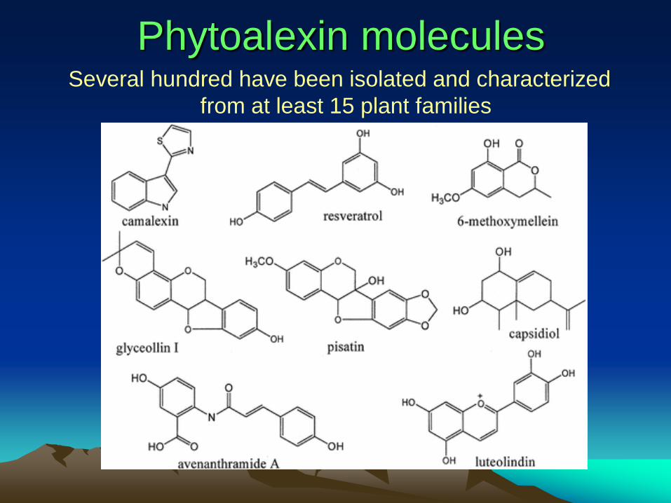
Phytoalexin molecules Several hundred have been isolated and characterized
from at least 15 plant families
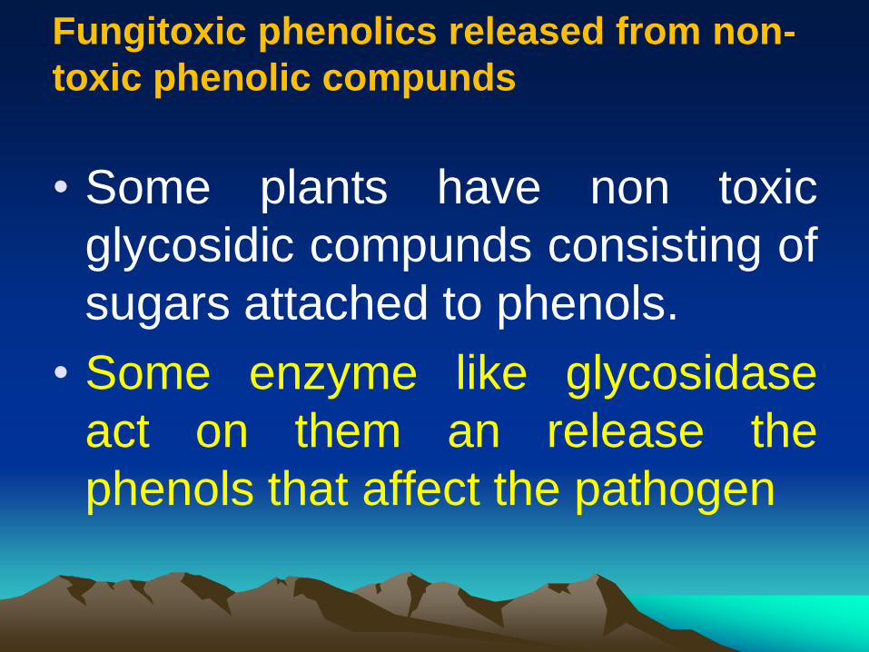
Fungitoxic phenolics released from non-
toxic phenolic compunds
• Some plants have non toxic
glycosidic compunds consisting of
sugars attached to phenols.
• Some enzyme like glycosidase
act on them an release the
phenols that affect the pathogen
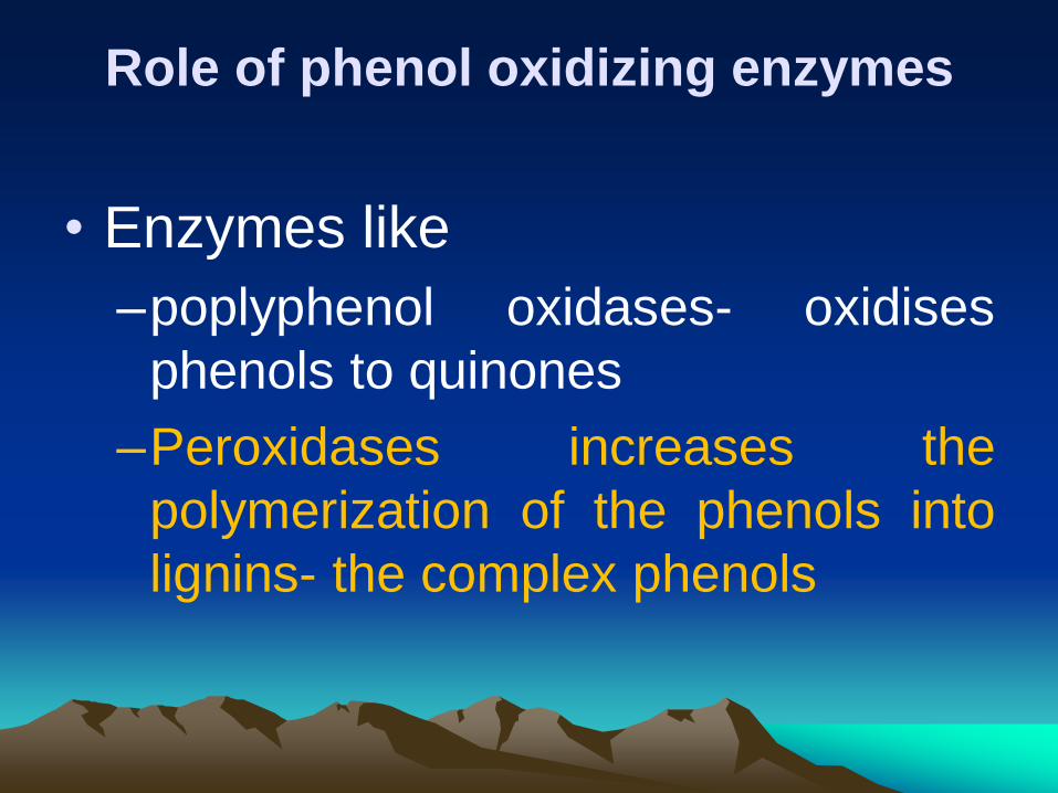
Role of phenol oxidizing enzymes
• Enzymes like
–poplyphenol oxidases- oxidises
phenols to quinones
–Peroxidases increases the
polymerization of the phenols into
lignins- the complex phenols
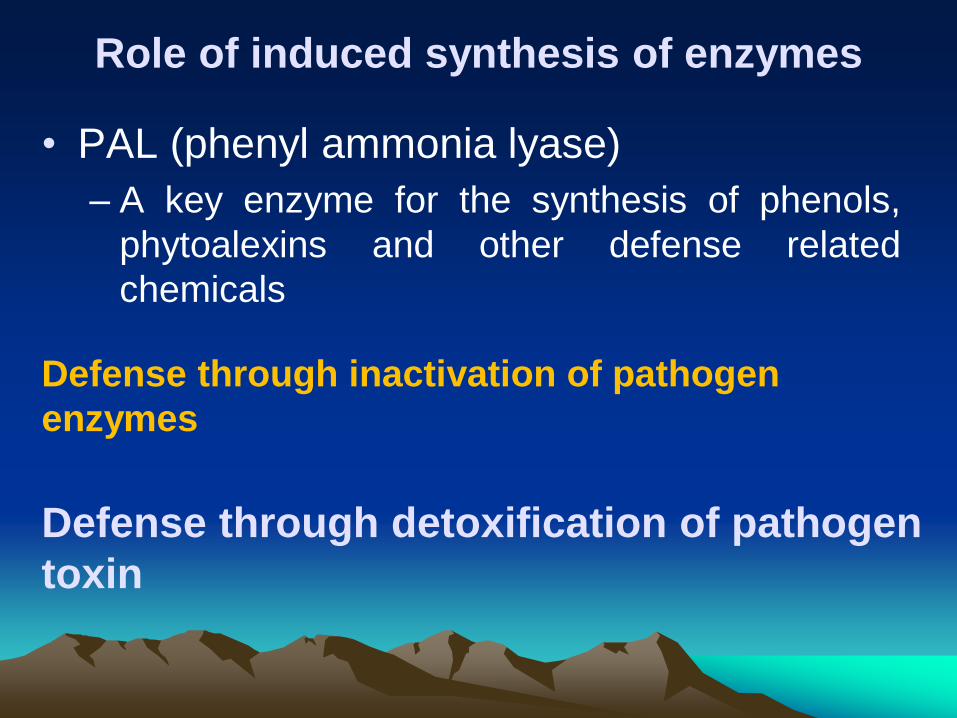
Role of induced synthesis of enzymes
• PAL (phenyl ammonia lyase)
– A key enzyme for the synthesis of phenols,
phytoalexins and other defense related
chemicals
Defense through inactivation of pathogen
enzymes
Defense through detoxification of pathogen
toxin
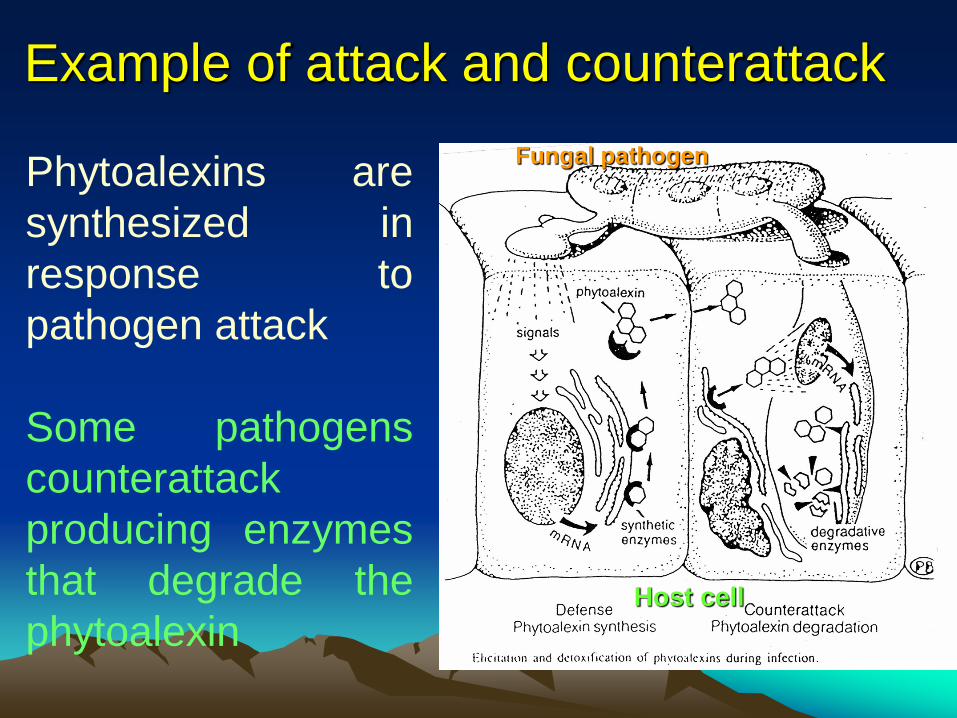
Example of attack and counterattack
Phytoalexins are
synthesized in
response to
pathogen attack
Some pathogens
counterattack
producing enzymes
that degrade the
phytoalexin
Fungal pathogen
Host cell
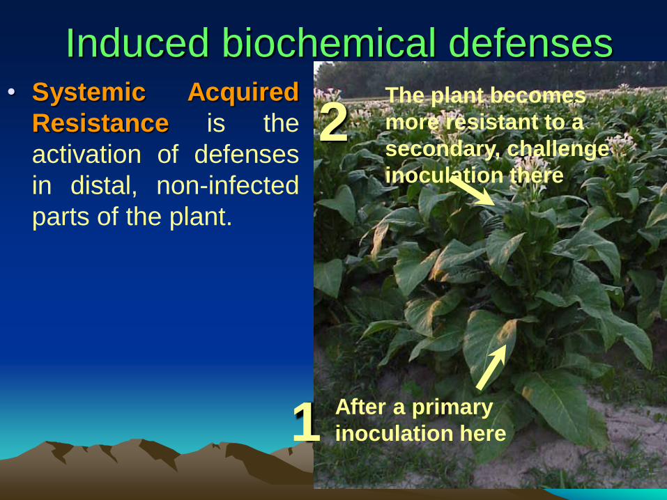
Induced biochemical defenses • Systemic Acquired
Resistance is the
activation of defenses
in distal, non-infected
parts of the plant.
After a primary
inoculation here
The plant becomes
more resistant to a
secondary, challenge
inoculation there
1
2
Systemic Acquired Resistance • SAR confers broad-based resistance to
different pathogens. For example, primary inoculation with a fungal leaf spot pathogen reduces susceptibility of the host plant to other fungi as well as to bacterial and viral pathogens
• Salicylic acid (chemical related to aspirin) is part of signaling pathway involved in transmission of the defense response throughout the plant to produce SAR. This has lead to the development of synthetic chemicals that mimic the role of salicylic acid
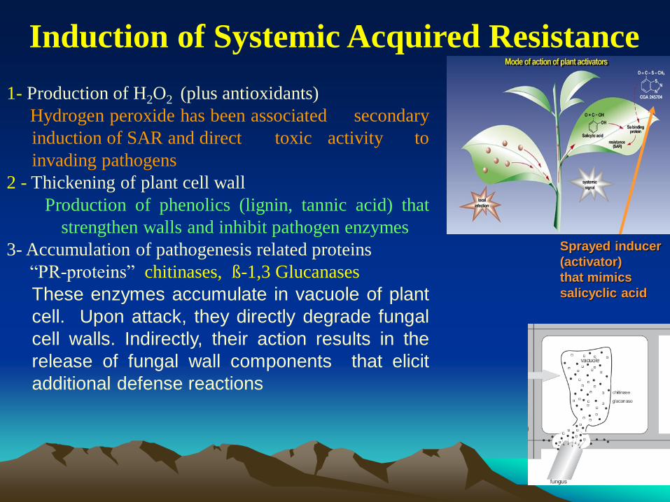
Induction of Systemic Acquired Resistance
1- Production of H2O2 (plus antioxidants)
Hydrogen peroxide has been associated secondary
induction of SAR and direct toxic activity to
invading pathogens
2 - Thickening of plant cell wall
Production of phenolics (lignin, tannic acid) that
strengthen walls and inhibit pathogen enzymes
3- Accumulation of pathogenesis related proteins
“PR-proteins” chitinases, ß-1,3 Glucanases
These enzymes accumulate in vacuole of plant
cell. Upon attack, they directly degrade fungal
cell walls. Indirectly, their action results in the
release of fungal wall components that elicit
additional defense reactions
Sprayed inducer
(activator)
that mimics
salicyclic acid
• The signal compounds responsible for
induction of PR proteins include
– salicylic acid,
– ethylene,
– xylanase,
– the polypeptide systemin,
– jasmonic acid and
– others such as poly acrylic acid, 2-
chloroethylphosphonic acid, acetyl- salicylic
acid, benzoic acid, indole-3-acetic acid, 2, 4-
dichlorophenoxycetic acid and b-benzyl
aminopurine (Bozarth and Ford, 1988).
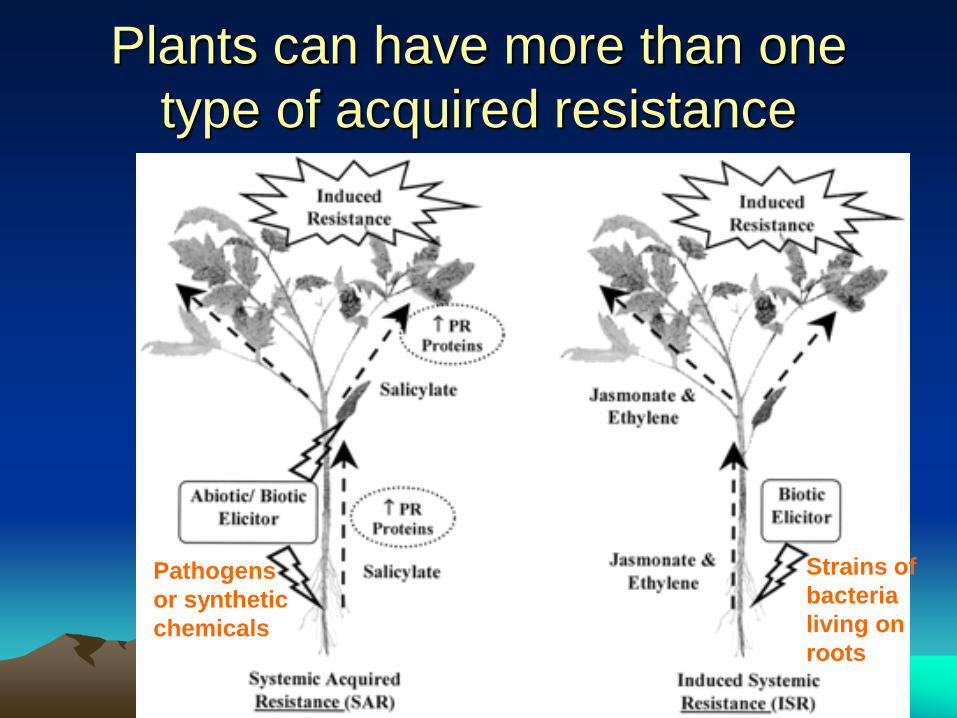
Plants can have more than one
type of acquired resistance
Strains of
bacteria
living on
roots
Pathogens
or synthetic
chemicals
Pathogenesis-Related Proteins
(PR-proteins) • Pathogenesis related proteins, called
PR-proteins- A group of plant coded proteins
• Are structurally diverse group toxic to invading pathogens.
• Produced under stress
• Pathogenesis related proteins of Antomiw et al. (1980) are also known as “B-proteins” by Gianinazzi and Ahl (1983).
• They are widely distributed in plants in
trace amounts but are produced in high
concentration following pathogen attack
or stress.
• The PR proteins exist in plant cells
intracellularly (acidic in Apoplast & basic
form in vacuoles) and also in the
intercellular spaces.
• Varying types of PR-proteins have been
isolated from several crop plants.
groups of PR-proteins
• The several groups of PR-proteins have
been classified according to their
function, serological relationship,
amino acid sequence, molecular weight
and certain other properties.
• The PR proteins are either extremely
acidic or extremely basic and therefore
are highly soluble and reactive.
• PR-proteins are low molecular weight
(approximately 14000 - 30000)
compounds, extractable at low pH (2-
3),
• rich in aromatic amino acids and
resistant to trypsin and chemo trypsin
action.
• The better known PR proteins are:
– PR 1 proteins, B-1, 3-glucanases, chitinases,
lysozymes,
– PR 4 proteins, thaumatinelike proteins,
osmotinlike proteins, cysteine-rich proteins,
glycine-rich proteins, proteinase inhibitors,
proteinases, chitosanases and peroxidases.
There are often numerous isoforms of each
PR-protein in various host plants.
• Although healthy plants may contain
trace amounts of several PR proteins,
attack by pathogens, treatment with
elicitors, wounding, or stress induces
transcription of a battery of genes that
code for PR-proteins.
• This occurs as a part of a massive switch
in the over all pattern of gene expression
during which normal protein production
nerely ceases.
• The signal compounds responsible for
induction of PR proteins include
– salicylic acid,
– ethylene,
– xylanase,
– the polypeptide systemin,
– jasmonic acid and
– others such as poly acrylic acid, 2-
chloroethylphosphonic acid, acetyl- salicylic
acid, benzoic acid, indole-3-acetic acid, 2, 4-
dichlorophenoxycetic acid and b-benzyl
aminopurine (Bozarth and Ford, 1988).
Significance of PR-proteins • The significance of PR-proteins lies in the
fact that they show strong antifungal and
other antimicrobial activity.
• Some of them inhibit spore release and
germination, whereas others are
associated with strengthening of the host
cell wall and its out growths and papillae.
• Some of the PR-proteins, for example, B-
1, 3-glucanase and chitinase, diffuse
towards and affect (break down) the
chitin-supported structure of the cell walls
of several plant pathogenic fungi, whereas
lysozymes degrade the glucosmine and
muramic acid components of bacterial cell
walls.
• Plants genetically engineered to
express chitinase genes show good
resistance against the soil-borne
pathogen Rhizoctonia solani.
• Signal molecules that induce PR-
proteins synthesis seem to be
transported systemically to other parts
of the plant and to reduce disease
initiation and intensity in those parts for
several days or even weeks.
Acknowledgements
• I gratefully acknowledge the use of some
very important photographs given in text
book “Plant Pathology” by G N Agrios.
• I also acknowledge the scientists who spent
valuable time in generating information on
various aspects of plant pathology and
displayed the same on internet for use by
students, teachers and researchers