Embed Size (px)
Citation preview
Defece ntionle NCLASSIFIED
.iSUFFIELD REPORT
N SlfLE
A METHOD FOR THE QUANTITATION OF TRACE LEVELS OF(iI?:DIMETHYL SULFOXIDE IN URINE BY
HIGH PERFORMANCE LIQUID CHROMATOGRAPHY (U)
OTIC byELECT
AUG 16G199*80bC.L. Chenier and J.R. Hancock
ApP!,ovA PCN 351 SPz u on U
May 1989
DEFENCE RESEARCH ESTABLISHMENT SUFFIELD: -RALSTON :ALBERTA
I IN11.1 I F i Infrm iitted suV~Cmaua 7wcgno por 8 a9 8etrgt' 16 043
UNCLASSIFIED
DEFENCE RESEARCH ESTABLISHMENT SUFFIELD
RALSTON ALBERTA
SUFFIELD REPORT NO. 495
A METHOD FOR THE QUANTITATION OF TRACE LEVELS OF DIMETHYL SULFOXIDE
IN URINE BY HIGH PERFORMANCE LIQUID CHROMATOGRAPHY
by
C.L. Chenier and J.R. Hancock
PCN 351SP
nC ndLpAtSnI ightIEtD
UNCLASSIFIED
UNCLASSIFIED
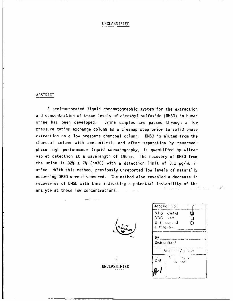
ABSTRACT
A semi-automated liquid chromatographic system for the extraction
and concentration of trace levels of dimethyl sulfoxide (DMSO) in human
urine has been developed. Urine samples are passed through a low
pressure cation-exchange column as a cleanup step prior to solid phase
extraction on a low pressure charcoal column. DMSO is eluted from the
charcoal column with acetonitrile and after separation by reversed-
phase high performance liquid chomatography, is quantified by ultra-
violet detection at a wavelength of 196nm. The recovery of DMSO from
the urine is 82% ± 7% (n=36) with a detection limit of 0.1 pg/mL in
urine. With this method, previously unreported low levels of naturally
occurring DMS were discovered. The method also revealed a decrease in
recoveries of DMSO with time indicating a potential instability of the
analyte at these low concentrations.
Accesor f o0NTIS CRAC %)
DTIC TAB 0
co ,y
Oistribo'i,w
Av~v , " ''.:,'.:
i Dist
UNCLASSIFIED L_____
UNCLASSIFIED
TABLE OF CONTENTSPAGE
ABSTRACT ..................................................... 1i
TABLE OF CONTENTS ............................................ 1ii
KTABLES ...................................... ............... ii
FIGURES...................................................... iv
INTRODUCTION ................................................. 1
EXPERIMENTAL ................................................. 2
Materials .................................................... 2Solvents and Reagents ...................................... 2Instrumentation ......................... .................. 2DMSO Spiked Urine Samples .................................. 3
Procedure .................................................... 3
Ion-Exchange Column........................................... 5Column Packing Procedure ................................... 5DMSO Elution Profile ....................................... 6
Charcoal Column............................................... 6Column Packing Procedure ................................... 6DMSO Elution Profile ....................................... 8
RESULTS AND DISCUSSION........................................ 8
Ion-Exchange Column ..................... ..................... 8
Charcoal Column........................... ................... 14
Sample Preparation Apparatus.................................. 16HPLC Calibration Curves.................................. 16System Recovery and Precision ............................. 17
Dietary DMSO in Urine Blanks.................................. 21
Stability of DMSO in Urine.................................... 21
CONCLUSIONS.................................................. 22
REFERENCES........................... ........................ 23
ii
UNCLASSIFIED
UNCLASSIFIED
TABLES
PAGE
TABLE 1 - Percent of Total DMSO Recovered in EachFraction off the Charcoal Column ....................... 16
TABLE II - System Reproducibility and Recovery .................... 20
iii
UNCLASSIFIED
UNCLASSIFIED
FIGURESPAGE
Figure 1 - Schematic of the Sample PreparationApparatus ............................................. 4
Figure 2 - Custom Fittings:a) Ion Exchange Column ............................... 7b) Charcoal Column ................................... 7
Figure 3 - HPLC chromatogram of fraction #19 from the ionexchange column using a DMSO spiked water sample ......
Figure 4 - A pseudo 3-dimensional plot of the DMSO profilefor a DMSO spiked water sample eluting from theion exchange column ................................... 9
Figure 5 - Comparison of ion-exchange column elutionprofiles for DMSO spiked water samples ofvarying concentrations ................................ 11
Figure 6 - Recovery profiles using the 110 pg/mL DMSO inwater sample in triplicate ........................... 12
Figure 7 - HPLC chromatogram of ion-exchange columnfraction #19 using a DMSO spiked urine sample ........ 13
Figure 8 - A pseudo 3-dimensional plot of the DMSOprofile for a DMSO in urine sample elutingfrom the ion-exchange column ......................... 13
Figure 9 - A pseudo 3-dimensional plot of the DMSOprofile for a DMSO spiked water sample elutingfrom the charcoal column ............................. 15
Figure 10 - Comparison of Area vs Concentration curvesfor DMSO in three different solvents ................. 18
Figure 11 - Influence of solvent composition on theretention time of DMSO ............................... 18
Figure 12 - HPLC chromatogram of a DMSO spiked urine sample ...... 19
Figure 13 - Comparison of six DMSO in urine samples runthrough one ion exchange column ...................... 19
iv
UNCLASSIFIED
UNCLASSIFIED 1
INTRODUCTION
According to western intelligence reports, the Eastern Bloc
countries are well in the lead in research and development of chemical
warfare agents (1-3). Therefore, the NATO countries are interested in
designing, testing, and improving protective equipment (PE), and their
associated standard operating procedures (SOPs) as a defence against
these agents.
One method to evaluate the protection level of the PE and SOPs
would be to use, in a realistic fashion, an intake simulant of a
chemical warfare agent. Should the intake simulant enter the body and
subsequently be detected in the body fluids then this would indicate
faulty equipment and/or procedures.
An ideal intake simulant is a chemical that emulates the agent
in all physical properties, is medically safe for absorption into the
body and can be readily detected in trace amounts in body fluids.
Dimethyl sulfoxide (DMSO) has been proposed as a candidate
intake simulant (4). Its physical properties are similar to those of a
G-agent and it is considered to be a relatively non-toxic compound.
The toxicology, pharmacology, clinical uses, fate and metabolism of
DMSO in humans and animals is well documented (5-11).
Due to the high toxicity of the G-agents, it was necessary to be
able to determine free DMSO concentration in the urine at levels as low
as 1.0 pg/mL. The few methods that have been reported for the
determination of DMSO in biological fluids (12-15), involve time
consuming sample preparation and are only suitable for the determina-
tion of DMSO at relatively high concentrations (>20 pg/mL). They
normally require protein precipitation either by perchloric acid or by
UNCLASSIFIED
UNCLASSIFIED 2
an organic solvent such as methanol or acetone followed by isolation
and quantitation by gas chromatography.
This report describes a methud capable of determining DMSO at
concentrations as low as 0.1 pg/mL in urine. A semi-automated liquid
chromatographic system was developed for the sample cleanup and
concentration, followed by separation by reversed phase high
performance liquid chromatography.
EXPERIMENTAL
Materials
Solvents and Reagents
Distilled water was used as the mobile phase on the sample
preparation apparatus. For the HPLC, the distilled water was further
purified daily using a commercially available photo- oxidizer
(Barnstead) and then filtered through a 0.45 pM filter (Millipore).
HPLC grade acetonitrile (BOH Chemicals, Omnisolve) was used for both
the HPLC mobile phase and the extraction solvent on the sample prepara-
tion apparatus. Gold label DMSO (Aldrich Chemical Company) was used
for the preparation of standards and spiked urine samples.
Instrumentation
A Hewlett-Packard HP1090 liquid chromatograph equipped with
autosampler module and a Kratos 783 UV-VIS variable wavelength detector
was used for DMSO analysis. The data was acquired and processed using
a Nelson Analytical Inc. Model 6000 laboratory data system.
UNCLASSIFIED
UNCLASSIFIED 3
DMSO Spiked Urine Samples
Urine was collected from an individual over a period of four
hours in order to prepare DMSO spiked urine samples. Three concentra-
tions of OMSO in urine (1.068, 1.091, and 10.91 pg/mL) were prepared by
standard addition of DMSO in water to 200 mL of urine.
The DMSO spiked urine samples were stored at room temperature in
polypropylene containers in a fume hood. The urine tended to become
basic with time. When necessary, HCl was added to the sample to make
it acidic and thereby compatible with the ion-exchange resin. Basic
samples tended to interact with the resin and form air bubbles in the
column which disrupted the flow and made it impossible to collect the
DMSO fraction.
Procedure
Figure 1 is a schematic of the sample preparation apparatus used
for the cleanup and enrichment of urine samples.
An electronic, microprocessor-based timer (Control Model CD-4)
was used to control the pumps P1 and P2 and valve V2 for reproducible
and accurate heart-cutting of the DMSO from the ion-exchange column.
All other valve rotations and fraction collection were performed
manually.
The sample was introduced into the 4.2 mL sample loop of the low
pressure sampling valve V1 (Rheodyne Model 5042 teflon rotary valve)
using a 5 mL disposable plastic syringe. The valve was placed in the
inject position and the timer started. This activated pump P1 (Eldex
Model B-100-S) which drove the sample through the ion-exchange column
UNCLASSIFIED

UNCLASSIFIED 4
z0'A-
z w -
z E
00
zj
L) 0.Jl w
0 C-0
ww0,
E0inw
CL,
Ml
M-
z4
04
UNCLASSIFIED
UNCLASSIFIED 5
at 4.25 mL/min using a 100% distilled water mobile phase. The exit of
the column was attached to the 10 mL sample loop of the low pressure
sampling valve V2 (Rheodyne Model 5042P) which was controlled by the
timer. As soon as the DMSO fraction was in the 10 mL sample loop this
valve was switched and pump P2 started. This introduced the OMSO
fraction onto the charcoal column at a rate of -2.0 mL/min, using a
100% distilled water mobile phase. As soon as the DMSO fraction was
completely loaded on the charcoal column, pump P2 stopped. The flow
setting was reduced to -i.5 mL/min, the mobile phase changed to 100%
acetonitrile and the pump restarted manually. A 2 mL glass pipette was
used to collect the first 0.5 mL of the acetonitrile front eluting from
the charcoal column. This aliquot was then drawn into a 1 mL glass
syringe, and filtered (Millipore SJHV 0.45 pm) into a 2 mL autosampler
vial.
Thirty six (36) aliquots from a single DMSO spiked sample (1.068
pg/mL) were used to determine the reproducibility and percent recovery
of the total sample preparation method. The ion exchange column was
replaced after every sixth sample while the charcoal column was
replaced after every sample.
In order to determine the proper fraction to heart-cut and
collect for HPLC analysis, the DMSO profiles and recovery were studied
on both the ion-exchange and charcoal columns.
Ion-Exchange Column
Column Packing Procedure
Ten milliliter disposable serological pipettes (Kimble), with
cotton plug were used as the columns and were packed with cation
UNCLASSIFIED
UNCLASSIFIED 6
exchange resin, AG 50W-X8, 200-400 mesh, hydrogen form (Bio-Rad Analy-
tical Grade).
The pipette cotton plug was packed tightly into the tip using a
1/8 inch steel rod. The bulb end of the pipette was placed into an ion
exchange res'i slurry. Vacuum was applied until the pipette was com-
pletely filled with resin. The filled pipette was carefully placed on
the clean up apparatus so that it was always full of resin and that no
air entered the resin bed. A customized Swagelok fitting was used to
connect the glass pipette column to the teflon tubing of the Rheodyne
valve (Figure 2a). Sealing was accomplished by compression of the
rubber O-ring around the glass tubing, and this method worked well
under the pressures generated by the system.
DMSO Elution Profile
The DMSO elution profile for the ion-exchange column was
determined by collecting 1.0 mL fractions from the outlet of the column
using a manual fraction collector (a teflon block with holes drilled
accurately to 1.0 ± .03 mL) and analyzing the fractions by HPLC.
Five DMSO spiked water samples of varying concentrations (1.05
to 110.0 pg/mL) were used to test the effects of concentration on the
elution profile and recovery efficiency. The effect of sample matrix
on the profile and recovery was also determined using a DMSO spiked
urine sample of moderately high concentration (10.92 pg/mL).
Charcoal Column
Column Packing Procedure
One milliliter disposable serological borosilicate glass
UNCLASSIFIED

UNCLASSIFIED 7
FLOW
118" O.D. TUBE TO1/8" NPT FITTING
RUBBER 0-RING
ICOLLET
318" O.0. TUBE TO1/8" NPT FITTING
10 ml. DISPOSABLEPIPETTE
(b)1 mL DISPOSABLE
PIPETTE
COL LET
RUBBER O-RING 1/4"O.D. TUBE TO
1/8" O.D. TUBE FITTING
BORED THRU
1/8" TFE TUBING 114 x 28 1/8" TUBEI TFE FITTING
FLOW
Figure 2
CUSTC, V Fr rINGS: a) ION-EXCHANGE COLUMN CONNECTOR,b) CHARCOAL COLUMN CONNECTOR
UNCLASSIFIED
UNCLASSIFIED 8
pipettes (Kimble) were used as columns and were packed with activated
carbon, 50-200 mesh (Fisher Scientific).
The cotton plug in the pipette was packed tightly into the tip
using a 1/8 inch steel rod. The pipette was completely filled with
carbon and packed using a laboratory vibrator (Mettler). The carbon
was held in place with a second cotton plug.
A customized Swagelok fitting was used to join this glass
pipette column to the teflon tubing of the Rheodyne valve (Figure 2b).
This fitting sealed well under the pressures generated by the system.
DMSO Elution Profile
The elution profile for the charcoal column was determined using
DMSO spiked water samples. Four OMSO spiked water samples of varying
concentrations (0.53 to 35.4 pg/mL) were used to test the effects of
concentration on the elution profile and recovery efficiency. The
sample was manually loaded into the sample loop of the charcoal column
and injected under the normal sample preparation conditions. As soon
as the acetonitrile elution was started, 0.5 mL fractions were
collected from the outlet of the charcoal column using a manual
fraction collector (a teflon block with holes drilled accurately to 0.5
± .03 mL). The fractions were then analysed for DMSO content by HPLC.
RESULTS AND DISCUSSION
Ion-Exchange Column
The DMSO elution profile from tha ion-exchange column was
characterized using DMSO spiked water samples. Figure 3 illustrates a
UNCLASSIFIED

UNCLASSIFIED 9
15000
SDMSO
4-
-',1O
30
80000 3.5
Time (min)
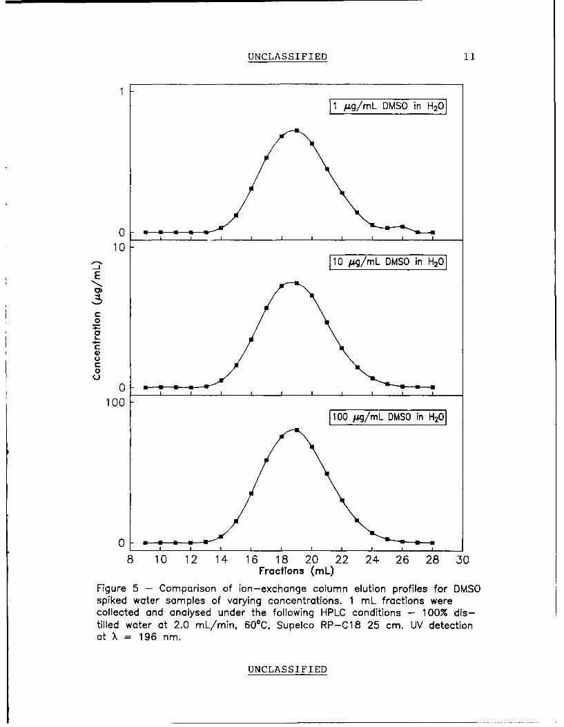
Figure 3 - HPLC chromatogram of fraction #19 from the ion exchangecolumn using a DMSO spiked water sample. HPLC conditions - 100% distilledwater; 2.0 mL/min; 25 cm Supelco RP-C18 column; 60°C; UV detectionat X = 196 nm; 10 usL injection. Retention time for DMSO is 2.37 min.
15000 - FRACTION #19
0
80000 3.5
Ttme (min)
Figure 4 - A pseudo 3-dimensional plot of the DMSO profile for a DMSOspiked water sample eluting from the ion exchange columr. Z-axis isfractions #12 to 26. Retention time of DMSO is 2.37 min.
UNCLASSIFIED
UNCLASSIFIED 10
typical chromatogram of a DMSO containing fraction collected from the
ion-exchange column. In total, 28 fractions of 1 mL each were
collected and Figure 4 shows a pseudo 3-dimensional plot of the chroma-
tograms of fractions 12 to 26. DMSO eluted in fractions 13 to 26 and
the elution profile for this compound was symmetrical and centered on
fraction 19.
As only 10 mL was heart-cut from the ion-exchange column
effluent for introduction onto the charcoal column, it was necessary to
test for any changes in the elution profile due to DMSO concentration.
Figure 5 compares the elution profiles of 3 water samples of differing
DMSO concentration. In each sample the DMSO eluted in fractions 13 to
26, and it was concluded that the elution profile was not DMSO
concentration dependent. Integration of the area under these curves
enabled recovery efficiency to be calculated. From the elution
profiles of all five available water samples the average DMSO recovery
was calculated to be 94% with a relative standard deviation of 3%.
Therefore the recovery was also independent of the concentration. The
most concentrated sample (110.0 pg DMSO/mL H20) was run in triplicate
to determine the reproducibility of the method on the ion-exchange
column and gave a relative standard deviation for the area of 1.2%
(Figure 6).
In order to determine if the elution profile would be altered by
the complexity of the urine matrix, a DMSO spiked urine sample was
prepared and an elution profile was obtained using the same procedure
as for the DMSO spiked water samples. Figure 7 illustrates a HPLC
chromatogram of one DMSO containing fraction (#19) acquired from the
ion-exchange column using a DMSO spiked urine sample. Many other UV
adsorbing constituents were observed but DMSO was well separated from
these potential interferences. A pseudo 3-dimensional profile is shown
UNCLASSIFIED
UNCLASSIFIED 1
1 usg/mL DMSO in H201
102 1 ~c/mnl DMSO in H-20
E
C
0
08 0 1 4 1 8 20 2 4 2 8 3
Frcins-L
Fiue5 C maio finecageclm lto rflsfrD Sspkdwtrsmlsofvrigcnetains Lfatoswrcolce n nlsdudrtefllwn PCcniin 0%dstildwtra . Lmn 0C SploRC1 5c .U eeto0tX=19 m
UNLSSFE
0. ....... ....

UNCLASSIFIED 12
100
0
v
8 10 12 14 16 18 20 22 24 26 28 30
Fractions (mL)
Figure 6 -Recovery profiles us ing the 110 A.kg/mL DMVSO in water samplein triplicate. Relative standard deviation for the area under the curve is 1.2%.
UNCLASSIFIED
0J) m m m m m m m

UNCLASSIFIED 13
8500
"-DMSO
0
49000 6
Time (min)
Figure 7 - HPLC chromatogram of ion-exchange column fraction #19using a DMSO spiked urine sample. HPLC conditions - 100% distilledwater; 1.0 mL/min; 6 cm Dupont Golden Series RP-C18 column; 500 C;UV detection at X = 196; 10 /L injection. Retention time for DMSO is4.3 min.
8500 ---- Fraction #I19
4--0
49000 6
Time (min)
Figure 8 - A pseudo 3-dimensional plot of the DMSO profile for a DMSOin urine sample eluting from the ion-exchange column. Z-axis is fractions#12 to 26. Retention time for DMSO is 4.3 min.
UNCLASSIFIED
UNCLASSIFIED 14
in Figure 8 Although more complicated than the DMSO spiked water
profile, the spiked urine profile remains symmetrical with the DMSO
eluting between fractions 13 to 26. This indicates that the DMSO
elution profile from the ion-exchange column was independent of sample
matrix.
Charcoal Column
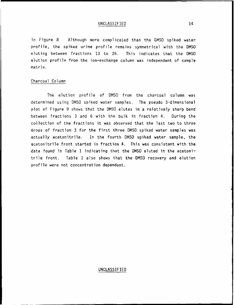
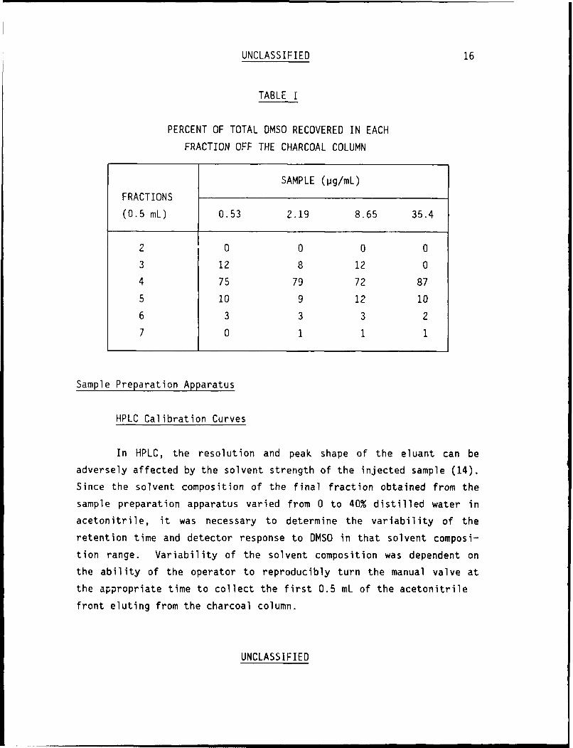
The elution profile of DMSO from the charcoal column was
determined using DMSO spiked water samples. The pseudo 3-dimensional
plot of Figure 9 shows that the DMSO elutes in a relatively sharp band
between fractions 3 and 6 with the bulk in fraction 4. During the
collection of the fractions it was observed that the last two to three
drops of fraction 3 for the first three DMSO spiked water samples was
actually acetonitrile. In the fourth DMSO spiked water sample, the
acetonitrile front started in fraction 4. This was consistent with the
data found in Table I indicating that the DMSO eluted in the acetoni-
trile front. Table I also shows that the DMSO recovery and elution
profile were not concentration dependant.
UNCLASSIFIED

UNCLASSIFIED 15
75000
--C
0
450
40 2
Time (min)
Figure 9 - A pseudo 3-dimensional plot of the DMSO profile for aDMVSO spiked water sample eluting from the charcoal column. Z-axisis fractions #2 to 7. Retention time for DMVSO is 1 .52 min.
UNCLASSIFIED
UNCLASSIFIED 16
TABLE I
PERCENT OF TOTAL DMSO RECOVERED IN EACH
FRACTION OFF THE CHARCOAL COLUMN
SAMPLE (pg/mL)
FRACTIONS
(0.5 mL) 0.53 2.19 8.65 35.4
2 0 0 0 0
3 12 8 12 0
4 75 79 72 87
5 10 9 12 10
6 3 3 3 2
7 0 1 1 1
Sample Preparation Apparatus
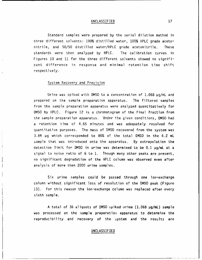
HPLC Calibration Curves
In HPLC, the resolution and peak shape of the eluant can be
adversely affected by the solvent strength of the injected sample (14).
Since the solvent composition of the final fraction obtained from the
sample preparation apparatus varied from 0 to 40% distilled water in
acetonitrile, it was necessary to determine the variability of the
retention time and detector response to DMSO in that solvent composi-
tion range. Variability of the solvent composition was dependent on
the ability of the operator to reproducibly turn the manual valve at
the appropriate time to collect the first 0.5 mL of the acetonitrile
front eluting from the charcoal column.
UNCLASSIFIED
UNCLASSIFIED 17
Standard samples were prepared by the serial dilution method in
three different solvents: 100% distilled water, 100% HPLC grade aceto-
nitrile, and 50/50 distilled water/HPLC grade acetonitrile. These
standards were then analyzed by HPLC. The calibration curves in
Figures 10 and 11 for the three different solvents showed no signifi-
cant difference in response and minimal retention time shift
respectively.
System Recovery and Precision
Urine was spiked with DMSO to a concentration of 1.068 pg/mL and
prepared on the sample preparation apparatus. The filtered samples
from the sample preparation apparatus were analyzed quantitatively for
DMSO by HPLC. Figure 12 is a chromatogram of the final fraction from
the sample preparation apparatus. Under the given conditions, DMSO had
a retention time of 4.65 minutes and was adequately resolved for
quantitative purposes. The mass of DMSO recovered from the system was
3.84 pg which corresponded to 86% of the total DMSO in the 4.2 mL
sample that was introduced onto the apparatus. By extrapolation the
detection limit for DMSO in urine was determined to be 0.1 pg/mL at a
signal to noise ratio of 6 to 1. Though many other peaks are present,
no significant degradation of the HPLC column was observed even after
analysis of more then 2000 urine samples.
Six urine samples could be passed through one ion-exchange
column without significant loss of resolution of the DMSO peak (Figure
13). For this reason the ion-exchange column was replaced after every
sixth sample.
A total of 36 aliquots of DMSO spiked urine (1.068 pg/mL) sample
was processed on the sample preparation apparatus to determine the
reproducibility and recovery of the system and the results are
UNCLASSIFIED

UNCLASSIFIED 18
10-- A-- 100% H20
--- 50/50 H20/CH3CN
---- 100% CH3CN
0
0 5 10Concentration (jug/mL)
Figure 10 - Comparison of Area vs Concentration curves for DMSOin three different solvents.
3.9-- 1-- 100% CH3 CN
• --- 50/50 H20/CH3CN"----
100% H20
ES3.7
E. .... .......... ...............................C p"00
- 3.5
3.30 4 8 12
Concentration (j",g/mL)
Figure 11 - Influence of solvent composition on the retentiontime of DMSO.
UNCLASSIFIED
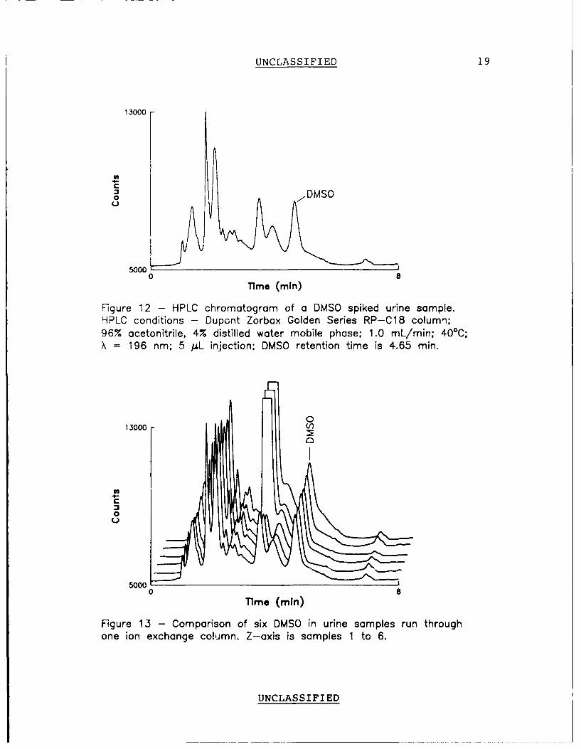
UNCLASSIFIED 19
13000
0,
0 8Time (min)
Figure 12 - HPLC chromatogram of a DMSO spiked urine sample.HPLC conditions - Dupont Zorbax Golden Series RP-C18 colum,1;96% acetonitrile, 4% distilled water mobile phase; 1.0 mL/min; 40°C;X= 196 nm; 5 uL injection; DMSO retention time is 4.65 min.
013000 V
00
5000
0
5000 I
Time (min)
Figure 13 - Comparison of six DMSO in urine samples run throughone ion exchange column. Z-axis is samples 1 to 6.
UNCLASSIFIED

UNCLASSIFIED 20
was processed on the sample preparation apparatus to determine the
reproducibility and recovery of the system and the results are
summarized in Table II.
TABLE II
SYSTEM REPRODUCIBILITY AND RECOVERY
Ion-Exchange Average Concentration Recovery
Column of 6 Replicates
(pg/mL)
1 7.70 ± 2.53% 86%
2 7.84 ± 2.38% 87%
3 7.22 ± 5.11% 80%
4 7.14 ± 3.65% 80%
5 7.55 ± 4.96% 84%
6 6.54 ± 5.03% 73%
mean 82% ± 7%
The recovery was calculated using the following equation:
CA VA
Recovery = C V * 100% [I]u u
Where:
CA= concentration of the processed sample in pg/mL
VA= volume of the processed sample = 0.5 mL
CU= concentration of urine sample = 1.068 pg/mL
VU= volume or urine sample = 4.2 mL
UNCLASSIFIED
UNCLASSIFIED 21
The variation of the apparatus was 7% with a combined precent recovery
of 82% for a 'fresh' DMSO spiked urine sample.
Dietary DMSO in Urine Blanks
During development of the method for DMSO in urine, low levels
of DMSO were detected in the blank urine samplec. This could be
attributed to dietary sources, as a variety of foods contain DMSO, as
do some beverages including milk (15, 16).
Stability of DMSO in Urine
There is some question as to the stability of DMSO in urine.
Considering that the urine is a complex matrix of organic and inorganic
materials (17) it is possible that the concentration of the "free" DMSO
could be reduced over time either by some chemical or biological
degradation or by becoming physically bound to other constituents in
the sample. To test this theory, an "old" DMSO urinary sample (spiked
with DMSO 52 days prior to sample preparation and stored at room
temperature) was prepared and analyzed concurrently with the "fresh"
DMSO urinary samples. This "old" sample yielded in DMSO recovery of
65% as compared to 82% for the "fresh" samples. Th - ;cates some
DMSO loss with time. A decrease in recovery was also observed in
samples (Table II) which were run over a period of only three days (87%
on day 1 to 73% on day 3).
The DMSO spiked urine sample which was used to determine the
elution profile of the ion exchange column produced a 73% recovery of
DMSO. This could also be an indication of DMSO loss as this sample was
prepared on the ion-exchange column 9 days after being spiked with
DMSO.
UNCLASSIFIED

UNCLASSIFIED 22
CONCLUSIONS
A method, based on solid phase extraction and concentration was
developed for the quantitative determination of trace amounts of
dimethyl sulfoxide (DMSO) in urine. The detection limit for DMSO in
urine was 0.1 pg/mL with a recovery of 82% ± 7%.
The discovery of naturally occurring DMSO in urine would make
the determination of exposure to DMSO from external sources inconclu-
sive. One method to alleviate this problem would be to determine the
rate at which the naturally occurring DMSO entered the urine and
assuming that it was constant, any significant increase to this rate
would indicate a DMSO intake from an external source.
The storage and handling of urine samples must be investigated
further as there was soin, indication of DMSO loss with time. This
could prove to be a significant problem in data interpretation from
samples analyzed for trials, especially if the samples were stored for
prolonged periods and/or degraded at different rates.
UNCLASSIFIED
UNCLASSIFIED 23
REFERENCES
1. Ingo Urban, Ostblick, (East View), October 1985, pp 15-19,
ISSN: 0177-0128
2. NBC Defence and Technology International Yearbook, Vol. 2, No.
1, NBC Defence International, Ltd., New York, N.Y., 1987.
3. Jane's Defence Weekly, Vol. 7, No. 4, January 31, 1987.
4. Lockwood, P.A., personnel communication.
5. Leake, C.D., (ed.) "Biological Actions of DMSO", Ann. NY Acad.
Sci., 141, pp 1-671 (1967).
6. Jacob, S.W., Rosenbaum, E.E., Wood, D.C. (ed.), "Dimethyl
Sulfoxide", Vol. 1., Basic Concepts of DMSO, 1971, Marcel
Dekker, Inc., New York.
7. Jacob, S.W. and Herschler, R., (ed.), "Biological Actions of
DMSO", Ann. NY Acad. Sci., 243, pp 1-508 (1975).
8. de la Torre, J.C., (ed.) "Biological Actions and Medical
Applications of Dimethyl Sulfoxide", Ann. NY Acad. Sci., 411, pp
1-402 (1983)
9. McCann, J., Choi, E., Yamasaki, E., and Ames, B.N., "Detection
of Carcinogens as Mutagens in the Salmonella/Microsome Test:
Assay of 300 Chemical Substances", Proc. Natl. Acad. Sci.,
U.S.A., 72, pp 5135 (1975).
UNCLASSIFIED

UNCLASSIFIED 24
10. Lobs, K., Damerau, W., and Schramm, T., "Carcinogenic Action of
Dimethyl Sulfoxide", Arch. Gischwulstforsch., 37, pp 1 (1971).
Chemical Abstract 75:3014 (s) (1971).
11. Survey of Compounds Which Have Been Tested for Carcinogenic
Activity, United States Public Health Service Publication #149,
1972-73 (cited in 10).
12. Garretson, S.E., Aitchison, J.P., "Determination of Dimethyl
Sulfoxide in Serum and Other Body Fluids by Gas Chromatography",
Journal of Analytical Toxicology. Vol. 6, March/April, 1982, pp
76-81.
13. Mehta, A.C., Peaker, S., Acomb, C., Calvert, R.T., "Rapid Gas
Chromatographic Determination of Dimethyl Sulfoxide and its
Metabolite Dimethyl Sulphone in Plasma and Urine", Journal of
Chromatography, Biomedical Applications, 383, 1986, pp 400-404.
14. Snyder, L.R., Kirkland, J.J., Introduction to Modern Liquid
Chromatography, 2nd Edition, 1979.
15. Jacob, S., Herschler, R., Knowles, R., "Proceedings on the
Symposium on Dimethyl Sulfoxide", Vet. Med. Small Anim. Clin.,
77, pp 365-373, March 1982.
16. Williams, K.I., Burnstein, S.H. and Layne, D.S. Proc. Soc. Exp.
Biol. Med., 122: pp 865-866 (1966), Arch. Biochem. Biophys.,
113, pp 251-252 (1966).
17. Hawk's Physiological Chemistry, 14th Edition, pp 1153-1205, 1965
McGraw-Hill Inc.
UNCLASSIFIED
UNCLASSIFIEDSECURITY CLASSIFICATION OF FORM
(highest classification of Title. Abstract, Keywords)
DOCUMENT CONTROL DATA(Security classification of title, body of abstract and indexing annototion must be entered when the overall document is classified)
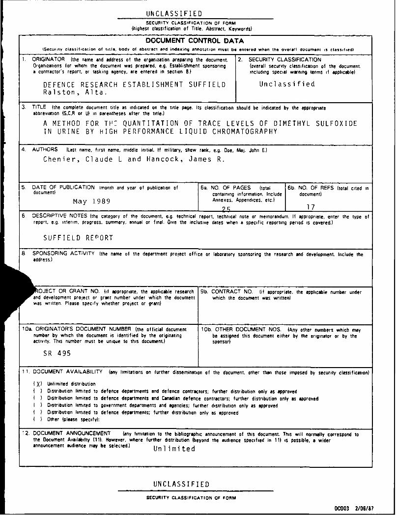
1. ORIGINATOR (the name and address of the organization preparing the document 2. SECURITY CLASSIFICATIONOrganizations for whom the document was prepared, e.g. Establishment sponsoring (overall security classification of the document.a contractor s report, or tasking agency, are entered in section 8.) including special warning terms if applicable)
DEFENCE RESEARCH ESTABLISHMENT SUFFIELD UnclassifiedRalston, Alta.
3. TITLE (the complete document title as indicated on the title page. Its classification should be indicated by the appropriate
abbreviation fS,C,R or U) in parentheses after the title.)
A METHOD FOR TH QUANTITATION OF TRACE LEVELS OF DIMETHYL SULFOXIDEIN URINE BY HIGH PERFORMANCE LIQUID CHROMATOGRAPHY
4. AUTHORS (Last name, first name, middle initial. If military, show rank, e.g. Doe, Maj. John E.)
Chenier, Claude L and Hancock, James R.
5. DATE OF PUBLICATION (month and year of publication of 6a. NO. OF PAGES (total 6b. NO. OF REFS (total cited in
document) containing information. Include document)
May 1989 Annexes, Appendices, etc.)
25 176. DESCRIPTIVE NOTES (the category of the document, e.g. technical report, technical note or memorandum. If appropriate, enter the type of
report, e.g. interim, progress, summary, annual or final. Give the inclusive dates when a specific reporting period is covered.)
SUFFIELD RErPORT
8 SPONSORING ACTIVITY (the name of the department project office or laboratory sponsoring the research and development Include theaddress.)
OJECT OR GRANT NO. (if appropriate, the applicable research 9b. CONTRACT NO. (if appropriate, the applicable number underand development project or grant number under which the document which the document was written)was written. Please specify whether project or grant)
10a. ORIGINATOR'S DOCUMENT NUMBER (the official document 10b. OTHER DOCUMENT NOS. (Any other numbers which maynumber by which the document is identified by the originating be assigned this document either by the originator or by theactivity. This number must be unique to this document.) sponsor)
SR 495
1 1. DOCUMENT AVAILABILITY (any limitations on further dissemination of the document, other than those imposed by security classification)
X) Unlimited distributionDistribution limited to defence departments and defence contractors; further distribution only as approved
I Distribution limited to defence departments and Canadian defence contractors; further distribution only as approvedI Distribution limited to government departments and agencies; further distribution only as approved
I Distribution limited to defence departments; further distribution only as approvedI Other (please specify):
12. DOCUMENT ANNOUNCEMENT (any limitation to the bibliographic announcement of this document. This will normally correspond tothe Document Availabilty (11). However, where further distribution (beyond the audience specified in 11) is possible, a widerannouncement audience may be selected.) Unlimited
UNCLASSIFIED
SECURITY CLASSIFICATION OF FORM
DC003 2/06/87
SECURITY CLASSIFICATION OF FORM
13. ABSTRACT ( a brief and factual summary of the document It may also appear elsewhere in the body of the document itself. It is highlydesirable that the abstract of classified documents be unclassified. Each paragraph of the abstract shall begin with an indication of thesecurity classification of the information in the paragraph (unless the document itself is unclassified) represented as (S), (C), (R), or (U).It is not necessary to include here abstracts in both offical languages unless the text is bilingual).
1 4. KEYWORDS. DESCRIPTORS or IDENTIFIERS (technically meaningful terms or Short phrases that characterize a document and coild behelpful in cataloguing the document They should be selected so that no security classification is required. Identifiers. Su,h as equipmentmodel designation, trade name, military project code name, geographic location may also be included. If possible keywords should be selectedfrom a published thesaurus. e.g. Thesaurus of Engineering and Scientific Terms (TEST) and that thesaurus-identified. If it is not possible toselect indexing terms which are Unclassified, the classification of each should be indicated as with the title.)
SECURITY CLASSIFICATION OF FORM