Embed Size (px)
Citation preview
1
Iahn Gonsenhauser, MD, MBAAssociate Chief Quality OfficerAssistant Professor - ClinicalDivision of Hospital Medicine
The Ohio State University Wexner Medical Center
Deep Venous Thrombosis/Pulmonary
Embolism
Objectives
• Recognize common signs and symptoms of venous thromboembolism (VTE)
• Select appropriate diagnostic testing to identify VTE
• Appropriately assess risk for VTE
• Apply evidence based interventions in the treatment of VTE
2
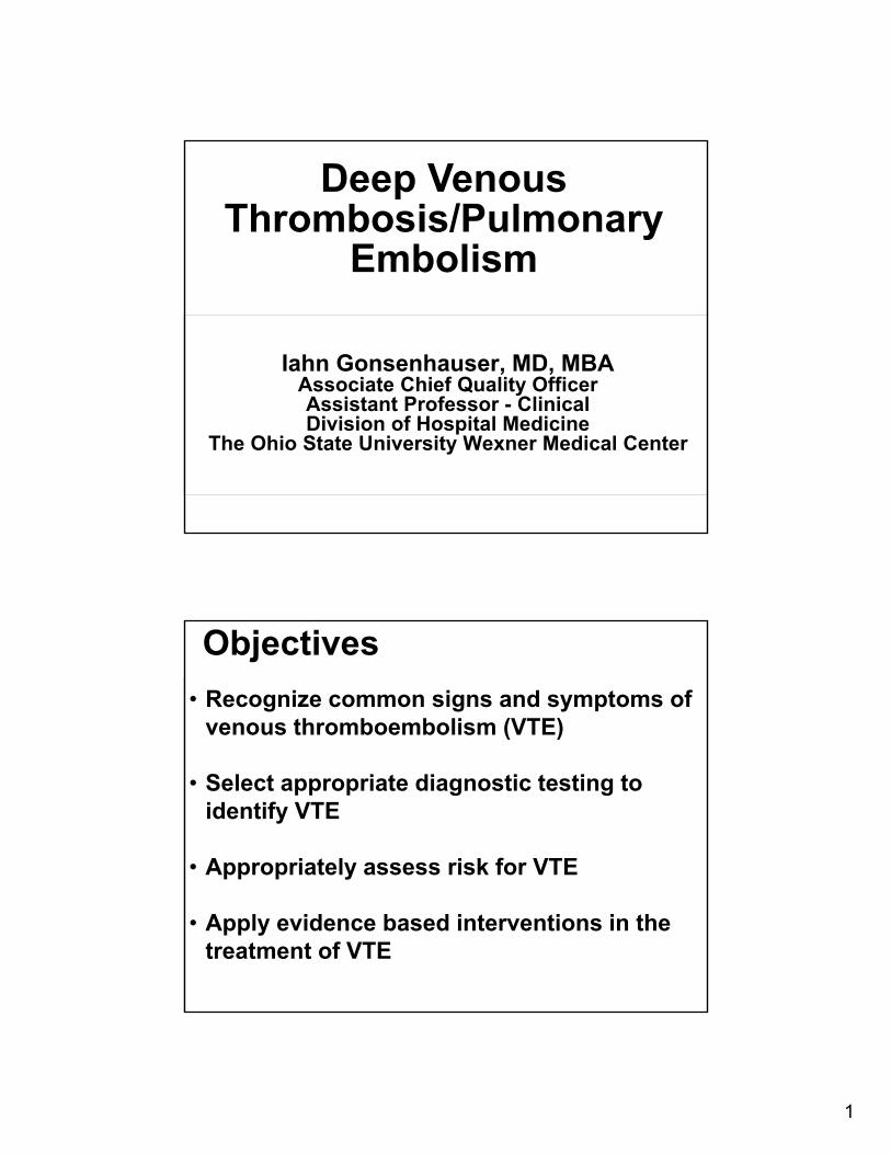
BackgroundVenous Thromboembolism (VTE) encompasses: Deep Venous Thrombosis (DVT) Pulmonary Embolism (PE)
Superficial Phlebitis is not included in this term
Blausen.com staff (2014). "Medical gallery of Blausen Medical 2014". WikiJournal of Medicine 1 (2).DOI:10.15347/wjm/2014.010. ISSN 2002-4436.
James Heilman, MD
Background• 350,000 – 600,000 US cases annually
• Hospitalization is a major risk factor
• Among the leading causes of preventable hospital death
• 10-15% Mortality
• Requires extended therapeutic anticoagulation
3
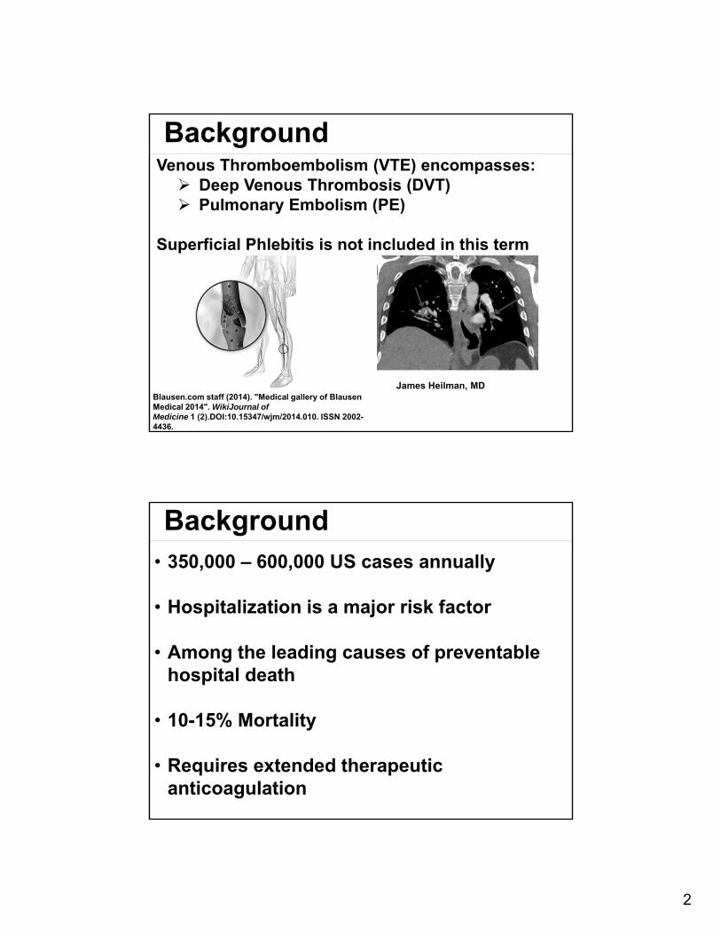
Pathophysiology
Virchow’s Triad
Stasis Hypercoagulability Endothelial Damage
SmokingHypertensionSurgeryCatheterizationTrauma
ImmobilityPolycythemia
Factor V LeidenProthrombin G20210AProthrombin C&S DeficiencyCancer/ChemotherapyPregnancyHITObesity
Clinical Pearls
96% of DVTs occur in the Lower Extremities
90% of Pulmonary Emboli originate from DVTs
50% of proximal LE DVT will result in PE
About 1/3 of DVTs result in post-thrombotic syndrome 5yrs post event
4
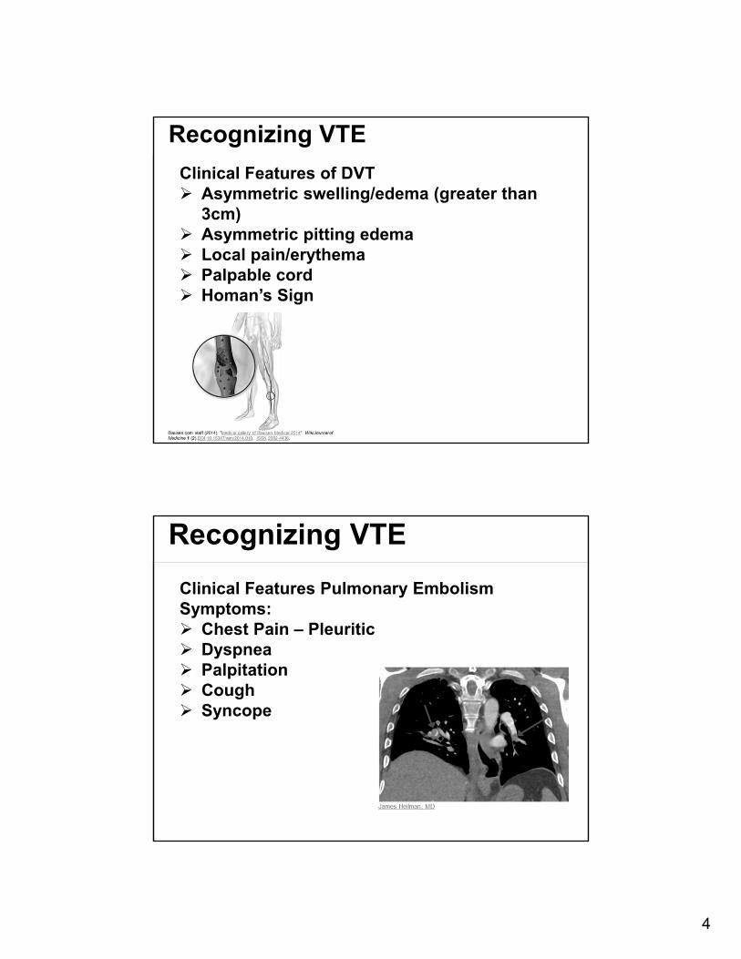
Recognizing VTE
Clinical Features of DVT Asymmetric swelling/edema (greater than
3cm) Asymmetric pitting edema Local pain/erythema Palpable cord Homan’s Sign
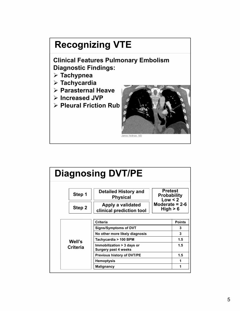
Recognizing VTE
Clinical Features Pulmonary EmbolismSymptoms: Chest Pain – Pleuritic Dyspnea Palpitation Cough Syncope
5
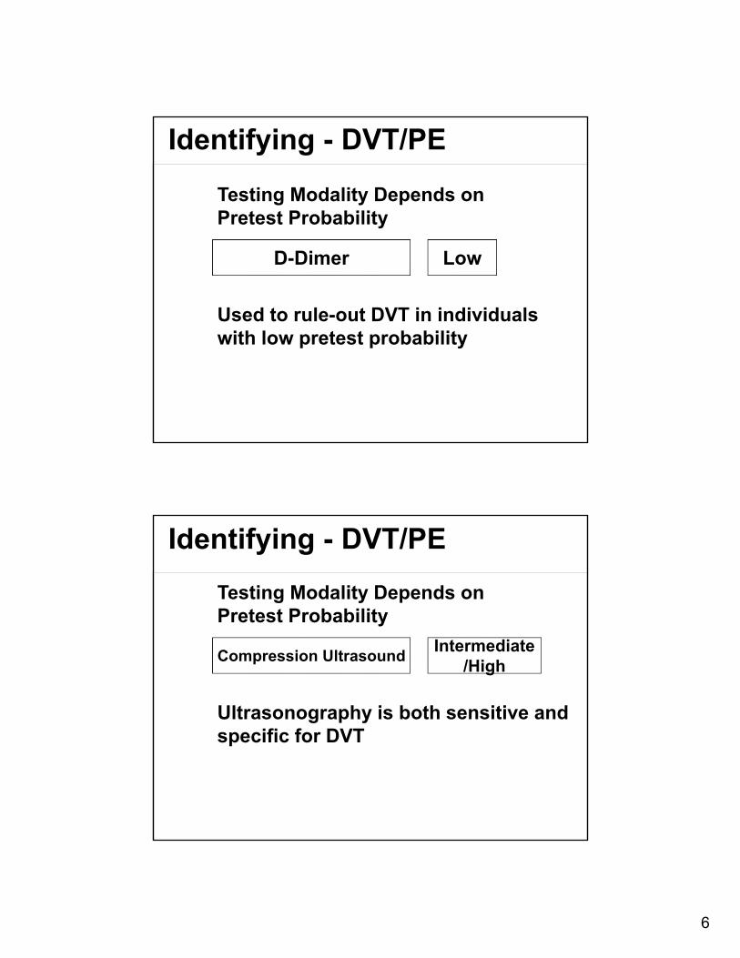
Recognizing VTE
Clinical Features Pulmonary EmbolismDiagnostic Findings: Tachypnea Tachycardia Parasternal Heave Increased JVP Pleural Friction Rub
Diagnosing DVT/PE
Step 1Detailed History and
Physical
Step 2Apply a validated
clinical prediction tool
Well’s Criteria
Criteria Points
Signs/Symptoms of DVT 3
No other more likely diagnosis 3
Tachycardia > 100 BPM 1.5
Immobilization > 3 days orSurgery past 4 weeks
1.5
Previous history of DVT/PE 1.5
Hemoptysis 1
Malignancy 1
Pretest Probability
Low < 2Moderate = 2-6
High > 6
6
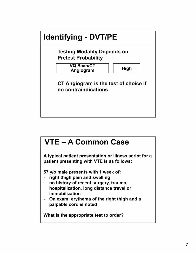
Identifying - DVT/PE
D-Dimer
Testing Modality Depends on Pretest Probability
Used to rule-out DVT in individuals with low pretest probability
Low
Identifying - DVT/PE
Compression Ultrasound
Testing Modality Depends on Pretest Probability
Ultrasonography is both sensitive and specific for DVT
Intermediate/High
7
Identifying - DVT/PE
VQ Scan/CT Angiogram
Testing Modality Depends on Pretest Probability
CT Angiogram is the test of choice if no contraindications
High
VTE – A Common Case
A typical patient presentation or illness script for a patient presenting with VTE is as follows:
57 y/o male presents with 1 week of:- right thigh pain and swelling - no history of recent surgery, trauma,
hospitalization, long distance travel or immobilization
- On exam: erythema of the right thigh and a palpable cord is noted
What is the appropriate test to order?
8
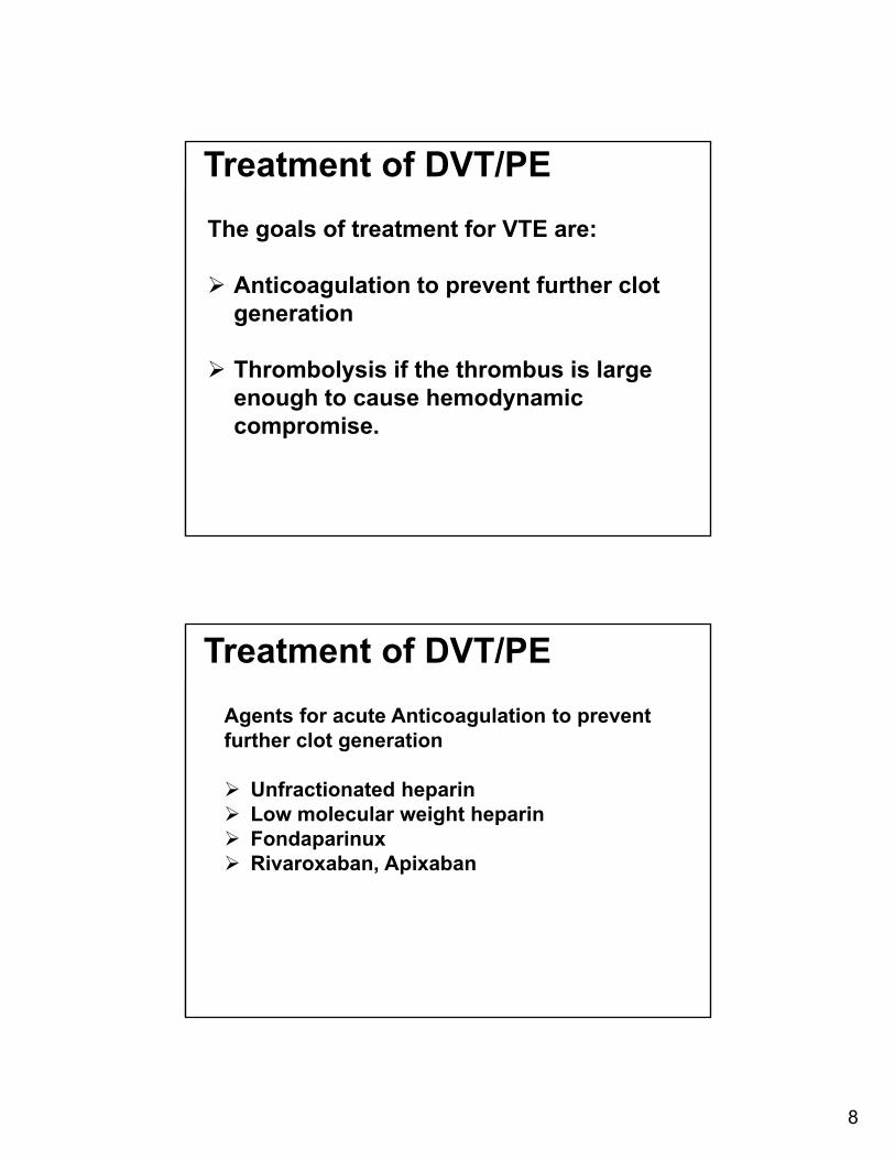
Treatment of DVT/PE
The goals of treatment for VTE are:
Anticoagulation to prevent further clot generation
Thrombolysis if the thrombus is large enough to cause hemodynamic compromise.
Treatment of DVT/PE
Agents for acute Anticoagulation to prevent further clot generation
Unfractionated heparin Low molecular weight heparin Fondaparinux Rivaroxaban, Apixaban
9
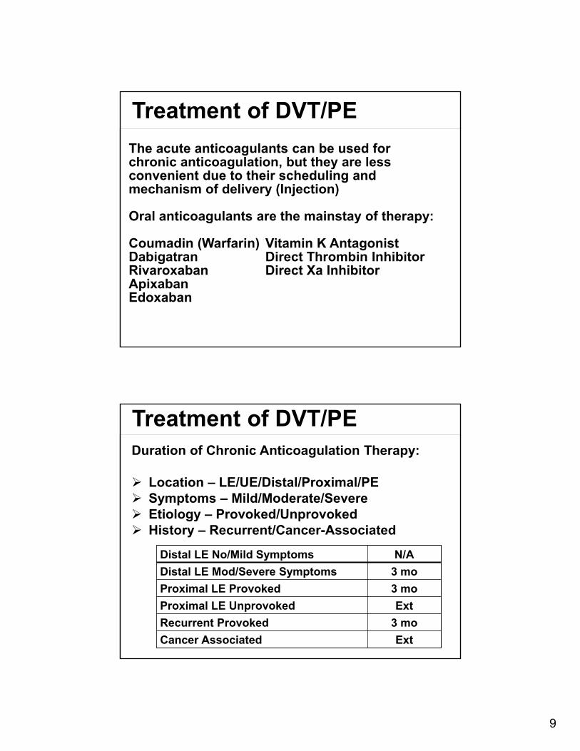
Treatment of DVT/PE
The acute anticoagulants can be used for chronic anticoagulation, but they are less convenient due to their scheduling and mechanism of delivery (Injection)
Oral anticoagulants are the mainstay of therapy:
Coumadin (Warfarin) Vitamin K AntagonistDabigatran Direct Thrombin InhibitorRivaroxaban Direct Xa InhibitorApixabanEdoxaban
Treatment of DVT/PEDuration of Chronic Anticoagulation Therapy:
Location – LE/UE/Distal/Proximal/PE Symptoms – Mild/Moderate/Severe Etiology – Provoked/Unprovoked History – Recurrent/Cancer-Associated
Distal LE No/Mild Symptoms N/A
Distal LE Mod/Severe Symptoms 3 mo
Proximal LE Provoked 3 mo
Proximal LE Unprovoked Ext
Recurrent Provoked 3 mo
Cancer Associated Ext
10
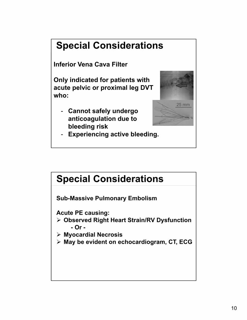
Special Considerations
Inferior Vena Cava Filter
Only indicated for patients with acute pelvic or proximal leg DVT who:
- Cannot safely undergo anticoagulation due to bleeding risk
- Experiencing active bleeding.
Special Considerations
Sub-Massive Pulmonary Embolism
Acute PE causing: Observed Right Heart Strain/RV Dysfunction
- Or - Myocardial Necrosis May be evident on echocardiogram, CT, ECG
11
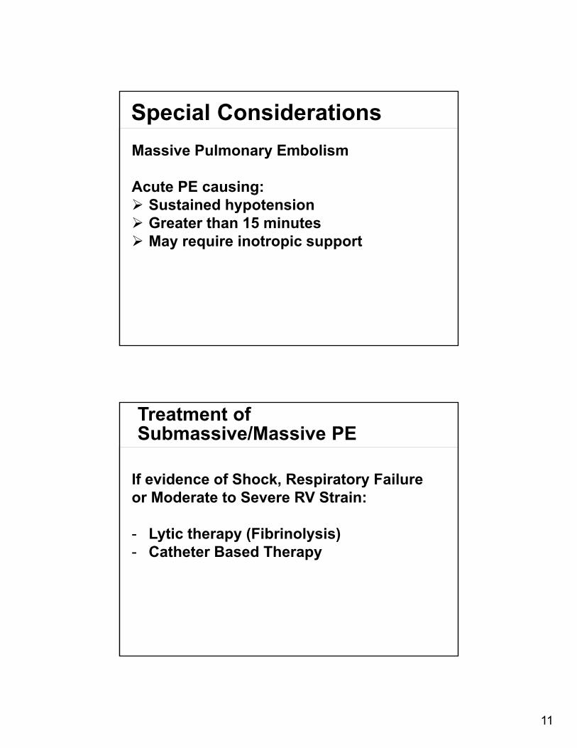
Special Considerations
Massive Pulmonary Embolism
Acute PE causing: Sustained hypotension Greater than 15 minutes May require inotropic support
Treatment of Submassive/Massive PE
If evidence of Shock, Respiratory Failure or Moderate to Severe RV Strain:
- Lytic therapy (Fibrinolysis)- Catheter Based Therapy
12
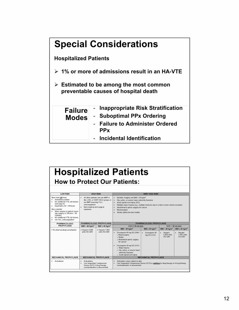
Special ConsiderationsHospitalized Patients
1% or more of admissions result in an HA-VTE
Estimated to be among the most common preventable causes of hospital death
Failure Modes
- Inappropriate Risk Stratification
- Suboptimal PPx Ordering
- Failure to Administer Ordered PPx
- Incidental Identification
Hospitalized PatientsHow to Protect Our Patients:
13
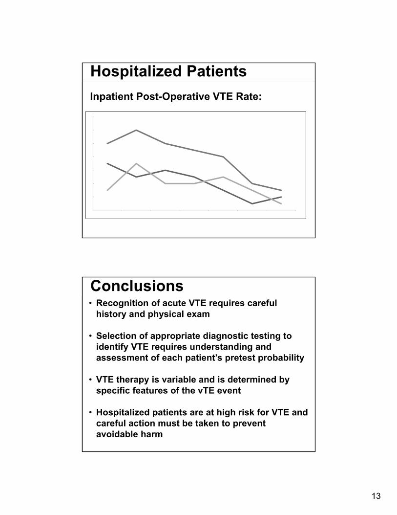
Hospitalized Patients
Inpatient Post-Operative VTE Rate:
Conclusions• Recognition of acute VTE requires careful
history and physical exam
• Selection of appropriate diagnostic testing to identify VTE requires understanding and assessment of each patient’s pretest probability
• VTE therapy is variable and is determined by specific features of the vTE event
• Hospitalized patients are at high risk for VTE and careful action must be taken to prevent avoidable harm
14
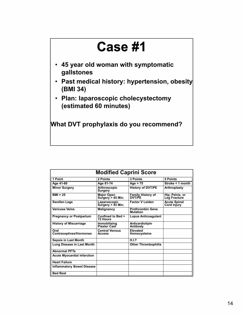
Case #1Case #1• 45 year old woman with symptomatic
gallstones
• Past medical history: hypertension, obesity (BMI 34)
• Plan: laparoscopic cholecystectomy (estimated 60 minutes)
What DVT prophylaxis do you recommend?
1 Point 2 Points 3 Points 5 PointsAge 41-60 Age 61-74 Age > 75 Stroke < 1 monthMinor Surgery Arthroscopic
SurgeryHistory of DVT/PE Arthroplasty
BMI > 25 Major Open Surgery > 45 Min.
Family History of DVT/PE
Hip, Pelvis, or Leg Fracture
Swollen Legs Laparoscopic Surgery > 45 Min.
Factor V Leiden Acute SpinalCord Injury
Varicose Veins Malignancy Prothrombin Gene Mutation
Pregnancy or Postpartum Confined to Bed > 72 Hours
Lupus Anticoagulant
History of Miscarriage ImmobilizingPlaster Cast
AnticardiolipinAntibody
Oral Contraceptives/Hormones
Central VenousAccess
Elevated Homocysteine
Sepsis in Last Month H.I.TLung Disease in Last Month Other Thrombophilia
Abnormal PFTsAcute Myocardial Infarction
Heart FailureInflammatory Bowel Disease
Bed Rest
Modified Caprini Score
15
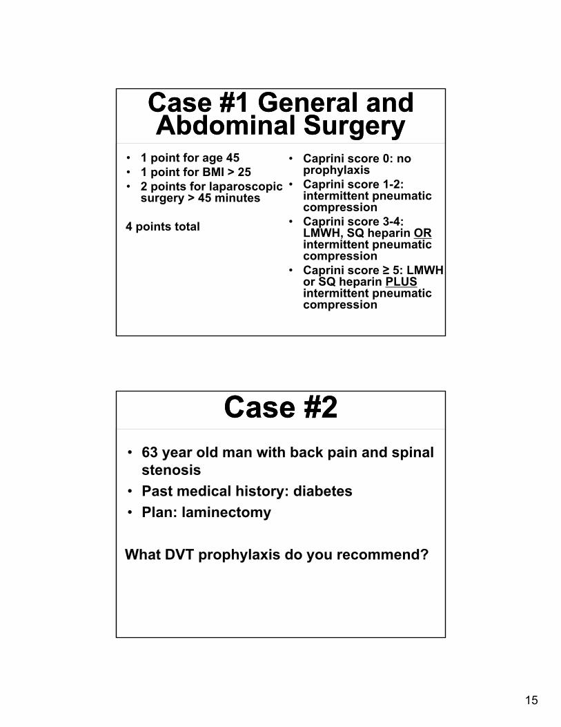
Case #1 General and Abdominal Surgery
Case #1 General and Abdominal Surgery
• 1 point for age 45• 1 point for BMI > 25• 2 points for laparoscopic
surgery > 45 minutes
4 points total
• Caprini score 0: no prophylaxis
• Caprini score 1-2: intermittent pneumatic compression
• Caprini score 3-4: LMWH, SQ heparin ORintermittent pneumatic compression
• Caprini score ≥ 5: LMWH or SQ heparin PLUSintermittent pneumatic compression
Case #2Case #2• 63 year old man with back pain and spinal
stenosis
• Past medical history: diabetes
• Plan: laminectomy
What DVT prophylaxis do you recommend?
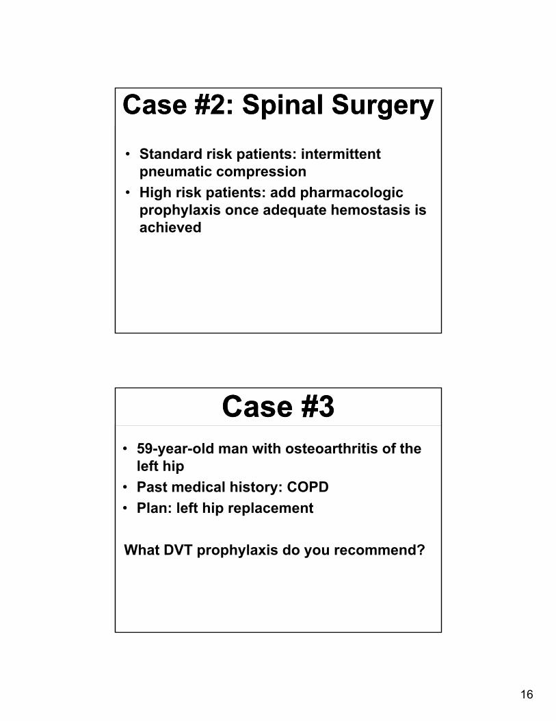
16
Case #2: Spinal SurgeryCase #2: Spinal Surgery
• Standard risk patients: intermittent pneumatic compression
• High risk patients: add pharmacologic prophylaxis once adequate hemostasis is achieved
Case #3Case #3• 59-year-old man with osteoarthritis of the
left hip
• Past medical history: COPD
• Plan: left hip replacement
What DVT prophylaxis do you recommend?
17
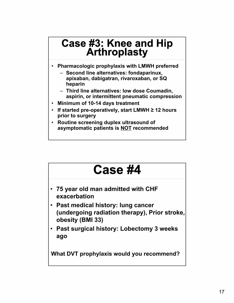
Case #3: Knee and Hip Arthroplasty
Case #3: Knee and Hip Arthroplasty
• Pharmacologic prophylaxis with LMWH preferred‒ Second line alternatives: fondaparinux,
apixaban, dabigatran, rivaroxaban, or SQ heparin
‒ Third line alternatives: low dose Coumadin, aspirin, or intermittent pneumatic compression
• Minimum of 10-14 days treatment• If started pre-operatively, start LMWH ≥ 12 hours
prior to surgery• Routine screening duplex ultrasound of
asymptomatic patients is NOT recommended
Case #4Case #4• 75 year old man admitted with CHF
exacerbation
• Past medical history: lung cancer (undergoing radiation therapy), Prior stroke, obesity (BMI 33)
• Past surgical history: Lobectomy 3 weeks ago
What DVT prophylaxis would you recommend?
18
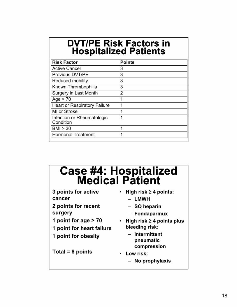
DVT/PE Risk Factors in Hospitalized Patients
DVT/PE Risk Factors in Hospitalized Patients
Risk Factor PointsActive Cancer 3Previous DVT/PE 3Reduced mobility 3Known Thrombophilia 3Surgery in Last Month 2Age > 70 1Heart or Respiratory Failure 1MI or Stroke 1Infection or RheumatologicCondition
1
BMI > 30 1Hormonal Treatment 1
Case #4: Hospitalized Medical Patient
Case #4: Hospitalized Medical Patient
3 points for active cancer
2 points for recent surgery
1 point for age > 70
1 point for heart failure
1 point for obesity
Total = 8 points
• High risk ≥ 4 points:
‒ LMWH
‒ SQ heparin
‒ Fondaparinux
• High risk ≥ 4 points plus bleeding risk:
‒ Intermittent pneumatic compression
• Low risk:
‒ No prophylaxis
19
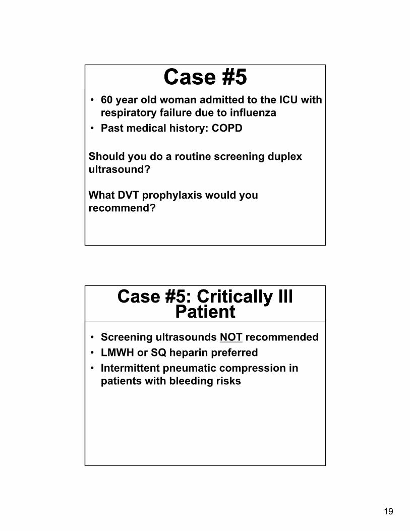
Case #5Case #5• 60 year old woman admitted to the ICU with
respiratory failure due to influenza
• Past medical history: COPD
Should you do a routine screening duplexultrasound?
What DVT prophylaxis would you recommend?
Case #5: Critically Ill Patient
Case #5: Critically Ill Patient
• Screening ultrasounds NOT recommended
• LMWH or SQ heparin preferred
• Intermittent pneumatic compression in patients with bleeding risks
20
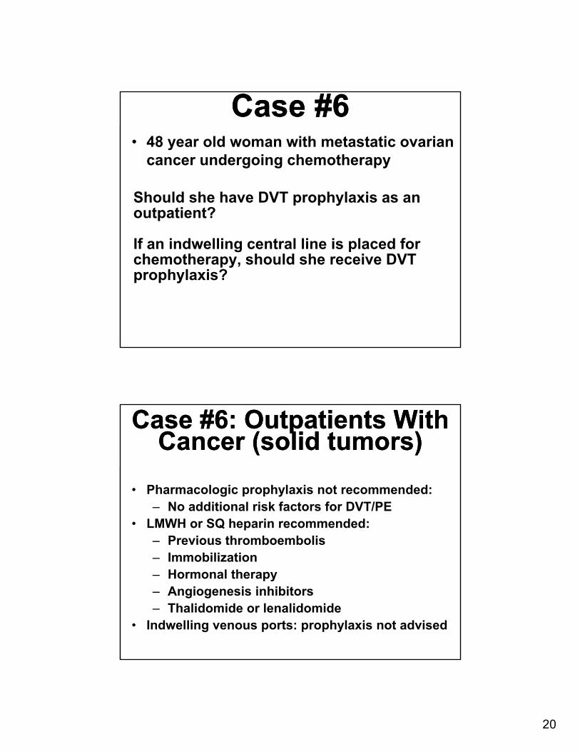
Case #6Case #6• 48 year old woman with metastatic ovarian
cancer undergoing chemotherapy
Should she have DVT prophylaxis as an outpatient?
If an indwelling central line is placed for chemotherapy, should she receive DVT prophylaxis?
Case #6: Outpatients With Cancer (solid tumors)
Case #6: Outpatients With Cancer (solid tumors)
• Pharmacologic prophylaxis not recommended:‒ No additional risk factors for DVT/PE
• LMWH or SQ heparin recommended:‒ Previous thromboembolis‒ Immobilization‒ Hormonal therapy‒ Angiogenesis inhibitors‒ Thalidomide or lenalidomide
• Indwelling venous ports: prophylaxis not advised