Embed Size (px)
Citation preview
Deep learning of the sectional appearances of 3D CT images for anatomicalstructure segmentation based on an FCN voting method
Xiangrong Zhoua) and Ryosuke TakayamaDepartment of Intelligent Image Information, Graduate School of Medicine, Gifu University, Gifu 501-1194, Japan
Song WangDepartment of Computer Science and Engineering, University of South Carolina, Columbia, SC 29208, USA
Takeshi Hara and Hiroshi FujitaDepartment of Intelligent Image Information, Graduate School of Medicine, Gifu University, Gifu 501-1194, Japan
(Received 31 October 2016; revised 3 July 2017; accepted for publication 10 July 2017;published 31 August 2017)
Purpose: We propose a single network trained by pixel-to-label deep learning to address the generalissue of automatic multiple organ segmentation in three-dimensional (3D) computed tomography(CT) images. Our method can be described as a voxel-wise multiple-class classification scheme forautomatically assigning labels to each pixel/voxel in a 2D/3D CT image.Methods: We simplify the segmentation algorithms of anatomical structures (including multipleorgans) in a CT image (generally in 3D) to a majority voting scheme over the semantic segmentationof multiple 2D slices drawn from different viewpoints with redundancy. The proposed method inher-its the spirit of fully convolutional networks (FCNs) that consist of “convolution” and “deconvolu-tion” layers for 2D semantic image segmentation, and expands the core structure with 3D-2D-3Dtransformations to adapt to 3D CT image segmentation. All parameters in the proposed network aretrained pixel-to-label from a small number of CT cases with human annotations as the ground truth.The proposed network naturally fulfills the requirements of multiple organ segmentations in CT casesof different sizes that cover arbitrary scan regions without any adjustment.Results: The proposed network was trained and validated using the simultaneous segmentation of 19anatomical structures in the human torso, including 17 major organs and two special regions (lumenand content inside of stomach). Some of these structures have never been reported in previousresearch on CT segmentation. A database consisting of 240 (95% for training and 5% for testing) 3DCT scans, together with their manually annotated ground-truth segmentations, was used in our exper-iments. The results show that the 19 structures of interest were segmented with acceptable accuracy(88.1% and 87.9% voxels in the training and testing datasets, respectively, were labeled correctly)against the ground truth.Conclusions: We propose a single network based on pixel-to-label deep learning to address the chal-lenging issue of anatomical structure segmentation in 3D CT cases. The novelty of this work is thepolicy of deep learning of the different 2D sectional appearances of 3D anatomical structures for CTcases and the majority voting of the 3D segmentation results from multiple crossed 2D sections toachieve availability and reliability with better efficiency, generality, and flexibility than conventionalsegmentation methods, which must be guided by human expertise. © 2017 The Authors. Medical
Physics published by Wiley Periodicals, Inc. on behalf of American Association of Physicists in
Medicine. [https://doi.org/10.1002/mp.12480]
Key words: 2D semantic segmentation, anatomical structure segmentation, CT images, deep learn-ing, fully convolutional network (FCN)
1. INTRODUCTION
Three-dimensional (3D) computed tomography (CT) imagesprovide useful internal information about the human anatomythat can be used to support diagnosis, surgery, and therapy.The recognition and segmentation of anatomical structuresplay critical roles in the quantitative interpretation of CTimages, and, conventionally, the image processing is accom-plished through human interpretation and manual annotationsby expert radiologists. However, human interpretation is oftenqualitative and subjective, with relatively high intra and
interobserver variability. Manual annotations also have limitedreproducibility and are very time-consuming. Therefore, auto-matic image segmentation would offer improved efficiency andreliability, as well as reducing the burden on radiologists.1,2
Fully automatic image segmentation, which transfers thephysical image signal to a useful abstraction, is a crucial pre-requisite for computer-based image analysis of 3D CT cases.3
Nevertheless, this task is challenging for several reasons.First, there is a large variation in the appearance of anatomi-cal structures in clinical image data (abnormal in most cases),and this is difficult to represent using mathematical rules.
5221 Med. Phys. 44 (10), October 2017 0094-2405/2017/44(10)/5221/13
© 2017 The Authors. Medical Physics published by Wiley Periodi-cals, Inc. on behalf of American Association of Physicists inMedicine. This is an open access article under the terms of the
Creative Commons Attribution-NonCommercial License, which permitsuse, distribution and reproduction in any medium, provided the original
work is properly cited and is not used for commercial purposes.
5221
Second, low intensity contrast and high image noise usuallylead to ambiguous and blurred boundaries between differentorgan or tissue regions in CT images. These boundaries aredifficult to identify using low-level digital signal/image pro-cessing. Third, different CT cases may cover different parts ofthe human body using different spatial image resolutionsaccording to specific clinical requirements. It is difficult andcostly to prepare models that are applicable to all possible CTcases and apply these models to CT image segmentation.Therefore, accurate CT image segmentation has become thebottleneck in many applications of computer-based medicalimage analysis and image interpretation.
CT image segmentation3 covers a wide range of researchfields. In this paper, we focus on simultaneous multipleorgan segmentation across a wide range of the human bodyin 3D CT images, which is a major research topic in com-puter-aided diagnosis. Conventional approaches for multipleorgan segmentation typically use pixel-based methods. Inthese approaches, image segmentations are divided into anumber of functional modules, and numerous hand-craftedsignal processing algorithms and image features are exam-ined according to human experience and knowledge.Although some mathematical models4–11 have recently beenintroduced, conventional CT image segmentation methodsstill attempt to emulate limited human-specified rules oroperations in segmenting CT images directly. These methodscan achieve reasonable segmentation results on CT imagesfor a special organ type within a known narrow scan range.However, they may fail in many clinical cases that includeanatomical structures that are seriously deformed and gener-ally cannot deal with scan ranges that are unknown a priori.To further improve the accuracy and robustness of CT imagesegmentation, the developed segmentation methods areexpected to handle a larger variety of ambiguous imageappearances, shapes, and relationships of anatomical struc-tures. It is difficult to achieve this goal by defining and con-sidering human knowledge and rules explicitly. Instead,data-driven approaches using large sets of image data —
such as a deep convolutional neural network (CNN) — aremore appropriate for solving this segmentation problem.
Recently, deep CNNs have been applied to medical imageanalysis in several studies. Most of them have used deepCNNs for lesion detection or classification,12–15 while othershave embedded CNNs into conventional organ-segmentationprocesses to reduce the false positive rate in the segmentationresults or to predict the likelihood of the image patches.16–18
Studies of this type usually divide CT images into numeroussmall 2D/3D patches at different locations, and then classifythese patches into multiple predefined categories. DeepCNNs can also be used to learn a set of optimized image fea-tures (sometimes combined with a classifier) to achieve theoptimal classification rate for these image patches. However,the anatomical segmentation of CT images over a wide regionof the human body is still challenging because of similaritiesin the images of different structures, as well as the difficultyof ensuring global spatial consistency in the labeling ofpatches in different CT cases.
This paper proposes a deep learning-based approach tothe general multiple organ-segmentation problem in 2D/3DCT images. The initial idea of this approach was presentedin a conference with a preliminary result.19 Our method tack-les three critical issues in CT image segmentation. First,efficiency and generality: our approach uses pixel-to-labellearning to train all of the variable arguments together forgeneral multiple organ segmentations. This is much moreconvenient to use and extend than conventional methods,which require specific models and algorithms to be preparedfor each type of organ. Second, performance and complex-ity: our method tackles 3D image segmentation as an itera-tion of single 2D CNN, which is implemented as GPU-based parallel processing. This greatly speeds up the seg-mentation process compared with CPU-based systems.Third, applicability and flexibility: the core component ofimage segmentation uses a fully convolutional network(FCN), which is naturally adaptive to segmenting differentcontent from different-size images that may cover arbitraryCT scan ranges (e.g., body, chest, abdomen). No CT imagesegmentation technique with this capability has previouslybeen published.
2. METHODS
2.A. Overview
The basis of our method for CT image segmentation canbe summarized as “multiple 2D proposals followed by 3Dintegration.”19 This comes from the way in which a radiolo-gist interprets CT cases — observing many 2D sections andthen reconstructing/imagining the 3D anatomical structure.Multiple organ segmentations on 2D CT sections are muchmore difficult than direct segmentation on a 3D CT vol-ume, because 2D sectional fragments of 3D anatomicalstructures from different perspectives may appear to havelittle in common and are difficult to integrate using conven-tional modeling approaches. The reasons we use a 2D sec-tional image segmentation as the basic processing unit,rather than directly segmenting the 3D CT volumes, are to(a) learn a model with features in a pixel-to-label way thatcan successfully represent anatomical structures as com-pletely as possible under the current computer hardwareresources (NVIDIA graphics processing units (GPUs) in aLinux environment), (b) gain better segmentation perfor-mance by employing majority voting over multiple 2D seg-mentation results (increasing redundancy) on the samelocation in 3D, and (c) satisfy the needs of clinical medi-cine regarding 3D or 2D images over an arbitrary CT scanrange (e.g., body, chest, abdomen). To model the large vari-ance of 2D sectional image appearances, we train a deepCNN to encode the anatomical structures from a relativelysmall number of 3D CT images, and accomplish CT imagesegmentation using pixel-wise labeling and decoding theinput image based on the trained deep CNN.
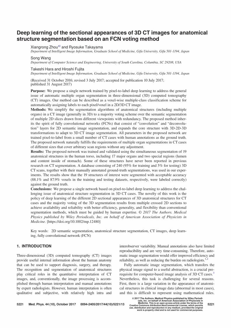
The proposed segmentation method is illustrated in Fig. 1.The input is a 3D CT image (a 2D case can be regarded as a
Medical Physics, 44 (10), October 2017
5222 Zhou et al.: CT image segmentation based on deep learning 5222
degenerate 3D case with only one section), and the output isa label map of the same size and dimensions in which thelabels are an annotated set of anatomical structures. Our seg-mentation process is repeated to sample 2D sections from a3D CT image, pass them to the deep CNN for pixel-wiseannotation, and stack the 2D labeled results back into 3D.Finally, the anatomical structure label at each voxel is deter-mined based on majority voting from multiple 2D labeledresults crossed at the voxel. The 2D segmentation uses anFCN20 for the anatomical segmentation of 2D CT image sec-tions. This FCN is trained on a set of 3D CT images, withhuman annotations as the ground truth. The processing stepsin our segmentation are integrated into a single networkunder a simple architecture without the need for conventionalimage-processing algorithms such as smoothing, filtering,and level-set methods. The parameters of the network are
learnable and optimized based on a pixel-to-label trainingscheme.
2.B. 2D CT image segmentation using FCN
FCNs have achieved state-of-the-art performance on natu-ral image segmentation tasks and feature representation fordense classification.20 This dense classification can also beused to predict the probability of each pixel belonging to aspecific anatomical structure in a CT image. The architectureof FCNs includes two modules (down-sampling path and up-sampling path) that are integrated into a simple networktrained in a pixel-to-label way. The motivation behind theFCN is that the down-sampling path extracts high-levelabstraction information (target location and type), while theup-sampling path predicts the low-level (pixel-wise)
FIG. 1. Network of the proposed anatomical structure segmentation for 3D CT image. See Fig. 2 for the details of the FCN part.
Medical Physics, 44 (10), October 2017
5223 Zhou et al.: CT image segmentation based on deep learning 5223
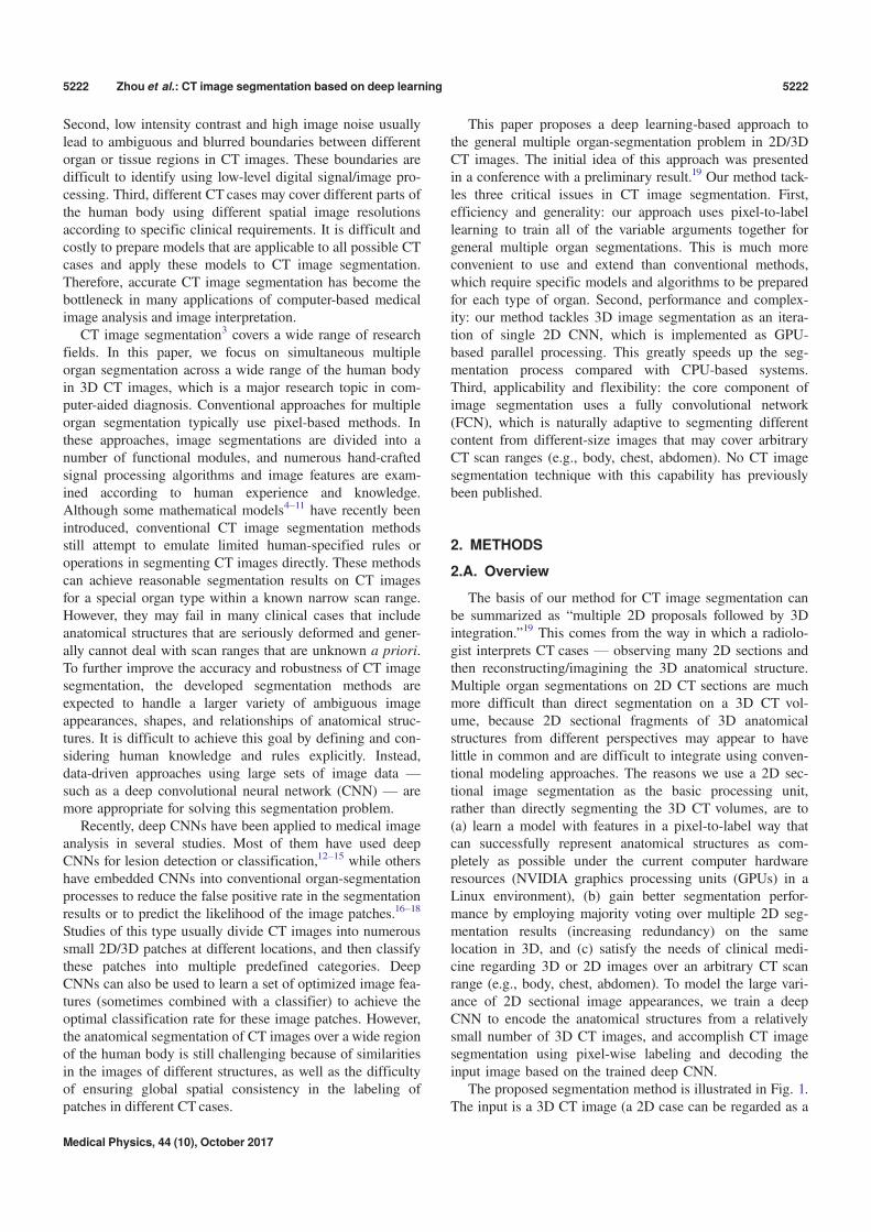
information (target shape and contour). The parameters inthese two modules of the FCN are globally optimized in thetraining stage. The structure of the FCN used in the proposedmethod for 2D CT image segmentation is shown in Fig. 2.
The FCN predicts scores (probability of each label class)based on intensity–texture information within a given recep-tive field (e.g., an image patch with a predefined size). Toavoid confusing similar patches that belong to differentorgans in CT images, our method uses a variable-size recep-tive field and regards the whole region of a 2D CT sectionalimage as one receptive field. This enables all information in a2D CT section to be used directly to predict complex struc-tures (multiple labels). Using the FCN architecture, our seg-mentation network provides the capability to adapt naturallyto input CT images of any size and any scan range across thehuman body, producing an output with the correspondingspatial dimensions.
The down-sampling path of our FCN uses the VGG16 netstructure21 (16 3 9 3 convolution layers interleaved with fivemaximum pooling layers plus three fully connected layers),as shown in Fig. 2. We change the three fully connected lay-ers of VGG16 to convolutional layers.20 The final fully con-nected classifier is a 1 9 1 convolution layer whose channeldimension is fixed according to the number of labels (22 inour case). The up-sampling path contains five deconvolu-tional (backward-stride convolution) and convolutional lay-ers. These have a skip structure that passes information lostin the lower convolution layers of VGG16 directly into thedeconvolution process, enabling detailed contours to berecovered sequentially under a higher image resolution.20
Rectified Linear Unit (ReLU) is used as the activation func-tion in both the up- and down-sampling paths. A graph for
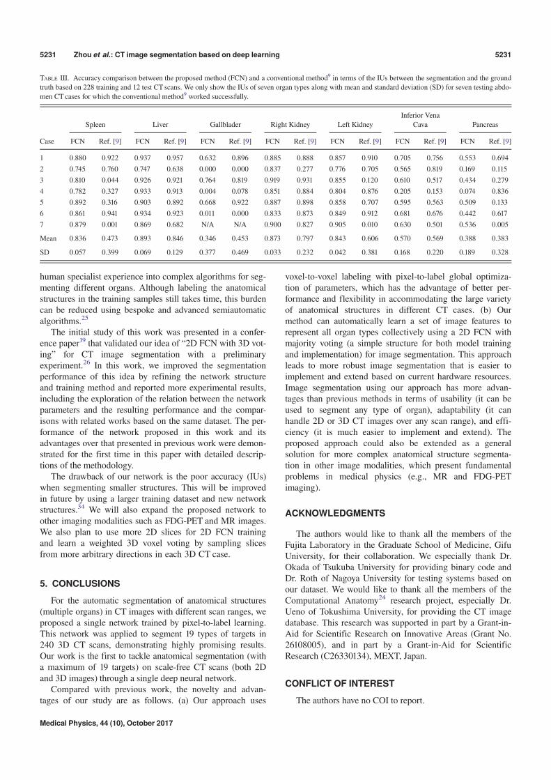
easily visualizing the details of our FCN structure is pre-sented in the Appendix.
2.C. 3D CT image segmentation by expanding FCNwith 3D-2D-3D transformation
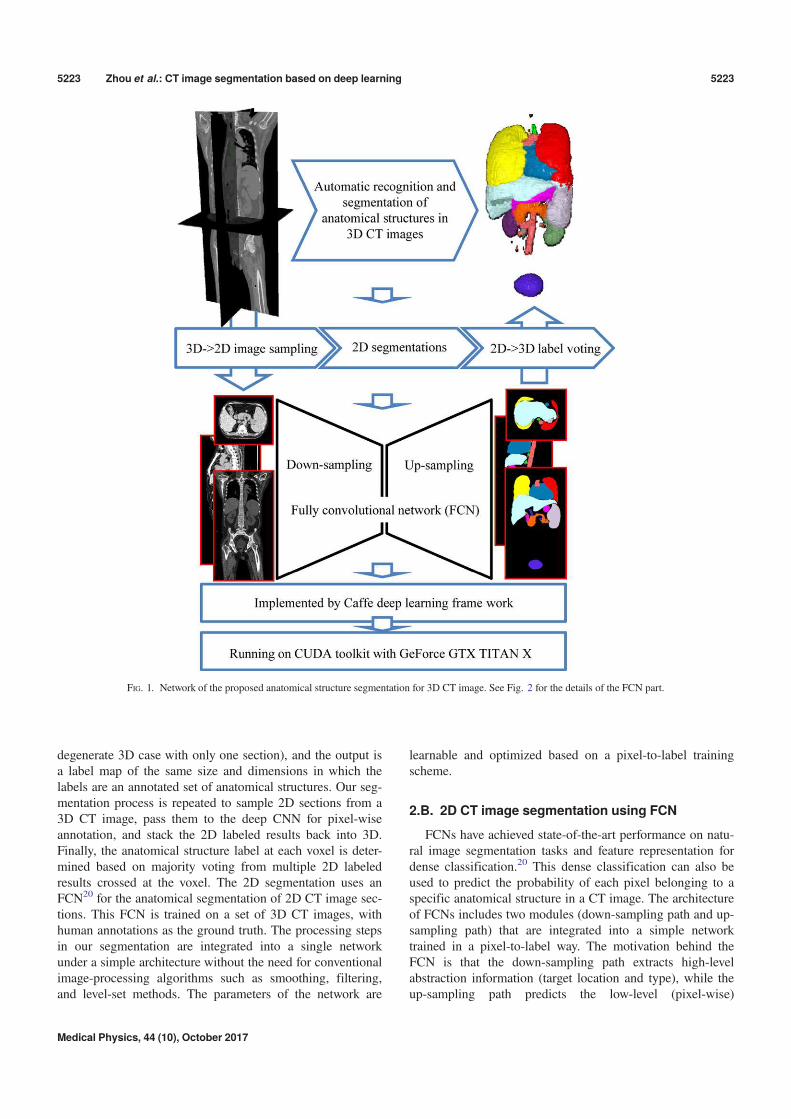
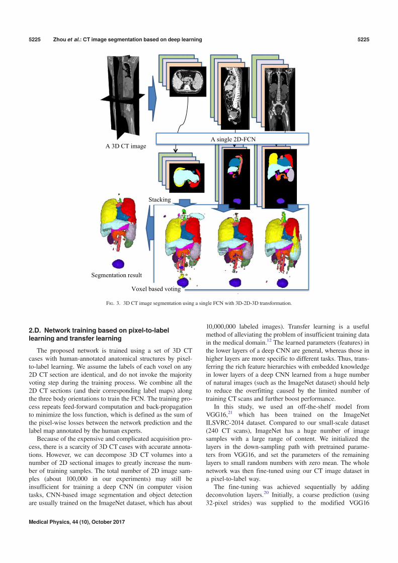
Each voxel in a 3D CT image can lie on different 2Dsections that pass through the voxel with different orienta-tions. Our idea of 3D CT image segmentation is to use therich image information of the entire 2D section to predictthe anatomical label of this voxel. The robustness and accu-racy of this technique are increased by redundantly labelingthis voxel on the multiple 2D sections with different orien-tations. We sample a 3D CT case over numerous sections(2D images) with different orientations, segment each 2Dsection using the FCN, and then assemble the output of thesegmentation (i.e., labeled 2D maps) back into 3D. In thiswork, we select all the 2D sections in three orthogonaldirections (axial, sagittal, and coronal body); this ensuresthat each voxel in the 3D image is located on three 2D CTsections. After the 2D image segmentation, each voxel isredundantly annotated three times, once for each 2D CTsection. The annotated results for each voxel should ideallybe identical, but may differ because of mislabeling by theFCN during the 2D image segmentation. The labels arefused using majority voting (selecting the mode of the threelabels) to improve the stability and accuracy of the finaldecision (refer to Fig. 3). When there is no duplicationamong the three labels, our method selects the label of anorgan type with the largest volume (the highest prior forthe voxel appearances in CT images based on anatomicalknowledge) as the output.
FIG. 2. Semantic image segmentation of 2D CT slice using a fully convolutional network (FCN).20 (K kernel size, S: stride).
Medical Physics, 44 (10), October 2017
5224 Zhou et al.: CT image segmentation based on deep learning 5224
2.D. Network training based on pixel-to-labellearning and transfer learning
The proposed network is trained using a set of 3D CTcases with human-annotated anatomical structures by pixel-to-label learning. We assume the labels of each voxel on any2D CT section are identical, and do not invoke the majorityvoting step during the training process. We combine all the2D CT sections (and their corresponding label maps) alongthe three body orientations to train the FCN. The training pro-cess repeats feed-forward computation and back-propagationto minimize the loss function, which is defined as the sum ofthe pixel-wise losses between the network prediction and thelabel map annotated by the human experts.
Because of the expensive and complicated acquisition pro-cess, there is a scarcity of 3D CT cases with accurate annota-tions. However, we can decompose 3D CT volumes into anumber of 2D sectional images to greatly increase the num-ber of training samples. The total number of 2D image sam-ples (about 100,000 in our experiments) may still beinsufficient for training a deep CNN (in computer visiontasks, CNN-based image segmentation and object detectionare usually trained on the ImageNet dataset, which has about
10,000,000 labeled images). Transfer learning is a usefulmethod of alleviating the problem of insufficient training datain the medical domain.12 The learned parameters (features) inthe lower layers of a deep CNN are general, whereas those inhigher layers are more specific to different tasks. Thus, trans-ferring the rich feature hierarchies with embedded knowledgein lower layers of a deep CNN learned from a huge numberof natural images (such as the ImageNet dataset) should helpto reduce the overfitting caused by the limited number oftraining CT scans and further boost performance.
In this study, we used an off-the-shelf model fromVGG16,21 which has been trained on the ImageNetILSVRC-2014 dataset. Compared to our small-scale dataset(240 CT scans), ImageNet has a huge number of imagesamples with a large range of content. We initialized thelayers in the down-sampling path with pretrained parame-ters from VGG16, and set the parameters of the remaininglayers to small random numbers with zero mean. The wholenetwork was then fine-tuned using our CT image dataset ina pixel-to-label way.
The fine-tuning was achieved sequentially by addingdeconvolution layers.20 Initially, a coarse prediction (using32-pixel strides) was supplied to the modified VGG16
Voxel based voting
Stacking
A single 2D-FCNA 3D CT image
Segmentation result
FIG. 3. 3D CT image segmentation using a single FCN with 3D-2D-3D transformation.
Medical Physics, 44 (10), October 2017
5225 Zhou et al.: CT image segmentation based on deep learning 5225
network with one deconvolution layer (called FCN32s). Afiner training sample was then added after inserting one fur-ther deconvolution layer at the end of the network. This wasdone using skips that combine the final prediction layer and alower layer with a finer stride in the modified VGG16 net-work. This fine-grained training was repeated with more net-work layers trained from the predictions of 16, 8, 4, and 2strides on the CT images to build FCN16s, 8s, 4s, and 2s,respectively. The detailed network structure of FCN2s can befound in the Appendix.
2.E. Implementation details
Our network was developed based on the open-sourcelibrary Caffe22 and the reference implementation of FCN.20
In the training stage, we used two optimization functions:stochastic gradient descent (SGD) with a momentum of 0.9and ADAM23 for comparison. A mini-batch size of 20images, learning rate of 10!4, and weight decay of 2!4 wereused as the training parameters. In addition, we incorporateddropout layers (drop rate 0.5) and local contrast normaliza-tion (LCN) layers (local size 3, alpha = 5 9 10!5,beta = 0.75) into the deconvolution layers to validate the per-formance. A workstation with the CUDA Library on a GPU(NVIDIA GeForce TITAN-X with 12 GB of memory) wasused for network training and testing.
3. EXPERIMENTS AND RESULTS
3.A. Dataset
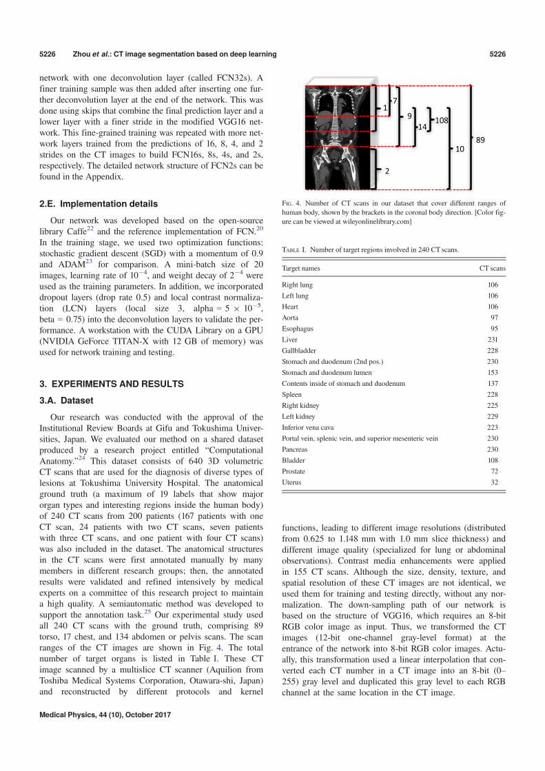
Our research was conducted with the approval of theInstitutional Review Boards at Gifu and Tokushima Univer-sities, Japan. We evaluated our method on a shared datasetproduced by a research project entitled “ComputationalAnatomy.”24 This dataset consists of 640 3D volumetricCT scans that are used for the diagnosis of diverse types oflesions at Tokushima University Hospital. The anatomicalground truth (a maximum of 19 labels that show majororgan types and interesting regions inside the human body)of 240 CT scans from 200 patients (167 patients with oneCT scan, 24 patients with two CT scans, seven patientswith three CT scans, and one patient with four CT scans)was also included in the dataset. The anatomical structuresin the CT scans were first annotated manually by manymembers in different research groups; then, the annotatedresults were validated and refined intensively by medicalexperts on a committee of this research project to maintaina high quality. A semiautomatic method was developed tosupport the annotation task.25 Our experimental study usedall 240 CT scans with the ground truth, comprising 89torso, 17 chest, and 134 abdomen or pelvis scans. The scanranges of the CT images are shown in Fig. 4. The totalnumber of target organs is listed in Table I. These CTimage scanned by a multislice CT scanner (Aquilion fromToshiba Medical Systems Corporation, Otawara-shi, Japan)and reconstructed by different protocols and kernel
functions, leading to different image resolutions (distributedfrom 0.625 to 1.148 mm with 1.0 mm slice thickness) anddifferent image quality (specialized for lung or abdominalobservations). Contrast media enhancements were appliedin 155 CT scans. Although the size, density, texture, andspatial resolution of these CT images are not identical, weused them for training and testing directly, without any nor-malization. The down-sampling path of our network isbased on the structure of VGG16, which requires an 8-bitRGB color image as input. Thus, we transformed the CTimages (12-bit one-channel gray-level format) at theentrance of the network into 8-bit RGB color images. Actu-ally, this transformation used a linear interpolation that con-verted each CT number in a CT image into an 8-bit (0–255) gray level and duplicated this gray level to each RGBchannel at the same location in the CT image.
2
1
89
9108
14
7
10
FIG. 4. Number of CT scans in our dataset that cover different ranges of
human body, shown by the brackets in the coronal body direction. [Color fig-
ure can be viewed at wileyonlinelibrary.com]
TABLE I. Number of target regions involved in 240 CT scans.
Target names CT scans
Right lung 106
Left lung 106
Heart 106
Aorta 97
Esophagus 95
Liver 231
Gallbladder 228
Stomach and duodenum (2nd pos.) 230
Stomach and duodenum lumen 153
Contents inside of stomach and duodenum 137
Spleen 228
Right kidney 225
Left kidney 229
Inferior vena cava 223
Portal vein, splenic vein, and superior mesenteric vein 230
Pancreas 230
Bladder 108
Prostate 72
Uterus 32
Medical Physics, 44 (10), October 2017
5226 Zhou et al.: CT image segmentation based on deep learning 5226
3.B. Experimental setting
We randomly picked 5% of the samples from the torso,chest, and abdomen CT scans as the testing dataset (total of12 CT scans) and used the remaining 228 CT cases to trainthe network. Multiple CT scans from the same patients wereonly used for training. We repeated this procedure three timesto train three networks, applied them to three testing datasets,and obtained segmentation results for a total of 36 (3 9 12)CT scans without overlap.
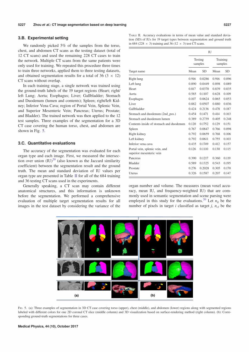
In each training stage, a single network was trained usingthe ground-truth labels of the 19 target regions (Heart; right/left Lung; Aorta; Esophagus; Liver; Gallbladder; Stomachand Duodenum (lumen and contents); Spleen; right/left Kid-ney; Inferior Vena Cava; region of Portal Vein, Splenic Vein,and Superior Mesenteric Vein; Pancreas; Uterus; Prostate;and Bladder). The trained network was then applied to the 12test samples. Three examples of the segmentation for a 3DCT case covering the human torso, chest, and abdomen areshown in Fig. 5.
3.C. Quantitative evaluations
The accuracy of the segmentation was evaluated for eachorgan type and each image. First, we measured the intersec-tion over union (IU)20 (also known as the Jaccard similaritycoefficient) between the segmentation result and the groundtruth. The mean and standard deviation of IU values perorgan type are presented in Table II for all of the 684 trainingand 36 testing CT scans used in the experiments.
Generally speaking, a CT scan may contain differentanatomical structures, and this information is unknownbefore the segmentation. We performed a comprehensiveevaluation of multiple target segmentation results for allimages in the test dataset by considering the variance of the
organ number and volume. The measures (mean voxel accu-racy, mean IU, and frequency-weighted IU) that are com-monly used in semantic segmentation and scene parsing wereemployed in this study for the evaluations.20 Let nij be thenumber of pixels in target i classified as target j, ncl be the
(a) (b)
FIG. 5. (a): Three examples of segmentation in 3D CT case covering torso (upper), chest (middle), and abdomen (lower) regions along with segmented regions
labeled with different colors for one 2D coronal CT slice (middle column) and 3D visualization based on surface-rendering method (right column). (b): Corre-
sponding ground-truth segmentations for three cases.
TABLE II. Accuracy evaluations in terms of mean value and standard devia-
tion (SD) of IUs for 19 target types between segmentation and ground truth
in 684 (228 9 3) training and 36 (12 9 3) test CT scans.
Target name
IU
Testing
samples
Training
samples
Mean SD Mean SD
Right lung 0.916 0.0286 0.916 0.096
Left lung 0.890 0.0449 0.898 0.089
Heart 0.817 0.0370 0.839 0.035
Aorta 0.585 0.1107 0.628 0.109
Esophagus 0.107 0.0624 0.065 0.053
Liver 0.882 0.0507 0.880 0.036
Gallbladder 0.424 0.2136 0.478 0.187
Stomach and duodenum (2nd_pos.) 0.454 0.1471 0.414 0.163
Stomach and duodenum lumen 0.389 0.2739 0.405 0.248
Contents inside of stomach and duodenum 0.120 0.1752 0.129 0.151
Spleen 0.767 0.0847 0.766 0.098
Right kidney 0.792 0.0659 0.768 0.106
Left kidney 0.792 0.0611 0.755 0.103
Inferior vena cava 0.435 0.1749 0.412 0.157
Portal vein, splenic vein, and
superior mesenteric vein
0.126 0.1110 0.130 0.115
Pancreas 0.390 0.1217 0.360 0.119
Bladder 0.589 0.1325 0.543 0.195
Prostate 0.276 0.2028 0.305 0.170
Uterus 0.326 0.1587 0.207 0.147
Medical Physics, 44 (10), October 2017
5227 Zhou et al.: CT image segmentation based on deep learning 5227
total number of different targets in a CT case, and ti ¼P
j nijbe the total number of pixels in target i. These measures aredefined as:
• Mean voxel accuracy:P
i nii=ti! "#
ncl ð1Þ
• Mean IU:P
i nii#
ti þP
j nji ! nii
$ %$ %
#
ncl ð2Þ
• Frequency-weighted IU:
X
ktk
$ %!1X
itinii
#
ti þX
jnji ! nii
$ %ð3Þ
The evaluation results for the mean voxel accuracy andfrequency-weighted IU were 87.9% and 80.5%, respectively,when averaged over all the segmentation results of the testdataset. These results mean that 87.9% of the voxels withinthe anatomical structures (constructed using multiple targetregions) were labeled correctly, with an overlap ratio of80.5% for the test dataset. We conducted the same evaluationon the training dataset, and found corresponding values of88.1% and 79.9%.
3.D. Segmentation performance
We validated the performance of the proposed network byevaluating the segmentation results of FCN 8s. The networkwas trained over 160,000 iterations using the ADAM opti-mizer with the training protocol described above. The trainednetwork was then applied to the test CT cases. Except for onegallbladder and one prostate, our network recognized andextracted all organs correctly. Because our segmentation tar-gets cover a wide range of shapes, volumes, and sizes, eitherwith or without contrast enhancement, and come from differ-ent locations in the human body, these experimental resultsoffer an excellent demonstration of the capability of ourapproach to recognize anatomical structures in the types ofCT images actually used in clinical medicine. Table II pre-sents the mean per-target IUs between the segmentationresults and ground truth in both the testing and training data.We found that the mean IUs of organs with larger volumes(e.g., lung) were comparable to those achieved by previousmethods.6–11 For some smaller organs (e.g., gallbladder) andstomach contents (which have not previously been reported),our segmentation did not produce particularly high IUs. Thelimited image resolution is likely to be the major cause of thispoor performance for these organs. Our evaluation shows thatthe average segmentation accuracy of all targets over both thetest and training CT images is approximately 80.5% and79.9% in terms of the frequency-weighted IUs. Because theperformance of deep learning is highly dependent to theamount of training data, we reduced the number of CT scansin the training stage from 95% to 75% and increased thenumber CT scans for testing from 5% to 25% by using thesame dataset and experimental setting and used a fourfoldcross-validation to evaluate the performance again. Theseadditional experimental results demonstrated that the averagesegmentation accuracy of all targets over the 240 (4 9 60)
test CT scans was approximately 78.3% in terms of the fre-quency-weighted IUs and 86.1% in terms of the mean voxelaccuracy, which are comparable to the performance (80.5%and 87.9%) in the previous experiment. The IUs of mostorgan types showed similar values, except that the spleen,prostate, and uterus showed a large decrease in the accuracyof more than 10% in terms of the IU. These decreases in theperformance were caused by the shortage of the training sam-ples and may be improved by increasing the number of CTscans in the training stage. Thus, our approach can recognizeand extract all types of major anatomical structures simulta-neously, achieving a reasonable accuracy according to theorgan volume in the CT images.
4. DISCUSSION
4.A. Transfer learning and training protocols
For comparison, we trained our network to “learn fromscratch” by initializing all parameters of an FCN to small ran-dom numbers with zero mean. No convergence was observedwithin 80,000 learning iterations, and the network then failedto segment any of the targets in the test dataset. These resultsindicate that the size of our CT dataset with current trainingprotocols is insufficient to train the proposed network suc-cessfully when starting from scratch. However, when we fine-tuned our network using VGG16,21 which is pretrained usingImageNet, convergence was achieved after 22,000 iterations.The trained network fine-tuned from VGG16 in 80,000 learn-ing iterations could segment multiple organs successfully inCT images from both the testing and training datasets. Thisdemonstrates that comprehensive knowledge learned fromlarge-scale, well-annotated datasets can be transferred to ournetwork to accomplish the CT image segmentation task.
We also compared the performance of networks optimizedby SGD and ADAM with the same training protocolsdescribed in Section 2.E. The segmentation results on the testdata indicate that the network trained by ADAM offersslightly better performance (up by 0.3% in voxel accuracy,0.15% in frequency-weighted IU) than that trained by SGD.Because the learning rate does not need to be tuned inADAM and the default parameters are likely to achieve goodresults, we used this function as the default optimizer for ournetwork in subsequent experiments.
The performance of the network may be affected by thenumber of training iterations. We compared the segmentationresults on the test dataset given by networks after 80,000,160,000, and 550,000 training iterations. We found that160,000 iterations was sufficient to train the network. Furthertraining iterations may improve the segmentation accuracy ofsome organ types, but could not improve the overall perfor-mance across all organ types.
4.B. Network structure
For comparison, we incorporated dropout layers with eachdeconvolution layer in the up-sampling path and retrained the
Medical Physics, 44 (10), October 2017
5228 Zhou et al.: CT image segmentation based on deep learning 5228
network. We found that the network performance with the testdataset decreased (down by 23% in voxel accuracy and 28%in frequency-weighted IU) after inserting these dropout lay-ers. We also tried to incorporate LCN layers in the same way,but did not observe any significant improvement in perfor-mance. Based on these results, we do not include dropoutand LCN layers in the up-sampling path of the proposednetwork.
The up-sampling path consists of five deconvolutional lay-ers (FCN32s to FCN2s). We investigated the segmentationresults in the test samples after applying each FCN layer tothe network sequentially. We found that the frequency-weighted IUs increased monotonically (69.8%, 81.1%, 84.9%and 88.0% at FCN32s, 16s, 8s, and 4s, respectively), and nofurther improvement was observed by FCN2s. This resultdemonstrates that diminishing returns of gradient descentoccurred from FCN4s in the training stage. A similarphenomenon was noted in the original FCN structures20 andconfirmed in our preliminary reports.19,26
Some alterations to the deep CNN architecture haverecently been reported in the field of computer vision andmedical image analysis.27–29 The well-known SegNet27 net-work achieved better segmentation performance than FCN,20
especially for small targets in natural images. We replaced theFCN part of our network with SegNet and examined its per-formance in CT image segmentation. This experimentshowed that the original SegNet implementation could onlydeal with predefined input image sizes. Even when all CTcases were re-scaled accordingly, the preliminary experimen-tal results did not suggest better performance than the FCN.It is possible that a customized version of SegNet is neededfor CT image segmentation. However, further investigation ofthe differences in performance offered by FCN and SegNet isbeyond the scope of this paper. Our current results show thatFCN is the best deep learning network for our CT image seg-mentation task.
4.C. Insights offered by the trained FCN
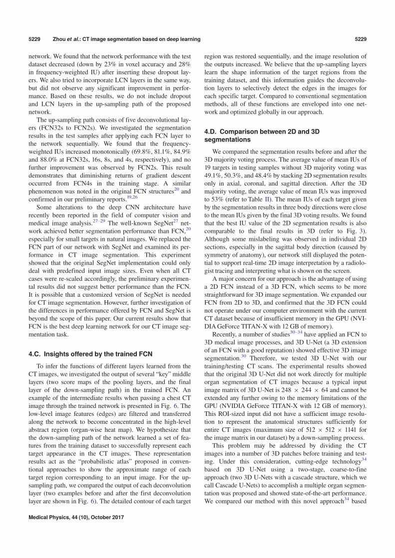
To infer the functions of different layers learned from theCT images, we investigated the output of several “key”middlelayers (two score maps of the pooling layers, and the finallayer of the down-sampling path) in the trained FCN. Anexample of the intermediate results when passing a chest CTimage through the trained network is presented in Fig. 6. Thelow-level image features (edges) are filtered and transferredalong the network to become concentrated in the high-levelabstract region (organ-wise heat map). We hypothesize thatthe down-sampling path of the network learned a set of fea-tures from the training dataset to successfully represent eachtarget appearance in the CT images. These representationresults act as the “probabilistic atlas” proposed in conven-tional approaches to show the approximate range of eachtarget region corresponding to an input image. For the up-sampling path, we compared the output of each deconvolutionlayer (two examples before and after the first deconvolutionlayer are shown in Fig. 6). The detailed contour of each target
region was restored sequentially, and the image resolution ofthe outputs increased. We believe that the up-sampling layerslearn the shape information of the target regions from thetraining dataset, and this information guides the deconvolu-tion layers to selectively detect the edges in the images foreach specific target. Compared to conventional segmentationmethods, all of these functions are enveloped into one net-work and optimized globally in our approach.
4.D. Comparison between 2D and 3Dsegmentations
We compared the segmentation results before and after the3D majority voting process. The average value of mean IUs of19 targets in testing samples without 3D majority voting was49.1%, 50.3%, and 48.4% by stacking 2D segmentation resultsonly in axial, coronal, and sagittal direction. After the 3Dmajority voting, the average value of mean IUs was improvedto 53% (refer to Table II). The mean IUs of each target givenby the segmentation results in three body directions were closeto the mean IUs given by the final 3D voting results. We foundthat the best IU value of the 2D segmentation results is alsocomparable to the final results in 3D (refer to Fig. 3).Although some mislabeling was observed in individual 2Dsections, especially in the sagittal body direction (caused bysymmetry of anatomy), our network still displayed the poten-tial to support real-time 2D image interpretation by a radiolo-gist tracing and interpreting what is shown on the screen.
A major concern for our approach is the advantage of usinga 2D FCN instead of a 3D FCN, which seems to be morestraightforward for 3D image segmentation. We expanded ourFCN from 2D to 3D, and confirmed that the 3D FCN couldnot operate under our computer environment with the currentCT dataset because of insufficient memory in the GPU (NVI-DIAGeForce TITAN-X with 12 GB of memory).
Recently, a number of studies30–34 have applied an FCN to3D medical image processes, and 3D U-Net (a 3D extensionof an FCN with a good reputation) showed effective 3D imagesegmentation.30 Therefore, we tested 3D U-Net with ourtraining/testing CT scans. The experimental results showedthat the original 3D U-Net did not work directly for multipleorgan segmentation of CT images because a typical inputimage matrix of 3D U-Net is 248 9 244 9 64 and cannot beextended any further owing to the memory limitations of theGPU (NVIDIA GeForce TITAN-X with 12 GB of memory).This ROI-sized input did not have a sufficient image resolu-tion to represent the anatomical structures sufficiently forentire CT images (maximum size of 512 9 512 9 1141 forthe image matrix in our dataset) by a down-sampling process.
This problem may be addressed by dividing the CTimages into a number of 3D patches before training and test-ing. Under this consideration, cutting-edge technology34
based on 3D U-Net using a two-stage, coarse-to-fineapproach (two 3D U-Nets with a cascade structure, which wecall Cascade U-Nets) to accomplish a multiple organ segmen-tation was proposed and showed state-of-the-art performance.We compared our method with this novel approach34 based
Medical Physics, 44 (10), October 2017
5229 Zhou et al.: CT image segmentation based on deep learning 5229
on the same training (228) and testing (12) CT scans. Theexperiment showed the accuracies (IUs) in each target typeon average of 12 testing CT scans obtained using our methodwere better than a single 3D U-Net with ROI-sized inputs,and still better than the Cascade U-Nets for nine target types,except for the other nine types of target (having a small vol-ume or tube structures). The performance in terms of the fre-quency-weighted IUs of our method (80.5%) was comparableto the Cascade U-Nets (80.3%) for the test dataset.
Considering the difference between the two structures(two 3D U-Nets versus a single 2D FCN + voting), we mustconclude that our 2D FCN is currently a realistic method forCT image segmentation.
4.E. Comparison to previous work
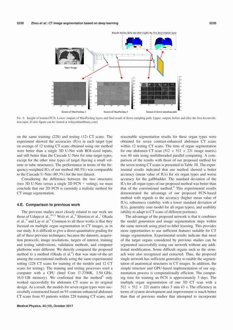
The previous studies most closely related to our work arethose of Udupa et al.,10,11 Wolz et al.,8 Shimizu et al.,7 Okadaet al.,9 and Lay et al.6 Common to all these works is that theyfocused on multiple organ segmentation in CT images, as inour study. It is difficult to give a direct quantitative grading forall of these previous techniques, because the datasets, acquisi-tion protocols, image resolutions, targets of interest, trainingand testing subdivisions, validation methods, and computerplatforms were different. We directly compared the proposedmethod to a method (Okada et al.9) that was state-of-the-artamong the conventional methods using the same experimentalsetting (228 CT scans for training of the models and 12 CTscans for testing). The training and testing processes used acomputer with a CPU (Intel Core i7-2700K, 3.50 GHz,16.0 GB memory). We confirmed that the method9 onlyworked successfully for abdomen CT scans as its originaldesign. As a result, the models for seven organ types were suc-cessfully constructed based on 93 contrast-enhanced abdomenCT scans from 93 patients within 228 training CT scans, and
reasonable segmentation results for these organ types wereobtained for seven contrast-enhanced abdomen CT scanswithin 12 testing CT scans. The time of organ segmentationfor one abdomen CT scan (512 9 512 9 221 image matrix)was 40 min using multithreaded parallel computing. A com-parison of the results with those of our proposed method forthe seven testing CTscans is presented in Table III. The exper-imental results indicated that our method showed a betteraccuracy (mean value of IUs) for six organ types and worseaccuracy for the gallbladder. The standard deviation of theIUs for all organ types of our proposed method was better thanthat of the conventional method.9 This experimental resultsdemonstrated the advantage of our proposed FCN-basedmethod with regards to the accuracy (higher mean value ofIUs), robustness (stability with a lower standard deviation ofIUs), generality (one model for all organ types), and usability(ability to adapt to CT scans of different portions).
The advantage of the proposed network is that it combinesthe model generation and image segmentation steps withinthe same network using pixel-to-label learning. This providesmore opportunities to use sufficient features suitable for CTimage segmentation. Experimental results indicate that mostof the target organs considered by previous studies can besegmented successfully using our network without any addi-tional modification. Some difficult organs such as the stom-ach were also recognized and extracted. Thus, the proposedsingle network has sufficient generality to enable the segmen-tation of anatomical structures in CT images. In addition, thesimple structure and GPU-based implementation of our seg-mentation process is computationally efficient. The comput-ing time for training an FCN is approximately 3 days. Themultiple organ segmentation of one 3D CT scan with a512 9 512 9 221 matrix takes 3 min 43 s. The efficiency interms of system development and improvement is much betterthan that of previous studies that attempted to incorporate
FIG. 6. Insight of learned FCN. Lower: outputs of MaxPooling layers and final result of down-sampling path; Upper: outputs before and after the first deconvolu-
tion layer. [Color figure can be viewed at wileyonlinelibrary.com]
Medical Physics, 44 (10), October 2017
5230 Zhou et al.: CT image segmentation based on deep learning 5230
human specialist experience into complex algorithms for seg-menting different organs. Although labeling the anatomicalstructures in the training samples still takes time, this burdencan be reduced using bespoke and advanced semiautomaticalgorithms.25
The initial study of this work was presented in a confer-ence paper19 that validated our idea of “2D FCN with 3D vot-ing” for CT image segmentation with a preliminaryexperiment.26 In this work, we improved the segmentationperformance of this idea by refining the network structureand training method and reported more experimental results,including the exploration of the relation between the networkparameters and the resulting performance and the compar-isons with related works based on the same dataset. The per-formance of the network proposed in this work and itsadvantages over that presented in previous work were demon-strated for the first time in this paper with detailed descrip-tions of the methodology.
The drawback of our network is the poor accuracy (IUs)when segmenting smaller structures. This will be improvedin future by using a larger training dataset and new networkstructures.34 We will also expand the proposed network toother imaging modalities such as FDG-PET and MR images.We also plan to use more 2D slices for 2D FCN trainingand learn a weighted 3D voxel voting by sampling slicesfrom more arbitrary directions in each 3D CT case.
5. CONCLUSIONS
For the automatic segmentation of anatomical structures(multiple organs) in CT images with different scan ranges, weproposed a single network trained by pixel-to-label learning.This network was applied to segment 19 types of targets in240 3D CT scans, demonstrating highly promising results.Our work is the first to tackle anatomical segmentation (witha maximum of 19 targets) on scale-free CT scans (both 2Dand 3D images) through a single deep neural network.
Compared with previous work, the novelty and advan-tages of our study are as follows. (a) Our approach uses
voxel-to-voxel labeling with pixel-to-label global optimiza-tion of parameters, which has the advantage of better per-formance and flexibility in accommodating the large varietyof anatomical structures in different CT cases. (b) Ourmethod can automatically learn a set of image features torepresent all organ types collectively using a 2D FCN withmajority voting (a simple structure for both model trainingand implementation) for image segmentation. This approachleads to more robust image segmentation that is easier toimplement and extend based on current hardware resources.Image segmentation using our approach has more advan-tages than previous methods in terms of usability (it can beused to segment any type of organ), adaptability (it canhandle 2D or 3D CT images over any scan range), and effi-ciency (it is much easier to implement and extend). Theproposed approach could also be extended as a generalsolution for more complex anatomical structure segmenta-tion in other image modalities, which present fundamentalproblems in medical physics (e.g., MR and FDG-PETimaging).
ACKNOWLEDGMENTS
The authors would like to thank all the members of theFujita Laboratory in the Graduate School of Medicine, GifuUniversity, for their collaboration. We especially thank Dr.Okada of Tsukuba University for providing binary code andDr. Roth of Nagoya University for testing systems based onour dataset. We would like to thank all the members of theComputational Anatomy24 research project, especially Dr.Ueno of Tokushima University, for providing the CT imagedatabase. This research was supported in part by a Grant-in-Aid for Scientific Research on Innovative Areas (Grant No.26108005), and in part by a Grant-in-Aid for ScientificResearch (C26330134), MEXT, Japan.
CONFLICT OF INTEREST
The authors have no COI to report.
TABLE III. Accuracy comparison between the proposed method (FCN) and a conventional method9 in terms of the IUs between the segmentation and the ground
truth based on 228 training and 12 test CT scans. We only show the IUs of seven organ types along with mean and standard deviation (SD) for seven testing abdo-
men CT cases for which the conventional method9 worked successfully.
Case
Spleen Liver Gallblader Right Kidney Left Kidney
Inferior Vena
Cava Pancreas
FCN Ref. [9] FCN Ref. [9] FCN Ref. [9] FCN Ref. [9] FCN Ref. [9] FCN Ref. [9] FCN Ref. [9]
1 0.880 0.922 0.937 0.957 0.632 0.896 0.885 0.888 0.857 0.910 0.705 0.756 0.553 0.694
2 0.745 0.760 0.747 0.638 0.000 0.000 0.837 0.277 0.776 0.705 0.565 0.819 0.169 0.115
3 0.810 0.044 0.926 0.921 0.764 0.819 0.919 0.931 0.855 0.120 0.610 0.517 0.434 0.279
4 0.782 0.327 0.933 0.913 0.004 0.078 0.851 0.884 0.804 0.876 0.205 0.153 0.074 0.836
5 0.892 0.316 0.903 0.892 0.668 0.922 0.887 0.898 0.858 0.707 0.595 0.563 0.509 0.133
6 0.861 0.941 0.934 0.923 0.011 0.000 0.833 0.873 0.849 0.912 0.681 0.676 0.442 0.617
7 0.879 0.001 0.869 0.682 N/A N/A 0.900 0.827 0.905 0.010 0.630 0.501 0.536 0.005
Mean 0.836 0.473 0.893 0.846 0.346 0.453 0.873 0.797 0.843 0.606 0.570 0.569 0.388 0.383
SD 0.057 0.399 0.069 0.129 0.377 0.469 0.033 0.232 0.042 0.381 0.168 0.220 0.189 0.328
Medical Physics, 44 (10), October 2017
5231 Zhou et al.: CT image segmentation based on deep learning 5231
APPENDIX
A graph that shows the network structure of FCN2s for the image segmentation of CT images.
Medical Physics, 44 (10), October 2017
5232 Zhou et al.: CT image segmentation based on deep learning 5232
a)Author to whom correspondence should be addressed. Electronic mail:[email protected].
REFERENCES
1. Doi K. Computer-aided diagnosis in medical imaging: historical review,current status and future potential. Comput Med Imaging Graph.2007;31:198–211.
2. Giger ML, Karssemeijer N, Schnabel JA. Breast image analysis for riskassessment, detection diagnosis, and treatment of cancer. Annu RevBiomed Eng. 2013;15:327–357.
3. Pham DL, Xu C, Prince JL. Current methods in medical image segmen-tation. Biomed Eng. 2000;2:315–333.
4. Heimann T, Meinzer HP. Statistical shape models for 3D medical imagesegmentation: a review. Med Image Anal. 2009;13:543–563.
5. Xu Y, Xu C, Kuang X, et al. 3D-SIFT-Flow for atlas-based CT liverimage segmentation. Med Phys. 2016;43:2229–2241.
6. Lay N, Birkbeck N, Zhang J, Zhou SK. Rapid multi-organ segmentationusing context integration and discriminative models. Proc IPMI.2013;7917:450–462.
7. Shimizu A, Ohno R, Ikegami T, Kobatake H, Nawano S, Smutek D.Segmentation of multiple organs in non-contrast 3D abdominal CTimages. Int J Comput Assist Radiol Surg. 2007;2:135–142.
8. Wolz R, Chu C, Misawa K, Fujiwara M, Mori K, Rueckert D. Auto-mated abdominal multi-organ segmentation with subject-specific atlasgeneration. IEEE Trans Med Imaging. 2013;32:1723–1730.
9. Okada T, Linguraru MG, Hori M, Summers RM, Tomiyama N, Sato Y.Abdominal multi-organ segmentation from CT images using conditionalshape-location and unsupervised intensity priors. Med Image Anal.2015;26:1–18.
10. Bagci U, Udupa JK, Mendhiratta N, et al. Joint segmentation of anatom-ical and functional images: applications in quantification of lesions fromPET, PET-CT, MRI-PET, and MRI-PET-CT images. Med Image Anal.2013;17:929–945.
11. Sun K, Udupa JK, Odhner D, Tong Y, Zhao L, Torigian DA. Automaticthoracic anatomy segmentation on CT images using hierarchical fuzzymodels and registration. Med Phys. 2016;43:1882–1896.
12. Shin HC, Roth HR, Gao M, et al. Deep convolutional neural net-works for computer-aided detection: CNN architectures, dataset char-acteristics and transfer learning. IEEE Trans Med Imaging.2016;35:1285–1298.
13. Ciompi F, de Hoop B, van Riel SJ, et al. Automatic classification of pul-monary peri-fissural nodules in computed tomography using an ensem-ble of 2D views and a convolutional neural network out-of-the-box. MedImage Anal. 2015;26:195–202.
14. Teramoto A, Fujita H, Yamamuro O, Tamaki T. Automated detection ofpulmonary nodules in PET/CT images: ensemble false-positive reduc-tion using a convolutional neural network technique. Med Phys.2016;43:2821–2827.
15. N€appi JJ, Hironaka T, Regge D, Yoshida H. Deep transfer learning ofvirtual endoluminal views for the detection of polyps in CT colonogra-phy. Proc SPIE. 2016;9785:97852B-1–97852B-8.
16. Brebisson D, Montana G, “Deep neural networks for anatomical brainsegmentation,” Proc. CVPRWorkshops; 2015, 20–28.
17. Roth HR, Farag A, Lu L, Turkbey EB, Summers RM. Deep convolu-tional networks for pancreas segmentation in CT imaging. Proc SPIE.2015;9413:94131G-1–94131G-8.
18. Cha KH, Hadjiiski L, Samala RK, Chan HP, Caoili EM, Cohan RH.Urinary bladder segmentation in CT urography using deep-learningconvolutional neural network and level sets. Med Phys. 2016;43:1882–1896.
19. Zhou X, Ito T, Takayama R, Wang S, Hara T, Fujita H. Three-dimen-sional CT image segmentation by combining 2D fully convolutionalnetwork with 3D majority voting. Proc. 2nd Workshop on Deep Learn-ing in Medical Image Analysis, MICCAI 2016, LNCS 10008, 111-120;2016.
20. Long J, Shelhamer E, Darrell T. Fully convolutional networks for seman-tic segmentation. Proc CVPR. 2015;3431–3440.
21. Simonyan K, Zisserman A. Very deep convolutional networks for large-scale image recognition. Proc ICLR. 2015; arXiv:1409.1556.
22. Deep learning framework. http://caffe.berkeleyvision.org.23. Kingma DP, Ba JL. ADAM: a method for stochastic optimization. Proc
ICLR. 2015; arXiv:1412.6980.24. Computational Anatomy for Computer-aided Diagnosis and Therapy.
http://www.comp-anatomy.org/wiki/.25. Watanabe H, Shimizu A, Ueno J, Umetsu S, Nawano S, Kobatake H.
Semi-automated organ segmentation using 3-dimensional medical ima-gery through sparse representation. Trans Jpn Soc Med Biol Eng.2013;51:300–312.
26. Zhou X, Ito T, Takayama R, Wang S, Hara T, Fujita H. First trialand evaluation of anatomical structure segmentations in 3D CTimages based only on deep learning. Med Image Inform Sci.2016;33:69–74.
27. Badrinarayanan V, Kendall A, Cipolla R. SegNet: a deep convolutionalencoder-decoder architecture for image segmentation; 2015, arXiv.http://arxiv.org/abs/1511.00561.
28. Lerouge J, Herault R, Chatelain C, Jardin F, Modzelewski R. IODA: aninput/output deep architecture for image labeling. Pattern Recog.2015;48:2847–2858.
29. Hoo-Chang S, Orton MR, Collins DJ, Doran SJ, Leach MO. Stackedautoencoders for unsupervised feature learning and multiple organdetection in a pilot study using 4D patient data. IEEE Trans Pattern AnalMach Intell. 2013;35:1930–1943.
30. C! ic!ek €O, Abdulkadir A, Lienkamp SS, Ronneberger O. 3D U-Net:learning dense volumetric segmentation from sparse annotation. MIC-CAI 2016. Vol. 9901. Athens, Greece: LNCS; 2016:424–432.
31. Yang L, Zhang Y, Guldner IH, Zhang S, Chen DZ. 3D Segmentation ofglial cells using fully convolutional networks and k-terminal cut. MIC-CAI 2016. Vol. 9901. Athens, Greece: LNCS; 2016:658–666.
32. Christ PF, Elshaer MEA, Ettlinger F, et al. Automatic liver and lesionsegmentation in CT using cascaded fully convolutional neural networksand 3D conditional random fields. MICCAI 2016. Vol. 9901. Athens,Greece: LNCS; 2016: 415–423.
33. Dou Q, Chen H, Yu L, et al. Automatic detection of cerebral microb-leeds from MR images via 3D convolutional neural networks. IEEETrans Med Imaging. 2016;35:1182–1195.
34. Roth HR, Oda H, Hayashi Y, et al. Hierarchical 3D fully convolutionalnetworks for multi-organ segmentation; 2017, arXiv. https://arxiv.org/abs/1704.06382.
Medical Physics, 44 (10), October 2017
5233 Zhou et al.: CT image segmentation based on deep learning 5233

![Cross-Sectional Regressions in Event Studies · 2017. 2. 16. · 3 Christie shows [2] is misspecified, yet regressions like this make frequent appearances in the literature. One motivation](https://img.pdfslide.us/doc/110x75/60c5e24ef5cf5521d821b4c8/cross-sectional-regressions-in-event-2017-2-16-3-christie-shows-2-is-misspecified.jpg)