Embed Size (px)
Citation preview

© 2013 Elsevier Ltd. All rights reserved. http://dx.doi.org/10.1016/B978-0-7020-4601-8.00008-6
8
Introduction
Arm pain syndromes constitute a complex entity which can arise from a wide range of different
conditions. Symptoms in the upper quadrant, including the neck, shoulder, arm, forearm, or hand not related to an acute trauma or underly-ing systemic diseases, can be provoked by trigger points (TrPs). In fact, there are several neck and shoulder muscles with referred pain pattern being perceived throughout the upper extremity, e.g. the scalenes, subclavius, pectoralis minor, supra-spinatus, infraspinatus, subscapularis, pectoralis major, latissimus dorsi, serratus posterior supe-rior and serratus anterior muscles ( Simons et al. 1999 ). For instance, Qerama et al. (2009) dem-onstrated that 49% of individuals with normal electrophysiological findings in the median nerve, but with symptoms mimicking carpal tunnel syn-drome, presented with active TrPs in the infra-spinatus muscle with paresthesia and referred pain to the arm and fingers. In the same study, patients with mild electrophysiological signs of carpal tunnel syndrome exhibited a significantly higher occurrence of infraspinatus muscle TrPs in the symptomatic arm as compared with patients with moderate to severe electrophysiological signs (33% vs 20%). Dry needling of these mus-cles has been covered in Chapters 6 (scalene) and 7 (shoulder).
Additionally, TrP taut bands in the musculature of the upper quadrant can be related to neural or articular dysfunctions. For instance, since the bra-chial plexus runs anatomically between the ante-rior and the medial scalene muscles, TrPs in the scalene muscles may be related to entrapment of
Deep dry needling of the arm and hand muscles
César Fernández-de-las-Peñas Javier González Iglesias Christian Gröbli Ricky Weissmann
CHAPTER CONTENT
Introduction . . . . . . . . . . . . . . . . . . . 107
Clinical relevance of TrPs in arm and hand pain syndromes . . . . . . . . . . . 108
Dry needling of the arm and hand muscles . . 108
Coracobrachialis muscle. . . . . . . . . . 108Biceps brachii muscle (short head) . . . . 109Triceps brachii muscle (lower portion) . . . 109Anconeus muscle . . . . . . . . . . . . . 110Brachialis muscle . . . . . . . . . . . . . 110Brachioradialis muscle . . . . . . . . . . . 111Supinator muscle . . . . . . . . . . . . . 111Wrist and fi nger extensor muscles. . . . . 112Pronator teres muscle . . . . . . . . . . . 113Wrist and fi nger fl exor muscles . . . . . . 113Flexor pollicis longus, extensor pollicis longus, and abductor pollicis longus . . . 114Extensor indicis muscle . . . . . . . . . . 115Adductor pollicis, opponens pollicis, fl exor pollicis brevis, and abductor pollicis brevis muscles . . . . . . . . . . . 116Interosseous, lumbricals, and abductor digiti minimus muscles. . . . . . . . . . . 117
Clinical and evidence-informed approach of TrP dry needlingP A R T T W O
108
the brachial plexus (Chen et al. 1998). Similarly, shortening of the scalene muscles induced by TrPs taut bands may be related to first rib dysfunctions ( Ferguson & Gerwin 2005 ), which means that cli-nicians should integrate TrP dry needling within the overall clinical reasoning process and management. In the current chapter we will cover deep dry nee-dling of TrPs in the arm and hand muscles.
Clinical relevance of TrPs in arm and hand pain syndromes
There are several studies demonstrating the rel-evance of TrPs in the etiology of different arm pain syndromes. The most accepted muscle pain syn-drome in the arm is lateral epicondylalgia ( Slater et al. 2003 ). Fernández-Carnero et al. (2007) found that active TrPs in the extensor wrist musculature reproduced the pain symptoms in subjects with lateral epicondylalgia (65% extensor carpi radia-lis brevis, 55% extensor carpi radialis longus, 50% brachioradialis, 25% extensor digitorum communis muscle). In a subsequent study, Fernández-Carnero et al. (2008) reported that subjects with unilateral lateral epicondylalgia also exhibited latent TrPs within the unaffected elbow (88% extensor carpi radialis brevis, 80% extensor carpi radialis longus), which may be related to the development of bilat-eral symptoms in this patient population. A recent study found that active TrPs in the extensor carpi radialis brevis were very prevalent (68% right side; 57% left side) in women with fibromyalgia syn-drome ( Alonso-Blanco et al. 2011 ). These studies support the role of TrPs in arm pain syndromes, although further studies are needed. Addition-ally, when TrPs are present in the brachioradialis ( Mekhail et al. 1999 ) or extensor carpi radialis bre-vis muscle ( Clavert et al. 2009 ), entrapment of the radial nerve is feasible.
In clinical practice, an association between TrPs in the wrist flexor muscles and medial epicon-dylalgia is commonly seen, particularly in individu-als with high muscular demands in the forearm, i.e., climbers ( González-Iglesias et al. 2011 ), or with low-load but repetitive load, i.e., manual or office workers (Fernández-de-las-Peñas et al. 2012 ). Again, TrPs in the wrist flexor muscula-ture can be also related to different nerve entrap-ments. For instance, as the pronator teres muscle is a common place for median nerve entrapment,
commonly referred to as pronator syndrome ( Lee & LaStayo 2004 ), tension induced by TrP taut bands may be relevant for symptoms associ-ated with median nerve compression ( Simons et al. 1999 ). Similarly, the median nerve can be entrapped by TrPs in the flexor digitorum profun-dus and superficialis muscles, whereas the ulnar nerve can be entrapped by TrPs in the flexor carpi ulnaris and flexor digitorum profundus ( Chaitow & Delany 2008 ). Therefore, clinicians should con-sider muscle-nerve interrelations into their daily practice even though no study has confirmed the clinical observations.
Finally, TrPs within the intrinsic muscles of the hand, i.e., the interossei and lumbricals, can also be clinically relevant for unspecific wrist-hand pain. For instance, manual laborers or boxers who suf-fered a traumatic event over the wrist or the hand frequently develop TrPs in these muscles. There is clinical evidence that TrP dry needling of the intrin-sic hand muscles, such as the dorsal interossei, is highly effective in these patients. TrPs in the thenar muscles are commonly seen in complaints of pre-sumed arthritic changes in the joints of the thumb. Dry needling of TrPs in the abductor pollicis bre-vis may relieve the pain associated with these joint problems. Again, no scientific study has been pub-lished confirming these clinical observations.
It is important for clinicians to combine scien-tific and clinical-based evidence as there is no sci-entific evidence yet for several approaches that clinically are found to be effective. In this chap-ter we cover dry needling of TrPs in the arm and hand musculature based on clinical and scientific reasoning.
Dry needling of the arm and hand muscles
Coracobrachialis muscle
• Anatomy: The muscle originates from the cora-coid process and inserts to the mid-portion of the humerus bone.
• Function: It assists in flexion and adduction of the arm at the glenohumeral joint.
• Innervation: Musculocutaneous nerve, via the lateral cord from the C5 and C6 roots. It should be noted that the musculocutaneous nerve crosses the muscle belly of the coracobrachialis underneath the pectoralis major muscle.

Deep dry needling of the arm and hand muscles C H A P T E R 8
109
• Referred pain: It is projected over the anterior aspect of the shoulder and also extends down the back of the arm and dorsum of the forearm to the back of the hand.
• Needling technique: The patient lies supine with lateral rotation at the shoulder. The muscle is needled via flat palpation. The needle is inserted perpendicular to the skin from medial to lateral side toward the upper third of the humerus bone ( Figure 8.1 ). The muscle can also be nee-dled near the coracoid process just medial to the tendon of the short head of the biceps brachii muscle.
• Precautions: The neurovascular bundle, which includes the median nerve, the musculocutaneous nerve which passes through the muscle, the ulnar nerve, and the brachial artery, is located dorsally and medially to the muscle and must be avoided.
Biceps brachii muscle (short head)
• Anatomy: The long head of the muscle originates from the superior margin of the glenoid cavity, whereas the short head originates from the cora-coid process of the scapula. Both heads attach distally to the lesser tuberosity of the radius.
• Function: This muscle flexes the forearm at the elbow, assists flexion of the arm at the shoulder, and assists supination of the forearm when the elbow is not fully extended.
• Innervation: Musculocutaneous nerve, via the lateral cord from the C5 and C6 roots. It should be noted that the median nerve runs
anatomically medial to the muscle belly of the biceps brachii ( Maeda et al. 2009 ).
• Referred pain: It is projected mainly upward, over the muscle to the front of the shoulder (mimicking symptoms of long head bicipital tendonitis) and the common tendon of the bicep brachii muscle.
• Needling technique: The patient lies in supine. The muscle is needled via a pincer palpation. The needle is inserted perpendicular to the skin from medial to lateral side of the short head, and directed towards the practitioner’s finger ( Figure 8.2 ). Otherwise, the needle can be also inserted from lateral to medial side of the muscle.
• Precautions: The neurovascular bundle, which includes the median nerve, the musculocuta-neous nerve, the ulnar nerve and the brachial artery, is located medially to the biceps brachii muscle and must be avoided.
Triceps brachii muscle (lower portion)
• Anatomy: The long head of the muscle origi-nates from the scapula inferior to the glenoid fossa, the medial head originates from the medial portion of the humerus and the lateral head originates from the lateral side of the humerus. All three heads insert to the olecranon process on the ulna via a common tendon.
• Function: This muscle extends the forearm at the elbow (antagonist of biceps brachii). The long head may extend the arm at the shoulder joint.
• Innervation: Radial nerve, via the posterior cord of the brachial plexus from spinal roots C7 and C8.
Figure 8.1 • Dry needling of the coracobrachialis muscle.
Figure 8.2 • Dry needling of the short head of the biceps brachii muscle.

Clinical and evidence-informed approach of TrP dry needlingP A R T T W O
110
• Referred pain: It is projected up and down the posterior aspect of the shoulder, spreading occa-sionally to the upper trapezius region, and some-times down the dorsum of the forearm, to the posterior part of the arm, spreading to the dor-sum of the forearm or the fourth and fifth digits (lateral head), and to the lateral, sometimes to the medial, epicondyle (medial head).
• Needling technique: For the lower portion, the patient lies in prone. The muscle is needled via flat palpation. The needle is inserted perpendicu-lar to the skin from lateral to medial side of the lateral head directed towards the posterior part of the humerus ( Figure 8.3 ). If the muscle belly can be grasped with pincer palpation, the needling procedure can be also conducted with pincer pal-pation. The upper portion (which is needled via pincer palpation) is covered in Chapter 7.
• Precautions: The radial nerve runs deep to the lateral head of the triceps muscle ( Rezzouk et al. 2002 ) and must be avoided. This is of particular relevance for dry needling of the upper portion of the muscle (see Ch.7).
Anconeus muscle
• Anatomy: The muscle originates from the side of the olecranon process and to the dorsal surface of the ulna, and inserts to the lateral epicondyle.
• Function: It assists the extension movement of the forearm at the elbow (agonist of the triceps brachii).
• Innervation: Radial nerve, via the posterior cord of the brachial plexus from spinal roots C7 and C8.
• Referred pain: It induces pain and tenderness locally to the lateral epicondyle.
• Needling technique: The patient lies in prone, with the forearm flexed about 45° at the elbow. The muscle is needled via flat palpation. The needle is inserted perpendicular to the skin directed towards the ulna bone ( Figure 8.4 ).
• Precautions: None
Brachialis muscle
• Anatomy: The muscle originates from the distal two thirds of the humerus and inserts at the coronoid process of the ulnar tuberosity. This muscle extends into the anterior part of the joint capsule of the elbow.
• Function: This muscle flexes the forearm at the elbow.
• Innervation: Musculocutaneous nerve, via the lateral cord and by spinal roots C5 and C6.
• Referred pain: It is projected to the base of the thumb, and often to the ante-cubital region of the elbow.
• Needling technique: The patient lies supine with the elbow relaxed and slightly flexed. The mus-cle is needled via a flat palpation. The muscle is needled only from the lateral aspect of the arm to avoid hitting the neurovascular bundle. The
Figure 8.3 • Dry needling of the lower portion of the triceps brachii muscle.
Figure 8.4 • Dry needling of the anconeus muscle

Deep dry needling of the arm and hand muscles C H A P T E R 8
111
needle is directed medially between the biceps and triceps brachii ( Figure 8.5 ).
• Precautions: The neurovascular bundle should be avoided over the medial head of the muscle.
Brachioradialis muscle
• Anatomy: The muscle starts from the upper two-thirds of the supracondylar ridge of the humerus and attaches over the distal radius at the styloid process.
• Function: In the neutral position of the forearm, the muscle flexes the forearm at the elbow.
• Innervation: Radial nerve, via the posterior cord of the brachial plexus from spinal roots C7 and C8.
• Referred pain: It is projected to the lateral epi-condyle, the radial aspect of the forearm, the wrist, and the base of the thumb.
• Needling technique: The patient lies in supine position. The muscle is needled via pincer pal-pation. The needle is inserted from either the medial or the lateral aspect of the forearm and directed towards the practitioner’s finger ( Figure 8.6 ). In patients with a very thin muscle, needling in between the fingers may be a safer option for the clinician to avoid needling the opposing finger.
• Precautions: This muscle is the most superficial muscle over the lateral elbow. The radial nerve passes close to it ( Mekhail et al. 1999 ) and must be avoided. Clinicians should be aware of needling their opposing finger in patients with a very thin brachioradialis muscle when they used the pincer procedure.
Supinator muscle
• Anatomy: The muscle originates from the lat-eral humeral epicondyle, the radial collateral ligament, and the annular ligament and the supinator crest of the ulna. The muscle inserts over the radial tuberosity and upper third of the radial shaft.
• Function: This muscle supinates the forearm. It may assist flexion at the elbow.
• Innervation: Radial nerve, via the posterior cord of the brachial plexus from spinal roots C7 and C8. In fact, the radial nerve crosses the fibrous arch of the supinator muscle, called the arcade of Frohse. Muscle tension induced by TrPs taut bands in this muscle can entrap the radial nerve, particularly the motor branch (posterior interosseous) ( Schneider 2005, Tatar et al. 2009 ).
• Referred pain: It is projected mainly to the lat-eral epicondyle, the lateral area of the elbow, and sometimes can project spillover pain to the dorsal aspect of the web of the thumb.
• Needling technique: The patient is in supine position. The muscle is needled via flat palpa-tion. The needle is inserted perpendicular to the skin at the dorsal aspect of the forearm at the level of upper third of the radial bone ( Figure 8.7 ). TrP at the ventral part of the mus-cles can be needled in a similar fashion.
• Precautions: There is a risk of hitting the super-ficial branch of the radial nerve over the muscle or the posterior interosseous nerve, between the two heads of the muscle.
Figure 8.5 • Dry needling of the brachialis muscle
Figure 8.6 • Dry needling of the brachioradialis muscle

Clinical and evidence-informed approach of TrP dry needlingP A R T T W O
112
Wrist and fi nger extensor muscles
• Anatomy: The wrist-finger extensors (extensor carpi radialis longus, extensor carpi radialis bre-vis, extensor digitorum communis and extensor carpi ulnaris muscles) originate from the lateral supracondylar ridge of the humerus bone, the lateral epicondyle, the radial ligament of the elbow and the inter-muscular septa through a common tendon. The attachments are at the base of the second metacarpal bone (extensor carpi radialis longus), base of the third meta-carpal bone (extensor carpi radialis brevis), the ulnar aspect of the base of the fifth metacarpal bone (extensor carpi ulnaris) and the distal phalanx of the second-fourth fingers (extensor digitorum communis).
• Function: These muscles extend (all muscles) and deviate the hand at the wrist to either radial (extensor carpi radialis longus) or ulnar (exten-sor carpi ulnaris) side. The extensor digitorum communis extends the phalanges.
• Innervation: Deep branch of the radial nerve (posterior interosseous nerve), via the posterior cord of the brachial plexus from spinal roots C7 and C8. In fact, the radial nerve may get entrapped in the superior-lateral aspect of the extensor carpi radialis brevis muscle ( Clavert et al. 2009 ).
• Referred pain: The extensor carpi radialis longus muscle refers pain to the lateral epicondyle and to the dorsum of the hand next to the thumb; the extensor carpi radialis brevis muscle proj-ects pain to the radial and posterior aspects of
the hand and the wrist; the extensor digitorum communis refers pain downward to the forearm, reaching the same digit that the fibers activate, and the extensor carpi ulnaris refers pain to the ulnar side of the back of the wrist.
• Needling technique: The patient lies in supine with the forearm pronated. The extensor carpi radialis longus can be needled with a pincer palpation (similar to the brachioradialis muscle). Both the extensor carpi radialis brevis and extensor digitorum muscles are needled with flat palpation. The needle is again inserted per-pendicular to the skin and directed towards the radius bone. The extensor carpi radialis brevis is medial ( Figure 8.8 ) to the extensor digitorum ( Figure 8.9 ) muscle. For the extensor carpi ulnaris the needle is inserted perpendicular to
Figure 8.7 • Dry needling of the supinator muscle
Figure 8.8 • Dry needling of the extensor carpi radialis brevis muscle
Figure 8.9 • Dry needling of the extensor digitorum muscle

Deep dry needling of the arm and hand muscles C H A P T E R 8
113
the skin and directed towards the ulnar bone ( Figure 8.10 ). Since these are relatively flat muscles, needling directly over a TrP may not be as accurate as inserting the needle 1 cm or so away from the TrP and needle towards the TrP.
• Precautions: The radial nerve crosses over the extensor digitorum muscle and the extensor carpi radialis brevis, and should be avoided.
Pronator teres muscle
• Anatomy: The humeral head originates from the medial epicondyle, whereas the ulnar head originates from the medial side of the coronoid process of the ulnar bone. Both heads insert over the radius distally from the insertion of the supinator muscle.
• Function: This muscle pronates the forearm and assists the pronator quadratus, the primary pronator, in fast movements and to overcome resistance.
• Innervation: Median nerve, via the lateral cord and upper and middle trunks of the brachial plexus from spinal roots C6 and C7. In fact, the median nerve runs in between the two heads of the pronator teres muscle ( Lee & LaStayo 2004 ).
• Referred pain : It is projected deep in the volar radial region of the wrist and of the forearm, over the carpal tunnel.
• Needling technique: The patient lies in supine with the forearm supinated. The muscle can be
needled at the proximal, medial portion approxi-mately 1 – 2 cm below the medial epicondyle to avoid the median and ulnar nerves. The muscle is palpated with flat palpation. The needle is inserted perpendicular to the skin and directed towards the ulna ( Figure 8.11 ). The muscle can also be needled in its distal portion with nee-dling toward the radius.
• Precautions: The median nerve runs between the two heads of the muscle and should be avoided.
Wrist and fi nger fl exor muscles
• Anatomy: The wrist-finger flexors (flexor carpi radialis, palmaris longus, flexor digitorum superficialis, flexor digitorum profundus and flexor carpi ulnaris) originate from the lateral supracondylar ridge of the humerus bone, the medial epicondyle, and the inter-muscular septa via a common tendon. The attachments are: the palmar aspect of the second metacarpal bone (flexor carpi radialis); the base of the palmar fascia (palmaris longus); the ulnar aspect of the base of the fifth metacarpal bone (flexor carpi ulnaris); the second phalanx of the second-fourth fingers (flexor digitorum superficialis) and the third phalanx of the second-fourth fingers (flexor digitorum profundus). It should be noted that the palmaris longus muscle is not present in all subjects.
• Function: These muscles flex (all muscles) and deviate the hand at the wrist to either the radial
Figure 8.10 • Dry needling of the extensor carpi ulnaris muscle
Figure 8.11 • Dry needling of the pronator teres muscle

Clinical and evidence-informed approach of TrP dry needlingP A R T T W O
114
(flexor carpi radialis) or ulnar (flexor carpi ulna-ris) side. The flexor digitorum communis flexes the phalanges.
• Innervation: Median nerve, via the lateral cord and upper-middle trunks of the brachial plexus from spinal roots C6 and C7; and ulnar nerve, via the medial cord and lower trunk of the brachial plexus from spinal roots C8 and T1. In fact, the median nerve can be entrapped by the flexor digitorum profundus and superficialis muscles and the ulnar nerve can be entrapped by the flexor carpi ulnaris and flexor digitorum profundus muscles ( Chaitow & Delany 2008 ).
• Referred pain: The flexor carpi radialis muscle refers pain to the volar aspect of the wrist; the palmaris longus muscle projects superficial, needle-like pain over the volar area of the palm; the flexor carpi ulnaris muscle projects pain to the ulnar side of the volar aspect of the wrist; whereas the flexors digitorum superficialis and profundus muscles refer pain to the same digit that the fibers activate, e.g. the fibers of the middle finger flexor muscle refer pain through the length of the middle finger.
• Needling technique: The patient lies in supine with the forearm supinated. The muscles are needled with a flat palpation. The needle is inserted perpendicular to the skin and directed towards the radius for the flexor carpi radialis ( Figure 8.12 ). The palmaris longus is slightly medial to the flexor carpi radialis. For the flexor digitorum muscles, the needle is inserted perpendicular to the skin and directed towards the interosseous
membrane ( Figure 8.13 ). For the flexor carpi ulnaris, the needle is inserted directed towards the ulnar bone ( Figure 8.14 ).
• Precautions: The median nerve runs between the flexor digitorum profundus and superficialis, and the ulnar nerve runs between the flexor carpi ulnaris and flexor digitorum profundus muscles. Both nerves should be avoided.
Flexor pollicis longus, extensor pollicis longus, and abductor pollicis longus muscles
• Anatomy: The flexor pollicis longus muscle originates from the proximal part of the radius,
Figure 8.12 • Dry needling of the fl exor carpi radia-lis muscle
Figure 8.13 • Dry needling of the fl exor digitorum muscle
Figure 8.14 • Dry needling of the fl exor carpi ulna-ris muscle
Deep dry needling of the arm and hand muscles C H A P T E R 8
115
the adjacent interosseous membrane, and by a slip to the humerus, and it inserts over the base of the distal phalanx of the thumb. The extensor pollicis longus muscle extends from the dorsal surface of the ulna bone and the interosseous membrane to the base of the dis-tal phalanx of the thumb. The abductor pollicis longus muscle originates from the ulnar side of the middle third of the radius, the lateral side of the dorsal surface of the ulna and it inserts into the radial side of the base of the first metacarpal bone.
• Function: The flexor pollicis longus muscle flexes the terminal phalanx and adducts the proximal phalanx of the thumb; the extensor pollicis longus muscle extends the terminal phalanx of the thumb and it helps to extend and abduct the wrist, whereas the abductor pollicis longus muscle abducts the first metacarpal bone and it also helps to abduct the wrist.
• Innervation: The flexor pollicis longus muscle is innervated by the median nerve, via the lateral cord and upper-middle trunks of the brachial plexus from spinal roots C6 and C7; whereas the extensor pollicis longus and abductor pol-licis longus muscles are innervated by the deep branch of the radial nerve (posterior interosse-ous nerve), via the posterior cord of the brachial plexus from spinal roots C7 and C8.
• Referred pain: The flexor pollicis longus muscle refers pain to the proximal and distal phalanxes of the thumb; the extensor pollicis longus muscle refers pain to the dorsal aspect of the thumb; and the abductor pollicis longus muscle refers pain to the radial aspect of the wrist and the dorsal aspect of the third and fourth fingers ( Hwang et al. 2005 )
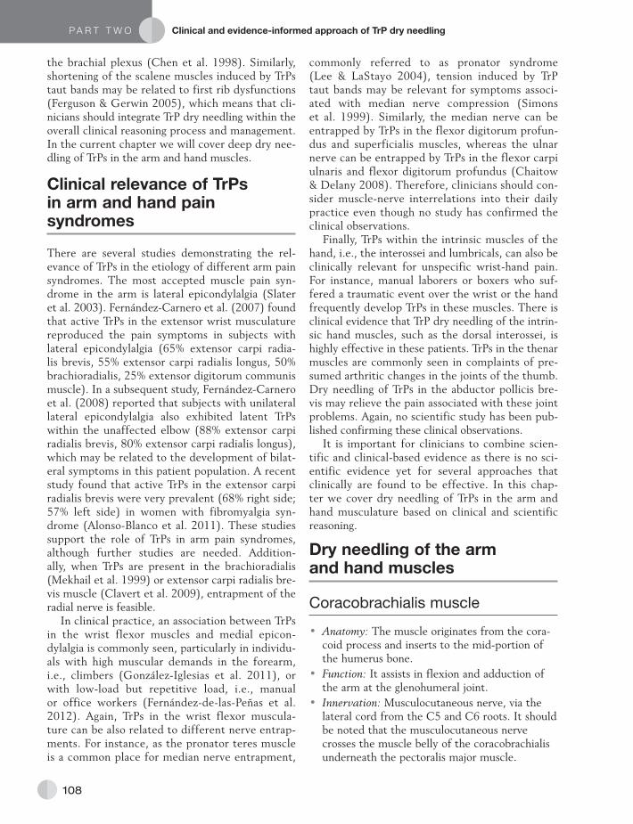
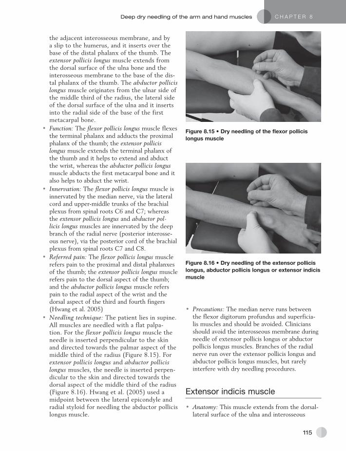
• Needling technique: The patient lies in supine. All muscles are needled with a flat palpa-tion. For the flexor pollicis longus muscle the needle is inserted perpendicular to the skin and directed towards the palmar aspect of the middle third of the radius ( Figure 8.15 ). For extensor pollicis longus and abductor pollicis longus muscles, the needle is inserted perpen-dicular to the skin and directed towards the dorsal aspect of the middle third of the radius ( Figure 8.16 ). Hwang et al. (2005) used a midpoint between the lateral epicondyle and radial styloid for needling the abductor pollicis longus muscle.
• Precautions: The median nerve runs between the flexor digitorum profundus and superficia-lis muscles and should be avoided. Clinicians should avoid the interosseous membrane during needle of extensor pollicis longus or abductor pollicis longus muscles. Branches of the radial nerve run over the extensor pollicis longus and abductor pollicis longus muscles, but rarely interfere with dry needling procedures.
Extensor indicis muscle
• Anatomy: This muscle extends from the dorsal-lateral surface of the ulna and interosseous
Figure 8.15 • Dry needling of the fl exor pollicis longus muscle
Figure 8.16 • Dry needling of the extensor pollicis longus, abductor pollicis longus or extensor indicis muscle

Clinical and evidence-informed approach of TrP dry needlingP A R T T W O
116
membrane to the dorsal aspect of the second metacarpal bone.
• Function : This muscle extends the second finger.
• Innervation: This muscle is innervated by the deep branch of the radial nerve (posterior inter-osseous nerve), via the posterior cord of the brachial plexus from spinal roots C7 and C8.
• Referred pain: It refers pain to the dorsal aspect of the second finger.
• Needling technique: The needle is inserted per-pendicular to the skin towards the dorsal aspect of the middle third of the radius, similarly to the extensor pollicis longus and abductor pollicis lon-gus muscles ( Figure 8.16 ). The referred pain and the muscle contraction will assist the clinician to focus the needling in one or other muscle.
• Precautions: Clinicians should avoid the interos-seous membrane during needle.
Adductor pollicis, opponens pollicis, fl exor pollicis brevis and abductor pollicis brevis muscles
• Anatomy: The adductor pollicis muscle originates in the carpometacarpal region of the index and middle fingers, to the base of the proximal pha-lanx of the thumb. The opponens pollicis muscle extends from the trapezium bone of the wrist and the flexor retinaculum in the heel of the hand to wrap partially around, and attaches to the first metacarpal bone. The flexor pollicis brevis muscle extends from the trapezium, trapezoid, and capitate bones, and the flexor retinaculum to the palmar aspect of the first metacarpal and sesamoid bone. The abductor pollicis brevis mus-cle originates in the scaphoid bone and the flexor retinaculum and inserts to the lateral aspect of the first metacarpal and sesamoid bones.
• Function: The adductor pollicis muscle adducts the thumb toward the index finger, the opponens pollicis opposites the thumb pad across the palm to touch the pads of the ring or little fingers; the flexor pollicis brevis muscle flexes the thumb toward the palm; and the abductor pollicis bre-vis muscle abducts the thumb away the palm.
• Innervation: The adductor pollicis muscle is supplied by the deep palmar branch of the ulnar nerve, via the medial cord and lower trunk of the brachial plexus from spinal roots
C8 and T1, whereas the opponens pollicis, flexor pollicis brevis, and abductor pollicis brevis muscles are supplied by a branch of the median nerve, via the lateral cord and upper-middle trunks of the brachial plexus from spi-nal roots C6 and C7.
• Referred pain: The a dductor pollicis muscle refers pain to the ulnar aspect of the thumb; the opponens pollicis muscle projects pain to the pal-mar aspect of the thumb, the flexor pollicis bre-vis muscle refers pain to the palmar aspect of the thumb, and the abductor pollicis brevis muscle projects pain to the radial aspect of the thumb.
• Needling technique: Generally, a short but thin needle is used for the thenar muscles such as a 0.14 × 15 mm needle. The patient lies in supine with the forearm pronated (for the adductor pollicis muscle) or supinated (for the remaining muscles). These muscles are needled with a pincer palpation. The needle is inserted perpendicular to the skin and directed to the muscle. For the adductor pollicis muscle the needle is inserted between the first and sec-ond metacarpal bones ( Figure 8.17 ); for the opponens and flexor pollicis brevis muscles the needle is inserted via the radial aspect of the first metacarpal (to avoid palmar fascia) towards the thenar eminency ( Figure 8.18 ); and for the abductor pollicis brevis muscle the needle is directed towards the radial aspect of the first metacarpal ( Figure 8.19 ).
• Precautions: The flexor pollicis longus ten-don crosses between the adductor pollicis and opponens pollicis muscles and should be avoided.
Figure 8.17 • Dry needling of the adductor pollicis muscle
Deep dry needling of the arm and hand muscles C H A P T E R 8
117
Interosseous, lumbricals, and abductor digiti minimus muscles
• Anatomy: The interosseous muscles (dorsal or palmar) lie between the adjacent metacarpal bones. The lumbrical muscle s attach proximally to the four tendons of the flexor digitorum pro-fundus in the mid-palm, distally to the radial side of the aponeurosis on each of the four fin-gers. The abductor digiti minimus muscle arises proximally from the pisiform bone, and attaches distally to the ulnar side of the base of the first phalanx of the little finger.
• Function: The dorsal interosseous muscles moves a finger away from the midline of the middle finger (abduction); the palmar interosseous muscles adduct each of the other fingers toward
the middle finger (adduction); the lumbricals inhibit flexion of a distal phalanx via the exten-sor mechanism; whereas the abductor digiti minimus abducts the little finger.
• Innervation: The interosseous, the abductor digiti minimus and the third-fourth lumbricals muscles are supplied by branches of ulnar nerve, via the medial cord and lower trunk of the bra-chial plexus from spinal roots C8 and T1. The first and second lumbricals are supplied by the median nerve, via the lateral cord and upper-middle trunks of the brachial plexus from spinal roots C6 and C7.
• Referred pain: The dorsal and palmar interos-seous muscles project their pain along the side of the finger to which that interosseous muscle attaches. The first dorsal interosseous also proj-ects to the dorsum of the hand and ulnar side of the little finger. Referred pain from the lumbri-cals is similar to the referred pain of the interos-sei. The abductor digiti minimus muscle pain extends along the lateral side of the little finger.
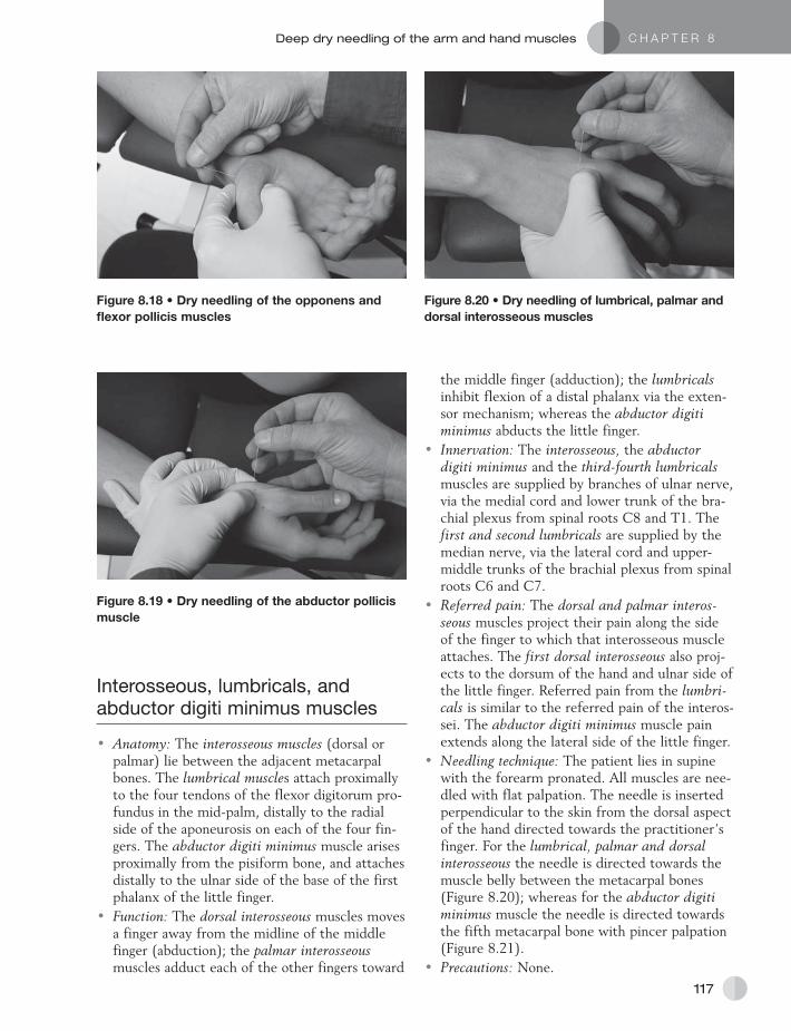
• Needling technique: The patient lies in supine with the forearm pronated. All muscles are nee-dled with flat palpation. The needle is inserted perpendicular to the skin from the dorsal aspect of the hand directed towards the practitioner’s finger. For the lumbrical, palmar and dorsal interosseous the needle is directed towards the muscle belly between the metacarpal bones ( Figure 8.20 ); whereas for the abductor digiti minimus muscle the needle is directed towards the fifth metacarpal bone with pincer palpation ( Figure 8.21 ).
• Precautions: None.
Figure 8.18 • Dry needling of the opponens and fl exor pollicis muscles
Figure 8.19 • Dry needling of the abductor pollicis muscle
Figure 8.20 • Dry needling of lumbrical, palmar and dorsal interosseous muscles

Clinical and evidence-informed approach of TrP dry needlingP A R T T W O
118
Alonso-Blanco , C. , Fernández-de-las Peñas , C. , Morales-Cabezas , M. , et al., 2011 . Multiple active myo-fascial trigger points reproduce the overall spontaneous pain pattern in women with fibromyalgia and are related to widespread mechanical hypersensitivity . Clin J Pain 27 , 405 – 413 .
Chaitow , L. , Delany , J. , 2008 . Clinical application of neuromuscular tech-niques: The upper body , Shoulder, arm and hand . Second ed . Elsevier , London, 445 .
Clavert , P. , Lutz , J.C. , Adam , P. , et al., 2009 . Frohse’s arcade is not the exclusive compression site of the radial nerve in its tunnel . Orthopae-dics & Traumatology: Surg & Res. 95 , 114 – 118 .
Ferguson , L. , Gerwin , R. , 2005 . Shoul-der dysfunction and frozen shoulder . In: Ferguson , L. , Gerwin , R. (Eds.), Clinical mastery in the treatment of myofascial pain , Lippincott Williams & Wilkins , Baltimore , pp. 91 – 121 .
Fernández-Carnero , J. , Fernández-de-las Peñas , C. , De-la-Llave-Rincón , A.I. , et al., 2007 . Prevalence of and referred pain from myofascial trig-ger points in the forearm muscles in patients with lateral epicondylalgia . Clin J Pain 23 , 353 – 360 .
Fernández-Carnero , J. , Fernández-de-las Peñas , C. , De-la-Llave-Rincón , A.I. , et al., 2008 . Bilateral myofas-cial trigger points in the forearm muscles in chronic unilateral lateral epicondylalgia: A blinded controlled study . Clin J Pain 24 , 802 – 807 .
Fernández-de-las Peñas , C. , Gröbli , C. , Ortega Santiago , R. , et al., 2012 . Referred pain from myofascial trig-ger points in head, neck, shoulder and arm muscles reproduces pain symptoms in blue-collar (manual) and white-collar (office) workers . Clin J Pain 28, 511-518 .
González-Iglesias , J. , Cleland , J.A. , Gutiérrez-Vega , M.R. , Fernández-de-las Peñas , C. , 2011 . Multimodal management of lateral epicondylal-gia in rock climbers: A prospective case series . J Manipul Physiol Ther. 34 , 635 – 642 .
Hwang , M. , Kang , Y.K. , Shin , J.Y. , Kim , D.H. , 2005 . Referred pain pat-tern of the abductor pollicis longus muscle . Am J Phys Med Rehabil. 84 , 593 – 597 .
Lee , M.J. , LaStayo , D. , 2004 . Pronator syndrome and other nerve compres-sions that mimic carpal tunnel syn-drome . J Orthopaedics Sports Phys Ther. 34 , 601 – 609 .
Maeda , S. , Kawai , K. , Koizumi , M. , et al., 2009 . Morphological study of the communication between the musculo-cutaneous and median nerves . Anatomical Sci Internat. 84 , 34 – 40 .
Mekhail , A.O. , Checroun , A.J. , Ebra-heim , N.A. , et al., 1999 . Extensile approach to the anterolateral surface of the humerus and the radial nerve . J Shoulder Elbow Sur. 8 , 112 – 118 .
Qerama , E. , Kasch , H. , Fuglsang-Frederiksen , A. , 2009 . Occurrence of myofascial pain in patients with possible carpal tunnel syndrome: A single-blinded study . Eur J Pain 13 , 588 – 591 .
Rezzouk , J. , Durandeau , A. , Vital , J.M. , Fabre , T. , 2002 . Long head of the triceps brachii in axillary nerve injury: anatomy and clinical aspects . Rev Chir Orthop Reparatrice Appar Mot. 88 , 561 – 564 .
Schneider , M. , 2005 . Tennis elbow . In: Ferguson , L. , Gerwin , R. (Eds.), Clinical mastery in the treatment of myofascial pain , Lippincott Williams & Wilkins , Baltimore , pp. 122 – 144 .
Simons , D.G. , Travell , J.G. , Simons , L.S. , 1999 . Second ed . Travell & Simons’ Myofascial pain and dys-function: the trigger point manual . Vol. 1, Lippincott William & Wilkins , Baltimore, pp278 – 307 .
Slater , H. , Arendt-Nielsen , L. , Wright , A. , Graven-Nielsen , T. , 2003 . Experimental deep tissue pain in wrist extensors: a model of lateral epicondylalgia . Eur J Pain 7 , 277 – 288 .
Tatar , I. , Kocabiyik , N. , Gayretli , O. , Ozan , H. , 2009 . The course and branching pattern of the deep branch of the radial nerve in rela-tion to the supinator muscle in fetus elbow . Surg Radiol Anat. 31 , 591 – 596 .
Figure 8.21 • Dry needling of the abductor digiti minimus muscle.
References