Embed Size (px)
Citation preview
1
Deep Convolutional Neural Networks forComputer-Aided Detection: CNN Architectures,Dataset Characteristics and Transfer Learning
Hoo-Chang Shin, Member, IEEE, Holger R. Roth, Mingchen Gao, Le Lu, Senior Member, IEEE, Ziyue Xu,Isabella Nogues, Jianhua Yao, Daniel Mollura, Ronald M. Summers*
Abstract—Remarkable progress has been made in image recog-nition, primarily due to the availability of large-scale annotateddatasets (i.e. ImageNet) and the revival of deep convolutionalneural networks (CNN). CNNs enable learning data-driven,highly representative, layered hierarchical image features fromsufficient training data. However, obtaining datasets as compre-hensively annotated as ImageNet in the medical imaging domainremains a challenge. There are currently three major techniquesthat successfully employ CNNs to medical image classification:training the CNN from scratch, using off-the-shelf pre-trainedCNN features, and conducting unsupervised CNN pre-trainingwith supervised fine-tuning. Another effective method is transferlearning, i.e., fine-tuning CNN models (supervised) pre-trainedfrom natural image dataset to medical image tasks (althoughdomain transfer between two medical image datasets is alsopossible).
In this paper, we exploit three important, but previouslyunderstudied factors of employing deep convolutional neuralnetworks to computer-aided detection problems. We first exploreand evaluate different CNN architectures. The studied modelscontain 5 thousand to 160 million parameters, and vary innumbers of layers. We then evaluate the influence of dataset scaleand spatial image context on performance. Finally, we examinewhen and why transfer learning from pre-trained ImageNet (viafine-tuning) can be useful. We study two specific computer-aided detection (CADe) problems, namely thoraco-abdominallymph node (LN) detection and interstitial lung disease (ILD)classification. We achieve the state-of-the-art performance onthe mediastinal LN detection, with 85% sensitivity at 3 falsepositive per patient, and report the first five-fold cross-validationclassification results on predicting axial CT slices with ILD cate-gories. Our extensive empirical evaluation, CNN model analysisand valuable insights can be extended to the design of highperformance CAD systems for other medical imaging tasks.
I. INTRODUCTION
Tremendous progress has been made in image recogni-tion, primarily due to the availability of large-scale anno-tated datasets (i.e. ImageNet [1], [2]) and the recent revivalof deep convolutional neural networks (CNN) [3], [4]. For
Hoo-Chang Shin, Holger R. Roth, Le Lu, Isabella Nogues, Jianhua Yao andRonald M. Summers are with the Imaging Biomarkers and Computer-AidedDiagnosis Laboratory; Mingchen Gao, Ziyue Xu and Daniel Mollura arewith Center for Infectious Disease Imaging, Le Lu, Jianhua Yao and RonaldM. Summers are also with Clinical Image Processing Service, Radiologyand Imaging Sciences Department, National Institutes of Health ClinicalCenter, Bethesda, MD 20892-1182, USA. Holger R. Roth and Mingchen Gaocontributed equally to this work. Asterisk indicates corresponding author.e-mail: {hoochang.shin, le.lu, rms}@nih.gov. Copyright (c) 2010 IEEE.Personal use of this material is permitted. However, permission to use thismaterial for any other purposes must be obtained from the IEEE by sendinga request to [email protected].
data-driven learning, large-scale well-annotated datasets withrepresentative data distribution characteristics are crucial tolearning more accurate or generalizable models [5], [4]. Unlikeprevious image datasets used in computer vision, ImageNet[1] offers a very comprehensive database of more than 1.2million categorized natural images of 1000+ classes. The CNNmodels trained upon this database serve as the backbonefor significantly improving many object detection and imagesegmentation problems using other datasets [6], [7], e.g.,PASCAL [8] and medical image categorization [9], [10], [11],[12]. However, there exists no large-scale annotated medicalimage dataset comparable to ImageNet, as data acquisition isdifficult, and quality annotation is costly.
There are currently three major techniques that successfullyemploy CNNs to medical image classification: 1) training the“CNN from scratch” [13], [14], [15], [16], [17]; 2) using“off-the-shelf CNN” features (without retraining the CNN) ascomplementary information channels to existing hand-craftedimage features, for Chest X-rays [10] and CT lung noduleidentification [9], [12]; and 3) performing unsupervised pre-training on natural or medical images and fine-tuning on med-ical target images using CNN or other types of deep learningmodels [18], [19], [20], [21]. A decompositional 2.5D viewresampling and an aggregation of random view classificationscores are used to eliminate the “curse-of-dimensionality”issue in [22], in order to acquire a sufficient number of trainingimage samples.
Previous studies have analyzed three-dimensional patchcreation for LN detection [23], [24], atlas creation from chestCT [25] and the extraction of multi-level image features [26],[27]. At present, there are several extensions or variations ofthe decompositional view representation introduced in [22],[28], such as: using a novel vessel-aligned multi-planar imagerepresentation for pulmonary embolism detection [29], fusingunregistered multiview for mammogram analysis [16] andclassifying pulmonary peri-fissural nodules via an ensembleof 2D views [12].
Although natural images and medical images differ signif-icantly, conventional image descriptors developed for objectrecognition in natural images, such as the scale-invariantfeature transform (SIFT) [30] and the histogram of orientedgradients (HOG) [31], have been widely used for object de-tection and segmentation in medical image analysis. Recently,ImageNet pre-trained CNNs have been used for chest pathol-ogy identification and detection in X-ray and CT modalities
arX
iv:1
602.
0340
9v1
[cs
.CV
] 1
0 Fe
b 20
16
2
[10], [9], [12]. They have yielded the best performance resultsby integrating low-level image features (e.g., GIST [32], bag ofvisual words (BoVW) and bag-of-frequency [12]). However,the fine-tuning of an ImageNet pre-trained CNN model onmedical image datasets has not yet been exploited.
In this paper, we exploit three important, but previouslyunder-studied factors of employing deep convolutional neuralnetworks to computer-aided detection problems. Particularly,we explore and evaluate different CNN architectures varying inwidth (ranging from 5 thousand to 160 million parameters) anddepth (various numbers of layers), describe the effects of vary-ing dataset scale and spatial image context on performance,and discuss when and why transfer learning from pre-trainedImageNet CNN models can be valuable. We further verifyour hypothesis by inheriting and adapting rich hierarchicalimage features [5], [33] from the large-scale ImageNet datasetfor computer aided diagnosis (CAD). We also explore CNNarchitectures of the most studied seven-layered “AlexNet-CNN” [4], a shallower “Cifar-CNN” [22], and a much deeperversion of “GoogLeNet-CNN” [33] (with our modificationson CNN structures). This study is partially motivated byrecent studies [34], [35] in computer vision. The thoroughquantitative analysis and evaluation on deep CNN [34] orsparsity image coding methods [35] elucidate the emergingtechniques of the time and provide useful suggestions for theirfuture stages of development, respectively.
Two specific computer-aided detection (CADe) problems,namely thoraco-abdominal lymph node (LN) detection andinterstitial lung disease (ILD) classification are studied in thiswork. On mediastinal LN detection, we surpass all currentlyreported results. We obtain 86% sensitivity on 3 false positives(FP) per patient, versus the prior state-of-art sensitivities of78% [36] (stacked shallow learning) and 70% [22] (CNN),as prior state-of-the-art. For the first time, ILD classificationresults under the patient-level five-fold cross-validation proto-col (CV5) are investigated and reported. The ILD dataset [37]contains 905 annotated image slices with 120 patients and6 ILD labels. Such sparsely annotated datasets are generallydifficult for CNN learning, due to the paucity of labeledinstances.
Evaluation protocols and details are critical to derivingsignificant empirical findings [34]. Our experimental resultssuggest that different CNN architectures and dataset re-sampling protocols are critical for the LN detection taskswhere the amount of labeled training data is sufficient andspatial contexts are local. Since LN images are more flexiblethan ILD images with respect to resampling and reformatting,LN datasets may be more readily augmented by such imagetransformations. As a result, LN datasets contain more trainingand testing data instances (due to data auugmentation) thanILD datasets. They nonetheless remain less comprehensivethan natural image datasets, such as ImageNet. Fine-tuningImageNet-trained models for ILD classification is clearlyadvantageous and yields early promising results, when theamount of labeled training data is highly insufficient and multi-class categorization is used, as opposed to the LN dataset’sbinary class categorization. Another significant finding is thatCNNs trained from scratch or fine-tuned from ImageNet mod-
els consistently outperform CNNs that merely use off-the-shelfCNN features, in both the LN and ILD classification problems.We further analyze, via CNN activation visualizations, whenand why transfer learning from non-medical to medical imagesin CADe problems can be valuable.
II. DATASETS AND RELATED WORK
We employ CNNs (with the characteristics defined above)to thoraco-abdominal lymph node (LN) detection (evaluatedseparately on the mediastinal and abdominal regions) andinterstitial lung disease (ILD) detection. For LN detection, weuse randomly sampled 2.5D views in CT [22]. We use 2D CTslices [38], [39], [40] for ILD detection. We then evaluate andcompare CNN performance results.
Until the detection aggregation approach [22], [41], thora-coabdominal lymph node (LN) detection via CADe mecha-nisms has yielded poor performance results. In [22], each 3DLN candidate produces up to 100 random 2.5D orthogonallysampled images or views which are then used to train aneffective CNN model. The best performance on abdominalLN detection is achieved at 83% recall on 3FP per patient[22], using a “Cifar-10” CNN. Using the thoracoabdominalLN detection datasets [22], we aim to surpass this CADeperformance level, by testing different CNN architectures,exploring various dataset re-sampling protocols, and applyingtransfer learning from ImageNet pre-trained CNN models.
Interstitial lung disease (ILD) comprises more than 150 lungdiseases affecting the interstitium, which can severely impairthe patient’s ability to breathe. Gao et al. [40] investigatethe ILD classification problem in two scenarios: 1) slice-level classification: assigning a holistic two-dimensional axialCT slice image with its occurring ILD disease label(s); and2) patch-level classification: a/ sampling patches within the2D ROIs (Regions of Interest provided by [37]), then b/classifying patches into seven category labels ( six diseaselabels and one “healthy” label). Song et al. [38], [39] onlyaddress the second sub-task of patch-level classification underthe “leave-one-patient-out” (LOO) criterion. By training on themoderate-to-small scale ILD dataset [37], our main objectiveis to exploit and benchmark CNN based ILD classificationperformances under the CV5 metric (which is more realisticand unbiased than LOO [38], [39] and hard-split [40]), withand without transfer learning.
Thoracoabdominal Lymph Node Datasets. We use thepublicly available dataset from [22], [41]. There are 388mediastinal LNs labeled by radiologists in 90 patient CT scans,and 595 abdominal LNs in 86 patient CT scans. To facilitatecomparison, we adopt the data preparation protocol of [22],where positive and negative LN candidates are sampled withthe fields-of-view (FOVs) of 30mm to 45mm, surrounding theannotated and detected LN centers (obtained by a candidategeneration process). More precisely, [22], [41], [36] followa coarse-to-fine CADe scheme, partially inspired by [42],which operates with ∼ 100% detection recalls at the cost ofapproximately 40 false or negative LN candidates per patientscan. In this work, positive and negative LN candidate arefirst sampled up to 200 times with translations and rotations.
3
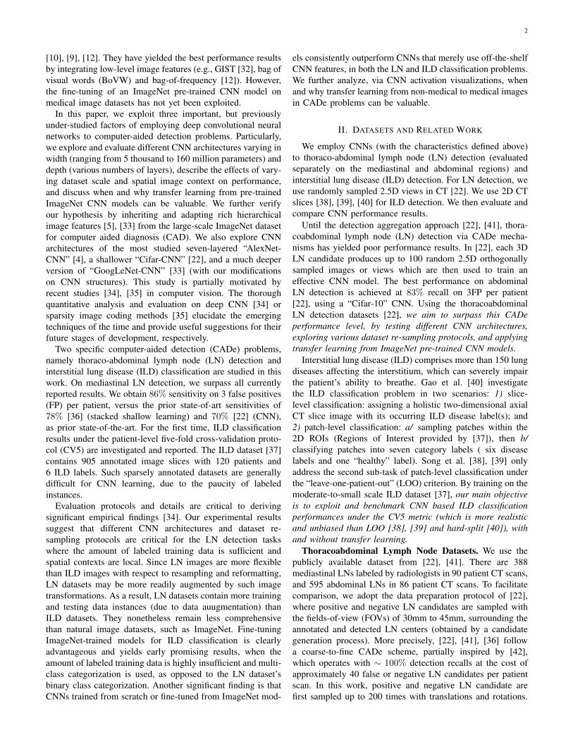
Fig. 1. Some examples of abdominal and mediastinal lymph nodes sampledon axial (ax), coronal (co), and sagittal (sa) views, with four different fields-of-views (30mm: orange; 45mm: red; 85mm: green; 128mm: blue) surroundinglymph nodes.
Afterwards, negative LN samples are randomly re-selected at alower rate close to the total number of positives. LN candidatesare randomly extracted from fields-of-view (FOVs) spanning35mm to 128mm in soft-tissue window [-100, 200HU]. Thisallows us to capture multiple spatial scales of image context[43], [44]). The samples are then rescaled to a 64× 64 pixelresolution via B-spline interpolation. A few examples of LNswith axial, coronal, and sagittal views encoded in RGB colorimages [22] are shown in Figure 1.
Unlike the heart or the liver, lymph nodes have no pre-determined anatomic orientation. Hence, the purely randomimage resampling (with respect to scale, displacement andorientation) and reformatting (the axial, coronal, and sagittalviews are in any system randomly resampled coordinates)is a natural choice, which also happens to yield high CNNperformance. Although we integrate three channels of informa-tion from three orthogonal views for LN detection, the pixel-wise spatial correlations between or among channels are notnecessary. The convolutional kernels in the lower level CNNarchitectures can learn the optimal weights to linearly combinethe observations from the axial, coronal, and sagittal channelsby computing their dot-products. Transforming axial, coronal,and sagittal representations to RGB also facilitates transferlearning from CNN models trained on ImageNet.
This learning representation (i.e., “built-in CNN”) is flexi-ble, in that it naturally combines multiple sources or channelsof information. In the recent literature [45], even heteroge-neous class-conditional probability maps can be combinedwith raw images to improve performance. This set-up issimilar to that of other works in computer vision, suchas [46], where heterogeneous image information channelsare jointly fed into the CNN convolutional layers for high-accuracy human parsing and segmentation. Finally, if thereare correlations among CNN input channels, one may observethe corresponding correlated patterns in the learned filters.
In summary, the assumption that there are or must be pixel-wise spatial correlations among input channels does not applyto the CNN model representation. For other medical imagingproblems, such as pulmonary embolism detection [29], in
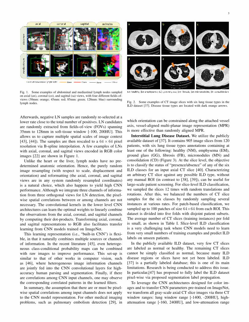
Fig. 2. Some examples of CT image slices with six lung tissue types in theILD dataset [37]. Disease tissue types are located with dark orange arrows.
which orientation can be constrained along the attached vesselaxis, vessel-aligned multi-planar image representation (MPR)is more effective than randomly aligned MPR.
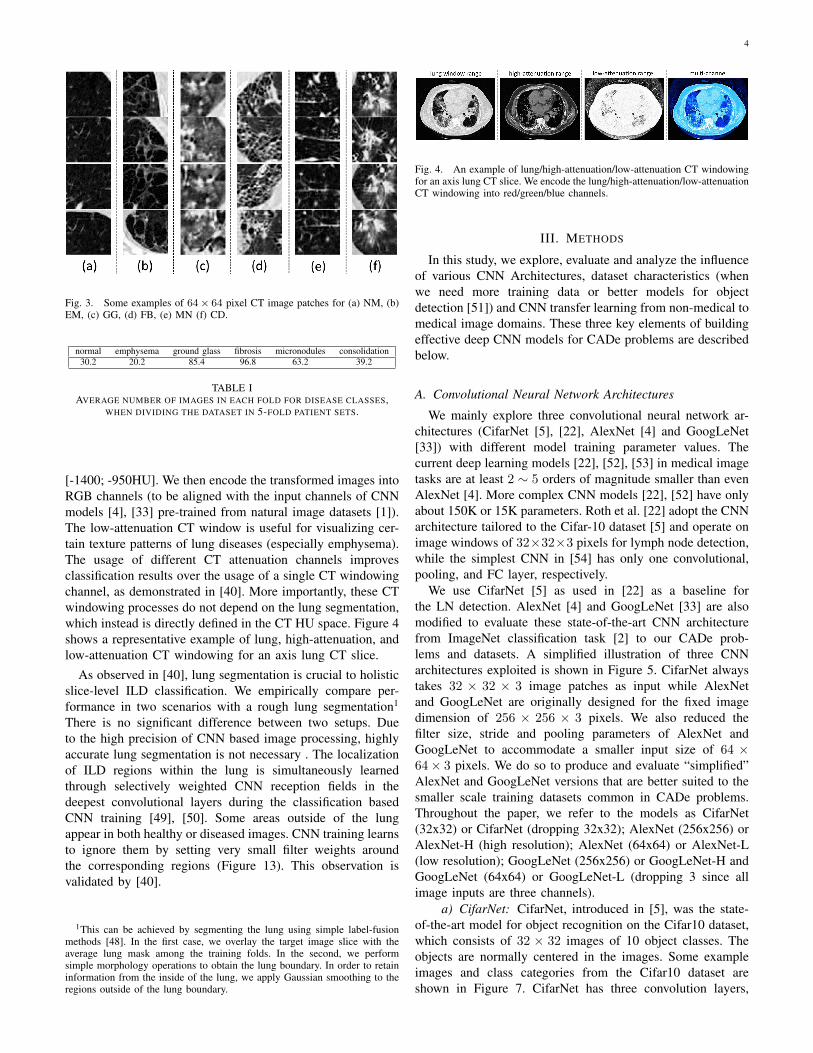
Interstitial Lung Disease Dataset. We utilize the publiclyavailable dataset of [37]. It contains 905 image slices from 120patients, with six lung tissue types annotations containing atleast one of the following: healthy (NM), emphysema (EM),ground glass (GG), fibrosis (FB), micronodules (MN) andconsolidation (CD) (Figure 3). At the slice level, the objectiveis to classify the status of “presence/absence” of any of the sixILD classes for an input axial CT slice [40]. Characterizingan arbitrary CT slice against any possible ILD type, withoutany manual ROI (in contrast to [38], [39]), can be useful forlarge-scale patient screening. For slice-level ILD classification,we sampled the slices 12 times with random translations androtations. After this, we balanced the numbers of CT slicesamples for the six classes by randomly sampling severalinstances at various rates. For patch-based classification, wesampled up to 100 patches of size 64×64 from each ROI. Thisdataset is divided into five folds with disjoint patient subsets.The average number of CT slices (training instances) per foldis small, as shown in Table I. Slice-level ILD classificationis a very challenging task where CNN models need to learnfrom very small numbers of training examples and predict ILDlabels on unseen patients.
In the publicly available ILD dataset, very few CT slicesare labeled as normal or healthy. The remaining CT slicescannot be simply classified as normal, because many ILDdisease regions or slices have not yet been labeled. ILD[37] is a partially labeled database; this is one of its mainlimitations. Research is being conducted to address this issue.In particular,[47] has proposed to fully label the ILD datasetpixel-wise via proposed segmentation label propagation.
To leverage the CNN architectures designed for color im-ages and to transfer CNN parameters pre-trained on ImageNet,we transform all gray-scale axial CT slice images via three CTwindow ranges: lung window range [-1400, -200HU], high-attenuation range [-160, 240HU], and low-attenuation range
4
Fig. 3. Some examples of 64× 64 pixel CT image patches for (a) NM, (b)EM, (c) GG, (d) FB, (e) MN (f) CD.
normal emphysema ground glass fibrosis micronodules consolidation30.2 20.2 85.4 96.8 63.2 39.2
TABLE IAVERAGE NUMBER OF IMAGES IN EACH FOLD FOR DISEASE CLASSES,
WHEN DIVIDING THE DATASET IN 5-FOLD PATIENT SETS.
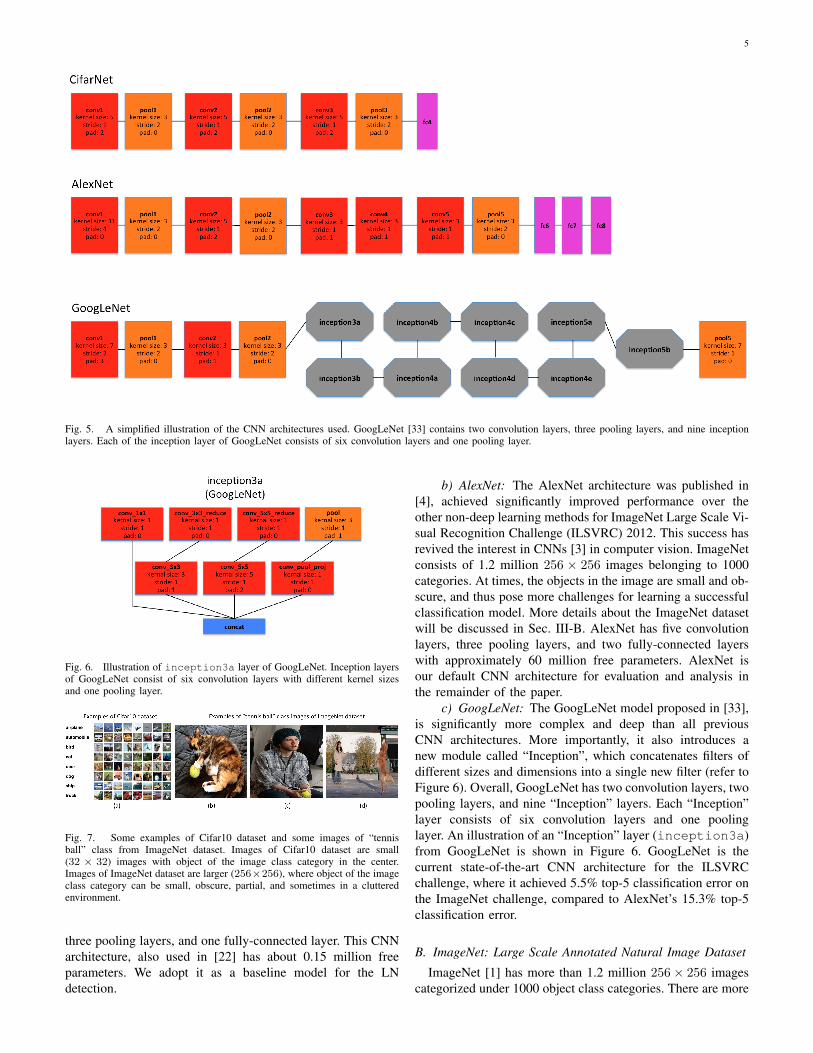
[-1400; -950HU]. We then encode the transformed images intoRGB channels (to be aligned with the input channels of CNNmodels [4], [33] pre-trained from natural image datasets [1]).The low-attenuation CT window is useful for visualizing cer-tain texture patterns of lung diseases (especially emphysema).The usage of different CT attenuation channels improvesclassification results over the usage of a single CT windowingchannel, as demonstrated in [40]. More importantly, these CTwindowing processes do not depend on the lung segmentation,which instead is directly defined in the CT HU space. Figure 4shows a representative example of lung, high-attenuation, andlow-attenuation CT windowing for an axis lung CT slice.
As observed in [40], lung segmentation is crucial to holisticslice-level ILD classification. We empirically compare per-formance in two scenarios with a rough lung segmentation1
There is no significant difference between two setups. Dueto the high precision of CNN based image processing, highlyaccurate lung segmentation is not necessary . The localizationof ILD regions within the lung is simultaneously learnedthrough selectively weighted CNN reception fields in thedeepest convolutional layers during the classification basedCNN training [49], [50]. Some areas outside of the lungappear in both healthy or diseased images. CNN training learnsto ignore them by setting very small filter weights aroundthe corresponding regions (Figure 13). This observation isvalidated by [40].
1This can be achieved by segmenting the lung using simple label-fusionmethods [48]. In the first case, we overlay the target image slice with theaverage lung mask among the training folds. In the second, we performsimple morphology operations to obtain the lung boundary. In order to retaininformation from the inside of the lung, we apply Gaussian smoothing to theregions outside of the lung boundary.
Fig. 4. An example of lung/high-attenuation/low-attenuation CT windowingfor an axis lung CT slice. We encode the lung/high-attenuation/low-attenuationCT windowing into red/green/blue channels.
III. METHODS
In this study, we explore, evaluate and analyze the influenceof various CNN Architectures, dataset characteristics (whenwe need more training data or better models for objectdetection [51]) and CNN transfer learning from non-medical tomedical image domains. These three key elements of buildingeffective deep CNN models for CADe problems are describedbelow.
A. Convolutional Neural Network Architectures
We mainly explore three convolutional neural network ar-chitectures (CifarNet [5], [22], AlexNet [4] and GoogLeNet[33]) with different model training parameter values. Thecurrent deep learning models [22], [52], [53] in medical imagetasks are at least 2 ∼ 5 orders of magnitude smaller than evenAlexNet [4]. More complex CNN models [22], [52] have onlyabout 150K or 15K parameters. Roth et al. [22] adopt the CNNarchitecture tailored to the Cifar-10 dataset [5] and operate onimage windows of 32×32×3 pixels for lymph node detection,while the simplest CNN in [54] has only one convolutional,pooling, and FC layer, respectively.
We use CifarNet [5] as used in [22] as a baseline forthe LN detection. AlexNet [4] and GoogLeNet [33] are alsomodified to evaluate these state-of-the-art CNN architecturefrom ImageNet classification task [2] to our CADe prob-lems and datasets. A simplified illustration of three CNNarchitectures exploited is shown in Figure 5. CifarNet alwaystakes 32 × 32 × 3 image patches as input while AlexNetand GoogLeNet are originally designed for the fixed imagedimension of 256 × 256 × 3 pixels. We also reduced thefilter size, stride and pooling parameters of AlexNet andGoogLeNet to accommodate a smaller input size of 64 ×64× 3 pixels. We do so to produce and evaluate “simplified”AlexNet and GoogLeNet versions that are better suited to thesmaller scale training datasets common in CADe problems.Throughout the paper, we refer to the models as CifarNet(32x32) or CifarNet (dropping 32x32); AlexNet (256x256) orAlexNet-H (high resolution); AlexNet (64x64) or AlexNet-L(low resolution); GoogLeNet (256x256) or GoogLeNet-H andGoogLeNet (64x64) or GoogLeNet-L (dropping 3 since allimage inputs are three channels).
a) CifarNet: CifarNet, introduced in [5], was the state-of-the-art model for object recognition on the Cifar10 dataset,which consists of 32 × 32 images of 10 object classes. Theobjects are normally centered in the images. Some exampleimages and class categories from the Cifar10 dataset areshown in Figure 7. CifarNet has three convolution layers,
5
Fig. 5. A simplified illustration of the CNN architectures used. GoogLeNet [33] contains two convolution layers, three pooling layers, and nine inceptionlayers. Each of the inception layer of GoogLeNet consists of six convolution layers and one pooling layer.
Fig. 6. Illustration of inception3a layer of GoogLeNet. Inception layersof GoogLeNet consist of six convolution layers with different kernel sizesand one pooling layer.
Fig. 7. Some examples of Cifar10 dataset and some images of “tennisball” class from ImageNet dataset. Images of Cifar10 dataset are small(32 × 32) images with object of the image class category in the center.Images of ImageNet dataset are larger (256×256), where object of the imageclass category can be small, obscure, partial, and sometimes in a clutteredenvironment.
three pooling layers, and one fully-connected layer. This CNNarchitecture, also used in [22] has about 0.15 million freeparameters. We adopt it as a baseline model for the LNdetection.
b) AlexNet: The AlexNet architecture was published in[4], achieved significantly improved performance over theother non-deep learning methods for ImageNet Large Scale Vi-sual Recognition Challenge (ILSVRC) 2012. This success hasrevived the interest in CNNs [3] in computer vision. ImageNetconsists of 1.2 million 256 × 256 images belonging to 1000categories. At times, the objects in the image are small and ob-scure, and thus pose more challenges for learning a successfulclassification model. More details about the ImageNet datasetwill be discussed in Sec. III-B. AlexNet has five convolutionlayers, three pooling layers, and two fully-connected layerswith approximately 60 million free parameters. AlexNet isour default CNN architecture for evaluation and analysis inthe remainder of the paper.
c) GoogLeNet: The GoogLeNet model proposed in [33],is significantly more complex and deep than all previousCNN architectures. More importantly, it also introduces anew module called “Inception”, which concatenates filters ofdifferent sizes and dimensions into a single new filter (refer toFigure 6). Overall, GoogLeNet has two convolution layers, twopooling layers, and nine “Inception” layers. Each “Inception”layer consists of six convolution layers and one poolinglayer. An illustration of an “Inception” layer (inception3a)from GoogLeNet is shown in Figure 6. GoogLeNet is thecurrent state-of-the-art CNN architecture for the ILSVRCchallenge, where it achieved 5.5% top-5 classification error onthe ImageNet challenge, compared to AlexNet’s 15.3% top-5classification error.
B. ImageNet: Large Scale Annotated Natural Image Dataset
ImageNet [1] has more than 1.2 million 256× 256 imagescategorized under 1000 object class categories. There are more
6
than 1000 training images per class. The database is organizedaccording to the WordNet [55] hierarchy, which currentlycontains only nouns in 1000 object categories. The image-object labels are obtained largely through crowd-sourcing,e.g., Amazon Mechanical Turk, and human inspection. Someexamples of object categories in ImageNet are “sea snake”,“sandwich”, “vase”, “leopard”, etc. ImageNet is currently thelargest image dataset among other standard datasets for visualrecognition. Indeed, the Caltech101, Caltech256 and Cifar10dataset merely contain 60000 32 × 32 images and 10 objectclasses. Furthermore, due to the large number (1000+) ofobject classes, the objects belonging to each ImageNet classcategory can be occluded, partial and small, relative to those inthe previous public image datasets. This significant intra-classvariation poses greater challenges to any data-driven learningsystem that builds a classifier to fit given data and generalizeto unseen data. For comparison, some example images ofCifar10 dataset and ImageNet images in the “tennis ball”class category are shown in Figure 7. The ImageNet datasetis publicly available, and the ImageNet Large Scale VisualRecognition Challenge (ILSVRC) has become the standardbenchmark for large-scale object recognition.
C. Training Protocols and Transfer Learning
When learned from scratch, all the parameters of CNNmodels are initialized with random Gaussian distributionsand trained for 30 epochs with the mini-batch size of 50image instances. Training convergence can be observed within30 epochs. The other hyperparameters are momentum: 0.9;weight decay: 0.0005; (base) learning rate: 0.01, decreased bya factor of 10 at every 10 epochs. We use the Caffe framework[56] and NVidia K40 GPUs to train the CNNs.
AlexNet and GoogLeNet CNN models can be either learnedfrom scratch or fine-tuned from pre-trained models. Gir-shick et al. [6] find that, by applying ImageNet pre-trainedALexNet to PASCAL dataset [8], performances of semantic20-class object detection and segmentation tasks significantlyimprove over previous methods that use no deep CNNs.AlexNet can be fine-tuned on the PASCAL dataset to sur-pass the performance of the ImageNet pre-trained AlexNet,although the difference is not as significant as that betweenthe CNN and non-CNN methods. Similarly, [57], [58] alsodemonstrate that better performing deep models are learnedvia CNN transfer learning from ImageNet to other datasets oflimited scales.
Our hypothesis on CNN parameter transfer learning is thefollowing: despite the disparity between natural images andnatural images, CNNs comprehensively trained on the largescale well-annotated ImageNet may still be transferred to makemedical image recognition tasks more effective. Collectingand annotating large numbers of medical images still posessignificant challenges. On the other hand, the mainstream deepCNN architectures (e.g., AlexNet and GoogLeNet) containtens of millions of free parameters to train, and thus requiresufficiently large numbers of labeled medical images.
For transfer learning, we follow the approach of [57],[6] where all CNN layers except the last are fine-tuned at
a learning rate 10 times smaller than the default learningrate. The last fully-connected layer is random initialized andfreshly trained, in order to accommodate the new objectcategories in our CADe applications. Its learning rate is keptat the original 0.01. We denote the models with randominitialization or transfer learning as AlexNet-RI and AlexNet-TL, and GoogLeNet-RI and GoogLeNet-TL. We found thatthe transfer learning strategy yields the best performanceresults. Determining the optimal learning rate for differentlayers is challenging, especially for very deep networks suchas GoogLeNet.
We also perform experiments using “off-the-shelf” CNNfeatures of AlexNet pre-trained on ImageNet and training onlythe final classifier layer to complete the new CADe classifica-tion tasks. Parameters in the convolutional and fully connectedlayers are fixed and are used as deep image extractors, as in[10], [9], [12]. We refer to this model as AlexNet-ImNet in theremainder of the paper. Note that [10], [9], [12] train supportvector machines and random forest classifiers using ImageNetpre-trained CNN features. Our simplified implementation isintended to determine whether fine-tuning the “end-to-end”CNN network is necessary to improve performance, as op-posed to merely training the final classification layer. This isa slight modification from the method described in [10], [9],[12].
Finally, transfer learning in CNN representation, as empiri-cally verified in previous literature [59], [60], [61], [11], [62],can be effective in various cross-modality imaging settings(RGB images to depth images [59], [60], natural images togeneral CT and MRI images [11], and natural images toneuroimaging [61] or ultrasound [62] data). More thoroughtheoretical studies on cross-modality imaging statistics andtransferability will be needed for future studies.
IV. EVALUATIONS AND DISCUSSIONS
In this section, we evaluate and compare the performancesof nine CNN model configurations (CifarNet, AlexNet-ImNet,AlexNet-RI-H, AlexNet-TL-H, AlexNet-RI-L, GoogLeNet-RI-H, GoogLeNet-TL-H, GoogLeNet-RI-L and combined)on two important CADe problems using publicly availabledatasets [22], [41], [37].
A. Thoracoabdominal Lymph Node Detection
We train and evaluate CNNs using three-fold cross-validation (folds are split into disjoint sets of patients), with thedifferent CNN architectures described above. In testing, eachLN candidate has multiple random 2.5D views tested by CNNclassifiers to generate LN class probability scores. We followthe random view aggregation by averaging probabilities, as in[22].
We first sample the LN image patches at a 64 × 64 pixelresolution. We then up-sample the 64 × 64 pixel LN imagesvia bi-linear interpolation to 256 × 256 pixels, in order toaccommodate AlexNet-RI-L, AlexNet-TL-H, GoogLeNet-RI-H and GoogLeNet-TL-H. For the modified AlexNet-RI-L at(64×64) pixel resolution, we reduce the number of first layerconvolution filters from 96 to 64 and reduce the stride from 4
7
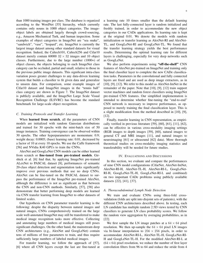
Fig. 8. FROC curves averaged on three-fold CV for the abdominal (left) and mediastinal (right) lymph nodes using different CNN models.
Region Mediastinum AbdomenMethod AUC TPR/3FP AUC TPR/3FP
[41] - 0.63 - 0.70[22] 0.92 0.70 0.94 0.83[36] - 0.78 - 0.78
CifarNet 0.91 0.70 0.81 0.44AlexNet-ImNet 0.89 0.63 0.80 0.41AlexNet-RI-H 0.94 0.79 0.92 0.67AlexNet-TL-H 0.94 0.81 0.92 0.69
GoogLeNet-RI-H 0.85 0.61 0.80 0.48GoogLeNet-TL-H 0.94 0.81 0.92 0.70
AlexNet-RI-L 0.94 0.77 0.88 0.61GoogLeNet-RI-L 0.95 0.85 0.91 0.69
Combined 0.95 0.85 0.93 0.70
TABLE IICOMPARISON OF MEDIASTINAL AND ABDOMINAL LN DETECTION
RESULTS USING VARIOUS CNN MODELS. NUMBERS IN BOLD INDICATETHE BEST PERFORMANCE VALUES ON CLASSIFICATION ACCURACY.
to 2. For the modified GoogLeNet-RI (64× 64), we decreasethe number of first layer convolution filters from 64 to 32,the pad size from 3 to 2, the kernel size from 7 to 5, stridefrom 2 to 1 and the stride of the subsequent pooling layerfrom 2 to 1. We slightly reduce the number of convolutionalfilters in order to accommodate the smaller input image sizesof target medical image datasets [22], [37], while preventingover-fitting. This eventually improves performance on patch-based classification. CifarNet is used in [22] to detect LNsamples of 32 × 32 × 3 images. For consistency purposes,we down-sample 64× 64× 3 resolution LN sample images tothe dimension of 32× 32× 3.
Results for lymph node detection in the mediastinum andabdomen are reported in Table II. FROC curves are illustratedin Figure 8. The area-under-the-FROC-curve (AUC) and truepositive rate (TPR, recall or sensitivity) at three false positivesper patient (TPR/3FP) are used as performance metrics. Of
the nine investigated CNN models, CifarNet, AlexNet-ImNetand GoogLeNet-RI-H generally yielded the least competitivedetection accuracy results. Our LN datasets are significantlymore complex (i.e., display much larger within-class appear-ance variations), especially due to the extracted fields-of-view(FOVs) of (35mm-128mm) compared to (30mm-45mm) in[22], where CifarNet is also employed. In this experiment,CifarNet is under-trained with respect to our enhanced LNdatasets, due to its limited input resolution and parameter com-plexity. The inferior performance of AlexNet-ImNet impliesthat using the pre-trained ImageNet CNNs alone as “off-the-shelf” deep image feature extractors may not be optimal oradequate for mediastinal and abdominal LN detection tasks.To complement “off-the-shelf” CNN features, [10], [9], [12]all add and integrate various other hand-crafted image featuresas hybrid inputs for the final CADe classification.
GoogLeNet-RI-H performs poorly, as it is susceptibleto over-fitting. No sufficient data samples are available totrain GoogLeNet-RI-H with random initialization. Indeed,due to GoogLeNet-RI-H’s complexity and 22-layer depth,million-image datasets may be required to properly trainthis model. However, GoogLeNet-TL-H significantly improvesupon GoogLeNet-RI-H (0.81 versus 0.61 TPR/3FP in me-diastinum; 0.70 versus 0.48 TPR/3FP in abdomen). Thisindicates that transfer learning offers a much better initial-ization of CNN parameters than random initialization. Like-wise, AlexNet-TL-H consistently outperforms AlexNet-RI-H,though by smaller margins (0.81 versus 0.79 TPR/3FP inmediastinum; 0.69 versus 0.67 TPR/3FP in abdomen). Thisis also consistent with the findings reported for ILD detectionin Table III and Figure 11.
GoogLeNet-TL-H yields results similar to AlexNet-TL-H’sfor the mediastinal LN detection, and slightly outperformsAlex-Net-H for abdominal LN detection. AlexNet-RI-H ex-hibits less severe over-fitting than GoogLeNet-RI-H. We alsoevaluate a simple ensemble by averaging the probability scoresfrom five CNNs: AlexNet-RI-H, AlexNet-TL-H, AlexNet-RI-
8
H, GoogLeNet-TL-H and GoogLeNet-RI-L. This combinedensemble outputs the classification accuracies matching orslightly exceeding the best performing individual CNN modelson the mediastinal or abdominal LN detection tasks, respec-tively.
Many of our CNN models achieve notably better (FROC-AUC and TPR/3FP) results than the previous state-of-the-artmodels [36] for mediastinal LN detection: GoogLeNet-RI-Lobtains an AUC=0.95 and 0.85 TPR/3FP, versus AUC=0.92and 0.70 TPR/3FP [22] and 0.78 TPR/3FP [36] which usesstacked shallow learning. This difference lies in the fact thatannotated lymph node segmentation masks are required tolearn a mid-level semantic boundary detector [36], whereasCNN approaches only need LN locations for training [22].In abdominal LN detection, [22] obtains the best trade-off between its CNN model complexity and sampled dataconfiguration. Our best performing CNN model is GoogLeNet-TL (256x256) which obtains an AUC=0.92 and 0.70 TPR/3FP.
The main difference between our dataset preparation pro-tocol and that from [22] is a more aggressive extraction ofrandom views within a much larger range of FOVs. Theusage of larger FOVs to capture more image spatial context isinspired by deep zoom-out features [44] that improve semanticsegmentation. This image sampling scheme contributes to ourbest reported performance results in both mediastinal LNdetection (in this paper) and automated pancreas segmentation[45]. As shown in Figure 1, abdominal LNs are surrounded bymany other similar looking objects. Meanwhile, mediastinalLNs are more easily distinguishable, due to the images’larger spatial contexts. Finally, from the perspective of thedata-model trade-off: “Do We Need More Training Data orBetter Models?” [51], more abdomen CT scans from distinctpatient populations need to be acquired and annotated, inorder to take full advantage of deep CNN models of highcapacity. Nevertheless, deeper and wider CNN models (e.g.,GoogLeNet-RI-L and GoogLeNet-TL-H versus Cifar-10 [22])have shown improved results in the mediastinal LN detection.
Figure 9 provides examples of misclassified lymph nodes(in axial view) (both false negatives (Left) and false posi-tives(Right)), from the Abdomen and Mediastinum datasets.The overall reported LN detection results are clinically signif-icant, as indicated in [63].
B. Interstitial Lung Disease ClassificationThe CNN models evaluated in this experiment are 1)
AlexNet-RI (training from scratch on the ILD dataset withrandom initialization); 2) AlexNet-TL (with transfer learn-ing from [4]); 3) AlexNet-ImNet: pre-trained ImageNet-CNNmodel [4] with only the last cost function layer retrained fromrandom initialization, according to the six ILD classes (similarto [9] but without using additional hand-crafted non-deepfeature descriptors, such as GIST and BoVW); 4) GoogLeNet-RI (random initialization); 5) GoogLeNet-TL (GoogLeNetwith transfer learning from [33]). All ILD images (patchesof 64× 64 and CT axial slices of 512× 512) are re-sampledto a fixed dimension of 256× 256 pixels.
We evaluate the ILD classification task with five-fold CVon patient-level split, as it is more informative for real clinical
Fig. 9. Examples of misclassified lymph nodes (in axial view) of both falsenegatives (Left) and false positives (Right). Mediastinal LN examples areshown in the upper row, and abdominal LN examples in the bottom row.
NM EM GG FB MN CDPatch-LOO [38] 0.84 0.75 0.78 0.84 0.86 -Patch-LOO [39] 0.88 0.77 0.80 0.87 0.89 -Patch-CV10 [54] 0.84 0.55 0.72 0.76 0.91 -
Patch-CV5 0.64 0.81 0.74 0.78 0.82 0.64Slice-Test [40] 0.40 1.00 0.75 0.80 0.56 0.50
Slice-CV5 0.22 0.35 0.56 0.75 0.71 0.16Slice-Random 0.90 0.86 0.85 0.94 0.98 0.83
TABLE IVCOMPARISON OF INTERSTITIAL LUNG DISEASE CLASSIFICATION RESULTS
USING F-SCORES: NM, EM, GG, FB, MN AND CD.
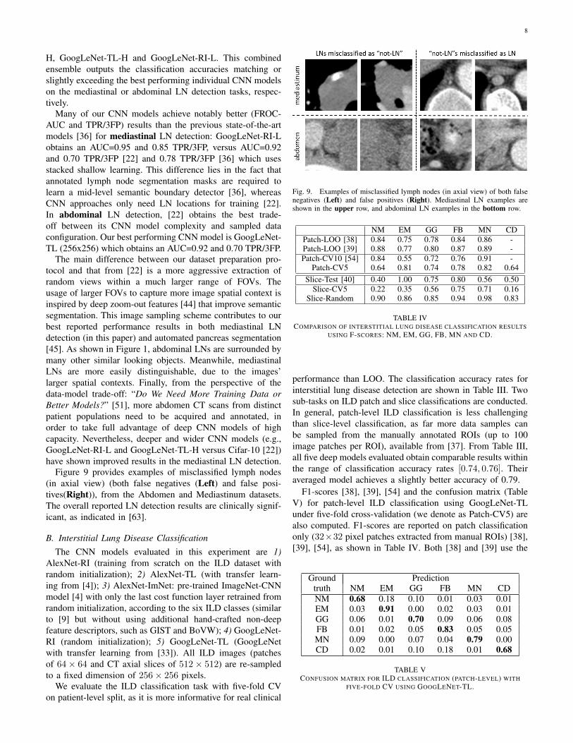
performance than LOO. The classification accuracy rates forinterstitial lung disease detection are shown in Table III. Twosub-tasks on ILD patch and slice classifications are conducted.In general, patch-level ILD classification is less challengingthan slice-level classification, as far more data samples canbe sampled from the manually annotated ROIs (up to 100image patches per ROI), available from [37]. From Table III,all five deep models evaluated obtain comparable results withinthe range of classification accuracy rates [0.74, 0.76]. Theiraveraged model achieves a slightly better accuracy of 0.79.
F1-scores [38], [39], [54] and the confusion matrix (TableV) for patch-level ILD classification using GoogLeNet-TLunder five-fold cross-validation (we denote as Patch-CV5) arealso computed. F1-scores are reported on patch classificationonly (32×32 pixel patches extracted from manual ROIs) [38],[39], [54], as shown in Table IV. Both [38] and [39] use the
Ground Predictiontruth NM EM GG FB MN CDNM 0.68 0.18 0.10 0.01 0.03 0.01EM 0.03 0.91 0.00 0.02 0.03 0.01GG 0.06 0.01 0.70 0.09 0.06 0.08FB 0.01 0.02 0.05 0.83 0.05 0.05MN 0.09 0.00 0.07 0.04 0.79 0.00CD 0.02 0.01 0.10 0.18 0.01 0.68
TABLE VCONFUSION MATRIX FOR ILD CLASSIFICATION (PATCH-LEVEL) WITH
FIVE-FOLD CV USING GOOGLENET-TL.
9
Method AlexNet-ImNet AlexNet-RI AlexNet-TL GoogLeNet-RI GoogLeNet-TL Avg-AllSlice-CV5 0.45 0.44 0.46 0.41 0.57 0.53Patch-CV5 0.76 0.74 0.76 0.75 0.76 0.79
TABLE IIICOMPARISON OF INTERSTITIAL LUNG DISEASE CLASSIFICATION ACCURACIES ON BOTH SLICE-LEVEL (SLICE-CV5) AND PATCH-BASED (PATCH-CV5)
CLASSIFICATION USING FIVE-FOLD CV. BOLD NUMBERS INDICATE THE BEST PERFORMANCE VALUES ON CLASSIFICATION ACCURACY.
evaluation protocol of “leave-one-patient-out” (LOO), whichis arguably much easier and not directly comparable to 10-foldCV [54] or our Patch-CV5. In this study, we classify six ILDclasses by adding a consolidation (CD) class to five classesof healthy (normal - NM), emphysema (EM), ground glass(GG), fibrosis (FB), and micronodules (MN) in [38], [39],[54]. Patch-CV10 [54] and Patch-CV5 report similar mediumto high F-scores. This implies that the ILD dataset (althoughone of the mainstream public medical image datasets) may notadequately represent ILD disease CT lung imaging patterns,over a population of only 120 patients. Patch-CV5 yieldshigher F-scores than [54] and classifies the extra consolidation(CD) class. At present, the most pressing task is to drasticallyexpand the dataset or to explore across-dataset deep learningon the combined ILD and LTRC datasets [64].
Recently, Gao et al. [40] have argued that a new CADeprotocol on holistic classification of ILD diseases directly,using axial CT slice attenuation patterns and CNN, may bemore realistic for clinical applications. We refer to this asslice-level classification, as image patch sampling from manualROIs can be completely avoided (hence, no manual ROIinputs will be provided). The experimental results in [40] areconducted with a patient-level hard split of 100 (training) and20 (testing). The method’s testing F-scores (i.e., Slice-Test)are given in Table IV. Note that the F-scores in [40] are notdirectly comparable to our results, due to different evaluationcriteria. Only Slice-Test is evaluated and reported in [40], andwe find that F-scores can change drastically from differentrounds of the five-fold CV.
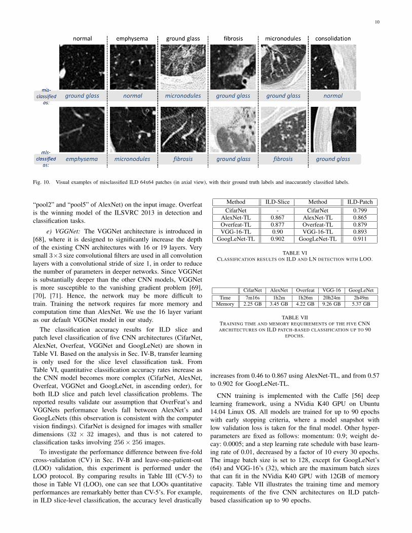
While it is a more practical CADe scheme, slice-levelCNN learning [40] is very challenging, as it is restrictedto only 905 CT image slices with tagged ILD labels. Weonly benchmark the slice-level ILD classification results inthis section. Even with the help of data augmentation (de-scribed in Sec. II), the classification accuracy of GoogLeNet-TL from Table III is only 0.57. However, transfer learningfrom ImageNet pre-trained model is consistently beneficial,as evidenced by AlexNet-TL (0.46) versus AlexNet-RI (0.44),and GoogLeNet-TL (0.57) versus GoogLeNet-RI (0.41). Itespecially prevents GoogLeNet from over-fitting on the limitedCADe datasets. Finally, when the cross-validation is conductedby randomly splitting the set of all 905 CT axial slices into fivefolds, markedly higher F-scores are obtained (Slice-Randomin Table IV). This further validates the claim that the datasetpoorly generalizes ILDs for different patients. Figure 10 showsexamples of misclassified ILD patches (in axial view), withtheir ground truth labels and inaccurately classified labels.
No existing work has reached the performance requirementsfor a realistic clinical setting [40], in which simple ROI-guided
image patch extraction and classification (which requires man-ual ROI selection by clinicians) is implemented. The main goalof this paper is to investigate the three factors (CNN architec-tures, dataset characteristics and transfer learning) that affectperformance on a specific medical image analysis problemand to ultimately deliver clinically relevant results. For ILDclassification, the most critical performance bottlenecks arethe challenge of cross-dataset learning and the limited patientpopulation size. We attempt to overcome these obstacles bymerging the ILD [37] and LTRC datasets. Although the ILD[37] and LTRC datasets [64] (used in [19]) were generatedand annotated separately, they contain many common diseaselabels. For instance, the ILD disease classes emphysema (EM),ground glass (GG), fibrosis (FB), and micronodules (MN)belong to both datasets, and thus can be jointly trained/testedto form a larger and unified dataset.
Adapting fully convolutional CNN or FCNN to parse everypixel location in the ILD lung CT images or slices, or adaptingother methods from CNN based semantic image segmentationusing PASCAL or ImageNet, may improve accuracy andefficiency. However, current FCNN approaches [65], [66]lack adequate spatial resolution in their directly output labelspace. A segmentation label propagation method was recentlyproposed [47] to provide full pixel-wise labeling of the ILDdata images. In this work, we sample image patches from theslice using the ROIs for the ILD provided in the dataset, inorder to be consistent with previous methods in patch-level[38], [39], [54] and slice-level classification [40].
C. Evaluation of Five CNN Models using ILD Classification
In this work, we mainly focus on AlexNet and GoogLeNet.AlexNet is the first notably successful CNN architecture onthe ImageNet challenge and has rekindled significant researchinterests on CNN. GoogLeNet is the state-of-the-art deepmodel, which has outperformed other notable models, such asAlexNet, OverFeat, and VGGNet [67], [68] in various com-puter vision benchmarks. Likewise, a reasonable assumptionis that OverFeat and VGGNet may generate quantitative per-formance results ranked between AlexNet’s and GoogLeNet’s.For completeness, we include the Overfeat and VGGNet in thefollowing evaluations, to bolster our hypothesis.
d) Overfeat: OverFeat is described in [67] as an inte-grated framework for using CNN for classification, localiza-tion and detection. Its architecture is similar to that of AlexNet,but contains far more parameters (e.g., 1024 convolution filtersin both “conv4” and “conv5” layers compared to 384 and256 convolution kernels in the “conv4” and “conv5” layers ofAlexNet), and operates more densely (e.g., smaller kernel sizeof 2 in “pool2” layer “pool5” compared to the kernel size 3 in
10
Fig. 10. Visual examples of misclassified ILD 64x64 patches (in axial view), with their ground truth labels and inaccurately classified labels.
“pool2” and “pool5” of AlexNet) on the input image. Overfeatis the winning model of the ILSVRC 2013 in detection andclassification tasks.
e) VGGNet: The VGGNet architecture is introduced in[68], where it is designed to significantly increase the depthof the existing CNN architectures with 16 or 19 layers. Verysmall 3×3 size convolutional filters are used in all convolutionlayers with a convolutional stride of size 1, in order to reducethe number of parameters in deeper networks. Since VGGNetis substantially deeper than the other CNN models, VGGNetis more susceptible to the vanishing gradient problem [69],[70], [71]. Hence, the network may be more difficult totrain. Training the network requires far more memory andcomputation time than AlexNet. We use the 16 layer variantas our default VGGNet model in our study.
The classification accuracy results for ILD slice andpatch level classification of five CNN architectures (CifarNet,AlexNet, Overfeat, VGGNet and GoogLeNet) are shown inTable VI. Based on the analysis in Sec. IV-B, transfer learningis only used for the slice level classification task. FromTable VI, quantitative classification accuracy rates increase asthe CNN model becomes more complex (CifarNet, AlexNet,Overfeat, VGGNet and GoogLeNet, in ascending order), forboth ILD slice and patch level classification problems. Thereported results validate our assumption that OverFeat’s andVGGNets performance levels fall between AlexNet’s andGoogLeNets (this observation is consistent with the computervision findings). CifarNet is designed for images with smallerdimensions (32 × 32 images), and thus is not catered toclassification tasks involving 256× 256 images.
To investigate the performance difference between five-foldcross-validation (CV) in Sec. IV-B and leave-one-patient-out(LOO) validation, this experiment is performed under theLOO protocol. By comparing results in Table III (CV-5) tothose in Table VI (LOO), one can see that LOOs quantitativeperformances are remarkably better than CV-5’s. For example,in ILD slice-level classification, the accuracy level drastically
Method ILD-Slice Method ILD-PatchCifarNet - CifarNet 0.799
AlexNet-TL 0.867 AlexNet-TL 0.865Overfeat-TL 0.877 Overfeat-TL 0.879VGG-16-TL 0.90 VGG-16-TL 0.893
GoogLeNet-TL 0.902 GoogLeNet-TL 0.911
TABLE VICLASSIFICATION RESULTS ON ILD AND LN DETECTION WITH LOO.
CifarNet AlexNet Overfeat VGG-16 GoogLeNetTime 7m16s 1h2m 1h26m 20h24m 2h49m
Memory 2.25 GB 3.45 GB 4.22 GB 9.26 GB 5.37 GB
TABLE VIITRAINING TIME AND MEMORY REQUIREMENTS OF THE FIVE CNNARCHITECTURES ON ILD PATCH-BASED CLASSIFICATION UP TO 90
EPOCHS.
increases from 0.46 to 0.867 using AlexNet-TL, and from 0.57to 0.902 for GoogLeNet-TL.
CNN training is implemented with the Caffe [56] deeplearning framework, using a NVidia K40 GPU on Ubuntu14.04 Linux OS. All models are trained for up to 90 epochswith early stopping criteria, where a model snapshot withlow validation loss is taken for the final model. Other hyper-parameters are fixed as follows: momentum: 0.9; weight de-cay: 0.0005; and a step learning rate schedule with base learn-ing rate of 0.01, decreased by a factor of 10 every 30 epochs.The image batch size is set to 128, except for GoogLeNet’s(64) and VGG-16’s (32), which are the maximum batch sizesthat can fit in the NVidia K40 GPU with 12GB of memorycapacity. Table VII illustrates the training time and memoryrequirements of the five CNN architectures on ILD patch-based classification up to 90 epochs.
11
D. Training with “Equal Prior” vs. “Biased Prior”
Medical datasets are often “biased”, in that the number ofhealthy samples is much larger than the number of diseasedinstances, or that the numbers of images per class are uneven.In ILD dataset, the number of fibrosis samples is about3.5 times greater than the number of emphysema samples.The number of non-LNs is 3 ∼ 4 times greater than thenumber of LNs in lymph node detection. Different samplingor resampling rates are routinely applied to both ILD and LNdetection to balance the data sample number or scale per class,as in[22]. We refer this as “Equal Prior”. If we use the samesampling rate, that will lead to a “Biased Prior” across differentclasses.
Without loss of generality, after GoogLeNet is trained onthe training sets under “Equal” or “Biased” priors, we com-pare its classification results on the balanced validation sets.Evaluating a classifier on a biased validation set will causeunfair assessment of its performance. For instance, a classifierthat predicts every image patch as “non-LN” will still achieve a70% accuracy rate on a biased set with 3.5 times as many non-LN samples as LN samples. The classification accuracy resultsof GoogLeNet trained under two configurations are shown inTable VIII. Overall, it achieves lower accuracy results whentrained with a “biased prior” in both tasks, and the accuracydifference for ILD patch-based classification is small.
ILD-Slice ILD-PatchEqual Prior 0.902 0.953Biased Prior 0.872 0.952
TABLE VIIICLASSIFICATION ACCURACIES FOR ILD SLICE AND LN PATCH-LEVEL
DETECTION WITH “EQUAL PRIOR” AND “BIASED PRIOR”, USINGGOOGLENET-TL.
V. ANALYSIS VIA CNN LEARNING TRACES &LULVISUALIZATION
In this section, we determine and analyze, via CNN visu-alization, the reasons for which transfer learning is beneficialto achieve better performance on CAD applications.
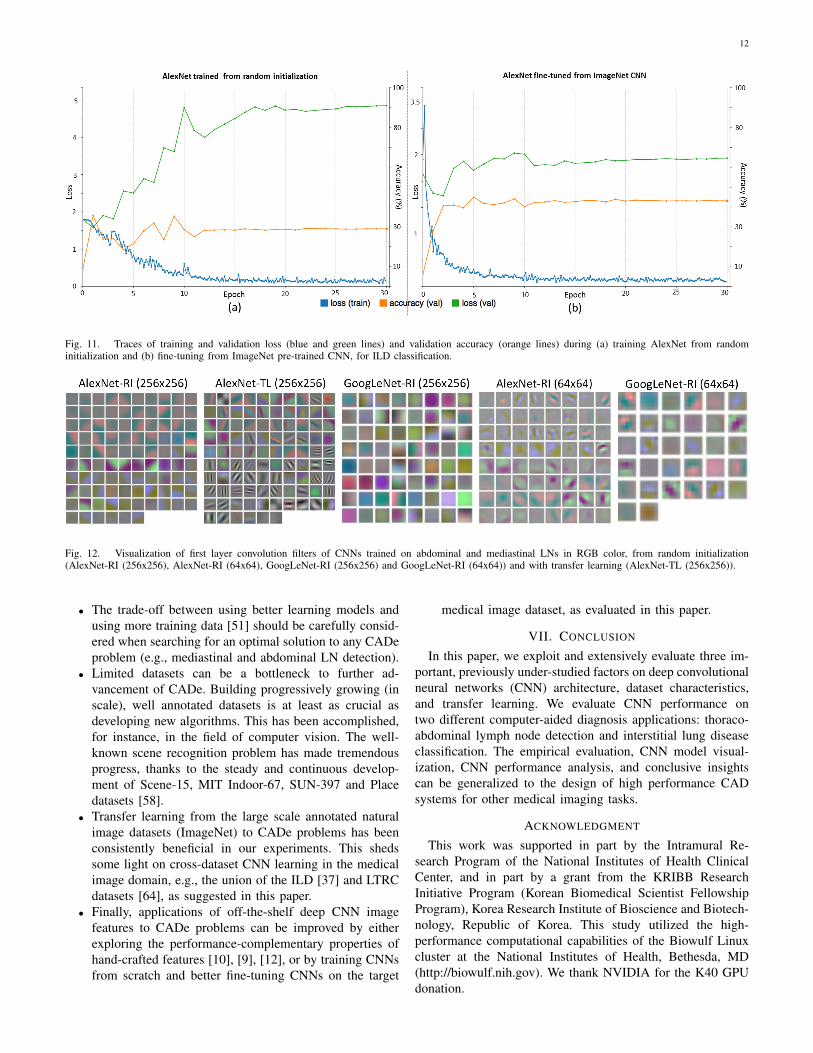
Thoracoabdominal LN Detection. In Figure 12, thefirst layer convolution filters from five different CNN ar-chitectures are visualized. We notice that without trans-fer learning [57], [6], somewhat blurry filters are learned(AlexNet-RI (256x256), AlexNet-RI (64x64), GoogLeNet-RI (256x256) and GoogLeNet-RI (64x64)). However, inAlexNet-TL (256x256), many higher orders of contrast- oredge-preserving patterns (that enable capturing image ap-pearance details) are evidently learned through fine-tuningfrom ImageNet. With a smaller input resolution, AlexNet-RI(64x64) and GoogLeNet-RI (64x64) can learn image contrastfilters to some degree; whereas, GoogLeNet-RI (256x256)and AlexNet-RI (256x256) have over-smooth low-level filtersthroughout.
ILD classification. We focus on analyzing visual CNNoptimization traces and activations from the ILD dataset, asits slice-level setting is most similar to ImageNet’s. Indeed,
both datasets use full-size images. The traces of the trainingloss, validation loss and validation accuracy of AlexNet-RI andAlexNet-TL, are shown in Figure 11. For AlexNet-RI in Figure11 (a), the training loss significantly decreases as the numberof training epochs increases, while the validation loss notablyincreases and the validation accuracy does not improve muchbefore reaching a plateau. With transfer learning and fine-tuning, much better and consistent performances of trainingloss, validation loss and validation accuracy traces are obtained(see Figure 11 (b)). We begin the optimization problem – thatof fine-tuning the ImageNet pre-trained CNN to classify acomprehensive set of images – by initializing the parametersclose to an optimal solution. One could compare this processto making adults learn to classify ILDs, as opposed to babies.During the process, the validation loss, having remained atlower values throughout, achieves higher final accuracy levelsthan the validation loss on a similar problem with randominitialization. Meanwhile, the training losses in both casesdecrease to values near zero. This indicates that both AlexNet-RI and AlexNet-TL over-fit on the ILD dataset, due to its smallinstance size. The quantitative results in Table III indicatethat AlexNet-TL and GoogLeNet-TL have consistently betterclassification accuracies than AlexNet-RI and GoogLeNet-RI,respectively.
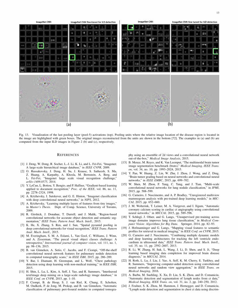
The last pooling layer (pool-5) activation maps of the Ima-geNet pre-trained AlexNet [4] (analogical to AlexNet-ImNet)and AlexNet-TL, obtained by processing two input images ofFigure 2 (b,c), are shown in Figure 13 (a,b). The last poolinglayer activation map summarizes the entire input image byhighlighting which relative locations or neural reception fieldsrelative to the image are activated. There are a total of 256(6x6) reception fields in AlexNet [4]. Pooling units where therelative image location of the disease region is present in theimage are highlighted with green boxes. Next, we reconstructthe original ILD images using the process of de-convolution,back-propagating with convolution and un-pooling from theactivation maps of the chosen pooling units [72]. From thereconstructed images (Figure 13 bottom), we observe thatwith fine-tuning, AlexNet-TL detects and localizes objects ofinterest (ILD disease regions depicted in in Figure 2 (b) and(c)) better than AlexNet-ImNet. The filters shown in Figure13 that better localize regions on the input images (Figure 2(b) and (c)) respectively, produce relatively higher activations(in the top 5%) among all 512 reception field responses inthe fine-tuned AlexNet-TL model. As observed in [73], thefinal CNN classification score can not be driven solely by asingle strong activation in the receptions fields, but often by asparse set of high activations (i.e., varying selective or sparseactivations per input image).
VI. FINDINGS AND FUTURE DIRECTIONS
We summarize our findings as follows.• Deep CNN architectures with 8, even 22 layers [4],
[33], can be useful even for CADe problems where theavailable training datasets are limited. Previously, CNNmodels used in medical image analysis applications haveoften been 2 ∼ 5 orders of magnitude smaller.
12
Fig. 11. Traces of training and validation loss (blue and green lines) and validation accuracy (orange lines) during (a) training AlexNet from randominitialization and (b) fine-tuning from ImageNet pre-trained CNN, for ILD classification.
Fig. 12. Visualization of first layer convolution filters of CNNs trained on abdominal and mediastinal LNs in RGB color, from random initialization(AlexNet-RI (256x256), AlexNet-RI (64x64), GoogLeNet-RI (256x256) and GoogLeNet-RI (64x64)) and with transfer learning (AlexNet-TL (256x256)).
• The trade-off between using better learning models andusing more training data [51] should be carefully consid-ered when searching for an optimal solution to any CADeproblem (e.g., mediastinal and abdominal LN detection).
• Limited datasets can be a bottleneck to further ad-vancement of CADe. Building progressively growing (inscale), well annotated datasets is at least as crucial asdeveloping new algorithms. This has been accomplished,for instance, in the field of computer vision. The well-known scene recognition problem has made tremendousprogress, thanks to the steady and continuous develop-ment of Scene-15, MIT Indoor-67, SUN-397 and Placedatasets [58].
• Transfer learning from the large scale annotated naturalimage datasets (ImageNet) to CADe problems has beenconsistently beneficial in our experiments. This shedssome light on cross-dataset CNN learning in the medicalimage domain, e.g., the union of the ILD [37] and LTRCdatasets [64], as suggested in this paper.
• Finally, applications of off-the-shelf deep CNN imagefeatures to CADe problems can be improved by eitherexploring the performance-complementary properties ofhand-crafted features [10], [9], [12], or by training CNNsfrom scratch and better fine-tuning CNNs on the target
medical image dataset, as evaluated in this paper.
VII. CONCLUSION
In this paper, we exploit and extensively evaluate three im-portant, previously under-studied factors on deep convolutionalneural networks (CNN) architecture, dataset characteristics,and transfer learning. We evaluate CNN performance ontwo different computer-aided diagnosis applications: thoraco-abdominal lymph node detection and interstitial lung diseaseclassification. The empirical evaluation, CNN model visual-ization, CNN performance analysis, and conclusive insightscan be generalized to the design of high performance CADsystems for other medical imaging tasks.
ACKNOWLEDGMENT
This work was supported in part by the Intramural Re-search Program of the National Institutes of Health ClinicalCenter, and in part by a grant from the KRIBB ResearchInitiative Program (Korean Biomedical Scientist FellowshipProgram), Korea Research Institute of Bioscience and Biotech-nology, Republic of Korea. This study utilized the high-performance computational capabilities of the Biowulf Linuxcluster at the National Institutes of Health, Bethesda, MD(http://biowulf.nih.gov). We thank NVIDIA for the K40 GPUdonation.
13
Fig. 13. Visualization of the last pooling layer (pool-5) activations (top). Pooling units where the relative image location of the disease region is located inthe image are highlighted with green boxes. The original images reconstructed from the units are shown in the bottom [72]. The examples in (a) and (b) arecomputed from the input ILD images in Figure 2 (b) and (c), respectively.
REFERENCES
[1] J. Deng, W. Dong, R. Socher, L.-J. Li, K. Li, and L. Fei-Fei, “Imagenet:A large-scale hierarchical image database,” in IEEE CVPR, 2009.
[2] O. Russakovsky, J. Deng, H. Su, J. Krause, S. Satheesh, S. Ma,Z. Huang, A. Karpathy, A. Khosla, M. Bernstein, A. Berg, andL. Fei-Fei, “Imagenet large scale visual recognition challenge,”arXiv:1409.0575, 2014.
[3] Y. LeCun, L. Bottou, Y. Bengio, and P. Haffner, “Gradient-based learningapplied to document recognition,” Proc. of the IEEE, vol. 86, no. 11,pp. 2278–2324, 1998.
[4] A. Krizhevsky, I. Sutskever, and G. E. Hinton, “Imagenet classificationwith deep convolutional neural networks,” in NIPS, 2012.
[5] A. Krizhevsky, “Learning multiple layers of features from tiny images,”in Master’s Thesis. Dept. of Comp. Science, University of Toronto,2009.
[6] R. Girshick, J. Donahue, T. Darrell, and J. Malik, “Region-basedconvolutional networks for accurate object detection and semantic seg-mentation,” IEEE Trans. Pattern Anal. Mach. Intell., 2015.
[7] K. He, X. Zhang, S. Ren, and J. Sun, “Spatial pyramid pooling indeep convolutional networks for visual recognition,” IEEE Trans. PatternAnal. Mach. Intell., 2015.
[8] M. Everingham, S. M. A. Eslami, L. Van Gool, C. Williams, J. Winn,and A. Zisserman, “The pascal visual object classes challenge: Aretrospective,” International journal of computer vision, vol. 111, no. 1,pp. 98–136, 2015.
[9] B. van Ginneken, A. Setio, C. Jacobs, and F. Ciompi, “Off-the-shelfconvolutional neural network features for pulmonary nodule detectionin computed tomography scans,” in IEEE ISBI, 2015, pp. 286–289.
[10] Y. Bar, I. Diamant, H. Greenspan, and L. Wolf, “Chest pathologydetection using deep learning with non-medical training,” in IEEE ISBI,2015.
[11] H. Shin, L. Lu, L. Kim, A. Seff, J. Yao, and R. Summers, “Interleavedtext/image deep mining on a large-scale radiology image database,” inIEEE Conf. on CVPR, 2015, pp. 1–10.
[12] F. Ciompi, B. de Hoop, S. J. van Riel, K. Chung, E. Scholten,M. Oudkerk, P. de Jong, M. Prokop, and B. van Ginneken, “Automaticclassification of pulmonary peri-fissural nodules in computed tomogra-
phy using an ensemble of 2d views and a convolutional neural networkout-of-the-box,” Medical Image Analysis, 2015.
[13] B. Menze, M. Reyes, and K. Van Leemput, “The multimodal brain tumorimage segmentation benchmark (brats),” Medical Imaging, IEEE Trans.on, vol. 34, no. 10, pp. 1993–2024, 2015.
[14] Y. Pan, W. Huang, Z. Lin, W. Zhu, J. Zhou, J. Wong, and Z. Ding,“Brain tumor grading based on neural networks and convolutional neuralnetworks,” in IEEE EMBC, 2015, pp. 699–702.
[15] W. Shen, M. Zhou, F. Yang, C. Yang, and J. Tian, “Multi-scaleconvolutional neural networks for lung nodule classification,” in IPMI,2015, pp. 588–599.
[16] G. Carneiro, J. Nascimento, and A. P. Bradley, “Unregistered multiviewmammogram analysis with pre-trained deep learning models,” in MIC-CAI, 2015, pp. 652–660.
[17] J. M. Wolterink, T. Leiner, M. A. Viergever, and I. Isgum, “Automaticcoronary calcium scoring in cardiac ct angiography using convolutionalneural networks,” in MICCAI, 2015, pp. 589–596.
[18] T. Schlegl, J. Ofner, and G. Langs, “Unsupervised pre-training acrossimage domains improves lung tissue classification,” in Medical Com-puter Vision: Algorithms for Big Data. Springer, 2014, pp. 82–93.
[19] J. Hofmanninger and G. Langs, “Mapping visual features to semanticprofiles for retrieval in medical imaging,” in IEEE Conf. on CVPR, 2015.
[20] G. Carneiro and J. Nascimento, “Combining multiple dynamic modelsand deep learning architectures for tracking the left ventricle endo-cardium in ultrasound data,” IEEE Trans. Pattern Anal. Mach. Intell.,vol. 35, no. 11, pp. 2592–2607, 2013.
[21] R. Li, W. Zhang, H. Suk, L. Wang, J. Li, D. Shen, and S. Ji, “Deeplearning based imaging data completion for improved brain diseasediagnosis,” in MICCAI, 2014.
[22] H. Roth, L. Lu, J. Liu, J. Yao, A. Seff, K. M. Cherry, E. Turkbey, andR. Summers, “Improving computer-aided detection using convolutionalneural networks and random view aggregation,” in IEEE Trans. onMedical Imaging, 2016.
[23] A. Barbu, M. Suehling, X. Xu, D. Liu, S. K. Zhou, and D. Comaniciu,“Automatic detection and segmentation of lymph nodes from ct data,”Medical Imaging, IEEE Trans. on, vol. 31, no. 2, pp. 240–250, 2012.
[24] J. Feulner, S. K. Zhou, M. Hammon, J. Hornegger, and D. Comaniciu,“Lymph node detection and segmentation in chest ct data using discrim-
14
inative learning and a spatial prior,” Medical image analysis, vol. 17,no. 2, pp. 254–270, 2013.
[25] M. Feuerstein, B. Glocker, T. Kitasaka, Y. Nakamura, S. Iwano, andK. Mori, “Mediastinal atlas creation from 3-d chest computed tomogra-phy images: application to automated detection and station mapping oflymph nodes,” Medical image analysis, vol. 16, no. 1, pp. 63–74, 2012.
[26] L. Lu, P. Devarakota, S. Vikal, D. Wu, Y. Zheng, and M. Wolf,“Computer aided diagnosis using multilevel image features on large-scale evaluation,” in Medical Computer Vision. Large Data in MedicalImaging. Springer, 2014, pp. 161–174.
[27] L. Lu, J. Bi, M. Wolf, and M. Salganicoff, “Effective 3d object detectionand regression using probabilistic segmentation features in ct images,”in IEEE CVPR, 2011.
[28] L. Lu, A. Barbu, M. Wolf, J. Liang, M. Salganicoff, and D. Comaniciu,“Accurate polyp segmentation for 3d ct colonography using multi-stagedprobabilistic binary learning and compositional model,” in IEEE CVPR,2008.
[29] N. Tajbakhsh, M. B. Gotway, and J. Liang, “Computer-aided pulmonaryembolism detection using a novel vessel-aligned multi-planar imagerepresentation and convolutional neural networks,” in MICCAI, 2015.
[30] D. G. Lowe, “Distinctive image features from scale-invariant keypoints,”International journal of computer vision, vol. 60, no. 2, pp. 91–110,2004.
[31] N. Dalal and B. Triggs, “Histograms of oriented gradients for humandetection,” in IEEE CVPR, vol. 1, 2005, pp. 886–893.
[32] A. Torralba, R. Fergus, and Y. Weiss, “Small codes and large imagedatabases for recognition,” in IEEE CVPR, 2008, pp. 1–8.
[33] C. Szegedy, W. Liu, Y. Jia, P. Sermanet, S. Reed, D. Anguelov, D. Erhan,and A. Rabinovich, “Going deeper with convolutions,” in IEEE Conf.on CVPR, 2015.
[34] K. Chatfield, K. Simonyan, A. Vedaldi, and A. Zisserman, “Return ofthe devil in the details: Delving deep into convolutional nets,” in BMVC,2014.
[35] K. Chatfield, V. S. Lempitsky, A. Vedaldi, and A. Zisserman, “The devilis in the details: an evaluation of recent feature encoding methods.” inBMVC, 2011.
[36] A. Seff, L. Lu, A. Barbu, H. Roth, H.-C. Shin, and R. Summers, “Lever-aging mid-level semantic boundary cues for computer-aided lymph nodedetection,” in MICCAI, 2015.
[37] A. Depeursinge, A. Vargas, A. Platon, A. Geissbuhler, P.-A. Poletti, andH. Muller, “Building a reference multimedia database for interstitial lungdiseases,” Computerized medical imaging and graphics, vol. 36, no. 3,pp. 227–238, 2012.
[38] Y. Song, W. Cai, Y. Zhou, and D. D. Feng, “Feature-based image patchapproximation for lung tissue classification,” Medical Imaging, IEEETrans. on, vol. 32, no. 4, pp. 797–808, 2013.
[39] Y. Song, W. Cai, H. Huang, Y. Zhou, D. Feng, Y. Wang, M. Fulham,and M. Chen, “Large margin local estimate with applications to medicalimage classification.” IEEE Trans. on Medical Imaging, 2015.
[40] M. Gao, U. Bagci, L. Lu, A. Wu, M. Buty, H.-C. Shin, H. Roth,Z. G. Papadakis, A. Depeursinge, R. Summers, Z. Xu, and J. D.Mollura, “Holistic classification of ct attenuation patterns for interstitiallung diseases via deep convolutional neural networks,” in MICCAI firstWorkshop on Deep Learning in Medical Image Analysis, 2015.
[41] A. Seff, L. Lu, K. M. Cherry, H. R. Roth, J. Liu, S. Wang, J. Hoffman,E. B. Turkbey, and R. Summers, “2d view aggregation for lymph nodedetection using a shallow hierarchy of linear classifiers,” in MICCAI,2014, pp. 544–552.
[42] L. Lu, M. Liu, X. Ye, S. Yu, and H. Huang, “Coarse-to-fine classificationvia parametric and nonparametric models for computer-aided diagnosis,”in ACM Conf. on CIKM, 2011, pp. 2509–2512.
[43] C. Farabet, C. Couprie, L. Najman, and Y. LeCun, “Learning hierarchicalfeatures for scene labeling,” IEEE Trans. Pattern Anal. Mach. Intell.,vol. 35, no. 8, pp. 1915–1929, 2013.
[44] M. Mostajabi, P. Yadollahpour, and G. Shakhnarovich, “Feedfor-ward semantic segmentation with zoom-out features,” arXiv preprintarXiv:1412.0774, 2014.
[45] H. Roth, L. Lu, A. Farag, H.-C. Shin, J. Liu, E. Turkbey, andR. Summers, “Deeporgan: Multi-level deep convolutional networks forautomated pancreas segmentation,” in MICCAI, 2015.
[46] X. Liang, C. Xu, X. Shen, J. Yang, S. Liu, J. Tang, L. Lin, and S. Yan,“Human parsing with contextualized convolutional neural network,” inIEEE ICCV, 2015, pp. 1386–1394.
[47] M. Gao, Z. Xu, L. Lu, I. Nogues, R. Summers, and D. Mollura, “Seg-mentation label propagation using deep convolutional neural networksand dense conditional random field,” in IEEE ISBI, 2016.
[48] H. Wang, J. W. Suh, S. R. Das, J. B. Pluta, C. Craige, P. Yushkevichet al., “Multi-atlas segmentation with joint label fusion,” IEEE Trans.Pattern Anal. Mach. Intell., vol. 35, no. 3, pp. 611–623, 2013.
[49] M. Oquab, L. Bottou, I. Laptev, and J. Sivic, “Is object localization forfree?–weakly-supervised learning with convolutional neural networks,”in IEEE CVPR, 2015, pp. 685–694.
[50] M. Oquab, L. Bottou, I. Laptev, and S. Josef, “Learning and transferringmid-level image representations using convolutional neural networks,”in IEEE CVPR, 2015, pp. 1717–1724.
[51] X. Zhu, C. Vondrick, D. Ramanan, and C. Fowlkes, “Do we need moretraining data or better models for object detection?” in BMVC, 2012.
[52] D. Ciresan, A. Giusti, L. Gambardella, and J. Schmidhuber, “Mitosisdetection in breast cancer histology images with deep neural networks,”in MICCAI, 2013.
[53] W. Zhang, R. Li, H. Deng, L. Wang, W. Lin, S. Ji, and D. Shen, “Deepconvolutional neural networks for multi-modality isointense infant brainimage segmentation,” NeuroImage, vol. 108, pp. 214–224, 2015.
[54] Q. Li, W. Cai, X. Wang, Y. Zhou, D. D. Feng, and M. Chen, “Med-ical image classification with convolutional neural network,” in IEEEICARCV, 2014, pp. 844–848.
[55] G. A. Miller, “Wordnet: a lexical database for english,” Communicationsof the ACM, vol. 38, no. 11, pp. 39–41, 1995.
[56] Y. Jia, E. Shelhamer, J. Donahue, S. Karayev, J. Long, R. B. Girshick,S. Guadarrama, and T. Darrell, “Caffe: Convolutional architecture forfast feature embedding.” in ACM Multimedia, vol. 2, 2014, p. 4.
[57] A. S. Razavian, H. Azizpour, J. Sullivan, and S. Carlsson, “Cnn featuresoff-the-shelf: an astounding baseline for recognition,” in IEEE CVPRW,2014, pp. 512–519.
[58] B. Zhou, A. Lapedriza, J. Xiao, A. Torralba, and A. Oliva, “Learningdeep features for scene recognition using places database,” in NIPS,2014, pp. 487–495.
[59] S. Gupta, R. Girshick, P. Arbelez, and J. Malik, “Learning rich featuresfrom rgb-d images for object detection and segmentation,” in ECCV,2014, pp. 345–360.
[60] S. Gupta, P. Arbelez, R. Girshick, and J. Malik, “Indoor scene under-standing with rgb-d images: Bottom-up segmentation, object detectionand semantic segmentation,” International Journal of Computer Vision,vol. 112, no. 2, pp. 133–149, 2015.
[61] A. Gupta, M. Ayhan, and A. Maida, “Natural image bases to representneuroimaging data,” in ICML, 2013, pp. 987–994.
[62] H. Chen, Q. Dou, D. Ni, J. Cheng, J. Qin, S. Li, and P. Heng, “Automaticfetal ultrasound standard plane detection using knowledge transferredrecurrent neural networks,” in MICCAI, 2015, pp. 507–514.
[63] L. Kim, H. Roth, L. Lu, S. Wang, E. Turkbey, and R. Summers,“Performance assessment of retroperitoneal lymph node computer-assisted detection using random forest and deep convolutional neuralnetwork learning algorithms in tandem,” in the 102nd Annual Meetingof Radiological Society of North America, 2014.
[64] D. Holmes III, B. Bartholmai, R. Karwoski, V. Zavaletta, and R. Robb,“The lung tissue research consortium: an extensive open databasecontaining histological, clinical, and radiological data to study chroniclung disease,” in 2006 MICCAI Open Science Workshop, 2006.
[65] J. Long, E. Shelhamer, and T. Darrell, “Fully convolutional networksfor semantic segmentation,” in IEEE CVPR, 2015.
[66] L.-C. Chen, G. Papandreou, I. Kokkinos, K. Murphy, and A. L. Yuille,“Semantic image segmentation with deep convolutional nets and fullyconnected crfs,” ICLR, 2015.
[67] P. Sermanet, D. Eigen, X. Zhang, M. Mathieu, R. Fergus, and Y. Le-Cun, “Overfeat: Integrated recognition, localization and detection usingconvolutional networks,” in ICLR, 2014.
[68] K. Simonyan and A. Zisserman, “Very deep convolutional networks forlarge-scale image recognition,” ICLR, 2014.
[69] S. Hochreiter, “The vanishing gradient problem during learning recurrentneural nets and problem solutions,” Int. J. of Uncertainty, Fuzziness andKnowledge-Based Systems, vol. 6, no. 02, pp. 107–116, 1998.
[70] G. E. Hinton, S. Osindero, and Y.-W. Teh, “A fast learning algorithm fordeep belief nets,” Neural computation, vol. 18, no. 7, pp. 1527–1554,2006.
[71] Y. Bengio, P. Simard, and P. Frasconi, “Learning long-term dependencieswith gradient descent is difficult,” Neural Networks, IEEE Transactionson, vol. 5, no. 2, pp. 157–166, 1994.
[72] M. D. Zeiler and R. Fergus, “Visualizing and understanding convolu-tional networks,” in ECCV, 2014, pp. 818–833.
[73] P. Agrawal, R. Girshick, and J. Malik, “Analyzing the performance ofmultilayer neural networks for object recognition,” in ECCV, 2014.