Embed Size (px)
Citation preview
ANRV278-NE29-08 ARI 8 May 2006 15:49
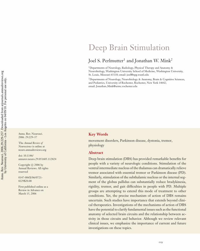
Deep Brain StimulationJoel S. Perlmutter1 and Jonathan W. Mink2
1Departments of Neurology, Radiology, Physical Therapy and Anatomy &Neurobiology, Washington University School of Medicine, Washington University,St. Louis, Missouri 63110; email: [email protected] of Neurology, Neurobiology & Anatomy, Brain & Cognitive Sciences,and Pediatrics, University of Rochester, Rochester, New York 14642;email: Jonathan [email protected]
Annu. Rev. Neurosci.2006. 29:229–57
The Annual Review ofNeuroscience is online atneuro.annualreviews.org
doi: 10.1146/annurev.neuro.29.051605.112824
Copyright c© 2006 byAnnual Reviews. All rightsreserved
0147-006X/06/0721-0229$20.00
First published online as aReview in Advance onMarch 15, 2006
Key Words
movement disorders, Parkinson disease, dystonia, tremor,physiology
AbstractDeep brain stimulation (DBS) has provided remarkable benefits forpeople with a variety of neurologic conditions. Stimulation of theventral intermediate nucleus of the thalamus can dramatically relievetremor associated with essential tremor or Parkinson disease (PD).Similarly, stimulation of the subthalamic nucleus or the internal seg-ment of the globus pallidus can substantially reduce bradykinesia,rigidity, tremor, and gait difficulties in people with PD. Multiplegroups are attempting to extend this mode of treatment to otherconditions. Yet, the precise mechanism of action of DBS remainsuncertain. Such studies have importance that extends beyond clini-cal therapeutics. Investigations of the mechanisms of action of DBShave the potential to clarify fundamental issues such as the functionalanatomy of selected brain circuits and the relationship between ac-tivity in those circuits and behavior. Although we review relevantclinical issues, we emphasize the importance of current and futureinvestigations on these topics.
229
Ann
u. R
ev. N
euro
sci.
2006
.29:
229-
257.
Dow
nloa
ded
from
arj
ourn
als.
annu
alre
view
s.or
gby
Uni
vers
ity o
f C
alif
orni
a -
Los
Ang
eles
on
04/0
9/10
. For
per
sona
l use
onl
y.
ANRV278-NE29-08 ARI 8 May 2006 15:49
Contents
INTRODUCTION. . . . . . . . . . . . . . . . . 230HISTORY OF DEEP BRAIN
STIMULATION . . . . . . . . . . . . . . . . 230CLINICAL APPLICATIONS OF
DEEP BRAINSTIMULATION . . . . . . . . . . . . . . . . 231Deep Brain Stimulation for
Essential Tremor . . . . . . . . . . . . . . 231Deep Brain Stimulation for
Parkinson Disease . . . . . . . . . . . . . 231Deep Brain Stimulation for
Dystonia . . . . . . . . . . . . . . . . . . . . . . 232Deep Brain Stimulation for
Tourette Syndrome . . . . . . . . . . . . 233Deep Brain Stimulation for Pain . . 233Deep Brain Stimulation for
Depression and ObsessiveCompulsive Disorder . . . . . . . . . . 233
NEUROPHYSIOLOGY OF DEEPBRAIN STIMULATION . . . . . . . . 234Ventral Thalamic Nuclei . . . . . . . . . . 235Subthalamic Nucleus . . . . . . . . . . . . . 237Globus Pallidus . . . . . . . . . . . . . . . . . . 241Release of Neurotransmitters by
Deep Brain Stimulation . . . . . . . . 242Synthesis of Neurophysiologic
Data . . . . . . . . . . . . . . . . . . . . . . . . . . 242FUNCTIONAL IMAGING OF
DEEP BRAINSTIMULATION–INDUCEDCHANGES IN BRAINCIRCUITS . . . . . . . . . . . . . . . . . . . . . . 244Positron Emission Tomography . . . 244Functional Magnetic Resonance
Imaging Studies of Deep BrainStimulation . . . . . . . . . . . . . . . . . . . 246
CONCLUSIONS. . . . . . . . . . . . . . . . . . . 246
INTRODUCTION
Deep brain stimulation (DBS) has provided
DBS: deep brainstimulation
ET: essential tremor
PD: Parkinsondisease
dramatic clinical benefit for people with es-sential tremor (ET) and Parkinson disease(PD). Placement of high frequency stimulat-ing electrodes in the region of the ventral in-
termediate nucleus of the thalamus (VIM) canmarkedly reduce tremor in these conditions,and stimulation of either the subthalamic nu-cleus (STN) or the internal segment of theglobus pallidus (GPi) may not only reducetremor, but also decrease bradykinesia, rigid-ity, and gait impairment, which plague peoplewith PD. Furthermore, many have touted thepotential benefit of DBS of selected brain re-gions for other movement disorders such asdystonia or Tourette syndrome, as well as a va-riety of disorders such as pain, depression, andobsessive compulsive disorder (OCD). De-spite these realized and potential advances intreatment, controversy swirls around a num-ber of clinically relevant and basic mechanisticissues. What conditions are amenable to treat-ment by DBS? What are the mechanisms ofaction of DBS? What effect does DBS haveon the function of brain circuits? We addressthese controversial issues and emphasize theneed for future investigations. To set the stage,however, we first review the history of the de-velopment of DBS as a therapeutic tool.
HISTORY OF DEEP BRAINSTIMULATION
Ever since Fritsch & Hitzig’s (1870) clas-sical demonstration of the localized electri-cal excitability of the motor cortex, electricalstimulation of the brain has played a majorrole in investigations of brain function. Thefirst report of human cortical stimulation ap-peared four years later (Bartholow 1874). Al-though electrical stimulation was used to mapcortical function in the 1930s (Penfield &Boldrey 1937), it was not until humanstereotaxic devices were developed that neu-rosurgeons could begin to investigate theeffects of stimulating deeper structures(Spiegel et al. 1947). By the early 1950s, in-traoperative stimulation was used to identifydeep structures such as the corticospinal tractprior to lesioning the globus pallidus or tha-lamus (Spiegel & Wycis 1952). Most reportsin the 1950s focused on positive phenomenathat were elicited by stimulation. In the early
230 Perlmutter · Mink
Ann
u. R
ev. N
euro
sci.
2006
.29:
229-
257.
Dow
nloa
ded
from
arj
ourn
als.
annu
alre
view
s.or
gby
Uni
vers
ity o
f C
alif
orni
a -
Los
Ang
eles
on
04/0
9/10
. For
per
sona
l use
onl
y.
ANRV278-NE29-08 ARI 8 May 2006 15:49
1960s, it was reported that high-frequency(100-Hz) stimulation of the ventrolateralthalamus could diminish tremor (Hassler et al.1960, Ohye et al. 1964).
The idea of treating neurologic disorderswith chronic stimulation began to emergein the 1960s, but stimulation was largelyused for targeting surgical lesions (Bergstromet al. 1966). Sem-Jacobsen (1966) developeda method of implanting a bundle of multipleelectrode wires deep in the brain and leav-ing them in place for weeks, during whichstimulation could be delivered. The goal ofthe stimulation was to delineate the “best”target for a subsequent lesion. With the im-planted wires, a lesion could be made in smallsteps over a span of days to weeks to tryto achieve maximum benefit without unto-ward effects. Although the goal was still lesionguidance, this is perhaps the earliest reportof stimulation through chronically implantedelectrodes.
In the early 1970s, reports of using chronicstimulation therapeutically emerged for treat-ing pain (Hosobuchi et al. 1973), movementdisorders, or epilepsy (Cooper 1973). Cooperet al. (1976) published the first large se-ries of chronic cerebellar stimulation stud-ies for cerebral palsy. In those cases, stimula-tion was delivered transcutaneously throughinductive coupling devices to electrodes im-planted on the surface of the cerebellar cor-tex. Benefit was said to occur in 49 of 50patients. However, cerebellar stimulation incerebral palsy eventually fell out of favor whenblinded studies failed to show consistent ben-efits (Penn 1982). By 1980, other reportsof treating movement disorders with chronicstimulation had appeared (Brice & McLellan1980).
Although the first long-term internallyimplanted cardiac pacemaker was devel-oped by 1960, it was not until the 1990sthat implantable pacemaker technology wascombined with chronically implanted deepbrain electrodes for long-term chronic DBS(Benabid et al. 1991, 1996). Since then, DBShas become increasingly used for treating a
VIM: ventralintermediate nucleusof the thalamus
STN: subthalamicnucleus
GPi: internalsegment of theglobus pallidus
variety of disorders. These are summarizedbriefly in the section below.
CLINICAL APPLICATIONS OFDEEP BRAIN STIMULATION
Deep Brain Stimulation for EssentialTremor
The first widespread use of DBS in the UnitedStates and Europe was for the treatment of ETor the tremor of PD. Benabid and colleagues(1991) first reported the efficacy of VIM stim-ulation with implantable pulse generators.Subsequently, they reported a larger series ofpatients with VIM stimulation for the treat-ment of tremor, with significant benefit in themajority of patients (Benabid et al. 1996). Sin-gle and multicenter studies have consistentlyreported substantial benefit of VIM stimula-tion for ET with an average tremor reductionof over 80% in the majority of patients (Kolleret al. 1999a, Ondo et al. 1998, Rehncrona et al.2003).
Deep Brain Stimulation forParkinson Disease
Different sites of stimulation provide differ-ent clinical effects in PD. Thalamic stimu-lation in the region of the VIM may reducelimb tremor (Kumar et al. 2003, Putzke et al.2003) but has little effect on other manifes-tations of the disease (Benabid et al. 1996).Stimulation of the GPi may reduce all of themajor motor manifestations of PD, includingthe reduction of dopa-induced dyskinesias, in-voluntary movements produced by individ-ual doses of dopaminergic medications thatcan limit treatment efficacy (Anderson et al.2005, Peppe et al. 2001). GPi stimulation alsomay reduce painful cramps and sensory symp-toms that may occur when the benefit fromindividual doses of levodopa abates (Loheret al. 2002). However, GPi stimulation doesnot typically permit the reduction of medica-tion, and this may be a serious limitation forthose having drug-induced side effects such as
www.annualreviews.org • Deep Brain Stimulation 231
Ann
u. R
ev. N
euro
sci.
2006
.29:
229-
257.
Dow
nloa
ded
from
arj
ourn
als.
annu
alre
view
s.or
gby
Uni
vers
ity o
f C
alif
orni
a -
Los
Ang
eles
on
04/0
9/10
. For
per
sona
l use
onl
y.

ANRV278-NE29-08 ARI 8 May 2006 15:49
orthostasis, psychosis, daytime lethargy, orcognitive impairment. STN DBS pro-vides similar reduction of motor symptoms(Benabid et al. 1998; Burchiel et al. 1999;Koller et al. 1999b, 2000, 2001; Kumar et al.1998b; Taha et al. 1999). Several studies in-dicate that bilateral STN DBS improves gait,tremor, and bradykinesia (Bastian et al. 2003;Kumar et al. 1998a, 1999b; Rizzone et al.2002; Ferrarin et al. 2005) and also permitsthe reduction of dopaminergic medicationsleading to fewer drug-induced adverse events(Kumar et al. 1998a, 1998b; Nutt et al. 2001;Pollak et al. 2002; Russmann et al. 2004). Di-rect, uncontrolled comparisons of GPi DBSwith STN DBS have been done (Volkmannet al. 2001), but a preliminary report of a con-trolled comparison of the benefit from GPiDBS versus STN DBS (Anderson et al. 2005)confirms the comparable clinical benefit fromstimulation at either site with little change inpreoperative medications in the GPi group asopposed to the STN group.
The degree of benefit from STN DBSor GPi DBS does not usually exceed thatfound from individual doses of levodopa ineach patient (Pahwa et al. 2005), but DBSaffords two main advantages: (a) It reducesthe time a patient spends in the “off ” statewhen the benefit from an individual dose ofmedication has diminished—for some this offstate leaves a person slow, shaky, stiff, and un-able to rise from a chair, and (b) it permitsthe reduction of medications and their atten-dant untoward effects ( Jaggi et al. 2004). Thebenefit from surgery appears sustained for atleast 4 years (Rodriguez-Oroz et al. 2004,Visser-Vandewalle et al. 2005) although somecomplications appear to be cumulative (Lyonset al. 2004). Several studies have demonstratedan improved quality of life from STN DBS(Diamond & Jankovic 2005, Lyons & Pahwa2005). The best candidates for DBS are thosewith a short duration of benefit from indi-vidual doses of levodopa, those who have asubstantial motor benefit from oral medica-tion, and those who may be limited by dopa-induced side effects. Cognitive impairment
such as disorientation or memory deficits maybe exacerbated by DBS and is a relative con-traindication for the procedure.
Interestingly, STN DBS may impaircertain aspects of cognitive processing. Stim-ulation settings optimized for motor ben-efit may impair spatial delayed recall orresponse inhibition (Hershey et al. 2004).Others have found STN DBS may improvesome executive functions, whereas GPi DBSmay produce deleterious effects ( Jahanshahiet al. 2000). However, relatively simple cogni-tive tasks may be unchanged or improved bySTN DBS, whereas more difficult demand-ing tasks could be impaired (Hershey et al.2004). Socially important activities such as theidentification of the emotional tone of an an-gry face may be impaired (Schroeder et al.2004). STN DBS also may produce unto-ward emotional responses, including manicresponses (Herzog et al. 2003), hallucinations(Diederich et al. 2000), and decreased mood(Berney et al. 2002), and yet at other times mayprovide an antidepressant effect (Takeshitaet al. 2005).
Deep Brain Stimulation for Dystonia
With the emergence of DBS for treating PDand tremor, there was a natural temptationto try it for dystonia. Stereotaxic ablations ofthe globus pallidus or thalamus had been usedfor many years in the treatment of medicallyrefractory generalized dystonia; however,their performance was not widespread. Earlyreports of DBS for dystonia involved the tha-lamus (Sellal et al. 1993) and the globus pal-lidus internal segment (Kumar et al. 1999a).With the increasing success of pallidotomyfor generalized dystonia caused by the DYT1mutation, the globus pallidus became the pri-mary target for primary dystonia, but the tha-lamic target is still used (Eltahawy et al. 2004,Lozano et al. 1997, Vitek et al. 1998, Yoshoret al. 2001). In a recent controlled trial ofpallidal DBS in 22 patients with primarygeneralized dystonia, there was a 30%–50%improvement in symptoms (Vidailhet et al.
232 Perlmutter · Mink
Ann
u. R
ev. N
euro
sci.
2006
.29:
229-
257.
Dow
nloa
ded
from
arj
ourn
als.
annu
alre
view
s.or
gby
Uni
vers
ity o
f C
alif
orni
a -
Los
Ang
eles
on
04/0
9/10
. For
per
sona
l use
onl
y.
ANRV278-NE29-08 ARI 8 May 2006 15:49
2005). Uncontrolled trials have also producedpromising results for primary generalized dys-tonia (Coubes et al. 2004). Smaller series ofcase reports have suggested potential efficacyfor treating primary cervical dystonia (Kisset al. 2004) and some forms of secondary dys-tonia (Castelnau et al. 2005). Although DBSfor treating dystonia requires further investi-gation, early results are promising.
Deep Brain Stimulation for TouretteSyndrome
There have been a few recent reports ofDBS for Tourette Syndrome (Diederich et al.2005, Temel & Visser-Vandewalle 2004).The centromedian-parafascicular complexof the thalamus has been targeted bilaterallyin the majority of those cases (Houeto et al.2005, Visser-Vandewalle et al. 2003), butthe GPi (Diederich et al. 2005, Houetoet al. 2005) and the anterior limb of theinternal capsule (Flaherty et al. 2005) alsohave been targeted. To date, six cases of DBSfor Tourette Syndrome have been published,and there are insufficient data to compareefficacy across targets. However, all patientshave had some degree of tic reduction withDBS in these targets.
Deep Brain Stimulation for Pain
DBS has been used for more than 50 years totreat a variety of intractable pain syndromes,including neuropathic pain, phantom-limbpain, failed low back pain, and cluster-headache pain. A variety of papers based onanectodal experience or open-label studiessuggest DBS provides short- or long-termbenefit in a variety of these syndromes (Tasker& Vilela 1995). The benefit varies depend-ing upon length of follow-up, the conditiontreated, the definition of adequate pain re-lief, and the site of stimulation (Bittar et al.2005). Sites of stimulation have varied fromthe sensory thalamus to the periaquaductalgray, periventricular gray, posterior hypotha-lamus (Franzini et al. 2003), internal capsule(Kumar et al. 1997), and the motor cortex
fMRI: functionalmagnetic resonanceimaging
(Tirakotai et al. 2005). Some believe that stim-ulation of the periaquaductal gray or periven-tricular gray is particularly efficacious for no-ciceptive pain, whereas DBS of the sensorythalamus is more effective for deafferentationpain (Levy et al. 1987). A study in six pa-tients with cluster headaches suggested thatDBS of the ipsilateral ventroposterior hy-pothalamus reduces cluster headache attacks,but one of the patients died from a perisurgi-cal intracerebral hemorrhage (Schoenen et al.2005). Clearly the risk is not benign. Higherpoints of stimulation such as cortical targetsmay be more likely to reduce pain in post-stroke pain syndromes based on open-labelreports (Katayama et al. 2001b). Similarly,DBS of the thalamus may reduce pain inphantom-limb syndrome based on open-labelevaluation (Katayama et al. 2001a). Inter-estingly one study used functional magneticresonance imaging (f MRI) to identify activa-tion in the posterior inferior hypothalamus inpeople with facial pain associated with short-lasting unilateral neuralgiform headache at-tacks with conjunctival injection and tearingand then targeted DBS in that area to pro-vide pain relief for those patients (Leone et al.2005). Mapping evoked responses to painfulstimuli may be a way to identify nociceptivecells in the brain that could be appropriatetargets for a site of DBS to relieve that typeof pain (Hanajima et al. 2004, Pralong et al.2004). Similarly local field potential responsesassociated with pain and recorded at the timeof surgery may predict stimulation variablesthat relieve pain (low frequency relieved pain;greater than 50 Hz) (Nandi et al. 2003).
Deep Brain Stimulation forDepression and ObsessiveCompulsive Disorder
Although the studies are currently limited,DBS may in the future play a role in thetreatment of refractory depression. A recentstudy found that DBS of the subgenual cin-gulate white matter improved mood in four ofsix people with treatment-resistant depression
www.annualreviews.org • Deep Brain Stimulation 233
Ann
u. R
ev. N
euro
sci.
2006
.29:
229-
257.
Dow
nloa
ded
from
arj
ourn
als.
annu
alre
view
s.or
gby
Uni
vers
ity o
f C
alif
orni
a -
Los
Ang
eles
on
04/0
9/10
. For
per
sona
l use
onl
y.
ANRV278-NE29-08 ARI 8 May 2006 15:49
PET: positronemissiontomography
(Mayberg et al. 2005). The investigators tar-geted this region because they had previouslydemonstrated increased fluorodeoxyglucose(FDG) uptake measured with positron emis-sion tomography (PET) in this area in peoplewith depression. A single case report sug-gested stimulation of the inferior thalamic pe-duncle also may relieve depressive symptoms( Jimenez et al. 2005). Some have suggestedthe improvement of quality of life producedby STN DBS in patients with PD is primar-ily a reflection of the reduction of depressionrather than the improvement in motor symp-toms (Troster et al. 2003).
DBS of the bilateral anterior limbs of theinternal capsules may reduce symptoms inOCD as found in three patients in one study(Gabriels et al. 2003). Another small, short-term, blinded study reported that two of fourpatients with OCD had either dramatic ormoderate benefit after stimulation of the an-terior limb of the internal capsule (Abelsonet al. 2005). An open-label study found im-provement of OCD in three of four patients(Cosyns et al. 2003). Stimulation of the ven-tral caudate nucleus relieved depressive andOCD symptoms in an open-label case reportof a single patient (Aouizerate et al. 2005).
NEUROPHYSIOLOGY OF DEEPBRAIN STIMULATION
Electrical stimulation of the brain has beenshown to influence a variety of mechanismsinvolved in neuronal function and signaling.The sensitivity of different elements dependson the amplitude and temporal characteris-tics of the stimulation, physiologic propertiesof individual cells, geometry of the stimulusfield, geometry of the stimulated elements,and possibly the underlying pathophysiologyof different disease states. No single mecha-nism has emerged to account for the effectof DBS in different brain regions and in dif-ferent diseases. However, it is becoming in-creasingly clear that different types of centralnervous system (CNS) neurons possess dif-ferent types of ion channels that may have
different voltage-sensitive activation and in-activation properties. Therefore, the effect ofDBS on neurons in different nuclei may bequite different. Nonetheless, the net effectresulting from different mechanisms may becomparable.
What elements of the CNS are affected byDBS under the usual clinical conditions? Al-though there are few data from human studies,general principles from work in other animalslikely apply with few modifications. Ranck(1975) outlined many of the primary princi-ples. One of the most important principles isthe relationship between stimulus amplitudeand duration. Weiss (1901) first described thisrelationship over 100 years ago. As currentamplitude decreases, duration must increaseto produce a constant effect. Similarly, as du-ration decreases, amplitude must increase toproduce the same effect. For most neural el-ements, the form of the amplitude-durationcurve is usually an exponential decay. Theamplitude asymptote (threshold) at very longdurations is called the rheobase. The relation-ship between the amplitude and pulse widthis described by the following equation:
Ith = Irh(1 + τad/PW),
where Ith is the threshold current, Irh is therheobase, τad is the chronaxie, and PW is thepulse width (duration). The chronaxie distin-guishes different types of neural tissues or el-ements. The larger the chronaxie, the higherthe current or pulse width must be to activatethe neuronal element.
The chronaxie is substantially different formyelinated axons than for dendrites or cellbodies. Large myelinated CNS fibers havechronaxies of 30–200 μs, whereas the chron-axie of dendrites and cell bodies may be inthe 1–10-ms range (Ranck 1975). Compa-rable findings come from rat visual cortexwhere the chronaxie was 271μs for subcorticalwhite matter, 380 μs for cortical gray matter,and 15 ms for cortical cell bodies (Nowak &Bullier 1998a). Thus, with usual stimula-tion parameters, postsynaptic responses from
234 Perlmutter · Mink
Ann
u. R
ev. N
euro
sci.
2006
.29:
229-
257.
Dow
nloa
ded
from
arj
ourn
als.
annu
alre
view
s.or
gby
Uni
vers
ity o
f C
alif
orni
a -
Los
Ang
eles
on
04/0
9/10
. For
per
sona
l use
onl
y.
ANRV278-NE29-08 ARI 8 May 2006 15:49
electrical stimulation of the cortical gray mat-ter result from the activation of axons (initialsegments or branches) rather than from cellbodies. These findings were confirmed andextended with experiments inducing depolar-ization block in cell bodies and the adjoininginitial axon segment. Even when cells wereblocked with NMDA-induced depolarization,stimulation in the neocortex elicited ortho-dromic reponses that were only reduced by15%–20% from the control condition. Thus,postsynaptic effects of cortical stimulation arelikely to result primarily from the activationof efferent axons (Nowak & Bullier 1998b).A study to determine chronaxies in humanVIM and GPi based on clinical efficacy foundcomparable results and suggested the effectof VIM DBS and GPi DBS is most likelymediated through afferent and efferent axonsrather than through stimulation of cell bodies(Holsheimer et al. 2000).
The orientation of the cell body and ax-ons in relation to current flow is an importantdeterminant of responsiveness (Ranck 1975).For axons, the voltage gradient parallel tothe axon is most important for eliciting a re-sponse. Gray matter and white matter havedifferent resistivities as do myelinated and un-myelinated fibers. Thus the response to stim-ulation in a nucleus containing a mixture ofelements is likely to be complex dependingon the geometry of the neural elements, thestimulating electrode configuration, and thenucleus.
A final factor in determining responsive-ness is the distance of the neural element fromthe electrode. Rheobase and chronaxie rise inproportion to the distance from the electrode(Holsheimer et al. 2000, Weiss 1901). Fur-thermore, currents from monopolar cathodesmore than eight times threshold may block ac-tion potentials in axons. Thus at high currentsnearby elements may be blocked, and distantelements may not receive sufficient stimula-tion, but elements in a intermediate “shell”will be activated.
The response to high-frequency stimula-tion in the context of therapeutic DBS has
been studied most extensively in the ventraltier nuclei of the thalamus, the STN, and theglobus pallidus. Studies have suggested thephysiologic response to high-frequency stim-ulation may differ across nuclei.
Ventral Thalamic Nuclei
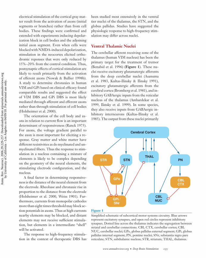
The cerebellar afferent receiving zone of thethalamus (human VIM nucleus) has been theprimary target for the treatment of tremor(Benabid et al. 1996) (Figure 1). These nu-clei receive excitatory glutamatergic afferentsfrom the deep cerebellar nuclei (Asanumaet al. 1983, Kultas-Ilinsky & Ilinsky 1991),excitatory glutamatergic afferents from thecerebral cortex (Bromberg et al. 1981), and in-hibitory GABAergic inputs from the reticularnucleus of the thalamus (Ambardekar et al.1999, Ilinsky et al. 1999). In some species,they also receive inputs from GABAergic in-hibitory interneurons (Kultas-Ilinsky et al.1985). The output from these nuclei primarily
Cerebral Cortex
STR
GPi /SNr
CBLNUC
GPe CBLCTX
STN PNTHAL
Figure 1Simplified schematic of subcortical motor systems circuitry. Blue arrowsrepresent excitatory synapses, and open red circles represent inhibitorysynapses. Dotted line across the thalamus indicates the segregation betweenstriatal and cerebellar connections. CBL CTX, cerebellar cortex; CBLNUC, cerebellar nuclei; GPe, globus pallidus external segment; GPi, globuspallidus internal segment; PN, pontine nuclei; SNr, substantia nigra parereticulate; STN, subthalamic nucleus; STR, striatum; THAL, thalamus.
www.annualreviews.org • Deep Brain Stimulation 235
Ann
u. R
ev. N
euro
sci.
2006
.29:
229-
257.
Dow
nloa
ded
from
arj
ourn
als.
annu
alre
view
s.or
gby
Uni
vers
ity o
f C
alif
orni
a -
Los
Ang
eles
on
04/0
9/10
. For
per
sona
l use
onl
y.
ANRV278-NE29-08 ARI 8 May 2006 15:49
targets motor areas of cerebral cortex (Hoover& Strick 1999, Strick et al. 1993) but has alsobeen shown to project to striatum (Hoshi et al.2005, McFarland & Haber 2001). Thus, al-though it is common to view VIM as a simplerelay for information from the cerebellum tocerebral cortex, the synaptic connections arecomplex and DBS likely influences multipleelements.
Rodent in vitro thalamic slice prepara-tions. To investigate the cellular mechanismby which DBS might work, Kiss and col-leagues have employed a slice preparationfrom rat thalamus (Anderson et al. 2004, Kisset al. 2002). The rat homologues of humanVIM are the ventrolateral and ventroposte-rior nuclei. Using simulated DBS (sDBS) withvariables comparable with that used in humanDBS, Kiss et al (2002) have shown the ef-fect of stimulation of ventral lateral ventralposterior thalamic nuclei (VL-VP) on neu-rons is both amplitude and frequency depen-dent. Response to stimulation was seen atfrequencies above 20 Hz, it increased with in-creasing stimulation frequency, and it reacheda maximum at 200 Hz. This is comparablewith the frequency response characteristics ofVIM DBS for ET (Ushe et al. 2004). Whenrhythmic pulse trains were injected intoVL-VP neurons to simulate tremor-likebursting, sDBS eliminated the rhythmic fir-ing (Kiss et al. 2002). At moderate currents,the rhythmic firing was replaced by nonrhyth-mic firing, but higher currents induced blockand eliminated firing.
Using bipolar stimulation with parametersto mimic DBS (125 Hz, 60-μs pulse width)and limit current spread to the VL-VP tha-lamus, 10-s trains of sDBS induced depolar-ization in VL-VP neurons (Anderson et al.2004). For each neuron, the time course ofthe depolarization followed one of two pat-tern types. Type I (43 of 62 neurons) quicklyreached a depolarization plateau and began torepolarize after 1 s with a moderate sustaineddepolarization of 8.2 ± 6.1 mV with no spikeactivity and no apparent excitatory postsynap-
tic potentials (EPSPs) after the initial depo-larization. Type II (19 of 62 neurons) quicklyreached a depolarization plateau but did notrepolarize and maintained a larger plateau po-tential (28.8 ± 8 mV). Action potential oc-currence in Type II responses was variable,usually with a period of quiescence followedby reemergence of firing. If stimulus trainswere prolonged, both types of responses weremaintained for up to 5 min of sDBS. Type Iand Type II responses were thought to occurin the same cell type because the rat ventralthalamus is made of a homogeneous popula-tion of cells. Changing the stimulation cur-rent did not convert one response type to theother, and there was no relationship betweenresponse type and current amplitude or dis-tance from stimulating electrode. Both typesof depolarization responses were blockedby tetrodotoxin, kynurenate, or a mix-ture of 2-amino-5-phosphonovaleric acid or6,7-dinitroquinoxaline-2,3-dione. The block-ade of presynaptic Ca2+ channels similarlyblocked the depolarization response to sDBS.There was no effect of GABA blockade oneither type of response. Thus, the depolariza-tion response is dependent on action potentialgeneration and glutamate neurotransmissionvia ionotropic receptors. The difference be-tween Type I and Type II responses mightreflect differences in the proportion of corti-cal and cerebellar afferents to individual cellsin the slice preparation. The apparent lack ofEPSPs during Type I responses suggests theremight be a functional deafferentation. Type IIresponses would be associated with the lossof any rhythmic firing and might representa mechanism by which pathological signalswould be disrupted.
In addition to activating excitatory presy-naptic terminals, sDBS in rat thalamic slicealso produced increased excitability of tha-lamic neurons (Anderson et al. 2004). Thethreshold for triggering Na+-dependent ac-tion potentials was decreased by sDBS, evenin the presence of ionotropic glutamate block-ade, causing a 30% increased probabilityof firing action potentials in response to
236 Perlmutter · Mink
Ann
u. R
ev. N
euro
sci.
2006
.29:
229-
257.
Dow
nloa
ded
from
arj
ourn
als.
annu
alre
view
s.or
gby
Uni
vers
ity o
f C
alif
orni
a -
Los
Ang
eles
on
04/0
9/10
. For
per
sona
l use
onl
y.
ANRV278-NE29-08 ARI 8 May 2006 15:49
injected depolarizing currents. The decreasedthreshold was not a result of changes in mem-brane resistance. These nonsynaptic effectswere dependent on current and distance fromthe stimulating electrode. Thus, regardless ofpresynaptic effects, the increased excitabilityof cell bodies suggests there also may be in-creased excitability of efferent axons.
The limitations of the rat thalamic slicepreparation include the following: (a) spon-taneous afferent activity is lost; (b) anypathological changes associated with neuro-logic disorders will not be represented; and(c) GABAergic inhibitory interneurons arepresent in human but not rat thalamus. Theabsence of inhibitory synaptic influences inthe rat may limit the ability to extend thesefindings to human thalamus. In a preliminaryreport of human VIM neurons, 1-s trains ofmicrostimulation at 100–300 Hz induced pro-longed inhibition in 40% of recorded neurons(Dostrovsky et al. 2002). The inhibition wasmore common in neurons that were firing inbursts. The field of microstimulation effect islikely to be substantially smaller than that oftypical DBS, and there is possibly a higherprobability of activating local inhibitory neu-rons than excitatory afferents with microstim-ulation. Nonetheless, in VIM DBS, inhibitorysynaptic mechanisms may be important con-tributors to the local effects of stimulation.
Subthalamic Nucleus
The STN has become the most commonlyused target for DBS in the treatment of PD(Rodriguez-Oroz et al. 2004) (Figure 1). TheSTN is an important node in basal gangliacircuits, serving as a major target for corticalafferents and also receiving multiple inputsfrom other basal ganglia components (Mink1996, Parent & Hazrati 1995). The STN re-ceives glutamatergic excitatory afferents fromthe frontal lobe of the cerebral cortex (Mon-akow et al. 1978, Rouzaire-Dubois & Scarnati1987), GABAergic inhibitory afferents fromthe globus pallidus external segment (Bolamet al. 2000, Rouzaire-Dubois et al. 1980),
and excitatory afferents from the parafasci-cular nucleus of the thalamus (Mouroux &Feger 1993). There are also inputs from thepedunculopontine nucleus (Lavoie & Parent1994) and from substantia nigra pars com-pacta (Cossette et al. 1999). The output fromthe STN is glutamatergic and excitatory toboth segments of the globus, to the substan-tia nigra pars reticulata (SNr), and to the pe-dunculopontine area (Smith et al. 1990). Out-puts appear to arise from different types ofneurons, but classification schemes have notagreed on how many types of neurons exist inthe STN. It appears there are at least two typesof neurons in the STN as defined by baselinefiring pattern and morphology (Magarinos-Ascone et al. 2002). Thus DBS in the STN hasthe potential to influence a variety of afferentand efferent targets and may have differenteffects on different neurons.
Rodent in vitro subthalamic nucleus slicepreparations. The effect of high-frequencystimulation has been studied in rat STN slicesby several investigators. The studies have in-volved different stimulation methodologiesand have focused on different time periodsmaking direct comparison difficult. Bipolarmicrostimulation (0.1–1.0 μA) with trains ofpulses produced a response that dependedon the type of neuron (Magarinos-Asconeet al. 2002). The current was selected to pro-duce subthreshold EPSPs in STN neurons.The two most frequently encountered neu-ron types were (a) tonically active neuronsthat had a round soma and extensive radialdendritic field (68%) and (b) bursting neu-rons with a triangular soma and less exten-sive dendritic field (25%). Tonically activecells followed 130-Hz stimulation for 5–15 s,then developed a bursting pattern, beforeceasing to fire after 25 s of stimulation. At fre-quencies less than 90 Hz, the cells followedfor 5–15 s and then changed to bursting thatpersisted for the duration of the stimulation(40 s). Bursting cells responded to stimula-tion trains with a brief burst of action poten-tials followed by prolonged silence. There was
www.annualreviews.org • Deep Brain Stimulation 237
Ann
u. R
ev. N
euro
sci.
2006
.29:
229-
257.
Dow
nloa
ded
from
arj
ourn
als.
annu
alre
view
s.or
gby
Uni
vers
ity o
f C
alif
orni
a -
Los
Ang
eles
on
04/0
9/10
. For
per
sona
l use
onl
y.
ANRV278-NE29-08 ARI 8 May 2006 15:49
no frequency dependence in bursting cells.A major limitation of this study was that thestimulation current was low and primarily af-fected presynaptic axons rather than cell bod-ies. Nevertheless, these findings suggest thatpresynaptic driving of STN neurons may failat sustained high frequencies.
Beurrier et al. (2001) also reported a pro-longed inactivation of STN neurons. Theydelivered bipolar stimulation to rat STNslice preparations in 1-min trains of 100-μsecpulses at a variety of frequencies. At fre-quencies ≤100 Hz, there was no effect. Athigher frequencies, there was a slowing ofthe post-stimulation firing rate and with fre-quencies between 166 and 250 Hz, there wasa complete and prolonged cessation of fir-ing for an average of 5.8 min. During thesilent period, action potentials could still beevoked but at a slightly higher threshold. Thesilent period was not a result of hyperpolar-ization and was not influenced by chemicalsthat blocked ionotropic glutamate receptorsor GABA receptors. Similarly, the blockadeof presynaptic Ca2+ entry had no effect. Dur-ing the silent period, the persistent Na+ cur-rent was 99% blocked and T- and l-type Ca2+
currents were transiently depressed. Thus,it appeared the post-stimulation silence wasa result of changes in membrane propertiesand not synaptic effects. It should be notedthat prolonged post-stimulation silencing oc-curred only at frequencies higher than thosethat produce maximum benefit from STNDBS in PD patients (Moro et al. 2002).
Using stimulation parameters that moreclosely simulate clinical DBS, Garcia andcolleagues (2003) have studied the effect ofhigh-frequency stimulation in rat STN slicepreparations from normal and dopamine-depleted rats. Monopolar stimulation withfrequencies in the range of 80–185 Hz blockedspontaneous firing in STN neurons but in-duced stimulus-driven firing. The effect wasseen regardless of whether the neurons weretonically active or bursting at baseline. Thestimulus-driven firing was single spikes atlower currents or recurrent bursting at higher
currents. The frequency of spikes withinbursts followed reliably at 80 Hz, had somefailure at 135 Hz, and only occurred every2–3 pulses at 185 Hz, firing with a mean in-traburst frequency of 64–85 Hz. The patternof response varied among neurons but didnot depend on the distance from the stim-ulating electrode, suggesting cell geometryin relation to the stimulation field might de-termine the response to stimulation (Garciaet al. 2003, Ranck 1975). There was no dif-ference between the slices from intact ordopamine-depleted rats. The blockade ofionotropic or metabotropic glutamate recep-tors or of GABA receptors had no effect onthe stimulus-driven firing. The stimulation-driven firing appeared to be a result of theactivation of voltage-sensitive Na+ and l-type Ca2+ channels. However, consistent withthe findings of Beurrier et al. (2001), afterthe stimulus train ended, STN neurons weresilent for as long as several minutes. Thus,although there was likely to be a reductionof certain Na+ and Ca2+ conductances, thestimulation trains were sufficient to overcomethose to induce firing. In a subsequent study,Garcia et al. (2005) confirmed their previousfindings and showed by systematically vary-ing pulse width and stimulation frequencythat combinations in the range used in humanSTN DBS never silenced STN neurons butrather drove firing. Combinations in the ther-apeutic range replaced baseline firing withstimulus-driven spikes in a stable oscillatorypattern time locked to the stimuli.
The lack of presynaptic effect with sDBS inSTN slices contrasts with the results of Ander-son et al. (2004) in thalamic slices. It is possiblethe difference relates to the method of stim-ulation. Garcia et al. (2003) used a monopo-lar configuration, and Anderson et al. (2004)used a bipolar configuration. When Andersonet al. (2004) used a monopolar configura-tion, they found a substantial reduction ofpresynaptic activation unless the current wasincreased three- to fivefold. It is also possi-ble the predominately synaptic effect seen byAnderson and the predominately cellular
238 Perlmutter · Mink
Ann
u. R
ev. N
euro
sci.
2006
.29:
229-
257.
Dow
nloa
ded
from
arj
ourn
als.
annu
alre
view
s.or
gby
Uni
vers
ity o
f C
alif
orni
a -
Los
Ang
eles
on
04/0
9/10
. For
per
sona
l use
onl
y.
ANRV278-NE29-08 ARI 8 May 2006 15:49
membrane effect seen by Garcia are results ofdifferences between the ventral thalamus andSTN neurons.
In vivo animal studies. Although in vitrostudies in slices can investigate synaptic andmembrane physiology, they have limited abil-ity to evaluate downstream effects of stim-ulation. Although few studies have beenperformed in intact experimental animals, thestudies have indicated that DBS may activateefferent axons independent of any local synap-tic or cellular effects.
In urethane-anesthetized rats, high-frequency STN stimulation caused post-stimulation depression of firing in themajority of neurons (Benazzouz et al. 2000).It also caused an inhibition of the majorityof neurons recorded in the SNr and anincrease in the majority of cells recorded inthe ventrolateral thalamus. However, thisstudy was limited by the inability to recordduring the time of stimulation, so all resultswere during the post-stimulation period.Nevertheless, in anesthetized rats, trains ofhigh-frequency stimulation appear to causea prolonged post-stimulation inactivation ofSTN neurons, and postsynaptic effects wereconsistent with inactivation of the excitatorySTN to SNr projection.
In rats anesthetized with chloral hydrate,Maurice et al. (2003) investigated the effectof high-frequency (50–200-Hz) STN stimu-lation on spontaneous SNr firing and on SNractivity evoked by motor cortex stimulation.Low intensity microstimulation (20–80 μA) at130 Hz with 30-s trains of 60-μs pulses pro-duced three types of effects on spontaneouslyactive SNr neurons. The firing of 84 of 129SNr cells was inhibited by an average of 79%.The amount of inhibition was the same forfrequencies ranging from 50–200 Hz. Theinhibition was blocked by the application ofthe GABA antagonist bicuculline. Because theprojection from the STN to the SNr is en-tirely excitatory, it is likely the stimulationactivated inhibitory striatonigral or pallidon-igral fibers (Windels et al. 2005). Excitation
was seen in 28 of 129 SNr cells with firing ratesincreasing up to 400%. In 13 cells, inhibitionwas seen at low-stimulation currents and exci-tation was seen at higher currents. Excitatoryresponses were frequency dependent, increas-ing in a linear relationship for stimulation fre-quencies from 50 to 130 Hz. These responseswere likely a result of the direct activation ofsubthalamonigral neurons or axons. Twentyof 129 neurons were activated antidromically,suggesting the stimulation effect was not con-fined to STN but spread to the nigrothalamicpathway.
In the absence of STN stimulation, motorcortex stimulation typically elicits a triphasicresponse in SNr neurons with early excitation,inhibition, then late excitation. The early ex-citation is mediated by the activation of theexcitatory projection from the STN to theSNr and the inhibition by the activation ofinhibitory striatonigral neurons (“direct path-way”), and the late excitation is mediated bythe disinhibition of subthalamonigral neurons(“indirect pathway”). In SNr neurons inhib-ited by STN stimulation, the early and lateexcitatory phases of cortically evoked activitywere inhibited by 56% and 35%, respectively,consistent with the activation of inhibitory in-puts to the SNr. In SNr neurons excited bySTN stimulation, both the early and late exci-tation were completely blocked during STNstimulation but the inhibitory response waspreserved. Thus, high-frequency STN stim-ulation blocks the transmission of informationthrough the STN. In summary, these resultsshow that STN stimulation can activate mul-tiple pathways but also that high-frequencySTN stimulation activates excitatory projec-tions from the STN to the SNr. Furthermore,STN stimulation blocks the flow of informa-tion through the STN, potentially prevent-ing aberrant signals from being propogatedin disease states. Indeed, STN stimulation inrats rendered cataleptic with dopamine antag-onists reverses abnormal patterns in SNr neu-rons (Degos et al. 2005).
Two studies of the DBS effect on down-stream neurons have been performed in
www.annualreviews.org • Deep Brain Stimulation 239
Ann
u. R
ev. N
euro
sci.
2006
.29:
229-
257.
Dow
nloa
ded
from
arj
ourn
als.
annu
alre
view
s.or
gby
Uni
vers
ity o
f C
alif
orni
a -
Los
Ang
eles
on
04/0
9/10
. For
per
sona
l use
onl
y.
ANRV278-NE29-08 ARI 8 May 2006 15:49
On-stimulation period
PS
TH
(in
cid
en
ce
/sti
m)
Fir
ing
ra
te (
sp
ike
s/s
)
(Sec)
Pre-stimulation On-stimulation
0.30
0.20
00
250
200
150
100
50
00 10 20 30 40 50 60 70 80
7 ms
0.30
0.20
0.10 0.10
00 7 ms
**
* *
*
*†† †††
*
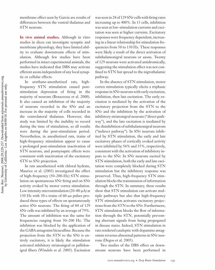
Figure 2The neuronal response of a GPi cell during subthalamic nucleusstimulation. Top trace shows analog signal overlays of 100 sweeps madeby triggering at 10-ms intervals in the prestimulation period and bytriggering on the stimulation pulse in the on-stimulation period. Arrowsindicate residual stimulation artifacts after artifact-template subtraction.Middle traces display peristimulus time histograms (PSTHs)reconstructed from successive 7.0-ms time periods in the prestimulationperiod and from the interstimulus periods in the on-stimulation period.The first PSTH bin is omitted in the on-stimulation period because ofsignal saturation and residual stimulation artifacts. Asterisks representsignificant increase at p ≤ 0.01; daggers represent significant decrease atp ≤ 0.01; Wilcoxon signed rank test. Bottom plot represents the meanfiring rate calculated every 1 s on the basis of the PSTH illustrating thetime course of the firing rate. From Hashimoto et al. 2003.
monkeys (Hashimoto et al. 2003, Kita et al.2005). Using a scaled-down version of theDBS electrode used clinically, Hashimotoet al. (2003) studied the effect of low- andhigh-frequency STN stimulation on pallidalneuron firing in 1-methyl-4-phenyl-1,2,3,6-tetrahydropyridine (MPTP) treated parkin-
sonian monkeys (Hashimoto et al. 2003).Stimulation at 136 Hz reliably reducedparkinsonian signs when sufficient currentwas delivered. Neurons in both the exter-nal pallidum (GPe) and GPi were recordedduring stimulation at 2, 136, and 157 Hzwith both effective and ineffective voltages.Short latency, multiphasic responses with al-ternating periods of inhibition and excitationwere seen in GPe and GPi neurons following2-Hz STN stimulation. These short-latencyresponses also were present at 136-Hz stimu-lation, voltages effective for the alleviation ofparkinsonian signs. The later components ofthe response were obscured by stimulation at157 Hz, but the early components remainedintact. The response persisted for up to 5 minof stimulation, producing a significant in-crease in mean discharge rate and a stimulus-synchronized regular firing pattern in themajority of GPe and GPi neurons (Figure 2).The preservation of the response pattern andoverall increase in firing rate indicated thathigh-frequency STN stimulation using clin-ically relevant DBS parameters causes theactivation of STN efferent fibers. The mul-tiphasic response pattern suggests there wasdi- and trisynaptic activation of other compo-nents of basal ganglia circuitry. There was alsoevidence for antidromic activation of someGPe neurons. Kita et al. (2005) reported com-parable results using shorter bursts of stimula-tion (10 pulses at 100 Hz) but found that morecomplex disynaptic responses in the GPi ex-ceeded simple monosynaptic excitation.
Human studies. Human studies have beenperformed in patients undergoing DBS elec-trode implantation for the treatment of PD.Theses studies offer the advantage of beingable to study physiology in the relevant dis-ease state using stimulation parameters thatelicit clinical benefit. However, there are con-straints as to what can be studied. Thus mostrecordings have been made in the region ofstimulation and not in downstream structures.
Using paired electrodes separated by600 μm, Filali et al. (2004) recorded the
240 Perlmutter · Mink
Ann
u. R
ev. N
euro
sci.
2006
.29:
229-
257.
Dow
nloa
ded
from
arj
ourn
als.
annu
alre
view
s.or
gby
Uni
vers
ity o
f C
alif
orni
a -
Los
Ang
eles
on
04/0
9/10
. For
per
sona
l use
onl
y.
ANRV278-NE29-08 ARI 8 May 2006 15:49
activity of STN neurons in response to brieftrains (500 ms) of high-frequency STN stim-ulation. Their artifact suppression methodprecluded recording for several millisecondsafter each pulse. However, following stimulustrains at 100–300 Hz, 25 of 60 STN cells wereinhibited. No post-stimulus change in firingrate was observed in the other 35 cells. In 15cells it was possible to detect inhibition duringthe train; 13 of these were inhibited. Further-more, 8 of the 25 inhibited neurons also wereinhibited by single pulses. Welter et al. (2004)confirmed these results by recording 21 STNcells during high-frequency STN stimulationtrains of 20-s duration. Fifteen of the 21 cellshad decreased firing, and six had complete ces-sation of firing during the stimulation period.No increases were seen.
Globus Pallidus
The GPi is the second most commonlyused DBS target for the treatment of PD(Anderson et al. 2005) and is increasinglytargeted for DBS treatment of dystonia(Vidailhet et al. 2005). The GPi is one ofthe primary output nuclei of the basal gan-glia and is considered the main output rep-resentation of limb movements (Mink 1996).The GPi receives excitatory glutamatergicafferents from the STN (Hazrati & Parent1992, Rinvik & Ottersen 1993), inhibitoryGABAergic afferents from striatum (Kita &Kitai 1988), inhibitory inputs from the GPe(Bolam & Smith 1992), and nigral dopamineafferents (Smith et al. 1989). The inhibitoryGABAergic output of the GPi projects tothe ventral anterior and ventral lateral thala-mus, intralaminar thalamus, and the pedun-culopontine area (Parent & De Bellefeuille1982). Owing to its size and geometry, theeffect of stimulation in the GPi is more likelyto be restricted to the nucleus, but the poten-tial remains for the possible spread to adjacentstructures and pathways, especially the GPeand internal capsule.
The rodent homologue of GPi is the en-topeduncular nucleus, which is embedded in
the internal capsule. Thus it is not possibleto simulate GPi DBS in slice preparationsor in whole brain studies in rodents withoutconfounding effects from stimulating fibers ofpassage.
In vivo animal studies of GPi stimulation.In an MPTP parkinsonian monkey, stimula-tion of the anterior GPi with 20-s trains of100–120-Hz stimulation reduced the activityin 48 of 56 GPi neurons recorded during thestimulation trains (Boraud et al. 1996). Noactivity increases were reported. In that an-imal, the GPi firing rate increased above nor-mal baseline rates in response to MPTP treat-ment. High-frequency GPi stimulation re-duced the average firing rate to normal range.No cells were completely inhibited.
In a subsequent study, Bar-Gad et al.(2004) recorded GPi activity during micros-timulation of the GPi using short trains ofhigh-frequency stimuli (10–40 pulse). Theyalso reported an overall decrease in GPi firingrates but also reported some increases. Analy-sis with a higher temporal resolution revealeda complex locking of responses to the stim-uli in most neurons. The locking displayed astereotypical temporal structure consisting ofthree phases: an initial excitation followed byan inhibition and a second excitation. Thesedata suggest the response of local neurons tohigh-frequency stimulation is complex. How-ever, only short trains were used in that studyand the response to chronic high-frequencystimulation may be different.
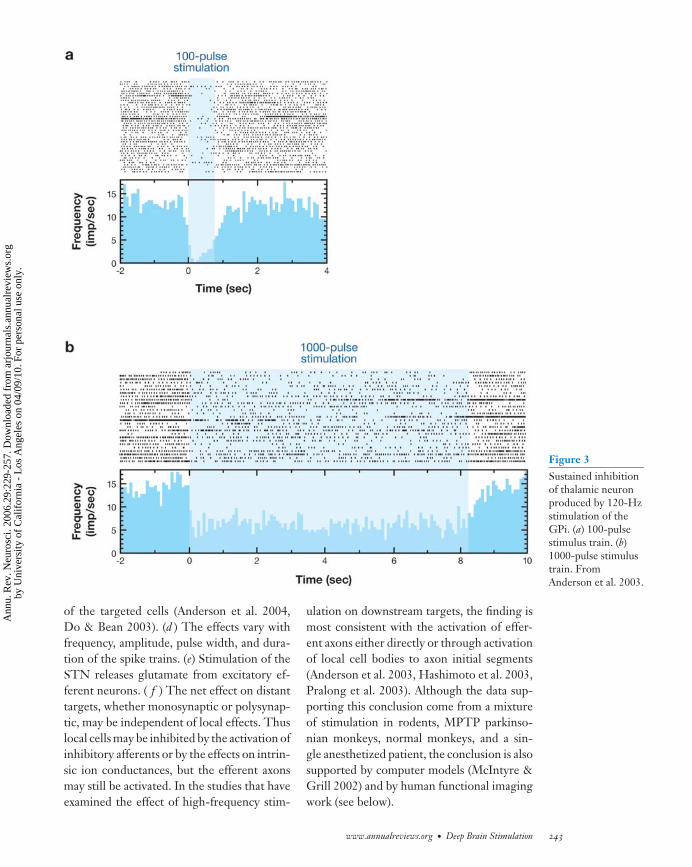
Anderson et al. (2003) recorded the ac-tivity of neurons in the pallidal-receivingzone of the thalamus during short trains(<10 s) of 120-Hz stimulation in nonparkin-sonian monkeys. Thirty-three of 73 recordedthalamic neurons were inhibited by high-frequency GPi stimulation, and seven wereexcited (Figure 3). At least one of the ex-citation responses recorded during stimula-tion also evoked muscle contraction at thecontralateral shoulder, suggesting spread tothe internal capsule. Low-amplitude stimu-lation produced inhibition but did not block
www.annualreviews.org • Deep Brain Stimulation 241
Ann
u. R
ev. N
euro
sci.
2006
.29:
229-
257.
Dow
nloa
ded
from
arj
ourn
als.
annu
alre
view
s.or
gby
Uni
vers
ity o
f C
alif
orni
a -
Los
Ang
eles
on
04/0
9/10
. For
per
sona
l use
onl
y.
ANRV278-NE29-08 ARI 8 May 2006 15:49
movement-related firing increases; however,higher amplitude stimulation did blockthese movement-related changes. These datasuggest that in addition to any effect onGPi cell bodies, high-frequency GPi stimu-lation activates efferent axons. Furthermore,stimulation changes baseline firing rates butalso has the potential to disrupt normal (orabnormal) task-related patterns of activity inpostsynaptic cells.
Human studies of GPi stimulation. In hu-man subjects undergoing the implantation ofGPi DBS electrodes, Dostrovsky et al. (2000)recorded the response of GPi neurons to low-frequency (5–50-Hz), low-amplitude stimula-tion delivered 250–600 μm from the record-ing site. The response in 22 of 23 cells wasinhibition lasting 15–25 ms after each pulse,consistent with the activation of presynapticinhibitory terminals. At higher frequencies upto 300 Hz, stimulus trains produced decreasedfiring but did not completely block firing.
Pralong et al. (2003) reported the responseof thalamic neurons to GPi DBS in a uniquesituation. A patient with postanoxic dystoniahad previously undergone the implantationof GPi DBS electrodes without benefit. Thepatient subsequently consented to thalamicstimulation, in the putative pallidal-receivingzone. While recording prior to implantationof the thalamic DBS electrodes, the authorsexamined the response of seven thalamic neu-rons to GPi DBS while the patient was anes-thetized with propofol. Four tonically activecells were inhibited by GPi stimulation; threelow frequency cells did not change. Althoughlimited, these results are consistent with thosereported by Anderson et al. (2003) in themonkey.
Release of Neurotransmitters byDeep Brain Stimulation
An early study suggested that high-frequencystimulation of the STN in rodents increasesextracellular glutamate in the GPi and adownstream target of STN projections, and
that release may be dependent upon stimu-lation frequency (Windels et al. 2003). Al-though a similar increase was not foundin humans with PD, there was an increasein cyclic guanosine monophosphate (cGMP)in the GPi (Stefani et al. 2005). Interest-ingly Windels et al. (2005) found that high-frequency STN stimulation in vivo in ratsincreased GABA in the SNr, and this effectwas abolished by ibotenic acid lesioning of theglobus pallidus. STN DBS also increases ex-tracellular striatal glutamate and GABA in rats(Windels et al. 2003). Together these findingssupport the notion that STN DBS drives out-put neurons. A similar effect may be importantfor other sites of stimulation. For example, theeffects of high-frequency stimulation of thala-mic slices were blocked by glutamate receptorantagonists (Anderson et al. 2004).
PET measures of striatal [11C]racloprideuptake did not change with STN DBS, sug-gesting release of striatal dopamine did notchange enough to produce either an in-creased striatal [11C]raclopride uptake (ev-idence of reduced competition from lessreleased dopamine) or a decreased striatal[11C]raclopride (suggesting increased releaseof striatal dopamine) (Hilker et al. 2003). Thiscontrasts with a previous rodent study indicat-ing that STN DBS increases striatal dopaminerelease (Meissner et al. 2002) in both normaland denervated (nigrostriatal lesioned) rats(Bruet et al. 2001).
Synthesis of Neurophysiologic Data
Differences in techniques, anatomy, cell type,and experimental setting limit the ability tomake direct comparisons across the studies re-viewed above. However, several conclusionsare possible. (a) High-frequency stimulationaffects multiple elements, including afferentaxons, cell bodies, efferent axons, and fibersof passage. (b) The stimulated elements maydiffer depending on the anatomy of the tar-get (e.g., the VIM thalamus, STN, GPi, orothers). (c) The effects may vary depend-ing on the intrinsic physiologic properties
242 Perlmutter · Mink
Ann
u. R
ev. N
euro
sci.
2006
.29:
229-
257.
Dow
nloa
ded
from
arj
ourn
als.
annu
alre
view
s.or
gby
Uni
vers
ity o
f C
alif
orni
a -
Los
Ang
eles
on
04/0
9/10
. For
per
sona
l use
onl
y.
ANRV278-NE29-08 ARI 8 May 2006 15:49
Figure 3Sustained inhibitionof thalamic neuronproduced by 120-Hzstimulation of theGPi. (a) 100-pulsestimulus train. (b)1000-pulse stimulustrain. FromAnderson et al. 2003.
of the targeted cells (Anderson et al. 2004,Do & Bean 2003). (d ) The effects vary withfrequency, amplitude, pulse width, and dura-tion of the spike trains. (e) Stimulation of theSTN releases glutamate from excitatory ef-ferent neurons. ( f ) The net effect on distanttargets, whether monosynaptic or polysynap-tic, may be independent of local effects. Thuslocal cells may be inhibited by the activation ofinhibitory afferents or by the effects on intrin-sic ion conductances, but the efferent axonsmay still be activated. In the studies that haveexamined the effect of high-frequency stim-
ulation on downstream targets, the finding ismost consistent with the activation of effer-ent axons either directly or through activationof local cell bodies to axon initial segments(Anderson et al. 2003, Hashimoto et al. 2003,Pralong et al. 2003). Although the data sup-porting this conclusion come from a mixtureof stimulation in rodents, MPTP parkinso-nian monkeys, normal monkeys, and a sin-gle anesthetized patient, the conclusion is alsosupported by computer models (McIntyre &Grill 2002) and by human functional imagingwork (see below).
www.annualreviews.org • Deep Brain Stimulation 243
Ann
u. R
ev. N
euro
sci.
2006
.29:
229-
257.
Dow
nloa
ded
from
arj
ourn
als.
annu
alre
view
s.or
gby
Uni
vers
ity o
f C
alif
orni
a -
Los
Ang
eles
on
04/0
9/10
. For
per
sona
l use
onl
y.
ANRV278-NE29-08 ARI 8 May 2006 15:49
FUNCTIONAL IMAGING OFDEEP BRAINSTIMULATION–INDUCEDCHANGES IN BRAIN CIRCUITS
Positron Emission Tomography
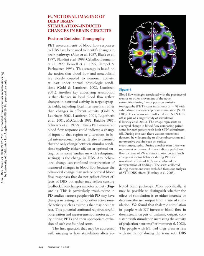
PET measurements of blood flow responsesto DBS have been used to identify changes inbrain pathways (Aiko et al. 1987, Black et al.1997, Blandini et al. 1999, Ceballos-Baumannet al. 1999, Feiwell et al. 1999, Tempel &Perlmutter 1993). This strategy is based onthe notion that blood flow and metabolismare closely coupled to neuronal activity,at least under normal physiologic condi-tions (Gold & Lauritzen 2002, Lauritzen2001). Another key underlying assumptionis that changes in local blood flow reflectchanges in neuronal activity in target synap-tic fields, including local interneurons, ratherthan changes in efferent activity (Gold &Lauritzen 2002, Lauritzen 2001, Logothetiset al. 2001, McCulloch 1982, Raichle 1987,Schwartz et al. 1979). Thus a PET-measuredblood flow response could indicate a changeof input to that region or alterations in lo-cal interneuronal activity. It also is assumedthat the only change between stimulus condi-tions (typically either off, on at optimal set-ting, or in some studies on with suboptimalsettings) is the change in DBS. Any behav-ioral change can confound interpretation ofmeasured changes in blood flow because thebehavioral change may induce cortical bloodflow responses that do not reflect direct ef-fects of DBS but rather may reflect sensoryfeedback from changes in motor activity (Fig-ure 4). This is particularly troublesome inPD studies because people with PD may havechanges in resting tremor or other active mus-cle activity such as dystonia that may occur atrest. This potential confound requires carefulobservation and measurement of motor activ-ity during PETs and then appropriate exclu-sion of such confounded scans.
The first question that may be addressedwith imaging is how stimulation alters se-
Figure 4Blood flow changes associated with the presence oftremor or other movement of the upperextremities during 1-min positron emissiontomography (PET) scans in patients (n = 8) withsubthalamic nucleus deep brain stimulation (STNDBS). These scans were collected with STN DBSoff as part of a larger study of stimulation(Hershey et al. 2003). The image represents anaveraged change in blood flow comparing pairedscans for each patient with both STN stimulatorsoff. During one scan there was no movementdetected by videography or direct observation andno excessive activity seen on surfaceelectromyography. During another scan there wasmovement or tremor. Arrows indicate peak bloodflow increase of 5% in sensorimotor cortex. Suchchanges in motor behavior during PETs toinvestigate effects of DBS can confound theinterpretation of findings. The scans collectedduring movement were excluded from our analysisof STN DBS effects (Hershey et al. 2003).
lected brain pathways. More specifically, itmay be possible to distinguish whether theeffect of stimulation is to either increase ordecrease the net output from a site of stim-ulation. We found that thalamic stimulationin people with ET increases blood flow indownstream targets of thalamic output, con-sistent with stimulation increasing the activityof projection neurons (Perlmutter et al. 2002).The people with ET had their arms at restwith no tremor during the scans with DBS
244 Perlmutter · Mink
Ann
u. R
ev. N
euro
sci.
2006
.29:
229-
257.
Dow
nloa
ded
from
arj
ourn
als.
annu
alre
view
s.or
gby
Uni
vers
ity o
f C
alif
orni
a -
Los
Ang
eles
on
04/0
9/10
. For
per
sona
l use
onl
y.

ANRV278-NE29-08 ARI 8 May 2006 15:49
on or off, so there was no behavioral changethat could have produced sensory feedback.Surface electromyography on the limbs, con-tinuous videography, and direct observationduring the scans ensured there was no tremoror other extraneous movements during thescans. Haslinger et al. (2003) also found thatVIM DBS in ET patients at rest increased re-gional blood flow at the site of stimulationand in the sensorimotor cortex in an increas-ing fashion corresponding to increasing stim-ulus frequency or amplitude. In contrast, asimilar study of thalamic stimulation in peo-ple with PD found that cortical flow was de-creased with DBS, but changes in behaviorsuch as the reduction or elimination of rest-ing tremor could reduce the flow as a re-sult of this behavioral change (Fukuda et al.2004).
Deiber et al. (1993) used PET to compareblood flow during parkinsonian tremor withVIM DBS off, during parkinsonian tremorwith ineffective DBS settings (frequency low-ered to 50–65 Hz) and during suppressedtremor with effective settings. Subtractionanalysis of effective DBS with suppressedtremor minus ineffective DBS revealed re-duced flow in the cerebellum, but this could bea result of the effects of reduced feedback fromthe presence of the tremor rather than a di-rect change induced by VIM DBS. IneffectiveDBS minus the stimulator off condition re-vealed reduction of homolateral cerebral cor-tex flow (likely a result of the effects of theineffective stimulation because there were noother changes between the two conditions).However, this small study of six subjects waslimited by an older data analysis method thatdid not consider differences in regional vari-ance in the PET data.
Neuroimaging studies in PD are morechallenging as a result of the potential be-havioral changes with stimulators either on oroff. For example, we had to eliminate at leastone-third of the PETs in PD patients becausethe subjects had either tremor or other po-tentially confounding extraneous movementsduring a 1-min blood flow scan in a study of
DBS responses to STN stimulation (Hersheyet al. 2003). Having done that, we then coulddemonstrate that STN DBS increased bloodflow in the thalamus and reduced blood flowin cortical areas (Hershey et al. 2003). Thesedata are consistent with the hypothesis thatSTN stimulation increases firing of STN out-put neurons, which increases the inhibitionof thalamocortical projections, ultimately de-creasing blood flow in cortical targets.
Increased thalamic metabolism also wasfound in another study with bilateral STNDBS in eight people with PD (Hilker et al.2004). However, this FDG PET study alsoreported increased FDG uptake in multi-ple cortical regions, and the investigatorsdid not mention behavioral changes thatlikely occurred during the two different PETconditions: on and off bilateral STN DBS.Therefore, this and other PET or single pho-ton emission computed tomography studies(Hilker et al. 2002, 2004; Fukuda et al. 2001b;Sestini et al. 2002) that do not adequately as-sess and consider behavioral condition of sub-jects during PETs must be interpreted with agreat deal of caution. Monitoring surface elec-tromyographic activity, directly observing andvideotaping all subjects during PETs, and ex-cluding scans with these confounds may helpto avoid these potential pitfalls (Hershey et al.2003).
Several studies have reported changes inregional blood flow during motor tasks withand without DBS of the STN or the GPi,but these do not directly identify the effectsof DBS alone (Fukuda et al. 2001a, 2002;Strafella et al. 2003). In these types of studiesthere are two potential behavioral confounds.First, if the motor task is performed differ-ently with the stimulators on versus off, thenthe sensory feedback to the brain and subse-quent flow or metabolic response might bealtered. Second, even the same performancemay be actuated differently if there is differ-ent resistance or power needed to perform thesame task in the two DBS conditions. Thus,these types of studies must be interpretedcautiously.
www.annualreviews.org • Deep Brain Stimulation 245
Ann
u. R
ev. N
euro
sci.
2006
.29:
229-
257.
Dow
nloa
ded
from
arj
ourn
als.
annu
alre
view
s.or
gby
Uni
vers
ity o
f C
alif
orni
a -
Los
Ang
eles
on
04/0
9/10
. For
per
sona
l use
onl
y.
ANRV278-NE29-08 ARI 8 May 2006 15:49
Functional Magnetic ResonanceImaging Studies of Deep BrainStimulation
Could fMRI of blood oxygenation level–dependent signals be used for these studies?Rezai et al. (1999) demonstrated the feasibil-ity of this approach for thalamic stimulation,which produces clinical effects within 30 s ofstarting stimulation. They studied patientsafter the implantation of the electrode intothe thalamus but before surgical placementof the pulse generator, requiring a studybetween the two surgeries. However, thisapproach does not permit an opportunityto optimize programming of DBS or to letany lesion effect of surgery abate, which maysubstantially limit its practicality. Further-more, because the time to maximal benefitfrom STN DBS in people with PD takesas much as 30 min or longer, this wouldbe difficult for an fMRI study that requiresrepeated on–off cycles because of issues ofshifting baseline. Near-infrared spectroscopymeasurements found considerable variationsin the blood oxygenation in frontal cortexduring either thalamic or GPi stimulation,which raises questions about the potential offMRI for these studies (Murata et al. 2000,Sakatani et al. 1999). Thorough evaluationto ensure safety must also be done prior toexposing patients to this research procedure.At least one study found that structural MRIin people with implanted DBS electrodescan be done safely (Uitti et al. 2002), butothers suggest that substantial caution mustbe exercised when doing magnetic resonancescanning with active DBS electrodes inthe magnetic resonance field (Georgi et al.2004). fMRI pulse sequences produce largermagnitude magnetic fields that may poseadditional risks for active DBS contacts andpulse generators. However, it is possible todo fMRI studies with externalized leads andpulse generators removed from the magneticresonance field (Stefurak et al. 2003), butthis permits only peri-operative studies withlimited time for patient evaluations. Finally,
one must be careful during such researchstudies as a slightly frayed wire carries anincreased risk of heating surrounding tissue.
A single case report suggested that fMRIblood oxygenation level–dependent signalsincreased in different cortical regions de-pending upon the position within the re-gion of the STN of the stimulating electrodeand associated behavioral response (Stefuraket al. 2003). Specifically, stimulation throughthe left active electrode in the left inferiorSTN provided good motor benefit and in-creased flow in primary motor areas but de-creased flow in supplementary motor area.Such strategies may help to identify functionalconnections among basal ganglia and corti-cal loops. However, interpretation of thesetypes of studies is critically dependent uponaccurate identification of the site and effectsof DBS.
CONCLUSIONS
DBS has the potential to provide substan-tial benefit for a variety of neuropsychiatricconditions. Despite the marked clinical ben-efit, we still have much to learn about themechanism of action of DBS. However, wehave come a long way in our understand-ing of the effects of DBS on neurons, trans-mitters, and brain pathways. Physiologic andimaging studies support the notion that thenet effect of DBS is to increase the fir-ing of neurons projecting from the site ofstimulation. This may be mediated primarilyvia the stimulation of axons rather than cellbodies.
If DBS drives efferent axons, how does itexert its clinical effect? DBS seems to mimicthe effect of destructive lesions, suggestingthat despite the activation of efferent axons,there is interruption of information flow orprocessing. The data of Maurice et al. (2003)from rats and of Anderson et al. (2003) frommonkeys indicate that high-frequency stim-ulation can prevent the normal pattern ofactivity whether driven by electrical cortical
246 Perlmutter · Mink
Ann
u. R
ev. N
euro
sci.
2006
.29:
229-
257.
Dow
nloa
ded
from
arj
ourn
als.
annu
alre
view
s.or
gby
Uni
vers
ity o
f C
alif
orni
a -
Los
Ang
eles
on
04/0
9/10
. For
per
sona
l use
onl
y.
ANRV278-NE29-08 ARI 8 May 2006 15:49
stimulation or related to a limb-movementtask. If normal circuits are disrupted in thisway, it makes sense that abnormal circuit ac-tivity also would be disrupted. Indeed, STNDBS has been shown to eliminate abnormalrhythmic oscillation of GPi local field poten-tials (Brown et al. 2004), and impairing abnor-mal firing patterns may be more critical thanchanging net firing rates (McIntyre & Thakor2002, Vitek 2002).
Future studies may continue to distinguishvariations in the effects of DBS on differentnuclei and different neuronal cell types. Fur-thermore, patients with implanted DBS elec-trodes afford an outstanding opportunity toinvestigate behavioral effects of functional cir-cuits (Hershey et al. 2004, Schroeder et al.2003), but it will be critical to carefully controlbehavioral confounds to properly interpretsuch studies.
DISCLOSURE OF BIAS
J.P.’ s lab/section receives partial fellowship support from Medtronics, Inc. each year.
ACKNOWLEDGMENTS
The authors thank Dr. Tamara Hershey for expert assistance and providing Figure 4. Thisresearch was supported by NIH grants NS050425, NS39913, NS39821, and NS41509 and theGreater St. Louis Chapter of the American Parkinson Disease Association (APDA), the APDACenter for Advanced PD Research at Washington University, the Barnes-Jewish HospitalFoundation (the Jack Buck Fund and the Elliot H. Stein Family Fund), the Missouri Chapterof the Dystonia Medical Research Foundation, and the Sam & Barbara Murphy Fund.
LITERATURE CITED
Abelson JL, Curtis GC, Sagher O, Albucher RC, Harrigan M, et al. 2005. Deep brain stimu-lation for refractory obsessive-compulsive disorder. Biol. Psychiatry 57:510–16
Aiko Y, Shima F, Hosokawa S, Kato M, Kitamura K. 1987. Altered local cerebral glucoseutilization induced by electrical stimulations of the thalamic sensory and parafascicularnuclei in rats. Brain Res. 408:47–56
Ambardekar AV, Ilinsky IA, Forestl W, Bowery NG, Kultas-Ilinsky K. 1999. Distributionand properties of GABA(B) antagonist [3H]CGP 62349 binding in the rhesus monkeythalamus and basal ganglia and the influence of lesions in the reticular thalamic nucleus.Neuroscience 93:1339–47
Anderson ME, Postupna N, Ruffo M. 2003. Effects of high-frequency stimulation in theinternal globus pallidus on the activity of thalamic neurons in the awake monkey. J.Neurophysiol. 89:1150–60
Anderson T, Hu B, Pittman Q, Kiss ZH. 2004. Mechanisms of deep brain stimulation: anintracellular study in rat thalamus. J. Physiol. 559:301–13
Anderson VC, Burchiel KJ, Hogarth P, Favre J, Hammerstad JP. 2005. Pallidal vs subthalamicnucleus deep brain stimulation in Parkinson disease. Arch. Neurol. 62:554–60
Aouizerate B, Martin-Guehl C, Cuny E, Guehl D, Amieva H, et al. 2005. Deep brain stimula-tion of the ventral striatum in the treatment of obsessive-compulsive disorder and majordepression. Med. Sci. (Paris) 21:811–13
Asanuma C, Thach WT, Jones EG. 1983. Distribution of cerebellar terminations and theirrelation to other afferent terminations in the ventral lateral thalamic region of the monkey.Brain Res. 286:237–65
www.annualreviews.org • Deep Brain Stimulation 247
Ann
u. R
ev. N
euro
sci.
2006
.29:
229-
257.
Dow
nloa
ded
from
arj
ourn
als.
annu
alre
view
s.or
gby
Uni
vers
ity o
f C
alif
orni
a -
Los
Ang
eles
on
04/0
9/10
. For
per
sona
l use
onl
y.
ANRV278-NE29-08 ARI 8 May 2006 15:49
Bar-Gad I, Elias S, Vaadia E, Bergman H. 2004. Complex locking rather than completecessation of neuronal activity in the globus pallidus of a 1-methyl-4-phenyl-1,2,3,6-tetrahydropyridine-treated primate in response to pallidal microstimulation. J. Neurosci.24:7410–19
Bartholow R. 1874. Experimental investigations into the functions of the human brain. Am. J.Med. Sci. 67:305–13
Bastian AJ, Kelly VE, Revilla FJ, Perlmutter JS, Mink JW. 2003. Different effects of unilateralversus bilateral subthalamic nucleus stimulation on walking and reaching in Parkinson’sdisease. Mov. Disord. 18:1000–7
Benabid AL, Benazzouz A, Hoffmann D, Limousin P, Krack P, Pollak P. 1998. Long-termelectrical inhibition of deep brain targets in movement disorders. Mov. Disord. 13(Suppl.3):119–25
Benabid AL, Pollak P, Gao D, Hoffmann D, Limousin P, et al. 1996. Chronic electrical stim-ulation of the ventralis intermedius nucleus of the thalamus as a treatment of movementdisorders. J. Neurosurg. 84:203–14
Benabid AL, Pollak P, Gervason C, Hoffmann D, Gao DM, et al. 1991. Long-term suppressionof tremor by chronic stimulation of the ventral intermediate thalamic nucleus. Lancet337:403–6
Benazzouz A, Gao DM, Ni ZG, Piallat B, Bouali-Benazzouz R, Benabid AL. 2000. Effectof high-frequency stimulation of the subthalamic nucleus on the neuronal activities ofthe substantia nigra pars reticulata and ventrolateral nucleus of the thalamus in the rat.Neuroscience 99:289–95
Bergstrom MR, Johansson GG, Laitinen LV, Sipponen P. 1966. Electrical stimulation of thethalamic and subthalamic area in cerebral palsy. Acta Physiol. Scand. 67:208–13
Berney A, Vingerhoets F, Perrin A, Guex P, Villemure JG, et al. 2002. Effect on mood ofsubthalamic DBS for Parkinson’s disease: a consecutive series of 24 patients. Neurology59:1427–29
Beurrier C, Bioulac B, Audin J, Hammond C. 2001. High-frequency stimulation producesa transient blockade of voltage-gated currents in subthalamic neurons. J. Neurophysiol.85:1351–56
Bittar RG, Kar-Purkayastha I, Owen SL, Bear RE, Green A, et al. 2005. Deep brain stimulationfor pain relief: a meta-analysis. J. Clin. Neurosci. 12:515–19
Black KJ, Gado MH, Perlmutter JS. 1997. PET measurement of dopamine D2 receptor-mediated changes in striatopallidal function. J. Neurosci. 17:3168–77
Blandini F, Conti G, Martignoni E, Colangelo V, Nappi G, et al. 1999. Modifications of localcerebral metabolic rates for glucose and motor behavior in rats with unilateral lesion ofthe subthalamic nucleus. J. Cereb. Blood Flow Metab. 19:149–54
Bolam JP, Hanley JJ, Booth PA, Bevan MD. 2000. Synaptic organisation of the basal ganglia.J. Anat. 196(Pt. 4):527–42
Bolam JP, Smith Y. 1992. The striatum and the globus pallidus send convergent synaptic inputsonto single cells in the entopeduncular nucleus of the rat: a double anterograde labelingstudy combined with postembedding immunocytochemistry for GABA. J. Comp. Neurol.321:456–76
Boraud T, Bezard E, Bioulac B, Gross C. 1996. High frequency stimulation of the internalglobus pallidus (GPi) simultaneously improves parkinsonian symptoms and reduces thefiring frequency of GPi neurons in the MPTP-treated monkey. Neurosci. Lett. 215:17–20
Brice J, McLellan L. 1980. Suppression of intention tremor by contingent deep-brain stimu-lation. Lancet 1:1221–22
248 Perlmutter · Mink
Ann
u. R
ev. N
euro
sci.
2006
.29:
229-
257.
Dow
nloa
ded
from
arj
ourn
als.
annu
alre
view
s.or
gby
Uni
vers
ity o
f C
alif
orni
a -
Los
Ang
eles
on
04/0
9/10
. For
per
sona
l use
onl
y.
ANRV278-NE29-08 ARI 8 May 2006 15:49
Bromberg MB, Penney JBJ, Stephenson BS, Young AB. 1981. Evidence for glutamate as theneurotransmitter of corticothalamic and corticorubral pathways. Brain Res. 215:369–74
Brown P, Mazzone P, Oliviero A, Altibrandi MG, Pilato F, et al. 2004. Effects of stimulationof the subthalamic area on oscillatory pallidal activity in Parkinson’s disease. Exp. Neurol.188:480–90
Bruet N, Windels F, Bertrand A, Feuerstein C, Poupard A, Savasta M. 2001. High frequencystimulation of the subthalamic nucleus increases the extracellular contents of striataldopamine in normal and partially dopaminergic denervated rats. J. Neuropathol. Exp. Neu-rol. 60:15–24
Burchiel KJ, Anderson VC, Favre J, Hammerstad JP. 1999. Comparison of pallidal and sub-thalamic nucleus deep brain stimulation for advanced Parkinson’s disease: results of arandomized, blinded pilot study. Neurosurgery 45:1375–82
Castelnau P, Cif L, Valente EM, Vayssiere N, Hemm S, et al. 2005. Pallidal stimulation im-proves pantothenate kinase-associated neurodegeneration. Ann. Neurol. 57:738–41
Ceballos-Baumann AO, Boecker H, Bartensetin P, von Falkenhayn I, Riescher H, et al. 1999.A positron emission tomographic study of subthalamic nucleus stimulation in Parkinson’sdisease: enhanced movement-related activity of motor-association cortex and decreasedmotor cortex resting activity. Arch. Neurol. 56:997–1003
Cooper IS. 1973. Effect of chronic stimulation of anterior cerebellum on neurological disease.Lancet 1:206
Cooper IS, Riklan M, Amin I, Waltz JM, Cullinan T. 1976. Chronic cerebellar stimulation incerebral palsy. Neurology 26:744–53
Cossette M, Levesque M, Parent A. 1999. Extrastriatal dopaminergic innervation of humanbasal ganglia. Neurosci. Res. 34:51–54
Cosyns P, Gabriels L, Nuttin B. 2003. Deep brain stimulation in treatment refractory obsessivecompulsive disorder. Verh. K. Acad. Geneeskd. Belg. 65:385–99
Coubes P, Cif L, El Fertit H, Hemm S, Vayssiere N, et al. 2004. Electrical stimulation of theglobus pallidus internus in patients with primary generalized dystonia: long-term results.J. Neurosurg. 101:189–94
Degos B, Deniau JM, Thierry AM, Glowinski J, Pezard L, Maurice N. 2005. Neuroleptic-induced catalepsy: electrophysiological mechanisms of functional recovery induced byhigh-frequency stimulation of the subthalamic nucleus. J. Neurosci. 25:7687–96
Deiber MP, Pollak P, Passingham R, Landais P, Gervason C, et al. 1993. Thalamic stimula-tion and suppression of parkinsonian tremor. Evidence of a cerebellar deactivation usingpositron emission tomography. Brain 116(Pt. 1):267–79
Diamond A, Jankovic J. 2005. The effect of deep brain stimulation on quality of life in movementdisorders. J. Neurol. Neurosurg. Psychiatr. 76:1188–93
Diederich NJ, Alesch F, Goetz C. 2000. Visual hallucinations induced by deep brain stimulationin Parkinson’s disease. Clin. Neuropharmacol. 23:287–89
Diederich NJ, Kalteis K, Stamenkovic M, Pieri V, Alesch F. 2005. Efficient internal pallidalstimulation in Gilles de la Tourette syndrome: a case report. Mov. Disord. 20:1496–99
Do MT, Bean BP. 2003. Subthreshold sodium currents and pacemaking of subthalamic neu-rons: modulation by slow inactivation. Neuron 39:109–20
Dostrovsky JO, Levy R, Wu JP, Hutchison WD, Tasker RR, Lozano AM. 2000.Mirostimulation-induced inhibition of neuronal firing in human globus pallidus. J. Neu-rophysiol. 84:570–74
Dostrovsky JO, Patra S, Hutchison WD, Palter VN, Filali M, Lozano AM. 2002. Effects ofstimulation in human thalamus on activity of nearby thalamic neurons. Society Neurosci.Abstr. 62.14
www.annualreviews.org • Deep Brain Stimulation 249
Ann
u. R
ev. N
euro
sci.
2006
.29:
229-
257.
Dow
nloa
ded
from
arj
ourn
als.
annu
alre
view
s.or
gby
Uni
vers
ity o
f C
alif
orni
a -
Los
Ang
eles
on
04/0
9/10
. For
per
sona
l use
onl
y.
ANRV278-NE29-08 ARI 8 May 2006 15:49
Eltahawy HA, Saint-Cyr J, Giladi N, Lang AE, Lozano AM. 2004. Primary dystonia is moreresponsive than secondary dystonia to pallidal interventions: outcome after pallidotomyor pallidal deep brain stimulation. Neurosurgery 54:613–19
Feiwell RJ, Black KJ, McGee-Minnich LA, Snyder AZ, MacLeod AMK, Perlmutter JS. 1999.Diminished regional cerebral blood flow response to vibration in patients with ble-pharospasm. Neurology 52:291–97
Ferrarin M, Rizzone M, Bergamasco B, Lanotte M, Recalcati M, et al. 2005. Effects of bilateralsubthalamic stimulation on gait kinematics and kinetics in Parkinson’s disease. Exp. BrainRes. 160:517–27
Filali M, Hutchison WD, Palter VN, Lozano AM, Dostrovsky JO. 2004. Stimulation-inducedinhibition of neuronal firing in human subthalamic nucleus. Exp. Brain Res. 156:274–81
Flaherty AW, Williams ZM, Amirnovin R, Kasper E, Rauch SL, et al. 2005. Deep brain stim-ulation of the anterior internal capsule for the treatment of Tourette syndrome: technicalcase report. Neurosurgery 57:E403
Franzini A, Ferroli P, Leone M, Broggi G. 2003. Stimulation of the posterior hypothalamusfor treatment of chronic intractable cluster headaches: first reported series. Neurosurgery52:1095–99
Fritsch G, Hitzig E. 1870. Uber die elektrische Erregbarkeit des Grosshirns. Arch. Anat. Physiol.37:300–32
Fukuda M, Barnes A, Simon ES, Holmes A, Dhawan V, et al. 2004. Thalamic stimulation forparkinsonian tremor: correlation between regional cerebral blood flow and physiologicaltremor characteristics. Neuroimage 21:608–15
Fukuda M, Ghilardi MF, Carbon M, Dhawan V, Ma Y, et al. 2002. Pallidal stimulation forparkinsonism: improved brain activation during sequence learning. Ann. Neurol. 52:144–52
Fukuda M, Mentis M, Ghilardi MF, Dhawan V, Antonini A, et al. 2001a. Functional correlatesof pallidal stimulation for Parkinson’s disease. Ann. Neurol. 49:155–64
Fukuda M, Mentis MJ, Ma Y, Dhawan V, Antonini A, et al. 2001b. Networks mediating theclinical effects of pallidal brain stimulation for Parkinson’s disease: a PET study of resting-state glucose metabolism. Brain 124:1601–9
Gabriels L, Cosyns P, Nuttin B, Demeulemeester H, Gybels J. 2003. Deep brain stimulation fortreatment-refractory obsessive-compulsive disorder: psychopathological and neuropsy-chological outcome in three cases. Acta. Psychiatr. Scand. 107:275–82
Garcia L, Audin J, D’Alessandro G, Bioulac B, Hammond C. 2003. Dual effect of high-frequency stimulation on subthalamic neuron activity. J. Neurosci. 23:8743–51
Garcia L, D’Alessandro G, Fernagut PO, Bioulac B, Hammond C. 2005. Impact of high-frequency stimulation parameters on the pattern of discharge of subthalamic neurons. J.Neurophysiol. 94:3662–69
Georgi JC, Stippich C, Tronnier VM, Heiland S. 2004. Active deep brain stimulation duringMRI: a feasibility study. Magn. Reson. Med. 51:380–88
Gold L, Lauritzen M. 2002. Neuronal deactivation explains decreased cerebellar blood flow inresponse to focal cerebral ischemia or suppressed neocortical function. Proc. Natl. Acad.Sci. USA 99:7699–704
Hanajima R, Dostrovsky JO, Lozano AM, Hutchison WD, Davis KD, et al. 2004. Somatosen-sory evoked potentials (SEPs) recorded from deep brain stimulation (DBS) electrodes inthe thalamus and subthalamic nucleus (STN). Clin. Neurophysiol. 115:424–34
Hashimoto T, Elder CM, Okun MS, Patrick SK, Vitek JL. 2003. Stimulation of the subthalamicnucleus changes the firing pattern of pallidal neurons. J. Neurosci. 23:1916–23
250 Perlmutter · Mink
Ann
u. R
ev. N
euro
sci.
2006
.29:
229-
257.
Dow
nloa
ded
from
arj
ourn
als.
annu
alre
view
s.or
gby
Uni
vers
ity o
f C
alif
orni
a -
Los
Ang
eles
on
04/0
9/10
. For
per
sona
l use
onl
y.
ANRV278-NE29-08 ARI 8 May 2006 15:49
Haslinger B, Boecker H, Buchel C, Vesper J, Tronnier VM, et al. 2003. Differential modulationof subcortical target and cortex during deep brain stimulation. Neuroimage 18:517–24
Hassler R, Riechart T, Munginer F, Umbach W, Ganglberger JA. 1960. Physiological obser-vations in stereotaxic operations in extrapyramidal motor disturbances. Brain 83:337–50
Hazrati LN, Parent A. 1992. Convergence of subthalamic and striatal efferents at pallidal levelin primates: an anterograde double-labeling study with biocytin and PHA-L. Brain Res.569:336–40
Hershey T, Revilla F, Wernle A, Schneider-Gibson P, Dowling J, Perlmutter JS. 2004. Stim-ulation of STN impairs aspects of cognitive control in PD. Neurology 62:1110–14
Hershey T, Revilla FJ, Wernle A, McGee-Minnich L, Antenor JV, et al. 2003. Cortical andsubcortical blood flow effects of subthalamic nucleus stimulation in PD. Neurology 61:816–21
Herzog J, Reiff J, Krack P, Witt K, Schrader B, et al. 2003. Manic episode with psychoticsymptoms induced by subthalamic nucleus stimulation in a patient with Parkinson’s dis-ease. Mov. Disord. 18:1382–84
Hilker R, Voges J, Ghaemi M, Lehrke R, Rudolf J, et al. 2003. Deep brain stimulation of thesubthalamic nucleus does not increase the striatal dopamine concentration in parkinsonianhumans. Mov. Disord. 18:41–48
Hilker R, Voges J, Thiel A, Ghaemi M, Herholz K, et al. 2002. Deep brain stimulation of thesubthalamic nucleus versus levodopa challenge in Parkinson’s disease: measuring the on-and off-conditions with FDG-PET. J. Neural Transm. 109:1257–64
Hilker R, Voges J, Weisenbach S, Kalbe E, Burghaus L, et al. 2004. Subthalamic nucleus stim-ulation restores glucose metabolism in associative and limbic cortices and in cerebellum:evidence from a FDG-PET study in advanced Parkinson’s disease. J. Cereb. Blood FlowMetab. 24:7–16
Holsheimer J, Demeulemeester H, Nuttin B, de Sutter P. 2000. Identification of the targetneuronal elements in electrical deep brain stimulation. Eur. J. Neurosci. 12:4573–77
Hoover JE, Strick PL. 1999. The organization of cerebellar and basal ganglia outputs to primarymotor cortex as revealed by retrograde transneuronal transport of herpes simplex virustype 1. J. Neurosci. 19:1446–63
Hoshi E, Tremblay L, Feger J, Carras PL, Strick PL. 2005. The cerebellum communicateswith the basal ganglia. Nat. Neurosci. 8:1491–93
Hosobuchi Y, Adams JE, Rutkin B. 1973. Chronic thalamic stimulation for the control of facialanesthesia dolorosa. Arch. Neurol. 29:158–61
Houeto JL, Karachi C, Mallet L, Pillon B, Yelnik J, et al. 2005. Tourette’s syndrome and deepbrain stimulation. J. Neurol. Neurosurg. Psychiatr. 76:992–95
Ilinsky IA, Ambardekar AV, Kultas-Ilinsky K. 1999. Organization of projections from the an-terior pole of the nucleus reticularis thalami (NRT) to subdivisions of the motor thalamus:light and electron microscopic studies in the rhesus monkey. J. Comp. Neurol. 409:369–84
Jaggi JL, Umemura A, Hurtig HI, Siderowf AD, Colcher A, et al. 2004. Bilateral stimulation ofthe subthalamic nucleus in Parkinson’s disease: surgical efficacy and prediction of outcome.Stereotact. Funct. Neurosurg. 82:104–14
Jahanshahi M, Ardouin CM, Brown RG, Rothwell JC, Obeso J, et al. 2000. The impact of deepbrain stimulation on executive function in Parkinson’s disease. Brain 123(Pt. 6):1142–54
Jimenez F, Velasco F, Salin-Pascual R, Hernandez JA, Velasco M, et al. 2005. A patient witha resistant major depression disorder treated with deep brain stimulation in the inferiorthalamic peduncle. Neurosurgery 57:585–93
www.annualreviews.org • Deep Brain Stimulation 251
Ann
u. R
ev. N
euro
sci.
2006
.29:
229-
257.
Dow
nloa
ded
from
arj
ourn
als.
annu
alre
view
s.or
gby
Uni
vers
ity o
f C
alif
orni
a -
Los
Ang
eles
on
04/0
9/10
. For
per
sona
l use
onl
y.
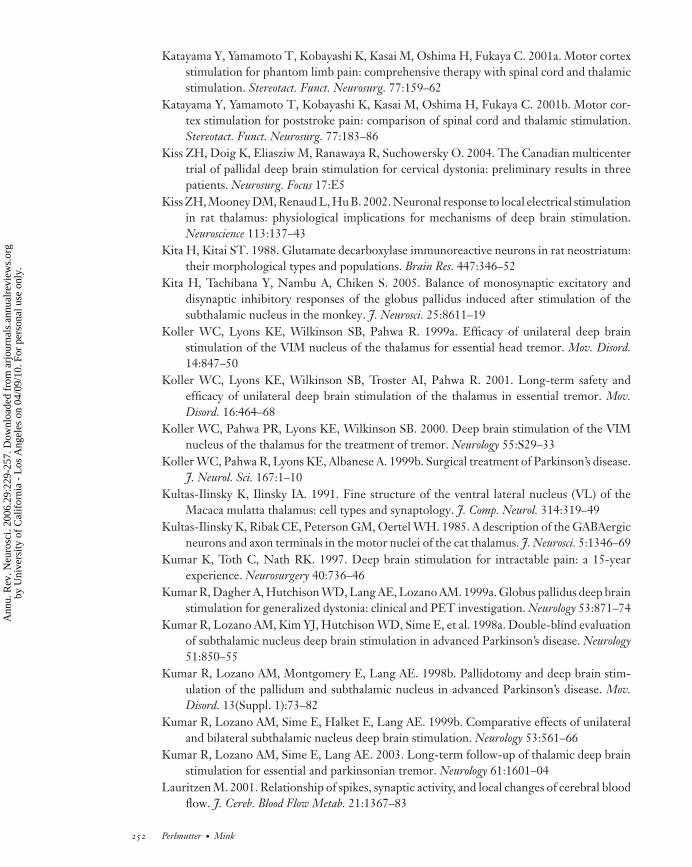
ANRV278-NE29-08 ARI 8 May 2006 15:49
Katayama Y, Yamamoto T, Kobayashi K, Kasai M, Oshima H, Fukaya C. 2001a. Motor cortexstimulation for phantom limb pain: comprehensive therapy with spinal cord and thalamicstimulation. Stereotact. Funct. Neurosurg. 77:159–62
Katayama Y, Yamamoto T, Kobayashi K, Kasai M, Oshima H, Fukaya C. 2001b. Motor cor-tex stimulation for poststroke pain: comparison of spinal cord and thalamic stimulation.Stereotact. Funct. Neurosurg. 77:183–86
Kiss ZH, Doig K, Eliasziw M, Ranawaya R, Suchowersky O. 2004. The Canadian multicentertrial of pallidal deep brain stimulation for cervical dystonia: preliminary results in threepatients. Neurosurg. Focus 17:E5
Kiss ZH, Mooney DM, Renaud L, Hu B. 2002. Neuronal response to local electrical stimulationin rat thalamus: physiological implications for mechanisms of deep brain stimulation.Neuroscience 113:137–43
Kita H, Kitai ST. 1988. Glutamate decarboxylase immunoreactive neurons in rat neostriatum:their morphological types and populations. Brain Res. 447:346–52
Kita H, Tachibana Y, Nambu A, Chiken S. 2005. Balance of monosynaptic excitatory anddisynaptic inhibitory responses of the globus pallidus induced after stimulation of thesubthalamic nucleus in the monkey. J. Neurosci. 25:8611–19
Koller WC, Lyons KE, Wilkinson SB, Pahwa R. 1999a. Efficacy of unilateral deep brainstimulation of the VIM nucleus of the thalamus for essential head tremor. Mov. Disord.14:847–50
Koller WC, Lyons KE, Wilkinson SB, Troster AI, Pahwa R. 2001. Long-term safety andefficacy of unilateral deep brain stimulation of the thalamus in essential tremor. Mov.Disord. 16:464–68
Koller WC, Pahwa PR, Lyons KE, Wilkinson SB. 2000. Deep brain stimulation of the VIMnucleus of the thalamus for the treatment of tremor. Neurology 55:S29–33
Koller WC, Pahwa R, Lyons KE, Albanese A. 1999b. Surgical treatment of Parkinson’s disease.J. Neurol. Sci. 167:1–10
Kultas-Ilinsky K, Ilinsky IA. 1991. Fine structure of the ventral lateral nucleus (VL) of theMacaca mulatta thalamus: cell types and synaptology. J. Comp. Neurol. 314:319–49
Kultas-Ilinsky K, Ribak CE, Peterson GM, Oertel WH. 1985. A description of the GABAergicneurons and axon terminals in the motor nuclei of the cat thalamus. J. Neurosci. 5:1346–69
Kumar K, Toth C, Nath RK. 1997. Deep brain stimulation for intractable pain: a 15-yearexperience. Neurosurgery 40:736–46
Kumar R, Dagher A, Hutchison WD, Lang AE, Lozano AM. 1999a. Globus pallidus deep brainstimulation for generalized dystonia: clinical and PET investigation. Neurology 53:871–74
Kumar R, Lozano AM, Kim YJ, Hutchison WD, Sime E, et al. 1998a. Double-blind evaluationof subthalamic nucleus deep brain stimulation in advanced Parkinson’s disease. Neurology51:850–55
Kumar R, Lozano AM, Montgomery E, Lang AE. 1998b. Pallidotomy and deep brain stim-ulation of the pallidum and subthalamic nucleus in advanced Parkinson’s disease. Mov.Disord. 13(Suppl. 1):73–82
Kumar R, Lozano AM, Sime E, Halket E, Lang AE. 1999b. Comparative effects of unilateraland bilateral subthalamic nucleus deep brain stimulation. Neurology 53:561–66
Kumar R, Lozano AM, Sime E, Lang AE. 2003. Long-term follow-up of thalamic deep brainstimulation for essential and parkinsonian tremor. Neurology 61:1601–04
Lauritzen M. 2001. Relationship of spikes, synaptic activity, and local changes of cerebral bloodflow. J. Cereb. Blood Flow Metab. 21:1367–83
252 Perlmutter · Mink
Ann
u. R
ev. N
euro
sci.
2006
.29:
229-
257.
Dow
nloa
ded
from
arj
ourn
als.
annu
alre
view
s.or
gby
Uni
vers
ity o
f C
alif
orni
a -
Los
Ang
eles
on
04/0
9/10
. For
per
sona
l use
onl
y.
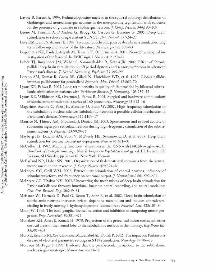
ANRV278-NE29-08 ARI 8 May 2006 15:49
Lavoie B, Parent A. 1994. Pedunculopontine nucleus in the squirrel monkey: distribution ofcholinergic and monoaminergic neurons in the mesopontine tegmentum with evidencefor the presence of glutamate in cholinergic neurons. J. Comp. Neurol. 344:190–209
Leone M, Franzini A, D’Andrea G, Broggi G, Casucci G, Bussone G. 2005. Deep brainstimulation to relieve drug-resistant SUNCT. Ann. Neurol. 57:924–27
Levy RM, Lamb S, Adams JE. 1987. Treatment of chronic pain by deep brain stimulation: longterm follow-up and review of the literature. Neurosurgery 21:885–93
Logothetis NK, Pauls J, Augath M, Trinath T, Oeltermann A. 2001. Neurophysiological in-vestigation of the basis of the fMRI signal. Nature 412:150–57
Loher TJ, Burgunder JM, Weber S, Sommerhalder R, Krauss JK. 2002. Effect of chronicpallidal deep brain stimulation on off period dystonia and sensory symptoms in advancedParkinson’s disease. J. Neurol. Neurosurg. Psychiatr. 73:395–99
Lozano AM, Kumar R, Gross RE, Giladi N, Hutchison WD, et al. 1997. Globus pallidusinternus pallidotomy for generalized dystonia. Mov. Disord. 12:865–70
Lyons KE, Pahwa R. 2005. Long-term benefits in quality of life provided by bilateral subtha-lamic stimulation in patients with Parkinson disease. J. Neurosurg. 103:252–55
Lyons KE, Wilkinson SB, Overman J, Pahwa R. 2004. Surgical and hardware complicationsof subthalamic stimulation: a series of 160 procedures. Neurology 63:612–16
Magarinos-Ascone C, Pazo JH, Macadar O, Buno W. 2002. High-frequency stimulation ofthe subthalamic nucleus silences subthalamic neurons: a possible cellular mechanism inParkinson’s disease. Neuroscience 115:1109–17
Maurice N, Thierry AM, Glowinski J, Deniau JM. 2003. Spontaneous and evoked activity ofsubstantia nigra pars reticulata neurons during high-frequency stimulation of the subtha-lamic nucleus. J. Neurosci. 23:9929–36
Mayberg HS, Lozano AM, Voon V, McNeely HE, Seminowicz D, et al. 2005. Deep brainstimulation for treatment-resistant depression. Neuron 45:651–60
McCulloch J. 1982. Mapping functional alterations in the CNS with [14C]deoxyglucose. InHandbook of Psychopharmacology: New Techniques in Psychopharmacology, ed. LL Iverson, SDIverson, SH Snyder, pp 321–410. New York: Plenum
McFarland NR, Haber SN. 2001. Organization of thalamostriatal terminals from the ventralmotor nuclei in the macaque. J. Comp. Neurol. 429:321–36
McIntyre CC, Grill WM. 2002. Extracellular stimulation of central neurons: influence ofstimulus waveform and frequency on neuronal output. J. Neurophysiol. 88:1592–604
McIntyre CC, Thakor NV. 2002. Uncovering the mechanisms of deep brain stimulation forParkinson’s disease through functional imaging, neural recording, and neural modeling.Crit. Rev. Biomed. Eng. 30:249–81
Meissner W, Harnack D, Paul G, Reum T, Sohr R, et al. 2002. Deep brain stimulation ofsubthalamic neurons increases striatal dopamine metabolism and induces contralateralcircling in freely moving 6-hydroxydopamine-lesioned rats. Neurosci. Lett. 328:105–8
Mink JW. 1996. The basal ganglia: focused selection and inhibition of competing motor pro-grams. Prog. Neurobiol. 50:381–425
Monakow KH, Akert K, Kunzle H. 1978. Projections of the precentral motor cortex and othercortical areas of the frontal lobe to the subthalamic nucleus in the monkey. Exp Brain Res.33:395–403
Moro E, Esselink RJ, Xie J, Hommel M, Benabid AL, Pollak P. 2002. The impact on Parkinson’sdisease of electrical parameter settings in STN stimulation. Neurology 59:706–13
Mouroux M, Feger J. 1993. Evidence that the parafascicular projection to the subthalamicnucleus is glutamatergic. Neuroreport 4:613–15
www.annualreviews.org • Deep Brain Stimulation 253
Ann
u. R
ev. N
euro
sci.
2006
.29:
229-
257.
Dow
nloa
ded
from
arj
ourn
als.
annu
alre
view
s.or
gby
Uni
vers
ity o
f C
alif
orni
a -
Los
Ang
eles
on
04/0
9/10
. For
per
sona
l use
onl
y.
ANRV278-NE29-08 ARI 8 May 2006 15:49
Murata Y, Katayama Y, Oshima H, Kawamata T, Yamamoto T, et al. 2000. Changes in cerebralblood oxygenation induced by deep brain stimulation: study by near-infrared spectroscopy(NIRS). Keio J. Med. 49(Suppl. 1):A61–63
Nandi D, Aziz T, Carter H, Stein J. 2003. Thalamic field potentials in chronic central paintreated by periventricular gray stimulation—a series of eight cases. Pain 101:97–107
Nowak LG, Bullier J. 1998a. Axons, but not cell bodies, are activated by electrical stimulation incortical gray matter. I. Evidence from chronaxie measurements. Exp. Brain Res. 118:477–88
Nowak LG, Bullier J. 1998b. Axons, but not cell bodies, are activated by electrical stimulationin cortical gray matter. II. Evidence from selective inactivation of cell bodies and axoninitial segments. Exp. Brain Res. 118:489–500
Nutt JG, Rufener SL, Carter JH, Anderson VC, Pahwa R, et al. 2001. Interactions betweendeep brain stimulation and levodopa in Parkinson’s disease. Neurology 57:1835–42
Ohye C, Kubota K, Hongo T, Nagao T, Narabayashi H. 1964. Ventrolateral and subventro-lateral thalamic stimulation. Arch. Neurol. 11:427–34
Ondo W, Jankovic J, Schwartz K, Almaguer M, Simpson RK. 1998. Unilateral thalamic deepbrain stimulation for refractory essential tremor and Parkinson’s disease tremor. Neurology51:1063–69
Pahwa R, Wilkinson SB, Overman J, Lyons KE. 2005. Preoperative clinical predictors of re-sponse to bilateral subthalamic stimulation in patients with Parkinson’s disease. Stereotact.Funct. Neurosurg. 83:80–83
Parent A, De Bellefeuille L. 1982. Organization of efferent projections from the internalsegment of globus pallidus in primate as revealed by fluorescence retrograde labelingmethod. Brain Res. 245:201–13
Parent A, Hazrati LN. 1995. Functional anatomy of the basal ganglia. II. The place of subtha-lamic nucleus and external pallidum in basal ganglia circuitry. Brain Res. Brain Res. Rev.20:128–54
Penfield W, Boldrey E. 1937. Somatic motor and sensory representation in the cerebral cortexof man as studied by electrical stimulation. Brain 60:389–443
Penn RD. 1982. Chronic cerebellar stimulation for cerebral palsy: a review. Neurosurgery10:116–21
Peppe A, Pierantozzi M, Altibrandi MG, Giacomini P, Stefani A, et al. 2001. Bilateral GPiDBS is useful to reduce abnormal involuntary movements in advanced Parkinson’s diseasepatients, but its action is related to modality and site of stimulation. Eur. J. Neurol. 8:579–86
Perlmutter JS, Mink JW, Bastian AJ, Zackowski K, Hershey T, et al. 2002. Blood flow responsesto deep brain stimulation of thalamus. Neurology 15:1388–94
Pollak P, Fraix V, Krack P, Moro E, Mendes A, et al. 2002. Treatment results: Parkinson’sdisease. Mov. Disord. 17(Suppl. 3):S75–83
Pralong E, Debatisse D, Maeder M, Vingerhoets F, Ghika J, Villemure JG. 2003. Effect ofdeep brain stimulation of GPI on neuronal activity of the thalamic nucleus ventralis oralisin a dystonic patient. Neurophysiol. Clin. 33:169–73
Pralong E, Pollo C, Bloch J, Villemure JG, Daniel RT, et al. 2004. Recording of ventralposterior lateral thalamus neuron response to contact heat evoked potential in patientwith neurogenic pain. Neurosci. Lett. 367:332–35
Putzke JD, Wharen REJ, Wszolek ZK, Turk MF, Strongosky AJ, Uitti RJ. 2003. Thalamicdeep brain stimulation for tremor-predominant Parkinson’s disease. Parkinsonism Relat.Disord. 10:81–88
Raichle ME. 1987. Circulatory and metabolic correlates of brain function in normal humans. InHandbook of Physiology: The Nervous System, ed. F Plum, pp 643–74. Bethesda: Am. Physiol.Soc.
254 Perlmutter · Mink
Ann
u. R
ev. N
euro
sci.
2006
.29:
229-
257.
Dow
nloa
ded
from
arj
ourn
als.
annu
alre
view
s.or
gby
Uni
vers
ity o
f C
alif
orni
a -
Los
Ang
eles
on
04/0
9/10
. For
per
sona
l use
onl
y.
ANRV278-NE29-08 ARI 8 May 2006 15:49
Ranck JBJ. 1975. Which elements are excited in electrical stimulation of mammalian centralnervous system: a review. Brain Res. 98:417–40
Rehncrona S, Johnels B, Widner H, Tornqvist AL, Hariz M, Sydow O. 2003. Long-termefficacy of thalamic deep brain stimulation for tremor: double-blind assessments. Mov.Disord. 18:163–70
Rezai AR, Lozano AM, Crawley AP, Joy ML, Davis KD, et al. 1999. Thalamic stimulation andfunctional magnetic resonance imaging: localization of cortical and subcortical activationwith implanted electrodes. Technical note. J. Neurosurg. 90:583–90
Rinvik E, Ottersen OP. 1993. Terminals of subthalamonigral fibres are enriched withglutamate-like immunoreactivity: an electron microscopic, immunogold analysis in thecat. J. Chem. Neuroanat. 6:19–30
Rizzone M, Ferrarin M, Pedotti A, Bergamasco B, Bosticco E, et al. 2002. High-frequency elec-trical stimulation of the subthalamic nucleus in Parkinson’s disease: kinetic and kinematicgait analysis. Neurol Sci. 23(Suppl. 2):S103–4
Rodriguez-Oroz MC, Zamarbide I, Guridi J, Palmero MR, Obeso JA. 2004. Efficacy of deepbrain stimulation of the subthalamic nucleus in Parkinson’s disease 4 years after surgery:double blind and open label evaluation. J. Neurol. Neurosurg. Psychiatr. 75:1382–85
Rouzaire-Dubois B, Hammond C, Hamon B, Feger J. 1980. Pharmacological blockade ofthe globus palidus-induced inhibitory response of subthalamic cells in the rat. Brain Res.200:321–29
Rouzaire-Dubois B, Scarnati E. 1987. Pharmacological study of the cortical-induced exci-tation of subthalamic nucleus neurons in the rat: evidence for amino acids as putativeneurotransmitters. Neuroscience 21:429–40
Russmann H, Ghika J, Combrement P, Villemure JG, Bogousslavsky J, et al. 2004. l-dopa-induced dyskinesia improvement after STN-DBS depends upon medication reduction.Neurology 63:153–55
Sakatani K, Katayama Y, Yamamoto T, Suzuki S. 1999. Changes in cerebral blood oxygenationof the frontal lobe induced by direct electrical stimulation of thalamus and globus pallidus:a near infrared spectroscopy study. J. Neurol. Neurosurg. Psychiatr. 67:769–73
Schoenen J, Di Clemente L, Vandenheede M, Fumal A, De Pasqua V, et al. 2005. Hypothalamicstimulation in chronic cluster headache: a pilot study of efficacy and mode of action. Brain128:940–47
Schroeder U, Kuehler A, Hennenlotter A, Haslinger B, Tronnier VM, et al. 2004. Facialexpression recognition and subthalamic nucleus stimulation. J. Neurol. Neurosurg. Psychiatr.75:648–50
Schroeder U, Kuehler A, Lange KW, Haslinger B, Tronnier VM, et al. 2003. Subthalamicnucleus stimulation affects a frontotemporal network: a PET study. Ann. Neurol. 54:445–50
Schwartz WJ, Smith CB, Davidsen L, Savaki H, Sokoloff L. 1979. Metabolic mapping of func-tional activity in the hypothalamo-neurohypophysial system of the rat. Science 205:723–25
Sellal F, Hirsch E, Barth P, Blond S, Marescaux C. 1993. A case of symptomatic hemidystoniaimproved by ventroposterolateral thalamic electrostimulation. Mov. Disord. 8:515–18
Sem-Jacobsen CW. 1966. Depth-electrographic observations related to Parkinson’s disease.Recording and electrical stimulation in the area around the third ventricle. J. Neurosurg.24(Suppl.):388–402
Sestini S, Scotto di Luzio A, Ammannati F, De Cristofaro MTR, Passeri A, et al. 2002. Changesin regional cerebral blood flow caused by deep-brain stimulation of the subthalamic nu-cleus in Parkinson’s disease. J. Nucl. Med. 43:725–32
www.annualreviews.org • Deep Brain Stimulation 255
Ann
u. R
ev. N
euro
sci.
2006
.29:
229-
257.
Dow
nloa
ded
from
arj
ourn
als.
annu
alre
view
s.or
gby
Uni
vers
ity o
f C
alif
orni
a -
Los
Ang
eles
on
04/0
9/10
. For
per
sona
l use
onl
y.
ANRV278-NE29-08 ARI 8 May 2006 15:49
Smith Y, Hazrati LN, Parent A. 1990. Efferent projections of the subthalamic nucleus in thesquirrel monkey as studied by the PHA-L anterograde tracing method. J. Comp. Neurol.294:306–23
Smith Y, Lavoie B, Dumas J, Parent A. 1989. Evidence for a distinct nigropallidal dopaminergicprojection in the squirrel monkey. Brain Res. 482:381–86
Spiegel EA, Wycis HT. 1952. Thalamotomy and pallidotomy for treatment of choreic move-ments. Acta. Neurochir. (Wien) 2:417–22
Spiegel EA, Wycis HT, Marks M, Lee AJ. 1947. Stereotaxic apparatus for operations on thehuman brain. Science 106:349–50
Stefani A, Fedele E, Galati S, Pepicelli O, Frasca S, et al. 2005. Subthalamic stimulation activatesinternal pallidus: evidence from cGMP microdialysis in PD patients. Ann. Neurol. 57:448–52
Stefurak T, Mikulis D, Mayberg H, Lang AE, Hevenor S, et al. 2003. Deep brain stimulationfor Parkinson’s disease dissociates mood and motor circuits: a functional MRI case study.Mov. Disord. 18:1508–16
Strafella AP, Dagher A, Sadikot AF. 2003. Cerebral blood flow changes induced by subthalamicstimulation in Parkinson’s disease. Neurology 60:1039–42
Strick PL, Hoover JE, Mushiake H. 1993. Evidence for ‘output channels’ in the basal gangliaand cerebellum. In Role of the Cerebellum and Basal Ganglia in Voluntary Movement, ed. NMano, I Hamada, pp. 171–82. Amsterdam: Elsevier Science
Taha JM, Janszen MA, Favre J. 1999. Thalamic deep brain stimulation for the treatment ofhead, voice, and bilateral limb tremor. J. Neurosurg. 91:68–72
Takeshita S, Kurisu K, Trop L, Arita K, Akimitsu T, Verhoeff NP. 2005. Effect of subthalamicstimulation on mood state in Parkinson’s disease: evaluation of previous facts and problems.Neurosurg. Rev. 28:179–86
Tasker RR, Vilela FO. 1995. Deep brain stimulation for neuropathic pain. Stereotact. Funct.Neurosurg. 65:122–24
Temel Y, Visser-Vandewalle V. 2004. Surgery in Tourette syndrome. Mov. Disord. 19:3–14Tempel LW, Perlmutter JS. 1993. Abnormal cortical responses in patients with writer’s cramp.
Neurology 43:2252–57Tirakotai W, Reigel T, Sure U, Rohlfs J, Gharabaghi A, et al. 2005. Image-guided motor cortex
stimulation in patients with central pain. Minim. Invas. Neurosurg. 47:273–77Troster A, Fields JA, Wilkinson S, Pahwa R, Koller WC, Lyons KE. 2003. Effect of motor
improvement on quality of life following subthalamic stimulation is mediated by changesin depressive symptomatology. Stereotact. Funct. Neurosurg. 80:43–47
Uitti RJ, Tsuboi Y, Pooley RA, Putzke JD, Turk MF, et al. 2002. Magnetic resonance imagingand deep brain stimulation. Neurosurgery 51:1423–28
Ushe M, Mink JW, Revilla FJ, Wernle A, Schneider GP, et al. 2004. Effect of stimulationfrequency on tremor suppression in essential tremor. Mov. Disord. 19:1163–68
Vidailhet M, Vercueil L, Houeto JL, Krystkowiak P, Benabid AL, et al. 2005. Bilateral deep-brain stimulation of the globus pallidus in primary generalized dystonia. N. Engl. J. Med.352:459–67
Visser-Vandewalle V, Temel Y, Boon P, Vreeling F, Colle H, et al. 2003. Chronic bilateralthalamic stimulation: a new therapeutic approach in intractable Tourette syndrome. Reportof three cases. J. Neurosurg. 99:1094–100
Visser-Vandewalle V, van der Linden C, Temel Y, Celik H, Ackermans L, et al. 2005. Long-term effects of bilateral subthalamic nucleus stimulation in advanced Parkinson disease: afour year follow-up study. Parkinsonism Relat. Disord. 11:157–65
256 Perlmutter · Mink
Ann
u. R
ev. N
euro
sci.
2006
.29:
229-
257.
Dow
nloa
ded
from
arj
ourn
als.
annu
alre
view
s.or
gby
Uni
vers
ity o
f C
alif
orni
a -
Los
Ang
eles
on
04/0
9/10
. For
per
sona
l use
onl
y.
ANRV278-NE29-08 ARI 8 May 2006 15:49
Vitek JL. 2002. Mechanisms of deep brain stimulation: excitation or inhibition. Mov. Disord.17:S69–72
Vitek JL, Zhang J, Evatt M, Mewes K, DeLong MR, et al. 1998. GPi pallidotomy for dystonia:clinical outcome and neuronal activity. Adv. Neurol. 78:211–19
Volkmann J, Allert N, Voges J, Weiss PH, Freund HJ, Sturm V. 2001. Safety and efficacy ofpallidal or subthalamic nucleus stimulation in advanced PD. Neurology 56:548–51
Weiss G. 1901. Sur la possibilite de rendre comparable entre eux les appareils servant al’excitation electrique. Arch. Ital. Biol. 35:413–46
Welter ML, Houeto JL, Bonnet AM, Bejjani PB, Mesnage V, et al. 2004. Effects of high-frequency stimulation on subthalamic neuronal activity in parkinsonian patients. Arch.Neurol. 61:89–96
Windels F, Bruet N, Poupard A, Feuerstein C, Bertrand A, Savasta M. 2003. Influence of thefrequency parameter on extracellular glutamate and gamma-aminobutyric acid in substan-tia nigra and globus pallidus during electrical stimulation of subthalamic nucleus in rats.J. Neurosci. Res. 72:259–67
Windels F, Carcenac C, Poupard A, Savasta M. 2005. Pallidal origin of GABA release withinthe substantia nigra pars reticulata during high-frequency stimulation of the subthalamicnucleus. J. Neurosci. 25:5079–86
Yoshor D, Hamilton WJ, Ondo W, Jankovic J, Grossman RG. 2001. Comparison of thalam-otomy and pallidotomy for the treatment of dystonia. Neurosurgery 48:818–24
www.annualreviews.org • Deep Brain Stimulation 257
Ann
u. R
ev. N
euro
sci.
2006
.29:
229-
257.
Dow
nloa
ded
from
arj
ourn
als.
annu
alre
view
s.or
gby
Uni
vers
ity o
f C
alif
orni
a -
Los
Ang
eles
on
04/0
9/10
. For
per
sona
l use
onl
y.
Contents ARI 25 May 2006 20:33
Annual Reviewof Neuroscience
Volume 29, 2006Contents
Adaptive Roles of Programmed Cell Death During Nervous SystemDevelopmentRobert R. Buss, Woong Sun, and Ronald W. Oppenheim � � � � � � � � � � � � � � � � � � � � � � � � � � � � � � � � � � � � � 1
Endocannabinoid-Mediated Synaptic Plasticity in the CNSVivien Chevaleyre, Kanji A. Takahashi, and Pablo E. Castillo � � � � � � � � � � � � � � � � � � � � � � � � � � � � �37
Noncoding RNAs in the Mammalian Central Nervous SystemXinwei Cao, Gene Yeo, Alysson R. Muotri, Tomoko Kuwabara,and Fred H. Gage � � � � � � � � � � � � � � � � � � � � � � � � � � � � � � � � � � � � � � � � � � � � � � � � � � � � � � � � � � � � � � � � � � � � � � � � � � � � �77
The Organization of Behavioral Repertoire in Motor CortexMichael Graziano � � � � � � � � � � � � � � � � � � � � � � � � � � � � � � � � � � � � � � � � � � � � � � � � � � � � � � � � � � � � � � � � � � � � � � � � � � � 105
TRP Ion Channels and Temperature SensationAjay Dhaka, Veena Viswanath, and Ardem Patapoutian � � � � � � � � � � � � � � � � � � � � � � � � � � � � � � � � � 135
Early Events in Olfactory ProcessingRachel I. Wilson and Zachary F. Mainen � � � � � � � � � � � � � � � � � � � � � � � � � � � � � � � � � � � � � � � � � � � � � � � � � � 163
Cortical Algorithms for Perceptual GroupingPieter R. Roelfsema � � � � � � � � � � � � � � � � � � � � � � � � � � � � � � � � � � � � � � � � � � � � � � � � � � � � � � � � � � � � � � � � � � � � � � � � � � 203
Deep Brain StimulationJoel S. Perlmutter and Jonathan W. Mink � � � � � � � � � � � � � � � � � � � � � � � � � � � � � � � � � � � � � � � � � � � � � � � � 229
RNA-Mediated Neuromuscular DisordersLaura P.W. Ranum and Thomas A. Cooper � � � � � � � � � � � � � � � � � � � � � � � � � � � � � � � � � � � � � � � � � � � � � � � 259
Locomotor Circuits in the Mammalian Spinal CordOle Kiehn � � � � � � � � � � � � � � � � � � � � � � � � � � � � � � � � � � � � � � � � � � � � � � � � � � � � � � � � � � � � � � � � � � � � � � � � � � � � � � � � � � � � 279
Homeostatic Control of Neural Activity: From Phenomenology toMolecular DesignGraeme W. Davis � � � � � � � � � � � � � � � � � � � � � � � � � � � � � � � � � � � � � � � � � � � � � � � � � � � � � � � � � � � � � � � � � � � � � � � � � � � 307
Organelles and Trafficking Machinery for Postsynaptic PlasticityMatthew J. Kennedy and Michael D. Ehlers � � � � � � � � � � � � � � � � � � � � � � � � � � � � � � � � � � � � � � � � � � � � � � 325
Noncanonical Wnt Signaling and Neural PolarityMireille Montcouquiol, E. Bryan Crenshaw, III, and Matthew W. Kelley � � � � � � � � � � � � � � � 363
v
Ann
u. R
ev. N
euro
sci.
2006
.29:
229-
257.
Dow
nloa
ded
from
arj
ourn
als.
annu
alre
view
s.or
gby
Uni
vers
ity o
f C
alif
orni
a -
Los
Ang
eles
on
04/0
9/10
. For
per
sona
l use
onl
y.
Contents ARI 25 May 2006 20:33
Pathomechanisms in Channelopathies of Skeletal Muscle and BrainStephen C. Cannon � � � � � � � � � � � � � � � � � � � � � � � � � � � � � � � � � � � � � � � � � � � � � � � � � � � � � � � � � � � � � � � � � � � � � � � � � � 387
Imaging Valuation Models in Human ChoiceP. Read Montague, Brooks King-Casas, and Jonathan D. Cohen � � � � � � � � � � � � � � � � � � � � � � � � � 417
Brain Work and Brain ImagingMarcus E. Raichle and Mark A. Mintun � � � � � � � � � � � � � � � � � � � � � � � � � � � � � � � � � � � � � � � � � � � � � � � � � � 449
Complete Functional Characterization of Sensory Neurons by SystemIdentificationMichael C.-K. Wu, Stephen V. David, and Jack L. Gallant � � � � � � � � � � � � � � � � � � � � � � � � � � � � � � 477
Neurotrophins: Mediators and Modulators of PainSophie Pezet and Stephen B. McMahon � � � � � � � � � � � � � � � � � � � � � � � � � � � � � � � � � � � � � � � � � � � � � � � � � � � � 507
The Hedgehog Pathway and Neurological DisordersTammy Dellovade, Justyna T. Romer, Tom Curran, and Lee L. Rubin � � � � � � � � � � � � � � � � � � 539
Neural Mechanisms of Addiction: The Role of Reward-RelatedLearning and MemorySteven E. Hyman, Robert C. Malenka, and Eric J. Nestler � � � � � � � � � � � � � � � � � � � � � � � � � � � � � � 565
INDEXES
Subject Index � � � � � � � � � � � � � � � � � � � � � � � � � � � � � � � � � � � � � � � � � � � � � � � � � � � � � � � � � � � � � � � � � � � � � � � � � � � � � � � � � � � 599
Cumulative Index of Contributing Authors, Volumes 20–29 � � � � � � � � � � � � � � � � � � � � � � � � � � � 613
Cumulative Index of Chapter Titles, Volumes 20–29 � � � � � � � � � � � � � � � � � � � � � � � � � � � � � � � � � � � � 617
ERRATA
An online log of corrections to Annual Review of Neuroscience chapters (if any, 1977 tothe present) may be found at http://neuro.annualreviews.org/
vi Contents
Ann
u. R
ev. N
euro
sci.
2006
.29:
229-
257.
Dow
nloa
ded
from
arj
ourn
als.
annu
alre
view
s.or
gby
Uni
vers
ity o
f C
alif
orni
a -
Los
Ang
eles
on
04/0
9/10
. For
per
sona
l use
onl
y.