Embed Size (px)
Citation preview

Brit. J. industr. Med., 1971, 28, 323-329
Decompression sickness in caissonworkers
SAMIR H. EL GHAWABI, MOHAMED B. MANSOUR, FATMA L.YOUSSEF, MOHAMED H. EL GHAWABI, and MOHAMED M. ABDEL LATIFThe National Institute of Occupational Safety and Health, Heliopolis, Cairo, U.A.R.
El Ghawabi, S. H., Mansour, M. B., Youssef, F. L., El Ghawabi, M. H., and Abd El Latif,M. M. (1971). Brit. J. industr. Med., 28, 323-329. Decompression sickness in caisson workers.An investigation of 55 bridge construction workers is reported. The overall bends rate was097 %. (The term 'bends' as used in this study is defined in the paper.) Chokes were encoun-tered in 67-27% of workers. A clinical, haematological, and radiological study was per-formed.
Definite bony changes were found in 43-6% of all workers; 91-6% of these had lesionsaround the elbow. The presence of dense areas in the neck of the scapula is reported in twocases for the first time. The relatively high haematocrit value is thought to play apart in the pathogenesis of bone infarction through its relation with blood viscosity.
Despite the marked improvements in diving dressesand the very comprehensive regulations governingwork in subaquatic civil engineering, sporadic casesof severe and fatal decompression sickness stilloccur as a result of inadequate attention to de-compression procedures (McCallum and Walder,1953).Bone necrosis, as a result of decompression sick-
ness, was first noted by Twynam in 1888 (Kahlstrom,Burton, and Phemister, 1939). Additional informa-tion has been supplied by Taylor (1944), James(1945), Sartor (1947), Parodi (1948), Raymond(1948), Cavigneaux, Charles, Fuchs, and Tara (1949),McCallum and Walder (1953), Ronald (1953),Behnke (1955), Roche, Devic, Genevois, and Marin(1956), Tillman (1961), and Giuntini (1967).Although a high rate of bone lesions has recentlybeen reported (Golding, Griffiths, Hempleman,Paton, and Walder, 1960; McCallum and Walder,1966), very little information exists about the in-cidence of caisson disease of bone in differentcountries.
Salah (1950) reported the first case of decom-pression sickness encountered in an Egyptian diverbut the sequelae of working in caissons, especiallybony changes, have not been investigated in theUnited Arab Republic. The aim of this study wasto evaluate the general health of caisson workers andto determine the frequency of bony and joint changesin such a group.
Industrial processPneumatic caissons, 26 x 7 x 2-1 m, were used in bridgework for plugging the bottom of the foundation withconcrete. Caissons were allowed to sink 20 m in thickslime until they reached a solid sandy layer and waterwas expelled from the working chamber by compressedair. The workers entered the chamber through an airlock, reached the caisson bottom by a vertical ladder,excavated the mud using buckets, and hoisted it upthrough a 'muck lock'. They securely plugged the bottomof the foundation with concrete after the caisson settledin the firm sand layer below. The air pressure in thecaissons was expressed as gauge pressure in kilogrammes
323
on 3 Novem
ber 2018 by guest. Protected by copyright.
http://oem.bm
j.com/
Br J Ind M
ed: first published as 10.1136/oem.28.4.323 on 1 O
ctober 1971. Dow
nloaded from
324 S. H. El Ghawabi, M. B. Mansour, F. L. Youssef, M. H. El Ghawabi, and M. M. Abd El Latif
per square centimetre and it was varied according to thedepth of caisson below water level to prevent water risinginto the caisson before it was sealed with concrete. Themaximum gauge pressure used was about 2-8 kg/cm2(280 kN/m2).The work in each caisson was in three shifts, each of
11 men. The duration of each shift was eight hours,shortened to six hours when the pressure exceeded2-5 kg/cm2 (250 kN/cm2). A general practitioner was incharge of the medical unit with two orderlies responsiblefor the first-aid room.
Material and technique
Clinical examination of 55 caisson workers at the Gizaand Ramses Bridges was carried out. This includedoccupational, past, and present medical histories as wellas family history, age, and smoking habits. The presentmedical history was verified by comparing it with thedata obtained by the retrospective study of the officialmedical book in the company. Haematological examina-tion included haemoglobin concentration (Sahli), erythro-cyte count, total and differential leucocyte counts, andhaematocrit values (standard Wintrobe method).
Antero-posterior and lateral x-rays of the shoulder,elbow, hip, wrist, knee, and ankle joints, exposing alarge part of the diaphysis in each case, were performed.A control group of 35 construction workers with no
previous experience of compressedairwork, was examinedradiologically and haematologically. The age distributionof this control group was nearly identical with that of thecaisson workers.
Results
The age distribution and duration of exposure of allworkers exposed to high pressure are shown inTable 1.
BendsOnly caisson workers with symptoms sufficientlysevere to bring them back for an injection ofnovalginhad been considered as having decompression
TABLE 1WORKERS DISTRIBUTED ACCORDING TO AGE, DURA-TION OF EXPOSURE AND POSITIVE BONY LESIONS
Period of exposure (yr)No. of
Age No. of workers(yr) workers <1 1- 2- 3- 4- 5- with
bonychanges
25- 16 1 1 3 7 2 2 530- 14 3 4 4 3 635- 16 - 1 2 11 2 840- 7 5 2 445- 1 . . . . . 150- 1. .1
sickness because the minor pains, 'niggles', afterdecompression are too indefinite for quantitativestudy (Golding et al., 1960). The type of pain experi-enced in our series is similar to that described byBehnke (1955).There were 330 attacks in an estimated total of
34 164 compressions giving an overall bends rateof 0-97%. Seven workers did not suffer from anattack of bends. Of this group, six weighed less than65 kg (Table 2). Four workers (all weighing less than65 kg) suffered from a single attack but the rest of
TABLE 2DISTRIBUTION OF WORKERS BY BODY WEIGHT ANDNUMBER OF WORKERS NOT SUFFERING FROM BENDSIN EACH GROUP AND WITH TOTAL NUMBER OF BENDS
IN EACH GROUP
Total no.Body weight No. of No. of workers not ofbends in
(kg) workers suffering bends this group
45-54 15 2 4955-64 31 4 18665-74 8 1 91Over 75 1 4
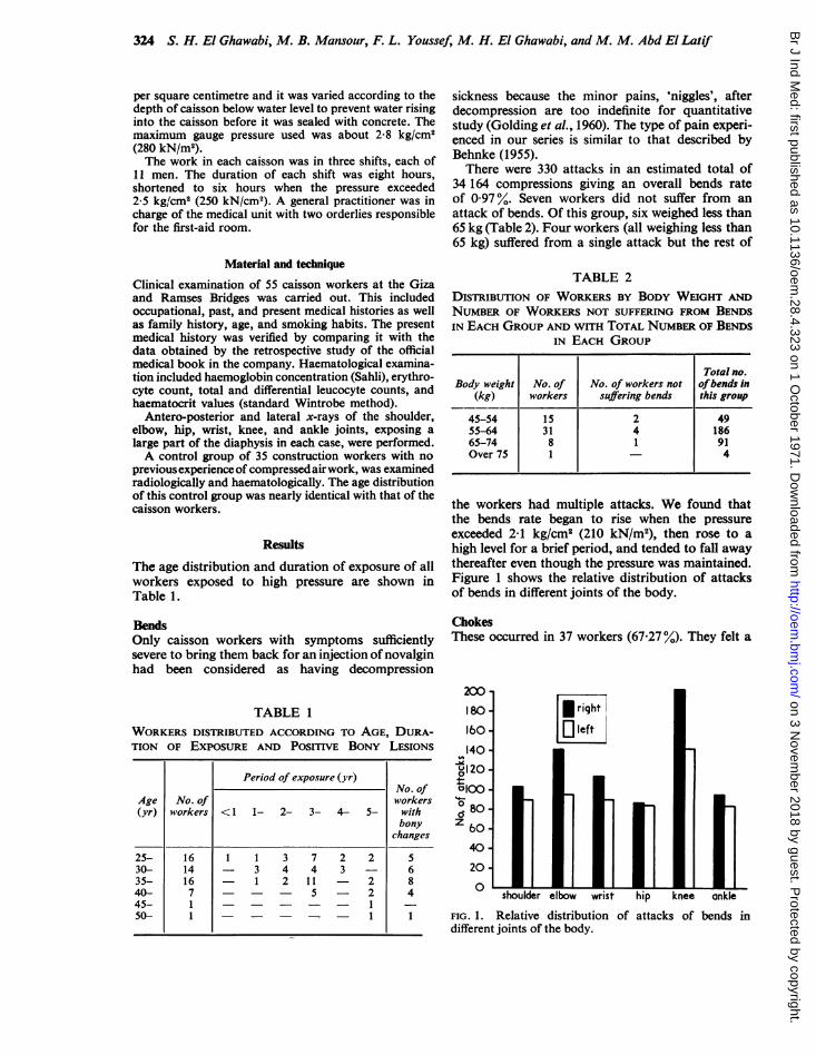
the workers had multiple attacks. We found thatthe bends rate began to rise when the pressureexceeded 2-1 kg/cm2 (210 kN/m2), then rose to ahigh level for a brief period, and tended to fall awaythereafter even though the pressure was maintained.Figure 1 shows the relative distribution of attacksof bends in different joints of the body.
ChokesThese occurred in 37 workers (67-27 %). They felt a
200180 *riht160 - left140 -
0U120-01008040-
40
20shoulder elbow wrist hip knee ankle
FIG. 1. Relative distribution of attacks of bends indifferent joints of the body.
on 3 Novem
ber 2018 by guest. Protected by copyright.
http://oem.bm
j.com/
Br J Ind M
ed: first published as 10.1136/oem.28.4.323 on 1 O
ctober 1971. Dow
nloaded from

Decompression sickntess in caisson workers 325
TABLE 3HAEMATOLOGICAL STUDIES IN 41 CAISSON WORKERS COMPARED WITH CONTROLS
Haemoglobin Total Polymorphs Lympho-Group Haemotocrit Red blood leucocyte Eosino- Basophils cvtes Monocytes
% g.100 ml value, % cells count phils % Staff Seg- % %x 103/mm3 /mm3 % mented
Caisson 89-9 14-4 44-14 4 501 6 205 2 5 0-7 2-8 59 3 31-9 3-17workers (69-117) (11-18-7) (38-48) (3 210- (3 200- (0-11) (0-2) (0-7) (50-72) (23-41) (0-7)
5 940) 11 200)
Controls 85 14-6 39-6 4 200 5 800 2-8 0-8 2-6 56 8 30 7 3 0(70-105) (11-1-16-8) (32-43) (3 680- (4 200- (0-10) (0-2) (0-7) (48-66) (26-40) (0-7)
5 109) 10 500)
Mean values; ranges in parentheses.
sensation of retrostemal distress lasting for a vari-able time. The sensation was usually relieved aftervomiting in 27 workers. The symptom occurred in14 workers once, in 13 workers twice, and in theremaining 10 workers more than twice. Behnke's(1955) sign was not observed in our series as theworkers were forbidden to inhale tobacco smoke fortwo hours after coming out of the air locks.
Cutaneous manifestationsThirty-eight (69 %) complained of itching aftercoming out of the air lock. This itching was usuallygeneralized and occurred especially when theweather was cool. Erythema and mottling of theskin was present in only five workers.
Blood examinationsThe blood of only 41 workers was examined as therest refused. The results were compared with thoseof the 35 controls. The blood picture was nearly thesame as in the control group (Table 3) except for the
mU
a--
, I
FIG. 2. Frequency of bony lesions in different regionsof the body.
haematocrit values which were significantly higher.The pooled standard deviation is 2-328, while thestandard error of the difference in the two means is0-536; t = 8-4, P < 0001.
Bony changesThe classification of McCallum and Walder (1966)was used. Figure 2 shows the frequency of bonylesions in different regions of the body. Definitebony changes were found in 24 men (43-6y%) whiledoubtful evidence was present in another 15 men(27 2%). This latter group will have to be followedup as such cases may become definite cases as timepasses.
Elbow lesions (Fig. 3)The elbow was affected in 22 men (91 6%) out of the24 positive cases. The lower ends of both humeriwere affected in 14 men. Unilateral affection of thelower end of the humerus occurred in seven men onthe right side and in one on the left side. The upperends of both ulnae were affected in 12 men, theupper end of the right ulna in nine men, and theupper end of the left ulna in one man. The upperends of both radii were affected in five men, theright radius in seven, and the left in two men. Theradius was not affected in every case compared withthe humerus or ulna.
Knee lesionsBony lesions in the knee region were present in 10men (41-6%) out of the 24 positive cases. The lowerends of both femora were affected in two men whilein three men one or other side was affected. Bilaterallesions in the upper ends of the tibiae were found intwo men, on the right side alone in three, and on theleft side alone in two men. Periosteal reaction(periostitis) along the lateral border of the upperpart of the metaphysis of the tibia occurred in sevenmen, four of whom had associated bony lesions inthe same tibia. The upper end of the fibula was
on 3 Novem
ber 2018 by guest. Protected by copyright.
http://oem.bm
j.com/
Br J Ind M
ed: first published as 10.1136/oem.28.4.323 on 1 O
ctober 1971. Dow
nloaded from

326 S. H. El Ghawabi, M. B. Mansour, F. L. Youssef, M. H. El Ghawabi, and M. M. Abd El Latif
Ankle region lesionsOf those cases with bony lesions, four (l6-6 %)showed lesions in the right ankle region. In one manthere was sequestration of the lateral part of thecortex of the lower end of the tibia. This juxta-articular lesion was accompanied by pain and slightdifficulty in walking. In the other three men therewere symptomless dense areas at the junction of theupper two-thirds of the tibial shaft with the lowerthird.
Hip lesionsOnly one (4-16%) of all the positive cases showeda radiotranslucent area in the neck of the femur onthe left side.
Feet and handsThere were no radiologically significant lesions.Our radiological findings in the 24 positive cases
could be summarized according to the McCallumand Walder classification as follows:
FiG. 3. Right elbowHumerus: juxta-articular dense area at junction of
trochlea and capitulum opposite epiphysealscar together with cyst in mfedial epicondyleand irregular calcified area in metaphysis ofshaft.
Ulna: juxta-articular linear opacity in middle ofupper surface of coronoid process withmultiple irregular dense areas mainly in meta-physis.
Radius: juxta-articular dense area in middle of headof radius with calcified area opposite radialtuberosity.
affected in five men; one case was bilateral and theremainder were on the right side. One man had adefinite small cyst in the head of the right fibula,with a well-defined sclerosed margin, especiallyalong its lateral border (Fig. 4). Biopsy was refused.
Shoulder region lesionsOf the cases with positive bony lesions, six (25%)were affected in the upper end of the humerus. Fourcases were left-sided,, the other two were right-sided. FIG. 4. Right knee.No bilateral lesions were detected. In two cases, the Juxta-articular linear opacity along both tibial condylesneck of the scapula showed irregular dense areas with dense area in its shaft. Presence of definite cyst ineasily differentiated from bone islands (Fig. 5). head of fibula with linear opacity along its lateral margin.
on 3 Novem
ber 2018 by guest. Protected by copyright.
http://oem.bm
j.com/
Br J Ind M
ed: first published as 10.1136/oem.28.4.323 on 1 O
ctober 1971. Dow
nloaded from

Decompression sickness in caisson workers 327
rio. 5. Left shoulder: a dense area in neck of scapula.
A. Juxta-articular lesions
Dense areasSpherical segmental opacitiesLinear opacitiesTransradiant subcortical bendsCollapse of articular cortexSequestration of part of cortexOsteoarthritis
B. Head, neck, and shaft lesionsDense areasIrregular calcified areasTransradiant areas and cysts
ases %55 1001 416
16 29-095 20-080 0
1 4-160 0
55 1005 20-08
11 45.8
Discussion
Several years ago caisson disease was believed to bepreventable by adequate prophylaxis and carefulattention to decompression procedures. But un-happily this prospect has not been achieved becausebony lesions appear to be more frequent than hadhitherto been realized. A great deal of attention hasrecently been focused on bony lesions in suchworkers chiefly because of the recent increase ininsurance costs for tunnel workers in some countries(Kleinfeld and Wilson, 1956).
Clinical manifestations of decompression sicknesshave been well described and its pathogenesis hasbeen generally accepted since Paul Bert published hisexperimental findings in 1878 (Kahlstrom et al.1939).The presenting symptom of the disease is usually
the pain (bends) and in our series it occurred in87-27% of workers. The overall bends rate variedfrom 0-87% at the Tyne Pedestrian Tunnel (Patonand Walder, 1954) to 4% at a caisson in the Thames(Lewis and Paton, 1957) compared with our overallbends rate which was 0 97 %. The rate is influencedby many variables such as length of shift, height ofpressure, high labour turnover, acclimatization,physical and mental characteristics of the workers,differences in temperature and humidity, and dis-ciplinary problems (McCallum, 1968). However, therole of acclimatization is doubtful. We agree withRozsahegyi (1959) that reduction in the sicknessincidence was due not to adaptation but to a naturalselection as those workers who frequently sufferedgave up the caisson work. Our group of workerswere previously ordinary construction workers andall of them were new to the job. Those who fre-quently suffered during the work in compressed airwere allowed to return to their former jobs.Chokes were the rather specific type of asphyxia
encountered in 67x27% of workers. The attacks ofchokes were relieved by vomiting in 73% of workers.
Cutaneous manifestations are early signs of com-pressed air illness. Most workers suffer from thiscomplaint from time to time (Farris and Sicca, 1952).The pruritus has been attributed to the nitrogenbubbles in the sweat glands of the skin. The erythemaand purplish mottling of the skin denote stasis ofblood in the cutaneous vessels. Thome (1941) hassuggested that this is a result of nitrogen emboli inthe skin capillaries.The prevalence of definite bony lesions in our
series was 43-6%. Cavigneaux and his colleagues(1949) gave a figure of 38%; Rozsahegyi and Fried(1963) reported the lesions in 22-2% of cases, whileMcCallum and Walder (1966) reported an incidenceof 30-4% in men who experienced more than 900decompressions. Thus a figure of 43 6% is relativelyhigh. It can be explained by incomplete adherence tothe British decompression table. We found that theforemen had modified the use of the British table togive a decompression schedule which was less byabout 25% than the minimum required by law.Walder (1967) stated that decompression given bythe correct use of the British table is probably in-ade4uate and that this may explain the high in-cidence of bony lesions.The minimum exposure necessary to produce
definite bony changes under these conditions is notknown, but one man in our series who had neverbeen exposed to high pressure before was found to
Ct
on 3 Novem
ber 2018 by guest. Protected by copyright.
http://oem.bm
j.com/
Br J Ind M
ed: first published as 10.1136/oem.28.4.323 on 1 O
ctober 1971. Dow
nloaded from

328 S. H. El Ghawabi, M. B. Mansour, F. L. Youssef, M. H. El Ghawabi, and M. M. Abd El Latif
have bony changes two months after the start ofwork in compressed air. In the series of McCallumand Walder (1966), the shortest period for bonychanges to occur was three months. The possibilityof bony lesions after a single exposure is supportedby the experience of James (1945), who foundaseptic bony necrosis in survivors from the sub-marine which sank in 1931; none of whom hadsuffered from caisson disease at any other time.No relationship could be established between the
number of decompressions and the prevalence ofbony lesions or between the site of bends and the siteof bony lesions. However, we agree with Rozsahegyiand Fried (1963) that the more attacks of bends aman has suffered the more likely he is to developbone necrosis.
In the present series, positive radiological findingswere mainly around the elbow (91 f6% in those withpositive bony lesions). Apart from the work of Bell,Edson, and Harnick (1942) quoted by McCallumand Walder (1966), we have found no reference tobony lesions in the lower end of the humerus. Allother authors described lesions in the proximal endof the humerus and in the proximal or distal ends ofthe femur (Cavigneaux et al., 1949; Jullien, 1956;Raymond, 1960; Golding et a., 1960; Rozsahegyiand Fried, 1963). This may be due partly to theirtechniques as some of them did not x-ray the elbowroutinely (Golding et al., 1960; McCallum andWalder, 1966). This also might explain why, whenarthrosis attributed to decompression sickness wasincluded in the list of notifiable occupationaldiseases in France in 1949, it was restricted to theshoulder and hip (Raymond, 1960). Thus theexplanation put forward by McCallum and Walderthat posture is a factor in determining the site of thelesion has to be revised, especially if those authorscan possibly obtain x-rays of the elbows in theirseries.A new finding in our series was the presence in
two men of dense areas in the neck of the scapula.These irregular dense areas could be easily differen-tiated from bone islands which are ovoid or oblong,uniformly dense, compact bone. The fact that theleft shoulder was affected in four out of six cases canbe explained by the mode of origin of the left sub-clavian artery as opposed to the right, as suggestedby James (1945).
Lesions around the ankle can easily be missed asdense areas in the lower end of the tibia occur usuallyfar from the joint at the junction of the upper two-thirds with the lower third of the shaft. No explana-tion can be offered why all the lesions occurred onthe right side.
In the present study a statistically significant highhaematocrit value was found in caisson workerscompared with the control group. The relationshipbetween blood viscosity and haematocrit value has
been emphasized by Miale (1967), and the increasedviscosity has been suggested as an aetiological factorin coronary thrombosis (Burch and DePasquale,1962). Experimentally it was shown that air em-bolism is not the only cause of bony infarction(Kahlstrom et al., 1939). James (1945) attributedbony infarction to an increased intravascularagglutination of erythrocytes, blood stasis resultingfrom air embolism, and the mechanical blockingeffect of gas-distended blood vessels. We believethat all these factors together with increased bloodviscosity may combine to produce the bony lesions.
ReferencesBehnke, A. R. (1955). Decompression sickness. Milit. Med.,
117, 257-271.Burch, G. E., and DePasquale, N. P. (1962). Editorial.
Hematocrit, blood viscosity and myocardial infarction.Amer. J. Med., 32, 161-163.
Cavigneaux, A., Charles, A., Fuchs, S., and Tara, S. (1949).Les l6sions osseuses ignor6es des tubistes. Arch. Mal.prof., 10, 359-361.
Farris, G., and Sicca, U. (1952). Manifestazioni cutanee neilavoratori dei cassoni pneumatici. Rass. Med. industr.,21, 350-366.
Golding, F. C., Griffiths, P., Hempleman, H. V., Paton,W. D. M., and Walder, D. N. (1960). Decompressionsickness during construction of the Dartford Tunnel.Brit. J. industr. Med., 17, 167-180.
Giuntini, C. N. (1967). Considerazioni sulle osteoartropatieda aeroembolismo disbarico. Med. d. Lavoro, 58, 161-200.
James, C. C. M. (1945). Late bone lesions in caisson disease.Lancet, 2, 6-8.
Jullien, G. (1956). ttude des reactions pathologiques con-s6cutives a la plongee sous-marine et au travail dans l'aircomprime. Arch. Mal. prof., 17, 228-236.
Kahlstrom, S. C., Burton, C. C., and Phemister, D. B. (1939).Aseptic necrosis of bone. Surg. Gynec. Obstet., 68, 129-146.
Kleinfeld, M., and Wilson, J. T. (1956). Decompression sick-ness (compressed-air illness) in a tunneling operation.Arch. indust,. Hlth, 14, 539-542.
Lewis, H. E., and Paton, W. D. M. (1957). Decompressionsickness during the sinking of a caisson. A study of someof the factors in the pathogenesis of caisson disease.Brit. J. industr. Med., 14, 5-12.
McCallum, R. I. (1968). Decompression sickness: a review.Brit. J. industr. Med., 25, 4-21.
__, and Walder, D. N. (1953). Compressed-air illness onTyneside. Lancet, 1, 464-467.- , and (1966). Bone lesions in compressed air
workers. J. Bone Jt Surg., 48B, 207-235.Miale, J. B. (1967). Laboratory Medicine, Hematology, 3rd
ed., C. V. Mosby, Saint Louis.Parodi, V. M. (1948). Rilievi semeiologici e clinici, nelle
forme osteomioartralgiche della malattia dei cassoni.Med. d. Lavoro, 39, 73-79.
Paton, W. D. M., and Walder, D. N. (1954). Compressed AirIllness. Spec. Rep. Ser. med. Res. Coun. (Lond.), No. 281.H.M.S.O., London.
Raymond, V. (1948). Les osteo-arthrites pneumatiques.Lesions osseuses des tubistes et des scaphandriers. Arch.Mal. prof., 9, 437-442.- (1960). Arthroses baro-traumatiques. Arch. Mal. prof.,
21, 609-621.Roche, L., Devic, M., Genevois, M., and Marin, A. (1956).
La tomographie osseuse danis l'osteoarthropathie des
on 3 Novem
ber 2018 by guest. Protected by copyright.
http://oem.bm
j.com/
Br J Ind M
ed: first published as 10.1136/oem.28.4.323 on 1 O
ctober 1971. Dow
nloaded from

Decompression sickness in caisson workers 329
caissons. Arch. Mal. prof., 17, 597-601.Ronald, J. (1953). Aseptic necrosis of bone in caisson disease.
Lancet, 2, 855-856.R6zsahegyi, I. (1959). Die Rolle der Disposition bei der
Entstehung der Dekompressionskrankheit. Arch. Gewer-bepath. Gewerbehyg., 17, 347-353.
, and Fried, L. (1963). Untersuchungen uber dieEntstehung und Dynamik der chronischen osteo-arthropathie der Caissonarbeiter. Z. ges. Hyg., 9, 915-924.
Salah, M. (1950). Compressed air disease (Caisson disease).Report of the 1st case encountered in an Egyptian diver.J. Egypt med. Ass., 33, 56-64.
Sartor, E. (1947). Skelettforandringar vid tryckluftsjuka(English summary). Nord. Med., 35, 1551-1554.
Taylor, H. K. (1944). Aseptic necrosis in adults: caissonworkers and others. Radiology, 42, 550-469.
Thorne, I. J. (1941). Caisson disease. A study based on threehundred cases observed at the Queens-Midtown Tunnelproject, 1938. Amer. med. Ass., 117, 585-588.
Tillmann, R. (1961). Knochennekrose der Schultergelenkebei Tauchern. Z. ges. Hyg., 7, 89-98,
Walder, D. N. (1967). Decompression sickness in Tunnelworkers. In: The Effects of Abnormal Physical Conditionsat Work. The Report of a Meeting held jointly by theBritish Occupational Hygiene Society, the ErgonomicsResearch Society and The So,iety of OccupationalMedicine, 1967. Edited by C. N. Davies, P. R. Davis,and F. H. Tyrer, pp. 101-110. Livingstone, Edinburghand London.
Received for publication September 9, 1970.
on 3 Novem
ber 2018 by guest. Protected by copyright.
http://oem.bm
j.com/
Br J Ind M
ed: first published as 10.1136/oem.28.4.323 on 1 O
ctober 1971. Dow
nloaded from