Embed Size (px)
Citation preview
Decoding olfaction in DrosophilaAndreas Keller� and Leslie B Vosshally
Recent experiments in Drosophila demonstrate striking
stereotypy in the neural architecture of the olfactory system.
Functional imaging experiments in mammals and honeybees
suggest a mechanism of odor coding that translates discrete
patterns of activity in olfactory glomeruli into an odor image.
Future experiments in Drosophila may permit a direct test of this
odor-coding hypothesis.
AddressesLaboratory of Neurogenetics and Behavior, Rockefeller University, 1230York Avenue, Box 63, New York NY 10021, USA�e-mail: [email protected]: [email protected]
Current Opinion in Neurobiology 2003, 13:103–110
This review comes from a themed issue on
Development
Edited by Magdalena Gotz and Samuel L Pfaff
0959-4388/03/$ – see front matter
� 2003 Elsevier Science Ltd. All rights reserved.
DOI 10.1016/S0959-4388(03)00011-4
AbbreviationsAL antennal lobe
DOR Drosophila odorant receptor
OB olfactory bulb
OR odorant receptor
OSN olfactory sensory neuron
PN projection neuron
IntroductionOlfaction is a primitive sense that permits all animals to
find food, identify conspecific mating partners, and avoid
predators, and, allows insects to identify suitable sub-
strates for egg-laying. The stimuli controlling these var-
ious olfactory-driven behaviors consist of blends of
volatile organic chemicals that differ in size, shape,
charge, and functional groups. For instance, the salient
olfactory constituents of the rose consist of 275 distinct
chemicals, whose ratio in the blend is carefully calibrated
to produce its characteristic scent [1]. The enduring
mystery of the olfactory system — unsolved to this day
— is how the brain parses complex and often contra-
dictory blends of odorous chemicals in the environment
into meaningful odor images.
The anatomy of the olfactory system is well understood.
In both vertebrates and insects, large numbers of func-
tionally distinct primary olfactory sensory neurons
(OSNs) extend dendrites that interact with odors from
the external world. Odorants, the chemicals that compose
odors, are recognized by distinct odorant receptor (OR)
proteins that reside in the dendritic membrane. These
receptor proteins, consisting of seven transmembrane
domains, transduce odor recognition into neuronal activa-
tion through G-protein-coupled second messenger signal-
ing pathways. Each OSN extends a single axon that
synapses with second-order neurons in the olfactory bulb
(OB) in vertebrates and the antennal lobe (AL) in insects.
From this first olfactory synapse, information is relayed to
higher brain centers, and ultimately to the descending
motor pathways that drive appropriate behaviors.
In this review, we discuss advances in understanding the
link between the neural circuitry of the olfactory system
and the mechanism that encodes odor images in the brain,
with special emphasis on studies in the fruit fly Drosophilamelanogaster. Recent functional expression experiments
have proven conclusively that the candidate odorant
receptors identified by molecular biology do indeed act
as ligand binding proteins that transduce odorant-specific
signals in the olfactory system. Odorant receptor genes
are a rapidly evolving gene family, and this may account
for variations in olfactory preferences among different
species. In the fly, stereotypy has emerged as the orga-
nizational principle in wiring both first order and second
order connections. Finally, functional imaging experi-
ments, coupled with behavior paradigms suggest that
the stereotypy in wiring may contribute to the process
of encoding the odor image.
Evidence that Drosophila odorant receptorsfunction in recognitionA large family of candidate odorant receptor (OR) genes
in the rat was reported more than ten years ago [2]. Each
OR gene encodes a different seven transmembrane
domain G-protein-coupled receptor, which is expressed
selectively in a subset of OSNs. Since this initial report,
ORs have been identified in many vertebrate species
(reviewed in [3]) and functional evidence that they bind
odors and transduce olfactory-specific signals has been
obtained in heterologous expression studies [4–6].
Odorant receptors in Drosophila are encoded by a distinct
gene family, containing at least 61 genes, with no
sequence similarity to vertebrate OR genes [7–9]. Each
Drosophila odorant receptor (DOR) gene, with the
exception of the broadly expressed receptor Or83b, is
expressed in a small, positionally invariant subset of
OSNs (Figure 1b). A second, distantly related gene
family of gustatory receptors includes some members
that are expressed in the olfactory system, although their
role in olfaction has not yet been investigated [10,11].
103
www.current-opinion.com Current Opinion in Neurobiology 2003, 13:103–110
Recent functional expression data confirm that DORs can
recognize odorants. Or43a was found to encode a low-
affinity receptor for cyclohexanol, cyclohexanone, benzyl
alcohol, and benzaldehyde, both by heterologous expres-
sion in Xenopus oocytes and by overexpression in the
Drosophila antenna [12��,13��]. Studies of the molecular
receptive range of Or43a and previous analysis of rat OR
I7 [14] suggest that a given OR can interact with several
different odorants with varying affinity. A given odorant is
also detected by more than one OR [15]. This receptor
promiscuity would give rise to a combinatorial code of
odors, such that an animal would be expected to detect
many more odorants than the number of OR genes it
possesses [15].
Rapid evolution of odorant receptor genesin insectsTo what extent is odor recognition species-specific?
Rapid divergence of OR genes is apparent in a compar-
ison between two dipterans, Drosophila and the malaria
mosquito Anopheles gambiae, and the moth Heliothisvirescens [16��,17�]. Anopheles has 79 ORs and, with the
exception of Or83b, no direct orthologue of any DOR
gene is apparent in the Anopheles genome sequence.
Phylogenetic analysis reveals large Drosophila-specific
and Anopheles-specific OR subfamilies, which may sub-
serve the recognition of the very different odors that are
ecologically relevant to these dipterans. Similarly, the
eight cloned Heliothis OR genes show a low degree of
Figure 1
Current Opinion in Neurobiology
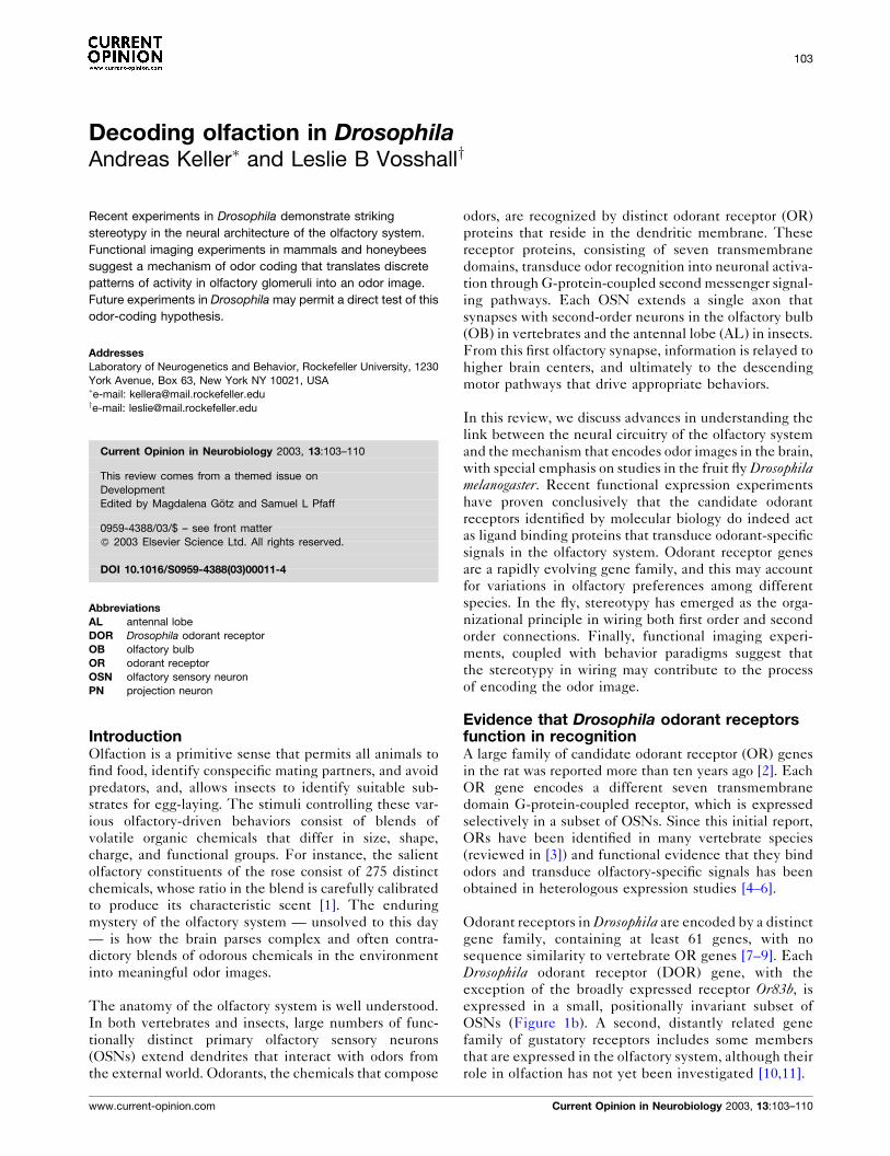
(b) Antenna(a) Model of the Drosophila olfactory system (c) Antennal lobe (d) Lateral horn
Stereotypy in the labeled line of Or47b at all levels of the Drosophila olfactory system. (a) A frontal schematic view of a Drosophila head that depicts
the olfactory circuitry displayed in Figure 1b–d. Dorsal is up; compound eyes are lateral and shaded red; antennae are medial and shaded grey; thecentral brain is visible through the head capsule; the AL is shaded blue. OSNs expressing Or47b are represented as blue dots on the antenna. These
neurons extend axons (in black) that fasciculate and extend dorsally toward the brain and synapse in the VA1l/m glomerulus (shaded green) in
the AL. The cell bodies of two dorsal PNs that synapse with Or47b-expressing OSNs in the AL are indicated in red. The dendrites of these cells
innervate the VA1l/m glomerulus (large green structure in lateral AL) and the axons (in green) extend dorsally to make synapses both in the
mushroom body calyx and the lateral horn. (b) Expression of the Or47b odorant receptor gene is restricted to a spatially conserved lateral-distal
domain of the third antennal segment. (c) All axons from Or47b-expressing olfactory sensory neurons converge upon the VA1l/m glomerulus in the
antennal lobe. (d) Axonal projections of a single dorsal group projection neuron, which sends dendrites to the Or47b (VA1l/m) glomerulus, are
stereotyped in the ventral region of the lateral horn of the protocerebrum. Data in Figure 1(d) were kindly provided by Allan Wong, Jing Wang, and
Richard Axel [30��].
104 Development
Current Opinion in Neurobiology 2003, 13:103–110 www.current-opinion.com
sequence similarity to the OR gene families of the two
dipterans [17�].
In marked contrast to this apparent species-specificity of all
other insect ORs, Or83b is exceptionally well conserved
from Drosophila to Anopheles to the moth Heliothis virescens[16��,17�]. This receptor is broadly expressed in most
OSNs in Drosophila, such that each neuron expresses a
conventional DOR along with Or83b. Whether Or83bfunctions as a heterodimer with conventional ORs to
modulate ligand specificity, plays a role in receptor traffick-
ing, or couples DOR activation to downstream signal
transduction machinery is not known. However, the
greater than 65% amino acid identity of Drosophila, mos-
quito, and moth Or83b suggests that this protein plays a
central, well-conserved role in insect olfaction.
Stereotypy in wiring the olfactory systemRemarkably, the functional architecture of the olfactory
system is similar in Drosophila and the mouse, despite
the apparently independent evolution of the OR genes
in these two species. In both mouse and Drosophila,
neurons expressing a given OR gene extend axons that
converge to form spatially discrete synapses with second-
order projection neurons (PNs) (Figure 1). These
synapses are organized into a spherical neuropil called
a glomerulus, which consists of OSN axons, projection
neuron dendrites, and input from a network of local
inhibitory interneurons. Individual glomeruli therefore
collect input of OSNs expressing a given OR gene
(Figure 1c; [18–22]). Convergent wiring of neurons
expressing the same OR, which therefore respond to
the same odorants, may provide the basis for the brain to
translate patterns of glomerular activity into perception
of a stimulus.
By what mechanisms do these stereotyped and precise
glomerular maps of OR axons form? Studies in the mouse
have implicated the OR protein itself in the guidance
process. Deleting an OR gene disrupts axon targeting,
and replacement of a given OR gene with an alternate OR
gene drives the axons to form synapses within an ectopic
glomerulus [18,23]. Invoking the OR itself as a guidance
molecule simplifies the problem of wiring millions of
OSNs expressing one of several hundred different
ORs. In fact, recent experiments that examined mice
expressing ectopic OR genes suggest that these OSNs
form ectopic glomeruli in the OB that are functionally
innervated by mitral cells, the vertebrate PNs [24��,25�].The formation of de novo functional glomeruli via OR-
based axon guidance would permit the brain to cope with
the rapid evolution of the OR gene family, enabling the
generation of novel OSNs with new chemical specificities
and novel synaptic connectivity in the OB.
Although direct evidence is lacking, it seems unlikely that
a similar OR-dependent wiring mechanism operates to
pattern glomerular connections in the Drosophila AL.
Drosophila OSNs are born early in pupal life and extend
axons and form glomeruli that are essentially complete by
the mid-pupal stage [26]. However, the DOR genes are
not expressed detectably until the end of pupal devel-
opment [7,27]. The late-onset of OR gene expression in
Drosophila suggests that OR-independent mechanisms
establish the glomerular map.
Olfactory information must be relayed from convergent
synapses in the AL and OB to higher brain centers, where
it is decoded to yield a coherent odor image. Recent work
in Drosophila has produced the remarkable finding that
the innervation of specific AL glomeruli by dendrites of a
given second-order PN is invariant, and that these pat-
terns are specified early in development before contact
with OSNs and long before onset of DOR gene expres-
sion in these OSNs [28��]. Thus, it seems likely that
independent pre-specification drives afferent OSN axons
and postsynaptic PN dendrites to converge onto a given
common glomerulus. This model appears to rule out any
activity-dependent communication between OSNs and
their targets in the AL that would serve to link up neurons
by common functional properties, and instead requires a
highly stereotyped genetic pre-programming of a neuro-
nal circuit.
Similar stereotypy is seen in the axonal projections of PNs
as they synapse with their targets in the mushroom body
and the lateral horn of the protocerebrum (Figure 1d;
[29��,30��]). PNs that innervate a given glomerulus have
an invariant pattern of axonal branching in the lateral
horn. They target diffuse and overlapping regions of the
lateral horn. Thus, the convergent olfactory wiring seen in
the AL is represented at higher levels of the brain as a
widely distributed but spatially invariant map in the
mushroom body and the lateral horn. Experiments in
mice that traced the olfactory circuitry of OSNs expres-
sing a given OR suggest a similarly distributed olfactory
code in the vertebrate olfactory cortex [31�]. These trac-
ing studies are consistent with functional studies that
demonstrate a characteristic response property of indivi-
dual mitral cells that reflects the molecular receptive
range of the glomerulus from which it receives olfactory
input [32,33].
Functional imaging techniques used todiscern mechanisms of odor codingThe stereotypy apparent at all levels of the olfactory
system (Figure 1) is highly suggestive of a mechanism of
odor coding that employs spatial patterns of glomerular
activation to represent olfactory stimuli, but direct proof
of this model is lacking. Recent efforts to optically record
olfactory glomeruli in diverse species confirm that the
glomerulus is a functional unit in the olfactory system
[34–38,39��,40�]. Using either intrinsic signal imaging or
calcium-sensitive dyes, these investigators have shown,
Decoding olfaction in Drosophila Keller and Vosshall 105
www.current-opinion.com Current Opinion in Neurobiology 2003, 13:103–110
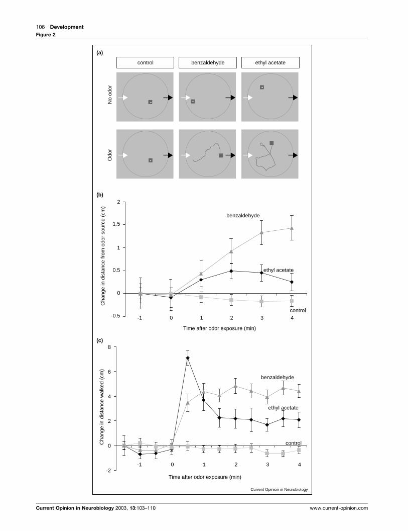
Figure 2
Current Opinion in Neurobiology
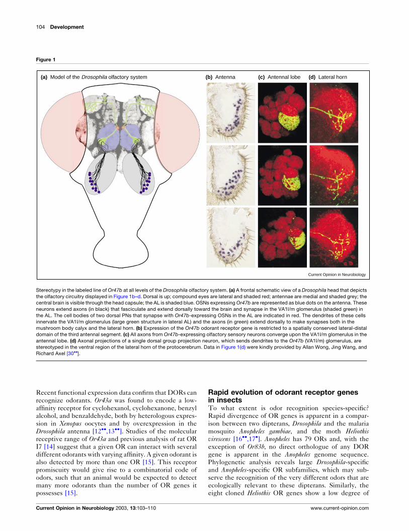
(a)
control benzaldehyde ethyl acetate
Odo
rN
o od
or
-0.5
0
0.5
1
1.5
2
-1 0 1 2 3 4
Time after odor exposure (min)
benzaldehyde
ethyl acetate
control
(b)
Cha
nge
in d
ista
nce
from
odo
r so
urce
(cm
)
-2
0
2
4
6
8
-1 0 1 2 3 4
Time after odor exposure (min)
benzaldehyde
ethyl acetate
control
(c)
Cha
nge
in d
ista
nce
wal
ked
(cm
)
106 Development
Current Opinion in Neurobiology 2003, 13:103–110 www.current-opinion.com
first, that a given odorant activates a reproducible subset
of glomeruli that is invariant between different indivi-
duals of a species; second, that with increasing concen-
tration additional glomeruli are recruited into the activity
pattern, and third that glomeruli responsive to chemi-
cally related odorants are clustered on the surface of the
vertebrate OB. The most recent direct proof that signals
obtained in functional imaging represent OR-specific
glomerular input was provided by experiments in geneti-
cally manipulated mice that express ectopic OR genes in
fluorescently tagged OSNs [24��,25�]. Experiments in
the honeybee and the moth [41�,42�] that examined
information flow in the AL suggested that inhibitory
interneurons play a major role in modulating the output
of glomerular activity. The inhibitory network filters and
processes the olfactory information that arrives at the
glomeruli from the OSNs and produces a coherent stimu-
lus-specific output.
How is olfactory information from the AL or OB repre-
sented at higher levels in the brain? The PN tracing
studies in Drosophila suggest that although PNs form
highly distributed synapses in the mushroom body and
lateral horn, the patterns of synapse distribution are
strongly conserved between different individuals. Elec-
trophysiological experiments in the locust and the zebra-
fish carried out by Laurent and co-workers [43��,44] have
led to an alternate hypothesis of odor coding that does not
rely solely on the hypothesis of glomerular encoding and
instead favors a temporal model. In this model, odor
stimulation induces stimulus-specific alterations in the
synchrony of local field potentials. It is proposed that
these temporal parameters are central to the process of
representing odorants, especially those that are structu-
rally similar and likely to produce overlapping patterns of
activation in the OB or AL.
Behavioral discrimination and the glomerularcodeUltimately, determining how odors are encoded in the
brain will require linking a specific behavioral output with
an olfactory input, and correlating this link with a measure
of synaptic activity in the brain. In practice, this can be
done by testing the ability of animals to discriminate
between odors. Behavioral experiments in the moth
Manduca sexta showed that this species can discriminate
between similar odors, although there is overlap in the
representation of these odors [45�]. Furthermore, both
honeybees and rats can efficiently discriminate enantio-
meric pairs of odorants, that is, chemicals that differ only
in their left/right handedness [46,47]. Functional imaging
of the responses of the rat OB in response to these stimuli
revealed activation of largely overlapping, yet different,
patterns. This is consistent with the rat’s ability to dis-
criminate these odors behaviorally. It seems that enan-
tiomeric pairs with very similar activation patterns are
only discriminated after reinforcement, whereas the pairs
evoking less similar activation patterns are distinguished
spontaneously [48��,49��]. A learning-dependent effect
on glomerular activity in the honeybee AL was described
several years ago [50], and illustrates the reciprocal
dependence of behavioral output and synaptic activity
in the brain. Pharmacological perturbations that affected
both the local field potential oscillation and the modula-
tion of PN output by local inhibitory interneurons caused
honeybees to lose the ability to discriminate between
closely related odorants, although they retained their
ability to discriminate distinct stimuli [51]. Many of these
experiments provide a good correlation between patterns
of glomerular activity and the animal’s behavioral perfor-
mance. However, they stop short of demonstrating that
these activity patterns are the salient information that the
animal uses to encode the odor. Drosophila provides a
unique system that may permit the linking of mechan-
isms that control the development of olfactory circuitry to
an understanding of how this circuitry serves to generate
and organize complex behaviors.
ConclusionsDespite the diversity of olfactory responses among dif-
ferent species, glomerular coding of olfactory stimuli
appears to be a central and conserved mechanism. In
Drosophila and mice, the wiring of OSNs to the first
olfactory relay is highly stereotyped and leads to a con-
vergence of all of the neurons expressing a given OR gene
to one or two glomeruli. The majority of the PNs extend
dendrites into a single glomerulus and elaborate axonal
processes that terminate in stereotyped patterns in the
mushroom body and lateral horn of the fly. Finally, flies
exhibit robust olfactory-driven behaviors that reveal strik-
ing differences in the responses of a single fly to distinct
odorants (see Figure 2). By utilizing the genetic tools
available in Drosophila, it should be possible to silence or
activate distinct parts of this simple olfactory circuit and
to monitor the resulting behavioral output. This approach
may reveal new insights into the mechanisms by which
Figure 2 Legend Behavioral responses of Drosophila to odors. (a) Tracks of individual flies in a circular arena measured by videography coupled to
tracking software. Left arrows depict the site of odor input and right arrows odor output. 30 s tracks of three flies before (above) and after (below)onset of odor delivery are shown. Both benzaldehyde and ethyl acetate but not pure air induce activity in resting flies. The squares represent the
position at the end of the 30 s assay period. (b) The change in distance from the source of ethyl acetate and benzaldehyde. The avoidance of
benzaldehyde is more pronounced than the avoidance of ethyl acetate. (c) The increase in activity after onset of odor exposure. Both benzaldehyde
and ethyl acetate induce activity. However, the temporal dynamics of these activities differ. Ethyl acetate induces a high initial activity that decreases
after 30 s to reach a plateau, whereas benzaldehyde induces a slow increase in activity that reaches a plateau higher than the one seen for ethyl
acetate activity. Means of 50 to 60 flies are given. Error bars indicate SEMs.
Decoding olfaction in Drosophila Keller and Vosshall 107
www.current-opinion.com Current Opinion in Neurobiology 2003, 13:103–110
organisms respond to the odorous environment in which
they live.
UpdateThree recent papers report the shattering of a technical
barrier that has prevented the imaging of synaptic activity
in the AL of living Drosophila while they are exposed to
odors [52��–54��]. The small size of Drosophila and the
relatively low number of OSNs converging upon a given
glomerulus in the AL have precluded the use of conven-
tional optical imaging techniques that employ Caþ2-sen-
sitive dyes or intrinsic signal imaging. Both groups
overcome the problem of size and signal strength in
Drosophila by the use of genetically encoded sensors of
neuronal activity, which permit the selective expression
under genetic control of high levels of fluorescent sensor
proteins in the neurons of choice in the olfactory network.
Ng and co-workers [52��] used synapto-pHluorin, which
is a pH-sensitive variant of GFP that is tethered to the
synaptic vesicle and produces pH-dependent fluores-
cence changes upon evoked synaptic release. They exam-
ine odor-evoked synaptic activity simultaneously in
OSNs, PNs, and inhibitory interneurons in the AL, by
selective expression of synapto-pHluorin in these differ-
ent cell populations. Similar to previous studies in the
honeybee, the Drosophila responses showed a combina-
torial logic, with odors activating distinct but overlapping
glomeruli in the AL. These responses were highly repro-
ducible between different individuals and the number of
glomeruli activated increased with increasing concentra-
tions of odorant. Unlike the honeybee, in which activity
patterns elicited by a given odorant were broader when
measured in OSNs than postsynaptic PNs [41�], Droso-phila responses appeared to be highly similar whether
measured in OSNs or PNs. In contrast, local interneurons
demonstrated much broader and more complex patterns
of activation.
Fiala et al. [53��] used cameleon as a genetically encoded
Ca2þ sensor to monitor odor-evoked changes in the activ-
ity of Drosophila PNs in the AL and in the calyx of the
mushroom body. They find a similar degree of stereotypy
in activation of discrete foci in the AL — which are likely
to be glomeruli — and show for the first time that spatially
conserved regions of the mushroom body calyx are acti-
vated in response to a given odorant. These results are
significant because they are a functional correlation of the
stereotypy in PN axonal connectivity demonstrated by the
Axel and Luo groups (Figure 1d; [29��, 30��]).
Finally, Wong et al. [54��] use a third genetically encoded
calcium sensor protein, G-CaMP, which produces fluor-
escent intensity changes of up to 120%, to characterize
the response properties of 23 glomeruli to 16 different
odorants. They find that the glomerular code is sparse at
low stimulus concentrations and that at higher concentra-
tions, a large number of glomeruli are recruited. They
argue that the sparse odor code is more likely to represent
the physiological state of the animal, than the highly
overlapping promiscuous code obtained at high stimulus
concentrations. Comparison of activity in OSNs and PNs
suggests that information is relayed faithfully, with mini-
mal processing or filtering, to higher brain centers. In a
genetically reprogrammed fly, the activity of a glomerulus
is shown to be a property of the OR gene expressed in
OSNs that synapse in that glomerulus.
AcknowledgementsWe thank Kevin Lee, Silke Sachse and Kristin Scott for comments on themanuscript. The authors’ research is supported by grants from the NationalInstitute of Health, National Science Foundation, John Merck Fund,Beckman Foundation, and the McKnight Endowment Fund forNeuroscience to Leslie B Vosshall.
References and recommended readingPapers of particular interest, published within the annual period ofreview, have been highlighted as:
� of special interest��of outstanding interest
1. Ohloff G: Scent and Fragrances: The Fascination of Odors and theirChemical Perspectives. Berlin: Springer-Verlag; 1994;154-158.
2. Buck L, Axel R: A novel multigene family may encode odorantreceptors: a molecular basis for odor recognition. Cell 1991,65:175-187.
3. Mombaerts P: Seven-transmembrane proteins as odorant andchemosensory receptors. Science 1999, 286:707-711.
4. Zhao H, Ivic L, Otaki JM, Hashimoto M, Mikoshiba K, Firestein S:Functional expression of a mammalian odorant receptor.Science 1998, 279:237-242.
5. Krautwurst D, Yau KW, Reed RR: Identification of ligands forolfactory receptors by functional expression of a receptorlibrary. Cell 1998, 95:917-926.
6. Touhara K, Sengoku S, Inaki K, Tsuboi A, Hirono J, Sato T, SakanoH, Haga T: Functional identification and reconstitution of anodorant receptor in single olfactory neurons. Proc Natl Acad SciUSA 1999, 96:4040-4045.
7. Clyne PJ, Warr CG, Freeman MR, Lessing D, Kim J, Carlson JR:A novel family of divergent seven-transmembrane proteins:candidate odorant receptors in Drosophila. Neuron 1999,22:327-338.
8. Gao Q, Chess A: Identification of candidate Drosophila olfactoryreceptors from genomic DNA sequence. Genomics 1999,60:31-39.
9. Vosshall LB, Amrein H, Morozov PS, Rzhetsky A, Axel R: A spatialmap of olfactory receptor expression in the Drosophilaantenna. Cell 1999, 96:725-736.
10. Scott K, Brady R Jr, Cravchik A, Morozov P, Rzhetsky A, Zuker C,Axel R: A chemosensory gene family encoding candidategustatory and olfactory receptors in Drosophila. Cell 2001,104:661-673.
11. Dunipace L, Meister S, McNealy C, Amrein H: Spatially restrictedexpression of candidate taste receptors in the Drosophilagustatory system. Curr Biol 2001, 11:822-835.
12.��
Stortkuhl KF, Kettler R: Functional analysis of an olfactoryreceptor in Drosophila melanogaster. Proc Natl Acad Sci USA2001, 98:9381-9385.
The authors provide the first functional demonstration that a DOR proteinhas selective ligand-binding activity for specific odorants. Electroanten-nogram recordings from Drosophila overexpressing the receptor Or43a inthe antenna are used to identify the ligands of the receptor as cyclohex-anol, cyclohexanone, benzyl alcohol, and benzaldehyde (See also [13��]).
108 Development
Current Opinion in Neurobiology 2003, 13:103–110 www.current-opinion.com
13.��
Wetzel CH, Behrendt H-J, Gisselmann G, Stortkuhl KF, HovemannB, Hatt H: Functional expression and characterization of aDrosophila odorant receptor in a heterologous cell system.Proc Natl Acad Sci USA 2001, 98:9377-9380.
Expression of Or43a in Xenopus oocytes, a heterologous system thatpermits the screening of ligands for receptors, confirms the ligandassignments made by misexpression studies in vivo (see [12��]).
14. Araneda RC, Kini AD, Firestein S: The molecular receptive rangeof an odorant receptor. Nat Neurosci 2000, 3:1248-1255.
15. Malnic B, Hirono J, Sato T, Buck LB: Combinatorial receptorcodes for odors. Cell 1999, 96:713-723.
16.��
Hill CA, Fox AN, Pitts RJ, Kent LB, Tan PL, Chrystal MA,Cravchik A, Collins FH, Robertson HM, Zwiebel LJ: G protein-coupled receptors in Anopheles gambiae. Science 2002,298:176-178.
The first glimpse of the entire repertoire of OR genes identified fromanalysis of genomic sequence databases of the economically and medi-cally relevant malaria mosquito (Anopheles gambiae). Bioinformaticapproaches identified 79 candidate odorant receptors in Anophelesgambiae. The majority show expression exclusively in olfactory tissues.Of the DORs only the broadly expressed Or83b has an orthologue inAnopheles (see also [17�]).
17.�
Krieger J, Raming K, Dewer YM, Bette S, Conzelmann S, Breer H:A divergent gene family encoding candidate olfactoryreceptors of the moth Heliothis virescens. Eur J Neurosci 2002,16:619-628.
A genomic approach was used to identify odorant receptors in thetobacco budworm (Heliothis virescens). As in Drosophila, the OR genesof these insects are divergent but are clearly members of the insect ORgene superfamily. A clear orthologue of Or83b, also expressed in themajority of OSNs was identified (see also [16��]).
18. Mombaerts P, Wang F, Dulac C, Chao SK, Nemes A, MendelsohnM, Edmondson J, Axel R: Visualizing an olfactory sensory map.Cell 1996, 87:675-686.
19. Ressler KJ, Sullivan SL, Buck LB: Information coding in theolfactory system: evidence for a stereotyped and highlyorganized epitope map in the olfactory bulb. Cell 1994,79:1245-1255.
20. Vassar R, Chao SK, Sitcheran R, Nunez JM, Vosshall LB, Axel R:Topographic organization of sensory projections to theolfactory bulb. Cell 1994, 79:981-991.
21. Gao Q, Yuan B, Chess A: Convergent projections of Drosophilaolfactory neurons to specific glomeruli in the antennal lobe.Nat Neurosci 2000, 3:780-785.
22. Vosshall LB, Wong AM, Axel R: An olfactory sensory map in thefly brain. Cell 2000, 102:147-159.
23. Wang F, Nemes A, Mendelsohn M, Axel R: Odorant receptorsgovern the formation of a precise topographic map. Cell 1998,93:47-60.
24.��
Belluscio L, Lodovichi C, Feinstein P, Mombaerts P, Katz LC:Odorant receptors instruct functional circuitry in the mouseolfactory bulb. Nature 2002, 419:296-300.
Characterization of the novel glomerulus that results from the substitutionof rat OR I7 in OSNs that normally express mouse OR M71. Theseexperiments demonstrate for the first time an overlap in the anatomicalmapping of OSNs to specific glomeruli and the functional map thatemerges from examining glomerular activation in response to specificligands. Furthermore, the data show that novel glomeruli are innervatedby postsynaptic neurons that acquire the functional identity of the glo-meruli and establish reciprocal projections between the two ectopicmirror-image glomeruli.
25.�
Bozza T, Feinstein P, Zheng C, Mombaerts P: Odorant receptorexpression defines functional units in the mouse olfactorysystem. J Neurosci 2002, 22:3033-3043.
The authors show that substituting one receptor for another in OSNs inmice changes the stimulus response profiles of the neurons and results inthe formation of ectopic glomeruli in the olfactory bulb. Therefore theodorant receptor is the principal determinant of the odor-specificity of agiven OSN.
26. Jhaveri D, Rodrigues V: Sensory neurons of the Atonal lineagepioneer the formation of glomeruli within the adult Drosophilaolfactory lobe. Development 2002, 129:1251-1260.
27. Elmore T, Smith DP: Putative Drosophila odor receptor OR43blocalizes to dendrites of olfactory neurons. Insect Biochem MolBiol 2001, 31:791-798.
28.��
Jefferis GS, Marin EC, Stocker RF, Luo L: Target neuronprespecification in the olfactory map of Drosophila. Nature2001, 414:204-208.
A systematic clonal analysis of the PNs of Drosophila revealed that PNsare derived from three neuroblasts. The glomerular choice of the PNs isprespecified by the lineage and birth order of the PNs.
29.��
Marin EC, Jefferis GS, Komiyama T, Zhu H, Luo L: Representationof the glomerular olfactory map in the Drosophila brain.Cell 2002, 109:243-255.
Labeling of single PNs sending dendrites to specific glomeruli revealedtheir stereotypical axon branching patterns and terminal fields in higherolfactory centers.
30.��
Wong AM, Wang JW, Axel R: Spatial representation of theglomerular map in the Drosophila protocerebrum. Cell 2002,109:229-241.
The authors visualized single Drosophila PNs connecting defined glo-meruli with higher brain centers to study their innervation patterns. Atopographical map of olfactory information is retained in higher sensorycenters in the brain.
31.�
Zou Z, Horowitz LF, Montmayeur J-P, Snapper S, Buck LB:Genetic tracing reveals a stereotyped sensory map in theolfactory cortex. Nature 2001, 414:173-179.
The authors use a transneuronal tracer to visualize neurons in theolfactory cortex receiving input from a particular OR. Thus, they are ableto describe a sensory map in the olfactory cortex, in which input from aparticular OR is targeted to clusters of neurons conserved betweendifferent animals. Signals from different ORs overlap spatially and signalsfrom the same OR are targeted to multiple cortical areas.
32. Luo M, Katz LC: Response correlation maps of neurons in themammalian olfactory bulb. Neuron 2001, 32:1165-1179.
33. King JR, Christensen TA, Hildebrand JG: Responsecharacteristics of an identified, sexually dimorphic olfactoryglomerulus. J Neurosci 2000, 20:2391-2399.
34. Friedrich RW, Korsching SI: Combinatorial and chemotopicodorant coding in the zebrafish olfactory bulb visualized byoptical imaging. Neuron 1997, 18:737-752.
35. Joerges J, Kuttner A, Galizia CG, Menzel R: Representation ofodours and odour mixtures visualized in the honeybee brain.Nature 1997, 387:285-288.
36. Galizia CG, Sachse S, Rappert A, Menzel R: The glomerular codefor odor representation is species specific in the honeybeeApis mellifera. Nat Neurosci 1999, 2:473-478.
37. Rubin BD, Katz LC: Optical imaging of odorant representationsin the mammalian olfactory bulb. Neuron 1999, 23:499-511.
38. Uchida N, Takahashi YK, Tanifuji M, Mori K: Odor maps in themammalian olfactory bulb: domain organization and odorantstructural features. Nat Neurosci 2000, 3:1035-1043.
39.��
Meister M, Bonhoeffer T: Tuning and topography in an odor mapon the rat olfactory bulb. J Neurosci 2001, 21:1351-1360.
The authors recorded responses of glomeruli in the rat olfactory bulb andshowed that the patterns of activated glomeruli are bilaterally symmetricand their extent varies with concentration. Glomeruli with similar tuningproperties are shown to be located near each other.
40.�
Belluscio L, Katz LC: Symmetry, stereotypy, and topography ofodorant representations in mouse olfactory bulbs. J Neurosci2001, 21:2113-2122.
Optical imaging of intrinsic signals in rodent olfactory bulbs is used toshow that odors generally activate a bilaterally symmetric pattern ofglomeruli. A systematic map of molecular chain length is produced onthe surface of the olfactory bulb and mixed odors activate a patternsimilar to the combination of the patterns elicited by each individualcomponent.
41.�
Sachse S, Galizia CG: Role of inhibition for temporal and spatialodor representation in olfactory output neurons: a calciumimaging study. J Neurophysiol 2002, 87:1106-1117.
A combination of optically recorded calcium responses of selectivelystained PNs and pharmacological experiments revealed that two sepa-rate inhibitory networks enhance the contrast between odor representa-tions in the antennal lobe of honeybees.
Decoding olfaction in Drosophila Keller and Vosshall 109
www.current-opinion.com Current Opinion in Neurobiology 2003, 13:103–110
42.�
Lei H, Christensen TA, Hildebrand JG: Local inhibition modulatesodor-evoked synchronization of glomerulus-specific outputneurons. Nat Neurosci 2002, 5:557-565.
The authors used simultaneous intracellular recordings from pairs of PNsto reveal that PNs innervating the same glomerulus show odor-evokedsynchrony of spike discharges, and that the extent of this synchrony ismodulated by inhibitory input from the neighbouring glomerulus.
43.��
Friedrich RW, Laurent G: Dynamic optimization of odorrepresentations by slow temporal patterning of mitral cellactivity. Science 2001, 291:889-894.
By investigating mitral cell ensembles in the olfactory bulb of the zebra-fish, the authors show that the similarity between distributed temporalpatterns that represent related odors reduces progressively over time,whereas the responses of individual cells do not become more specific.
44. Perez-Orive J, Mazor O, Turner GC, Cassanaer S, Wilson RI,Laurent G: Oscillations and sparsening of odor representationsin the mushroom body. Science 2002, 297:359-365.
45.�
Daly KC, Chandra S, Durtschi ML, Smith BH: The generalization ofan olfactory-based conditioned response reveals unique butoverlapping odour representations in the moth Manduca sexta.J Exp Biol 2001, 204:3085-3095.
The authors use behavioral experiments to assess how the moth Man-duca sexta can distinguish between odors that vary in one or moremolecular dimensions. They found that odor similarity is a function ofdifferences in the shape and length of the carbon chain and the functionalgroup attached to it.
46. Rubin BD, Katz LC: Spatial coding of enantiomers in the ratolfactory bulb. Nat Neurosci 2001, 4:355-356.
47. Laska M, Galizia CG: Enantioselectivity of odor perception inhoneybees (Apis mellifera carnica). Behav Neurosci 2001,115:632-639.
48.��
Linster C, Johnson BA, Yue E, Morse A, Xu Z, Hingco EE, Choi Y,Choi M, Messiha A, Leon M: Perceptual correlates of neuralrepresentations evoked by odorant enantiomers. J Neurosci2001, 21:9837-9843.
In this paper it is shown that enantiomeric pairs that have clearly differentglomerular representations but not those having very similar representa-tions are discriminated by rats spontaneously.
49.��
Linster C, Johnson BA, Morse A, Yue E, Leon M: Spontaneousversus reinforced olfactory discriminations. J Neurosci 2002,22:6842-6845.
Here it is shown that enantiomeric pairs that have barely distinguishableactivation patterns and are not discriminated spontaneously by rats arediscriminated if subjected to differential reinforcement.
50. Faber T, Joerges J, Menzel R: Associative learning modifiesneural representations of odors in the insect brain. Nat Neurosci1999, 2:74-78.
51. Stopfer M, Bhagavan S, Smith BH, Laurent G: Impaired odourdiscrimination on desynchronization of odour-encoding neuralassemblies. Nature 1997, 390:70-74.
52.��
Ng M, Roorda RD, Lima SQ, Zemelman BV, Morcillo P, MiesenbockG: Transmission of olfactory information between threepopulations of neurons in the antennal lobe of the fly.Neuron 2002, 36:463-474.
Using synapto-pHluorin, synaptic activity is measured in DrosophilaOSNs, PNs, and local interneurons. Unique combinatorials of glomeruliare activated by distinct odors and these activity patterns are hypothe-sized to underlie the odor code that permits the animal to smell.
53.��
Fiala A, Spall T, Diegelmann S, Eisermann B, Sachse S, Devaud JM,Buchner E, Galizia CG: Genetically expressed cameleon inDrosophila melanogaster is used to visualize olfactoryinformation in projection neurons. Curr Biol 2002, 12:1877-1884.
Cameleon is used as a genetic tracer of neuronal activity in PNs in the ALand in the calyx of the mushroom body. Odor-specific patterns ofactivation that are conserved between individuals are seen both in theAL and in the mushroom body.
54.��
Wang JW, Wong AM, Flores J, Vosshall LB, Axel R: Two-photoncalcium imaging reveals an odor-evoked map of activity in thefly brain. Cell 2003, In Press.
A thorough examination of the response properties of more than half ofthe AL glomeruli as stimulated by 16 different odorants, demonstrates asparse odor code in which only a few glomeruli are activated in responseto a given odorant. Dense odor coding and glomerular promiscuity isseen only at high odor concentrations. Olfactory information is shown tobe minimally processed from OSNs to PNs and the response propertiesof a glomerulus are demonstrated to depend largely on the OR geneexpressed in its afferent OSNs.
110 Development
Current Opinion in Neurobiology 2003, 13:103–110 www.current-opinion.com