Embed Size (px)
Citation preview
Purdue UniversityPurdue e-Pubs
Open Access Dissertations Theses and Dissertations
January 2016
Deciphering the role of Hsp31 as a multitaskingchaperoneKiran AslamPurdue University
Follow this and additional works at: https://docs.lib.purdue.edu/open_access_dissertations
This document has been made available through Purdue e-Pubs, a service of the Purdue University Libraries. Please contact [email protected] foradditional information.
Recommended CitationAslam, Kiran, "Deciphering the role of Hsp31 as a multitasking chaperone" (2016). Open Access Dissertations. 1242.https://docs.lib.purdue.edu/open_access_dissertations/1242
Graduate School Form 30 Updated 12/26/2015
PURDUE UNIVERSITY GRADUATE SCHOOL
Thesis/Dissertation Acceptance
This is to certify that the thesis/dissertation prepared
By
Entitled
For the degree of
Is approved by the final examining committee:
To the best of my knowledge and as understood by the student in the Thesis/Dissertation Agreement, Publication Delay, and Certification Disclaimer (Graduate School Form 32), this thesis/dissertation adheres to the provisions of Purdue University’s “Policy of Integrity in Research” and the use of copyright material.
Approved by Major Professor(s):
Approved by: Head of the Departmental Graduate Program Date
Kiran Aslam
DECIPHERING THE ROLE OF HSP31 AS A MULTITASKING CHAPERONE
Doctor of Philosophy
Tony R HazbunChair
Jean-Christophe Rochet
Douglas J. LaCount
Ruben C Aguilar
Tony R Hazbun
Jean-Christophe Rochet 06/07/16
i
i
DECIPHERING THE ROLE OF HSP31 AS A MULTITASKING CHAPERONE
Dissertation
Submitted to the Faculty
of
Purdue University
by
Kiran Aslam
In Partial Fulfillment of the
Requirements for the Degree
of
Doctor of Philosophy
August 2016
Purdue University
West Lafayette, Indiana

ii
ii
For my parents;
Aslam and Mukhtiar
And to my wonderful husband Khizar Rouf

iii
iii
ACKNOWLEDGEMENTS
Though only my name appears on the cover of this thesis, earning a doctorate and writing
a thesis is certainly not done single-handedly. I owe my gratitude to all those people who
have contributed to its production and because of them my experience at Purdue
University has been one that I will cherish forever. I would like to reflect on all those
people who have supported and inspired me not only in the scientific arena, but also on a
personal level throughout this period.
First of all, my deepest gratitude goes to my advisor, Dr. Tony Hazbun. I have been
fortunate to have an advisor who trained me how to question thoughts and deliver ideas.
He gave me the freedom to explore on my own, and at the same time guided me to
recover whenever I faced obstacles. I would like to thank him for his patience in
mentoring me as a scientist. Without his guidance, this work would not exist.
I extend my sincere gratitude to Dr. Jean-Christophe Rochet, who has been always there
to listen and give advice. I am sincerely grateful to him for the insightful feedback that
helped me sort out the technical details of enzymatic assay presented in this thesis. I am
also thankful to him for carefully reading and commenting on many revisions of my JBC
manuscript. I am indebted to him for his continuous encouragement and guidance that
will stay with me for the rest of my life. I am also thankful to Dr. Douglas J. LaCount for
reading my reports, providing thought-provoking comments that helped me move

iv
iv
forward in positive direction. I am grateful to him for constructive criticisms at different
level of graduate school that have helped me focus on my goals. I also like to thank him
and his group members for letting me use their lab’s equipment and reagents. Next, I
would like to thank Dr. Ruben C Aguilar who is one of the best teachers that I have had
in my life. I am grateful to him for teaching me important concepts in cellular biology
that help me understand and enrich my ideas.
Next, I would like to acknowledge past and present members of Dr. Hazbun’s group, for
their friendship, valuable discussion and technical help with scientific projects. I
appreciate the efforts of the many undergraduate students who helped me make the media
and buffers to run various experiments on time. I am also thankful to the faculty and staff
of Medicinal Chemistry and Molecular Pharmacology department for their various form
of support during my graduate study.
Most importantly, I would like to express my heart-felt gratitude to my family and friends
who have helped me stay sane through these difficult years. I greatly appreciate their love,
support and belief in me that has been strength for me. I have to give a special mention to
my husband Khizar Rouf who worries about me more than I worry about myself.
Finally, I appreciate the United State Education Foundation in Pakistan and International
Institute of Education for providing me Fulbright fellowship that was a huge financial
support for me.
v
v
TABLE OF CONTENTS
Page
LIST OF ABBREVIATIONS ............................................................................................ xi
ABSTRACT ..................................................................................................................... xiii
CHAPTER 1. INTRODUCTION .................................................................................... 1
1.1 High-fidelity protein quality control ...................................................................... 1
1.1.1 Consequences of Protein Misfolding .............................................................. 2
1.1.2 Role of chaperone in protein homeostasis ....................................................... 2
1.1.2.1 Heat Shock Proteins .................................................................................. 4
1.1.2.2 Small heat shock proteins .......................................................................... 6
1.2 Neurodegenerative diseases ................................................................................... 7
1.2.1 Mitochondrial dysfunction in neurodegenerative disease ............................. 11
1.2.2 Amyloidogenesis ........................................................................................... 12
1.3 Parkinson’s disease .............................................................................................. 15
1.3.1 Risk factors .................................................................................................... 16
1.3.2 Genetic factors ............................................................................................... 18
1.3.3 Diagnosis ....................................................................................................... 18
1.3.4 Clinical presentation ...................................................................................... 20
1.3.4.1 Motor symptoms ...................................................................................... 20
vi
vi
Page
1.3.4.2 Non-motor symptoms .............................................................................. 21
1.3.5 Management of Parkinson’s disease ............................................................. 23
1.3.6 Yeast Model of Parkinson’s disease .............................................................. 24
1.4 Prion disease ........................................................................................................ 25
1.4.1 Prions in yeast ............................................................................................... 29
1.4.2 Structural organization .................................................................................. 30
1.4.2.1 Prion forming domain .............................................................................. 30
1.4.2.2 De novo prion formation ......................................................................... 33
1.4.3 Effect of heat shock proteins on prion propagation ...................................... 34
1.4.4 Prion associated toxicity ................................................................................ 38
1.5 DJ-1/ThiJ/Pfp1 superfamily ................................................................................. 38
1.5.1 Human DJ-1 .................................................................................................. 40
1.5.2 The yeast Hsp31 mini family ........................................................................ 41
1.6 Role of Hsp31 in cellular stress response ............................................................ 41
1.7 Comparison of Hsp31 paralogs ........................................................................... 45
1.8 Hsp31 role in redox homeostasis ......................................................................... 48
CHAPTER 2. HSP31 IS A STRESS-RESPONSE CHAPERONE THAT
INTERVENES IN THE PROTEIN MISFOLDING PROCESS ...................................... 50
2.1 Abstract ................................................................................................................ 50
2.2 Introduction .......................................................................................................... 51
2.3 Results .................................................................................................................. 53
2.3.1 Hsp31 is an integral part of the yeast cellular stress response ...................... 53
vii
vii
Page
2.3.2 Hsp31 methylglyoxalase activity is not required for rescue of α-syn-mediated
toxicity ...................................................................................................................... 60
2.3.3 D-lactate supplementation suppresses the steady state level of α-syn .......... 69
2.3.4 Hsp31 prevents formation of large prion aggregates .................................... 73
2.3.5 Hsp31 rescue of α-syn -mediated toxicity is independent of the autophagy
pathway ..................................................................................................................... 82
2.4 Methods ............................................................................................................... 86
2.4.1 Yeast cell growth conditions ......................................................................... 86
2.4.2 Spotting assay / Dilution growth assays ........................................................ 86
2.4.3 Yeast strains construction .............................................................................. 86
2.4.4 HSP31 9myc tagging ..................................................................................... 91
2.4.5 Antibodies and immunoblotting .................................................................... 91
2.4.6 Protein purification ........................................................................................ 92
2.4.7 Dilution growth assays .................................................................................. 93
2.4.8 Fluorescence imaging analysis α-syn localization ........................................ 93
2.4.9 MGO addition and microscopy ..................................................................... 94
2.4.10 Assessment of intracellular ROS ................................................................. 94
2.4.11 Prion expression experiments ...................................................................... 94
2.4.12 Glutathione-independent glyoxalase biochemical Assay ............................ 95
2.4.13 Semi-denaturing detergent-agarose gel electrophoresis (SDD-AGE) ........ 96
2.5 Discussion ............................................................................................................ 97
2.5.1 Role of Hsp31 in redox homeostasis ............................................................. 97
viii
viii
Page
2.5.2 Hsp31 chaperone activity is independent of its methylglyoxalase activity .. 99
2.5.3 Autophagy pathway is not essential for chaperone activity of Hsp31 ........ 100
2.5.4 Hsp31 inhibits Sup35 prion aggregation ..................................................... 101
2.5.5 Yeast purified MORF-Hsp31 is more potent than recombinant Hsp31 or DJ-1
102
2.6 Conclusion and future directions ....................................................................... 103
CHAPTER 3. THE SMALL HEAT SHOCK PROTEIN HSP31 COOPERATES WITH
HSP104 TO MODULATE THE SUP35 PRION ........................................................... 107
3.1 Abstract .............................................................................................................. 107
3.2 Introduction ........................................................................................................ 108
3.3 Results ................................................................................................................ 111
3.3.1 Hsp31 antagonizes the Sup35 aggregation formation in [psi- PIN+] strain
background. ............................................................................................................. 111
3.3.2 Hsp31 transiently inhibits Sup35 prion induction in vivo ........................... 115
3.3.3 [PSI+] prion state is not affected by Hsp31 overexpression or deletion ..... 118
3.3.4 Hsp31 deletion impairs [PSI+] prion curing by Hsp104 overexpression .... 119
3.3.5 Hsp31 collaborates with Hsp104 to cure [PSI+] prion ................................ 122
3.3.6 Effect of [PSI+] curing in the hsp31Δ strain is not due to loss of Hsp104
thermotolerance function. ....................................................................................... 126
3.3.7 Hsp104 physically interacts with Hsp31 ..................................................... 126
3.3.8 Hsp31 together with Hsp104 antagonizes prion dependent toxicity of excess
Sup35. 130
ix
ix
Page
3.3.9 Hsp31 and Hsp104 modulate Sup35 aggregation in [PSI+] cells ................ 132
3.4 Material and Methods ........................................................................................ 134
3.4.1 Yeast strains and plasmids .......................................................................... 134
3.4.2 Yeast growth conditions .............................................................................. 138
3.4.3 SDD-AGE ................................................................................................... 138
3.4.4 Fluorescence Microscopy and flow-cytometry ........................................... 138
3.4.5 Sup35 prion curing ...................................................................................... 139
3.4.6 Sup35 prion induction ................................................................................. 139
3.4.7 Prion toxicity assay ..................................................................................... 140
3.4.8 Pull down assay ........................................................................................... 140
3.4.9 Thermotolerance assay ................................................................................ 141
3.4.10 Sedimentation assay .................................................................................. 141
3.5 Discussion .......................................................................................................... 142
CHAPTER 4. CONCLUSIONS AND FUTURE DIRECTIONS ................................ 148
4.1 Summary ............................................................................................................ 148
4.2 What makes Hsp31 a multifunctional chaperone protein? What is the role of the
C138 residue in catalytic triad of Hsp31? ................................................................... 148
4.3 What is the link between Hsp31 deglycase activity and aggregation activity? . 150
4.4 Determine the role and nature of the Hsp31 and Hsp104 interaction? What are
the roles of Hsp82/Hsp70 and other co-chaperones in conjunction with Hsp31? ...... 150
4.5 Functional diversity or overlap among Hsp31 paralogs in yeast. ...................... 151
4.6 Conclusion ......................................................................................................... 153
x
x
Page
REFERENCES ............................................................................................................... 154
Appendix A Overexpression of Hsp31 under native promoter in yeast ..................... 178
Appendix B Hsp31 pull down for partner proteins ..................................................... 180
VITA ............................................................................................................................... 188
PUBLICATIONS ............................................................................................................ 190
xi
xi
LIST OF ABBREVIATIONS
AD Alzheimer’s disease Ade Adenine AGE Advanced glycation end products ALS Amyotrophic Lateral Sclerosis ANOVA Analysis of variance ApoA1 Apolipoprotein A1 ApoE Apolipoprotein E APP Amyloid precursor protein Arg Arginine ATG Autophagy related ATP Adenosine 5’-triphosphate BBB Blood brain barrier BSA Bovine serum albumin BSE Bovine spongiform encephalopathy CFP Cyan fluorescence protein CJD Creutzfeldt-Jakob disease CNS Central nervous system COMT Catechol-O-methyl transferase CS Citrate synthase Cys Cysteine DHE dihydroethidium DRPLA dentato-rubral and pallido-luysian atrophy DsRed Red fluorescence protein from Discosoma sp DTT Dithiothreitol EDTA Ethylene di-amine tetra acetic acid EGF Epidermal growth factor ETC Electron transport chain FTD Fronto-temporal dementia Gal Galactose GC/MS Gas chromatography/Mass spectrometry GFP Green fluorescence protein GLO Glyoxalase
Glu Glutamine GSH Glutathione (reduced form) GSS Gerstmann-Straussler-Scheinker HCL Hydrochloric acid H2O2 Hydrogen peroxide HEPE 4-(2-hydroxyethyl)-1-piperazineethanesulfonic acid His Histidine Hr Hour HSP Heat shock protein Hz Hertz iCJD iatrogenic Creutzfeldt-Jakob disease IPOD Insoluble protein deposits IPTG Isopropyl β-D-1-thiogalactopyranoside kDa Kilo dalton KO Knocked out LB Luria-Bertani Broth LBs Lewy bodies L-dopa L-3,4-dihydroxyphenylalanine Leu Leucine Lys Lysine MAO Monoamine oxidase MgCl2 Magnesium chloride MGO Methylglyoxal Min minute mM mili molar MORF Movable open reading frame MRI Magnetic resonance imaging MS Mass spectrometry NaCl Sodium chloride NAT Nourseothricin N-acetyl-transferase NDD Neurodegenerative disorder N-terminal Amino-terminal
xii
xii
NMDA N-methyl-D-aspartate NMS Non-motor symptoms OD Optical density OPR Oligopeptide repeat region ORF Open reading frame PBS Phosphate buffered saline PCR Polymerase chain reaction PD Parkinson’s disease PEG Poly ethylene glycol PET Positron emission tomography PI3K Phospho inositol 3 kinase PolyQ Poly glutamine PQC Protein quality control PrD Prion forming domain PRNP prion protein PrPC Cellular prion protein PTEN Phosphate and tension homolog QNR QN rich region ROS Reactive oxygen species RPM Revolutions per minute
SBMA Spinal and bulbar muscular atrophy SCA Spino-cerebellar ataxia SD Synthetic dextrose SDD-AGE Semi-Denaturating Detergent Agarose Gel Electrophoresis SDS-PAGE Sodium dodecyl sulfate polyacrylamide gel electrophoresis sHSP Small heat shock protein TCA Tricarboxylic acid TDP-43 Transactive response DNA-binding protein 43 Trp Tryptophan UPS Ubiquitin proteasome system Ura Uracil USA Ureidosuccinate vCJD Variant Creutzfeldt-Jakob disease WT Wild type YFP Yellow fluorescence protein YPD Yeast extracts peptone dextrose α-syn Alpha-synuclein
xiii
xiii
ABSTRACT
Aslam Kiran. Ph.D., Purdue University, August 2016. Deciphering the Role of Hsp31 as a Multitasking Chaperone Protein Major Professor: Tony Hazbun.
Among different type of protein aggregation, amyloids are biochemically well
characterized state of protein aggregation that is commonly associated with a large
number of neurodegenerative diseases in mammals and cause heritable traits in
Saccharomyces cerevisiae. Among many other neurodegenerative diseases linked with
amyloids, Parkinson’s disease is the second most common disorder that is caused by
progressive deterioration of dopaminergic neurons in substantia nigra. Cellular stresses
such as accumulation of high level of reactive oxygen species, mitochondrial dysfunction
and α-syn aggregation lead to toxicity and neuronal cell death in Parkinson’s disease
patients. Mutations in certain genes are also involved in the development of a familial
form of PD including PARK7 that encodes DJ-1. DJ-1 is a member of ThiJ/DJ-1/PfpI
protein superfamily that are the quintessential multitasking or moonlighting protein
family as evidenced by their involvement in multiple cellular functions including
oxidative stress sensing, protein folding, proteasome degradation, mitochondrial complex
stabilization, methylglyoxalase and deglycation enzyme activities. The members of the
ThiJ/DJ-1/Pfp1 superfamily appear to have evolved to numerous mechanisms to manage
cellular stress. The protein superfamily members are present across the evolutionary
xiv
xiv
spectrum including prokaryotes and the budding yeast, S. cerevisiae, that has four
paralogs Hsp31, Hsp32, Hsp33, and Hsp34. Hsp31 consists of 237 amino acids with a
MW of 25.5 kDa and forms a homodimer in solution. It possesses the Cys-His-Glu
catalytic triad common to ThiJ/DJ-1/PfpI superfamily proteins. Previously, we have
shown that Hsp31 possesses chaperone properties with protective effects against α-syn
toxicity in yeast. Recently, it is shown that Hsp31 has a methylglyoxalase activity that
converts the toxic metabolite methylglyoxal into lactate. Here, we confirmed that Hsp31
is a robust methylglyoxalse that is more potent in activity than its human homolog DJ-1.
We demonstrated that Hsp31 chaperone activity to protect the cells from α-syn toxicity is
not under the influence of its enzymatic activity or autophagy pathway. Moreover, we
confirmed that Hsp31 expression is induced by H2O2 mediated oxidative stress and
further showed an increased expression of Hsp31 under α-syn mediated proteotoxic stress.
These results establish that Hsp31 molecular chaperone activity is self-sufficient to
protect the cells from stress conditions without requiring its enzymatic activities.
Another associated class of amyloid aggregation state includes prions, which are self-
replicating, misfolded proteins capable of adopting amyloid aggregates in cells. In yeast,
[PSI+] prion is the aggregated form of translation termination factor Sup35. Sup35, a
translation-termination factor, is one of the original and best-studied prions in yeast. In
the present study, we established the role of Hsp31 in preventing Sup35 aggregation both
in vivo and in vitro using fluorescence microscopy, flow cytometry and SDD-AGE
respectively. In addition, we provide evidence that Hsp31 act early on in the process of
protein aggregation, as we didn't observe any co-localization of Hsp31 with larger Sup35
prion aggregates. Moreover, Hsp31 transiently prevents prion induction with no
xv
xv
significant reduction over a prolonged induction of Sup35 aggregation indicating that
Hsp31 acts prior to the formation of larger aggregates. This was further confirmed, as an
elevated level of Hsp31 by itself was unable to cure [PSI+] prion with formerly present
large aggregates. We established that Hsp31 inhibit Sup35 [PSI+] prion formation in
collaboration with a well-known disaggregase, Hsp104. Hsp31 inhibits Sup35 aggregates
formation and potentiates [PSI+] prion curing by overexpression of Hsp104. Absence of
Hsp31 reduce the rate of [PSI+] prion curing by Hsp104 without influencing its ability to
rescue the cell by thermotolerance. We also showed that Hsp31 physically interact with
Hsp104 and together they prevent Sup35 prion toxicity to greater extends than if they
were expressed individually in the yeast. These results elucidate a mechanism of Hsp31
on prion modulation that could have implication in many neurodegenerative diseases.
Taken together, the results show that Hsp31 is a stress-inducible protein with chaperone
and glyoxylase activity that acts on a wide spectrum of misfolded proteins including α-
syn and Sup35. These studies set the stage for further mechanistic insight in the
biological roles of the Hsp31/DJ-1 chaperone family.
1
1
CHAPTER 1. INTRODUCTION
1.1 High-fidelity protein quality control
Biogenesis of protein is carefully monitored by protein quality control (PQC) process to
avoid sporadic errors or damage ring during the synthesis or life times of cellular proteins.
In order to maintained protein homeostasis, damaged proteins must be corrected or
degraded after synthesis (1-5). These two types of defense mechanisms are mediated by a
complex network of chaperones, the ubiquitin–proteasome system (UPS) and autophagy
mediated-lysosomal proteolysis (2,4,6). Chaperones bind newly synthesized proteins as
well as unfolded proteins to assist them in reaching a mature protein conformation at the
expense of ATP hydrolysis (2,7,8). Moreover, ubiquitin ligases that are recruited by
chaperones, will themselves degrade damaged proteins that are beyond repair (2,6,8,9). In
addition, PQC monitors the cell to ensure proper folding of mature proteins that have the
tendency to revert into native conformation under oxidative or proteotoxic stress (3,10).
Similarly, additional quality control systems will ensure the proper synthesis of other
macromolecules such as DNA and RNA (1,3,11,12). The PQC process occurs throughout
the cell and is classified according to the location of the misfolded substrate in different
cellular compartments (7). Therefore, the PQC system plays a vital role in maintaining
the proper folding of proteins and it has significance in
2
2
the pathophysiology of diseases associated with protein misfolding and aggregation
(13,14).
1.1.1 Consequences of Protein Misfolding
Virtually all aspect of biological life processes is determined by highly diverse enzymatic
and the structural characteristics of proteins. On the other hand, protein can be a
vulnerable entity for living cells if there is extensive change in their structural
conformation (15-18). The alpha helical spiral coils are the most common secondary
structure in proteins that must be maintained to their native conformation, in order to be
biochemically functional. Although, many functional native proteins contain beta sheets,
a protein becomes toxic if it acquires an abnormal conformational transition from alpha
helix to beta sheet (19-22). Partially folded or misfolded proteins expose their
hydrophobic amino acids and unstructured polypeptide to promote protein aggregation in
a concentration dependent manner. Such misfolding or partial folding leads to association
of proteins with each other to form protein aggregates that further accumulate together to
form larger aggregates (23,24). While, hydrophobic forces driving the formation of
smaller aggregates primarily leads to larger amorphous aggregates, it can also guide to
the formation of highly structured protofibrils known as amyloids that possess distinct
cross β-strands and are thermodynamically stable (16,21,25).
1.1.2 Role of chaperone in protein homeostasis
Amino acid sequences of any protein encoded by DNA, contains all the fundamental
information required to fold a protein into a three-dimensional structure, as small proteins
can refold in vitro from a denatured state without needing other components or energy
sources (3). However, research over the past 20 years has revealed that many proteins
3
3
particularly large proteins require molecular chaperones to fold effectively and in a
timely manner in vivo (26,27). Chaperones can be defined as a protein that binds and
stabilizes another protein to achieve its functionally active conformation. Chaperones
form a complex network of many different classes of structurally unrelated proteins that
cooperate together in cells to maintain protein homeostasis (28). Members of these
families are often up regulated under conditions of stress in which the concentration of
partially folded protein intermediates are increased. They are often known as heat shock
proteins (HSPs) or stress proteins and named after their molecular weights such as Hsp40,
Hsp60, Hsp70, Hsp90, Hsp100 and small HSPs (27,29-33). They are involved in multiple
functions of protein homeostasis including de novo protein folding, oligomeric assembly,
protein trafficking, refolding of denatured proteins as well as help in proteolytic
degradation and disaggregation of larger aggregates (26,33-40). The classes of chaperone,
such as Hsp70 and Hsp90 that are involved in the de novo folding of protein require ATP
hydrolysis and multiple binding and release of co-chaperones. They also cooperate with
ATP-independent chaperones, such as small HSPs to facilitate protein disaggregation
(33,34,36,37). In the de novo folding of proteins, ATP-dependent chaperones usually
bind to exposed or accessible hydrophobic amino acids of a non-native protein to
transiently prevent aggregation driven by ATP hydrolysis. ATP hydrolysis is facilitated
by co-chaperones such as Hsp40, that mediate Hsp70 recruitment to substrate proteins,
Hsp40 also interacts with partially folded protein substrate to unfolded peptides. After
release of the substrate protein from co-chaperones, Hsp70 rebinds to the peptides until
they achieve their functional folded state and therefore prevent them from aggregation
(26).
4
4
Another role of chaperones in maintaining protein homeostasis is to regulate protein
concentration in the cells. During the folding process, chaperones stabilize protein
molecules and ultimately increase their concentration to achieve the cells need and
similarly, when a particular protein is not in demand chaperones diminish the folding
process with the help of regulator proteins to decrease its concentration. Although the
primary role of the chaperone machinery is during the initial protein folding process, it is
now being accepted that many proteins depend on molecular chaperones assistance to
maintain or regain their functionally active conformations throughout their cellular life
(26,41).
1.1.2.1 Heat Shock Proteins
Italian geneticist Ferruccio Ritossa first discovered the first evidence of heat shock
proteins in 1962. He reported that heat shock treatment of Drosophila larvae induces a
puff pattern in the polytene chromosomes that was later was identified and associated
with the synthesis of heat-shock proteins that are now commonly known as molecular
chaperones (42,43). These proteins are highly upregulated under stress conditions such as
environmental, metabolic or pathophysiological stress and play a vital role in the survival
of cells under such conditions. These proteins are highly conserved across species and
generally classified according to their molecular masses into six major families i.e.
Hsp100, Hsp90, Hsp70, Hsp60, Hsp40 and small heat shock proteins (sHSPs) (44-46). A
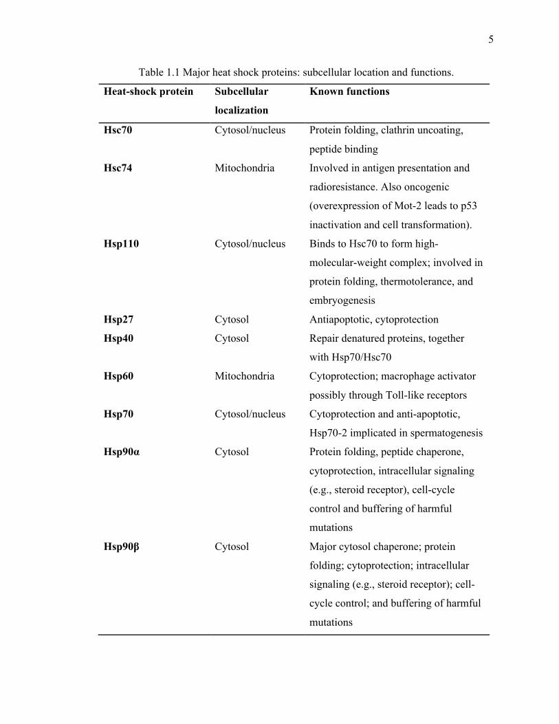
list of major HSPs along with their cellular location and brief description of primary
functions is outlined in Table 1.1.
5
5
Table 1.1 Major heat shock proteins: subcellular location and functions.
Heat-shock protein Subcellular
localization
Known functions
Hsc70 Cytosol/nucleus Protein folding, clathrin uncoating,
peptide binding
Hsc74 Mitochondria Involved in antigen presentation and
radioresistance. Also oncogenic
(overexpression of Mot-2 leads to p53
inactivation and cell transformation).
Hsp110 Cytosol/nucleus Binds to Hsc70 to form high-
molecular-weight complex; involved in
protein folding, thermotolerance, and
embryogenesis
Hsp27 Cytosol Antiapoptotic, cytoprotection
Hsp40 Cytosol Repair denatured proteins, together
with Hsp70/Hsc70
Hsp60 Mitochondria Cytoprotection; macrophage activator
possibly through Toll-like receptors
Hsp70 Cytosol/nucleus Cytoprotection and anti-apoptotic,
Hsp70-2 implicated in spermatogenesis
Hsp90α Cytosol Protein folding, peptide chaperone,
cytoprotection, intracellular signaling
(e.g., steroid receptor), cell-cycle
control and buffering of harmful
mutations
Hsp90β Cytosol Major cytosol chaperone; protein
folding; cytoprotection; intracellular
signaling (e.g., steroid receptor); cell-
cycle control; and buffering of harmful
mutations
6
6
1.1.2.2 Small heat shock proteins
The sHSPs are phylogenetically widespread and have been found in almost all organisms
ranging from archaebacteria, prokaryotes and eukaryotes. Within cells, they are found in
multiple subcellular locations where they bind to wide range of unfolded proteins
substrates to prevent their aggregation (44,47-49). Proteins in this family possess 12-43
kDa weights with the ability to form dynamic oligomers. Monomers of many sHSPs are
composed of a conserved 80-100 amino acid α-crystallin domain, accompanied with
beta-strands responsible for dimerization. The N-terminal domain is responsible for
oligomerization, phosphorylation and chaperone activity but it is not a highly conserved
domain, while the C-terminal domain is accountable for client protein interaction,
nucleotide binding and homodimerization (48). In vitro studies revealed that quaternary
structures of sHSPs are very stable and can binds to large number of unfolded protein
substrates compared to other major chaperones. sHSPs are consider as having ‘holdase’
activity because of their unusually high tendency to bind unfolded client proteins and to
assist subsequent refolding of substrates by ATP-dependent chaperone networks (44,48).
Structural and in vitro studies have led to a proposed model that under stress conditions,
oligomers of sHSPs undergo structural rearrangement and breakdown into monomers to
be functionally active. Although the exact mechanism is poorly understood, it is thought
that the hydrophobic regions of sHSPs may bind with hydrophobic surfaces of partially
misfolded proteins to make larger soluble complexes that inhibit further aggregation of
the misfolded proteins (50-52).
7
7
The sHSP family displays a functional diversity such as protecting the cells from stressful
conditions, involvement in stress tolerance, protein folding, protein degradation,
maintaining cytoskeletal integrity, cell cycle, signal transduction, cell differentiation and
cell death. Moreover, many members of sHSP family interact with their substrates to
display potent anti-apoptotic activity and anti-inflammatory property as well as exhibit
cardio and neuroprotection (44,47,53). Therefore, sHSP family members have important
implications in a broad range of health and disease conditions. However, many
unanswered questions remain regarding the underlying mechanisms of sHsps pleiotropic
functions and their promiscuous interactions.
1.2 Neurodegenerative diseases
Several common pathways have been proposed to cause the underlying etiology and
pathology of neurodegenerative diseases such as Alzheimer’s disease (AD), Parkinson’s
disease (PD), Fronto-temporal dementia (FTD), Amyotrophic Lateral Sclerosis (ALS),
and Prion diseases (54). Multiple factors have been implicated in causing
neurodegeneration such as protein misfolding leading to aggregation, mitochondrial
dysfunction, free radical formation caused by oxidative stress and environmental
exposure of metals and pesticides associated with age (20,22,55,56). Although there is a
partial overlap in the general mechanism of these neurodegenerative diseases, each
disease has its own distinct molecular mechanism and pathological manifestation
including degradation of specific brain regions and deposits of protein aggregates in
neurons (55,57). The most common characteristic of this group of diseases is the
accumulation and deposition of aggregated or misfolded proteins including α-synuclein
(α-syn) in PD, amyloid-β in AD, huntingtin protein in HD and transactive response

8
8
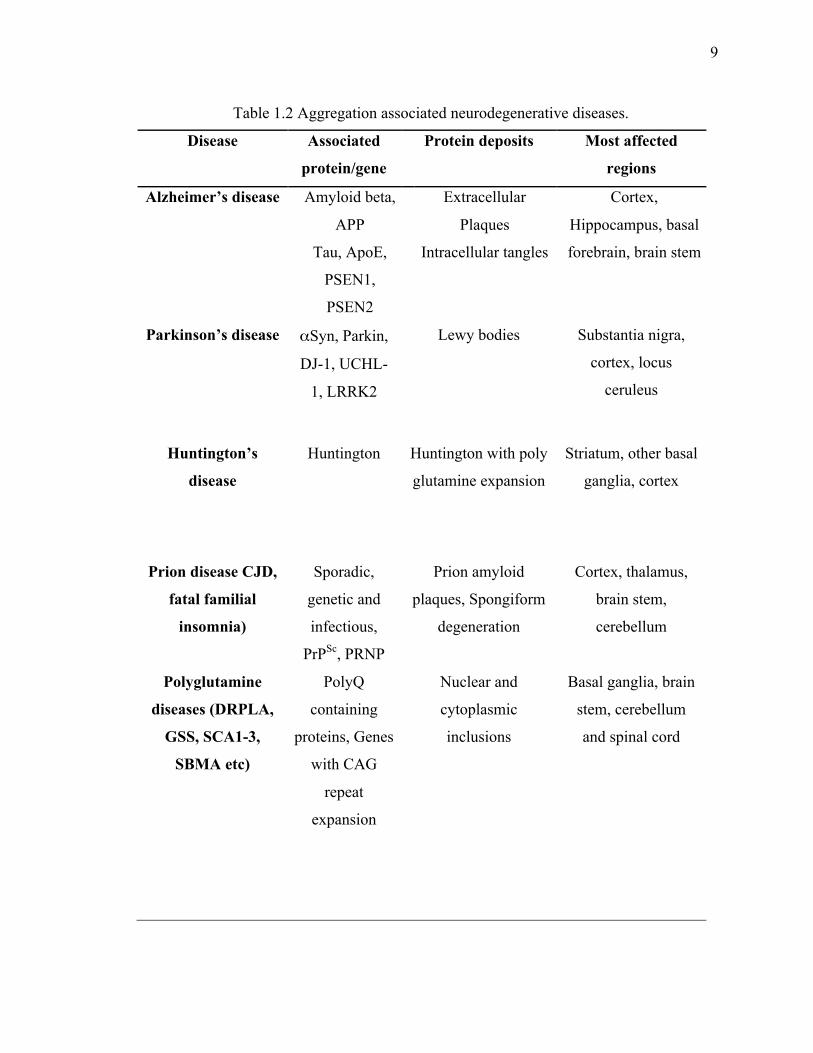
DNA-binding protein 43 (TDP-43) in ALS (54,56-61). A list of aggregation associated
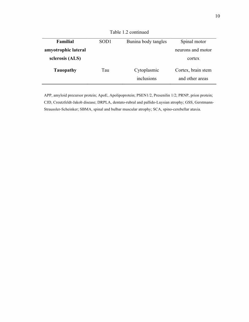
neurodegenerative diseases are outlined in Table 1.2.
9
9
Table 1.2 Aggregation associated neurodegenerative diseases.
Disease Associated
protein/gene
Protein deposits Most affected
regions
Alzheimer’s disease Amyloid beta,
APP
Tau, ApoE,
PSEN1,
PSEN2
Extracellular
Plaques
Intracellular tangles
Cortex,
Hippocampus, basal
forebrain, brain stem
Parkinson’s disease αSyn, Parkin,
DJ-1, UCHL-
1, LRRK2
Lewy bodies Substantia nigra,
cortex, locus
ceruleus
Huntington’s
disease
Huntington Huntington with poly
glutamine expansion
Striatum, other basal
ganglia, cortex
Prion disease CJD,
fatal familial
insomnia)
Sporadic,
genetic and
infectious,
PrPSc, PRNP
Prion amyloid
plaques, Spongiform
degeneration
Cortex, thalamus,
brain stem,
cerebellum
Polyglutamine
diseases (DRPLA,
GSS, SCA1-3,
SBMA etc)
PolyQ
containing
proteins, Genes
with CAG
repeat
expansion
Nuclear and
cytoplasmic
inclusions
Basal ganglia, brain
stem, cerebellum
and spinal cord
10
10
Table 1.2 continued
Familial
amyotrophic lateral
sclerosis (ALS)
SOD1 Bunina body tangles Spinal motor
neurons and motor
cortex
Tauopathy Tau Cytoplasmic
inclusions
Cortex, brain stem
and other areas
APP, amyloid precursor protein; ApoE, Apolipoprotein; PSEN1/2, Presenilin 1/2; PRNP, prion protein;
CJD, Creutzfeldt-Jakob disease; DRPLA, dentato-rubral and pallido-Luysian atrophy; GSS, Gerstmann-
Straussler-Scheinker; SBMA, spinal and bulbar muscular atrophy; SCA, spino-cerebellar ataxia.
11
11
1.2.1 Mitochondrial dysfunction in neurodegenerative disease
The mitochondria, most often known as powerhouses of the cells, are responsible for
most of the energy supply in eukaryotic cells. They play critical roles in regulating cell
processes including signaling pathways, calcium balances, reactive oxygen species (ROS)
formation, cell cycle regulation, thermogenesis and cell death. The loss of mitochondrial
function is associated with increased ROS production and oxidative stress that are
responsible for development of numerous neurodegenerative disorders. Mitochondria
generate adenosine triphosphate (ATP) during oxidative phosphorylation using metabolic
intermediates in tricarboxylic acid (TCA) cycle. Superoxide anion radicals (O-2) that are
produced during electron transport chain (ETC) activity are converted into hydrogen
peroxide by enzymatic action of superoxide dismutase in the mitochondria. Hydrogen
peroxide further detoxifies by the action of glutathione peroxidase into water in the
mitochondria. Defects in enzymes involved in the ETC leads to increased production of
ROS resulting in a decrease in the mitochondrial membrane potential, energy crisis and
finally cell death. In addition to antioxidant activity of mitochondrial enzymes mentioned
above, cells also possess glutathione (GSH), Vitamin E, Vitamin C and ubiquinone as
antioxidant agents that protect the cell from oxidative stress. An imbalance in the
antioxidant homeostasis leads to oxidative stress in cells (62-64). In the CNS, neurons are
particularly prone to oxidative stress resulting in accumulation of ROS that potentially
leads to the initiation of free radical chain reactions and finally cell death. Many classes
of macromolecules ranging from lipids to DNA and proteins are found to be oxidatively
damage in neurodegenerative diseases such as PD, AD, HD and ALS (56,65-67). Thus
mitochondrial dysfunction and defects in mitochondrial activity parameters including
12
12
alteration in mitochondrial dynamics, increase in ROS production and loss of energy
leads to neuronal death and are an important contributor in the manifestation of
neurodegenerative disorders (66). Therapeutic approaches to target mitochondrial
functions could certainly be useful however, using antioxidants, as sole therapy to treat
neurodegeneration appears to be not sufficient. In fact, several clinical studies have
demonstrated only little success with antioxidants in the treatment of neurodegeneration
(68). Targeting mitochondrial proteins to alter abnormal mitochondrial dynamics may
provide a potential therapeutic strategy against neurodegenerative diseases (64).
1.2.2 Amyloidogenesis
The term ‘amyloid’ has been used to describe the highly ordered cross beta proteinaceous
structures found in various pathological conditions. Amyloids are formed when a soluble
and innocuous protein transforms into insoluble protein aggregates that might not be
directly invasive but are associated with neuropathology. The tendency of amyloid
formation is multifactorial including amino acid composition, protein sequences and
concentration, posttranslational modification and environmental factors (16,19). Several
studies now clearly show that protein aggregation is a complex process that occurs in
several different steps, making different kinds of intermediates and finally turned into
larger aggregates that could be either amorphous or amyloids (69). However, there is
much to be learned as most of these studies are done in vitro and hence may not simulate
in vivo behavior in human diseases (70). Initiation of the multi-step pathway of protein
aggregation begins with modification of protein to an abnormal conformation that may be
a covalent modification such as cleavage or phosphorylation of the disease protein. These
modifications of proteins facilitate the conversion of monomers into smaller oligomers
13
13
that further assemble into protofibrils. Association of these protofibril intermediates can
form larger amyloid aggregates that can be visualized under the microscope (71-76). It is
hypothesized that intermediate species are more toxic than the precursor protein or final
amyloids (73,77,78) (Figure 1.1). Therefore, it would maybe beneficial to inhibit
aggregation formation at early stages of the pathway, as it may prevent the formation of
toxic oligomeric species.
14
14
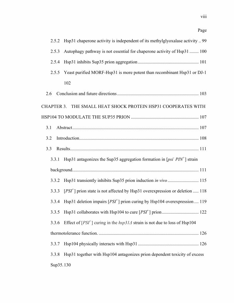
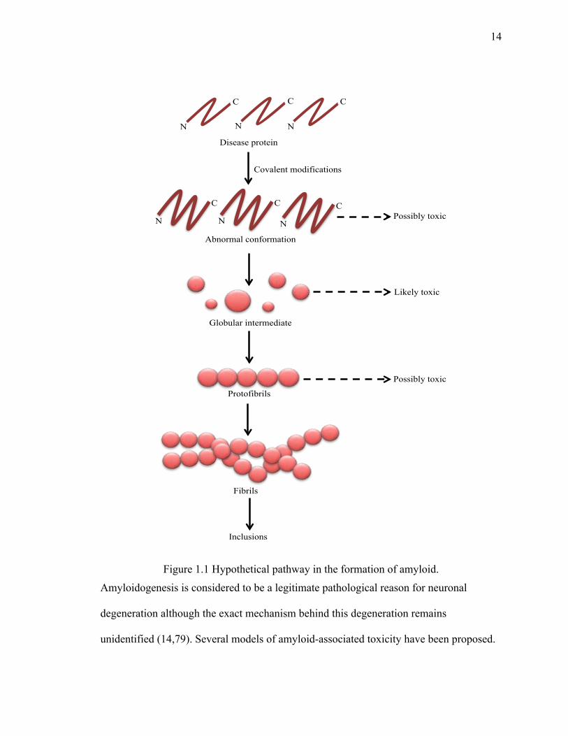
Figure 1.1 Hypothetical pathway in the formation of amyloid.
Amyloidogenesis is considered to be a legitimate pathological reason for neuronal
degeneration although the exact mechanism behind this degeneration remains
unidentified (14,79). Several models of amyloid-associated toxicity have been proposed.
Disease protein
N
C
N
C
N
C
N
C
N
C
N
C
Abnormal conformation
Globular intermediate
Protofibrils
Fibrils
Possibly toxic
Likely toxic
Possibly toxic
Covalent modifications
Inclusions
15
15
First, amyloids occupy the extracellular space that damages the normal architecture and
function of cells and thus leads to neuronal toxicity. It is also proposed that smaller
oligomers formed at initial stages of amyloidogenesis are more toxic to cells as they
destabilize cellular membranes (16,74,80,81). These intermediate oligomers assemble
into final amyloid fibrils that have been hypothesized as a non-invasive product of the
toxic intermediates that might rather be a detoxification mechanism. Another group of
studies suggest that generation of ROS by the incorporation of redox metals into
amyloids could affect cell viability (74). On the other hand, the larger aggregates could
recruit and thus diminish some essential proteins for cell survival (82).
1.3 Parkinson’s disease
PD is an idiopathic disease of the nervous system characterized by two neuropathological
hallmarks, the preferential loss of dopaminergic neurons in the substnatia nigra pars
compacta region of the brain and the presence of cytosolic inclusions called Lewy bodies
(LBs) in various brain regions (56,58,83). It is a chronic progressive disease that affects
the older population but can also occurs in much younger patients and is the second most
common neurodegenerative disorder after AD (84,85). Dr. James Parkinson, after whom
the disease is named, first recognized PD in the early 1800’s. PD affects 10 million
people worldwide and it is expected that its prevalence in America will increase
dramatically over the next 20 years as the proportion of the population ages (86,87). In
developing nations, the life expectancy is rising and cases of such age related
neurological disorders is also increasing resulting in a strong impact on the healthcare
system and economy due to direct and indirect costs associated with care of PD patients
(84,88,89). Therefore, it is critical to understand the molecular mechanisms of PD to
16
16
develop better therapeutic strategies, early detection of the disease and identify causative
genetic and environmental factors.
1.3.1 Risk factors
Age is the major risk factor for PD with an average age of disease onset of 60 years and
risk of diagnosis greatly increases after 85 years of age (90,91). Other risk factors include
family history and environmental stress such as exposure to pesticide toxins. Men are
more susceptible than women to develop the disease but the causative risk factor is still
unclear (92-94). Numerous other risk factors have been associated with PD though the
epidemiologic evidence is not robust. These include drinking of well water, excess milk
consumption, obesity, living in rural areas with exposure to copper, manganese, lead and
hydrocarbon solvents such as industrialized, farming or agricultural work (94-96). On the
other hand, cigarette smoking and caffeine intake have been linked with reduced risk of
PD (97). Table 1.3 describes the etiology of PD.
17
17
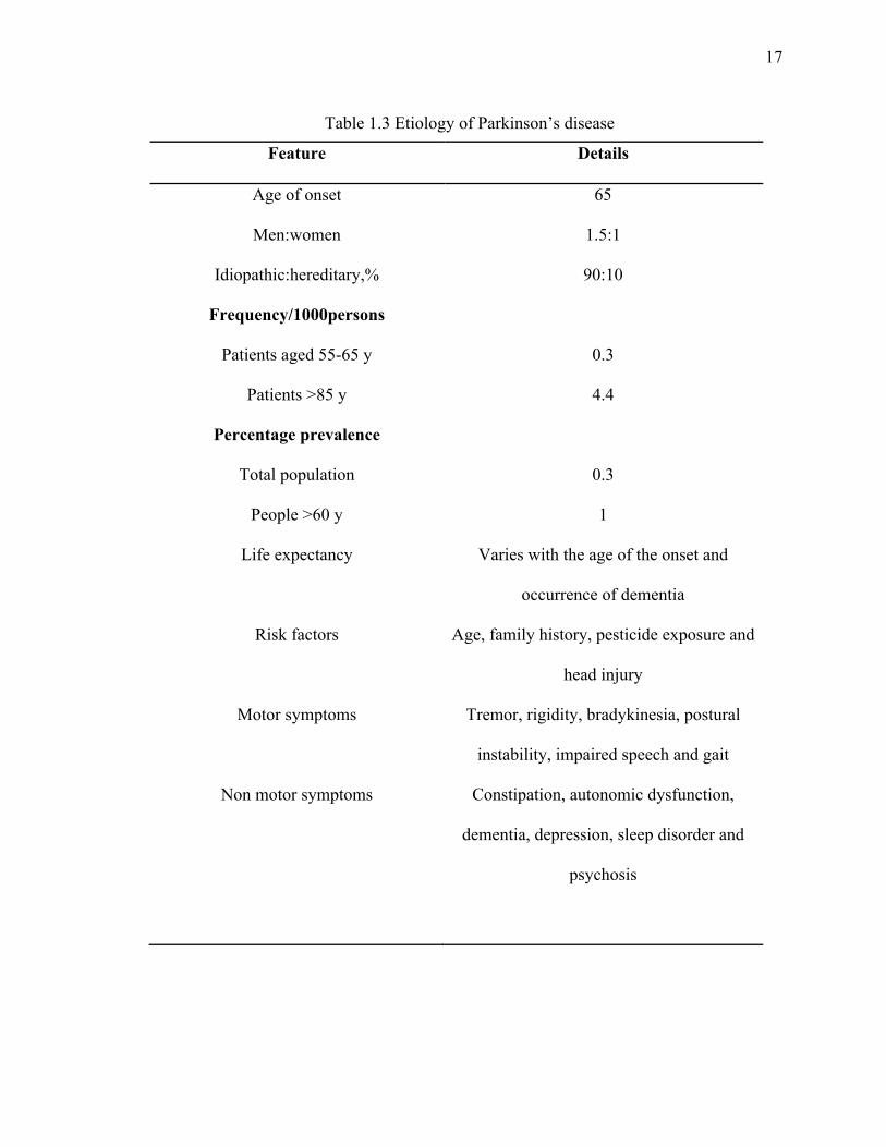
Table 1.3 Etiology of Parkinson’s disease
Feature Details
Age of onset 65
Men:women 1.5:1
Idiopathic:hereditary,% 90:10
Frequency/1000persons
Patients aged 55-65 y
Patients >85 y
0.3
4.4
Percentage prevalence
Total population
People >60 y
0.3
1
Life expectancy Varies with the age of the onset and
occurrence of dementia
Risk factors Age, family history, pesticide exposure and
head injury
Motor symptoms Tremor, rigidity, bradykinesia, postural
instability, impaired speech and gait
Non motor symptoms Constipation, autonomic dysfunction,
dementia, depression, sleep disorder and
psychosis
18
18
1.3.2 Genetic factors
Although the majority of PD cases are sporadic, about 5-10% PD cases are familial as
they are associated with gene mutation. The number of genes associated with the onset of
early or advanced PD have steadily increased over the last 15 years including mutations
or amplification in the LRRK2, PARK2, PARK7, PINK1, or SNCA (PARK1) gene and
possibly additional unidentified genes (92,94,98). Even though most cases of PD are
sporadic, examining the genes associated with PD is valuable as there is evidence to
show a strong correlation between sporadic and familial forms of PD. PARK1 encodes α-
syn consists of autosomal dominant gene, mutation or multiplication of this can lead to
PD (99,100). The α-syn protein is a 140 amino acid protein that exclusively expressed in
the CNS. Natively it is present mostly as membrane-bound α-helices in the neurons.
(101,102). In the disease form, α-syn mainly presents as an aggregated protein
component found in LBs (103). Several α-syn mutants including A53T, A30P, E46K and
H50Q have been shown to increase the aggregation formation that leads to form
potentially toxic protofibrils. High level of α-syn also sequestered several cytoskeletal
proteins, such as tubulin, that prevent the conversion of α-syn into fibrils leading to
increase in toxic protofibril species (99,100,104-107).
1.3.3 Diagnosis
Despite extensive research done on the pathophysiology of PD, its diagnosis is still
restricted to suboptimal methods for detection and prognosis. There is a critical need for
the development of highly sensitive and specific biomarkers as they are lacking currently
(108). The PD diagnostic decision in a clinic relies on the presence or manifestations of
symptoms associated with the disease such as bradykinesia, rest tremor, rigidity, postural
19
19
instability and impaired gait (93,109-111). If patient response to dopaminergic agents
such as levodopa and patient history reveals gradual progression of symptoms there is
likelihood of accurate diagnosis of PD. However, responsiveness to levodopa has been
seen in other parkinsonian syndromes and unrelated dystonias (108,112,113). To confirm
PD, presence of Lewy bodies (LBs), protein deposits in substantia nigra and loss of
neurons is a required diagnostic criteria upon examination of a deceased patient’s brain
section (110,113). As this approach is a post mortem examination, it has no implications
in clinical diagnosis.
There are several biomarkers currently being analyzed that can correlate with PD
prognosis with high accuracy and can be used as next generation diagnostic tools (114-
117). Among them the major constituent of PD such as oligomeric forms of α-syn and
DJ-1 protein level have been analyzed in the blood of PD patients with no success
(107,118,119). Several other biomarkers such as uric acid level, iron level in the
substantia nigra region of brains as well as epidermal growth factor (EGF) and
Apolipoprotein A1 (ApoA1) are under investigation (120-123). Currently there is no
single biomarker available to predict PD progression with reliability and validity
(114,124).
Neurologic imaging tools such as positron emission tomography (PET) scan, magnetic
resonance imaging (MRI), ultrasonography and others play small role in diagnosis and
are presently not accepted as diagnostic tool for PD (125-127).
20
20
1.3.4 Clinical presentation
There are four cardinal components of symptoms associated with PD: motor symptoms,
cognitive changes, behavioral/neuropsychiatry symptoms, and autonomic nervous system
failures. These can be divided into motor and non-motor symptoms.
1.3.4.1 Motor symptoms
The major motor features of PD are sometime represents by a mnemonic TRAP. It stands
for Tremors, Rigidity, Akinesia (or bradykinesia) and Postural instability. According to
pathological and neuroimaging studies, onset of motor symptoms occurs only after about
50-70% of neurodegeneration of substantia nigra (89,128). Rate of motor symptoms are
highly variable among patients. Bradykinesia is the most characteristic clinical
manifestation of PD that refers to slowness of movement. Patients often describe
bradykinesia as tiredness or weakness and commonly report difficulty from getting up
from a chair and opening packages or containers (129). Rigidity of idiopathic PD referred
to as ‘cogwheel’ phenomenon is when patients experience difficulty to initiate a
movement. At the beginning, it is unilateral but can move to other sides in the later stages
of the disease. Tremors at rest are the most easily recognizable symptom of PD that are
always prominent at the distal part in the extremities and occur at frequency between 4 to
6 Hz. At the late stages of PD usually after onset of other clinical features postural
instability develops due to loss of postural reflexes. Postural instability along with
freezing of gait significantly increases the risk of hip fracture due to sudden fall (130-
133).
21
21
In addition to classic motor features, other motor symptoms are also observed including
dysarthria, masked facial expression (hypomimia), dysphagia, blurred vision, decrease
eye blink, dystonia and difficulty turning in bed (93).
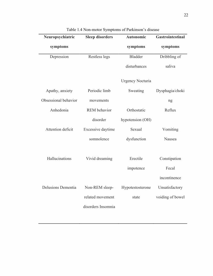
1.3.4.2 Non-motor symptoms
Along with the motor symptoms of PD, the non-motor symptoms (NMS) are major cause
of disability for PD patients but unfortunately unlike motor symptoms NMS are not well
recognized and undertreated (109,134-136). Recognition and treatment of NMS is an
important and fundamental aspect to consider in order to delivering a comprehensive
healthcare for PD patients. NMS arise with the progression of disease and are diverse
including neuropsychiatric symptom, sleep disorder, autonomic symptom,
gastrointestinal symptom and sensory symptom (Table 1.4). Sometime treatment of PD
leads to development or exacerbation of NMS in PD patients therefore it is important to
recognize, monitor and treat these symptoms (135-138).
22
22
Table 1.4 Non-motor Symptoms of Parkinson’s disease
Neuropsychiatric
symptoms
Sleep disorders Autonomic
symptoms
Gastrointestinal
symptoms
Depression Restless legs Bladder
disturbances
Urgency Nocturia
Dribbling of
saliva
Apathy, anxiety
Obsessional behavior
Periodic limb
movements
Sweating Dysphagia/choki
ng
Anhedonia REM behavior
disorder
Orthostatic
hypotension (OH)
Reflux
Attention deficit Excessive daytime
somnolence
Sexual
dysfunction
Vomiting
Nausea
Hallucinations Vivid dreaming Erectile
impotence
Constipation
Fecal
incontinence
Delusions Dementia Non-REM sleep-
related movement
disorders Insomnia
Hypotestosterone
state
Unsatisfactory
voiding of bowel
23
23
1.3.5 Management of Parkinson’s disease
Currently, there are only symptomatic treatments available with no proven
neuroprotective agents. For earlier stages of disease after diagnosis, it is important to take
time and educate the patient and relative about the condition and its implications. The
decision to start the treatment especially at earlier stages of disease when there is little
functional deficit is difficult. This decision should be made based on the age of the
patient, the consents of the patient, the presence of cognitive impairment, the likelihood
of complications associated with treatment and additional health problems (89,139,140).
The current gold standard regimen for managing symptoms is precursor of Dopamine i.e.
L-3,4- dihydroxyphenylalanine (L-Dopa or levodopa). Levodopa can cross the blood
brain barrier (BBB) easily to enter the central nervous system where it converts into
dopamine in the presence of an enzyme L-aromatic decarboxylase (133). Although L-
Dopa reduced the symptoms of PD and decreased the mortality rate associated with PD,
it is responsible for wide range of side effects due to peripheral conversion into dopamine.
To avoid these side-affects, dopa decarboxylase inhibitors, Carbidopa and Benserazide
are co-administered that cannot cross BBB and only inhibit peripheral dopamine
conversion. However, chronic use of L-dopa is associated with induction of dyskinesia
that is particularly problematic at later stages of disease with loss of large number of
dopaminergic neurons (141).
Another strategy to treat symptoms of PD is the use of dopamine agonists including ergot
derivatives such as bromocriptine, pergolide, cabergoline and lisuride; and non-ergot
derivatives as ropinirole and pramipexole. Dopamine agonists produced fewer of motor
24
24
complication compared with L-dopa but efficacy is much lower when used as mono-
therapy (142-144).
One more class of drug, the monoamine oxidase B (MAO-B) inhibitors, Selegiline and
Rasagiline, have demonstrated efficacy in PD disease and have potential to use as mono-
therapy in both early and advanced disease. An N-methyl-D- aspartate (NMDA) receptor
antagonist, amantadine, and anticholinergics such as benztropine have also been proven
efficacious in a small sub-population of PD patients (112,139,143,145). After few years
of consistent and effective response to L-dopa, the effect of a single L-dopa dose in most
patients fluctuates in terms of motor performance leading to wearing-off phenomenon.
Several catechol-O-methyl transferase (COMT) inhibitors and amantadine have been
used in combination with L-dopa for the treatment of dyskinesias and other motor
symptoms (144,146). A surgical procedure called deep brain stimulation has been
reserved for patients who are unresponsive to pharmacological treatment and have a high
degree of motor fluctuation and dyskinesias (147).
1.3.6 Yeast Model of Parkinson’s disease
Saccharomyces cerevisiae is a compatible cell model to understand the molecular
mechanisms of several human diseases including PD, for which many α-syn toxicity
models have been developed (148,149). This unicellular eukaryote is a well-suited model
for studying the disease related phenotypes, for instance, stress response, mitochondrial
and vesicular trafficking defects as well as oxidative stress response. In addition, this
simple genetic model has been extensively used for large-scale screening of drugs and
genes to uncover the underlying mechanism of disease (17,148-157). In this study we
used a budding yeast (Saccharomyces cerevisiae) model of PD that evaluates α-syn
25
25
misfolding, aggregation, and toxicity. In this model, at relatively low concentration of α-
syn localizes to membrane and is not toxic to cells. When two gene copies of α-syn are
expressed, an increase in the concentration of α-syn forms prominent intracellular
cytoplasmic inclusions or foci that are associated with toxicity to the cells. This
Saccharomyces cerevisiae model throws light on α-syn's role in PD pathogenesis (158).
This simple experimental model is very useful to dissect and understand the biochemical
mechanisms of many diseases, as there is high mechanistic similarity of many
physiological processes with human cells.
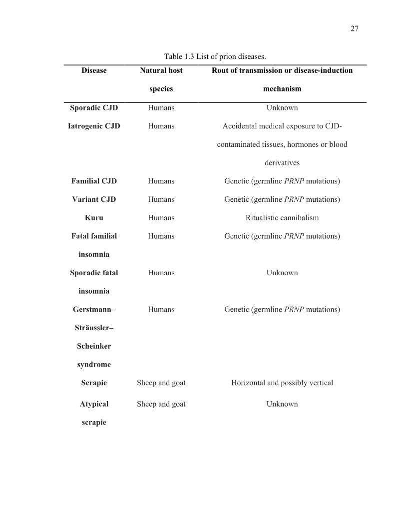
1.4 Prion disease
Prions are self-replicating proteins that are responsible for fatal neurodegenerative
disorder known as prion diseases or transmissible spongiform encephalopathies affecting
multiple mammalian species such as Kuru, bovine spongiform encephalopathy (BSE) and
scrapie, in human, cattle and sheep respectively (159-161). They are generally considered
a transmissible disease within and between different mammalian species as transfer of
brain extracts from affected persons into host species can spread the disease (162-164).
Transmission among humans could occur during surgical procedures or pituitary
hormone treatment, for example, 450 cases of iatrogenic Creutzfeldt–Jakob disease (iCJD)
have been reported during such treatments. Blood donors with undetected subclinical
symptoms could be a reason for variant CJD (vCJD) in humans. Cannibalistic rituals in
Papua New Guinea were also historically linked with transmission of Kuru (165-173).
Normal cellular prion protein (PrPC) is a 253 amino acid long protein encoded by a gene
PRNP on chromosome 20 mutations of which is associated to genetic prion disease. The
scrapie prion protein (PrPSc), is a pathological form of prion protein present in the brain
26
26
tissue of patients with transmissible spongiform encephalopathy (TSE). PrPSc differs with
normal cellular prion protein (PrPC) by translational modification and is formed by
recruiting PrPC conformers, which later perpetuate into larger aggregates that trigger
neurotoxic signals (162,163,174-177). These are the characteristics of typical spongiform
that are seen in the patient’s brain. A list of major prion diseases is provided in the table
1.5.
27
27
Table 1.3 List of prion diseases.
Disease Natural host
species
Rout of transmission or disease-induction
mechanism
Sporadic CJD Humans Unknown
Iatrogenic CJD Humans Accidental medical exposure to CJD-
contaminated tissues, hormones or blood
derivatives
Familial CJD Humans Genetic (germline PRNP mutations)
Variant CJD Humans Genetic (germline PRNP mutations)
Kuru Humans Ritualistic cannibalism
Fatal familial
insomnia
Humans Genetic (germline PRNP mutations)
Sporadic fatal
insomnia
Humans Unknown
Gerstmann–
Sträussler–
Scheinker
syndrome
Humans Genetic (germline PRNP mutations)
Scrapie Sheep and goat Horizontal and possibly vertical
Atypical
scrapie
Sheep and goat Unknown
28
28
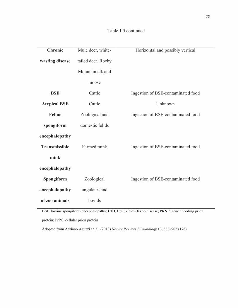
Table 1.5 continued
Chronic
wasting disease
Mule deer, white-
tailed deer, Rocky
Mountain elk and
moose
Horizontal and possibly vertical
BSE Cattle Ingestion of BSE-contaminated food
Atypical BSE Cattle Unknown
Feline
spongiform
encephalopathy
Zoological and
domestic felids
Ingestion of BSE-contaminated food
Transmissible
mink
encephalopathy
Farmed mink Ingestion of BSE-contaminated food
Spongiform
encephalopathy
of zoo animals
Zoological
ungulates and
bovids
Ingestion of BSE-contaminated food
BSE, bovine spongiform encephalopathy; CJD, Creutzfeldt–Jakob disease; PRNP, gene encoding prion
protein; PrPC, cellular prion protein
Adopted from Adriano Aguzzi et. al. (2013) Nature Reviews Immunology 13, 888–902 (178)
29
29
Like other neurodegeneration-associated amyloid proteins, PrPSc protein is rich in β-sheet
content and is protease resistant, thus it not easily degraded by cellular enzymes. These
protease resistant amyloids of PrPSc are then deposited in the brain tissue during
progression of the disease. Prion aggregation can occur both intracellularly and
extracellularly. Specific prion antibodies can detect amyloid plaques caused by prion
protein that appear similar to those of AD (162,164,179). On the other hand, how prions
destroy the neurons in CNS is still enigmatic. Numerous studies investigating the
mechanism of prion aggregate formation and propagation are performed in yeast and
many of the findings from these studies are applicable to human prion diseases.
1.4.1 Prions in yeast
Although no homologue of PrPC exists in yeast, many proteins have been discovered that
are present in different conformations including normal soluble or amyloid aggregated
forms. Different conformations of the same protein are linked with distinct phenotypes in
yeast. In yeast, prions act as epigenetic cytoplasmic elements that provide phenotypic
diversity in a heritable manner that operates at the level of protein conformation without
the need of nucleotide sequences (180-185).
Reed Wickner in 1994 proposed that the previously known yeast non-Mendelian
heritable elements [URE3] and [PSI+] are prions of Ure2 and Sup35 proteins
respectively(186,187). Prion are often linked with a loss of function phenotype in yeast.
Sup35 is a translational termination factor that loses its function in a way that translation
termination efficiency is compromised in the presence of the prion form of Sup35
(98,188-191). Similarly, the normal function of Ure2 is to regulate nitrogen catabolism,
preventing uptake of an intermediate, ureidosuccinate (USA), involved in the uracil
30
30
biosynthesis pathway. Thus the [URE3] prion form of Ure2, loses its normal function in
ura2 mutant cells and can grow on –Ura media by taking up USA (186,188,192-196).
Prion traits are dominant since aggregated forms of a prion can recruit the identical
soluble prion protein and convert it into the prion conformation. [PSI+] prion exists in
different variants similar to the observation in mammals that are genetically identical
showed different characteristics of prion in isolates of disease that can be stably
reproduced (164,197). Variation in the ratio of aggregated vs. soluble Sup35 protein is
linked with different variants of [PSI+] prion, known as weak or strong forms of the prion.
[PSI+] prion variants lead to different degree of loss of function for example, in the
presence of ade1-14 nonsense stop codon, strong variants will cause greater degrees of
translational read-through compared with weak variants resulting in accumulation of
characteristic red pigments indicating the absence of ADE1 (198-202).
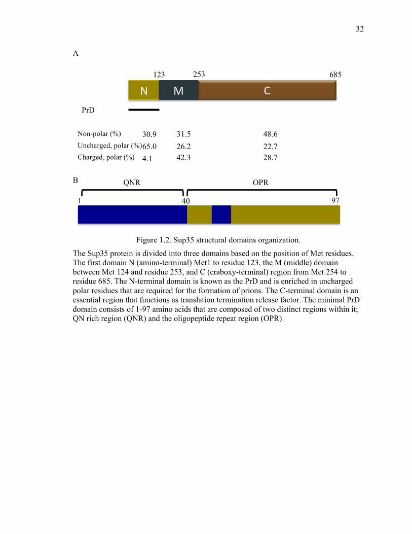
1.4.2 Structural organization
1.4.2.1 Prion forming domain
Most of the yeast prion proteins contain a region that is required for the formation and
propagation of the prion without requiring the remaining portions of the protein (203).
Such regions are known as prion-forming domains or PrDs. Sup35 PrD or Sup35N
domain is also involved in other cellular functions other than its role in prion formation
(204). A middle domain (Sup35M) links PrD to the C-terminal domain. Sup35M domain
is composed of highly charged amino acids residues that help to maintain prion
aggregation possibly by interaction with HSPs (205). Yeast PrDs are Q and N rich
domain, that when artificially recombined to different proteins may confer a prion state.

31
31
Sup35 prion forming domain (PrD) can be divided into two segments, the QNR stretch
and the OPR element based on the observation that the fragment of PrD required for
protein aggregation is shorter than segments needed for efficient propagation of prion
(Figure 1.2) (161,180,206,207).
32
32
A
B
Figure 1.2. Sup35 structural domains organization.
The Sup35 protein is divided into three domains based on the position of Met residues. The first domain N (amino-terminal) Met1 to residue 123, the M (middle) domain between Met 124 and residue 253, and C (craboxy-terminal) region from Met 254 to residue 685. The N-terminal domain is known as the PrD and is enriched in uncharged polar residues that are required for the formation of prions. The C-terminal domain is an essential region that functions as translation termination release factor. The minimal PrD domain consists of 1-97 amino acids that are composed of two distinct regions within it; QN rich region (QNR) and the oligopeptide repeat region (OPR).
C N M
123 253 685
PrD
Non-polar (%) Uncharged, polar (%) Charged, polar (%)
30.9
65.0
4.1
31.5
26.2 42.3
48.6
22.7 28.7
40 97 1
QNR OPR
33
33
1.4.2.2 De novo prion formation
The frequency of prion de novo appearance is considerably low unless prions or their
PrDs are transiently overexpressed resulting in the dramatic increase in the rate of prion
formation by up to 3000-fold. Interestingly, overexpression of just the PrD is more
efficient than transient overexpression of full-length protein (181,191,201,208). Still
elevated levels of PrD as such, is not sufficient for prion formation and requires the
presence of another QN rich prion, [PIN+] in aggregated state, for de novo prion
formation (209). [PIN+] is a prion form of the Rnq1 protein that was first discovered as a
non-Mendelian factor with prion like properties (181,201,208,210-212). The amino acid
composition of Rnq1 is similar to the PrD of Sup35 and it is proposed that [PIN+] acts as
an initial nucleus for de novo Sup35 prion aggregation formation. The de novo [PSI+]
prion formation in the presence of [PIN+] by transient overexpression of Sup35 or its PrD
is a multi-step process initiated by accumulation of misfolded protein in insoluble protein
deposits (IPODs) quality control (213,214). Fusing of Sup35 to GFP, allows the
visualization of formation of in vivo prion behavior, in which the fluorescent signal
appears as a ring-like and long filamentous structures at the periphery of cells. These
rings later collapse together to form internalized smaller rings that enclose the vacuoles.
After cell division the daughter cells appeared with dot-like foci of [PSI+] (213).
Aside from the presence of [PIN+], several other cellular components have been reported
to control de novo prion formation and propagation. Most of these components are
involved in stress response pathways such as chaperones, ubiquitin-proteasome system,
intracellular trafficking networks and members of actin cytoskeleton (215-221).
34
34
1.4.3 Effect of heat shock proteins on prion propagation
Although prion proteins require no cofactor to generate and propagate amyloid
aggregation in vitro, de novo prion propagation is modulated by chaperones such as
Hsp104, Hsp70, Hsp40 and sHSPs such as Hsp26 and Hsp42 (36,39,53,222-225).
Hsp104 is a member of the AAA ATPase chaperone family with homohexameric
structure and required for prion propagation in vivo. Hsp104 and its bacterial homolog,
ClpB, possess potent disaggregase activity against stress damage protein aggregates (226-
228). Deletion or overexpression of Hsp104 eliminates [PSI+] prion, thus an intermediate
level of Hsp104 is required for prion propagation (222,223,229,230). It was proposed that
Hsp104 generates smaller seeds by promoting fragmentation of larger prion fibers that
initiate propagation. It was also hypothesized that elevated Hsp104 cures [PSI+] prion by
monomerization of large aggregates but evidence for this hypothesis are based on the
indirect observation that overexpression of Sup35, leading to an increase in aggregate
size, partially compromises the curing effect of excess Hsp104 (231). Another model
proposes that elevated levels of Hsp104 cures [PSI+] by dissolution of the prion seeds,
the evidence for this model came from observation that Hsp104 overexpression induces a
diffuse localization signal for Sup35 tagged with GFP and a large fraction of soluble
Sup35 was observed in cell lysate (226,232,233). Recent studies also suggest that
dissolution of prion seed might be due to the trimming activity of Hsp104 in which
Sup35 dissociates from the seed terminus, hence that reducing its size without generating
new seeds. Trimming activity of Hsp104 is still present even when severing activity is
inhibited by treatment of guanidine (223). The effects of Hsp104 on prion curing are
strongly influenced by other chaperones or co-chaperones including the Hsp70 chaperone
35
35
system consisting of Ssa and its co-chaperones, Ydj1 or Sis1 and the Sse proteins
(38,40,208,234,235). The Hsp70 co-chaperone system has an essential role at the initial
steps of the disaggregation processes possibly by facilitating Hsp104 to recognize and
extract single polypeptide from aggregates at later stages (225). Remarkably, different
families of Hsp70 i.e. Ssa and Ssb shows contrary effects on [PSI+] prion. Ssb
overexpression enhances curing of [PSI+] prion by Hsp104 while Ssa protects [PSI+]
from curing by Hsp104. Ssa overexpression and Ssb deletion also increase de novo [PSI+]
prion formation. At the molecular level Ssa interacts with Sup35 and overexpression of
Ssa increases the polymer size of Sup35 (225,236-239). Altered expression of Hsp40
(Sis1 or Ydj1) by mutation, transient depletion or internal deletion, also influences the
dynamics of [PSI+] prion propagation. Sis1 overexpression promotes [PSI+] prion curing
by Hsp104. It is also proposed that Sis1 is responsible for recruiting Ssa and Hsp104 to
prion aggregates. Unlike Sis1, Ydj1 is non-essential and is shown to cure weak variants
of [PSI+] prion, but only when Ssa1 is also co-expressed (225,234). The exact mechanism
of action of Hsp70 and Hsp40 family members in prion propagation is still under
investigation but it is clear that they collaborate with Hsp104 and play crucial roles in
prion propagation. Deletion of co-chaperones of Hsp70/90 such as Sti1 or Cpr7, also
inhibit [PSI+] curing by Hsp104 over-expression (40).
In addition to Hsp104 and its assistant chaperones, sHSPs such as Hsp31, Hsp26 and
Hsp42 also play a role in disaggregation of misfolded proteins in yeast (53,240-242).
These proteins are highly expressed under moderate stress and during late growth phase
for transition to stationary phase. Hsp42 and Hsp26 work synergistically to inhibit prion
formation and potentiate dissolution of Sup35 prion aggregates by distinct mechanisms

36
36
(53). Furthermore, Hsp26 or Hsp42 collaborate with Hsp70 and or Hsp104 to reduce the
SDS-resistant polyglutamine aggregation (243). The diverse effects of chaperones on
prion propagation is summarized in table 1.6.
37
37
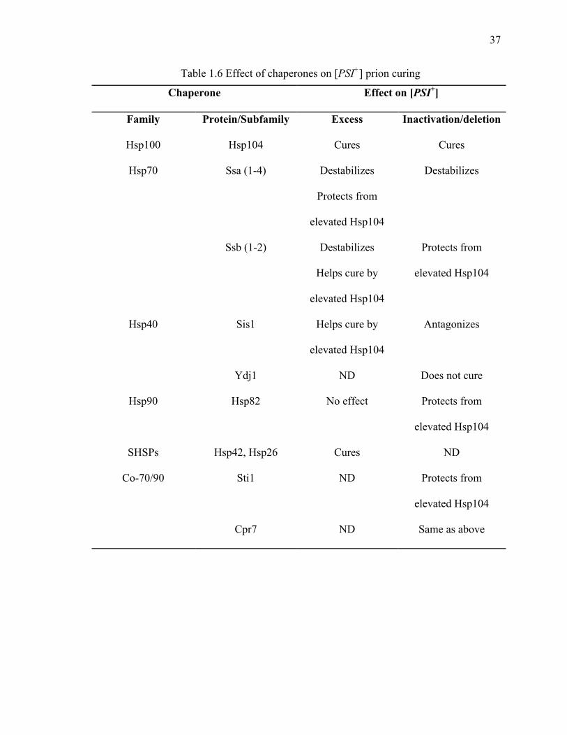
Table 1.6 Effect of chaperones on [PSI+] prion curing
Chaperone Effect on [PSI+]
Family Protein/Subfamily Excess Inactivation/deletion
Hsp100 Hsp104 Cures Cures
Hsp70 Ssa (1-4) Destabilizes
Protects from
elevated Hsp104
Destabilizes
Ssb (1-2) Destabilizes
Helps cure by
elevated Hsp104
Protects from
elevated Hsp104
Hsp40 Sis1 Helps cure by
elevated Hsp104
Antagonizes
Ydj1 ND Does not cure
Hsp90 Hsp82 No effect Protects from
elevated Hsp104
SHSPs Hsp42, Hsp26 Cures ND
Co-70/90 Sti1 ND Protects from
elevated Hsp104
Cpr7 ND Same as above
38
38
1.4.4 Prion associated toxicity
The unusual conformations of normal proteins into self-assembly leads to formation of
amyloid aggregates that has been implicated in both the acquisition of new traits and in
the emergence and progression of disease. In yeast, the presence of [PSI+] prion itself is
not toxic but overexpression of Sup35 or its PrD in such a strain is lethal (215,221,244).
It is proposed that toxicity is caused by recruitment of essential protein(s) including
another translational release factor Sup45 (245-247). Also, the combination of [PSI+]
prion together with tRNA suppressor induces the stress response (248,249). Some
variants of [PSI+] that are toxic can be rescued by overexpression of Sup35 derivative
that lack PrD and cannot be recruited by aggregates (250). Specific mutations in Hsp104
leads to [PSI+]-dependent cytotoxicity in yeast cells (251). Some variants of [PSI+]
prions that are not toxic per se may become toxic when combined with other factors such
as a polyQ stretch from human Huntingtin protein in [PIN+] or [PSI+] strains that are
otherwise non-toxic (245,252).
1.5 DJ-1/ThiJ/Pfp1 superfamily
The members of DJ-1 superfamily are large number of proteins with conserved three-
dimensional structure distributed across eukaryotes and prokaryotes (253-259). Members
of the family such as DJ-1, hchA and Hsp31 possess similar primary, secondary and
tertiary structures with the difference in the presence of main domains (P and A domains).
In addition, they also differ in the formation of homo-dimerization such as DJ-1
monomer interacts through α-helices while Hsp31 dimerization is stabilizes by β-sheets.
Another prominent feature of these family members is the presence of a conserved
cysteine catalytic triad (Figure 1.3).
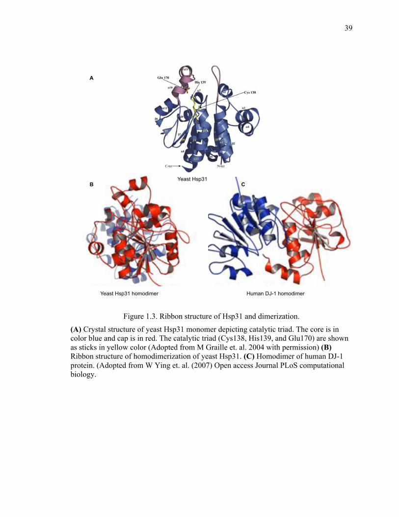
39
39
Figure 1.3. Ribbon structure of Hsp31 and dimerization.
(A) Crystal structure of yeast Hsp31 monomer depicting catalytic triad. The core is in color blue and cap is in red. The catalytic triad (Cys138, His139, and Glu170) are shown as sticks in yellow color (Adopted from M Graille et. al. 2004 with permission) (B) Ribbon structure of homodimerization of yeast Hsp31. (C) Homodimer of human DJ-1 protein. (Adopted from W Ying et. al. (2007) Open access Journal PLoS computational biology.
Yeast Hsp31
Yeast Hsp31 homodimer Human DJ-1 homodimer
A
B C
40
40
Despite highly conserved structure, members of this family are diverse in functions
including catalases, glyoxalases, proteases, transcriptional regulators and chaperones.
However, they are all involved in cellular stress response (158,260-266). Special
attention has been drawn to this family because of human member of the family, DJ-1,
that has a well-characterized involvement in two major diseases, cancer and PD (267-
270). A large number of studies have investigated the cellular function of DJ-1 and its
role in modulating disease pathogenesis. Other members of the family include E.coli
Hsp31, archeal Pyrococcus horikoshii proteins (PH1704, YajL and Yhbo) and Hsp31 in
the yeast Saccharomyces cerevisiae that has three other paralogs (Hsp32-34)
(255,258,271-274).
1.5.1 Human DJ-1
Human DJ-1 is a small protein of 189 amino acids that is expressed ubiquitously in
several tissues of body including brain and is mainly localized in the cytoplasm but also
present in mitochondria and the nucleus. It was first identified as an oncogene that is
elevated in several types of cancers such as leukemia, breast cancer, primary lung cancer,
prostate cancer, cervical cancer and pancreatic cancer (270,275-279). The oncogenic
potential of DJ-1 is thought to be related to its interaction with tumor suppressor
phosphate and tension homolog (PTEN) in the PI3K-Akt pathway that regulates cell
proliferation and transformation (280). Later studies showed its implication in
neurodegenerative disease such as mutations in the gene PARK7 cause early onset of
autosomal recessive PD (267,281-285). Mutations in DJ-1 that destabilize
homodimerization show the quaternary structure is critical for cellular function. The
conserved cysteine residue, C106, is also important to regulate cellular function and
41
41
localization of DJ-1. C106 is highly susceptible to oxidation and mutations affect its
function, oligomerization and localization(286-289). Interestingly, oxidatively damaged
proteins are found in the brains of patients with idiopathic PD, AD and HD. Like other
members of the family DJ-1 also function as methylglyoxalase and deglycase of glycated
proteins in the cell (290-292). In addition, DJ-1 positively regulates the antioxidant
transcription factor, erythroid 2-related factor 2 (Nrf2) (293). The multiple cellular roles
of DJ-1 and the different biochemical activity contributions to its overall role are under
investigation but compelling evidence indicates that it is a multifunctional chaperone that
protects cells from oxidative and associated stress similar to the yeast Hsp31 protein.
Interestingly, our understanding of DJ-1 function has been more advanced than the yeast
counterpart but new studies including ours are elucidating the function of yeast Hsp31
that will be applicable to DJ-1.
1.5.2 The yeast Hsp31 mini family
The yeast Saccharomyces cerevisiae Hsp31 mini-family consists of four paralogs, HSP31
(YDR533C), HSP32 (YMR322C), HSP33 (YOR391C), and HSP34 (YPL280W) (272).
Among the four members, HSP31 is the most divergent and is proposed to have been
duplicated during yeast evolution resulting in the paralogous genes. After resolving the
crystal structure of yeast Hsp31, the name was given because of structural similarity with
E. coli Hsp31. Hsp32, Hsp33 and Hsp34 share over 90% sequence homology. While,
Hsp31 is only about 70% similar to other members (271,272).
1.6 Role of Hsp31 in cellular stress response
Consistent with the main role of cellular stress response, Hsp31 expression is strongly
induced during late phases of growth and required for survival under conditions of
42
42
nutrient limitation (294). Hsp31 expression is also induced when yeast cells are treated
with H2O2 to produce oxidative stress and this up-regulation of Hsp31 is under the
control of stress responsive transcription factor, Yap1 (158,295). In addition, Hsp31
possesses robust glutathione independent glyoxalase activity that converts the toxic
metabolite, methylglyoxal (MGO), into D-lactate (158,260,261). The catalytic triad Cys-
His-Glu of Hsp31 is vital for this enzymatic activity and critical for suppressing the
elevated level of ROS by MGO (158,260). An overall view has emerged indicating
Hsp31 has important metabolic and regulatory roles in cytoprotective pathways (296).
In addition to the enzyme activity, we also demonstrated that Hsp31 has broad chaperone
activity in several classic protein aggregation assays indicating its ability to manage
misfolded proteins that initiate proteotoxic stress in the cell. We also demonstrated that
the yeast-purified protein was more active in preventing aggregation of several substrate
proteins including α-syn (158). The increased activity may be due to the difference in the
affinity tags used but may be a result of posttranslational modification(s) that occur in the
cell. Several reports have indicated that post-translational modifications or differing
levels of oxidation of the cysteine residue can alter activity of DJ-1 (297,298). In addition,
we showed that yeast Hsp31 is more active in preventing protein aggregation compared
to DJ-1. Interestingly, it has also been observed that Hsp31 rescues α-syn and huntingtin
toxicity to a greater extent than DJ-1 in vivo using yeast as a model system (106,299).
These results raise the intriguing possibility that Hsp31 is constitutively active whereas
DJ-1 must undergo an activation event to increase its activity although more studies must
be performed to confirm the mechanism for these differences.
43
43
Hsp31 inhibits the formation of aggregates of a wide range of proteins including insulin,
citrate synthase, α-syn and the Sup35 prion (158). The recombinant soluble α-syn and
Sup35 proteins can readily polymerize into amyloidogenic fibrils in vitro (300). Hsp31
inhibits protein aggregation formation in vitro and foci formation of GFP-tagged proteins
in vivo. In addition, over-expression of Hsp31 can rescue cells from toxicity associated
with overexpression of α-syn. The rescue phenotype could be mediated by several
different mechanisms including Hsp31’s methylglyoxalase or deglycation activities.
However, a Hsp31 mutant deficient in methylglyoxalase activity is still very active in
preventing α-syn in vitro aggregation and prevention of toxicity from overexpressed α-
syn (158). Another possible rescue mechanism could be the autophagy pathway,
particularly because deletion of Hsp31 impairs autophagy under carbon starvation
conditions (294). Autophagy does alleviate toxicity from α-syn overexpression because
we show a synthetic lethal relationship between α-syn overexpression and deletion of
ATG8. However, we demonstrated that overexpression of Hsp31 in the atg8Δ strain can
rescue α-syn-mediated toxicity (158). These data show that despite the multitasking
abilities and roles of Hsp31, the chaperone activity appears to have the ability to prevent
α-syn toxicity independent of other activities. In support of our finding, another study
found the autophagy pathway was not crucial in preventing the Hsp31 chaperone activity
against a cytoplasmic aggregation prone protein. The same study also demonstrated that
Hsp31 chaperone activity overlaps with the Ubr-dependent degradation pathway but is
independent of its function in the oxidative stress response (262). Further exploration of
this model would need to utilize mutants that abrogate chaperone function without
affecting enzyme activity and other biological functions.
44
44
The typical model of protein aggregation proposes that unfolded monomers as an
initiating event that progresses to unstable oligomeric intermediates, and finally elongates
to larger oligomers. The observed anti-aggregation activity of Hsp31 raises the question
of what stage Hsp31 intervenes in the protein aggregation process. On the bases of our in
vitro studies, Hsp31 likely interacts with early oligomeric intermediates of α-syn and
therefore prevents higher oligomer formation. For example, when soluble α-syn was
mixed with Hsp31, formation of precipitated SDS-resistant oligomers was markedly
reduced. Likewise, the soluble fraction of α-syn, which included SDS-resistant oligomers
in the size range of 25-50 KDa, was almost completely abolished in the presence of
Hsp31 indicating that Hsp31 likely interacts with the monomeric or early oligomers in
preventing the formation of higher order oligomers. We observed similar results with the
in vitro ThioT assay, in which incubating the Hsp31 with α-syn completely prevented the
increase in fluorescence intensity associated with increasing fibril formation again
supporting our model that Hsp31 acts at early stages of protein aggregation (158). Of
particular interest is that DJ-1 has been shown to interact with monomeric and oligomeric
form of α-syn as determined by pull-down assays (106). This same study also showed
that DJ-1 interacts with α-syn in vivo as well. Along with α-syn aggregation formation,
we also demonstrated the inhibitory effect of Hsp31 on the Sup35 prion based on
reduction of aggregates observed in cell lysates. These results support the notion that
Hsp31 acts at the early stages of oligomerization to prevent further protein
oligomerization. Interestingly, we previously showed that the overexpression of Hsp31
reduces the level of Sup35 aggregation but we also show that it is unable to cure prions
from a [PSI+] strain. [PSI+] strains contain Sup35 prion aggregates that can be cured by
45
45
chaperones such as Hsp104, but the lack of curing by Hsp31 suggests that it lacks
disaggregase activity and cannot intervene in an established prion cycle. Moreover, our
study showed no co-localization of Hsp31 and Sup35 aggregates as observed under
fluorescence microscope, rather Hsp31 is occluded from Sup35 prion aggregates,
indicating that Hsp31 acts on its substrates prior to the formation of large aggregates. The
strong chaperone activity of Hsp31 suggests that it may modulate prion aggregates but
might need to cooperate with other chaperones similar to what has been demonstrated
with other small HSPs such as Hsp26 and Hsp42 (53).
1.7 Comparison of Hsp31 paralogs
The paralog genes of the Hsp31 mini-family are located at the subtelomeric region of the
genome in Saccharomyces cerevisiae (272). All the members of Hsp31 family contain
the same Cys-His-Glu catalytic triad as present in the E. coli ortholog but interestingly no
protease activity has been detected so far for Hsp31 or other paralogs. Previously, it was
shown that mutation in the catalytic triad largely abolishes glyoxalase activity, but this
catalytic triad is not required for chaperone activity of Hsp31 (158). These results
indicate that the anti-aggregation activity of Hsp31 is not under the influence of its
enzymatic activity rather, it has a direct chaperone activity against misfolded proteins.
Intriguingly, all the paralogs of the Hsp31 minifamily possess comparable activity against
α-syn aggregation and toxicity when they are overexpressed from the GAL promoter
(106). Furthermore, the chaperone activity of Hsp31, Hsp32 and Hsp33 against a
cytoplasmic aggregation-prone protein is independent of their role in oxidative stress
response and the vacuolar degradation pathway (262). However, unlike Hsp31, the other
paralogs possess very little methylglyoxalase activity and are unable to protect the cells
46
46
from glyoxal toxicity (260). These results again support the notion that anti-aggregation
activity of Hsp31 mini-family is independent of its enzymatic activity. In addition, the
lack of methylglyoxalase activity in the paralogs is evidence that the paralogs are
diverging but additional studies dissecting the roles within this paralog group are needed
to further uncover these diverging functions. The Hsp31 protein family is broadly spread
across fungal species with varying levels of paralog duplications and additional evidence
of divergence including differences in localization in the Schizosaccharomyces pombe
Hsp31 family members (264). A functional comparison of Hsp31 and its paralogs are
summarized in Table 1.7 highlighting the similarities and differences among these
proteins.
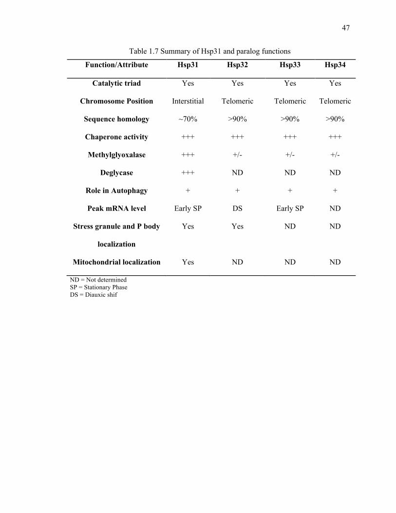
47
47
Table 1.7 Summary of Hsp31 and paralog functions
Function/Attribute Hsp31 Hsp32 Hsp33 Hsp34
Catalytic triad Yes Yes Yes Yes
Chromosome Position Interstitial Telomeric Telomeric Telomeric
Sequence homology ~70% >90% >90% >90%
Chaperone activity +++ +++ +++ +++
Methylglyoxalase +++ +/- +/- +/-
Deglycase +++ ND ND ND
Role in Autophagy + + + +
Peak mRNA level Early SP DS Early SP ND
Stress granule and P body
localization
Yes Yes ND ND
Mitochondrial localization Yes ND ND ND
ND = Not determined SP = Stationary Phase DS = Diauxic shif
48
48
1.8 Hsp31 role in redox homeostasis
Oxidative stress occurs when intra-cellular ROS overwhelms the anti-oxidative defense
system in the cell present during normal aerobic metabolism or by exposure to external
radical generating agents. ROS triggers damage to macromolecules in the form of
oxidative modifications and misfolding of proteins, associated with the development of
diseases and pathological conditions such as PD and prions (62,184,301,302). Hsp31 has
an important role in maintenance of redox homeostasis in yeast under oxidative stress
generated by MGO or H2O2 (260,303). Like many other heat-shock genes HSP26, HSP12,
HSP82, and SSA3, expression of HSP31 is strongly induced at diauxic shift when the
cells are stressed by nutrient limitation and by accumulation of oxidative metabolites
(158,294,303). Others and we also reported an elevated level of Hsp31 under oxidative
stress when cells were treated with H2O2 (158,303). Similarly, Hsp31 also plays a role in
the survival of cells during stationary phase and protects cells from oxidative stress
caused by MGO and H2O2 accumulation (158,303). In addition, we demonstrated that
Hsp31 expression was induced under proteotoxic stress such as overexpression of α-syn
(158). In support of the role of Hsp31 in managing this proteotoxic stress, we found that
deletion of HSP31 synergizes with α-syn expression to increasing toxicity. We reported
an increase in ROS level in the hsp31Δ strain that correlates with increased toxicity by α-
syn expression compared to wild type (WT) strain, indicating that the presence of Hsp31
is important in reducing ROS to basal levels. In agreement with our study,
overexpression of Hsp31 robustly suppresses both cytosolic and mitochondrial ROS
levels instigated by MGO and H2O2 and therefore provide cytoprotection (158,260). In
addition, Hsp31 localizes to mitochondria and preserves mitochondrial integrity by
49
49
redistributing glutathione to the cytoplasm under oxidative stress (260). Another study
examined the deglycase activity of Hsp31 and showed that it may efficiently deglycate
proteins with glycated Cys, Arg and Lys amino acid residues (265). Taken together, these
results suggest that Hsp31 is an integral part of the heat shock protein system and plays a
vital role in maintaining cellular homeostasis.
50
50
CHAPTER 2. HSP31 IS A STRESS-RESPONSE CHAPERONE THAT INTERVENES IN THE PROTEIN MISFOLDING PROCESS
This chapter contains parts of following publications:
Chia-Jui Tsai*, Kiran Aslam*, Holli M Drendel, Josephat M Asiago, Kourtney M
Goode, Lake N Paul, Jean-Christophe Rochet, Tony R Hazbun Hsp31 is a Stress-
Response Chaperone that Intervenes in the Protein Misfolding Process. The Journal of
biological chemistry, 290(41):24816-34, 2015
Kiran Aslam, Tony Hazbun Hsp31, a member of the DJ-1 superfamily, is a multitasking
stress responder with chaperone activity. Prion Apr 2016
2.1 Abstract
The Saccharomyces cerevisiae Hsp31 is a homodimeric protein that is highly induced
under stressful situations and involved in diauxic shift reprogramming. It is a protein with
multifunctional roles including functions as a chaperone, methylglyoxalase, deglycase
and is involved in the autophagy pathway. Hsp31 is homolog of human DJ-1 that has
implications in the pathophysiology of PD. We verified that Hsp31 are highly induced
under oxidative stress. Furthermore, we showed its induction under proteotoxic stress
condition. We confirmed that Hsp31 is a robust methylglyoxalase that is more potent in
activity than its human homolog DJ-1. We demonstrated that Hsp31 chaperone activity to
protect the cells from α-syn toxicity is not under the influence of its enzymatic activity or
autophagy pathway. We also showed that Hsp31 inhibit Sup35 PrD aggregations as
51
51
observed under fluorescence microscopy and flow cytometry. In addition, we provide
evidence that Hsp31 act early in the process of protein aggregation because Hsp31 does
not co-localize with larger Sup35 prion aggregates. These results establish that Hsp31
molecular chaperone activity is self-sufficient to protect the cells from stress conditions
without requiring its enzymatic activities.
2.2 Introduction
Yeast Hsp31 is encoded in the Saccharomyces cerevisiae genome and has drawn
attention after its structure was solved in 2003 indicating that it has structural similarity
with an evolutionarily conserved protein family the DJ-1/PfpI (ThiJ/PfpI) protein family
that includes human DJ-1 and E. coli Hsp31 proteins (271,272). The DJ-1/PfpI protein
family members including human DJ-1 and E. coli Hsp31 are moderately characterized
for their biological functions and role in pathologies but have many unanswered
questions with regards to their multifunctional roles and biochemical activities. Most
studies indicate an important role in response to stressed biological processes (303).
Mutated forms of DJ-1 are involved in early onset PD and DJ-1 is implicated to function
as a chaperone in response to oxidative stress by preventing α-syn fibrillation, a process
involved in PD pathogenesis (304). E. coli Hsp31 is a stress-inducible molecular
chaperone that plays an important role in protein misfolding and helps survival under
acidic stress (305). Initial characterization the yeast Hsp31 function showed that deletion
of HSP31 gene sensitized cells to ROS, suggesting a role for Hsp31 in protecting cells
against oxidative stress (303). Another study showed that yeast Hsp31 is important in
diauxic-shift where its expression is strongly induced and it has a survival role in
52
52
stationary phase of growth. In addition, deletion of HSP31 leads to impaired autophagy
under carbon starvation (294).
Furthermore, DJ-1 family proteins including human DJ-1, E. coli Hsp31 and yeast Hsp31
have been characterized as methylglyoxalases that detoxify by converting MGO into D-
lactate independent of GSH (261,264,290). MGO is a toxic metabolite that is mainly
produced as a byproduct of glycolysis. MGO is highly reactive with various amino acids
in proteins, nucleic acids and lipids to yield toxic AGE linked to several diseases. Cells
mainly catabolize MGO by two major enzymes including glutathione dependent GLO I
and GLO II (306,307). DJ-1 and its homologue proteins are characterized as GLO III that
do not require GSH as co-factor to convert MGO into D-lactate. Another study examined
the deglycase activity of DJ-1 and Hsp31 by showing that they efficiently deglycate
proteins with glycated Cys, Arg and Lys amino acid residues (265,292). Investigation of
the yeast Hsp31 is an excellent model to delineate and investigate the specific
biochemical activities of these proteins.
Previously in our lab, it was shown that Hsp31 protect cells against α-syn toxicity
associated with the formation of α-syn foci in a yeast model of PD. It also has been
shown that Hsp31 possesses a chaperone-like response against α-syn aggregation as
observed by an in vitro fibrillization assay as well as insulin and citrate synthase
aggregation assays (158). In the present study, we demonstrate that the protective effect
of Hsp31 against α-syn toxicity in yeast cells is independent of its methylglyoxalase
activity. DJ-1 and Hsp31 possess a conserved catalytic triad Glu-Cys-His, the first two
amino acids of this triad are important for GLOIII activity in DJ-1 (290). Here we tested
a mutation in Hsp31 C138D for methylglyoxalse activity and its activity against α-syn
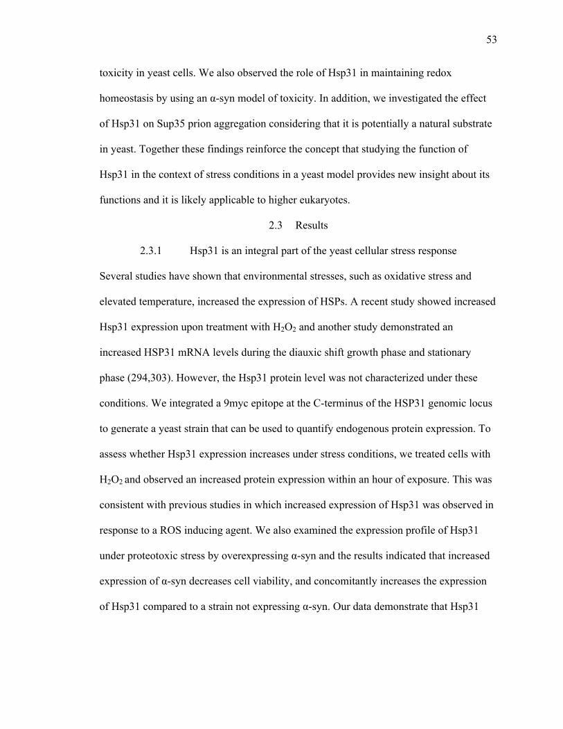
53
53
toxicity in yeast cells. We also observed the role of Hsp31 in maintaining redox
homeostasis by using an α-syn model of toxicity. In addition, we investigated the effect
of Hsp31 on Sup35 prion aggregation considering that it is potentially a natural substrate
in yeast. Together these findings reinforce the concept that studying the function of
Hsp31 in the context of stress conditions in a yeast model provides new insight about its
functions and it is likely applicable to higher eukaryotes.
2.3 Results
2.3.1 Hsp31 is an integral part of the yeast cellular stress response
Several studies have shown that environmental stresses, such as oxidative stress and
elevated temperature, increased the expression of HSPs. A recent study showed increased
Hsp31 expression upon treatment with H2O2 and another study demonstrated an
increased HSP31 mRNA levels during the diauxic shift growth phase and stationary
phase (294,303). However, the Hsp31 protein level was not characterized under these
conditions. We integrated a 9myc epitope at the C-terminus of the HSP31 genomic locus
to generate a yeast strain that can be used to quantify endogenous protein expression. To
assess whether Hsp31 expression increases under stress conditions, we treated cells with
H2O2 and observed an increased protein expression within an hour of exposure. This was
consistent with previous studies in which increased expression of Hsp31 was observed in
response to a ROS inducing agent. We also examined the expression profile of Hsp31
under proteotoxic stress by overexpressing α-syn and the results indicated that increased
expression of α-syn decreases cell viability, and concomitantly increases the expression
of Hsp31 compared to a strain not expressing α-syn. Our data demonstrate that Hsp31

54
54
expression is rapidly elevated at the protein level in the event of stress associated with
oxidative stress or proteotoxicity (Figure 2.1).
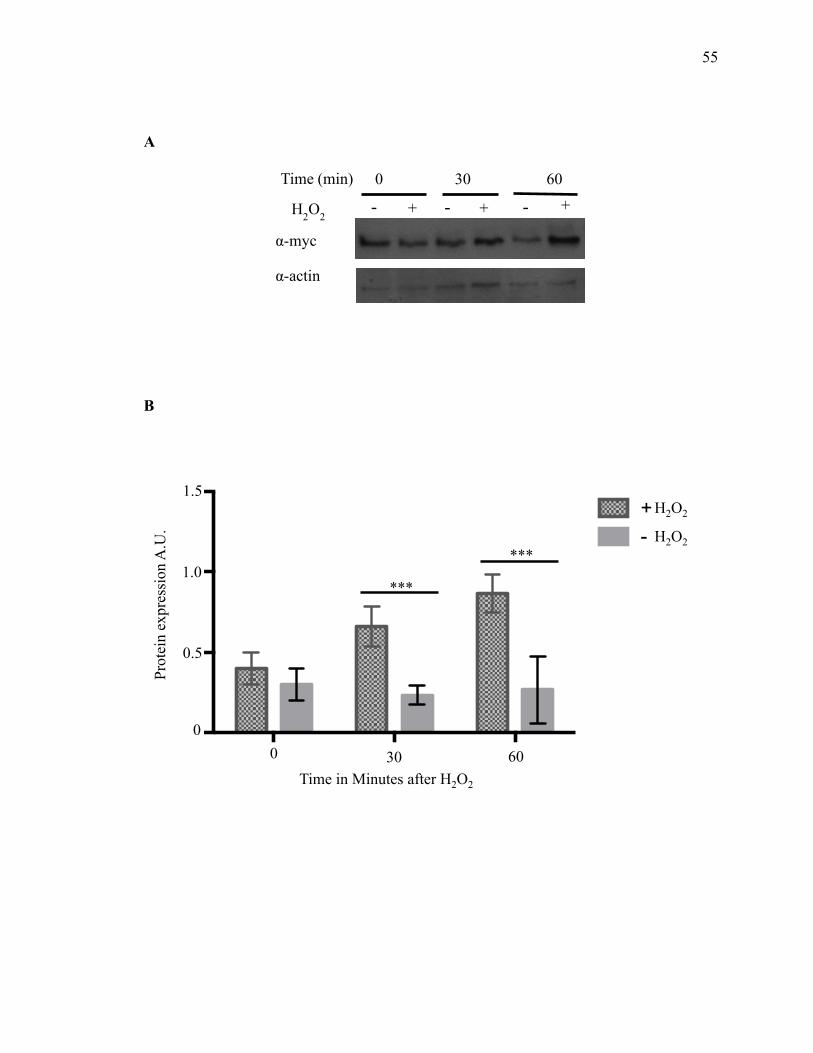
55
55
A
B
0 min
30 m
in
60 m
in0.0
0.5
1.0
1.5+-
Prot
ein
expr
essi
on A
.U.
***
***
Time in Minutes after H2O2
0 30 60
0
0.5
1.0
1.5 H2O2
H2O2
0 30 60
α-myc
α-actin
H2O2 + + + - - -
Time (min)
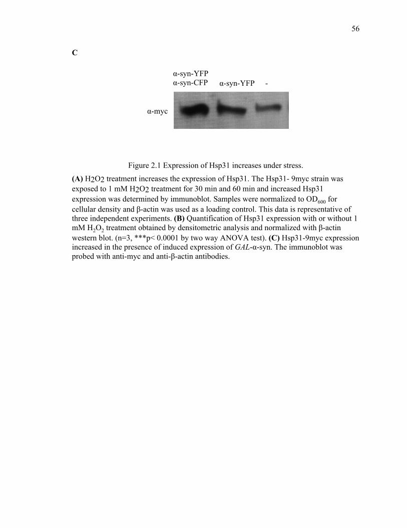
56
56
C
Figure 2.1 Expression of Hsp31 increases under stress.
(A) H2O2 treatment increases the expression of Hsp31. The Hsp31- 9myc strain was exposed to 1 mM H2O2 treatment for 30 min and 60 min and increased Hsp31 expression was determined by immunoblot. Samples were normalized to OD600 for cellular density and β-actin was used as a loading control. This data is representative of three independent experiments. (B) Quantification of Hsp31 expression with or without 1 mM H2O2 treatment obtained by densitometric analysis and normalized with β-actin western blot. (n=3, ***p< 0.0001 by two way ANOVA test). (C) Hsp31-9myc expression increased in the presence of induced expression of GAL-α-syn. The immunoblot was probed with anti-myc and anti-β-actin antibodies.
α-myc
α-syn-YFP α-syn-YFP α-syn-CFP -
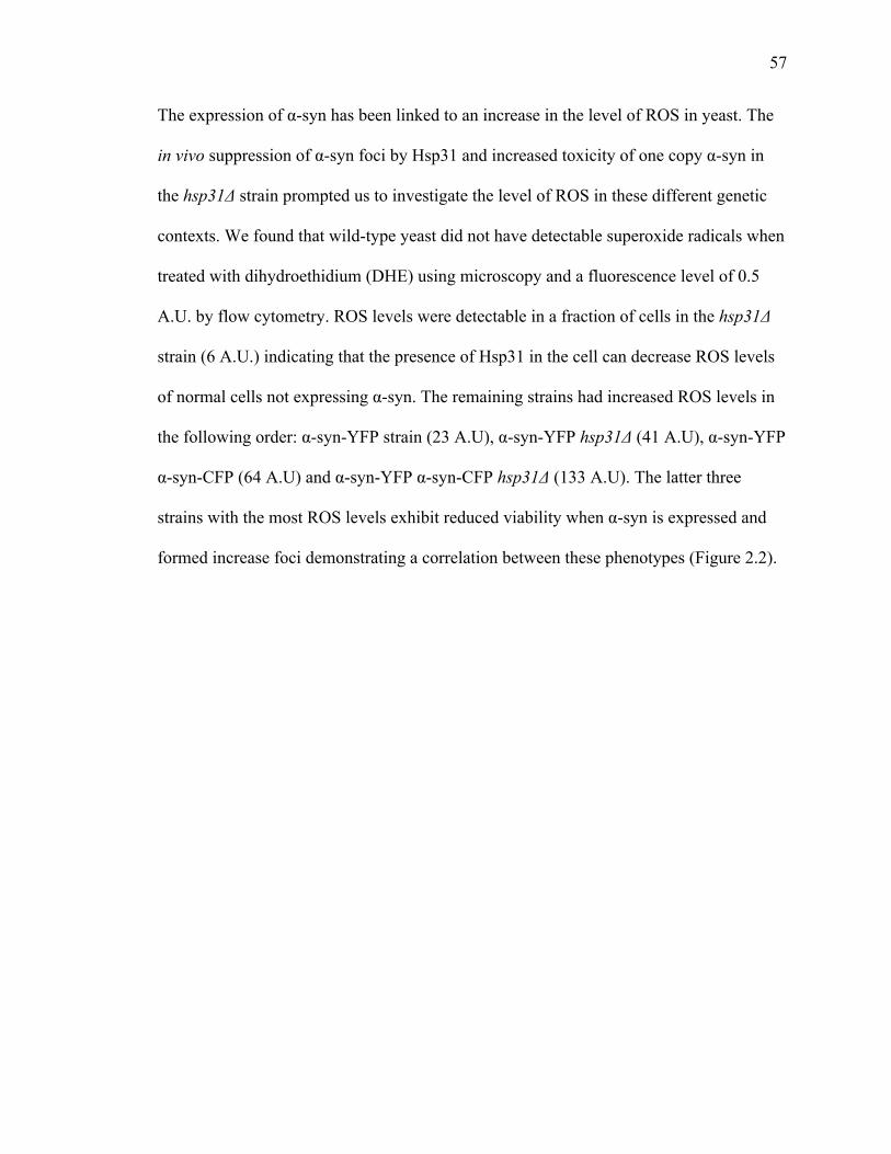
57
57
The expression of α-syn has been linked to an increase in the level of ROS in yeast. The
in vivo suppression of α-syn foci by Hsp31 and increased toxicity of one copy α-syn in
the hsp31Δ strain prompted us to investigate the level of ROS in these different genetic
contexts. We found that wild-type yeast did not have detectable superoxide radicals when
treated with dihydroethidium (DHE) using microscopy and a fluorescence level of 0.5
A.U. by flow cytometry. ROS levels were detectable in a fraction of cells in the hsp31Δ
strain (6 A.U.) indicating that the presence of Hsp31 in the cell can decrease ROS levels
of normal cells not expressing α-syn. The remaining strains had increased ROS levels in
the following order: α-syn-YFP strain (23 A.U), α-syn-YFP hsp31Δ (41 A.U), α-syn-YFP
α-syn-CFP (64 A.U) and α-syn-YFP α-syn-CFP hsp31Δ (133 A.U). The latter three
strains with the most ROS levels exhibit reduced viability when α-syn is expressed and
formed increase foci demonstrating a correlation between these phenotypes (Figure 2.2).
58
58
Bright field DHE Bright field DHE
W303
αSyn-YFP
αSyn-YFP αSyn-CFP
WT hsp31ΔA
a-Syn
-YFP
a-Syn
-CFP
hsp31Δ
B
WT
hsp31Δ
a-Syn
-YFP
a-Syn
-YFP
hsp31Δ
a-Syn
-YFP
a-Syn
-CFP
1150
1100
50
0
Fluo
resc
ence
inte
nsity
(A.U
. )
0.5 6.0
23
41
64
133
****"

59
59
Figure 2.2. Hsp31 decreases the ROS generated by α-syn. (A) Superoxide ions increase when HSP31 is deleted and when α-syn is expressed. In vivo presence of superoxide ions is detected by treatment with DHE and visualization by fluorescence microscopy. Representative fields of view are presented. (B) Quantification of superoxide ion increase in hsp31Δ and GAL-α-syn expressing strains. Flow cytometry was performed on three biological replicates. n=3 **** P ≤ 0.0001 based on the one-way ANOVA with multiple comparison t-test. Error bars = 1 SD.
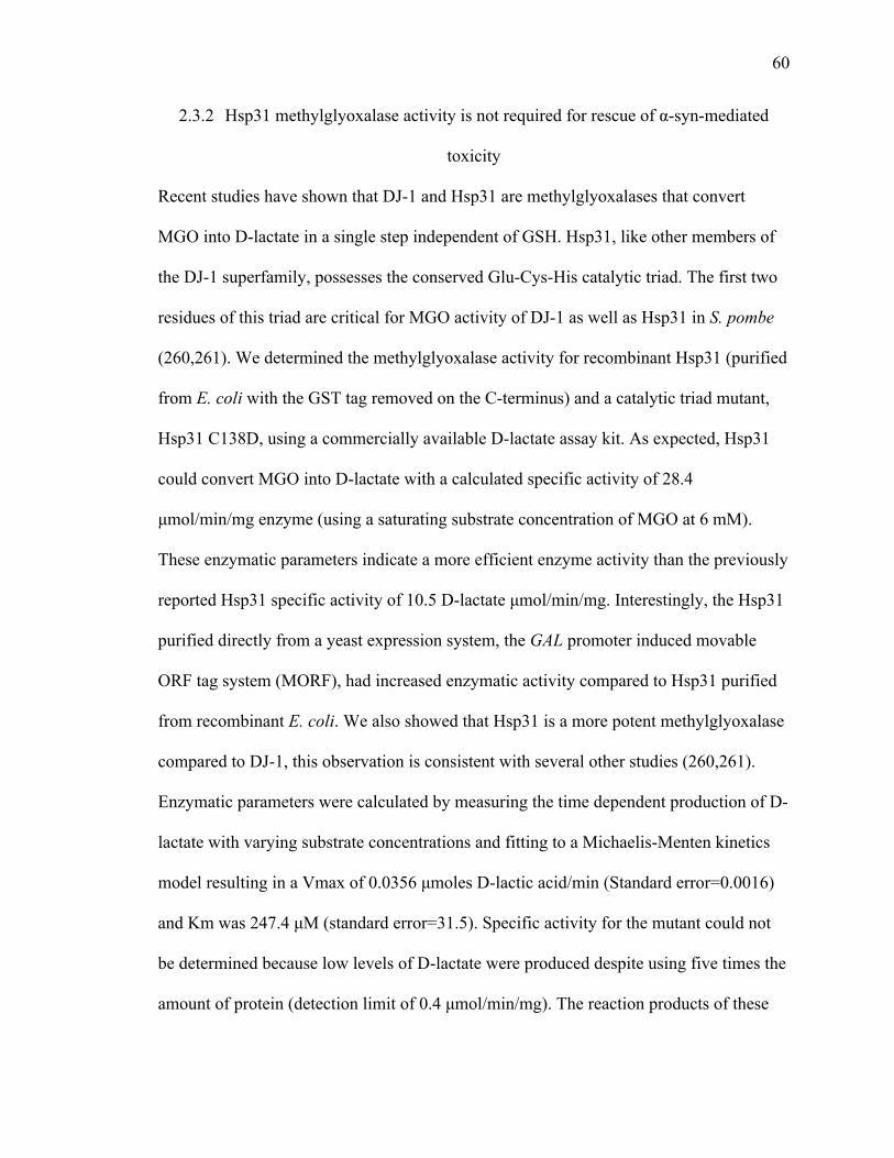
60
60
2.3.2 Hsp31 methylglyoxalase activity is not required for rescue of α-syn-mediated
toxicity
Recent studies have shown that DJ-1 and Hsp31 are methylglyoxalases that convert
MGO into D-lactate in a single step independent of GSH. Hsp31, like other members of
the DJ-1 superfamily, possesses the conserved Glu-Cys-His catalytic triad. The first two
residues of this triad are critical for MGO activity of DJ-1 as well as Hsp31 in S. pombe
(260,261). We determined the methylglyoxalase activity for recombinant Hsp31 (purified
from E. coli with the GST tag removed on the C-terminus) and a catalytic triad mutant,
Hsp31 C138D, using a commercially available D-lactate assay kit. As expected, Hsp31
could convert MGO into D-lactate with a calculated specific activity of 28.4
µmol/min/mg enzyme (using a saturating substrate concentration of MGO at 6 mM).
These enzymatic parameters indicate a more efficient enzyme activity than the previously
reported Hsp31 specific activity of 10.5 D-lactate µmol/min/mg. Interestingly, the Hsp31
purified directly from a yeast expression system, the GAL promoter induced movable
ORF tag system (MORF), had increased enzymatic activity compared to Hsp31 purified
from recombinant E. coli. We also showed that Hsp31 is a more potent methylglyoxalase
compared to DJ-1, this observation is consistent with several other studies (260,261).
Enzymatic parameters were calculated by measuring the time dependent production of D-
lactate with varying substrate concentrations and fitting to a Michaelis-Menten kinetics
model resulting in a Vmax of 0.0356 µmoles D-lactic acid/min (Standard error=0.0016)
and Km was 247.4 µM (standard error=31.5). Specific activity for the mutant could not
be determined because low levels of D-lactate were produced despite using five times the
amount of protein (detection limit of 0.4 µmol/min/mg). The reaction products of these

61
61
assays were examined by GC-MS and a peak identical to the lactic acid standard (6.09
min elution time) was identified, confirming the production of lactic acid by Hsp31.
Lactic acid was undetectable by GC-MS analysis in the Hsp31 C138D sample (Figure
2.3).
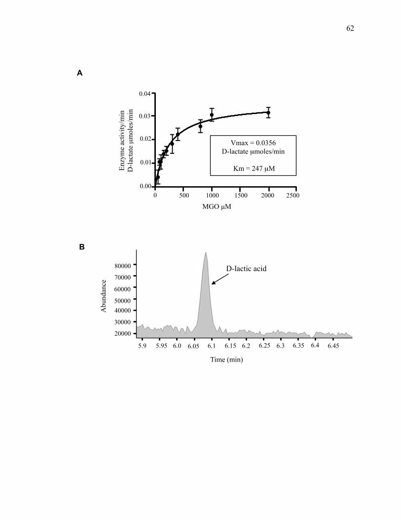
62
62
A
B
Time (min)
Abu
ndan
ce
80000
70000
60000
40000 50000
30000
20000
5.9 6.15 6.25 6.2 5.95 6.0 6.05 6.1 6.3 6.35 6.4 6.45
D-lactic acid
0 500 1000 1500 2000 2500 0.00
0.01
0.02
0.03
0.04
MGO µM
Enzy
me
activ
ity/m
in
D-la
ctat
e µ
mol
es/m
in
Vmax = 0.0356 D-lactate µmoles/min
Km = 247 µM
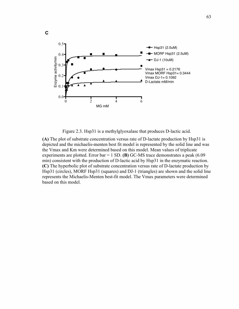
63
63
Figure 2.3. Hsp31 is a methylglyoxalase that produces D-lactic acid.
(A) The plot of substrate concentration versus rate of D-lactate production by Hsp31 is depicted and the michaelis-menten best fit model is represented by the solid line and was the Vmax and Km were determined based on this model. Mean values of triplicate experiments are plotted. Error bar = 1 SD. (B) GC-MS trace demonstrates a peak (6.09 min) consistent with the production of D-lactic acid by Hsp31 in the enzymatic reaction. (C) The hyperbolic plot of substrate concentration versus rate of D-lactate production by Hsp31 (circles), MORF Hsp31 (squares) and DJ-1 (triangles) are shown and the solid line represents the Michaelis-Menten best-fit model. The Vmax parameters were determined based on this model.
0 2 4 60.0
0.1
0.2
0.3
0.4
0.5
MGO mM
Enzy
me
activ
ity /m
inHsp31 2.5uMMORF Hsp31 2.5uMDJ-1 10uM
Hsp31 (2.5uM)MORF Hsp31 (2.5uM)DJ-1 (10uM)
MG mM
Enzy
me
activ
ity/m
in
Vmax Hsp31 = 0.2176Vmax MORF Hsp31= 0.3444 Vmax DJ-1= 0.1092D-Lactate mM/min
C
64
64
We next determined if methylglyoxalase activity is required for rescue of α-syn toxicity.
One copy of α-syn-YFP decreases fitness in the hsp31Δ strain but GPD driven expression
of Hsp31 rescues that toxicity. The expression of GPD driven Hsp31 C138D restored the
α-syn expressing strain to full viability comparable to expression of the wild-type Hsp31.
The lack of enzyme activity for this mutant and ability to rescue α-syn toxicity indicated
that the in vivo mechanism of rescue is not dependent on methylglyoxalase activity
(Figure 2.4).
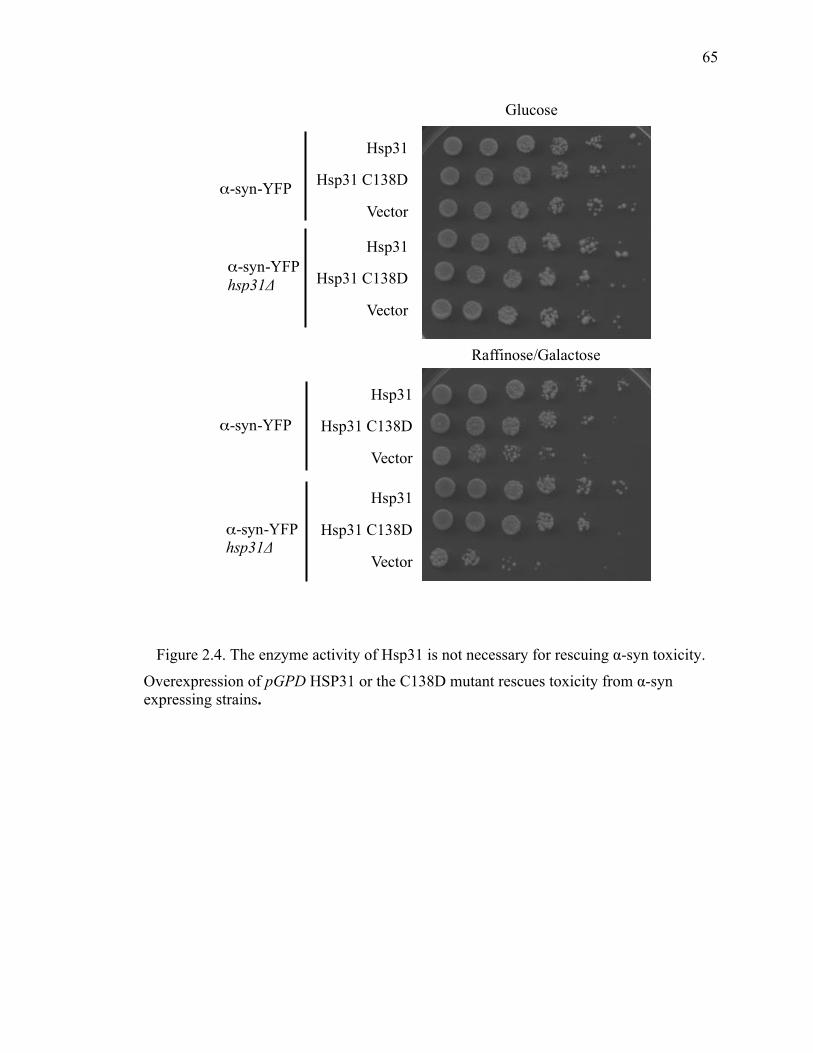
65
65
Figure 2.4. The enzyme activity of Hsp31 is not necessary for rescuing α-syn toxicity.
Overexpression of pGPD HSP31 or the C138D mutant rescues toxicity from α-syn expressing strains.
Glucose
Raffinose/Galactose
α-syn-YFP
Hsp31
Hsp31 C138D
Vector
α-syn-YFP hsp31Δ
Hsp31
Hsp31 C138D
Vector
Hsp31
Hsp31 C138D
Vector
Hsp31
Hsp31 C138D
Vector
α-syn-YFP
α-syn-YFP hsp31Δ
66
66
MGO is a toxic metabolite produced during glycolysis that reacts with proteins to yield
toxic AGE and has been associated with increased α-syn cross-linking and formation of
intracellular foci formation in neuroblastoma cells (261,264,290,306-308). We examined
if increased levels of MGO could increase the α-syn foci or toxicity by treating cells with
exogenous MGO and found no increased foci formation in wild-type yeast indicating that
foci formation is likely not influenced by MGO. In addition, the hsp31Δ strain also did
not have increased foci suggesting that lack of Hsp31 is not sufficient to increase foci
formation due to MGO treatment. We determined that MGO was toxic to wild-type yeast
at 10 and 20 mM MGO and viability in the hsp31Δ strains was similar to WT suggesting
that Hsp31 is not sufficient for detoxification. The concomitant expression of α-syn (one
or two copies) and treatment with MGO did not have differential effects on viability
(Figure 2.5). Consistent with these results is that cell lysates of the hsp31Δ strain had
equivalent levels of overall methylglyoxalase activity as wild-type because other yeast
methylgloxalase enzymes, Glo1 and Glo2, are present in yeast and have been shown to
detoxify MGO.
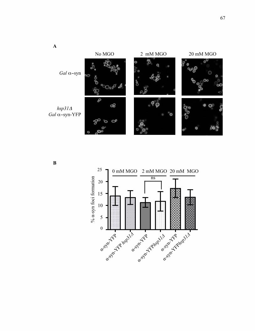
67
67
Gal α−syn
2 mM MGO No MGO
A
hsp31Δ Gal α−syn-YFP
20 mM MGO
0
15
10
20
5
25
% α
-syn
foci
form
atio
n
α-syn
-YFP
α-syn
-YFP
α-syn
-YFP
0 mM MGO 2 mM MGO 20 mM MGO ns
α-syn
-YFPhsp31Δ
α-syn
-YFPhsp31Δ
α-syn
-YFP hsp31Δ
B
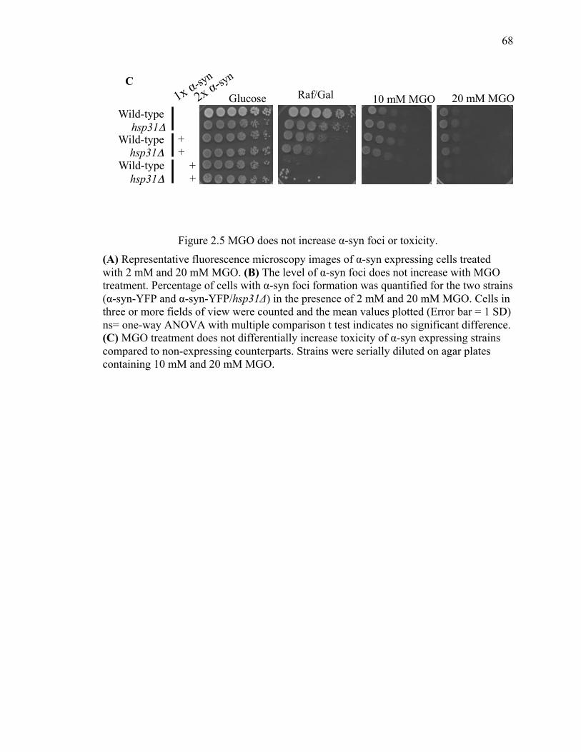
68
68
Figure 2.5 MGO does not increase α-syn foci or toxicity. (A) Representative fluorescence microscopy images of α-syn expressing cells treated with 2 mM and 20 mM MGO. (B) The level of α-syn foci does not increase with MGO treatment. Percentage of cells with α-syn foci formation was quantified for the two strains (α-syn-YFP and α-syn-YFP/hsp31Δ) in the presence of 2 mM and 20 mM MGO. Cells in three or more fields of view were counted and the mean values plotted (Error bar = 1 SD) ns= one-way ANOVA with multiple comparison t test indicates no significant difference. (C) MGO treatment does not differentially increase toxicity of α-syn expressing strains compared to non-expressing counterparts. Strains were serially diluted on agar plates containing 10 mM and 20 mM MGO.
Glucose Raf/Gal 10 mM MGO 20 mM MGO
hsp31Δ Wild-type
hsp31Δ Wild-type
hsp31Δ Wild-type
+ + + +
C
1x α-syn
2x α-syn
69
69
2.3.3 D-lactate supplementation suppresses the steady state level of α-syn
Since, Hsp31 is a methylglyoxalase that converts MGO into D-lactate we further
investigated the possibility that deletion of HSP31 causes the toxicity in α-syn expressing
strains due to compromise in the glutathione-independent methylglyoxalase activity of
Hsp31. We explored if the lack of enzyme activity leads to a build up toxic cellular MGO
or if the altered phenotype of the strain results from the lack in enzymatic product i.e. D-
lactate. More recently, it was shown that supplementing the cell with D-lactate restores
the mitochondrial membrane potential in DJ-1 deficient (RNAi-treated) HeLa cells (291).
We tested if external lactate supplementation could cure the α-syn toxicity similar to
Hsp31. First, we performed dilution growth assays on media containing lactic acid to
observe any rescue effect in α-syn expressing cells. Surprisingly, there was improved
growth of cells even in the presence of low levels of 0.2% lactic acid compared to cells
grown without lactic acid supplementation. In parallel to the dilution growth assay, we
performed microscopy on cells grown in liquid media containing lactic acid along with
galactose to induce α-syn expression and observed these cells under microscope. Cells
that were grown in media containing lactic acid showed reduced expression of α-syn
compared to non-treated cells. We further confirmed the reduced expression level of α-
syn protein in cells treated with lactic acid through western blot analysis using α-syn
specific antibody. These results showed that although supplementation with lactic acid
reduces α-syn toxicity the mechanism is due to a decreased expression of α-syn, which
has been demonstrated to be concentration dependent (1 copy of pGAL-α-syn is not as
toxic as 2 copies of pGAL-α-syn). It is not clear how lactate supplementation affected the
steady state level of α-syn. Further studies would be needed to determine if transcription

70
70
of α-syn was reduced or if post-translational regulation events affected the overall level
of α-syn (Figure 2.6).
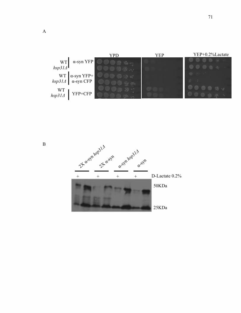
71
71
A
B
YFP+CFP
YEP+0.2%Lactate WT
hsp31Δ
WT hsp31Δ
WT hsp31Δ
YEP YPD
α-syn YFP+ α-syn CFP
α-syn YFP
50KDa
25KDa
D-Lactate 0.2%
2X α-syn hs
p31Δ
2X α-syn
α-syn hs
p31Δ
α-syn
+ + + +
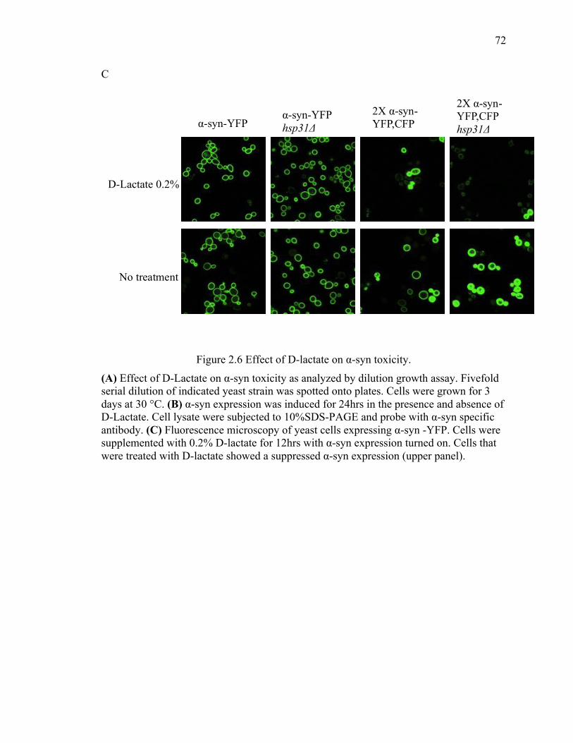
72
72
C
Figure 2.6 Effect of D-lactate on α-syn toxicity.
(A) Effect of D-Lactate on α-syn toxicity as analyzed by dilution growth assay. Fivefold serial dilution of indicated yeast strain was spotted onto plates. Cells were grown for 3 days at 30 °C. (B) α-syn expression was induced for 24hrs in the presence and absence of D-Lactate. Cell lysate were subjected to 10%SDS-PAGE and probe with α-syn specific antibody. (C) Fluorescence microscopy of yeast cells expressing α-syn -YFP. Cells were supplemented with 0.2% D-lactate for 12hrs with α-syn expression turned on. Cells that were treated with D-lactate showed a suppressed α-syn expression (upper panel).
D-Lactate 0.2%
2X α-syn-YFP,CFP hsp31Δ
2X α-syn-YFP,CFP
α-syn-YFP hsp31Δ α-syn-YFP
No treatment
73
73
2.3.4 Hsp31 prevents formation of large prion aggregates
The demonstration that Hsp31 possesses chaperone activity in a variety of anti-
aggregation assays prompted us to investigate if Hsp31 could also modulate the
oligomeric state of yeast prion proteins, such as the Sup35 prion. The aggregation state of
prions in living cells can be monitored by fusion of fluorescent protein tags to Sup35 PrD,
so we overexpressed Hsp31 to determine if it modulated the aggregation of Sup35 PrD.
Consistent with previous findings, cells overexpressing PrD in the presence of
endogenous Sup35 showed characteristic ribbon- like or punctate aggregates, which are
localized around the vacuoles or/and adjacent to plasma membranes. The cells co-
expressing pAG415-GPD-HSP31-DsRed and pAG424-GAL-PrD- Sup35-EYFP had
approximately 3 fold less Sup35 fluorescent aggregates compared with cells harboring
pAG424-GAL-PrD-Sup35-EYFP plus vector control, indicating that Hsp31 can inhibit
the formation of microscopically visible Sup35 prion aggregates (figure 2.7A-B). The
intrinsic fluorescence of Sup35-EYFP appears to increase upon aggregation, which
allowed us to utilize flow cytometry as an alternative method to quantitatively measure
and show decreased Sup35 aggregation. This phenomenon has not been exploited widely
but was noted in at least one previous study investigating the aggregation of a
Huntington-GFP fusion protein (309). Cell cultures co-expressing Sup35 and Hsp31 used
in the microscopy experiments were prepared for flow cytometry and analyzed for EYFP
fluorescence intensity. The flow cytometry results were similar to the microscopy results
because Hsp31 expressing cells consistently exhibited lower median Sup35-EYFP
fluorescence intensity when compared with cells containing Sup35-EYFP expression
plasmid and empty vector. Individual traces depicting the number of counting events for

74
74
each yeast strain also showed the decrease in cellular fluorescence intensity when Hsp31
is expressed (Figure 2.7).
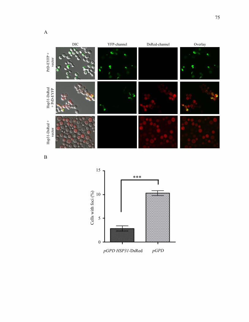
75
75
A
B
Hsp
31-D
sRed
+
vect
or
PrD
-EY
FP +
ve
ctor
H
sp31
-DsR
ed
+ Pr
D-E
YFP
DIC YFP-channel DsRed-channel Overlay
Cel
ls w
ith fo
ci (%
)
5
10
15
***
0
pGPD pGPD HSP31-DsRed
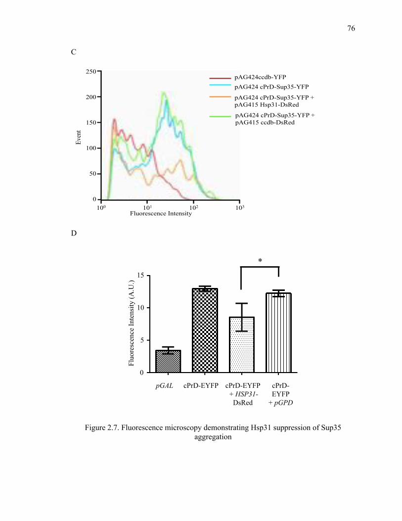
76
76
C
D
Figure 2.7. Fluorescence microscopy demonstrating Hsp31 suppression of Sup35 aggregation
100 101 102 103
0
50
100
150
200
250
Fluorescence Intensity
Even
t pAG424 cPrD-Sup35-YFP pAG424ccdb-YFP
pAG424 cPrD-Sup35-YFP + pAG415 Hsp31-DsRed
pAG424 cPrD-Sup35-YFP + pAG415 ccdb-DsRed
0
5
10
15
Fluo
resc
ence
Inte
nsity
(A.U
.)
cPrD-EYFP
+ pGPD
cPrD-EYFP cPrD-EYFP + HSP31-
DsRed
pGAL
*
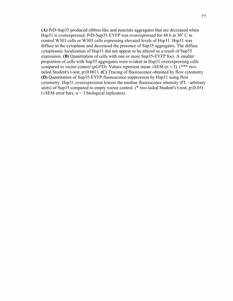
77
77
(A) PrD-Sup35 produced ribbon-like and punctate aggregates that are decreased when Hsp31 is overexpressed. PrD-Sup35-EYFP was overexpressed for 48 h at 30° C in control W303 cells or W303 cells expressing elevated levels of Hsp31. Hsp31 was diffuse in the cytoplasm and decreased the presence of Sup35 aggregates. The diffuse cytoplasmic localization of Hsp31 did not appear to be altered as a result of Sup35 expression. (B) Quantitation of cells with one or more Sup35-EYFP foci. A smaller proportion of cells with Sup35 aggregates were evident in Hsp31 overexpressing cells compared to vector control (pGPD). Values represent mean ±SEM (n = 3). (*** two-tailed Student's t-test; p≤0.001). (C) Tracing of fluorescence obtained by flow cytometry (D) Quantitation of Sup35-EYFP fluorescence suppression by Hsp31 using flow cytometry. Hsp31 overexpression lowers the median fluorescence intensity (FI – arbitrary units) of Sup35 compared to empty vector control. (* two-tailed Student's t-test; p≤0.05) (±SEM error bars; n = 3 biological replicates).
78
78
Expression of Hsp31 with the GPD promoter was diffuse and cytoplasmic, and varied in
intensity in individual cells across the total cell population. The cytoplasmic localization
profile of Hsp31 was not altered when PrD-Sup35 was expressed. Interestingly, we
observed overlapping localization in cells with Sup35 aggregates and relatively high
levels of Hsp31 but the proteins do not appear to mutually co-localize. In some cells it
was evident that the DsRed signal was decreased in the area where there was a Sup35
aggregate suggesting that Hsp31 is not incorporated in aggregates. The aggregate appears
to occlude Hsp31 and the diffuse overlapping signal observed in some cells is consistent
with the presence of Hsp31 throughout the cytoplasm surrounding the aggregate (Figure
2.8).
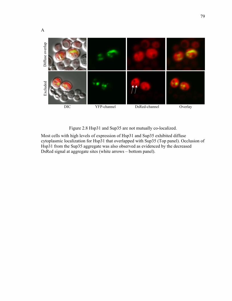
79
79
A
Figure 2.8 Hsp31 and Sup35 are not mutually co-localized.
Most cells with high levels of expression of Hsp31 and Sup35 exhibited diffuse cytoplasmic localization for Hsp31 that overlapped with Sup35 (Top panel). Occlusion of Hsp31 from the Sup35 aggregate was also observed as evidenced by the decreased DsRed signal at aggregate sites (white arrows – bottom panel).
DIC YFP-channel DsRed-channel Overlay
Diff
use
over
lap
Excl
uded
80
80
We also confirmed that Hsp31 overexpression reduces the formation of in vivo
aggregates based on assessing cell lysates by SDD-AGE. We found that SDS-resistant
Sup35 aggregates were greatly reduced when Hsp31 was over-expressed. The positive
control, Hsp104, completely eliminated detectable Sup35 aggregates. These results are
consistent with the ability of Hsp31 to inhibit the formation of large protein aggregates.
We also ascertained if other chaperones are induced upon overexpression of Hsp31. We
demonstrated Hsp70 and Hsp104 expression are not altered and hence elevated
expression of these chaperones is not responsible for the observed suppression of prion
aggregates (Figure 2.9). This is in agreement with our in vitro aggregation studies
establishing that purified and recombinant Hsp31 protein is solely sufficient to prevent
protein aggregation although we cannot exclude the possibility that Hsp31 could
collaborate with other chaperones to prevent prion aggregation in the cell.
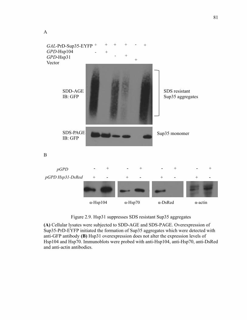
81
81
A
B
Figure 2.9. Hsp31 suppresses SDS resistant Sup35 aggregates (A) Cellular lysates were subjected to SDD-AGE and SDS-PAGE. Overexpression of Sup35-PrD-EYFP initiated the formation of Sup35 aggregates which were detected with anti-GFP antibody (B) Hsp31 overexpression does not alter the expression levels of Hsp104 and Hsp70. Immunoblots were probed with anti-Hsp104, anti-Hsp70, anti-DsRed and anti-actin antibodies.
pGPD Hsp31-DsRed
pGPD - +!
+ -!
α-Hsp104 α-actin α-Hsp70 α-DsRed
- +!
+ -!
- +!
+ -!
- +!
+ -!
SDD-AGE IB: GFP
SDS-PAGE IB: GFP
SDS resistant Sup35 aggregates
Sup35 monomer
GAL-PrD-Sup35-EYFP GPD-Hsp104 GPD-Hsp31 Vector
+ + + + - +
+ - + -
+
82
82
2.3.5 Hsp31 rescue of α-syn -mediated toxicity is independent of the autophagy
pathway
In several recent studies, autophagy has been identified as one of the main mechanisms to
clear and recycle misfolded proteins and aggregates in the cytosol (310,311). In addition
to the ubiquitin proteasome system, autophagy is a major cellular function involved in
clearance of misfolded and aggregated proteins including α-syn aggregates (310).
Deletion of autophagy genes such as ATG5 or ATG7 leads to neurodegenerative disease
in mice (312,313). In yeast, deletion of ATG1 gene that is required for induction of
autophagy reduced the clearance of α-syn aggregates (314). Deletion of HSP31 was also
demonstrated to impair autophagy under carbon starvation conditions (152). These
studies raise the question that Hsp31 could be promoting autophagy of aggregated protein.
In order to exclude the possibility that the Hsp31 rescue effect on α-syn toxicity depends
on autophagy, we deleted the ATG8 gene in the α-syn strains and assessed the synthetic
lethal effects of α-syn expression in these strains. As expected, expression of α-syn in the
atg8Δ strain resulted in decreased viability compared to the wild-type strain. The atg8Δ
hsp31Δ strain had similar viability to the atg8Δ strain when α-syn is expressed. These
results confirm that autophagy is a protective mechanism against α-syn toxicity. The lack
of synthetic lethal defect between ATG8 and HSP31 with and without α-syn expression is
consistent with genes that are in the same pathway and hence do not buffer each other
(Figure 2.9).
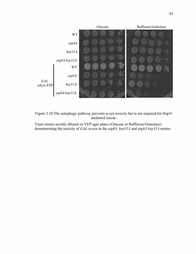
83
83
Figure 2.10 The autophagy pathway prevents α-syn toxicity but is not required for Hsp31 mediated rescue.
Yeast strains serially diluted on YEP agar plates (Glucose or Raffinose/Galactose) demonstrating the toxicity of GAL-α-syn in the atg8Δ, hsp31Δ and atg8Δ hsp31Δ strains.
Raffinose/Galactose Glucose
hsp31Δ
atg8Δ
atg8Δ hsp31Δ
WT
hsp31Δ
atg8Δ
atg8Δ hsp31Δ
WT
GAL αSyn-YFP
84
84
Expression of Hsp31 and the C138D mutant in the autophagy strains, atg8Δ α-syn or
atg8Δ hsp31Δ α-syn, increased viability in rich YEP media compared to vector controls.
We found that the rescue effect in synthetic media was not easily observed because
expression of α-syn was not toxic in the atg8Δ strain background but it was toxic in
hsp31Δ indicating media conditions are an important factor in this complex relationship.
These results demonstrate that Hsp31 can rescue α-syn toxicity in autophagy deficient
cells (Figure 2.10).
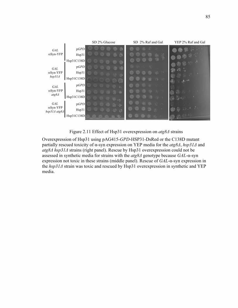
85
85
Figure 2.11 Effect of Hsp31 overexpression on atg8Δ strains Overexpression of Hsp31 using pAG415-GPD-HSP31-DsRed or the C138D mutant partially rescued toxicity of α-syn expression on YEP media for the atg8Δ, hsp31Δ and atg8Δ hsp31Δ strains (right panel). Rescue by Hsp31 overexpression could not be assessed in synthetic media for strains with the atg8Δ genotype because GAL-α-syn expression not toxic in these strains (middle panel). Rescue of GAL-α-syn expression in the hsp31Δ strain was toxic and rescued by Hsp31 overexpression in synthetic and YEP media.
SD 2% Glucose SD 2% Raf and Gal YEP 2% Raf and Gal
pGPD
Hsp31
Hsp31C138D
pGPD
Hsp31
Hsp31C138D
pGPD
Hsp31
Hsp31C138D
pGPD
Hsp31
Hsp31C138D
GAL αSyn-YFP
hsp31Δ atg8Δ
GAL αSyn-YFP
atg8Δ
GAL αSyn-YFP
hsp31Δ
GAL αSyn-YFP
86
86
2.4 Methods
2.4.1 Yeast cell growth conditions
Yeast media were prepared according to Amberg D C. et al. (315). Liquid yeast
extract/peptone dextrose (YPD) medium contained Bacto yeast extract (1%, Fisher
Scientific), Bacto peptone (2% Fisher Scientific) and glucose (2% Fisher Scientific).
Synthetic dextrose (SD) minimum liquid medium was made of 0.17% Difco yeast
nitrogen base (without amino acids), 2% glucose, and supplemented with necessary
amino acids for auxotrophic strains needed at concentrations described previously (315).
Solid medium plates were made with the same components of liquid medium plus 2%
agar (Fisher Scientific). To express galactose-inducible proteins, 2% raffinose
(Affymetrix, Cleveland OH) and 2% galactose (Affymetrix, Cleveland OH) were used to
replace glucose. Fractions of culture were obtained at designated times to monitor the cell
fitness and protein levels by OD600 and immunoblotting, respectively.
2.4.2 Spotting assay / Dilution growth assays
All the yeast strains were grown at 30 °C in 2% glucose minimal media overnight.
Strains were diluted to OD600 of 0.2 and grown in minimal media switching the carbon
source with 2% raffinose for overnight. Cultures were normalized to OD600 of 0.8 and 5X
serial dilutions were spotted onto the respected dropout plate containing 2% glucose or
Yeast strains construction galactose. Plates were grown at 30 °C for 2-3 days before
taking pictures.
2.4.3 Yeast strains construction
Yeast strains used in this study are listed in Table 2.1. The hsp31Δ and atg8Δ deletions
were generated in W303-1A or in a strain expressing one or two copies α-syn fused to
87
87
fluorescent proteins (CFP and YFP) using the primers listed in Table 2.2. The
nourseothricin N- acetyl-transferase (NAT) gene flanked by Hsp31 homology regions
was obtained by PCR with 70 nucleotide long forward primer and reverse primer. The
primers consisted of 20 nucleotides for amplifying the NAT gene from pFA6a-natNT2
(Euroscarf), and 50 nucleotides immediately preceding the Hsp31 or Atg8 start codon or
after the stop codon. The amplified product was integrated into W303 α-syn expressing
strains at ChrIV:1502160 to 1501447 as described previously. For double knockout
hsp31Δ atg8Δ strain KanMX4 resistance cassette was used to delete Hsp31 and than
NATMX4 resistance cassette for deletion of ATG8.
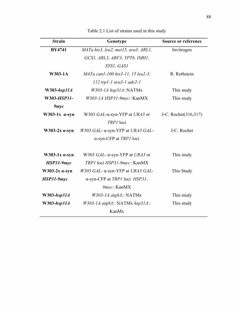
88
88
Table 2.1 List of strains used in this study
Strain Genotype Source or reference
BY4741 MATa his3, leu2, met15, ura3, ARL1,
GCS1, ARL3, ARF3, YPT6, IMH1,
SYS1, GAS1
Invitrogen
W303-1A MATa can1-100 his3-11, 15 leu2-3,
112 trp1-1 ura3-1 ade2-1
R. Rothstein
W303-hsp31Δ W303-1A hsp31Δ::NATMx This study
W303-HSP31-
9myc
W303-1A HSP31-9myc::KanMX This study
W303-1x α-syn W303 GAL-α-syn-YFP at URA3 or
TRP1 loci
J-C. Rochet(316,317)
W303-2x α-syn W303 GAL- α-syn-YFP at URA3 GAL-
α-syn-CFP at TRP1 loci
J-C. Rochet
W303-1x α-syn
HSP31-9myc
W303 GAL- α-syn-YFP at URA3 or
TRP1 loci HSP31-9myc::KanMX
This study
W303-2x α-syn
HSP31-9myc
W303 GAL- α-syn-YFP at URA3 GAL-
α-syn-CFP at TRP1 loci HSP31-
9myc::KanMX
This Study
W303-hsp31Δ W303-1A atg8Δ:: NATMx This study
W303-hsp31Δ W303-1A atg8Δ:: NATMx hsp31Δ::
KanMx
This study
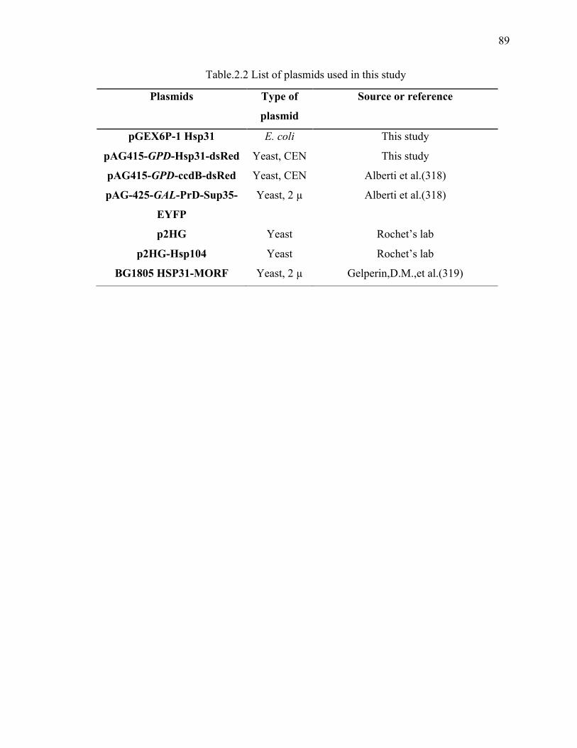
89
89
Table.2.2 List of plasmids used in this study
Plasmids Type of
plasmid
Source or reference
pGEX6P-1 Hsp31 E. coli This study
pAG415-GPD-Hsp31-dsRed Yeast, CEN This study
pAG415-GPD-ccdB-dsRed Yeast, CEN Alberti et al.(318)
pAG-425-GAL-PrD-Sup35-
EYFP
Yeast, 2 µ Alberti et al.(318)
p2HG Yeast Rochet’s lab
p2HG-Hsp104 Yeast Rochet’s lab
BG1805 HSP31-MORF Yeast, 2 µ Gelperin,D.M.,et al.(319)
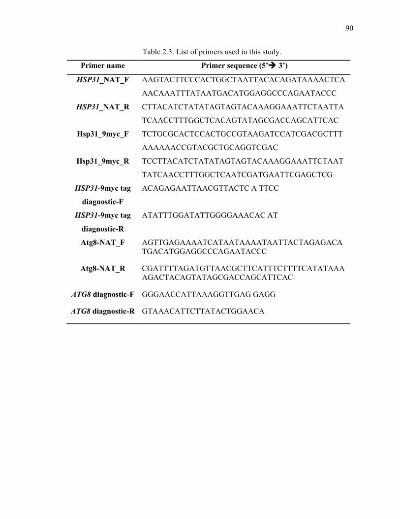
90
90
Table 2.3. List of primers used in this study.
Primer name Primer sequence (5’! 3’)
HSP31_NAT_F AAGTACTTCCCACTGGCTAATTACACAGATAAAACTCA
AACAAATTTATAATGACATGGAGGCCCAGAATACCC
HSP31_NAT_R CTTACATCTATATAGTAGTACAAAGGAAATTCTAATTA
TCAACCTTTGGCTCACAGTATAGCGACCAGCATTCAC
Hsp31_9myc_F TCTGCGCACTCCACTGCCGTAAGATCCATCGACGCTTT
AAAAAACCGTACGCTGCAGGTCGAC
Hsp31_9myc_R TCCTTACATCTATATAGTAGTACAAAGGAAATTCTAAT
TATCAACCTTTGGCTCAATCGATGAATTCGAGCTCG
HSP31-9myc tag
diagnostic-F
ACAGAGAATTAACGTTACTC A TTCC
HSP31-9myc tag
diagnostic-R
ATATTTGGATATTGGGGAAACAC AT
Atg8-NAT_F AGTTGAGAAAATCATAATAAAATAATTACTAGAGACATGACATGGAGGCCCAGAATACCC
Atg8-NAT_R CGATTTTAGATGTTAACGCTTCATTTCTTTTCATATAAAAGACTACAGTATAGCGACCAGCATTCAC
ATG8 diagnostic-F GGGAACCATTAAAGGTTGAG GAGG
ATG8 diagnostic-R GTAAACATTCTTATACTGGAACA
91
91
2.4.4 HSP31 9myc tagging
We endogenously tagged HSP31 with 9myc epitope using a PCR- based integration. The
pYM20 plasmid was used as template and appropriate primer set to obtain PCR product
with HSP31 genomic flanking sequence and the PCR product was transformed into
W303 and W303 α-syn-CFP + α-syn-YFP strains. The transformants were selected on
media containing hygromycin B (300mg/L) and correct integration was verified by PCR
using primers spanning the integration junctions and by DNA sequence analysis. DNA
manipulation - The plasmids used in this study are listed in Table 2.2. Hsp31 was shuttled
into pAG415- GPD-ccdB-DsRed (Addgene Cambridge MA (318) from pDONR221 with
LR clonase (Invitrogen, Grand Island, NY) to produce pAG415-GPD- HSP31-DsRed.
Both DJ-1 and Hsp31 were cloned into BamHI/XhoI sites of pGEX 6p-1. pESC-Leu
myc-HSP31 was constructed by cloning at the XhoI/NheI sites. The HSP31 gene was
amplified from yeast genomic DNA. The Hsp31 C138D mutant was prepared by PCR
amplification using pAG415-GPD-Hsp31-DsRed as template and set of primers listed in
Table 2.3 Each successfully mutated insert was sequenced to confirm the mutation.
2.4.5 Antibodies and immunoblotting
To determine protein expression yeast cells were collected by centrifuging at 5000 rpm
and pellets were resuspended in extraction buffer (50mM Tris-HCl pH 7.5, 1mM EDTA,
4mM MgCl2; 5mM DTT). Glass beads (Sigma, St. Louis, MO) were added into the
mixture, which was vortex 5 times for 10 s each, plus 1 min intervals on ice. Clear crude
protein lysate was obtained by spinning down the cell debris at 1000 xg at 4 °C for 10
min. The SDS- PAGE loading dye (4 % SDS, 40 % glycerol, 0.02% bromophenol blue,
Tris-Cl, pH6.8) was added to supernatant and samples were boiled, followed by SDS-
92
92
PAGE and immunoblotting with antibodies. Monoclonal anti-myc and anti-β-actin
antibody were purchased from Sigma-Aldrich (St. Louis, MO). Monoclonal anti-α-syn
was obtained from BD biosciences (San Jose, CA). The following antibodies were also
used to detect expression of Hsp104 (Abcam; ab69549), Hsp70 (Stressgen; SPA-82);
DsRed (Santa Cruz Biotechnology; Sc-33353) and GFP (Roche; anti- GFP).
2.4.6 Protein purification
BY4741 harboring yeast Hsp31 expression plasmid from the yeast moveable ORF
collection (Thermo Fisher Scientific Open Biosystems, Huntsville, AL) was used to
express and purify the protein. We did not see any evidence of co-purifying proteins on
SDS-PAGE gels and observed similar high activity from protein purified under high salt
conditions, which minimizes contaminant co-purifying proteins. Human DJ-1 was
encoded in pGEX 6p-1 (GE Healthcare Life Sciences), hchA was encoded in pNT-hchA,
and α-syn was expressed from the pT7 plasmid. The protein expression and purification
was described previously. Briefly, constructs were transformed into BL21 (DE3) cells.
The transformants were grown to an OD600 of 0.4 – 0.6 in LB medium supplemented with
100 µg/ml ampicillin at 37 °C, and Isopropyl β-D-1- thiogalactopyranoside (IPTG) (1mM
as final concentration) was added to induce protein expression. After 3 h of induction at
37 °C, cells were harvested by centrifugation at 2,000xg, and re-suspended in lysis buffer
(25 mM KPi pH 7.0, 200mM KCl) containing protease inhibitor cocktail set IV
(Calbiochem, Billerica, MA). Crude protein was prepared by sonicating cells and
clarifying the lysate by centrifugation at 10,000xg for 10 min. GST tagged DJ-1 was
immobilized by Glutathione agarose resin (Thermo scientific) and DJ-1 was eluted by
cleaving from the GST tag with PreScission Protease (GE Healthcare Life Sciences,
93
93
Pittsburgh, PA) (2 units of protease for every 100 µg of tagged DJ-1) leaving a linker
amino acid sequence: (NPAFLYKVVDVSRHHHGRIFYPYDVPDYAG LEVLFQ)
The protein was approximately 95% pure based on band intensity coomassie blue
staining with SDS- PAGE.
2.4.7 Dilution growth assays
Plasmids pAG415-GPD- ccdB-DsRed and pAG415-GPD-HSP31-DsRed were
transformed into W303 α-syn expressing strains with or without genomic copy of the
HSP31 gene knocked out using the PEG/lithium acetate transformation method. Single
colonies of transformants were grown in SD-Leu overnight, the cells were washed,
normalized and grown in synthetic complete without leucine (SC-Leu) + raffinose (2%)
to switch the carbon source. After incubating in the raffinose medium over night, the cell
number was normalized, and spotted on SD-Leu (suppressed expression) plate and SC-
Leu with 2% raffinose and 2% galactose (induced expression) with five-fold serial
dilutions. The plates were incubated at 30 °C for 3 days.
2.4.8 Fluorescence imaging analysis α-syn localization
pESC-Leu myc-Hsp31 and control empty vector were transformed into 2 copies of α-syn
expressing strain. Transformants were inoculated and grown as described in the growth
dilution assay. The α-syn expression was induced by growing the cells in synthetic
minimum medium with 2% raffinose and galactose for 8 h. The live cells were visualized
using Nikon TE2000-U inverted fluorescence microscope with Nikon Plan apochromat
60X (NA 1.4) oil immersion objective and YFP filters plus 300X magnification. In order
to differentiate the localization of protein, approximately 50 individual cells were
randomly counted in multiple regions of interest for each set of experiment. The ratio of
94
94
membrane to cytoplasmic localization of α-syn fluorescent fusion protein was presented
as the mean from three independent sets of experiments.
2.4.9 MGO addition and microscopy
α-syn expressing strains with or without the Hsp31 gene deleted were grown in YPD
media and transferred into media containing 2% galactose and MGO (0.5mM-2mM) for
6 h and 12 h. Cells were washed and subjected to confocal microscopy to observe α-syn
foci formation. The presence of MGO inhibited the growth of cells for the initial 12 h and
resulted in equal growth rate between strains regardless of the Hsp31 gene deletion.
Strains could not be grown in the presence of high concentrations of MGO (20mM) so α-
syn was induced for 10 h and then MGO was added and incubated for another 2 h before
confocal microscopy analysis.
2.4.10 Assessment of intracellular ROS
Cells were harvested after induction of α-syn expression for 12 h and 5 × 10⁶ of cells
were prepared to stain with dihydroethidium (DHE). Cells were suspended in 250 µl of
2.5 µg/ml DHE in PBS, and incubated in the dark for 10 min. Cells were washed in the
PBS and were subjected to fluorescence microscopy (excitation at 485 nm and emission
at 520 nm) and flow cytometry with FL-2 channel. FlowJo software was used to calculate
median fluorescence intensity.
2.4.11 Prion expression experiments
W303 WT strain was co-transformed with pAG424-GAL- PrD-Sup35-EYFP and
pAG415-GPD-HSP31-DsRed. Single transformations were also performed using the
same constructs and their corresponding control vector. Cells were grown at 30°C in
appropriate medium overnight (SD-Trp for pAG424, SD-Leu for pAG415 and SD-Trp-
95
95
Leu for co-transformants) and protein expression was induced for 24 h in SC 2%
Raffinose and 2% Galactose containing medium at 30 °C. After induction, cells were
subjected to fluorescence microscopy using a Nikon A1 confocal microscope with a
Nikon Plan apochromat 60X (NA 1.4) oil immersion objective to acquire fluorescence
and DIC images and were analyzed using Image J. The identical cultures used in
microscopy were used to prepare samples for flow cytometry, cells were washed and re-
suspended in PBS at a density of ~10x106 cells/ml. Cells were filtered and analyzed for
EYFP fluorescence intensity using a flow cytometer with the FL-1 channel. A total of
10,000 events were acquired for each sample and data was analyzed using FlowJo
software to calculate median fluorescence intensity after three biological replicates.
2.4.12 Glutathione-independent glyoxalase biochemical Assay
Purified recombinant Hsp31 and Hsp31 C138D with GST tag removed were used to
assay methylglyoxalase activities. The reaction was initiated by adding specified
concentration of proteins (1 µM Hsp31 and 5 µM Hsp31C138D) in reaction buffer (100
mM HEPES, pH 7.5, 50 mM KCl, 2 mM DTT) to 6 mM initial concentration of MGO
(Sigma, 40% solution), followed by incubation at 30 °C. The assay was performed, by
removing 50µl aliquots of the reaction at fixed time points (15, 30, 45, and 120 s) after
addition of protein. The amount of D-lactic acid produced by Hsp31 as a result of MGO
consumption was measured. The initial rate obtained was divided by amount of protein in
the reaction mixture to calculate the specific activity. Samples were also subjected to Gas
Chromatography-Mass Spectroscopy analysis (Agilent 5975C MSD) to identify the
presence of D-lactic acid. Samples were derivatized with N, O-bis (trimethylsilyl)
trifluoroacetamide) and trimethylchlorosilane and heated to 50 °C for 10 min and run in
96
96
the electron impact mode with scanning from 42-400 atomic mass units. A lactic acid
standard displayed a peak at 6.09 min identical to the peak detected in the Hsp31-treated
sample whereas Hsp31C138D did not display this peak. The spectral scan was visualized
and displayed using the OpenChrom software (320). To obtain the enzymatic parameters,
the reaction was initiated as described above by adding 1 µM Hsp31 or 5 µM
Hsp31C138D protein into reaction buffer and reactions were stopped after 1 min by
heating at 85 °C for 30 s. A range of MGO substrate from 50 µM to 2 mM and the
EnzyFluo D-Lactate Assay kit (Bioassay System) was used to determine the amount of
D-lactic acid produced in each reaction. Each rate was determined in triplicate and mean
values were plotted to obtain enzymatic parameters (Vmax and Km) by fitting to
michaelis-menten model using Graphpad prism.
2.4.13 Semi-denaturing detergent-agarose gel electrophoresis (SDD-AGE)
W303 cells harboring pAG424-GAL-cPrDSup35-EYFP and pAG415-GPD-HSP31-
DsRed plasmids were used and aggregates produced by inducing for 24 h in SC 2%
raffinose + 2% galactose media. Prion aggregates were analyzed using SDD-AGE (321).
Briefly, to prepare lysates, cells were harvested by centrifugation at 3,000'g for 2'min and
re- suspended in spheroplast solution (1.2 M D-sorbitol, 0.5 mM MgCl2, 20 mM Tris (pH
7.5), 50 mM β-mercaptoethanol and 0.5 mg/ml Zymolyase and incubated at 30 °C for 30
min with shaking. After spheroplast formation the samples were centrifuged at 800 rpm
for 5 min at room temperature and supernatant was removed. The pelleted spheroplasts
were resuspended into 100 µL lysis buffer (100 mM Tris 7.5, 50 mM NaCl, 10 mM β-
mercaptoethanol and protease inhibitor and vortexed at high speed for 2 min. The lysates
were collected and mixed with 4 x sample buffer (2 x TAE; 20 % glycerol; 4 % SDS;
97
97
0.01% bromophenol blue). The samples were incubated at room temperature for 15 min
and loaded onto a 1.8 % agarose gel containing 1 x TAE and 0.1 % SDS and run at 50 V,
followed by transfer onto nitrocellulose membrane using capillary transfer method. The
nitrocellulose membrane was subjected to western blot analysis using anti-GFP antibody
(Roche). A strain harboring p2HG-Hsp104 plasmid was included as a positive control.
2.5 Discussion
DJ-1/ThiJ/PfpI superfamily members composed of structurally similar proteins that are
present in prokaryotes and eukaryotes. Often these proteins are associated with stress
response. However, structural and biochemical studies have proven that they are diverse
in functions with the ability to perform multiple activities that include chaperone,
protease, methylglyoxalase, deglycase and role in autophagy. In this study we aimed to
delineate the functions of Hsp31 by using yeast as a model. Recently, we established that
Hsp31 is a potent chaperone that can inhibit the aggregate formation of large number of
proteins such as insulin, citrate synthase as well as prevent α-syn fibrillization in vitro
and α-syn toxicity in vivo. Because Hsp31 is a multifunctional protein, we sought to
identify the interaction between different functional pathways of Hsp31 and define its
role in protecting against cellular stress. We also obtained insight into the role of Hsp31
in Sup35 prion aggregations.
2.5.1 Role of Hsp31 in redox homeostasis
Previous studies showed the protective effect of Hsp31 under oxidative and proteotoxic
stress in that overexpression of Hsp31 protect yeast cell viability under these conditions
98
98
(260,303). In the past, our lab has shown that Hsp31 protects cells from α-syn mediated
toxicity. These observations led us to explore the expression profile of Hsp31 in response
to oxidative and proteotoxic stress. A genomically tagged strain shows an increase in
Hsp31 expression under both oxidative and proteotoxic stress (Figure 2.1). Previous
studies have shown that an increased level of Hsp31 at the diauxic shift and during
stationary phase of growth cycle. These phases in the growth cycle are associated with
stress due to accumulation of toxic metabolites and lack of nutrients (322). An elevated
level of Hsp31 at later phases of growth indicates that it could possibly be due to the
stress associated with these phases. HSP31 expression is also induced when yeast cells
are treated with H2O2 to produce oxidative stress and this up-regulation of HSP31 in
response to ROS has been reported to be under the control of stress responsive
transcription factor Yap1, as removal of Yap1 binding site (-363 and -353 bp relative to
the ATG) from HSP31 promoter has shown to reduce Hsp31 expression under oxidative
stress (303). However, we observed that this shortened promoter also eliminates another
stress response element (STRE, a CCCCT site at -379 to -383 from the ATG) in addition
to the Yap1 binding site. STREs are the binding site for Msn2/4, and several other stress-
related yeast genes have been documented to be transcriptionally activated by these
transcription factors, including genes up-regulated after the diauxic shift in stationary
phase cells (303).
Finally, we also reported an increase in ROS levels upon HSP31 deletion that correlates
with increased toxicity by α-syn expression compared to WT strains, as level of
superoxide was higher when we overexpressed α-syn indicating that the presence of
99
99
Hsp31 is important in reducing ROS to basal levels. A previous study has shown that α-
syn expression is associated with caspase-mediated ROS generation (323) (Figure 2.2). In
agreement with our study, overexpression of HSP31 robustly suppresses both cytosolic
and mitochondrial ROS levels instigated by MGO and H2O2 and therefore provide
cytoprotection. In addition, Hsp31 localizes to mitochondria and preserves mitochondrial
integrity by redistributing glutathione to the cytoplasm under oxidative stress (260).
Another study examined the deglycase activity of Hsp31 and showed that it may
efficiently deglycate proteins with glycated Cys, Arg and Lys amino acid residues (265).
Taken together, these results suggest that Hsp31 is an integral part of the heat shock
protein system and plays a vital role in maintaining cellular homeostasis. These results
establish that Hsp31 has a role in response to ROS and its expression is induced in
response to several types of stress.
2.5.2 Hsp31 chaperone activity is independent of its methylglyoxalase activity
Recently in our lab we demonstrated the robust chaperone activity of Hsp31 that can
rescue the α-syn toxicity in vivo. A question was raised that the in vivo rescue effects
could have been mediated by the Hsp31 methylglyoxalase activity or interaction with
other biological pathways such as the metabolic or autophagy pathway. First we
confirmed that Hsp31 does have methylglyoxalase activity and demonstrated that
mutation of the catalytic triad residue, C138D, results in greatly reduced
methylglyoxalase activity (Figure 2.3). However, this mutation did not inactivate the
chaperone activity in terms of rescuing the cells from α-syn-mediated toxicity when
expressed under GPD-HSP31 C138D system (Figure 2.4). Furthermore, we showed that
100
100
that exogenous MGO did not differentially affect viability suggesting that Hsp31 does not
rescue by reducing accumulation of this toxic metabolite in α-syn expressing cells
(Figure 2.5). Another study also reported that mutation in the catalytic triad largely
abolishes glyoxalase activity but this catalytic triad is not required for chaperone activity
of Hsp31 (260). These results indicate that the anti-aggregation activity of Hsp31 is not
under the influence of its known enzymatic activity rather; it has a direct chaperone
activity against misfolded proteins. Intriguingly, all the paralogs of the Hsp31 minifamily
possess comparable activity against α-syn aggregation and toxicity when they are
overexpressed from the GAL promoter (106). However, unlike Hsp31, the other paralogs
possess very little methylglyoxalase activity and are unable to protect the cells from
glyoxal toxicity (260). Furthermore, the chaperone activity of Hsp31, Hsp32 and Hsp33
against a cytoplasmic aggregation-prone protein is independent of their role in oxidative
stress response and the vacuolar degradation pathway (262). These results again support
the notion that anti-aggregation activity of Hsp31 mini-family is independent of its
enzymatic activity.
2.5.3 Autophagy pathway is not essential for chaperone activity of Hsp31
We also demonstrated the autophagy pathway is an important mediator in the α-syn
toxicity because deletion of ATG8 resulted in synthetic lethal interaction with α-syn
expression (Figure 2.10). This is consistent with atg1Δ or atg7Δ strains having synthetic
lethal interactions with α-syn expression and having defects in clearing α-syn foci (324).
However, we show that hsp31Δ atg8Δ strain does not have increased α-syn- mediated
toxicity when compared to atg8Δ, consistent with these genes occurring within the same
101
101
synthetic lethal pathway. A recent result indicates that hsp31Δ strains are defective in
autophagy (301) so the additional deletion of an autophagy gene should not increase
toxicity. Interestingly, overexpression of Hsp31 in these autophagy deficient strains can
rescue cells from α-syn toxicity indicating that autophagy pathway is not essential for
chaperone rescue (Figure 2.11). Autophagy may be beneficial for controlling α-syn
toxicity but it appears that chaperone activity of Hsp31 is also important.
2.5.4 Hsp31 inhibits Sup35 prion aggregation
Several orthologous experiments suggest that Hsp31 intervenes early in the misfolding
process and prevents the formation of larger oligomeric species. Previously, we showed
that the addition of Hsp31 to α-syn monomers in a fibrillization assay resulted in a
baseline level of ThioT fluorescence signal indicating that the formation of larger
oligomeric species was prevented. Analysis of the SDS-resistant oligomeric species by
SDS-PAGE demonstrated that the presence of Hsp31 prevented the formation of higher
order oligomeric α-syn species. Here, our studies demonstrate the ability of Hsp31 to
prevent prion aggregation in vivo and in vitro. Our results indicate that Hsp31 inhibits
prion assembly before the formation of visible subcellular YFP-tagged PrD aggregates
and those detected by SDD-AGE (Figure 2.7, Figure 2.9). Furthermore, the Sup35
aggregates occlude Hsp31, indicating that the anti-aggregation activity of Hsp31
commences prior to the formation of the visible aggregate and probably does not act to
remove or disassemble any preformed aggregates (Figure 2.8). This is in contrast to large
chaperones, Hsp104, Ssa1/2, Sis1 and Sse1, which mutually co-localize with prion
aggregates (325). A previous study has demonstrated that small HSPs can inhibit

102
102
formation of Sup35 aggregates and found that Hsp26 and Hsp42 inhibit rare transient
oligomers at distinctly different steps in the prion formation process (53). Further studies
are done to determine Hsp31 role in inhibiting prion aggregation. These results are listed
in Chapter 3. Overall, we demonstrate that Hsp31 can inhibit oligomerization or
aggregation of α-syn and Sup35 by intervening early in the process.
2.5.5 Yeast purified MORF-Hsp31 is more potent than recombinant Hsp31 or DJ-1
Intriguingly, Hsp31 purified directly from a yeast expression system, the GAL promoter
induced movable ORF tag system (MORF) (319), had increased enzymatic activity
compared to Hsp31 purified from recombinant E. coli (Figure 2.3). We also
demonstrated that the yeast-purified protein was more active in preventing aggregation of
several substrate proteins including α-syn. The increased activity may be due to the
difference in the affinity tags used or fusion tag orientation, that has been previously
observed for E coli Hsp31 (256,271) but, it may be a result of posttranslational
modification(s) that occur in the cell. Several reports have indicated that post-
translational modifications or differing levels of oxidation of the cysteine residue can
alter activity of DJ-1 (297,326). We also show that Hsp31 is a more potent
methylglyoxalase compared to DJ-1 consistent with several other studies (Figure 2.3)
(260,261). In addition, we have shown that yeast Hsp31 is more active in preventing
protein aggregation compared to DJ-1. Interestingly, it has also been observed that
Hsp31 rescues α-syn and Huntingtin’s toxicity to a greater extent than DJ-1 in vivo
(106,299). These results raise the intriguing possibility that Hsp31 is constitutively active
whereas DJ-1 must undergo an activation event to increase its activity.
103
103
2.6 Conclusion and future directions
Recent studies have demonstrated the facility of yeast to investigate pathogenic
mechanisms underlying α-syn toxicity including the identification of novel biological
pathways that impinge on α-syn biology and small molecule modulators of α-syn toxicity
(314,327). In addition, the action of Hsp31 on α-syn may provide insight into the mode of
toxicity of α-syn because recent evidence suggests that the α-syn toxic species is smaller
than the visible aggregate (314).
Our results together with other recent findings demonstrate the multitasking ability of
Hsp31, which is particularly important during stressful situations. It functions as a stress
response chaperone, glutathione independent methylglyoxalase, has a role in the
autophagy pathway and acts as a deglycase. Other possible functions have been observed
for this superfamily including a report of RNA binding for DJ-1 and protease activity for
other family members (328-330). These multiple functions can modulate the protein
misfolding and stress pathways at various points in the cellular network but our results
also highlight that Hsp31 has the ability to inhibit protein aggregation distinct of its
enzymatic activity (Figure 2.12). We believe that Hsp31 acts at the initial phases of
protein misfolding process and prevents the formation of larger aggregates but does not
possess disaggregase activity.
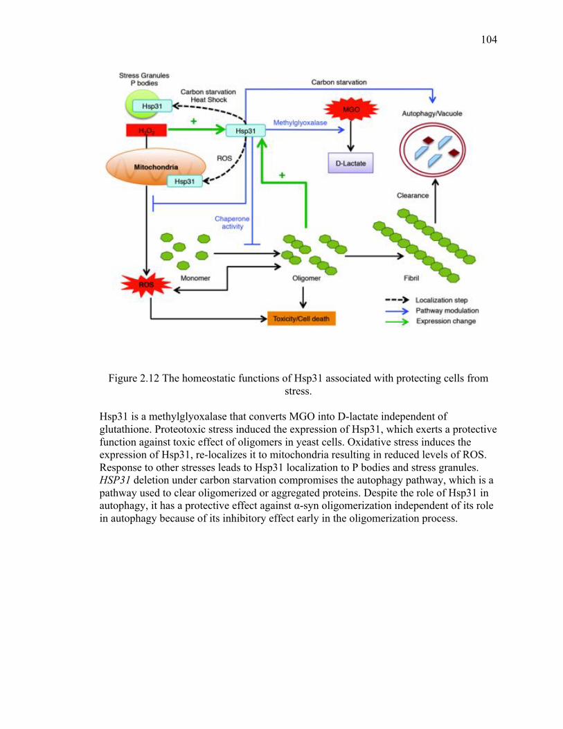
104
104
Figure 2.12 The homeostatic functions of Hsp31 associated with protecting cells from stress.
Hsp31 is a methylglyoxalase that converts MGO into D-lactate independent of glutathione. Proteotoxic stress induced the expression of Hsp31, which exerts a protective function against toxic effect of oligomers in yeast cells. Oxidative stress induces the expression of Hsp31, re-localizes it to mitochondria resulting in reduced levels of ROS. Response to other stresses leads to Hsp31 localization to P bodies and stress granules. HSP31 deletion under carbon starvation compromises the autophagy pathway, which is a pathway used to clear oligomerized or aggregated proteins. Despite the role of Hsp31 in autophagy, it has a protective effect against α-syn oligomerization independent of its role in autophagy because of its inhibitory effect early in the oligomerization process.
105
105
We have established a framework that extends the yeast model for investigating
mechanisms of α-syn toxicity in the context of the DJ-1/ThiJ/PfpI superfamily in yeast.
Extension of this work will assist elucidating the chaperone-like mechanisms of Hsp31,
and a comparison with DJ-1 may provide evolutionary insights into the activities of the
DJ-1/PfpI/ThiJ superfamily family. Our model may also be used to further delineate the
nature of oligomeric species in the pathogenesis of PD and possibly what species of α-
syn should be targeted therapeutically.
Given the apparent functional diversity of Hsp31 revealed so far, it is likely that there
might be other chaperone dependent and independent functions of this protein that may
exist. Many HSPs work in collaboration with other chaperone in order to be fully active.
The exploration and identification of protein-protein interaction partners of Hsp31 would
provide insight on mechanism and roles of Hsp31. There is s dearth of protein-protein
interaction information for Hsp31 although it was reported to interact with other
chaperones according to a large-scale proteomic study (28). Deletion of HSP31 down-
regulates the Ssa3, a Hsp70 paralog, mRNA level at stationary phase suggesting a
correlation between Hsp31 and Hsp70 activity (114). Human homolog DJ-1 is known to
interact with many chaperones including Hsp70 and mitochondrial Hsp70 indicating that
translocation of DJ-1 to mitochondria depends on these chaperones (304). A distinct
possibility is that relocation of Hsp31 to mitochondria, P bodies or stress granules under
oxidative stress is dependent on interactions with other chaperones. Our data support that
Hsp31 acts at early stages of protein aggregation do not delineate the stage of
intervention and therefore further investigation is needed. Hsp31 may interact with

106
106
unfolded monomers to sequester them from progressing to oligomers or alternatively, it
might become active only after smaller oligomers are formed.
107
107
CHAPTER 3. THE SMALL HEAT SHOCK PROTEIN HSP31 COOPERATES WITH HSP104 TO MODULATE THE SUP35 PRION
3.1 Abstract
The yeast homolog of DJ-1, Hsp31, is a multifunctional protein that is involved in several
cellular pathways including detoxification of the toxic metabolite MGO and as a protein
deglycase. Prior studies ascribed Hsp31 as a molecular chaperone that can inhibit α-syn
aggregation in vitro and alleviate its toxicity in vivo. It was also shown that Hsp31
inhibits Sup35 aggregate formation in yeast. However, it is unknown if Hsp31 can
modulate [PSI+] phenotype and Sup35 prionogenesis. Other small heat shock proteins
(sHSPs), Hsp26 and Hsp42 are known to be a part of a synergistic proteostasis network
that inhibits Sup35 prion formation and promotes its disaggregation. Here, we establish
that Hsp31 inhibits Sup35 [PSI+] prion formation in collaboration with a well-known
disaggregase, Hsp104. Hsp31 transiently prevents prion induction but does not suppress
induction upon prolonged expression of Sup35 indicating that Hsp31 can be overcome by
larger aggregates. In addition, elevated levels of Hsp31 do not cure [PSI+] strains
indicating that Hsp31 cannot intervene in a pre-existing prion oligomerization cycle.
However, Hsp31 can modulate prion status in cooperation with Hsp104 because it
inhibits Sup35 aggregate formation and potentiates [PSI+] prion curing upon
overexpression of Hsp104. The absence of Hsp31 reduces [PSI+] prion curing by Hsp104
without influencing its ability to rescue cellular thermotolerance. Hsp31 did not synergize
108
108
with Hsp42 to modulate the [PSI+] phenotype suggesting that both proteins act on similar
stages of the prion cycle. We also showed that Hsp31 physically interacts with Hsp104
and together they prevent Sup35 prion toxicity to greater extent than if they were
expressed individually. These results elucidate a mechanism for Hsp31 on prion
modulation that suggest it acts at a distinct step early in the Sup35 aggregation process
that is different from Hsp104. This is the first demonstration of the modulation of [PSI+]
status by the chaperone action of Hsp31. The delineation of Hsp31’s role in the
chaperone cycle has implications for understanding the role of the DJ-1 superfamily in
controlling misfolded proteins in neurodegenerative disease.
3.2 Introduction
Amyloids are highly ordered cross β-sheet protein polymers that are associated with a
broad range of neurodegenerative diseases including PD, AD, HD and Prion diseases
(16,331). Growth of amyloids occurs by the nucleated polymerization of soluble proteins
of a particular sequence (69,332). Many proteins can polymerize to form amyloid when
provided an appropriate environment in vitro, indicating this as inherent characteristic of
polypeptides. Indeed, many recent studies have shown the existence of beneficial
amyloids that are important for survival of a host organism (185). Furthermore, the
highly rigid structure of self-propagating amyloids provides a possible tool in designing a
unique nanomaterial (333). Therefore, it is important to study the process of amyloid
formation as well as its modulation.
The budding yeast Saccharomyces cerevisiae provides a useful model to understand the
formation, modulation and disaggregation of amyloids including prions (182). One of the
most extensively studied yeast prions is the translation termination factor Sup35, which
109
109
has the normal function of releasing polypeptide chains from the ribosome upon
encountering a stop codon. The prion form of Sup35 involves a self-perpetuating
conformational change that results in stop codon suppression and translational read-
through (180). This termination defect can be visualized easily in vivo by nonsense
suppression of a designed premature stop codon in a gene that affects colony color, thus
providing a convenient phenotypic assay to monitor [PSI+] prion (180).
The propagation of [PSI+] prion in yeast is rigorously controlled by molecular chaperone
machinery including, Hsp104, Hsp70, Hsp40 and their co-chaperones (40,232,334).
Hsp104 is a member of AAA+ ATPase superfamily and its expression is induced under
stress to facilitate refolding and dissociation of protein aggregates (228,335). A moderate
level of Hsp104 is required for [PSI+] prion propagation as overproduction or deletion of
Hsp104 cures the [PSI+] prion in yeast (45,222). Since Hsp104 disaggregates the
misfolded protein after heat shock, it is postulated that Hsp104 generates “propagons” by
fragmenting the prion polymers that are available for further polymerization and
therefore maintain the prion propagation (182). Another model proposes that elevated
levels of Hsp104 cure [PSI+] prion by dissolution of the prion seeds. The evidence for
this model came from the observation of the diffuse expression of Sup35 tagged with
GFP when Hsp104 was overexpressed in [PSI+] cells, (229) and a large fraction of
soluble Sup35 was observed in the cell lysate of [PSI+] with excess of Hsp104 expression
(187). Recent studies also suggests that dissolution of prion seed might be due to
trimming activity of Hsp104 in which Sup35 dissociates from the end of the prion seed
thus reduces its size without generating new seeds (223,233). Trimming activity of
Hsp104 is still present even when severing activity is inhibited by treatment with
110
110
guanidine (233). Hsp104 collaborates with Hsp70 and Hsp40 families in dissolution of
heat-damaged proteins as well as prion propagation (38,39,225,227,336,337).
Interestingly, different Hsp70 homologues have opposing effects on [PSI+] in which the
Ssa proteins antagonize while the Ssb proteins potentiate [PSI+] curing by elevated levels
of Hsp104 (40,224,238). Deletion of co-chaperones of Hsp70/90, such as Sti1 or Cpr7,
also inhibit [PSI+] curing by Hsp104 overexpression (40).
In addition to Hsp104 and its assistant chaperones, sHSPs such as Hsp31, Hsp26 and
Hsp42 also play a role in disaggregation of misfolded proteins in yeast
(53,240,242,243,262,299,338). These proteins are highly expressed under moderate stress
and during late growth phase for transition to stationary phase (262,294). Hsp42 and
Hsp26 work synergistically to inhibit prion formation and potentiate dissolution of Sup35
prion aggregates by distinct mechanisms (53). Furthermore, Hsp26 or Hsp42 collaborate
with Hsp70 and or Hsp104 to reduce the SDS-resistant polyglutamine aggregation
(234,243). Hsp31 inhibits the formation of α-syn aggregates in vitro and toxicity in vivo
(106,158). It was also demonstrated that Hsp31 inhibits Sup35 aggregation formation in
yeast (158) However, it is unknown whether Hsp31 interferes with [PSI+] prion induction
and propagation and if like other sHSPs it can coordinate with Hsp104.
In the present study, we aimed to explore the role of Hsp31 in prion propagation and
induction using [PSI+] prion model. We investigated the ability of Hsp31 to inhibit
Sup35 prion aggregation and induction by overexpression of the prion-forming domain
(PrD) in [psi-] strain. In this study we have delineated the collaboration between Hsp31
and Hsp104 on [PSI+] prion curing and prion associated toxicity. These results are the
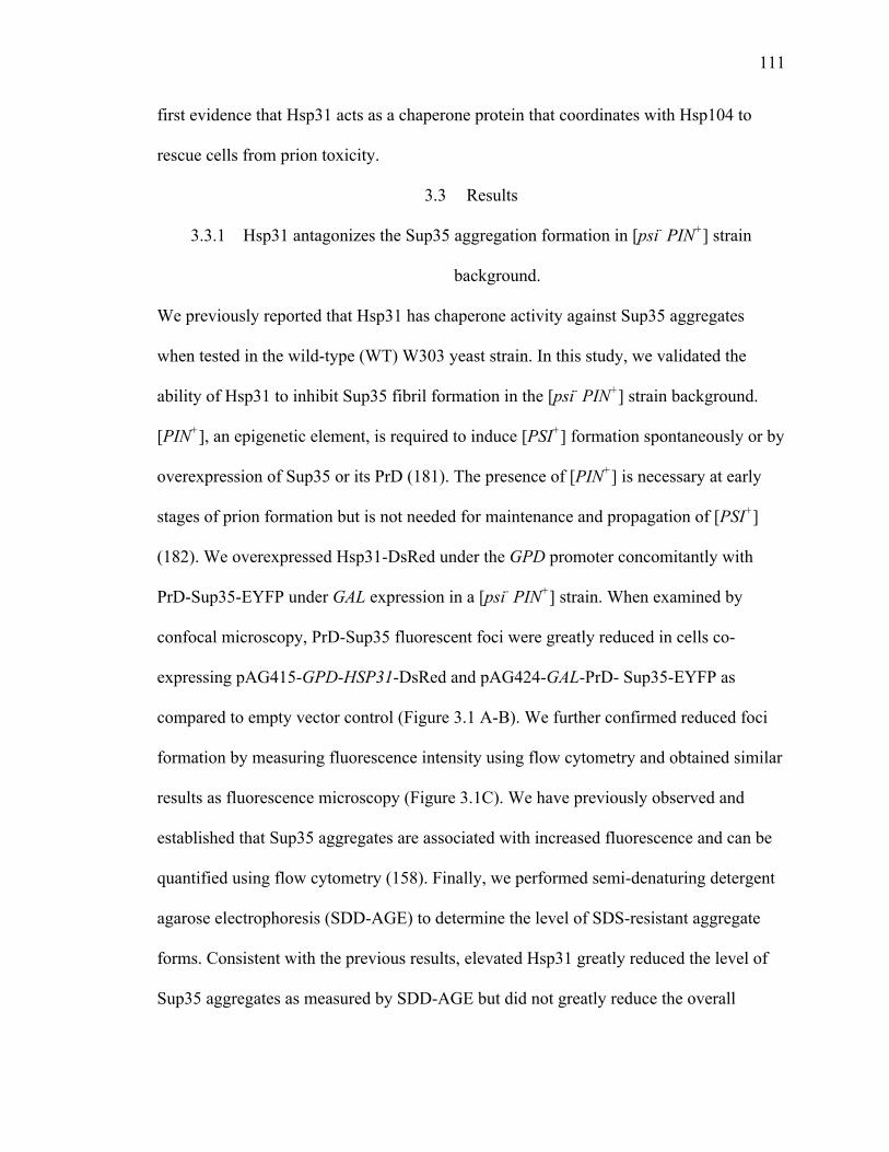
111
111
first evidence that Hsp31 acts as a chaperone protein that coordinates with Hsp104 to
rescue cells from prion toxicity.
3.3 Results
3.3.1 Hsp31 antagonizes the Sup35 aggregation formation in [psi- PIN+] strain
background.
We previously reported that Hsp31 has chaperone activity against Sup35 aggregates
when tested in the wild-type (WT) W303 yeast strain. In this study, we validated the
ability of Hsp31 to inhibit Sup35 fibril formation in the [psi- PIN+] strain background.
[PIN+], an epigenetic element, is required to induce [PSI+] formation spontaneously or by
overexpression of Sup35 or its PrD (181). The presence of [PIN+] is necessary at early
stages of prion formation but is not needed for maintenance and propagation of [PSI+]
(182). We overexpressed Hsp31-DsRed under the GPD promoter concomitantly with
PrD-Sup35-EYFP under GAL expression in a [psi- PIN+] strain. When examined by
confocal microscopy, PrD-Sup35 fluorescent foci were greatly reduced in cells co-
expressing pAG415-GPD-HSP31-DsRed and pAG424-GAL-PrD- Sup35-EYFP as
compared to empty vector control (Figure 3.1 A-B). We further confirmed reduced foci
formation by measuring fluorescence intensity using flow cytometry and obtained similar
results as fluorescence microscopy (Figure 3.1C). We have previously observed and
established that Sup35 aggregates are associated with increased fluorescence and can be
quantified using flow cytometry (158). Finally, we performed semi-denaturing detergent
agarose electrophoresis (SDD-AGE) to determine the level of SDS-resistant aggregate
forms. Consistent with the previous results, elevated Hsp31 greatly reduced the level of
Sup35 aggregates as measured by SDD-AGE but did not greatly reduce the overall

112
112
steady-state level of Sup35 in SDS-PAGE (Figure 3.1D). These results show that Hsp31
can prevent de novo [PSI+] aggregate formation in the presence of the [PIN+] genetic
element.
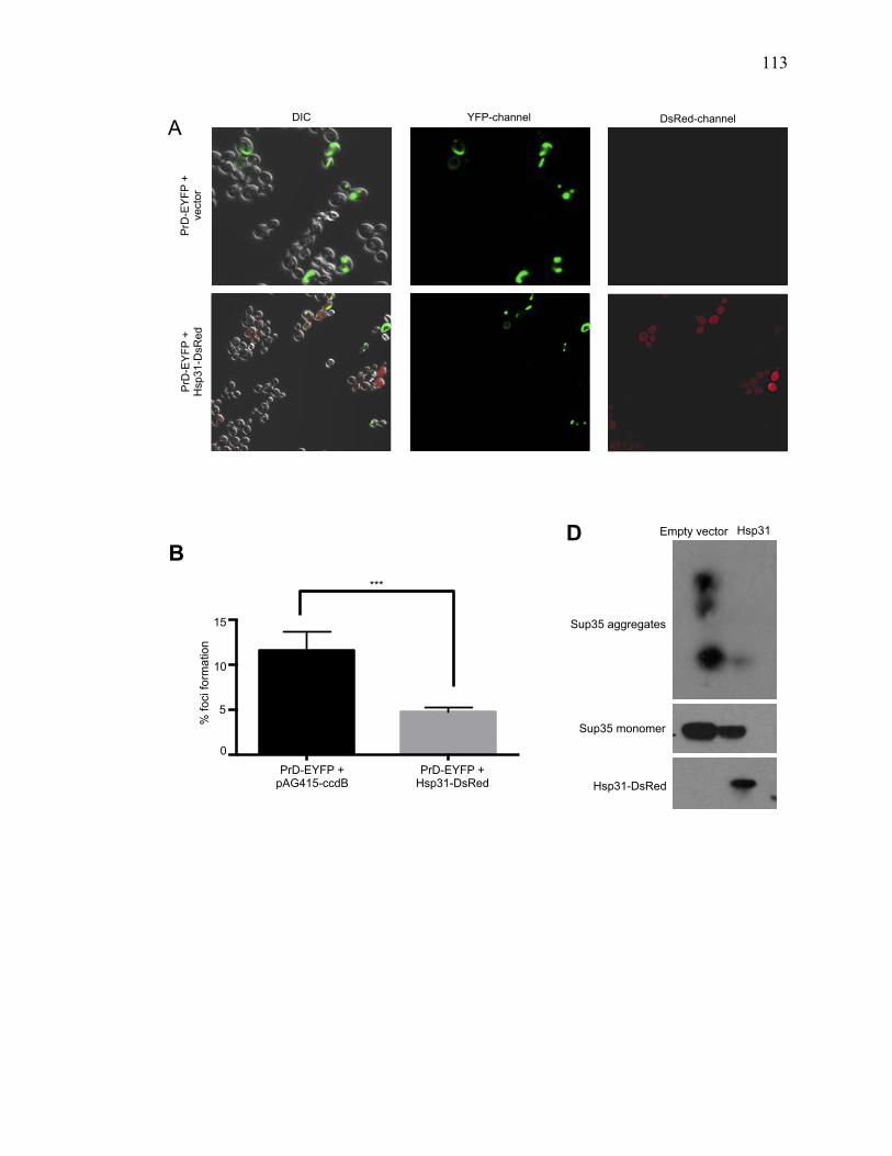
113
113
PrD
-EY
FP +
ve
ctor
P
rD-E
YFP
+
Hsp
31-D
sRed
DIC YFP-channel DsRed-channel A
PrD+EV
PrD + Hsp310
5
10
15
PrD-EYFP + pAG415-ccdB
PrD-EYFP + Hsp31-DsRed
***
15
10
5
0
B
% fo
ci fo
rmat
ion
Sup35 aggregates
Sup35 monomer
Hsp31-DsRed
Empty vector Hsp31 D
114
114
Figure 3.1. Hsp31 overexpression decreases the level of PrD-Sup35 aggregates. (A) GAL-driven PrD-Sup35-EYFP was overexpressed for 24 h at 30 °C in [psi- PIN+] cells with or without overexpression of DsRed tagged Hsp31. PrD-Sup35-EYFP aggregates appeared as ribbon-like vacuolar peripheral rings. Hsp31 remains cytoplasmically diffuse in these cells. Elevated levels of Hsp31 decreased the presence of Sup35 aggregates in individual cells. (B) Quantitation of the number of cells with one or more Sup35-EYFP foci. The average of at least three independent experiments was plotted; error bars represent mean ±SEM. (*** unpaired Student's t-test; p≤0.001). (C) Quantitation of the level of Sup35-EYFP fluorescence aggregates using flow cytometry. Aggregates are associated with higher fluorescence. Elevated levels of Hsp31 lowered the median fluorescence intensity (FI – arbitrary units) of Sup35-EYFP compared to empty vector control. Values represent mean ±SEM of three independent biological replicates (* unpaired Student's t-test; p≤0.01). (D) Cellular lysate of cells describe in A and B was analyzed by semi-denaturing agarose electrophoresis and SDS-PAGE. Overexpression of Hsp31 suppresses the level of SDS-resistant Sup35 aggregates as detected with anti-GFP antibody. Lower panel shows the expression of Hsp31 detected by anti-DsRed antibody.
Media
n Fl
inten
sity
psi- PrD + EV
psi- PrD
+ Hsp31
psi- EV
psi- PrD
0
5
10
15
20
15
10
5
20
0
*
PrD-EYFP + Hsp31-DsRed
pAG424-ccdB PrD-EYFP
C
PrD-EYFP + pAG415-ccdB
Fluo
resc
ence
inte
nsity
(A.U
.)
115
115
3.3.2 Hsp31 transiently inhibits Sup35 prion induction in vivo
The ability of Hsp31 to inhibit Sup35 aggregation in the [psi- PIN+] background led us to
investigate its ability to inhibit [PSI+] induction. Spontaneous de novo [PSI+] induction
frequency is extremely low unless Sup35 or its PrD is overexpressed (180,189). This
system is widely used to investigate the process of prion induction in yeast. To determine
whether Hsp31 can inhibit prion induction we used a [psi- PIN+] strain that forms red
color colonies on ¼ YPD plates with limited adenine. This strain forms white colonies in
the presence of [PSI+] because soluble Sup35 is depleted by aggregate formation
resulting in suppression of a premature stop codon and restoration of adenine prototrophy.
We observed that overexpression of Hsp31 antagonized [PSI+] prion induction triggered
by overexpression of Sup35 PrD for 6 h (Figure 3.2A-B). However, when Sup35 PrD
was expressed for 12 or 24 h, the [PSI+] prion induction rate between empty vector and
Hsp31 was statistically similar (Figure 3.2C). These findings suggested that Hsp31
antagonizes prion induction transiently but can be overcome by excess production of PrD
or cannot intervene when cells have established a full and more mature prion cycle.
These results are consistent with our previous proposal that Hsp31 intervenes early in the
protein misfolding processes (158,339).
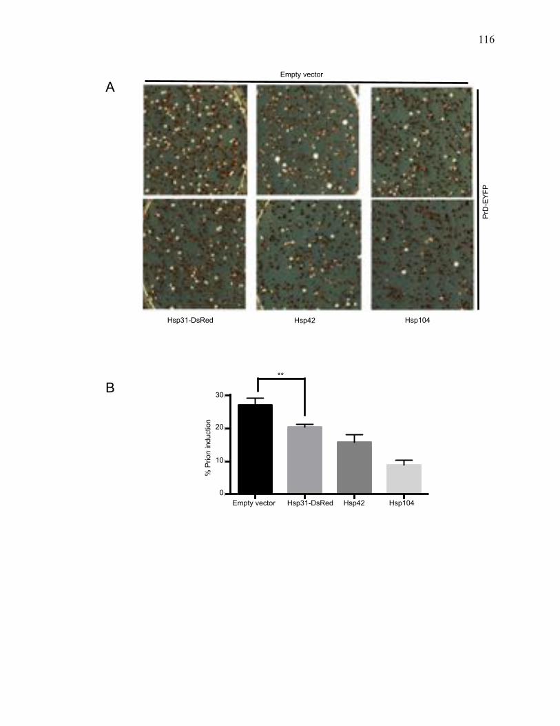
116
116
! 15!
(A)
!(B)
!!
Figure 6: Effect of Hsp31 on prion induction. (A)!PrD/Sup35-YFP was overexpressed for 6 h at 30°C in [psi−] cells expressing elevated levels of Hsp30, Hsp42, or Hsp104. Cells were plated on ¼ YPD plate and cells were grown for 3 days. (B) Proportion of prion induction was determined by number of white color colonies. Compared to the vector control there was significantly less [PSI+] prion induction in cells expressing Hsp30 (*p = 0.0086, two-tailed Student's t test), Values are represented as mean ±SEM (n = 3).
% P
rion
indu
ctio
n
Vector
Hsp31
Vector
Hsp42
Vector
Hsp10
40
10
20
30
40
PrD
-EY
FP
Hsp31-DsRed Hsp42 Hsp104
Empty vector
A
% P
rion
indu
ctio
n
Vector
Hsp31
-DsR
ed
Hsp42
Hsp10
40
10
20
30
40
30
20
10
0 Empty vector Hsp31-DsRed Hsp42 Hsp104
% P
rion
indu
ctio
n
**
B
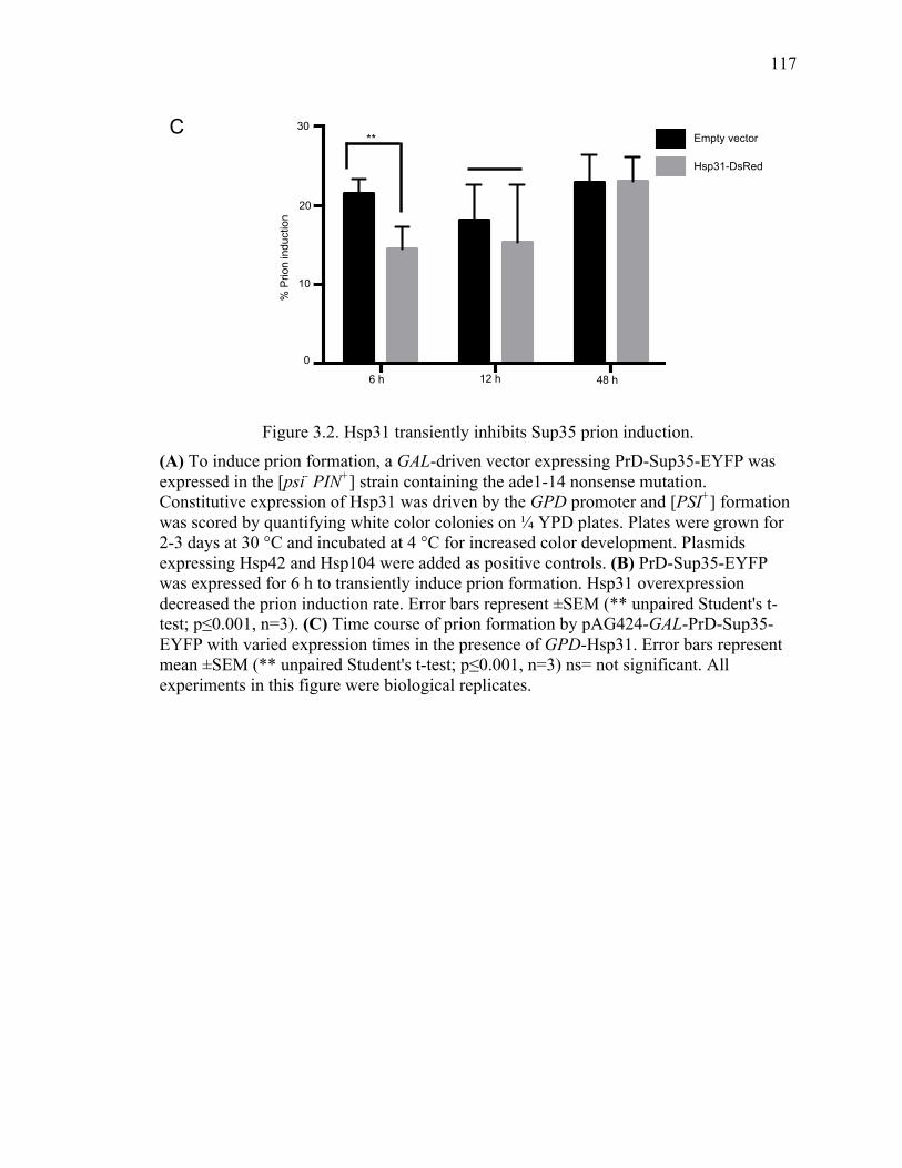
117
117
Figure 3.2. Hsp31 transiently inhibits Sup35 prion induction. (A) To induce prion formation, a GAL-driven vector expressing PrD-Sup35-EYFP was expressed in the [psi- PIN+] strain containing the ade1-14 nonsense mutation. Constitutive expression of Hsp31 was driven by the GPD promoter and [PSI+] formation was scored by quantifying white color colonies on ¼ YPD plates. Plates were grown for 2-3 days at 30 °C and incubated at 4 °C for increased color development. Plasmids expressing Hsp42 and Hsp104 were added as positive controls. (B) PrD-Sup35-EYFP was expressed for 6 h to transiently induce prion formation. Hsp31 overexpression decreased the prion induction rate. Error bars represent ±SEM (** unpaired Student's t-test; p≤0.001, n=3). (C) Time course of prion formation by pAG424-GAL-PrD-Sup35-EYFP with varied expression times in the presence of GPD-Hsp31. Error bars represent mean ±SEM (** unpaired Student's t-test; p≤0.001, n=3) ns= not significant. All experiments in this figure were biological replicates.
C
6hrs
12hrs
48hrs
0
10
20
30EVHsp31
** 30
20
10
0
Empty vector
Hsp31-DsRed
6hrs
12hrs
48hrs
0
10
20
30EVHsp31
6 h 12 h 48 h
% P
rion
indu
ctio
n
118
118
3.3.3 [PSI+] prion state is not affected by Hsp31 overexpression or deletion
Transient overexpression of sHSPs such as Hsp26 and Hsp42 cure the [PSI+] prion,
converting white colonies of [PSI+] into red colonies of [psi-] on ¼ YPD plates with
limited adenine (53). First, we tested if overexpression of Hsp31 can cure the [PSI+]
prion. Despite the fact that Hsp31 can prevent de novo prion aggregate formation in vivo
as detected by SDD-AGE (Figure 3.1), overexpression of Hsp31 is not sufficient to cure
the [PSI+] prion phenotype (Figure 3.3A). In addition, Hsp42 was used as a control and
was able to cure [PSI+] as previously reported (53). However, we found that Hsp26
overexpression using the identical plasmid vector from Duennwald et al. (53) could not
cure [PSI+] in this strain and under these experimental conditions. Our results are more
consistent with Wickner and colleagues (340) who reported lack of prion curing by
Hsp26 for both [Ure3-1] and [PSI+] phenotypes, which could be explained by differences
in strain genotypes and experimental conditions (340). We tested different plasmid
systems to express Hsp31 including the GPD and GAL promoters and none of them were
able to modulate the [PSI+] prion phenotype (data not shown). Next, we determined if
deletion of HSP31 influences the [PSI+] prion status. We constructed a [PSI+ PIN+]
hsp31Δ strain carrying the reporter nonsense allele ade1-14, so that the Sup35 read-
through caused by [PSI+] presence could be detected by development of white colonies.
The phenotype of the [PSI+] prion was similar in the hsp31Δ strain compared to WT with
no change in colony color (Figure 3.3B), suggesting that Hsp31 cannot intervene in an
established prion cycle.
119
119
3.3.4 Hsp31 deletion impairs [PSI+] prion curing by Hsp104 overexpression
A moderate level of Hsp104 is required for maintenance of [PSI+]; either deletion or
overexpression of Hsp104 cures the [PSI+] prion. Numerous chaperones such as Hsp70,
Hsp40 and Hsp90 along with its co-chaperones Sti1 and Cpr7 are known to modulate
prion curing by Hsp104 (39,40). To determine if Hsp31 altered Hsp104-mediated [PSI+]
prion curing, we transformed WT [PSI+] or hsp31Δ [PSI+] cells with p426-GPD-Hsp104
(low level of overexpression), pAG425-GAL-Hsp104-DsRed (medium overexpression) or
p2HG-Hsp104 (high overexpression GPD) plasmids (Figure 3.3C-E). The varied
overexpression level of Hsp104 under these different plasmid systems was confirmed by
western blotting (Figure 3.3F). The rate of [PSI+] prion curing was correlated to the level
of Hsp104 expression in WT [PSI+] cells. The rate of [PSI+] prion curing was
significantly reduced in hsp31Δ [PSI+] under the lowest overexpression condition of
GPD-Hsp104 (2% compared to 7% in WT) (Figure 3.3C). Under high overexpression of
Hsp104, a 100% curing rate was observed in the presence of HSP31 but the hsp31Δ strain
never achieved 100% curing rates and clearly white colonies were observed at 3%
frequency (Figure 3.3D). Induction of the GAL-Hsp104-DsRed construct for varied time
points from 2 h to 72 h also showed consistently less efficient prion curing by the hsp31Δ
strain (Figure 3.3E). The decreased prion curing in the hsp31Δ [PSI+] strain background
was not due to decreased Hsp104 expression because we observed a similar expression
level compared to WT (Figure 3.5B). These results demonstrate the presence of Hsp31 is
required for optimal prion curing efficiency at varied Hsp104 expression conditions.
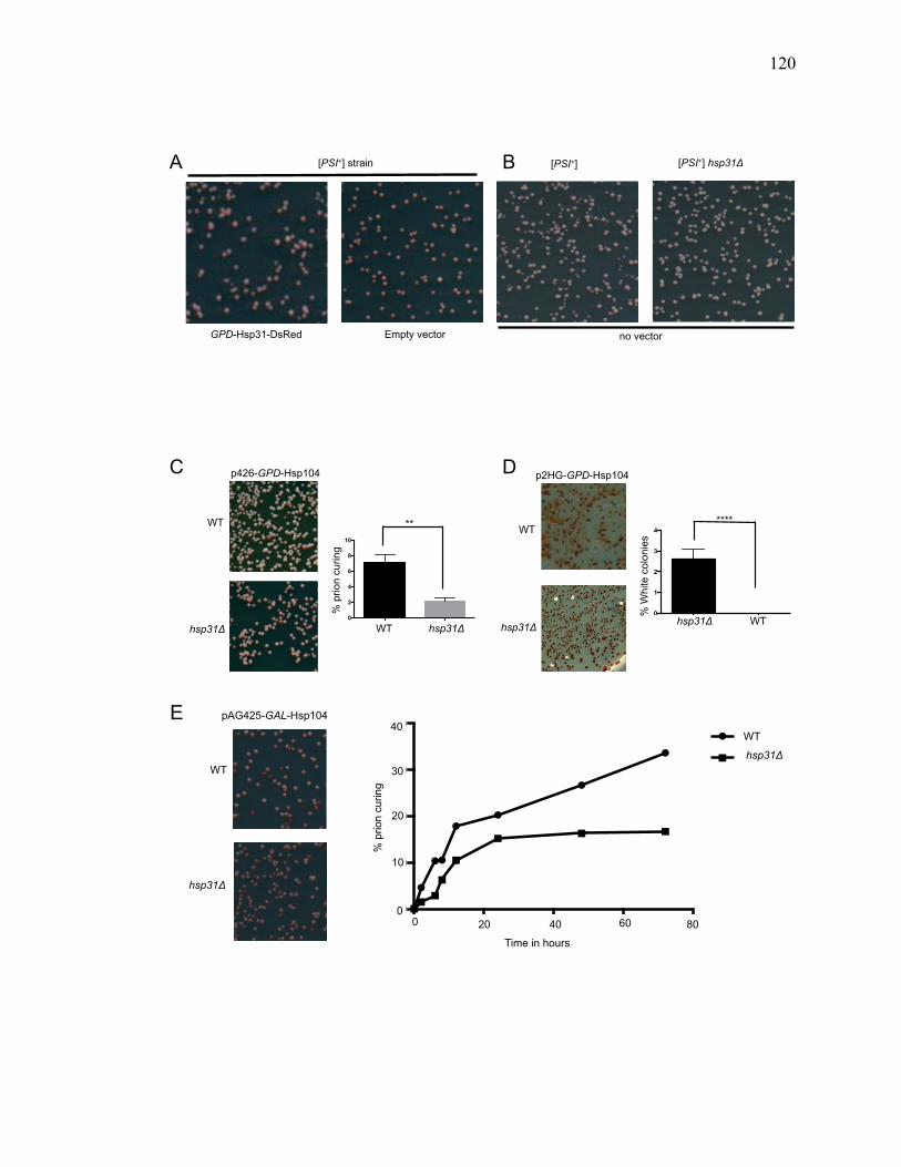
120
120
A
GPD-Hsp31-DsRed Empty vector
[PSI+] [PSI+] hsp31Δ [PSI+] strain
no vector
B
p2HG-GPD-Hsp104
% W
hite
col
onie
s
PSI+ hsp31
deleted
WT P
SI+0
1
2
3
4****
WT hsp31Δ
% W
hite
col
onie
s
WT
hsp31Δ
C D p426-GPD-Hsp104
WT
hsp31Δ WT
hsp31deleted
0
2
4
6
8
10
% p
rion
curin
g
WT
**
hsp31Δ
pAG425-GAL-Hsp104
WT
hsp31Δ
hsp31Δ
WT
Time in hours
% p
rion
curin
g
20 40 0 60 80 0
10
20
30
40 E
0 20 40 60 800
10
20
30
40
Time hours
WT[PSI+]hsp31 deleted [PSI+]
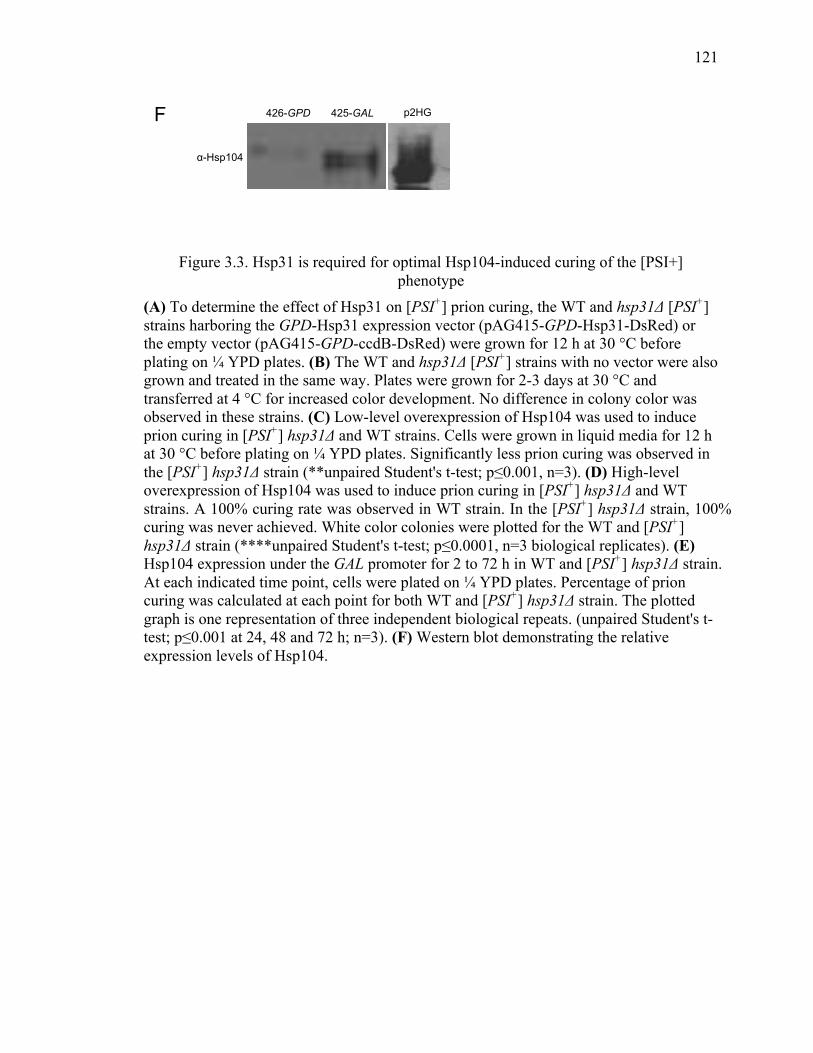
121
121
Figure 3.3. Hsp31 is required for optimal Hsp104-induced curing of the [PSI+] phenotype
(A) To determine the effect of Hsp31 on [PSI+] prion curing, the WT and hsp31Δ [PSI+] strains harboring the GPD-Hsp31 expression vector (pAG415-GPD-Hsp31-DsRed) or the empty vector (pAG415-GPD-ccdB-DsRed) were grown for 12 h at 30 °C before plating on ¼ YPD plates. (B) The WT and hsp31Δ [PSI+] strains with no vector were also grown and treated in the same way. Plates were grown for 2-3 days at 30 °C and transferred at 4 °C for increased color development. No difference in colony color was observed in these strains. (C) Low-level overexpression of Hsp104 was used to induce prion curing in [PSI+] hsp31Δ and WT strains. Cells were grown in liquid media for 12 h at 30 °C before plating on ¼ YPD plates. Significantly less prion curing was observed in the [PSI+] hsp31Δ strain (**unpaired Student's t-test; p≤0.001, n=3). (D) High-level overexpression of Hsp104 was used to induce prion curing in [PSI+] hsp31Δ and WT strains. A 100% curing rate was observed in WT strain. In the [PSI+] hsp31Δ strain, 100% curing was never achieved. White color colonies were plotted for the WT and [PSI+] hsp31Δ strain (****unpaired Student's t-test; p≤0.0001, n=3 biological replicates). (E) Hsp104 expression under the GAL promoter for 2 to 72 h in WT and [PSI+] hsp31Δ strain. At each indicated time point, cells were plated on ¼ YPD plates. Percentage of prion curing was calculated at each point for both WT and [PSI+] hsp31Δ strain. The plotted graph is one representation of three independent biological repeats. (unpaired Student's t-test; p≤0.001 at 24, 48 and 72 h; n=3). (F) Western blot demonstrating the relative expression levels of Hsp104.
α-Hsp104
F 426-GPD 425-GAL p2HG
122
122
3.3.5 Hsp31 collaborates with Hsp104 to cure [PSI+] prion
The decreased efficiency in Hsp104 curing [PSI+] in the absence of Hsp31 lead us to
further explore the relationship between these two chaperones. Small HSPs Hsp26 and
Hsp42 are known to collaborate with Hsp104 in rescuing the polyglutamine toxicity and
solubilization of amyloid aggregates (243). We aimed to test the effect of expressing
Hsp31 and Hsp104 together in curing of the [PSI+] prion. Expression of Hsp104 (p426-
GPD-Hsp104) in the [PSI+] strain resulted in a curing rate of 3 % that is lower than the
rate in Figure 3.3 because the co-existence of two different constructs decreases plasmid
copy numbers and affects expression levels. Co-expression of Hsp31 and Hsp104
increased the rate of prion curing to 6 % compared to the respective controls (Figure
3.4A-B). We also tested the collaboration between Hsp104 and Hsp31 in the hsp31Δ
strain and detected that co-expression of Hsp104 with Hsp31 was able to cure the [PSI+]
prion to a greater extent than individual chaperone expression (Figure 3.4C-D). These
results corroborate the inefficient curing in hsp31Δ and establish that Hsp31 is required
for optimal Hsp104 activity.
Previously, it was shown that sHSP, Hsp42, collaborates with Hsp26 to prevent [PSI+]
prionogenesis by distinct and synergistic mechanisms with Hsp104 (53), hence we tested
the interaction between Hsp42 and Hsp31 on [PSI+] prion curing. Elevated levels of
Hsp42 driven by the GPD promoter was sufficient to increase curing of [PSI+] in this
strain transformed with a single plasmid (5.4% compared to 1.4%). Introduction of a
second empty vector plasmid decreased the curing rate of the GPD-HSP42 construct to
background levels. We also co-expressed Hsp31 and Hsp42 in the [PSI+] strain and did
not detect any increased curing rate, in contrast to Hsp31 co-expression with Hsp104

123
123
(Figure 3.4E-F). This lack of synergy between these proteins implies that Hsp42 and
Hsp31 act at a similar stage of the prion cycle.
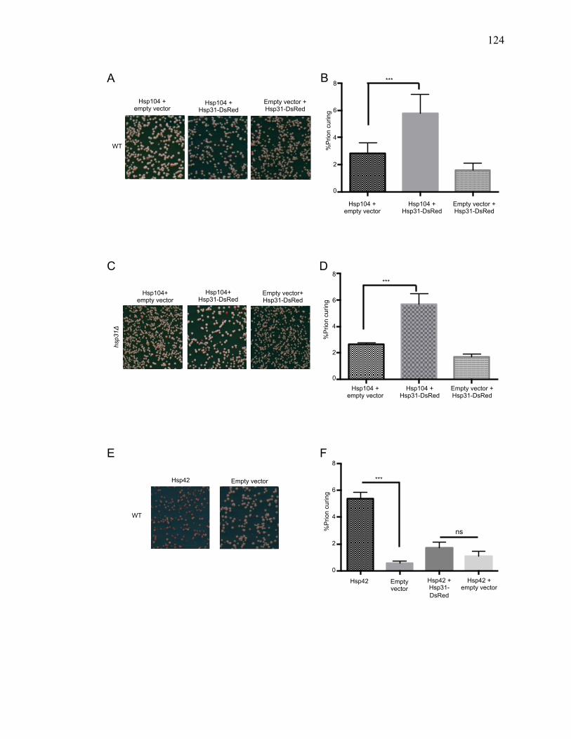
124
124
B
Hsp104 + empty vector
Hsp104 + Hsp31-DsRed
Empty vector + Hsp31-DsRed
WT
A
p426-GPD-Hsp104+pAG415-GPD-ccdB-DsRed
p426-GPD-Hsp104+pAG415-GPD-Hsp31-DsRed
p426-GPD+pAG415-GPD-Hsp31-DsRed
0
2
4
6
8
%P
rion
curin
g
***
0
2
4
6
8
Hsp104 + Hsp31-DsRed
Hsp104 + empty vector
Empty vector + Hsp31-DsRed
C D
hsp3
1Δ
Hsp104+ empty vector
Hsp104+ Hsp31-DsRed
Empty vector+ Hsp31-DsRed
Hsp10
4+EV
Hsp10
4+ H
sp31
Hsp31
+p42
6-GPD
0
2
4
6
8***
%P
rion
curin
g
0
2
4
6
8
Hsp104 + empty vector
Hsp104 + Hsp31-DsRed
Empty vector + Hsp31-DsRed
Hsp42
Empty Vec
tor
Hsp42
+Hsp
31-D
sRed
Hsp42
+empty
vecto
r0
2
4
6
8
Empty vector Hsp42
Hsp42 Empty vector
Hsp42 + empty vector
Hsp42 + Hsp31- DsRed
***
ns %Pr
ion
curin
g
0
2
4
6
8 F E
WT
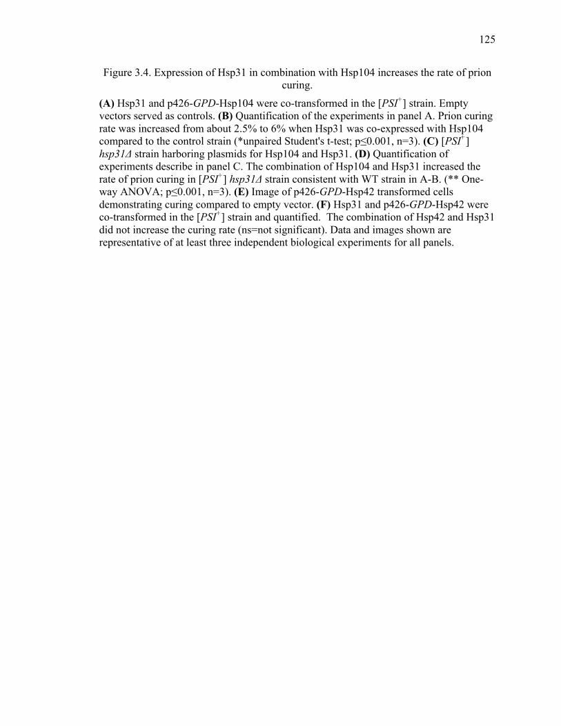
125
125
Figure 3.4. Expression of Hsp31 in combination with Hsp104 increases the rate of prion curing.
(A) Hsp31 and p426-GPD-Hsp104 were co-transformed in the [PSI+] strain. Empty vectors served as controls. (B) Quantification of the experiments in panel A. Prion curing rate was increased from about 2.5% to 6% when Hsp31 was co-expressed with Hsp104 compared to the control strain (*unpaired Student's t-test; p≤0.001, n=3). (C) [PSI+] hsp31Δ strain harboring plasmids for Hsp104 and Hsp31. (D) Quantification of experiments describe in panel C. The combination of Hsp104 and Hsp31 increased the rate of prion curing in [PSI+] hsp31Δ strain consistent with WT strain in A-B. (** One-way ANOVA; p≤0.001, n=3). (E) Image of p426-GPD-Hsp42 transformed cells demonstrating curing compared to empty vector. (F) Hsp31 and p426-GPD-Hsp42 were co-transformed in the [PSI+] strain and quantified. The combination of Hsp42 and Hsp31 did not increase the curing rate (ns=not significant). Data and images shown are representative of at least three independent biological experiments for all panels.
126
126
3.3.6 Effect of [PSI+] curing in the hsp31Δ strain is not due to loss of Hsp104
thermotolerance function.
A possible mechanism for the decrease in [PSI+] prion curing by Hsp104 in the hsp31Δ
background is decreased expression or activity of Hsp104. To measure the functional
competence of Hsp104 we tested the thermotolerance activity of Hsp104 in exponentially
growing cells. We induced Hsp104 expression in both strains by incubating the culture at
37 °C for 30 minute and then heat shocked at 50 °C. The survival of cells after heat shock
is dependent on Hsp104 induction, and we observed the rate of survival in hsp31Δ was
comparable to the isogenic WT [PSI+] strain (Figure 3.5A). Moreover, the basal
thermotolerance without induction of Hsp104 was not affected by deletion of Hsp31
(Figure 3.5A). In addition, we observed a slightly elevated level of Hsp104 in the hsp31Δ
strain compared to WT, hence the expression of Hsp104 is not compromised (Figure
3.5B). Thus, reduction in [PSI+] prion curing in the hsp31Δ strain is not due to general
impairment of Hsp104 activity or expression.
3.3.7 Hsp104 physically interacts with Hsp31
The close collaboration between Hsp104 and Hsp31 prompted us to test the physical
association between them. Co-immunoprecipitation followed by western blot analysis in
yeast lysates demonstrated that Hsp31 interacts with Hsp104. Immunoprecipitation was
performed using HSP31-9myc genomically tagged at the endogenous locus and
overexpressing Hsp104 either under the GPD or the GAL promoter. First, Hsp31-9myc
was pulled down using anti-myc antibody conjugated to agarose beads from
exponentially growing cell lysates. Western blots confirmed the successful pull down of
Hsp31-9myc (Figure 3.5C; middle panel). The upper panel demonstrates the successful
127
127
pull-down of Hsp104 in both GPD-HSP104 and GAL-HSP104 expressing lysates but not
in the empty vector control (Figure 3.5C). Similar results were obtained with an
alternative co-immunoprecipitation approach in which polyclonal anti-Hsp104 antibody
and protein G dynabeads were used to pull down Hsp104 followed by western blot
analysis with anti-myc antibody to detect Hsp31. This approach confirmed the
interactions and also demonstrated that Hsp31 is pulled down with strains having
endogenous levels of Hsp104 (Figure 3.5C; bottom panel empty vector lane). These
results demonstrate that Hsp31 is part of same complex with Hsp104 using two different
immunoprecipitation protocols and the Hsp31-Hsp104 interaction is detectable under
physiological expression levels.
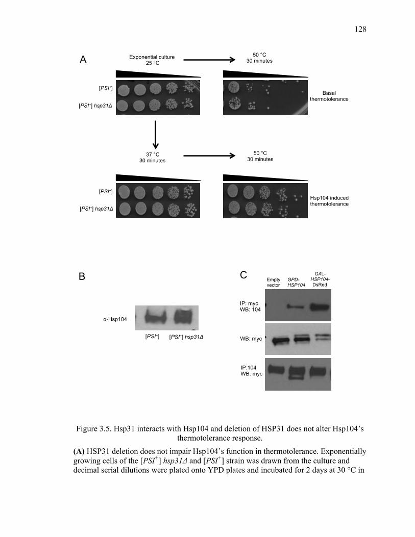
128
128
Figure 3.5. Hsp31 interacts with Hsp104 and deletion of HSP31 does not alter Hsp104’s thermotolerance response.
(A) HSP31 deletion does not impair Hsp104’s function in thermotolerance. Exponentially growing cells of the [PSI+] hsp31Δ and [PSI+] strain was drawn from the culture and decimal serial dilutions were plated onto YPD plates and incubated for 2 days at 30 °C in
A
Basal thermotolerance
Hsp104 induced thermotolerance
Exponential culture 25 °C
50 °C 30 minutes
37 °C 30 minutes
50 °C 30 minutes
[PSI+]
[PSI+]
[PSI+] hsp31Δ
[PSI+] hsp31Δ
IP:104 WB: myc
IP: myc WB: 104
GPD- HSP104
GAL- HSP104- DsRed
Empty vector
WB: myc
B C
α-Hsp104
[PSI+] hsp31Δ [PSI+]
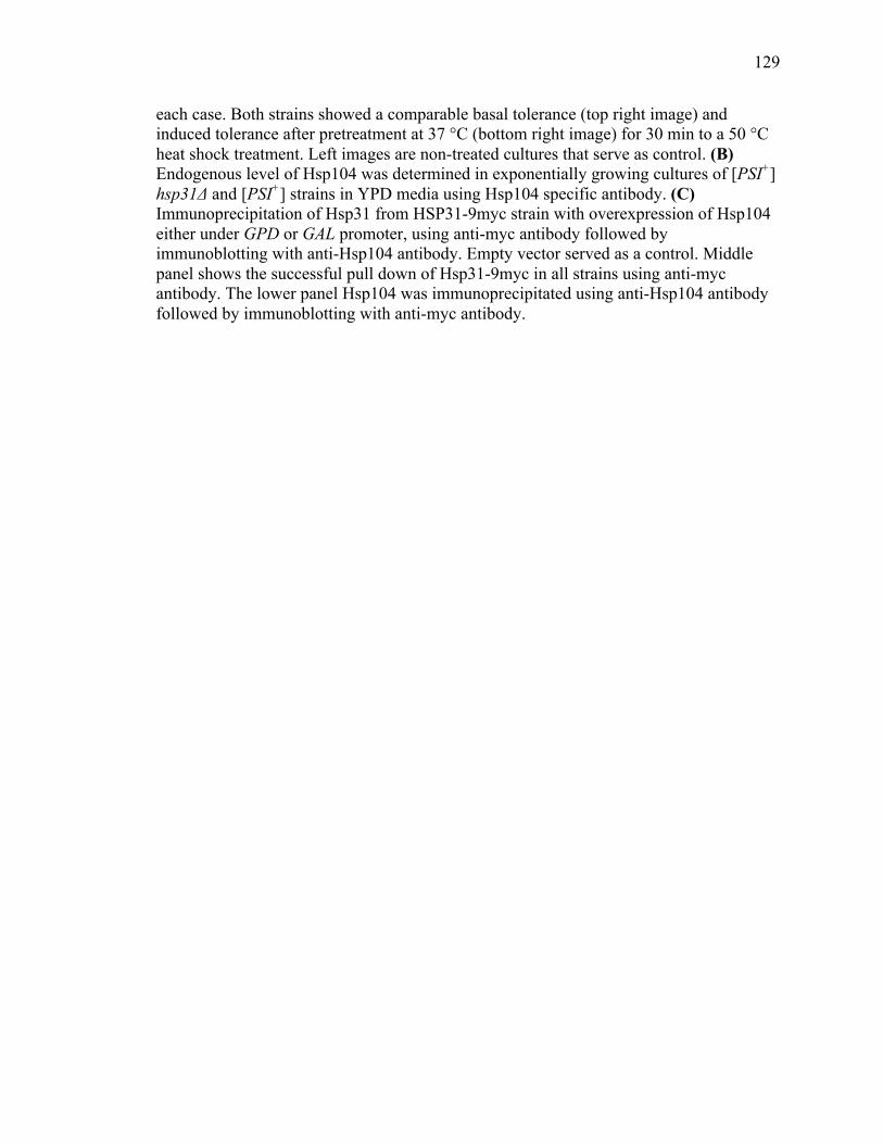
129
129
each case. Both strains showed a comparable basal tolerance (top right image) and induced tolerance after pretreatment at 37 °C (bottom right image) for 30 min to a 50 °C heat shock treatment. Left images are non-treated cultures that serve as control. (B) Endogenous level of Hsp104 was determined in exponentially growing cultures of [PSI+] hsp31Δ and [PSI+] strains in YPD media using Hsp104 specific antibody. (C) Immunoprecipitation of Hsp31 from HSP31-9myc strain with overexpression of Hsp104 either under GPD or GAL promoter, using anti-myc antibody followed by immunoblotting with anti-Hsp104 antibody. Empty vector served as a control. Middle panel shows the successful pull down of Hsp31-9myc in all strains using anti-myc antibody. The lower panel Hsp104 was immunoprecipitated using anti-Hsp104 antibody followed by immunoblotting with anti-myc antibody.
130
130
3.3.8 Hsp31 together with Hsp104 antagonizes prion dependent toxicity of excess
Sup35.
Overexpression of full length Sup35 or its PrD exhibits toxicity in the [PSI+] strain
(244,246). The toxicity of excess Sup35 in [PSI+] or [PSI+] hsp31Δ strain was
investigated. Deletion of HSP31 has no effect on Sup35 toxicity (Figure 3.6) in contrast
to our previous report of increased toxicity when α-syn is expressed in the hsp31Δ strain
background (158). However, elevated levels of Hsp31 expressed from the GPD promoter
rescued [PSI+] cells from Sup35 toxicity in both WT and deleted strains. As expected,
Hsp104 reduce the toxicity of Sup35 to greater extent than Hsp31. Strikingly, Hsp31
together with Hsp104 strongly reduced the toxicity of Sup35 in the [PSI+] strain (Figure
3.6). We also observed the rescue effect of sole expression of Hsp31 or Hsp104 and when
expressed together in the [PSI+] hsp31Δ strain. The level of rescue was not as dramatic as
in the WT strains suggesting that the expression of endogenous Hsp31 has a role in
reducing toxicity in conjunction with heterologous expression of Hsp31 or Hsp104. The
role of endogenous Hsp31 is not clear but could be a direct effect of chaperone activity or
because autophagy can be impaired in hsp31Δ strains (294) which may lead to increased
proteotoxicity of Sup35.
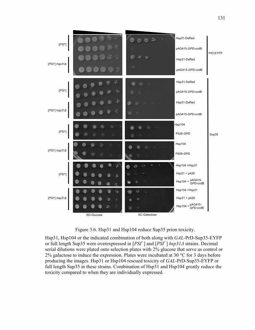
131
131
Figure 3.6. Hsp31 and Hsp104 reduce Sup35 prion toxicity. Hsp31, Hsp104 or the indicated combination of both along with GAL-PrD-Sup35-EYFP or full length Sup35 were overexpressed in [PSI+] and [PSI+] hsp31Δ strains. Decimal serial dilutions were plated onto selection plates with 2% glucose that serve as control or 2% galactose to induce the expression. Plates were incubated at 30 °C for 3 days before producing the images. Hsp31 or Hsp104 rescued toxicity of GAL-PrD-Sup35-EYFP or full length Sup35 in these strains. Combination of Hsp31 and Hsp104 greatly reduce the toxicity compared to when they are individually expressed.
SD-Glucose SC-Galactose
Hsp31-DsRed
pAG415-GPD-ccdB
Hsp31-DsRed
PrD-EYFP
[PSI+] hsp31Δ
[PSI+]
[PSI+] hsp31Δ
[PSI+]
Hsp31-DsRed
Hsp31-DsRed
[PSI+]
[PSI+] hsp31Δ
Hsp104
Hsp104
P426-GPD
P426-GPD
Sup35
[PSI+]
[PSI+] hsp31Δ
Hsp104 +Hsp31
Hsp31 + p426
Hsp104 +
Hsp104 +Hsp31
Hsp31 + p426
Hsp104 +
pAG415-GPD-ccdB
pAG415-GPD-ccdB
pAG415-GPD-ccdB
pAG415-GPD-ccdB
pAG415-GPD-ccdB
132
132
3.3.9 Hsp31 and Hsp104 modulate Sup35 aggregation in [PSI+] cells
Collaboration of Hsp31 with Hsp104 to reduce Sup35 induced prion toxicity prompted us
to investigate whether this activity correlates with protein disaggregation activity of
Hsp104. To assess the state of Sup35 in [PSI+] cells harboring Hsp31 and Hsp104, we
performed sedimentation analysis to determine the ratio of Sup35 in the soluble versus
aggregate forms. The individual expression of Hsp104 or Hsp31 resulted in a very strong
signal in the insoluble pellet fraction. However, the combination of Hsp104 and Hsp31
markedly reduced Sup35 aggregation found in the pellet fraction and increased the
amount of Sup35 found in the soluble fraction (Figure 3.7A). Soluble Sup35 was very
susceptible to proteolysis during processing of the samples as evident by the lower
molecular weight species, which is consistent with earlier reports (187,341). These results
demonstrate that sole overexpression of Hsp31 does not appear to intervene in the
established prion cycle present in a [PSI+] strain but can inhibit aggregate formation and
prionogenesis in a [psi-] strain (Figure 3.1). However, the results also show that Hsp31
can cooperate with Hsp104 to reduce Sup35 toxicity and simultaneously increase Sup35
solubility.
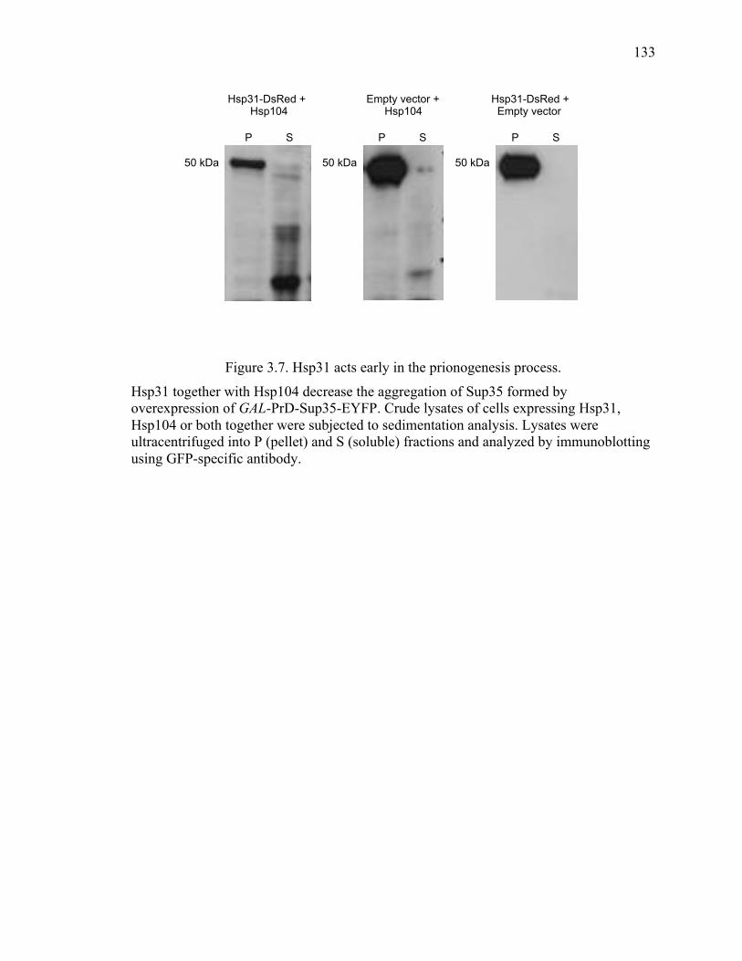
133
133
Figure 3.7. Hsp31 acts early in the prionogenesis process.
Hsp31 together with Hsp104 decrease the aggregation of Sup35 formed by overexpression of GAL-PrD-Sup35-EYFP. Crude lysates of cells expressing Hsp31, Hsp104 or both together were subjected to sedimentation analysis. Lysates were ultracentrifuged into P (pellet) and S (soluble) fractions and analyzed by immunoblotting using GFP-specific antibody.
Hsp31-DsRed + Hsp104
Empty vector + Hsp104
Hsp31-DsRed + Empty vector
P S
50 kDa 50 kDa 50 kDa
P S P S
134
134
3.4 Material and Methods
3.4.1 Yeast strains and plasmids
The [psi-, PIN+] and [PSI+] strains were used throughout the study for prion induction,
curing and toxicity assays. The W303 HSP31-9myc strain was used for pull down assays.
Details of the plasmid, strains and primers used in this study are provided in the tables
(Table 3.1-3.3). Deletion of HSP31 in [PSI+] strain was obtained by transforming a PCR
product containing the nourseothricin N- acetyl-transferase (NAT) gene flanked by
HSP31 homology regions. The primers consisted of 20 nucleotides for amplifying the
NAT gene from pFA6a-NATNT2 (Euroscarf), and 50 nucleotides immediately preceding
the HSP31 start codon or after the stop codon. The amplified product was integrated into
[PSI+] and [psi-, PIN+] strain. Successful integration and deletion was confirmed by
diagnostic PCR.
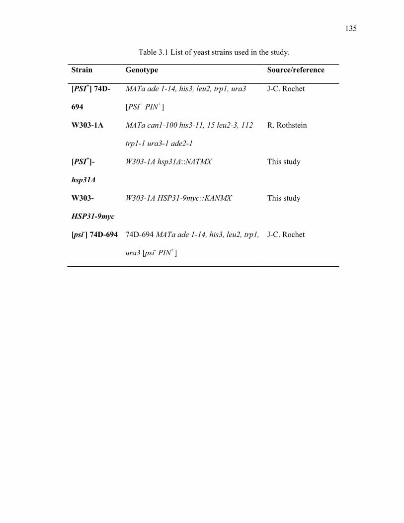
135
135
Table 3.1 List of yeast strains used in the study.
Strain Genotype Source/reference
[PSI+] 74D-
694
MATa ade 1-14, his3, leu2, trp1, ura3
[PSI+ PIN+]
J-C. Rochet
W303-1A MATa can1-100 his3-11, 15 leu2-3, 112
trp1-1 ura3-1 ade2-1
R. Rothstein
[PSI+]-
hsp31Δ
W303-1A hsp31Δ::NATMX This study
W303-
HSP31-9myc
W303-1A HSP31-9myc::KANMX This study
[psi-] 74D-694 74D-694 MATa ade 1-14, his3, leu2, trp1,
ura3 [psi- PIN+]
J-C. Rochet
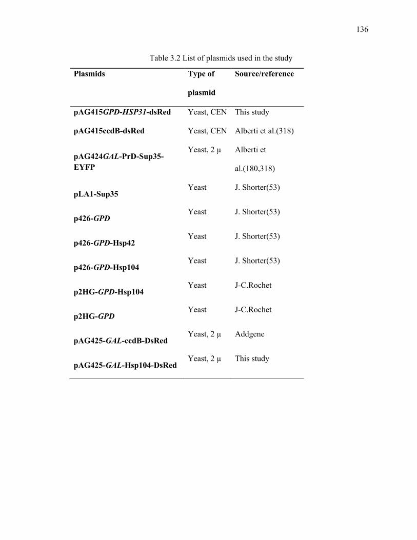
136
136
Table 3.2 List of plasmids used in the study
Plasmids Type of
plasmid
Source/reference
pAG415GPD-HSP31-dsRed Yeast, CEN This study
pAG415ccdB-dsRed Yeast, CEN Alberti et al.(318)
pAG424GAL-PrD-Sup35-EYFP
Yeast, 2 µ Alberti et
al.(180,318)
pLA1-Sup35 Yeast J. Shorter(53)
p426-GPD Yeast J. Shorter(53)
p426-GPD-Hsp42 Yeast J. Shorter(53)
p426-GPD-Hsp104 Yeast J. Shorter(53)
p2HG-GPD-Hsp104 Yeast J-C.Rochet
p2HG-GPD Yeast J-C.Rochet
pAG425-GAL-ccdB-DsRed Yeast, 2 µ Addgene
pAG425-GAL-Hsp104-DsRed Yeast, 2 µ This study
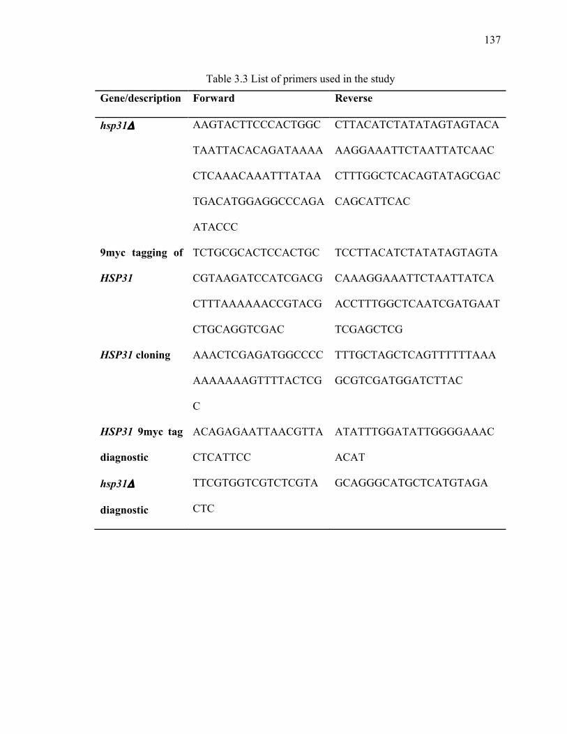
137
137
Table 3.3 List of primers used in the study
Gene/description Forward Reverse
hsp31Δ AAGTACTTCCCACTGGC
TAATTACACAGATAAAA
CTCAAACAAATTTATAA
TGACATGGAGGCCCAGA
ATACCC
CTTACATCTATATAGTAGTACA
AAGGAAATTCTAATTATCAAC
CTTTGGCTCACAGTATAGCGAC
CAGCATTCAC
9myc tagging of
HSP31
TCTGCGCACTCCACTGC
CGTAAGATCCATCGACG
CTTTAAAAAACCGTACG
CTGCAGGTCGAC
TCCTTACATCTATATAGTAGTA
CAAAGGAAATTCTAATTATCA
ACCTTTGGCTCAATCGATGAAT
TCGAGCTCG
HSP31 cloning AAACTCGAGATGGCCCC
AAAAAAAGTTTTACTCG
C
TTTGCTAGCTCAGTTTTTTAAA
GCGTCGATGGATCTTAC
HSP31 9myc tag
diagnostic
ACAGAGAATTAACGTTA
CTCATTCC
ATATTTGGATATTGGGGAAAC
ACAT
hsp31Δ
diagnostic
TTCGTGGTCGTCTCGTA
CTC
GCAGGGCATGCTCATGTAGA
138
138
3.4.2 Yeast growth conditions
We used isogenic [psi-] and [PSI+] derivatives of 74D-694 [MATa, his3, leu2, trp1, ura3;
suppressible marker ade1-14. Cells were grown at 30 °C on synthetic dextrose medium
(SD; 0.7% yeast nitrogen base, 2% glucose) with appropriate amino acid dropout mixture
for selection and maintenance of the particular plasmid. Synthetic complete (SC) medium
contains 2% raffinose in place of glucose and 2% galactose for induction of genes under
the GAL promoter. ¼ YPD solid medium used in the plating assays contains 0.5% yeast
extract, 2% peptone, and 2% glucose. Cultures were always maintained in actively
growing conditions and OD600 was used to measure the growth rate.
3.4.3 SDD-AGE
The [psi-] cells were co-transformed with pAG424-GAL-PrD-Sup35-EYFP and pAG415-
GPD-HSP31-DsRed plasmids. Cultures were grown in SD media overnight and induced
in SC 2% raffinose + 2% galactose media for 24 h. Prion aggregates were analyzed using
SDD-AGE as described previously(158). Briefly, cells were harvested by centrifugation
and spheroplasts were generated and lysed in 4 x SDS sample buffer at room temperature
for 15 min before loading onto a 1.8 % agarose gel followed by transfer to nitrocellulose
membrane. The membrane was immune-blotted using anti-GFP antibody (Roche;
11814460001) and anti-DsRed antibody (Santa Cruz Biotechnology; Sc-33353).
3.4.4 Fluorescence Microscopy and flow-cytometry
The [psi-] strain was co-transformed with pAG424-GAL-PrD-Sup35-EYFP and pAG415-
GPD-HSP31-DsRed. Successful transformation was selected and re-streaked on SD (-
tryptophan -leucine) agar plates. Cells were grown overnight at 30 °C in SD (-tryptophan
-leucine) medium and PrD-Sup35 expression was induced for 24 h in SC (-tryptophan -
139
139
leucine) medium with 2% raffinose and 2% galactose. Cells were examined under
fluorescence microscopy using a Nikon A1 confocal microscope with a Nikon Plan
apochromat 60 X (NA 1.4) oil immersion objective to acquire fluorescence and DIC
images and were analyzed using Image J. The identical cultures used in microscopy were
also subjected to flow cytometry. After induction cells were collected and washed with
PBS, filtered and analyzed for EYFP fluorescence intensity using the Beckman Coulter
FC500 flow cytometer with the FL-1 channel. A total of 10,000 events were acquired for
each sample and data was analyzed using FlowJo software to calculate median
fluorescence intensity.
3.4.5 Sup35 prion curing
Curing of [PSI+] was performed by transforming the strain with plasmids, p2HG-GPD-
Hsp104, pAG425-GAL-Hsp104, pAG415-GPD-HSP31-DsRed, p426-GPD-Hsp42 and
their corresponding empty vectors. For double transformation both plasmids were co-
transformed and selected on double dropout media simultaneously. Transferred cells
were grown in the SD medium at 30 °C for overnight growth and plated on ¼ YPD plates
which were incubated for three days at 30 °C and shifted for another day at 4 °C for
colony color development. To score for curing, colonies with red color were counted as
[psi-]. Cultures carrying plasmid with the GAL promoter were grown in SC medium
before plating on ¼ YPD.
3.4.6 Sup35 prion induction
For the induction experiment, at least three independent transformants with pAG424-
GAL-PrD-Sup35-EYFP and pAG415-GPD-HSP31-DsRed plasmid were grown at 30 °C
in SD medium overnight, centrifuges and washed three times with water, and shifted to
140
140
SC medium to induce prion formation. Aliquots were withdrawn at 6, 12 and 48 h and
diluted to a density of 50,000 cells per 100 µl for plating onto ¼ YPD plate. The plates
were then incubated for three days at 30 °C and another day at 4 °C for color
development. Percentage of [PSI+] induction was measured as the number of white ([PSI+]
colonies) colonies divided by the total number of colonies.
3.4.7 Prion toxicity assay
The [PSI+] strain was transformed with pAL1 Sup35 and plasmids expressing Hsp31,
Hsp42 and or Hsp104 along with their corresponding empty vector. After selection on SD
plates, cells were grown into 5 ml of SD liquid media with 2 % glucose at 30 °C
overnight. Cells were harvested and washed three times with water and a five fold serial
dilution was performed with a starting OD600 of 0.8. Diluted samples of 5 µl were spotted
onto SD and SC plates with the appropriate dropout selection. Plates were incubated at
30 °C for 3 days and imaged with a scanner.
3.4.8 Pull down assay
The HSP31-9myc tagged strain was transformed with plasmids pAG425-GAL-Hsp104 or
p426-GPD-Hsp104. Cells lysates were prepared in a buffer (50 mM Tris–HCl, 1 mM
EDTA, 5 mM DTT, 10% (v/v) glycerol, 0.5 M NaCl, at pH 7.5) with freshly added
protease inhibitor cocktail (Roche). Anti-myc antibody-conjugated agarose beads were
used to pull down Hsp31 protein by incubating the beads with the lysate at 4 °C for 1 h.
After the pull down, beads were washed three times with PBS-T and bound protein was
eluted by boiling the sample in SDS loading buffer before separating the proteins on
SDS-PAGE and western blotting using Hsp104 antibody (Abcam; ab69549).
Immunoprecipitation was also preformed in reverse by immobilizing Hsp104 antibody on
141
141
protein G dynabeads (Life Technology) and incubating the total cell lysate with this
complex for 1 h. In this case, western blotting was performed using anti-myc antibody
(Sigma; M4439).
3.4.9 Thermotolerance assay
WT [PSI+] and [PSI+] hsp31Δ strains were grown in YPD medium starting from OD600 of
0.2 until they reach exponential growth phase after 6 h. Equal number of cells from each
strain were incubated at 37 °C to induce Hsp104 expression and then heat shocked at
50 °C for 20 min. Aliquots were placed on ice before and after heat shock. A portion of
cultures were heat shocked at 50 °C without Hsp104 induction at 37 °C. Samples were
collected and five-fold dilutions were spotted on YPD medium.
3.4.10 Sedimentation assay
[PSI+] strains harboring Hsp31, Hsp104 and appropriate co-expression vector plasmids
were grown for 12 h and centrifuged to collect cells. Cells were washed in water and
lysed at 4 °C by bead beating in lysis buffer (50 mM Tris HCl pH 7.5, 50 mM NaCl, 2
mM EDTA and 5% Glycerol plus freshly added protease inhibitor cocktail(158)). Equal
volume of cold RIPA buffer (50 mM Tris HCl pH 7.0, 150 mM NaCl, 1% Triton X-100,
0.5% deoxycholate and 0.1 % SDS) was added to the lysate and the mixture was vortexed
for 10 s. Lysate was centrifuged at 800 rpm for 2 min at 4 °C in a Eppendorf
microcentrifuge. Lysate supernatant was subjected to ultracentrifugation at 80,000 rpm in
a TLA-120.2 rotor for 30 min using an Optima Max-XD Ultracentrifuge (Beckman
Coulter). Supernatant was collected and pellet was re-suspended in equal volume of lysis
and RIPA buffer. Supernatant and pellet fractions were subjected to SDS-PAGE and
immunoblotted using GFP antibody (Roche).
142
142
3.5 Discussion
Previous studies have demonstrated Hsp31 is a multitasking protein involved in several
cellular pathways ranging from functioning as a glutathione independent
methylglyoxalase to stress responder that acts as a molecular chaperone. In this study, we
have established the inhibitory role of Hsp31 in Sup35 prion formation and its
collaboration with Hsp104 to prevent prion aggregation and toxicity in yeast. In the [PSI+]
prion strain, soluble Sup35 protein is depleted into insoluble prion aggregates, hence, it is
no longer functional resulting in nonsense suppression. The stronger [PSI+] prion
phenotype is associated with larger amounts of protein aggregates. Overexpression of
Sup35 PrD-EYFP in a [psi-] strain efficiently induces de novo [PSI+] prion formation and
resulting aggregates, which appear as a peripheral ring associated with the vacuoles. The
first step in de novo prion induction is the formation of a single prion seed, also known as
a “propagon”. These seeds sequester the soluble Sup35 and grow at both ends into larger
aggregates that appeared as rings or dots under microscopy. Moreover, it has been
suggested that not all cells with fluorescent aggregates will transform into [PSI+] prions,
rather about 50% of the cells with fluorescent foci will die and some of the aggregate-
containing cells may not possess amyloids (342). Overexpression of Hsp31 in a [psi-]
strain inhibits Sup35 aggregate formation and this was confirmed by flow cytometry and
SDD-AGE. These results validate the previous observation that Hsp31 reduces Sup35
aggregates in the W303 strain (158). In addition, the inhibition of Sup35 aggregation by
Hsp31 could result in the inhibition of prion formation, because the [PIN+] element
required for prion induction is present in this strain. In fact, we observed that
overexpression of Hsp31 results in a significant reduction in prion induction from Sup35-
143
143
PrD overexpression. This only take place efficiently when Sup35-PrD was overexpressed
for a transient period of time and upon longer expression of Sup35-PrD, Hsp31 was
unable to reduce prion induction. Importantly, Hsp31 alone is unable to cure the [PSI+]
prion indicating that it has no disaggregase activity. We postulate that the inability of
curing but the concomitant ability to prevent the formation of de novo Sup35 SDS-
resistant aggregates suggests that Hsp31 acts early in the process of prion oligomerization
but once larger oligomers are formed it is not further active in preventing prion
propagation (Figure 3.8).
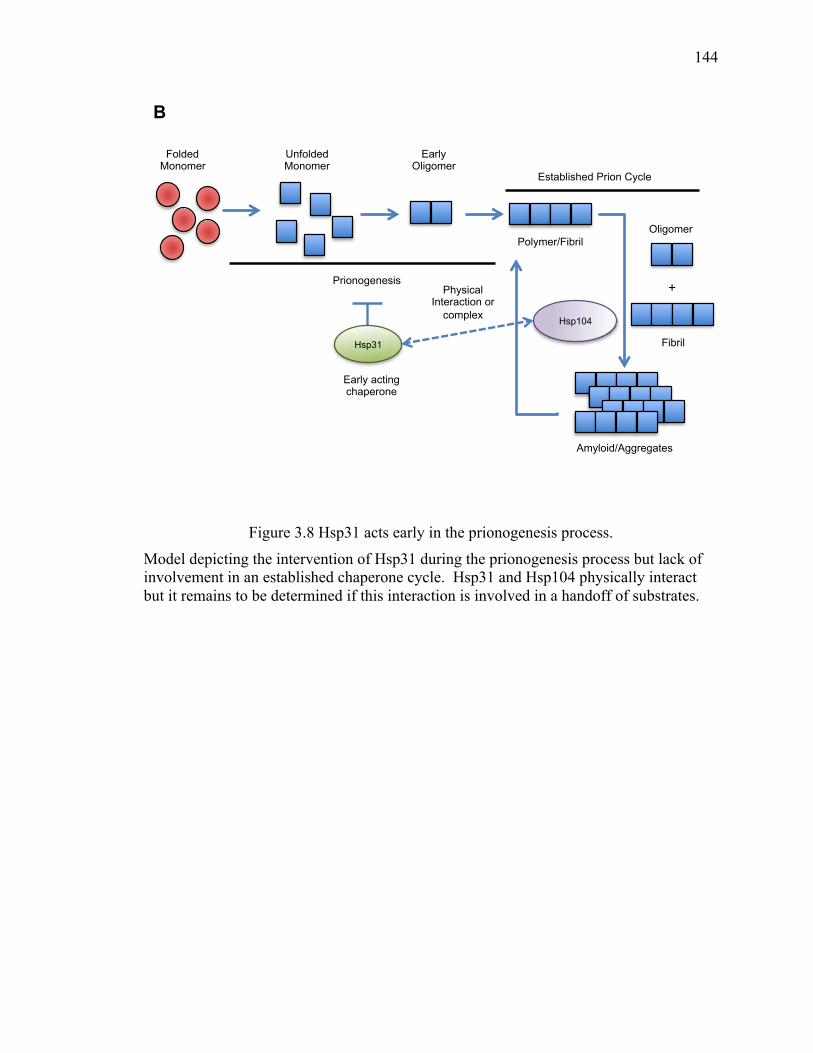
144
144
Figure 3.8 Hsp31 acts early in the prionogenesis process.
Model depicting the intervention of Hsp31 during the prionogenesis process but lack of involvement in an established chaperone cycle. Hsp31 and Hsp104 physically interact but it remains to be determined if this interaction is involved in a handoff of substrates.
Folded Monomer
Unfolded Monomer
+
Polymer/Fibril Oligomer
Fibril
Amyloid/Aggregates
Early acting chaperone
Prionogenesis
Established Prion Cycle
Early Oligomer
Hsp31
Hsp104
Physical Interaction or
complex
B
145
145
Hsp31 may not participate in modulating an established prion cycle by itself but does
appear to have a role in conjunction with Hsp104 because in a strain lacking Hsp31, the
rate of [PSI+] curing was reduced with Hsp104 overexpression. In addition, we showed
Hsp31 overexpression promotes the elimination of [PSI+] by Hsp104. In fact, another
small HSP Hsp26 was shown to potentiate protein disaggregation by Hsp104.
Interestingly, Hsp26 is only active as a disaggregase when it clusters together with the
protein substrate and not after protein aggregation. A factor in considering the
mechanism of action is that Hsp31 and the human ortholog, DJ-1, have protein deglycase
activity (265,292). Prion glycation can occur spontaneously (343) and glycation can
promote the stability of protein aggregates by covalent crosslinking(344) suggesting the
activity of a deglycase may abrogate prion propogation. Deciphering the contribution of
the enzyme activity versus chaperone function of Hsp31 would be revealing of the
biological role of this multi-functional enzyme. We have previously shown that
chaperone activity can be independent of the enzyme activity because overexpression of
an enzymatically inactive Hsp31 mutant prevents the toxicity of α-syn in yeast. While
there is enough evidence to propose Hsp31 acts early in the process of [PSI+] prion
oligomerization process, future investigation of Hsp31 binding to the Sup35 monomer or
other early oligomer and the role of deglycation in inhibiting prion induction would be
further revealing of the mechanism of action.
We eliminated several possible indirect mechanisms for the cooperation of Hsp31 with
Hsp104 including the possibility that disruption of Hsp31 might compromise Hsp104
expression and thermotolerance function (Figure 3.5). Taking into account that deletion
of Hsp31 down regulates the mRNA level of Ssa3, a possible effect of Hsp31 deletion on
146
146
[PSI+] prion curing by Hsp104 could be due to imbalances between Hsp104 and Hsp70
chaperone as these are required for efficient prion curing. However, this cannot be the
case when Hsp31 is co-expressed with Hsp104, as overexpression of Hsp31 does not alter
the level of Hsp70 protein (158).
We also showed that Hsp31 together with Hsp104 significantly reduced Sup35 prion
toxicity. These results indicate that Hsp31 cooperates with Hsp104 to potentiate Sup35
prion disaggregation and thereby prevent toxicity. Intriguingly, previous studies have
shown such cooperation between sHSPs and ATP dependent chaperones as Hsp104 and
Hsp70 (234,241,243). We also observed a significant reduction of Sup35 toxicity by
Hsp31 alone. A number of possible explanations could account for these results such as,
Hsp31 might prevent sequestration of soluble Sup35 into already present larger
aggregates and therefore inhibit toxicity. We have previously demonstrated that Hsp31
can reduce oxidative stress in cells (158) and this may be the reason for the rescue of
toxicity.
Intriguingly, we also found that Hsp31 physically interacts with Hsp104. Hsp104 is
known to interact with Ydj1, and co-chaperones of Hsp90; Sti1 and Cpr7 (40,225).
Interestingly, Sti1 and Cpr7 are not required for Sup35 prion propagation but deletion of
either of these reduced the Sup35 prion curing by Hsp104. Hsp31 has been documented
to interact with the yeast Hsp90, Hsp82 (345), based on affinity-mass spectrometry hence
the interaction may involve a bridging chaperone such as Hsp90. However, our data is
the first demonstration of Hsp31’s interaction with Hsp104 and could mean these
chaperones may pass substrates to each other. Although the molecular details of the

147
147
interaction are unknown, the involvement of Hsp31 in the prion modulation process and
the apparent close functional cooperation with Hsp104 is an important step in
understanding the biological roles of this multi-tasking protein.
148
148
CHAPTER 4. CONCLUSION AND FUTURE DIRECTIONS
4.1 Summary
Numerous human pathologies are associated with protein misfolding and aggregation
including several neurodegenerative diseases such as AD, PD and prion diseases. For
each such disease a different misfolded protein is responsible for neurodegeneration at
different regions of the brain that leads to differential clinical effects. Although there is
extensive understanding of these molecular mechanisms, there are still numerous
unanswered questions. Several studies show the protective role of HSPs in misfolding
processes that function as chaperones in modulating protein aggregation and their
involvement in neurodegenerative diseases. In this thesis, we used yeast as a model
system to gain insight into the role of Hsp31 as a molecular chaperone and characterized
its function in PD and prion modulation in yeast.
4.2 What makes Hsp31 a multifunctional chaperone protein? What is the role of the
C138 residue in catalytic triad of Hsp31?
An emerging view is that Hsp31 and its associated superfamily members each have
divergent multitasking functions that have the common theme of responding and
managing various types of cellular stress. Hsp31 is involved in multiple cellular functions
including oxidative stress sensing, chaperone and detoxifying enzyme activities such as
functions as methylglyoxalase and deglycation. In chapter 2, we have shown that
149
149
protective role of Hsp31’s chaperone activity can operate independent of detoxifying
enzyme activities in preventing the early stages of protein aggregate formation and
associated cellular toxicities. The catalytic triad present in the DJ-1 superfamily members
is the most common feature, mutation of which can lead to functional variation and
destabilization of dimerization.
In this thesis we showed that Hsp31 is capable of acting as a chaperone without
involvement of its enzymatic activity. Hsp31 possesses a conserved cysteine residue
C138 that is present in different species (272). Previously, it was shown that mutation in
C138 abrogates its enzymatic activity. However, in this thesis, we provide strong
evidence that mutation of C138 has no effect on the chaperone activity of Hsp31. Future
studies should be performed to further delineate the role of C138 in Hsp31. In DJ-1, the
oxidative state of C106 is involved in redox sensing, and the mutation of it destabilizes
the DJ-1 functions. Under oxidative stress condition DJ-1 homo-dimer destabilizes and
therefore loss its protective effect (346,347). Similarly, Hsp31 exists predominantly as a
homo-dimer in solution as shown previously (271,272), and it would be interesting to
determine what is the role of dimerization state in the chaperone activity of Hsp31? It
remains to be determined, what are the chaperone active sites, if C138 has a limited role
in chaperone function? Furthermore, in chapter 3 we provide significant evidence that
Hsp31 is involved in the modulation of prion aggregation, however, we never tested the
effect of C138 mutation in these experiments. Studying these questions will provide a
further mechanistic insight into the role of Hsp31 as a chaperone protein.
150
150
4.3 What is the link between Hsp31 deglycase activity and aggregation activity?
It is now well established that Hsp31 is a molecular chaperone that intervene in the
aggregation of a broad range of protein substrates. Our studies provide strong evidence to
delineating that Hsp31 intervenes early in the process of protein aggregation to prevent
larger aggregate or oligomer formation. Future studies will need to focus on dissection of
the mechanistic details of how Hsp31 prevents the formation of aggregates. Recent
studies showed that enzymatic activity of Hsp31 is capable to deglycate the damaged
proteins. One hypothesis is that Hsp31 binds to damaged or glycated substrate protein to
deglycate them and therefore prevent the formation of aggregates caused by glycation of
the monomeric proteins. It will be important to study that if Hsp31 binds to glycated
monomeric protein and therefore prevent further aggregation of protein. It is not clear if
MGO or glycated proteins are the prime substrate for Hsp31 or if both are important
hence, determination of the in vitro enzymatic activities of Hsp31 on these two different
substrates is important to determine. Further in vivo studies that explore the effect of
Hsp31 on glycated proteins will be needed to provided insight into the mode of action
and reveal the natural substrate(s) of Hsp31 with respect to its
methylyglyoxalase/deglycase activities.
4.4 Determine the role and nature of the Hsp31 and Hsp104 interaction? What are the
roles of Hsp82/Hsp70 and other co-chaperones in conjunction with Hsp31?
In chapter 3 we showed that Hsp31 cooperates with Hsp104 to prevent Sup35 prion
aggregation. Future studies will focus on the nature of interaction between Hsp31 and
Hsp104. Although, we have shown that Hsp31 can pull down endogenous levels of
Hsp104, it is not known if this interaction is direct based on the co-immunoprecipitation.
151
151
In order to determine if the interaction is direct, in vitro binding assays using recombinant
proteins would answer this question. If these two chaperones interact directly, we can
further explore the interaction sites or regions important for such an interaction.
Information on how the DJ-1/Hsp31 protein family interacts with other chaperones or co-
factors is severely lacking. Furthermore, identification of direct interaction sites would
allow a dissection and assessment of the role of this interaction on the overall activity of
Hsp104 or Hsp31? Based on affinity-mass spectrometry, it has been documented
previously, that Hsp31 interacts with the yeast Hsp90, Hsp82. It is also known that
Hsp104 interacts with co-chaperones of Hsp90 hence, the interaction between Hsp104
and Hsp31 may require a bridging chaperone as Hsp90 or its co-chaperones; Sti1 and
Cpr7. Alternatively, Hsp31 maybe a co-chaperone of Hsp90 or Hsp104 and may deliver
substrates to these other chaperones as part of an overall multi-step chaperone cycle.
4.5 Functional diversity or overlap among Hsp31 paralogs in yeast.
The Hsp31 mini-family is composed of four paralogs; HSP31 (YDR533C), HSP32
(YMR322C), HSP33 (YOR391C), and HSP34 (YPL280W). Genes of the Hsp31 mini
family are located at the subtelomeric region of the genome in Saccharomyces cerevisiae.
HSP31 is considered the parental gene with HSP32, HSP33 and HSP34 originating from
it during gene duplication events. Among all the members of this mini-family, Hsp31 is
most divergent and shares approximately 70% homology with the other members of the
family those possess more than 90% homology between them. All the members of Hsp31
family contain the same Cys-His-Glu catalytic triad including the E. coli ortholog.
Previously, it was shown that mutation in the catalytic triad largely abolishes glyoxalase
activity but this catalytic triad is not required for chaperone activity of Hsp31. These
152
152
results indicate that the anti-aggregation activity of Hsp31 is not under the influence of its
enzymatic activity rather, it has a direct chaperone activity against misfolded proteins.
Intriguingly, all the paralogs of the Hsp31 minifamily possess comparable activity against
α-syn aggregation and toxicity when they are overexpressed from the GAL promoter.
However, unlike Hsp31, the other paralogs possess very little methylglyoxalase activity
and are unable to protect the cells from glyoxal toxicity (260,348). These results support
the notion that anti-aggregation activity of Hsp31 mini-family is independent of its
enzymatic activity. Furthermore, all the members of the Hsp31 family contain the same
catalytic triad required for methylglyoxalse activity but so far only Hsp31 is determined
as the most active enzyme in vitro and in vivo. Despite the presence of the catalytic triad
Hsp34 is found to be a very weak methylglyoxalase, which indicates that other regions in
the protein sequence are also important for enzymatic activity. Hsp31 and Hsp34’s
divergent activity has only been shown for MGO, and the different paralogs could have
differential activity depending on the substrate type. In future research, it will be
important to determine the functional diversity between paralogs of Hsp31 in term of its
methylglyoxalase and other enzymatic activities. As, the lack of methylglyoxalase
activity in one of the paralogs is evidence that the paralogs are diverging but additional
studies dissecting the roles within this paralog group are needed to further uncover these
diverging functions. It is also important to note that other biomolecules such as nucleic
acids are also susceptible to oxidative damage and could also be substrates for the Hsp31
protein family.
In contrast, the chaperone activity of Hsp31 and its paralog are comparable (106). Also,
the chaperone activity of Hsp31, Hsp32 and Hsp33 against a cytoplasmic aggregation-
153
153
prone protein is independent of their role in oxidative stress response and the vacuolar
degradation pathway (262). It would be interesting to study the functional overlap of
these family members. The Hsp31 protein family is broadly spread across fungal species
with varying levels of paralog duplications and additional evidence of divergence
including differences in localization in the Schizosaccharomyces pombe Hsp31 family
members (272).
4.6 Conclusion
In conclusion, study presented here significantly contributed to understand the molecular
role of Hsp31 as a chaperone in protein misfolding diseases. Hsp31 promotes cell
survival under different stress conditions by participating in multiple cellular pathways.
Although, currently we know that Hsp31 is a multifunctional stress responder chaperone
proteins that have homologs in almost every organisms, the exact molecular mechanism
of Hsp31 remains enigmatic. Further studying the mode of action of Hsp31 provides a
promising model to understand the cytoprotective functions of DJ-1 that can be a
potential target for neurodegenerative disease as PD.

REFERENCES
154
154
REFERENCES
1. Ruggiano, A., Foresti, O., and Carvalho, P. (2014) Quality control: ER-associated degradation: protein quality control and beyond. J Cell Biol 204, 869-879
2. Zattas, D., and Hochstrasser, M. (2015) Ubiquitin-dependent protein degradation at the yeast endoplasmic reticulum and nuclear envelope. Crit Rev Biochem Mol Biol 50, 1-17
3. Wickner, S., Maurizi, M. R., and Gottesman, S. (1999) Posttranslational quality control: folding, refolding, and degrading proteins. Science 286, 1888-1893
4. Boban, M., and Foisner, R. (2016) Degradation-mediated protein quality control at the inner nuclear membrane. Nucleus, 0
5. Rusmini, P., Crippa, V., Cristofani, R., Rinaldi, C., Cicardi, M. E., Galbiati, M., Carra, S., Malik, B., Greensmith, L., and Poletti, A. (2015) The Role of the Protein Quality Control System in SBMA. J Mol Neurosci
6. Mukherjee, R., and Chakrabarti, O. (2016) Ubiquitin-mediated regulation of the E3 ligase GP78 by MGRN1 in trans affects mitochondrial homeostasis. J Cell Sci 129, 757-773
7. Webster, B. M., and Lusk, C. P. (2016) Border Safety: Quality Control at the Nuclear Envelope. Trends Cell Biol 26, 29-39
8. Halperin, L., Jung, J., and Michalak, M. (2014) The many functions of the endoplasmic reticulum chaperones and folding enzymes. IUBMB Life 66, 318-326
9. Kurland, C. G. (1992) Translational accuracy and the fitness of bacteria. Annu Rev Genet 26, 29-50
10. Hurtley, S. M., and Helenius, A. (1989) Protein oligomerization in the endoplasmic reticulum. Annu Rev Cell Biol 5, 277-307
11. Ibba, M., and Söll, D. (1999) Quality control mechanisms during translation. Science 286, 1893-1897
12. Lindahl, T., and Wood, R. D. (1999) Quality control by DNA repair. Science 286, 1897-1905
13. Hyttinen, J. M., Amadio, M., Viiri, J., Pascale, A., Salminen, A., and Kaarniranta, K. (2014) Clearance of misfolded and aggregated proteins by aggrephagy and implications for aggregation diseases. Ageing Res Rev 18C, 16-28
14. Shah, S. Z., Zhao, D., Khan, S. H., and Yang, L. (2015) Unfolded Protein Response Pathways in Neurodegenerative Diseases. J Mol Neurosci 57, 529-537
15. Amor, A. J., Castanzo, D. T., Delany, S. P., Selechnik, D. M., van Ooy, A., and Cameron, D. M. (2015) The ribosome-associated complex antagonizes prion formation in yeast. Prion 9, 144-164
155
155
16. Harrison, R. S., Sharpe, P. C., Singh, Y., and Fairlie, D. P. (2007) Amyloid peptides and proteins in review. Rev Physiol Biochem Pharmacol 159, 1-77
17. Franssens, V., Boelen, E., Anandhakumar, J., Vanhelmont, T., Büttner, S., and Winderickx, J. (2010) Yeast unfolds the road map toward alpha-synuclein-induced cell death. Cell Death Differ 17, 746-753
18. Wang, M., and Kaufman, R. J. (2016) Protein misfolding in the endoplasmic reticulum as a conduit to human disease. Nature 529, 326-335
19. Hammarström, P. (2009) Protein folding, misfolding and disease. FEBS Lett 583, 2579-2580
20. Cardinale, A., Chiesa, R., and Sierks, M. (2014) Protein misfolding and neurodegenerative diseases. Int J Cell Biol 2014, 217371
21. Ellisdon, A. M., and Bottomley, S. P. (2004) The role of protein misfolding in the pathogenesis of human diseases. IUBMB Life 56, 119-123
22. Penke, B., Bogár, F., and Fülöp, L. (2016) Protein Folding and Misfolding, Endoplasmic Reticulum Stress in Neurodegenerative Diseases: in Trace of Novel Drug Targets. Curr Protein Pept Sci 17, 169-182
23. Singh, J., and Udgaonkar, J. B. (2015) Molecular Mechanism of the Misfolding and Oligomerization of the Prion Protein: Current Understanding and Its Implications. Biochemistry 54, 4431-4442
24. Englander, S. W., Mayne, L., and Krishna, M. M. (2007) Protein folding and misfolding: mechanism and principles. Q Rev Biophys 40, 287-326
25. Borgia, A., Kemplen, K. R., Borgia, M. B., Soranno, A., Shammas, S., Wunderlich, B., Nettels, D., Best, R. B., Clarke, J., and Schuler, B. (2015) Transient misfolding dominates multidomain protein folding. Nat Commun 6, 8861
26. Kim, Y. E., Hipp, M. S., Bracher, A., Hayer-Hartl, M., and Hartl, F. U. (2013) Molecular chaperone functions in protein folding and proteostasis. Annu Rev Biochem 82, 323-355
27. Hubbard, T. J., and Sander, C. (1991) The role of heat-shock and chaperone proteins in protein folding: possible molecular mechanisms. Protein Eng 4, 711-717
28. Gong, Y., Kakihara, Y., Krogan, N., Greenblatt, J., Emili, A., Zhang, Z., and Houry, W. A. (2009) An atlas of chaperone-protein interactions in Saccharomyces cerevisiae: implications to protein folding pathways in the cell. Mol Syst Biol 5, 275
29. Burel, C., Mezger, V., Pinto, M., Rallu, M., Trigon, S., and Morange, M. (1992) Mammalian heat shock protein families. Expression and functions. Experientia 48, 629-634
30. Ellis, R. J., van der Vies, S. M., and Hemmingsen, S. M. (1989) The molecular chaperone concept. Biochem Soc Symp 55, 145-153
31. Horwich, A. L., and Willison, K. R. (1993) Protein folding in the cell: functions of two families of molecular chaperone, hsp 60 and TF55-TCP1. Philos Trans R Soc Lond B Biol Sci 339, 313-325; discussion 325-316
32. Kelley, W. L., and Georgopoulos, C. (1992) Chaperones and protein folding. Curr Opin Cell Biol 4, 984-991
156
156
33. Young, J. C. (2010) Mechanisms of the Hsp70 chaperone system. Biochem Cell Biol 88, 291-300
34. Pratt, W. B., Morishima, Y., Gestwicki, J. E., Lieberman, A. P., and Osawa, Y. (2014) A model in which heat shock protein 90 targets protein-folding clefts: rationale for a new approach to neuroprotective treatment of protein folding diseases. Exp Biol Med (Maywood) 239, 1405-1413
35. Miura, T., Minegishi, H., Usami, R., and Abe, F. (2006) Systematic analysis of HSP gene expression and effects on cell growth and survival at high hydrostatic pressure in Saccharomyces cerevisiae. Extremophiles 10, 279-284
36. Cyr, D. M., and Ramos, C. H. (2015) Specification of Hsp70 function by Type I and Type II Hsp40. Subcell Biochem 78, 91-102
37. Duncan, E. J., Cheetham, M. E., Chapple, J. P., and van der Spuy, J. (2015) The role of HSP70 and its co-chaperones in protein misfolding, aggregation and disease. Subcell Biochem 78, 243-273
38. Mogk, A., Kummer, E., and Bukau, B. (2015) Cooperation of Hsp70 and Hsp100 chaperone machines in protein disaggregation. Front Mol Biosci 2, 22
39. Reidy, M., Sharma, R., Shastry, S., Roberts, B. L., Albino-Flores, I., Wickner, S., and Masison, D. C. (2014) Hsp40s specify functions of Hsp104 and Hsp90 protein chaperone machines. PLoS Genet 10, e1004720
40. Reidy, M., and Masison, D. C. (2010) Sti1 regulation of Hsp70 and Hsp90 is critical for curing of Saccharomyces cerevisiae [PSI+] prions by Hsp104. Mol Cell Biol 30, 3542-3552
41. Walter, S., and Buchner, J. (2002) Molecular Chaperones—Cellular Machines for Protein Folding. Angewandte Chemie International Edition 41, 1098-1113
42. Ritossa, F. (1962) A new puffing pattern induced by temperature shock and DNP in drosophila. Experientia 18, 571-573
43. Rotossa, F. (1996) Discovery of the heat shock response. Cell stress Chaperones 1, 97-98
44. Bakthisaran, R., Tangirala, R., and Rao, C. M. (2015) Small heat shock proteins: Role in cellular functions and pathology. Biochim Biophys Acta 1854, 291-319
45. Mokry, D. Z., Abrahão, J., and Ramos, C. H. (2015) Disaggregases, molecular chaperones that resolubilize protein aggregates. An Acad Bras Cienc 87, 1273-1292
46. Schlesinger, M. J. (1990) Heat shock proteins. Journal of Biological Chemistry 265, 12111-12114
47. Clark, J. I., and Muchowski, P. J. (2000) Small heat-shock proteins and their potential role in human disease. Current Opinion in Structural Biology 10, 52-59
48. Eyles, S. J., and Gierasch, L. M. (2010) Nature’s molecular sponges: Small heat shock proteins grow into their chaperone roles. Proceedings of the National Academy of Sciences 107, 2727-2728
49. Jakob, U., Gaestel, M., Engel, K., and Buchner, J. (1993) Small heat shock proteins are molecular chaperones. Journal of Biological Chemistry 268, 1517-1520
157
157
50. Wehmeyer, N., and Vierling, E. (2000) The expression of small heat shock proteins in seeds responds to discrete developmental signals and suggests a general protective role in desiccation tolerance. Plant Physiol 122, 1099-1108
51. Zeng, L., Tan, J., Lu, T., Lei, Q., Chen, C., and Hu, Z. (2015) Small heat shock proteins and the endoplasmic reticulum: potential attractive therapeutic targets? Curr Mol Med 15, 38-46
52. Sun, Y., and MacRae, T. H. (2005) The small heat shock proteins and their role in human disease. FEBS Journal 272, 2613-2627
53. Duennwald, M. L., Echeverria, A., and Shorter, J. (2012) Small heat shock proteins potentiate amyloid dissolution by protein disaggregases from yeast and humans. PLoS Biol 10, e1001346
54. Kovacs, G. G. (2016) Molecular Pathological Classification of Neurodegenerative Diseases: Turning towards Precision Medicine. Int J Mol Sci 17
55. Carr, F. (2016) Neurodegenerative disease: Forming fragments. Nat Rev Neurosci 17, 74-75
56. Agid, Y. (1993) [Mechanism of cell death in neurodegenerative disorders: apropos of Parkinson disease]. C R Seances Soc Biol Fil 187, 37-46
57. Hamill, R. W., Caine, E., Eskin, T., Lapham, L., Shoulson, I., and McNeill, T. H. (1988) Neurodegenerative disorders and aging. Alzheimer's disease and Parkinson's disease--common ground. Ann N Y Acad Sci 515, 411-420
58. Ceballos, I., Javoy-Agid, F., Delacourte, A., Defossez, A., Nicole, A., and Sinet, P. M. (1990) Parkinson's disease and Alzheimer's disease: neurodegenerative disorders due to brain antioxidant system deficiency? Adv Exp Med Biol 264, 493-498
59. Crunkhorn, S. (2016) Neurodegenerative disease: Immunotherapy opportunity emerges for Alzheimer disease. Nat Rev Drug Discov
60. Iqbal, K., Liu, F., and Gong, C. X. (2016) Tau and neurodegenerative disease: the story so far. Nat Rev Neurol 12, 15-27
61. Rojas, J. C., and Boxer, A. L. (2016) Neurodegenerative disease in 2015: Targeting tauopathies for therapeutic translation. Nat Rev Neurol 12, 74-76
62. Dias, V., Junn, E., and Mouradian, M. M. (2013) The role of oxidative stress in Parkinson's disease. J Parkinsons Dis 3, 461-491
63. Jellinger, K. A. (2010) Basic mechanisms of neurodegeneration: a critical update. Journal of Cellular and Molecular Medicine 14, 457-487
64. Mandemakers, W., Morais, V. A., and De Strooper, B. (2007) A cell biological perspective on mitochondrial dysfunction in Parkinson disease and other neurodegenerative diseases. J Cell Sci 120, 1707-1716
65. Albers, D. S., and Beal, M. F. (2000) Mitochondrial dysfunction and oxidative stress in aging and neurodegenerative disease. J Neural Transm Suppl 59, 133-154
66. Butterfield, D. A., Palmieri, E. M., and Castegna, A. (2016) Clinical implications from proteomic studies in neurodegenerative diseases: lessons from mitochondrial proteins. Expert Rev Proteomics
67. Matsuda, N., and Tanaka, K. (2011) [Neurodegenerative disorder as "mitochondrial dysfunction disease"]. Rinsho Shinkeigaku 51, 988
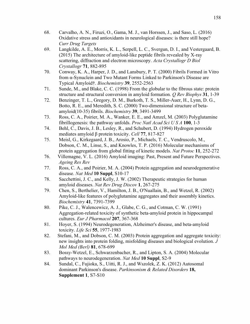
158
158
68. Carvalho, A. N., Firuzi, O., Gama, M. J., van Horssen, J., and Saso, L. (2016) Oxidative stress and antioxidants in neurological diseases: is there still hope? Curr Drug Targets
69. Langkilde, A. E., Morris, K. L., Serpell, L. C., Svergun, D. I., and Vestergaard, B. (2015) The architecture of amyloid-like peptide fibrils revealed by X-ray scattering, diffraction and electron microscopy. Acta Crystallogr D Biol Crystallogr 71, 882-895
70. Conway, K. A., Harper, J. D., and Lansbury, P. T. (2000) Fibrils Formed in Vitro from α-Synuclein and Two Mutant Forms Linked to Parkinson's Disease are Typical Amyloid†. Biochemistry 39, 2552-2563
71. Sunde, M., and Blake, C. C. (1998) From the globular to the fibrous state: protein structure and structural conversion in amyloid formation. Q Rev Biophys 31, 1-39
72. Benzinger, T. L., Gregory, D. M., Burkoth, T. S., Miller-Auer, H., Lynn, D. G., Botto, R. E., and Meredith, S. C. (2000) Two-dimensional structure of beta-amyloid(10-35) fibrils. Biochemistry 39, 3491-3499
73. Ross, C. A., Poirier, M. A., Wanker, E. E., and Amzel, M. (2003) Polyglutamine fibrillogenesis: the pathway unfolds. Proc Natl Acad Sci U S A 100, 1-3
74. Behl, C., Davis, J. B., Lesley, R., and Schubert, D. (1994) Hydrogen peroxide mediates amyloid β protein toxicity. Cell 77, 817-827
75. Meisl, G., Kirkegaard, J. B., Arosio, P., Michaels, T. C., Vendruscolo, M., Dobson, C. M., Linse, S., and Knowles, T. P. (2016) Molecular mechanisms of protein aggregation from global fitting of kinetic models. Nat Protoc 11, 252-272
76. Villemagne, V. L. (2016) Amyloid imaging: Past, Present and Future Perspectives. Ageing Res Rev
77. Ross, C. A., and Poirier, M. A. (2004) Protein aggregation and neurodegenerative disease. Nat Med 10 Suppl, S10-17
78. Sacchettini, J. C., and Kelly, J. W. (2002) Therapeutic strategies for human amyloid diseases. Nat Rev Drug Discov 1, 267-275
79. Chen, S., Berthelier, V., Hamilton, J. B., O'Nuallain, B., and Wetzel, R. (2002) Amyloid-like features of polyglutamine aggregates and their assembly kinetics. Biochemistry 41, 7391-7399
80. Pike, C. J., Walencewicz, A. J., Glabe, C. G., and Cotman, C. W. (1991) Aggregation-related toxicity of synthetic beta-amyloid protein in hippocampal cultures. Eur J Pharmacol 207, 367-368
81. Hoyer, S. (1994) Neurodegeneration, Alzheimer's disease, and beta-amyloid toxicity. Life Sci 55, 1977-1983
82. Stefani, M., and Dobson, C. M. (2003) Protein aggregation and aggregate toxicity: new insights into protein folding, misfolding diseases and biological evolution. J Mol Med (Berl) 81, 678-699
83. Bossy-Wetzel, E., Schwarzenbacher, R., and Lipton, S. A. (2004) Molecular pathways to neurodegeneration. Nat Med 10 Suppl, S2-9
84. Sundal, C., Fujioka, S., Uitti, R. J., and Wszolek, Z. K. (2012) Autosomal dominant Parkinson's disease. Parkinsonism & Related Disorders 18, Supplement 1, S7-S10
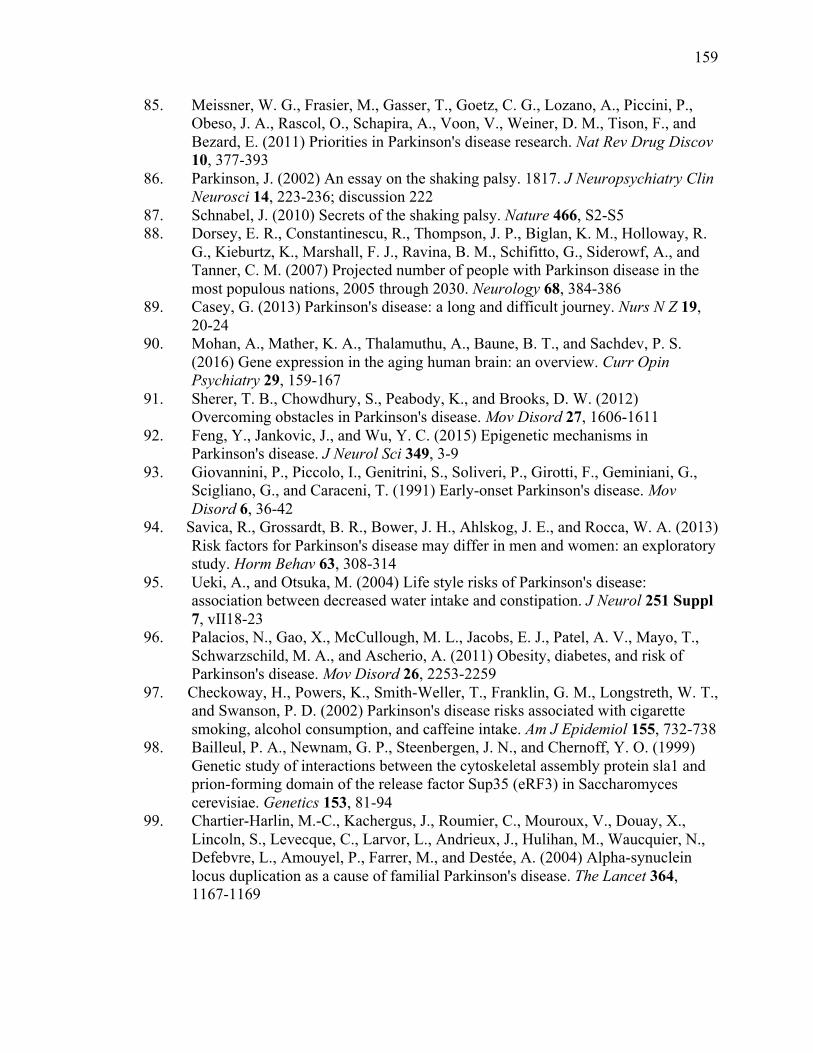
159
159
85. Meissner, W. G., Frasier, M., Gasser, T., Goetz, C. G., Lozano, A., Piccini, P., Obeso, J. A., Rascol, O., Schapira, A., Voon, V., Weiner, D. M., Tison, F., and Bezard, E. (2011) Priorities in Parkinson's disease research. Nat Rev Drug Discov 10, 377-393
86. Parkinson, J. (2002) An essay on the shaking palsy. 1817. J Neuropsychiatry Clin Neurosci 14, 223-236; discussion 222
87. Schnabel, J. (2010) Secrets of the shaking palsy. Nature 466, S2-S5 88. Dorsey, E. R., Constantinescu, R., Thompson, J. P., Biglan, K. M., Holloway, R.
G., Kieburtz, K., Marshall, F. J., Ravina, B. M., Schifitto, G., Siderowf, A., and Tanner, C. M. (2007) Projected number of people with Parkinson disease in the most populous nations, 2005 through 2030. Neurology 68, 384-386
89. Casey, G. (2013) Parkinson's disease: a long and difficult journey. Nurs N Z 19, 20-24
90. Mohan, A., Mather, K. A., Thalamuthu, A., Baune, B. T., and Sachdev, P. S. (2016) Gene expression in the aging human brain: an overview. Curr Opin Psychiatry 29, 159-167
91. Sherer, T. B., Chowdhury, S., Peabody, K., and Brooks, D. W. (2012) Overcoming obstacles in Parkinson's disease. Mov Disord 27, 1606-1611
92. Feng, Y., Jankovic, J., and Wu, Y. C. (2015) Epigenetic mechanisms in Parkinson's disease. J Neurol Sci 349, 3-9
93. Giovannini, P., Piccolo, I., Genitrini, S., Soliveri, P., Girotti, F., Geminiani, G., Scigliano, G., and Caraceni, T. (1991) Early-onset Parkinson's disease. Mov Disord 6, 36-42
94. Savica, R., Grossardt, B. R., Bower, J. H., Ahlskog, J. E., and Rocca, W. A. (2013) Risk factors for Parkinson's disease may differ in men and women: an exploratory study. Horm Behav 63, 308-314
95. Ueki, A., and Otsuka, M. (2004) Life style risks of Parkinson's disease: association between decreased water intake and constipation. J Neurol 251 Suppl 7, vII18-23
96. Palacios, N., Gao, X., McCullough, M. L., Jacobs, E. J., Patel, A. V., Mayo, T., Schwarzschild, M. A., and Ascherio, A. (2011) Obesity, diabetes, and risk of Parkinson's disease. Mov Disord 26, 2253-2259
97. Checkoway, H., Powers, K., Smith-Weller, T., Franklin, G. M., Longstreth, W. T., and Swanson, P. D. (2002) Parkinson's disease risks associated with cigarette smoking, alcohol consumption, and caffeine intake. Am J Epidemiol 155, 732-738
98. Bailleul, P. A., Newnam, G. P., Steenbergen, J. N., and Chernoff, Y. O. (1999) Genetic study of interactions between the cytoskeletal assembly protein sla1 and prion-forming domain of the release factor Sup35 (eRF3) in Saccharomyces cerevisiae. Genetics 153, 81-94
99. Chartier-Harlin, M.-C., Kachergus, J., Roumier, C., Mouroux, V., Douay, X., Lincoln, S., Levecque, C., Larvor, L., Andrieux, J., Hulihan, M., Waucquier, N., Defebvre, L., Amouyel, P., Farrer, M., and Destée, A. (2004) Alpha-synuclein locus duplication as a cause of familial Parkinson's disease. The Lancet 364, 1167-1169
160
160
100. Cookson, M. (2009) alpha-Synuclein and neuronal cell death. Molecular Neurodegeneration 4, 9
101. McLean, P. J., Kawamata, H., Ribich, S., and Hyman, B. T. (2000) Membrane association and protein conformation of alpha-synuclein in intact neurons. Effect of Parkinson's disease-linked mutations. J Biol Chem 275, 8812-8816
102. Taguchi, K., Watanabe, Y., Tsujimura, A., Tatebe, H., Miyata, S., Tokuda, T., Mizuno, T., and Tanaka, M. (2014) Differential expression of alpha-synuclein in hippocampal neurons. PLoS One 9, e89327
103. Cookson, M., and Brug, M. (2008) Cell systems and the toxic mechanism(s) of alpha-synuclein. Exp Neurol 209, 5 - 11
104. Cooper, A., Gitler, A., Cashikar, A., Haynes, C., Hill, K., Bhullar, B., Liu, K., Xu, K., Strathearn, K., Liu, F., Cao, S., Caldwell, K., Caldwell, G., Marsischky, G., Kolodner, R., Labaer, J., Rochet, J., Bonini, N., and Lindquist, S. (2006) Alpha-synuclein blocks ER-Golgi traffic and Rab1 rescues neuron loss in Parkinson's models. Science 313, 324 - 328
105. Hsu, L., Sagara, Y., Arroyo, A., Rockenstein, E., Sisk, A., Mallory, M., Wong, J., Takenouchi, T., Hashimoto, M., and Masliah, E. (2000) alpha-synuclein promotes mitochondrial deficit and oxidative stress. Am J Pathol 157, 401 - 410
106. Zondler, L., Miller-Fleming, L., Repici, M., Gonçalves, S., Tenreiro, S., Rosado-Ramos, R., Betzer, C., Straatman, K. R., Jensen, P. H., Giorgini, F., and Outeiro, T. F. (2014) DJ-1 interactions with α-synuclein attenuate aggregation and cellular toxicity in models of Parkinson's disease. Cell Death Dis 5, e1350
107. Atik, A., Stewart, T., and Zhang, J. (2016) Alpha-synuclein as a biomarker for Parkinson's disease. Brain Pathol
108. Scherfler, C., Göbel, G., Müller, C., Nocker, M., Wenning, G. K., Schocke, M., Poewe, W., and Seppi, K. (2016) Diagnostic potential of automated subcortical volume segmentation in atypical parkinsonism. Neurology
109. Postuma, R. B., Gagnon, J. F., and Montplaisir, J. (2010) Clinical prediction of Parkinson's disease: planning for the age of neuroprotection. J Neurol Neurosurg Psychiatry 81, 1008-1013
110. Burkhard, P. (2015) [Parkinson disease diagnosis: not so easy...]. Rev Med Suisse 11, 304-305
111. Martí, M. J., and Tolosa, E. (2013) Parkinson disease: New guidelines for diagnosis of Parkinson disease. Nat Rev Neurol 9, 190-191
112. Levin, J., Kurz, A., Arzberger, T., Giese, A., and Höglinger, G. U. (2016) The Differential Diagnosis and Treatment of Atypical Parkinsonism. Dtsch Arztebl Int 113, 61-69
113. Rajput, A. H., and Rajput, A. (2014) Accuracy of Parkinson disease diagnosis unchanged in 2 decades. Neurology 83, 386-387
114. Miller, D. B., and O'Callaghan, J. P. (2015) Biomarkers of Parkinson's disease: present and future. Metabolism 64, S40-46
115. Mattison, H. A., Stewart, T., and Zhang, J. (2012) Applying bioinformatics to proteomics: Is machine learning the answer to biomarker discovery for PD and MSA? Movement Disorders 27, 1595-1597
161
161
116. Hall, S., Surova, Y., Öhrfelt, A., Zetterberg, H., Lindqvist, D., and Hansson, O. (2015) CSF biomarkers and clinical progression of Parkinson disease. Neurology 84, 57-63
117. Perlmutter, J. S., and Norris, S. A. (2014) Neuroimaging biomarkers for Parkinson disease: facts and fantasy. Ann Neurol 76, 769-783
118. Foulds, P. G., Mitchell, J. D., Parker, A., Turner, R., Green, G., Diggle, P., Hasegawa, M., Taylor, M., Mann, D., and Allsop, D. (2011) Phosphorylated α-synuclein can be detected in blood plasma and is potentially a useful biomarker for Parkinson's disease. The FASEB Journal 25, 4127-4137
119. Bonifati, V., Rizzu, P., van Baren, M. J., Schaap, O., Breedveld, G. J., Krieger, E., Dekker, M. C., Squitieri, F., Ibanez, P., Joosse, M., van Dongen, J. W., Vanacore, N., van Swieten, J. C., Brice, A., Meco, G., van Duijn, C. M., Oostra, B. A., and Heutink, P. (2003) Mutations in the DJ-1 gene associated with autosomal recessive early-onset parkinsonism. Science 299, 256-259
120. Davis, J. W., Grandinetti, A., Waslien, C. I., Ross, G. W., White, L. R., and Morens, D. M. (1996) Observations on serum uric acid levels and the risk of idiopathic Parkinson's disease. Am J Epidemiol 144, 480-484
121. de Lau, L. M., Koudstaal, P. J., Hofman, A., and Breteler, M. M. (2005) Serum uric acid levels and the risk of Parkinson disease. Ann Neurol 58, 797-800
122. Chen-Plotkin, A. S., Hu, W. T., Siderowf, A., Weintraub, D., Goldmann Gross, R., Hurtig, H. I., Xie, S. X., Arnold, S. E., Grossman, M., Clark, C. M., Shaw, L. M., McCluskey, L., Elman, L., Van Deerlin, V. M., Lee, V. M., Soares, H., and Trojanowski, J. Q. (2011) Plasma epidermal growth factor levels predict cognitive decline in Parkinson disease. Ann Neurol 69, 655-663
123. Qiang, J. K., Wong, Y. C., Siderowf, A., Hurtig, H. I., Xie, S. X., Lee, V. M., Trojanowski, J. Q., Yearout, D., B Leverenz, J., Montine, T. J., Stern, M., Mendick, S., Jennings, D., Zabetian, C., Marek, K., and Chen-Plotkin, A. S. (2013) Plasma apolipoprotein A1 as a biomarker for Parkinson disease. Ann Neurol 74, 119-127
124. Hatano, T., Saiki, S., Okuzumi, A., Mohney, R. P., and Hattori, N. (2016) Identification of novel biomarkers for Parkinson's disease by metabolomic technologies. J Neurol Neurosurg Psychiatry 87, 295-301
125. Alongi, P., Iaccarino, L., and Perani, D. (2014) PET Neuroimaging: Insights on Dystonia and Tourette Syndrome and Potential Applications. Front Neurol 5, 183
126. Dekker, M. C. J., Eshuis, S. A., Maguire, R. P., Veenma-van der Duijn, L., Pruim, J., Snijders, P. J. L. M., Oostra, B. A., van Duijn, C. M., and Leenders, K. L. (2004) PET neuroimaging and mutations in the DJ-1 gene. Journal of Neural Transmission 111, 1575-1581
127. Zou, J., Weng, R. H., Chen, Z. Y., Wei, X. B., Wang, R., Chen, D., Xia, Y., and Wang, Q. (2016) Position Emission Tomography/Single-Photon Emission Tomography Neuroimaging for Detection of Premotor Parkinson's Disease. CNS Neurosci Ther 22, 167-177
128. Pagano, G., Ferrara, N., Brooks, D. J., and Pavese, N. (2016) Age at onset and Parkinson disease phenotype. Neurology
162
162
129. Schrag, A., and Quinn, N. (2000) Dyskinesias and motor fluctuations in Parkinson's disease. A community-based study. Brain 123 ( Pt 11), 2297-2305
130. Raffo De Ferrari, A., Lagravinese, G., Pelosin, E., Pardini, M., Serrati, C., Abbruzzese, G., and Avanzino, L. (2015) Freezing of gait and affective theory of mind in Parkinson disease. Parkinsonism Relat Disord 21, 509-513
131. Belin, J., Houéto, J. L., Constans, T., Hommet, C., de Toffol, B., and Mondon, K. (2015) [Geriatric particularities of Parkinson's disease: Clinical and therapeutic aspects]. Rev Neurol (Paris) 171, 841-852
132. Petrova, M., Mehrabian-Spasova, S., Aarsland, D., Raycheva, M., and Traykov, L. (2015) Clinical and Neuropsychological Differences between Mild Parkinson's Disease Dementia and Dementia with Lewy Bodies. Dement Geriatr Cogn Dis Extra 5, 212-220
133. Luquin, M. R., Scipioni, O., Vaamonde, J., Gershanik, O., and Obeso, J. A. (1992) Levodopa-induced dyskinesias in Parkinson's disease: clinical and pharmacological classification. Mov Disord 7, 117-124
134. Zis, P., Rizos, A., Martinez-Martin, P., Pal, S., Silverdale, M., Sharma, J. C., Sauerbier, A., and Chaudhuri, K. R. (2014) Non-motor symptoms profile and burden in drug naïve versus long-term Parkinson's disease patients. J Parkinsons Dis 4, 541-547
135. Lim, S. Y., and Lang, A. E. (2010) The nonmotor symptoms of Parkinson's disease--an overview. Mov Disord 25 Suppl 1, S123-130
136. Poewe, W., and Mahlknecht, P. (2009) The clinical progression of Parkinson's disease. Parkinsonism Relat Disord 15 Suppl 4, S28-32
137. Comella, C. L. (2007) Sleep disorders in Parkinson's disease: an overview. Mov Disord 22 Suppl 17, S367-373
138. Löhle, M., Storch, A., and Reichmann, H. (2009) Beyond tremor and rigidity: non-motor features of Parkinson's disease. J Neural Transm (Vienna) 116, 1483-1492
139. Lotia, M., and Jankovic, J. (2016) New and Emerging Medical Therapies in Parkinson's Disease. Expert Opin Pharmacother
140. Orme, R. P., Middleditch, C., Waite, L., and Fricker, R. A. (2016) The Role of Vitamin D3 in the Development and Neuroprotection of Midbrain Dopamine Neurons. Vitam Horm 100, 273-297
141. Olsen, J. H., Tangerud, K., Wermuth, L., Frederiksen, K., and Friis, S. (2007) Treatment with levodopa and risk for malignant melanoma. Mov Disord 22, 1252-1257
142. Fahn, S. (2015) The medical treatment of Parkinson disease from James Parkinson to George Cotzias. Mov Disord 30, 4-18
143. Chase, A. (2015) Parkinson disease: treatment needs vary between Parkinson disease subtypes. Nat Rev Neurol 11, 123
144. Ossig, C., and Reichmann, H. (2015) Treatment strategies in early and advanced Parkinson disease. Neurol Clin 33, 19-37
145. Connolly, B. S., and Lang, A. E. (2014) Pharmacological treatment of Parkinson disease: a review. JAMA 311, 1670-1683

163
163
146. Takeda, A. (2013) [Treatment & management guidelines 2011 for Parkinson disease]. Rinsho Shinkeigaku 53, 1346-1347
147. Duker, A. P., and Espay, A. J. (2013) Surgical treatment of Parkinson disease: past, present, and future. Neurol Clin 31, 799-808
148. Witt, S. N., and Flower, T. R. (2006) alpha-Synuclein, oxidative stress and apoptosis from the perspective of a yeast model of Parkinson's disease. FEMS Yeast Res 6, 1107-1116
149. Popova, B., Kleinknecht, A., and Braus, G. H. (2015) Posttranslational Modifications and Clearing of α-Synuclein Aggregates in Yeast. Biomolecules 5, 617-634
150. Bocharova, N., Chave-Cox, R., Sokolov, S., Knorre, D., and Severin, F. (2009) Protein aggregation and neurodegeneration: clues from a yeast model of Huntington's disease. Biochemistry (Mosc) 74, 231-234
151. Flower, T. R., Chesnokova, L. S., Froelich, C. A., Dixon, C., and Witt, S. N. (2005) Heat shock prevents alpha-synuclein-induced apoptosis in a yeast model of Parkinson's disease. J Mol Biol 351, 1081-1100
152. Miller-Fleming, L., Giorgini, F., and Outeiro, T. F. (2008) Yeast as a model for studying human neurodegenerative disorders. Biotechnology Journal 3, 325-338
153. Moosavi, B., Mousavi, B., and Macreadie, I. G. (2015) Yeast Model of Amyloid-β and Tau Aggregation in Alzheimer's Disease. J Alzheimers Dis 47, 9-16
154. Pereira, C., Costa, V., Martins, L. M., and Saraiva, L. (2015) A yeast model of the Parkinson's disease-associated protein Parkin. Exp Cell Res 333, 73-79
155. Smith, M. G., and Snyder, M. (2006) Yeast as a model for human disease. Curr Protoc Hum Genet Chapter 15, Unit 15.16
156. Wells, L., and Fridovich-Keil, J. L. (1996) The yeast, Saccharomyces cerevisiae, as a model system for the study of human genetic disease. SAAS Bull Biochem Biotechnol 9, 83-88
157. Verduyckt, M., Vignaud, H., Bynens, T., Van den Brande, J., Franssens, V., Cullin, C., and Winderickx, J. (2016) Yeast as a Model for Alzheimer's Disease: Latest Studies and Advanced Strategies. Methods Mol Biol 1303, 197-215
158. Tsai, C. J., Aslam, K., Drendel, H. M., Asiago, J. M., Goode, K. M., Paul, L. N., Rochet, J. C., and Hazbun, T. R. (2015) Hsp31 Is a Stress Response Chaperone That Intervenes in the Protein Misfolding Process. J Biol Chem 290, 24816-24834
159. Chase, A. (2014) Prion disease: New assays might facilitate the diagnosis of Creutzfeldt-Jakob disease in living patients. Nat Rev Neurol 10, 545
160. Appleby, B. S., Rincon-Beardsley, T. D., Appleby, K. K., Crain, B. J., and Wallin, M. T. (2014) Initial diagnoses of patients ultimately diagnosed with prion disease. J Alzheimers Dis 42, 833-839
161. Hofmann, J., Wolf, H., Grassmann, A., Arndt, V., Graham, J., and Vorberg, I. (2012) Creutzfeldt-Jakob disease and mad cows: lessons learnt from yeast cells. Swiss Med Wkly 142, 0
162. Crozet, C., Beranger, F., and Lehmann, S. (2008) Cellular pathogenesis in prion diseases. Vet Res 39, 44
163. McDonald, A. J., and Millhauser, G. L. (2014) PrP overdrive: does inhibition of α-cleavage contribute to PrP(C) toxicity and prion disease? Prion 8
164
164
164. Kovacs, G. G., and Budka, H. (2008) Prion diseases: from protein to cell pathology. Am J Pathol 172, 555-565
165. Kovács, G. G., Voigtländer, T., Gelpi, E., and Budka, H. (2004) Rationale for diagnosing human prion disease. World J Biol Psychiatry 5, 83-91
166. Bacchetti, P. (2003) Age and variant Creutzfeldt-Jakob disease. Emerg Infect Dis 9, 1611-1612
167. Basu, A. (2003) New variant Creutzfeldt-Jakob disease: a controversial but potential blood transfusion risk. Nepal Med Coll J 5, 51-52
168. Fontenot, A. B. (2003) The fundamentals of variant Creutzfeldt-Jakob disease. J Neurosci Nurs 35, 327-331
169. Saá, P., Yakovleva, O., de Castro, J., Vasilyeva, I., De Paoli, S. H., Simak, J., and Cervenakova, L. (2014) First demonstration of transmissible spongiform encephalopathy-associated prion protein (PrPTSE) in extracellular vesicles from plasma of mice infected with mouse-adapted variant Creutzfeldt-Jakob disease by in vitro amplification. J Biol Chem 289, 29247-29260
170. Will, R. G. (2003) The biology and epidemiology of variant Creutzfeldt-Jakob disease. Bull Mem Acad R Med Belg 158, 250-256; discussion 256-257
171. Sato, T. (2003) [Infectious prion disease: CJD with dura mater transplantation]. Rinsho Shinkeigaku 43, 870-872
172. Ironside, J. W. (1998) Prion diseases in man. J Pathol 186, 227-234 173. Johnson, R. T., and Gibbs, C. J. (1998) Creutzfeldt-Jakob disease and related
transmissible spongiform encephalopathies. N Engl J Med 339, 1994-2004 174. Zomosa-Signoret, V., Arnaud, J. D., Fontes, P., Alvarez-Martinez, M. T., and
Liautard, J. P. (2008) Physiological role of the cellular prion protein. Vet Res 39, 9
175. Sanders, D. W., Kaufman, S. K., Holmes, B. B., and Diamond, M. I. (2016) Prions and Protein Assemblies that Convey Biological Information in Health and Disease. Neuron 89, 433-448
176. Weissmann, C., and Flechsig, E. (2003) PrP knock-out and PrP transgenic mice in prion research. Br Med Bull 66, 43-60
177. Legname, G., Baskakov, I. V., Nguyen, H. O., Riesner, D., Cohen, F. E., DeArmond, S. J., and Prusiner, S. B. (2004) Synthetic mammalian prions. Science 305, 673-676
178. Aguzzi, A., Nuvolone, M., and Zhu, C. (2013) The immunobiology of prion diseases. Nat Rev Immunol 13, 888-902
179. Lee, I. Y., Westaway, D., Smit, A. F., Wang, K., Seto, J., Chen, L., Acharya, C., Ankener, M., Baskin, D., Cooper, C., Yao, H., Prusiner, S. B., and Hood, L. E. (1998) Complete genomic sequence and analysis of the prion protein gene region from three mammalian species. Genome Res 8, 1022-1037
180. Alberti, S., Halfmann, R., King, O., Kapila, A., and Lindquist, S. (2009) A systematic survey identifies prions and illuminates sequence features of prionogenic proteins. Cell 137, 146-158
181. Derkatch, I. L., Bradley, M. E., Zhou, P., Chernoff, Y. O., and Liebman, S. W. (1997) Genetic and environmental factors affecting the de novo appearance of the [PSI+] prion in Saccharomyces cerevisiae. Genetics 147, 507-519
165
165
182. Liebman, S. W., and Chernoff, Y. O. (2012) Prions in yeast. Genetics 191, 1041-1072
183. Kushnirov, V. V., and Ter-Avanesyan, M. D. (1998) Structure and replication of yeast prions. Cell 94, 13-16
184. Doronina, V. A., Staniforth, G. L., Speldewinde, S. H., Tuite, M. F., and Grant, C. M. (2015) Oxidative stress conditions increase the frequency of de novo formation of the yeast [PSI+] prion. Mol Microbiol 96, 163-174
185. Manogaran, A. L., Fajardo, V. M., Reid, R. J., Rothstein, R., and Liebman, S. W. (2010) Most, but not all, yeast strains in the deletion library contain the [PIN(+)] prion. Yeast 27, 159-166
186. Wickner, R. B. (1994) [URE3] as an altered URE2 protein: evidence for a prion analog in Saccharomyces cerevisiae. Science 264, 566-569
187. Paushkin, S. V., Kushnirov, V. V., Smirnov, V. N., and Ter-Avanesyan, M. D. (1996) Propagation of the yeast prion-like [psi+] determinant is mediated by oligomerization of the SUP35-encoded polypeptide chain release factor. EMBO J 15, 3127-3134
188. Wickner, R. B., Taylor, K. L., Edskes, H. K., Maddelein, M. L., Moriyama, H., and Roberts, B. T. (2000) Prions of yeast as heritable amyloidoses. J Struct Biol 130, 310-322
189. Chernoff, Y. O., Galkin, A. P., Lewitin, E., Chernova, T. A., Newnam, G. P., and Belenkiy, S. M. (2000) Evolutionary conservation of prion-forming abilities of the yeast Sup35 protein. Mol Microbiol 35, 865-876
190. Glover, J. R., Kowal, A. S., Schirmer, E. C., Patino, M. M., Liu, J. J., and Lindquist, S. (1997) Self-seeded fibers formed by Sup35, the protein determinant of [PSI+], a heritable prion-like factor of S. cerevisiae. Cell 89, 811-819
191. Zhou, P., Derkatch, I. L., and Liebman, S. W. (2001) The relationship between visible intracellular aggregates that appear after overexpression of Sup35 and the yeast prion-like elements [PSI(+)] and [PIN(+)]. Mol Microbiol 39, 37-46
192. Edskes, H. K., Gray, V. T., and Wickner, R. B. (1999) The [URE3] prion is an aggregated form of Ure2p that can be cured by overexpression of Ure2p fragments. Proc Natl Acad Sci U S A 96, 1498-1503
193. Fernandez-Bellot, E., Guillemet, E., and Cullin, C. (2000) The yeast prion [URE3] can be greatly induced by a functional mutated URE2 allele. EMBO J 19, 3215-3222
194. Brachmann, A., Baxa, U., and Wickner, R. B. (2005) Prion generation in vitro: amyloid of Ure2p is infectious. EMBO J 24, 3082-3092
195. Brachmann, A., Toombs, J. A., and Ross, E. D. (2006) Reporter assay systems for [URE3] detection and analysis. Methods 39, 35-42
196. Pieri, L., Bucciantini, M., Nosi, D., Formigli, L., Savistchenko, J., Melki, R., and Stefani, M. (2006) The yeast prion Ure2p native-like assemblies are toxic to mammalian cells regardless of their aggregation state. J Biol Chem 281, 15337-15344
166
166
197. Shelkovnikova, T. A., Kulikova, A. A., Tsvetkov, P. O., Peters, O., Bachurin, S. O., Buchman, V. L., and Ninkina, N. N. (2012) Proteinopathies, neurodegenerative disorders with protein aggregation-based pathology. Molecular Biology 46, 362-374
198. Kalastavadi, T., and True, H. L. (2010) Analysis of the [RNQ+] prion reveals stability of amyloid fibers as the key determinant of yeast prion variant propagation. J Biol Chem 285, 20748-20755
199. Bateman, D. A., and Wickner, R. B. (2013) The [PSI+] prion exists as a dynamic cloud of variants. PLoS Genet 9, e1003257
200. Wickner, R. B., Edskes, H. K., Shewmaker, F., Kryndushkin, D., and Nemecek, J. (2009) Prion variants, species barriers, generation and propagation. J Biol 8, 47
201. Bradley, M. E., and Liebman, S. W. (2003) Destabilizing interactions among [PSI(+)] and [PIN(+)] yeast prion variants. Genetics 165, 1675-1685
202. Sharma, J., and Liebman, S. W. (2012) [PSI(+) ] prion variant establishment in yeast. Mol Microbiol 86, 866-881
203. Ross, E. D., Minton, A., and Wickner, R. B. (2005) Prion domains: sequences, structures and interactions. Nat Cell Biol 7, 1039-1044
204. Inge-Vechtomov, S. G., Zhouravleva, G. A., and Chernoff, Y. O. (2007) Biological roles of prion domains. Prion 1, 228-235
205. Liu, J. J., Sondheimer, N., and Lindquist, S. L. (2002) Changes in the middle region of Sup35 profoundly alter the nature of epigenetic inheritance for the yeast prion [PSI+]. Proc Natl Acad Sci U S A 99 Suppl 4, 16446-16453
206. Crow, E. T., Du, Z., and Li, L. (2011) A small, glutamine-free domain propagates the [SWI(+)] prion in budding yeast. Mol Cell Biol 31, 3436-3444
207. Halfmann, R., Alberti, S., Krishnan, R., Lyle, N., O'Donnell, C. W., King, O. D., Berger, B., Pappu, R. V., and Lindquist, S. (2011) Opposing effects of glutamine and asparagine govern prion formation by intrinsically disordered proteins. Mol Cell 43, 72-84
208. Bradley, M. E., Edskes, H. K., Hong, J. Y., Wickner, R. B., and Liebman, S. W. (2002) Interactions among prions and prion "strains" in yeast. Proc Natl Acad Sci U S A 99 Suppl 4, 16392-16399
209. Derkatch, I. L., Bradley, M. E., Masse, S. V., Zadorsky, S. P., Polozkov, G. V., Inge-Vechtomov, S. G., and Liebman, S. W. (2000) Dependence and independence of [PSI(+)] and [PIN(+)]: a two-prion system in yeast? EMBO J 19, 1942-1952
210. Krishnan, R., and Lindquist, S. L. (2005) Structural insights into a yeast prion illuminate nucleation and strain diversity. Nature 435, 765-772
211. Derkatch, I. L., Chernoff, Y. O., Kushnirov, V. V., Inge-Vechtomov, S. G., and Liebman, S. W. (1996) Genesis and variability of [PSI] prion factors in Saccharomyces cerevisiae. Genetics 144, 1375-1386
212. Lancaster, A. K., Bardill, J. P., True, H. L., and Masel, J. (2010) The spontaneous appearance rate of the yeast prion [PSI+] and its implications for the evolution of the evolvability properties of the [PSI+] system. Genetics 184, 393-400
167
167
213. Tyedmers, J., Treusch, S., Dong, J., McCaffery, J. M., Bevis, B., and Lindquist, S. (2010) Prion induction involves an ancient system for the sequestration of aggregated proteins and heritable changes in prion fragmentation. Proc Natl Acad Sci U S A 107, 8633-8638
214. Tyedmers, J., Madariaga, M. L., and Lindquist, S. (2008) Prion switching in response to environmental stress. PLoS Biol 6, e294
215. Ganusova, E. E., Ozolins, L. N., Bhagat, S., Newnam, G. P., Wegrzyn, R. D., Sherman, M. Y., and Chernoff, Y. O. (2006) Modulation of prion formation, aggregation, and toxicity by the actin cytoskeleton in yeast. Mol Cell Biol 26, 617-629
216. Bailleul-Winslett, P. A., Newnam, G. P., Wegrzyn, R. D., and Chernoff, Y. O. (2000) An antiprion effect of the anticytoskeletal drug latrunculin A in yeast. Gene Expr 9, 145-156
217. Chernova, T. A., Romanyuk, A. V., Karpova, T. S., Shanks, J. R., Ali, M., Moffatt, N., Howie, R. L., O'Dell, A., McNally, J. G., Liebman, S. W., Chernoff, Y. O., and Wilkinson, K. D. (2011) Prion induction by the short-lived, stress-induced protein Lsb2 is regulated by ubiquitination and association with the actin cytoskeleton. Mol Cell 43, 242-252
218. Feliciano, D., and Di Pietro, S. M. (2012) SLAC, a complex between Sla1 and Las17, regulates actin polymerization during clathrin-mediated endocytosis. Mol Biol Cell 23, 4256-4272
219. Moosavi, B., Mousavi, B., and Yang, G. F. (2016) Actin, Membrane Trafficking and the Control of Prion Induction, Propagation and Transmission in Yeast. Traffic 17, 5-20
220. Cummings, C. J., Mancini, M. A., Antalffy, B., DeFranco, D. B., Orr, H. T., and Zoghbi, H. Y. (1998) Chaperone suppression of aggregation and altered subcellular proteasome localization imply protein misfolding in SCA1. Nat Genet 19, 148-154
221. Yang, Z., Stone, D. E., and Liebman, S. W. (2014) Prion-promoted phosphorylation of heterologous amyloid is coupled with ubiquitin-proteasome system inhibition and toxicity. Mol Microbiol 93, 1043-1056
222. Chernoff, Y. O., Lindquist, S. L., Ono, B., Inge-Vechtomov, S. G., and Liebman, S. W. (1995) Role of the chaperone protein Hsp104 in propagation of the yeast prion-like factor [psi+]. Science 268, 880-884
223. Park, Y. N., Zhao, X., Yim, Y. I., Todor, H., Ellerbrock, R., Reidy, M., Eisenberg, E., Masison, D. C., and Greene, L. E. (2014) Hsp104 overexpression cures Saccharomyces cerevisiae [PSI+] by causing dissolution of the prion seeds. Eukaryot Cell 13, 635-647
224. Masison, D. C., Kirkland, P. A., and Sharma, D. (2009) Influence of Hsp70s and their regulators on yeast prion propagation. Prion 3, 65-73
225. Shorter, J., and Lindquist, S. (2008) Hsp104, Hsp70 and Hsp40 interplay regulates formation, growth and elimination of Sup35 prions. EMBO J 27, 2712-2724
226. Doyle, S. M., and Wickner, S. (2009) Hsp104 and ClpB: protein disaggregating machines. Trends in biochemical sciences 34, 40-48
168
168
227. Romanova, N. V., and Chernoff, Y. O. (2009) Hsp104 and prion propagation. Protein Pept Lett 16, 598-605
228. Sweeny, E. A., and Shorter, J. (2015) Mechanistic and Structural Insights into the Prion-Disaggregase Activity of Hsp104. J Mol Biol
229. Wegrzyn, R. D., Bapat, K., Newnam, G. P., Zink, A. D., and Chernoff, Y. O. (2001) Mechanism of prion loss after Hsp104 inactivation in yeast. Mol Cell Biol 21, 4656-4669
230. Sweeny, E. A., Jackrel, M. E., Go, M. S., Sochor, M. A., Razzo, B. M., DeSantis, M. E., Gupta, K., and Shorter, J. (2015) The Hsp104 N-terminal domain enables disaggregase plasticity and potentiation. Mol Cell 57, 836-849
231. Borchsenius, A. S., Müller, S., Newnam, G. P., Inge-Vechtomov, S. G., and Chernoff, Y. O. (2006) Prion variant maintained only at high levels of the Hsp104 disaggregase. Curr Genet 49, 21-29
232. Bösl, B., Grimminger, V., and Walter, S. (2006) The molecular chaperone Hsp104--a molecular machine for protein disaggregation. J Struct Biol 156, 139-148
233. Park, Y. N., Morales, D., Rubinson, E. H., Masison, D., Eisenberg, E., and Greene, L. E. (2012) Differences in the curing of [PSI+] prion by various methods of Hsp104 inactivation. PLoS One 7, e37692
234. Haslbeck, M., Miess, A., Stromer, T., Walter, S., and Buchner, J. (2005) Disassembling protein aggregates in the yeast cytosol. The cooperation of Hsp26 with Ssa1 and Hsp104. J Biol Chem 280, 23861-23868
235. Moran, C., Kinsella, G. K., Zhang, Z. R., Perrett, S., and Jones, G. W. (2013) Mutational analysis of Sse1 (Hsp110) suggests an integral role for this chaperone in yeast prion propagation in vivo. G3 (Bethesda) 3, 1409-1418
236. Chernoff, Y. O., Newnam, G. P., Kumar, J., Allen, K., and Zink, A. D. (1999) Evidence for a protein mutator in yeast: role of the Hsp70-related chaperone ssb in formation, stability, and toxicity of the [PSI] prion. Mol Cell Biol 19, 8103-8112
237. Chernoff, Y. O., and Kiktev, D. A. (2016) Dual role of ribosome-associated chaperones in prion formation and propagation. Curr Genet
238. Allen, K. D., Wegrzyn, R. D., Chernova, T. A., Müller, S., Newnam, G. P., Winslett, P. A., Wittich, K. B., Wilkinson, K. D., and Chernoff, Y. O. (2005) Hsp70 chaperones as modulators of prion life cycle: novel effects of Ssa and Ssb on the Saccharomyces cerevisiae prion [PSI+]. Genetics 169, 1227-1242
239. Schwimmer, C., and Masison, D. C. (2002) Antagonistic interactions between yeast [PSI(+)] and [URE3] prions and curing of [URE3] by Hsp70 protein chaperone Ssa1p but not by Ssa2p. Mol Cell Biol 22, 3590-3598
240. Specht, S., Miller, S. B., Mogk, A., and Bukau, B. (2011) Hsp42 is required for sequestration of protein aggregates into deposition sites in Saccharomyces cerevisiae. J Cell Biol 195, 617-629
169
169
241. Walter, G. M., Smith, M. C., Wisén, S., Basrur, V., Elenitoba-Johnson, K. S., Duennwald, M. L., Kumar, A., and Gestwicki, J. E. (2011) Ordered assembly of heat shock proteins, Hsp26, Hsp70, Hsp90, and Hsp104, on expanded polyglutamine fragments revealed by chemical probes. J Biol Chem 286, 40486-40493
242. Haslbeck, M., Braun, N., Stromer, T., Richter, B., Model, N., Weinkauf, S., and Buchner, J. (2004) Hsp42 is the general small heat shock protein in the cytosol of Saccharomyces cerevisiae. EMBO J 23, 638-649
243. Cashikar, A. G., Duennwald, M., and Lindquist, S. L. (2005) A chaperone pathway in protein disaggregation. Hsp26 alters the nature of protein aggregates to facilitate reactivation by Hsp104. J Biol Chem 280, 23869-23875
244. Pezza, J. A., Villali, J., Sindi, S. S., and Serio, T. R. (2014) Amyloid-associated activity contributes to the severity and toxicity of a prion phenotype. Nat Commun 5, 4384
245. Zhao, X., Park, Y. N., Todor, H., Moomau, C., Masison, D., Eisenberg, E., and Greene, L. E. (2012) Sequestration of Sup35 by aggregates of huntingtin fragments causes toxicity of [PSI+] yeast. J Biol Chem 287, 23346-23355
246. Vishveshwara, N., Bradley, M. E., and Liebman, S. W. (2009) Sequestration of essential proteins causes prion associated toxicity in yeast. Mol Microbiol 73, 1101-1114
247. Vishveshwara, N., and Liebman, S. W. (2009) Heterologous cross-seeding mimics cross-species prion conversion in a yeast model. BMC Biol 7, 26
248. Eaglestone, S. S., Cox, B. S., and Tuite, M. F. (1999) Translation termination efficiency can be regulated in Saccharomyces cerevisiae by environmental stress through a prion-mediated mechanism. EMBO J 18, 1974-1981
249. Jung, G., Jones, G., Wegrzyn, R. D., and Masison, D. C. (2000) A role for cytosolic hsp70 in yeast [PSI(+)] prion propagation and [PSI(+)] as a cellular stress. Genetics 156, 559-570
250. McGlinchey, R. P., Kryndushkin, D., and Wickner, R. B. (2011) Suicidal [PSI+] is a lethal yeast prion. Proc Natl Acad Sci U S A 108, 5337-5341
251. Gokhale, K. C., Newnam, G. P., Sherman, M. Y., and Chernoff, Y. O. (2005) Modulation of prion-dependent polyglutamine aggregation and toxicity by chaperone proteins in the yeast model. J Biol Chem 280, 22809-22818
252. Gong, H., Romanova, N. V., Allen, K. D., Chandramowlishwaran, P., Gokhale, K., Newnam, G. P., Mieczkowski, P., Sherman, M. Y., and Chernoff, Y. O. (2012) Polyglutamine toxicity is controlled by prion composition and gene dosage in yeast. PLoS Genet 8, e1002634
253. Wilson, M., Collins, J., Hod, Y., Ringe, D., and Petsko, G. (2003) The 1.1-A resolution crystal structure of DJ-1, the protein mutated in autosomal recessive early onset Parkinson's disease. Proc Natl Acad Sci U S A 100, 9256 - 9261
254. Guo, P.-C., Zhou, Y.-Y., Ma, X.-X., and Li, W.-F. (2010) Structure of Hsp33/YOR391Cp from the yeast Saccharomyces cerevisiae. Acta Crystallographica Section F 66, 1557-1561
170
170
255. Quigley, P., Korotkov, K., Baneyx, F., and Hol, W. (2003) The 1.6-A crystal structure of the class of chaperones represented by Escherichia coli Hsp31 reveals a putative catalytic triad. Proc Natl Acad Sci U S A 100, 3137 - 3142
256. Marc Graille, Sophie Quevillon-Cheruel, Nicolas Leulliot, Cong-Zhao Zhou, Ines Li de La Sierra Gallay, Lilian Jacquamet, Jean-Luc Ferrer, Dominique Liger, Anne Poupon, Janin, J., and Tilbeurgh, H. v. (2004) Crystal Structure of the YDR533c S. cerevisiae Protein, a Class II Member of the Hsp31 Family. Structure 12, 839-847
257. Tao, X., and Tong, L. (2003) Crystal Structure of Human DJ-1, a Protein Associated with Early Onset Parkinson's Disease. Journal of Biological Chemistry 278, 31372-31379
258. Wilson, M. A., Ringe, D., and Petsko, G. A. (2005) The Atomic Resolution Crystal Structure of the YajL (ThiJ) Protein from Escherichia coli: A Close Prokaryotic Homologue of the Parkinsonism-associated Protein DJ-1. Journal of Molecular Biology 353, 678-691
259. Bandyopadhyay, S., and Cookson, M. (2004) Evolutionary and functional relationships within the DJ1 superfamily. BMC Evolutionary Biology 4, 6
260. Bankapalli, K., Saladi, S., Awadia, S. S., Goswami, A. V., Samaddar, M., and D'Silva, P. (2015) Robust Glyoxalase activity of Hsp31, a ThiJ/DJ-1/PfpI Family Member Protein, Is Critical for Oxidative Stress Resistance in Saccharomyces cerevisiae. J Biol Chem 290, 26491-26507
261. Hasim, S., Hussin, N. A., Alomar, F., Bidasee, K. R., Nickerson, K. W., and Wilson, M. A. (2014) A glutathione-independent glyoxalase of the DJ-1 superfamily plays an important role in managing metabolically generated methylglyoxal in Candida albicans. J Biol Chem 289, 1662-1674
262. Amm, I., Norell, D., and Wolf, D. H. (2015) Absence of the Yeast Hsp31 Chaperones of the DJ-1 Superfamily Perturbs Cytoplasmic Protein Quality Control in Late Growth Phase. PLoS One 10, e0140363
263. Malki, A., Caldas, T., Abdallah, J., Kern, R., Eckey, V., Kim, S. J., Cha, S.-S., Mori, H., and Richarme, G. (2005) Peptidase Activity of the Escherichia coli Hsp31 Chaperone. Journal of Biological Chemistry 280, 14420-14426
264. Zhao, Q., Su, Y., Wang, Z., Chen, C., Wu, T., and Huang, Y. (2014) Identification of glutathione (GSH)-independent glyoxalase III from Schizosaccharomyces pombe. BMC Evol Biol 14, 86
265. Mihoub, M., Abdallah, J., Gontero, B., Dairou, J., and Richarme, G. (2015) The DJ-1 superfamily member Hsp31 repairs proteins from glycation by methylglyoxal and glyoxal. Biochem Biophys Res Commun 463, 1305-1310
266. Subedi, K. P., Choi, D., Kim, I., Min, B., and Park, C. (2011) Hsp31 of Escherichia coli K-12 is glyoxalase III. Mol Microbiol 81, 926-936
267. Abou-Sleiman, P. M., Healy, D. G., Quinn, N., Lees, A. J., and Wood, N. W. (2003) The role of pathogenic DJ-1 mutations in Parkinson's disease. Annals of Neurology 54, 283-286
171
171
268. Aleyasin, H., Rousseaux, M. W. C., Phillips, M., Kim, R. H., Bland, R. J., Callaghan, S., Slack, R. S., During, M. J., Mak, T. W., and Park, D. S. (2007) The Parkinson's disease gene DJ-1 is also a key regulator of stroke-induced damage. Proceedings of the National Academy of Sciences 104, 18748-18753
269. Ariga, H., Takahashi-Niki, K., Kato, I., Maita, H., Niki, T., and Iguchi-Ariga, S. M. (2013) Neuroprotective function of DJ-1 in Parkinson's disease. Oxid Med Cell Longev 2013, 683920
270. Nagakubo, D., Taira, T., Kitaura, H., Ikeda, M., Tamai, K., Iguchi-Ariga, S. M., and Ariga, H. (1997) DJ-1, a novel oncogene which transforms mouse NIH3T3 cells in cooperation with ras. Biochem Biophys Res Commun 231, 509-513
271. Mark A. Wilson, C. V. S. A., Jennifer L. Collins, Dagmar Ringe, and Gregory A. Petsko. (2004) The 1.8-Å resolution crystal structure of YDR533Cp from Saccharomyces cerevisiae: A member of the DJ-1/ThiJ/PfpI superfamily. Proc Natl Acad Sci U S A. 101, 1531-1536
272. Graille, M., Quevillon-Cheruel, S., Leulliot, N., Zhou, C. Z., Li de la Sierra Gallay, I., Jacquamet, L., Ferrer, J. L., Liger, D., Poupon, A., Janin, J., and van Tilbeurgh, H. (2004) Crystal structure of the YDR533c S. cerevisiae protein, a class II member of the Hsp31 family. Structure 12, 839-847
273. Du, X., Choi, I. G., Kim, R., Wang, W., Jancarik, J., Yokota, H., and Kim, S. H. (2000) Crystal structure of an intracellular protease from Pyrococcus horikoshii at 2-A resolution. Proc Natl Acad Sci U S A 97, 14079-14084
274. Abdallah, J., Caldas, T., Kthiri, F., Kern, R., and Richarme, G. (2007) YhbO protects cells against multiple stresses. J Bacteriol 189, 9140-9144
275. Ariga, H. (2015) Common mechanisms of onset of cancer and neurodegenerative diseases. Biol Pharm Bull 38, 795-808
276. Liu, H., Wang, M., Li, M., Wang, D., Rao, Q., Wang, Y., Xu, Z., and Wang, J. (2008) Expression and role of DJ-1 in leukemia. Biochem Biophys Res Commun 375, 477-483
277. He, X., Zheng, Z., Li, J., Ben, Q., Liu, J., Zhang, J., Ji, J., Yu, B., Chen, X., Su, L., Zhou, L., Liu, B., and Yuan, Y. (2012) DJ-1 promotes invasion and metastasis of pancreatic cancer cells by activating SRC/ERK/uPA. Carcinogenesis 33, 555-562
278. Arnouk, H., Merkley, M. A., Podolsky, R. H., Stöppler, H., Santos, C., Alvarez, M., Mariategui, J., Ferris, D., Lee, J. R., and Dynan, W. S. (2009) Characterization of Molecular Markers Indicative of Cervical Cancer Progression. Proteomics Clin Appl 3, 516-527
279. Chen, Y., Kang, M., Lu, W., Guo, Q., Zhang, B., Xie, Q., and Wu, Y. (2012) DJ-1, a novel biomarker and a selected target gene for overcoming chemoresistance in pancreatic cancer. J Cancer Res Clin Oncol 138, 1463-1474
280. Le Naour, F., Misek, D. E., Krause, M. C., Deneux, L., Giordano, T. J., Scholl, S., and Hanash, S. M. (2001) Proteomics-based identification of RS/DJ-1 as a novel circulating tumor antigen in breast cancer. Clin Cancer Res 7, 3328-3335
281. Kahle, P. J., Waak, J., and Gasser, T. (2009) DJ-1 and prevention of oxidative stress in Parkinson's disease and other age-related disorders. Free Radical Biology and Medicine 47, 1354-1361
172
172
282. Bandopadhyay, R., Kingsbury, A. E., Cookson, M. R., Reid, A. R., Evans, I. M.,
Hope, A. D., Pittman, A. M., Lashley, T., Canet‐Aviles, R., Miller, D. W., McLendon, C., Strand, C., Leonard, A. J., Abou‐Sleiman, P. M., Healy, D. G., Ariga, H., Wood, N. W., de Silva, R., Revesz, T., Hardy, J. A., and Lees, A. J. (2004) The expression of DJ‐1 (PARK7) in normal human CNS and idiopathic Parkinson’s disease. Brain 127, 420-430
283. Hague, S., Rogaeva, E., Hernandez, D., Gulick, C., Singleton, A., Hanson, M., Johnson, J., Weiser, R., Gallardo, M., Ravina, B., Gwinn-Hardy, K., Crawley, A., St George-Hyslop, P., Lang, A., Heutink, P., Bonifati, V., and Hardy, J. (2003) Early-onset Parkinson's disease caused by a compound heterozygous DJ-1 mutation. Ann Neurol 54, 271 - 274
284. Lev N, R. D., Ickowicz D, Melamed E, Offen D. (2006) Role of DJ-1 in Parkinson's disease. J Mol Neurosci 29, 215-225
285. Lucas, J. I., and MarÃ-n, I. (2007) A New Evolutionary Paradigm for the Parkinson Disease Gene DJ-1. Molecular Biology and Evolution 24, 551-561
286. Zhao, J., Yu, S., Zheng, Y., Yang, H., and Zhang, J. (2016) Oxidative Modification and Its Implications for the Neurodegeneration of Parkinson's Disease. Mol Neurobiol
287. Ito, G., Ariga, H., Nakagawa, Y., and Iwatsubo, T. (2006) Roles of distinct cysteine residues in S-nitrosylation and dimerization of DJ-1. Biochemical and Biophysical Research Communications 339, 667-672
288. Hulleman, J. D., Chemistry, P. U. M., and Pharmacology, M. (2007) Regulation of DJ-1 Structure and Function: Implications for Parkinson's Disease, Purdue University
289. (2012) Effect of Single Amino Acid Substitution on Oxidative Modifications of the Parkinson’s Disease-Related Protein, DJ-1.
290. Lee, J. Y., Song, J., Kwon, K., Jang, S., Kim, C., Baek, K., Kim, J., and Park, C. (2012) Human DJ-1 and its homologs are novel glyoxalases. Hum Mol Genet 21, 3215-3225
291. Toyoda, Y., Erkut, C., Pan-Montojo, F., Boland, S., Stewart, M. P., Müller, D. J., Wurst, W., Hyman, A. A., and Kurzchalia, T. V. (2014) Products of the Parkinson's disease-related glyoxalase DJ-1, D-lactate and glycolate, support mitochondrial membrane potential and neuronal survival. Biol Open 3, 777-784
292. Richarme, G., Mihoub, M., Dairou, J., Bui, L. C., Leger, T., and Lamouri, A. (2015) Parkinsonism-associated protein DJ-1/Park7 is a major protein deglycase that repairs methylglyoxal- and glyoxal-glycated cysteine, arginine, and lysine residues. J Biol Chem 290, 1885-1897
293. Cuevas, S., Yang, Y., Konkalmatt, P., Asico, L. D., Feranil, J., Jones, J., Villar, V. A., Armando, I., and Jose, P. A. (2015) Role of nuclear factor erythroid 2-related factor 2 in the oxidative stress-dependent hypertension associated with the depletion of DJ-1. Hypertension 65, 1251-1257
173
173
294. Miller-Fleming, L., Antas, P., Pais, T. F., Smalley, J. L., Giorgini, F., and Outeiro, T. F. (2014) Yeast DJ-1 superfamily members are required for diauxic-shift reprogramming and cell survival in stationary phase. Proc Natl Acad Sci U S A 111, 7012-7017
295. Adrianna Skonecznaa, Miciałkiewiczb, A., and Skoneczny, M. (2007) Saccharomyces cerevisiae Hsp31p, a stress response protein conferring protection against reactive oxygen species. Free Radical Biology and Medicine 42, 1409-1420
296. Wilson, M. A. (2014) Metabolic role for yeast DJ-1 superfamily proteins. Proc Natl Acad Sci U S A 111, 6858-6859
297. Madian, A. G., Hindupur, J., Hulleman, J. D., Diaz-Maldonado, N., Mishra, V. R., Guigard, E., Kay, C. M., Rochet, J. C., and Regnier, F. E. (2012) Effect of single amino acid substitution on oxidative modifications of the Parkinson's disease-related protein, DJ-1. Mol Cell Proteomics 11, M111.010892
298. Hulleman, J. D., Mirzaei, H., Guigard, E., Taylor, K. L., Ray, S. S., Kay, C. M., Regnier, F. E., and Rochet, J.-C. (2007) Destabilization of DJ-1 by Familial Substitution and Oxidative Modifications: an Implications for Parkinson's Disease's. Biochemistry 46, 5776-5789
299. Sajjad, M. U., Green, E. W., Miller-Fleming, L., Hands, S., Herrera, F., Campesan, S., Khoshnan, A., Outeiro, T. F., Giorgini, F., and Wyttenbach, A. (2014) DJ-1 modulates aggregation and pathogenesis in models of Huntington's disease. Hum Mol Genet 23, 755-766
300. Krzewska, J., Tanaka, M., Burston, S. G., and Melki, R. (2007) Biochemical and functional analysis of the assembly of full-length Sup35p and its prion-forming domain. J Biol Chem 282, 1679-1686
301. Flower, T., Chesnokova, L., Froelich, C., Dixon, C., and Witt, S. (2005) Heat shock prevents alpha-synuclein-induced apoptosis in a yeast model of Parkinson's disease. J Mol Biol 351, 1081 - 1100
302. Grant, C. M. (2015) Sup35 methionine oxidation is a trigger for de novo [PSI(+)] prion formation. Prion 9, 257-265
303. Skoneczna, A., Miciałkiewicz, A., and Skoneczny, M. (2007) Saccharomyces cerevisiae Hsp31p, a stress response protein conferring protection against reactive oxygen species. Free Radic Biol Med 42, 1409-1420
304. Batelli S, A. D., Rametta R, Polito L, Prato F, Pesaresi M, Negro A, Forloni G. (2008) DJ-1 modulates alpha-synuclein aggregation state in a cellular model of oxidative stress: relevance for Parkinson's disease and involvement of HSP70. PLoS ONE 2, e1884
305. Mujacic, M., Bader, M. W., and Baneyx, F. (2004) Escherichia coli Hsp31 functions as a holding chaperone that cooperates with the DnaK-DnaJ-GrpE system in the management of protein misfolding under severe stress conditions. Mol Microbiol 51, 849-859
306. Angeloni, C., Zambonin, L., and Hrelia, S. (2014) Role of methylglyoxal in Alzheimer's disease. Biomed Res Int 2014, 238485
174
174
307. Vistoli, G., De Maddis, D., Cipak, A., Zarkovic, N., Carini, M., and Aldini, G. (2013) Advanced glycoxidation and lipoxidation end products (AGEs and ALEs): an overview of their mechanisms of formation. Free Radic Res 47 Suppl 1, 3-27
308. Shaikh, S., and Nicholson, L. F. (2008) Advanced glycation end products induce in vitro cross-linking of alpha-synuclein and accelerate the process of intracellular inclusion body formation. J Neurosci Res 86, 2071-2082
309. Kvam, E., Nannenga, B. L., Wang, M. S., Jia, Z., Sierks, M. R., and Messer, A. (2009) Conformational targeting of fibrillar polyglutamine proteins in live cells escalates aggregation and cytotoxicity. PLoS One 4, e5727
310. Nixon, R. A. (2013) The role of autophagy in neurodegenerative disease. Nat Med 19, 983-997
311. Qi, L., and Zhang, X. D. (2014) Role of chaperone-mediated autophagy in degrading Huntington's disease-associated huntingtin protein. Acta Biochim Biophys Sin (Shanghai) 46, 83-91
312. Au, A. K., Bayir, H., Kochanek, P. M., and Clark, R. S. (2010) Evaluation of autophagy using mouse models of brain injury. Biochim Biophys Acta 1802, 918-923
313. Juhász, G., Erdi, B., Sass, M., and Neufeld, T. P. (2007) Atg7-dependent autophagy promotes neuronal health, stress tolerance, and longevity but is dispensable for metamorphosis in Drosophila. Genes Dev 21, 3061-3066
314. Petroi, D., Popova, B., Taheri-Talesh, N., Irniger, S., Shahpasandzadeh, H., Zweckstetter, M., Outeiro, T. F., and Braus, G. H. (2012) Aggregate clearance of α-synuclein in Saccharomyces cerevisiae depends more on autophagosome and vacuole function than on the proteasome. J Biol Chem 287, 27567-27579
315. David C. Amberg, U. M. U., Syracuse, Daniel J. Burke, U. o. V. M. C., Charlottesville, and Strathern, J. N. (2005) Methods in Yeast Genetics: A Cold Spring Harbor Laboratory Course Manual, 2005 Edition.
316. Tardiff, D. F., Jui, N. T., Khurana, V., Tambe, M. A., Thompson, M. L., Chung, C. Y., Kamadurai, H. B., Kim, H. T., Lancaster, A. K., Caldwell, K. A., Caldwell, G. A., Rochet, J. C., Buchwald, S. L., and Lindquist, S. (2013) Yeast reveal a "druggable" Rsp5/Nedd4 network that ameliorates α-synuclein toxicity in neurons. Science 342, 979-983
317. Chung, C. Y., Khurana, V., Auluck, P. K., Tardiff, D. F., Mazzulli, J. R., Soldner, F., Baru, V., Lou, Y., Freyzon, Y., Cho, S., Mungenast, A. E., Muffat, J., Mitalipova, M., Pluth, M. D., Jui, N. T., Schüle, B., Lippard, S. J., Tsai, L. H., Krainc, D., Buchwald, S. L., Jaenisch, R., and Lindquist, S. (2013) Identification and rescue of α-synuclein toxicity in Parkinson patient-derived neurons. Science 342, 983-987
318. Alberti, S., Gitler, A. D., and Lindquist, S. (2007) A suite of Gateway® cloning vectors for high-throughput genetic analysis in Saccharomyces cerevisiae. Yeast 24, 913-919
175
175
319. Gelperin, D. M., White, M. A., Wilkinson, M. L., Kon, Y., Kung, L. A., Wise, K. J., Lopez-Hoyo, N., Jiang, L., Piccirillo, S., Yu, H., Gerstein, M., Dumont, M. E., Phizicky, E. M., Snyder, M., and Grayhack, E. J. (2005) Biochemical and genetic analysis of the yeast proteome with a movable ORF collection. Genes & Development 19, 2816-2826
320. Wenig, P., and Odermatt, J. (2010) OpenChrom: a cross-platform open source software for the mass spectrometric analysis of chromatographic data. BMC Bioinformatics 11, 405
321. Halfmann, R., and Lindquist, S. (2008) Screening for amyloid aggregation by Semi-Denaturing Detergent-Agarose Gel Electrophoresis. J Vis Exp
322. Galdieri L, M. S., Yu S, Vancura A. (2010) Transcriptional regulation in yeast during diauxic shift and stationary phase. OMICS 14, 629-638
323. Morano, K. A., Grant, C. M., and Moye-Rowley, W. S. (2012) The response to heat shock and oxidative stress in Saccharomyces cerevisiae. Genetics 190, 1157-1195
324. Chen, D., Toone, W. M., Mata, J., Lyne, R., Burns, G., Kivinen, K., Brazma, A., Jones, N., and Bähler, J. (2003) Global transcriptional responses of fission yeast to environmental stress. Mol Biol Cell 14, 214-229
325. Gitler, A. D., Bevis, B. J., Shorter, J., Strathearn, K. E., Hamamichi, S., Su, L. J., Caldwell, K. A., Caldwell, G. A., Rochet, J. C., McCaffery, J. M., Barlowe, C., and Lindquist, S. (2008) The Parkinson's disease protein alpha-synuclein disrupts cellular Rab homeostasis. Proc Natl Acad Sci U S A 105, 145-150
326. Hulleman, J. D., Mirzaei, H., Guigard, E., Taylor, K. L., Ray, S. S., Kay, C. M., Regnier, F. E., and Rochet, J. C. (2007) Destabilization of DJ-1 by familial substitution and oxidative modifications: implications for Parkinson's disease. Biochemistry 46, 5776-5789
327. Tenreiro, S., Reimão-Pinto, M. M., Antas, P., Rino, J., Wawrzycka, D., Macedo, D., Rosado-Ramos, R., Amen, T., Waiss, M., Magalhães, F., Gomes, A., Santos, C. N., Kaganovich, D., and Outeiro, T. F. (2014) Phosphorylation modulates clearance of alpha-synuclein inclusions in a yeast model of Parkinson's disease. PLoS Genet 10, e1004302
328. Chen, J., Li, L., and Chin, L. S. (2010) Parkinson disease protein DJ-1 converts from a zymogen to a protease by carboxyl-terminal cleavage. Hum Mol Genet 19, 2395-2408
329. Koide-Yoshida, S., Niki, T., Ueda, M., Himeno, S., Taira, T., Iguchi-Ariga, S. M., Ando, Y., and Ariga, H. (2007) DJ-1 degrades transthyretin and an inactive form of DJ-1 is secreted in familial amyloidotic polyneuropathy. Int J Mol Med 19, 885-893
330. van der Brug, M. P., Blackinton, J., Chandran, J., Hao, L. Y., Lal, A., Mazan-Mamczarz, K., Martindale, J., Xie, C., Ahmad, R., Thomas, K. J., Beilina, A., Gibbs, J. R., Ding, J., Myers, A. J., Zhan, M., Cai, H., Bonini, N. M., Gorospe, M., and Cookson, M. R. (2008) RNA binding activity of the recessive parkinsonism protein DJ-1 supports involvement in multiple cellular pathways. Proc Natl Acad Sci U S A 105, 10244-10249
176
176
331. Wickner, R. B., Edskes, H. K., Bateman, D. A., Kelly, A. C., Gorkovskiy, A., Dayani, Y., and Zhou, A. (2014) Amyloid diseases of yeast: prions are proteins acting as genes. Essays Biochem 56, 193-205
332. Portillo, A., Hashemi, M., Zhang, Y., Breydo, L., Uversky, V. N., and Lyubchenko, Y. L. (2015) Role of monomer arrangement in the amyloid self-assembly. Biochim Biophys Acta 1854, 218-228
333. Mankar, S., Anoop, A., Sen, S., and Maji, S. K. (2011) Nanomaterials: amyloids reflect their brighter side. Nano Rev 2
334. Helsen, C. W., and Glover, J. R. (2012) A new perspective on Hsp104-mediated propagation and curing of the yeast prion [PSI (+) ]. Prion 6, 234-239
335. Newnam, G. P., Birchmore, J. L., and Chernoff, Y. O. (2011) Destabilization and recovery of a yeast prion after mild heat shock. J Mol Biol 408, 432-448
336. Reidy, M., and Masison, D. C. (2011) Modulation and elimination of yeast prions by protein chaperones and co-chaperones. Prion 5, 245-249
337. Sporn, Z. A., and Hines, J. K. (2015) Hsp40 function in yeast prion propagation: Amyloid diversity necessitates chaperone functional complexity. Prion 9, 80-89
338. Haslbeck, M., Walke, S., Stromer, T., Ehrnsperger, M., White, H. E., Chen, S., Saibil, H. R., and Buchner, J. (1999) Hsp26: a temperature-regulated chaperone. EMBO J 18, 6744-6751
339. Aslam, K., and Hazbun, T. R. (2016) Hsp31, a member of the DJ-1 superfamily, is a multitasking stress responder with chaperone activity. Prion
340. Wickner, R. B., Bezsonov, E., and Bateman, D. A. (2014) Normal levels of the antiprion proteins Btn2 and Cur1 cure most newly formed [URE3] prion variants. Proc Natl Acad Sci U S A 111, E2711-2720
341. Kochneva-Pervukhova, N. V., Alexandrov, A. I., and Ter-Avanesyan, M. D. (2012) Amyloid-mediated sequestration of essential proteins contributes to mutant huntingtin toxicity in yeast. PLoS One 7, e29832
342. Arslan, F., Hong, J. Y., Kanneganti, V., Park, S. K., and Liebman, S. W. (2015) Heterologous aggregates promote de novo prion appearance via more than one mechanism. PLoS Genet 11, e1004814
343. Panza, G., Dumpitak, C., and Birkmann, E. (2010) Influence of the maillard reaction to prion protein aggregation. Rejuvenation research 13, 220-223
344. Vicente Miranda, H., and Outeiro, T. F. (2010) The sour side of neurodegenerative disorders: the effects of protein glycation. The Journal of pathology 221, 13-25
345. Zhao, R., Davey, M., Hsu, Y. C., Kaplanek, P., Tong, A., Parsons, A. B., Krogan, N., Cagney, G., Mai, D., Greenblatt, J., Boone, C., Emili, A., and Houry, W. A. (2005) Navigating the chaperone network: an integrative map of physical and genetic interactions mediated by the hsp90 chaperone. Cell 120, 715-727
346. Wilson, M. A. (2011) The role of cysteine oxidation in DJ-1 function and dysfunction. Antioxid Redox Signal 15, 111-122
347. Björkblom, B., Maple-Grødem, J., Puno, M. R., Odell, M., Larsen, J. P., and Møller, S. G. (2014) Reactive oxygen species-mediated DJ-1 monomerization modulates intracellular trafficking involving karyopherin β2. Mol Cell Biol 34, 3024-3040

177
177
348. Aslam, K., and Hazbun, T. R. (2016) Hsp31, a member of the DJ-1 superfamily, is a multitasking stress responder with chaperone activity. Prion.

APPENDICES
178
178
APPENDICES
Appendix A Overexpression of Hsp31 under native promoter in yeast
Proteins under different plasmid system exhibit differential expression and therefore
influence their activities. Most of the studies performed in this thesis are by expressing
Hsp31 under GPD promoter using pAG415-GPD-Hsp31-DsRed. This plasmid expressed
a steady state level of Hsp31 in different yeast strains. Next we ask if Hsp31 under its
native promoter can also overexpress the similar level of Hsp31 in different yeast strains.
We cloned Hsp31 native promoter into pESC-Leu plasmid by replacing the GAL
promoter. This is a 2 µ plasmid that contains myc tag attached to the Hsp31. We
transformed this plasmid into W303 and BY4741 yeast strains. Successful transformants
were selected and cells were allowed to grow into SD-Leu liquid media O/N. Next day,
yeast cell lysate were prepared and subjected to western blots for analysis of protein
expression. Hsp31 was detected by blotting the membrane with myc antibody (Sigma).
For positive control, we used pESC-Leu original plasmid under GAL promoter expressing
Hsp31-myc (Figure A1). We find this plasmid is expressing steady state level of Hsp31
in both strains therefore it provides a useful tool to obtain Hsp31 expression under native
condition.
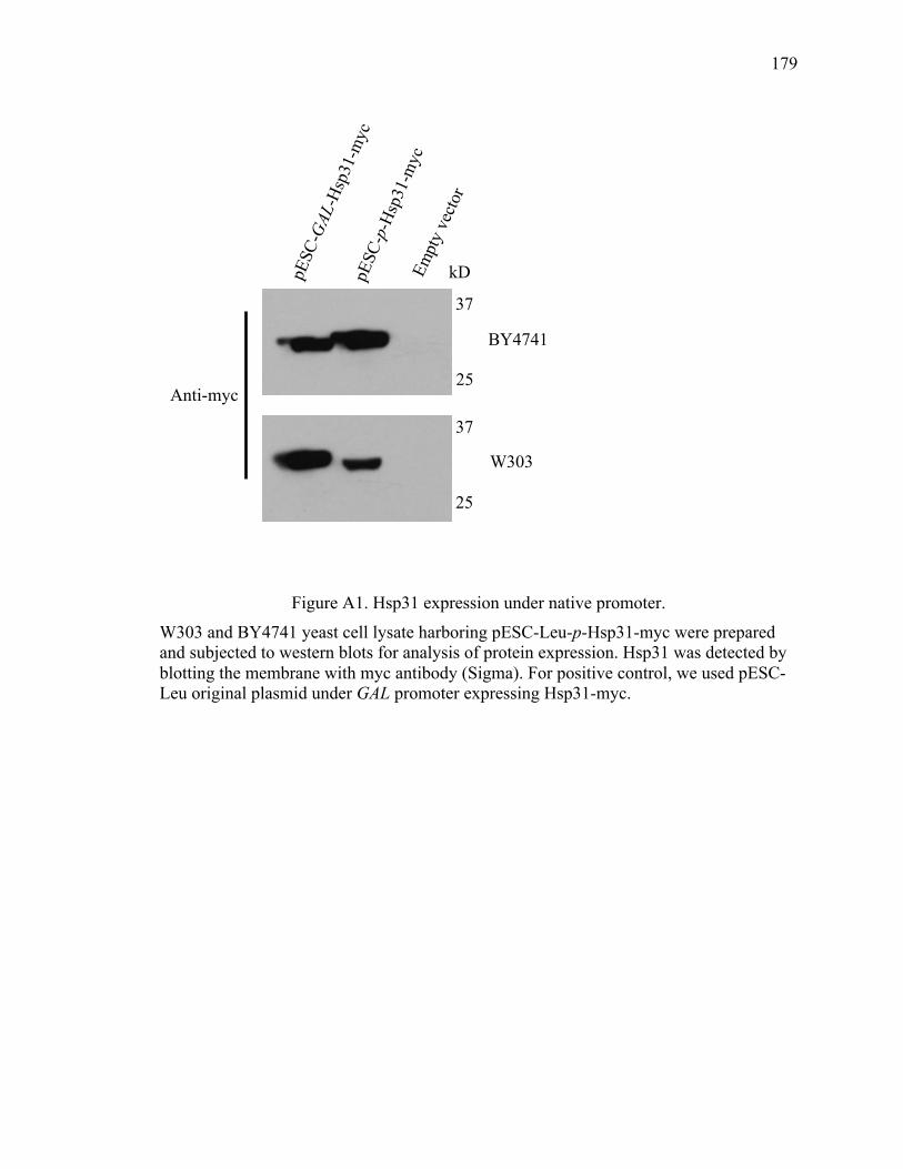
179
179
Figure A1. Hsp31 expression under native promoter.
W303 and BY4741 yeast cell lysate harboring pESC-Leu-p-Hsp31-myc were prepared and subjected to western blots for analysis of protein expression. Hsp31 was detected by blotting the membrane with myc antibody (Sigma). For positive control, we used pESC-Leu original plasmid under GAL promoter expressing Hsp31-myc.
Empt
y ve
ctor
pESC
-GAL
-Hsp31
-myc
pESC
-p-Hsp31
-myc
Anti-myc
37
25
25
37
kDa
BY4741
W303
180
180
Appendix B Hsp31 pull down for partner proteins
In order to further elucidate the mechanism of Hsp31 in preventing aggregation and to
narrow down its other possible role first it is important to determine the cellular
substrates of Hsp31 that we predict are either oxidatively damaged proteins or chaperones
or both. We proposed to take the approach to use a strain with endogenously tag Hsp31
with 9myc. We used this strain to affinity purify Hsp31 using agarose beads. Clear lysate
obtained from this strain was incubated with anti-myc agarose beads overnight at 4°C
with shaking. Successful pull down was confirmed by running a SDS-PAGE gel and
staining with Sypro Ruby stain followed by western blot using specific anti-myc antibody
(Figure B1). To increase the possibility of identifying Hsp31 client interactions we
digested the protein pull down on beads directly and subjected to mass spec analysis
(Table A1). Proteins that were present in a control sample (non-tagged strain lysate) or
with fewer peptide or spectral counts were removed and the final list is displayed in
Table 1. Hsp31 was the most predominantly identified protein and Hsp31 paralogs
(Hsp32 and Hsp33) were also identified. Potential co-chaperones were also observed
including several Hsp70 isoforms and Hsp104. We performed further verification of
some of these interactions by using various pull down experiments but results remains
inconclusive except for interaction between Hsp31 and Hsp104 that is described in
Chapter 3. For rest of the potential client proteins we performed pull down assay by
transforming client protein under MORF plasmid system into Hsp31-9myc tagged yeast
strain. First we performed the immunoprecipitation of Hsp31 by using myc tagged
agarose beads and subjected to western blot using anti HA antibody. Most of the clients
proteins showed positive signal that could be the reason of non specific binding of the
181
181
protein to agarose beads as these constructs are tagged with protein A along with HA
(Figure B2). In fact, we confirmed that possibility by using the WT stains without 9myc
tagged Hsp31 was still able to bind with the proteins (Figure B3). Due to high affinity of
protein-A binding with agarose beads we could not processed these experiments further.
Next, we proposed to use another technique by using purified GST-Hsp31 protein and
incubating it with lysate generated from cells (Hsp31-9myc) expressing client proteins
under MORF system. After incubation, samples were subjected to SDS-PAGE and
blotted with either GST or myc antibody. This experiment shows clear positive
interaction between Hsp31 and Hsp32 that was used as positive control. All other client
proteins came back negative (Figure B4). Based on these experiments none of the tested
client proteins directly interact with Hsp31 however, it is needed to confirm by using
other plasmid system such as Tap tagged. We also suspect that some of
Hsp31interactions with client proteins are transient in nature and different experimental
conditions are necessary to detect such interactions. Further verification of these
interactions and their role in the function of hsp31 will be pursued in future.
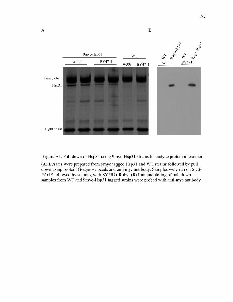
182
182
A B
Figure B1. Pull down of Hsp31 using 9myc-Hsp31 strains to analyze protein interaction.
(A) Lysates were prepared from 9myc tagged Hsp31 and WT strains followed by pull down using protein G-agarose beads and anti myc antibody. Samples were run on SDS-PAGE followed by staining with SYPRO-Ruby. (B) Immunobloting of pull down samples from WT and 9myc-Hsp31 tagged strains were probed with anti-myc antibody
!17!
Figure 7: Effect of Hsp31 on curing of prion. (A)!Hsp31, Hsp42, or Hsp104 were overexpressed in [PSI+] cells in liquid for 24hrs at 30°C. Empty vector served as the control. Cells were plated on ¼ YPD plates. Red color colonies indicate curing of [PSI+] prion. (B) Hsp31 was deleted in [PSI+] strain and Hsp104 was overexpressed to cure [PSI+] state. (C) Fluorescence microscopy demonstrating localization of Hsp31 and Sup35-YFP when overexpressed together. Hsp31 localized throughout the cytoplasm when overexpressed however, it is occluded from the Sup35-YFP foci as shown by white arrows. !
!!Figure 8: Atg8Δ elevate a-Syn toxicity analyzed by spotting assays. WT and atg8Δ yeast cells expressing a-Syn with or without hsp31Δ background were grown for 24(h in glucose media and spotted in fivefold serial dilutions onto glucose (expression repressed) and galactose (expression induced) agar plates. Cells were grown for two days at 30°C. (A) (B)
Figure 9: Pull down of Hsp31 using 9myc-Hsp31 strains to analyze protein interaction. (A) Lysates were prepared from 9myc tagged Hsp31 and WT strains followed by pull down using protein G-agarose beads and anti myc antibody. Samples were run on SDS-PAGE followed by staining with SYPRO-Ruby. (B) Immunobloting of pull down samples from WT and 9myc-Hsp31 tagged strains were probed with anti-myc antibody.
! 17!
Figure 7: Effect of Hsp31 on curing of prion. (A)! Hsp31, Hsp42, or Hsp104 were overexpressed in [PSI+] cells in liquid for 24hrs at 30°C. Empty vector served as the control. Cells were plated on ¼ YPD plates. Red color colonies indicate curing of [PSI+] prion. (B) Hsp31 was deleted in [PSI+] strain and Hsp104 was overexpressed to cure [PSI+] state. (C) Fluorescence microscopy demonstrating localization of Hsp31 and Sup35-YFP when overexpressed together. Hsp31 localized throughout the cytoplasm when overexpressed however, it is occluded from the Sup35-YFP foci as shown by white arrows. !
!!Figure 8: Atg8Δ elevate a-Syn toxicity analyzed by spotting assays. WT and atg8Δ yeast cells expressing a-Syn with or without hsp31Δ background were grown for 24(h in glucose media and spotted in fivefold serial dilutions onto glucose (expression repressed) and galactose (expression induced) agar plates. Cells were grown for two days at 30°C. (A) (B)
Figure 9: Pull down of Hsp31 using 9myc-Hsp31 strains to analyze protein interaction. (A) Lysates were prepared from 9myc tagged Hsp31 and WT strains followed by pull down using protein G-agarose beads and anti myc antibody. Samples were run on SDS-PAGE followed by staining with SYPRO-Ruby. (B) Immunobloting of pull down samples from WT and 9myc-Hsp31 tagged strains were probed with anti-myc antibody.
Heavy chain
Light chain
Hsp31
W303 BY4741 BY4741 W303
WT 9myc-Hsp31
9myc
-Hsp
31
9myc
-Hsp
31
WT WT
BY4741 W303
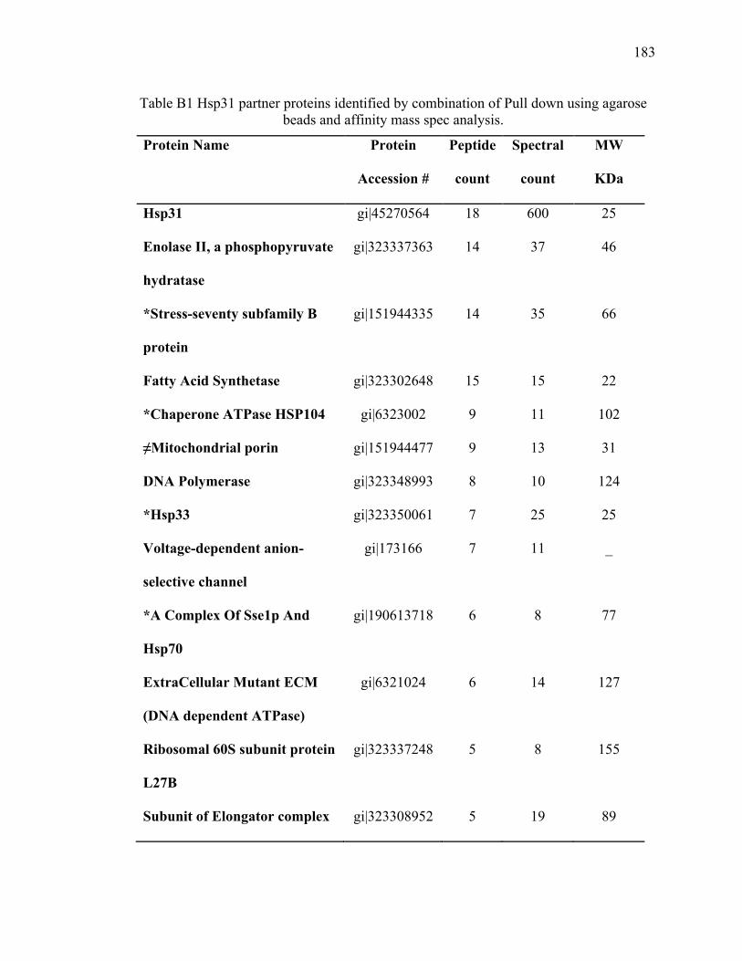
183
183
Table B1 Hsp31 partner proteins identified by combination of Pull down using agarose beads and affinity mass spec analysis.
Protein Name Protein
Accession #
Peptide
count
Spectral
count
MW
KDa
Hsp31 gi|45270564 18 600 25
Enolase II, a phosphopyruvate
hydratase
gi|323337363 14 37 46
*Stress-seventy subfamily B
protein
gi|151944335 14 35 66
Fatty Acid Synthetase gi|323302648 15 15 22
*Chaperone ATPase HSP104 gi|6323002 9 11 102
≠Mitochondrial porin gi|151944477 9 13 31
DNA Polymerase gi|323348993 8 10 124
*Hsp33 gi|323350061 7 25 25
Voltage-dependent anion-
selective channel
gi|173166 7 11 _
*A Complex Of Sse1p And
Hsp70
gi|190613718 6 8 77
ExtraCellular Mutant ECM
(DNA dependent ATPase)
gi|6321024 6 14 127
Ribosomal 60S subunit protein
L27B
gi|323337248 5 8 155
Subunit of Elongator complex gi|323308952 5 19 89
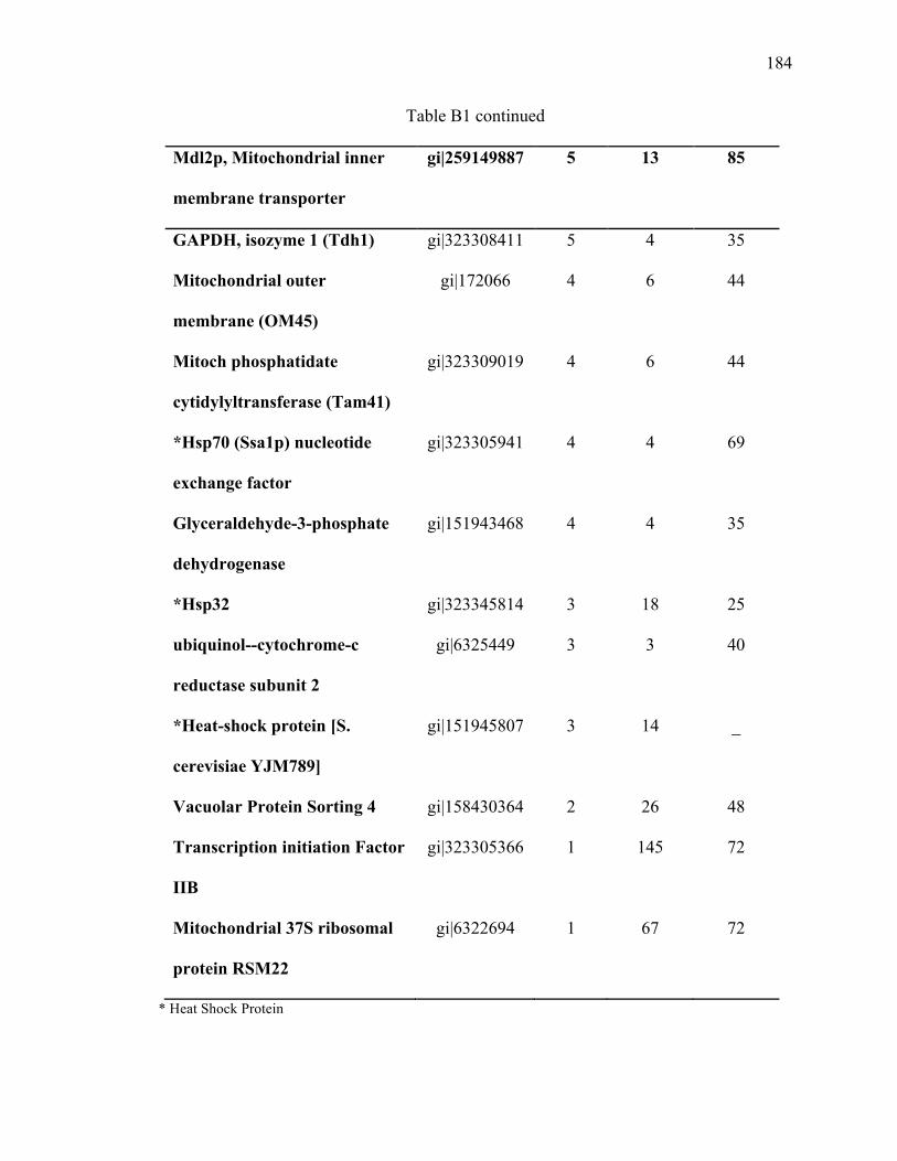
184
184
Table B1 continued
Mdl2p, Mitochondrial inner
membrane transporter
gi|259149887 5 13 85
GAPDH, isozyme 1 (Tdh1) gi|323308411 5 4 35
Mitochondrial outer
membrane (OM45)
gi|172066 4 6 44
Mitoch phosphatidate
cytidylyltransferase (Tam41)
gi|323309019 4 6 44
*Hsp70 (Ssa1p) nucleotide
exchange factor
gi|323305941 4 4 69
Glyceraldehyde-3-phosphate
dehydrogenase
gi|151943468 4 4 35
*Hsp32 gi|323345814 3 18 25
ubiquinol--cytochrome-c
reductase subunit 2
gi|6325449 3 3 40
*Heat-shock protein [S.
cerevisiae YJM789]
gi|151945807 3 14 _
Vacuolar Protein Sorting 4 gi|158430364 2 26 48
Transcription initiation Factor
IIB
gi|323305366 1 145 72
Mitochondrial 37S ribosomal
protein RSM22
gi|6322694 1 67 72
* Heat Shock Protein
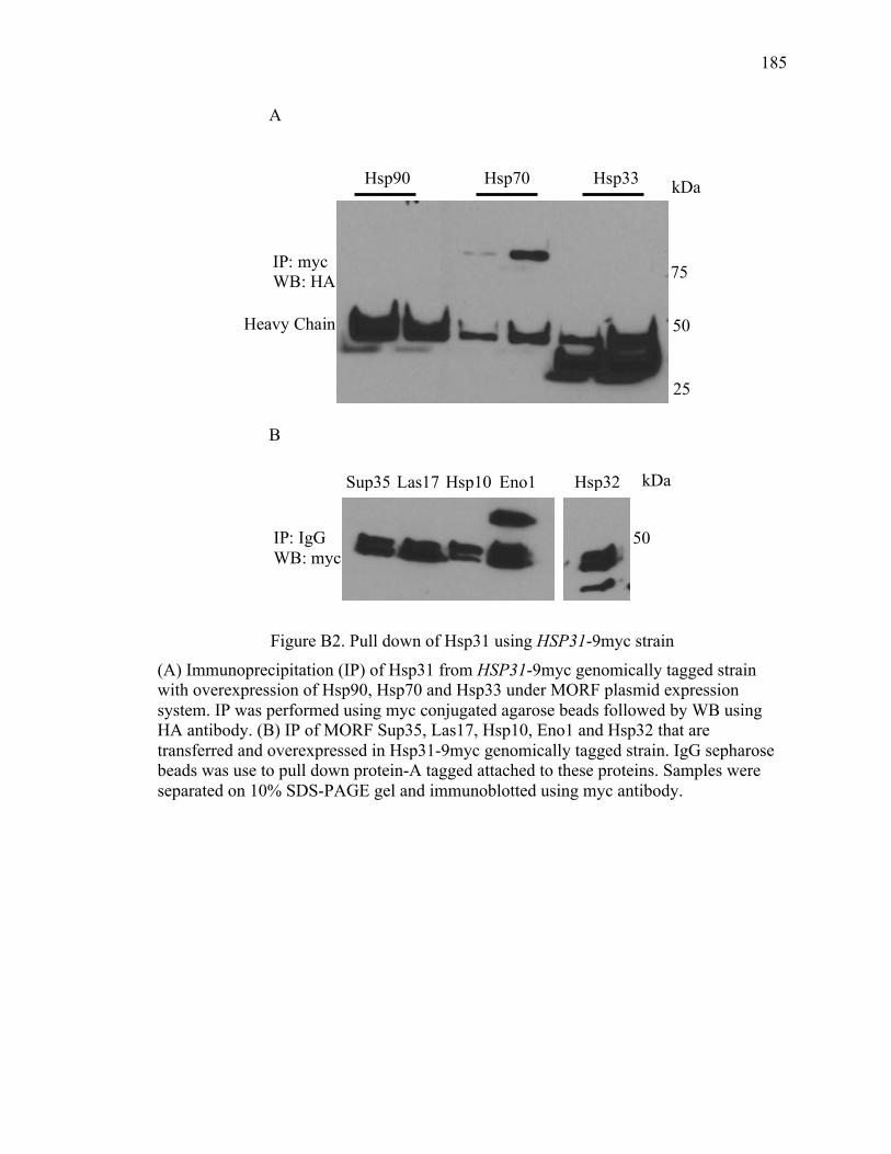
185
185
A
B
Figure B2. Pull down of Hsp31 using HSP31-9myc strain
(A) Immunoprecipitation (IP) of Hsp31 from HSP31-9myc genomically tagged strain with overexpression of Hsp90, Hsp70 and Hsp33 under MORF plasmid expression system. IP was performed using myc conjugated agarose beads followed by WB using HA antibody. (B) IP of MORF Sup35, Las17, Hsp10, Eno1 and Hsp32 that are transferred and overexpressed in Hsp31-9myc genomically tagged strain. IgG sepharose beads was use to pull down protein-A tagged attached to these proteins. Samples were separated on 10% SDS-PAGE gel and immunoblotted using myc antibody.
kDa Hsp70 Hsp33
50
75
25
Hsp90
Heavy Chain
IP: myc WB: HA
kDa Sup35 Las17 Hsp10 Eno1 Hsp32
50 IP: IgG WB: myc
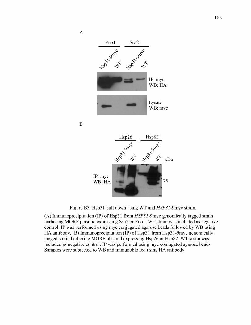
186
186
A
B
Figure B3. Hsp31 pull down using WT and HSP31-9myc strain. (A) Immunoprecipitation (IP) of Hsp31 from HSP31-9myc genomically tagged strain harboring MORF plasmid expressing Ssa2 or Eno1. WT strain was included as negative control. IP was performed using myc conjugated agarose beads followed by WB using HA antibody. (B) Immunoprecipitation (IP) of Hsp31 from Hsp31-9myc genomically tagged strain harboring MORF plasmid expressing Hsp26 or Hsp82. WT strain was included as negative control. IP was performed using myc conjugated agarose beads. Samples were subjected to WB and immunoblotted using HA antibody.
Eno1 Ssa2
Hsp31-9myc
WT
IP: myc WB: HA
Hsp31-9myc
WT
Lysate WB: myc
Hsp31-9myc
WT Hsp31-9myc
WT
Hsp26 Hsp82
75
kDa
IP: myc WB: HA
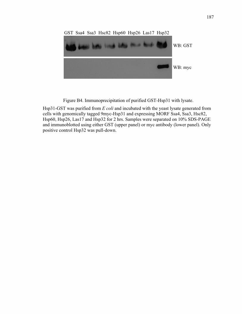
187
187
Figure B4. Immunoprecipitation of purified GST-Hsp31 with lysate.
Hsp31-GST was purified from E coli and incubated with the yeast lysate generated from cells with genomically tagged 9myc-Hsp31 and expressing MORF Ssa4, Ssa3, Hsc82, Hsp60, Hsp26, Las17 and Hsp32 for 2 hrs. Samples were separated on 10% SDS-PAGE and immunoblotted using either GST (upper panel) or myc antibody (lower panel). Only positive control Hsp32 was pull-down.
Hsp32 GST Las17 Hsp26 Hsp60 Hsc82 Ssa3 Ssa4
WB: GST
WB: myc

VITA
188
188
VITA
Kiran Aslam was born in Bahawalpur, Pakistan, the daughter of Muhammad Aslam and
Mukhtiar Bibi. After completing her high school degree she entered the Islamia
University of Bahawalpur, in Bahawalpur and received the degree of Bachelor of
Pharmacy in August 2005. She earned a fellowship to attend the graduate school at one of
the prestigious research institute of the country, HEJ research institute of Chemistry and
Molecular Medicine located inside the Karachi University in Karachi. She received
Master in Philosophy degree from the Department of Pharmacology in
Neuropharmacology in 2009. During her stay at the Karachi University she participated
in several national and international conferences. She achieved several accolades
including the best poster award, travel award from Japan Neuroscience Society and
International Brain Research Organization (IBRO) award to attend workshop in Bangkok.
Thailand. In 2010 she won IBRO studentship based on her proposed project to get trained
at the University College London (UCL) in London, UK. She joined the lab of Prof. Jim
Owen at division of medicine at Royal Free Campus in London, UK.
Later in 2010, she secured Fulbright fellowship offered by the United States of Education
Foundation in Pakistan and the International Institute of Education in USA to attend a
graduate school for PhD in the USA. She enrolled in the Department of Medicinal
Chemistry and Molecular Pharmacology at Purdue University. She joined the lab of Dr.
189
189
Tony Hazbun and started working on project to understand the molecular mechanism of
protein misfolding that is hallmark of many neurodegenerative diseases. The focus of the
project was to understand the molecular role of Hsp31 protein, a yeast homolog of human
DJ-1 that is involved in PD. During her time here she participated, presented and
published her work in many international conferences. She also published her work in the
Journal of Biological Chemistry and the Prion journal. She also received the Koo and
Paget travel award to attend the Keystone Symposium “Common mechanism of
Neurodegeneration” in June 2016.

PUBLICATIONS
190
190
PUBLICATIONS
• Chia-Jui Tsai*, Kiran Aslam*, Holli M Drendel, Josephat M Asiago, Kourtney
M Goode, Lake N Paul, Jean-Christophe Rochet, Tony R Hazbun Hsp31 is a
Stress-Response Chaperone that Intervenes in the Protein Misfolding Process. The
Journal of biological chemistry, 290(41):24816-34, 2015
*First Co-author
• Kiran Aslam, Tony Hazbun Hsp31, a member of the DJ-1 superfamily, is a
multitasking stress responder with chaperone activity. Prion Apr 2016
• Kiran Aslam, Chia-Jui Tsai, Tony Hazbun. Small heat shock protein Hsp31
cooperates with Hsp104 to modulate the Sup35 prion aggregation. Prion
Submitted March 2016.