Embed Size (px)
Citation preview
applied sciences
Article
Deciphering the Antitussive, Expectorant, andAnti-Inflammatory Potentials of ShashamKyeongok-Go andTheir Phytochemical Attributes: In Vivo Appraisal in ICR Mice
Jin-Ryul Hu 1, Chul-Jong Jung 2 , Seong-Min Ku 2, Dae-Hwa Jung 3, Sae-Kwang Ku 1,4 , Md. Mohibbullah 5 ,Hae-Jeung Lee 6,* and Jae-Suk Choi 7,*
�����������������
Citation: Hu, J.-R.; Jung, C.-J.; Ku,
S.-M.; Jung, D.-H.; Ku, S.-K.;
Mohibbullah, M.; Lee, H.-J.; Choi, J.-S.
Deciphering the Antitussive,
Expectorant, and Anti-Inflammatory
Potentials of ShashamKyeongok-Go
and Their Phytochemical Attributes:
In Vivo Appraisal in ICR Mice. Appl.
Sci. 2021, 11, 1349. https://doi.org/
10.3390/app11031349
Academic Editor: Carmela Spagnuolo
Received: 16 December 2020
Accepted: 28 January 2021
Published: 2 February 2021
Publisher’s Note: MDPI stays neutral
with regard to jurisdictional claims in
published maps and institutional affil-
iations.
Copyright: © 2021 by the authors.
Licensee MDPI, Basel, Switzerland.
This article is an open access article
distributed under the terms and
conditions of the Creative Commons
Attribution (CC BY) license (https://
creativecommons.org/licenses/by/
4.0/).
1 Department of Histology and Anatomy, Daegu Haany University, Gyeongsan-si 38610,Gyeongsangbuk-do, Korea; [email protected] (J.-R.H.); [email protected] (S.-K.K.)
2 Okchundang Inc., Ulju-gun 44900, Ulsan, Korea; [email protected] (C.-J.J.);[email protected] (S.-M.K.)
3 Department of Pharmaceutical Engineering, Daegu Haany University, Gyeongsan 38610,Gyeongsangbuk-do, Korea; [email protected]
4 The Medical Research Center for Herbal Convergence on Liver Disease, Daegu Haany University,Gyeongsan-si 38610, Gyeongsangbuk-do, Korea
5 Department of Fishing and Post Harvest Technology, Sher-e-Bangla Agricultural University,Dhaka 1207, Bangladesh; [email protected]
6 Department of Food and Nutrition, Gachon University, 1342 Seongnamdaero, Sujeong-gu,Seongnam 13120, Gyeonggi, Korea
7 Department of Food Biotechnology, Silla University, Sasang-gu 46958, Busan, Korea* Correspondence: [email protected] (H.-J.L.); [email protected] (J.-S.C.);
Tel.: +82-31-750-5968 (H.-J.L.); +82-51-248-7789 (J.-S.C.)
Abstract: In this paper, we hypothesized that ShashamKyeongok-go (SKOG) is a mixed preparationof Adenophorae Radix powder (AR) and Kyeongok-go (KOG). SKOG may be served as a novelpreventive and/or therapeutic agent for various respiratory diseases. SKOG were orally administeredto ICR mice at 400, 200, and 100 mg/kg once a day for 11 days to examine antitussive, expectorant,and anti-inflammatory effects. The NH4OH exposure-induced allergic acute inflammation withcoughing responses was dose-dependently and significantly (p < 0.01) inhibited by pretreatmentwith SKOG at doses of 400, 200, and 100 mg/kg. With these concentrations of SKOG, the thickness ofintrapulmonary secondary bronchus mucosa and the number of periodic acid Schiff stain-positivemucous-producing cells were significantly (p < 0.05 or p < 0.01) increased, as a result of the increasedamount of phenol red secretion. Subsequently, SKOG showed significant (p < 0.01) anti-inflammatoryactivities as characterized by reducing the effects of xylene-induced increases of ear weight, thicknessof total ear and ear dermis, and number of infiltrated inflammatory cells in the ear dermis, in adose-dependent manner. These results supported that SKOG might have potential therapeutic effectsto be used as an antitussive, expectorant, and anti-inflammatory agents in the prevention or treatmentof chronic bronchitis and asthma.
Keywords: shashamkyeongok-go; kyeongok-go; adenophorae radix; antitussive effects; expectoranteffects; anti-inflammation
1. Introduction
Airways bronchitis and asthma are a chronic inflammatory disease occupied in therespiratory tract and possibly caused by inherited predisposition and long-term effects ofenvironmental irritants such as dust mites, smoke, and various chemical irritants. Bron-choconstriction is one of the most frequent pathological events of having chronic bronchitisand asthma, where the airway tubes become narrower and harder, but which is supposedto be wider and smoother, in reaction to the introduced foreign particles [1]. Therefore,it affects patients with difficulty to breathe properly associated with cough induction. A
Appl. Sci. 2021, 11, 1349. https://doi.org/10.3390/app11031349 https://www.mdpi.com/journal/applsci
Appl. Sci. 2021, 11, 1349 2 of 20
cough is defined as, voluntary or involuntary acts, a forced expulsion behavior to clear thethroat, and breathing airways with a concomitant sound. In addition, it has two character-istic types including non-productive as dry and productive as chesty, which are exhaustiveand adversely affect the quality of life [2]. It represents global public health problemswhich are likely to be enormously higher, more than 339 million people currently sufferfrom asthma [3]. Moreover, compelling evidence suggested that inflammatory processes inairways were implicated in developing the pathogenesis of many respiratory diseases [4,5].Therefore, the present study is undertaken to search for new natural herb-based formula-tions acting as alternative therapeutics, which are rich in nature, deciphering the synergisticeffects in antitussive, expectorant, and anti-inflammatory activities in patients with airwaysbronchitis and asthma complications.
Adenophora triphylla var. japonica Hara is under the genus of Adenophora (Campanu-laceae), and has been documented as an oriental medicinal prescription in Korea, Japan,and China. Pharmacological potentials of A. triphylla are characterized by its antitussive,anti-inflammatory [6], and hepatoprotective properties [7,8]. Many of the pharmacologicalimportant compounds of dried root parts of A. triphylla are reported as triterpenoids, alka-loids, and various essential oil compounds including pyrrolidine and triphyllol [6], piperi-dine and heptacosane [9], lupenone [10,11], and saponine [12,13]. In addition, precedentresearch on the dried root parts of A. triphylla, alternatively known as Adenophorae Radix,have remarkable effects on in vitro mucus production [14], in vivo hepatoprotective [7],in vitro antitumor [12,13], and in vitro [8,10] and in vivo anti-obesity [8]. Kyeongok-go(KOG) is one of the widely accepted and most popular herbal recipes in oriental medicine ofKorea, comprising of Rehmanniae Radix Crudus, Pulvis Hoelen, Ginseng Radix Alba, andhoney [7,15]. Many experimental evidence showed the path of pharmacological importanceof KOG by the researchers on antioxidant [15], anti-inflammatory [16], immunomodula-tory [15], anti-fatigue and aerobic-capacity enhancing [16], and growth-promoting [17]potentials.
Therefore, we hypothesized and expected that the appropriate addition of AdenophoraeRadix powders (AR) to KOG can potentiate antitussive, expectorant, and anti-inflammatoryactivities, which may be utilized as a novel tonic agent for preventing varied respiratorydiseases. The present study was conducted in three different mouse models, such asNH4OH-induced coughing [18,19], phenol red secretion [18,20], and xylene-induced acuteinflammation in the ear [21,22], to perform antitussive, expectorant, and anti-inflammatoryassays, respectively. The obtained results from SKOG treatment groups were comparedside-by-side with a single dose of AR, KOG and/or standard drugs of theobromine (TB),ambroxol (AM), and dexamethasone (DEXA), respectively [14,23], aiming at examiningthe synergistic effects of SKOG on the mitigation of chronic bronchitis and asthma.
2. Materials and Methods2.1. Test Materials
Yellow-coloured and powdered forms of AR, KOG, and SKOG materials were sup-plied by Okchundang (Ulsan, Korea). To prepare KOG, briefly, appropriate amounts ofindividual herbs, namely Ginseng Radix Alba (6000 g), Pulvis Hoelen (12,000 g), Rehman-niae Radix Crudus (47,000 g), and honey (39,000 g), were mixed, heated at 60 ◦C for 72 h,and cooled at 20 ◦C; this process was repeated twice. To prepare SKOG, appropriateamounts of individual herbs, namely Ginseng Radix Alba (4500 g), Pulvis Hoelen (9000 g),AR (4500 g), Rehmanniae Radix Crudus (47,000 g), and honey (39,000 g) were mixed,heated in a water bath at 60 ◦C for 72 h, and cooled at 20 ◦C; this process was repeatedtwice (Table S1). Specimens of AR, KOG, and SKOG were deposited in the herbarium of theMedical Research Center, Daegu Haany University (Gyeongsan, Gyeongbuk, Korea) (codeno. AR2016Ku01, KOG2016Ku01, and SKOG2016Ku01, respectively). Unless otherwisestated, all chemicals used in the study were purchased from Sigma-Aldrich LLC (St. Louis,MO, USA) and kept at 4 ◦C until use.
Appl. Sci. 2021, 11, 1349 3 of 20
2.2. Analysis of Specific Ingredients of AR, KOG, and SKOG2.2.1. Instrument and Reagents
An ultra-performance liquid chromatography (UPLC) system (Waters Corp., Milford,MA, USA) was used, where the photodiode array detector (PDA; Waters Corp.), WatersACQUITYT SM (Sample Manager) Autosampler, Waters ACQUITYT CH-A Column Oven(20 ◦C), and C18 column with a dimension of 1.7 µm, 2.1 × 100 mm (ACQUITYTM BEH,Waters Corp.) were adjusted to the system [24]. The UPLC chromatogram was visualizedusing an Empower Software (Waters Corp.) equipped with the computer system. Thesample extractor was an 8210R-DHT ultrasonicator (Branson Ultrasonics, Danbury, CT,USA). The HPLC grade extraction solvents were methanol (MeOH; Junsei Chemical Co.,Ltd., Tokyo, Japan) and acetonitrile (BAKER, Center Valley, PA, USA). In case of the PDAanalysis, the detection wavelength of lupeol, acteoside, catalposide, and 5H2F was fixed at280 nm, and lobetyolin and syringaldehyde were analyzed in the wavelength of 310 and254 nm, respectively [25].
2.2.2. Preparation of Standard Solutions
Appropriate amounts of AR components (lupeol, lobetyolin, and syringaldehyde), Rehman-niae Radix Crudus components (containing acteoside, catalposide, and 5-hydroxymethyl-2-furfural (5H2F)), and Ginseng Radix Alba component (ginsenoside Rg3 (Rg3)) weremeasured accurately and dissolved in dimethyl sulfoxide (DMSO) or MeOH to preparestandard stock solutions with a concentration of 1 µg/mL. Next, appropriate amountsof standard solutions were diluted in MeOH to prepare three different concentrations 1,5, and 10 ng/mL. The standard curve of each compound was determined with a coeffi-cient (R2) value of more than 0.999. For standard preparations, lobetyolin was purchasedfrom the Extrasynthese (Genay Cedex, France) and all the other standards were fromSigma-Aldrich LLC.
2.2.3. Preparation of Test Samples for UPLC Analysis
For the quantitative analysis, 1 g of the samples were added to 10 mL of 30% MeOHand then microwave extraction was performed for 1 h. The supernatant was filtered usinga membrane filter paper of 0.2 µm in diameter. The filtered liquid was selected for UPLCanalysis.
2.2.4. Phytochemical Quantification by UPLC
Lupeol, lobetyolin, syringaldehyde acteoside, catalposide, 5H3F, and Rg3 contents inAR, KOG, and SKOG were quantified using UPLC. The column was kept at 20 ◦C for theanalysis. In the PDA analysis, lupeol, acteoside, catalposide, and 5H2F were analyzed at awavelength of 280 nm, whereas lobetyolin and syringaldehyde were analyzed at 310 and254 nm, respectively. The mobile phase was water and acetonitrile with formic acid (0.1%).Rg3 was analyzed at the wavelength of 203 nm and the mobile phase was a mixture ofwater and acetonitrile. A 2 µL of the sample was injected into UPLC with a flow rate of0.4 mL/min. Phytochemicals of the injected sample were identified by the retention timeand the same detectable wavelength value and quantified by measuring the area of thepeak (Figure S1).
2.3. Antitussive Assay2.3.1. Rodents and Husbandry
The Institute of Cancer Research (ICR) male mice, weighing 29–32 g upon received,were bought from OrientBio, Seungnum, Korea. Four mice were allocated to each cage andhoused at the temperature of 20–25 ◦C and relative humidity of 50–55% in a controlledroom of 12/12 h (light/dark) cycle, with free access to ad libitum of pelleted food (cat. no.38057; Purinafeed, Seungnam, Korea) and water. After 7 days of adaptation, all the animalswere selected for antitussive experiments, maintained as per the rules and regulations ofnational usage and welfare of laboratory animals based on ethical principles.
Appl. Sci. 2021, 11, 1349 4 of 20
2.3.2. Treatment and Grouping
After a week of adaptation, the eligible rodents were randomly chosen and dividedinto 8 groups of 10 rodents each, including an intact vehicle control (distilled water (DW),orally administered at 10 mL/kg), NH4OH control (DW and 25% NH4OH-induced cough-ing treated groups), TB [14] (a reference control drug, oral administration at 10 mL/kg,corresponding to 50 mg/kg and 25% NH4OH-treated groups), AR (oral administration at10 mL/kg, corresponding to 400 mg/kg and 25% NH4OH-treated groups), KOG (oral ad-ministration at 10 mL/kg, corresponding to 400 mg/kg and 25% NH4OH-treated groups),SKOG400 (oral administration at 10 mL/kg, corresponding to 400 mg/kg and 25% NH4OH-treated groups), SKOG200 (oral administration at 10 mL/kg, corresponding to 200 mg/kgand 25% NH4OH-treated groups), and SKOG100 (oral administration at 10 mL/kg, corre-sponding to 100 mg/kg and 25% NH4OH-treated groups). SKOG was dissolved in DW tomake working concentrations of 40, 20, and 10 mg/mL, and AR and KOG were preparedfollowing the same procedure to make a concentration of 40 mg/mL for oral administra-tion. Similarly, TB was suspended and prepared in DW to the working concentration of5 mg/mL. All the treatments were performed once a day for 11 days before the NH4OHexposure. To maintain the same restrain stress, DW was administered in all the controlgroups rather than AR, KOG, SKOG, or TB treatments.
2.3.3. Body Weight Measurement
Body weight (BW) changes in the treatment mice were measured once a day usingan electronic balance (Precisa Instrument, Dietikon, Switzland) during the entire adminis-tration period of AR, KOG, SKOG, or TB. The BW gain was calculated using a formula asfollows:
The BW gain after 11 days of oral administration of test substances = BW at sacrifice—BW at the initial treatment of the test substance (g/head).
2.3.4. Coughing Induction and Monitoring
Coughing was induced by an inhalation of 25% NH4OH (Sigma-Aldrich LLC), where0.3 mL was taken to a 1 L Erlenmeyer flask and inhaled to mice for 45 s at 1 h after thelast administration of the test substance. Following the NH4OH induction, the number ofcoughing responses was quantitated in 6 min using video observations [21,22]. Mice withan intact vehicle control were exposed to 0.3 mL saline for 45 s, in replacement of NH4OH.A cough in mice was characterized as opening the mouth with a sudden expulsion followedby the sound of coughing, contraction of thoracic and abdominal muscles, and shaking ofthe front body [20,21].
2.3.5. Histopathology
After obtaining video images, some portion of the left lateral lobes of individual lungsand trachea consisting of 3 mm from the thyroid cartilages were isolated and fixed in asolution of 10% neutral buffered formalin (NBF). The sample was then trimmed, embeddedin paraffin, sectioned at 3~4 µm thickness, stained with hematoxylin and eosin (H&E) forgeneral histopathology examination or with toluidine blue for mast cell examination, andobserved under a light microscope (Eclipse 80i; Nikon, Tokyo, Japan). Histopathologicalprofiles were analyzed with an image analysis program (iSolution FL ver 9.1; IMT i-solutionInc., Quebec, QC, Canada), as followed by the previously established methods [25,26].
2.4. Expectorant Assay2.4.1. Rodents and Husbandry
ICR mice (♂) of 86 weeks old, weighing 29–32 g upon their receival, were boughtfrom OrientBio, Seungnum, Korea. Animal husbandry was conducted the same as in theantitussive assay. Mice selected for expectorant experiments were maintained abiding bythe rules and regulations of national usage and welfare of laboratory animals based onethical principles.
Appl. Sci. 2021, 11, 1349 5 of 20
2.4.2. Treatment and Grouping
After 7 days of adaptation, 70 mice each were selected and divided into 7 groupseach, including an intact vehicle control (DW), AM (250 mg/kg administered mice), AR(administration at 400 mg/kg and AM-exposed group), KOG (administration at 400 mg/kgand AM-exposed group), SKOG (administration at 400 mg/kg and AM-exposed group),SKOG (administration at 200 mg/kg and AM-exposed group), SKOG (administration at100 mg/kg and AM-exposed group). AR, KOG, and SKOG were orally administered thesame as in the antitussive assay, once a day for 11 days before being treated with phenolred. To provide the same restrain stress, the intact vehicle control rodents were orallyadministered 10 mL/kg of DW, rather than AR, KOG, SKOG, or AM.
2.4.3. BW Measurement
The BW and BW gain were evaluated in a similar way as mentioned in the antitussiveassay.
2.4.4. Measurement of Mucous Secretion
To measure the mucous secretion, rodents were intraperitoneally injected with a singledose of 5% phenol red solution (Junsei Chemical Co. Ltd., Tokyo, Japan) at 10 mL/kgdissolved in a saline solution (w/v), after 30 min of the last (eleventh) administration oftest material. Gross images of individual mice were then obtained to investigate the phenolred-induced body-surface redness. Thereafter, the trachea was dissected after the sacrificeof mice by cervical dislocation and homogenized in normal saline for 15 min to preparethe trachea lavage fluid (TLF) after adding 1 mL of 5% NaHCO3 solution (w/v). Theoptical density (OD) of TLF was recorded at the wavelength of 546 mm, as followed by theprevious studies [20,22].
2.4.5. Histopathology
At trachea excisions, individual left lateral lobes of the lung were collected. Then,the lung lobes were fixed (10% NBF), crossly trimmed, embedded in paraffin, sectioned(3~4 µm thickness), stained (H&E) for general histopathology examination or with aPeriodic acid Schiff stain for mucous-producing cell examination, and observed undera light microscope. Histopathological profiles were examined using an image analysisprogram, as described formerly [25–28].
2.5. Anti-Inflammatory Assay2.5.1. Rodents and Husbandry
ICR mice (♂)of 96 weeks old, weighing 29–32 g upon their receival, were boughtfrom OrientBio, Seungnum, Korea. Animal husbandry was conducted the same as inthe antitussive assay. Mice selected for anti-inflammatory experiments were maintainedfollowing the rules and regulations of national usage and welfare of laboratory animalsbased on ethical principles.
2.5.2. Treatment and Grouping
After 7 days of adaptation, 80 mice each were selected and divided into 8 groupseach, including an intact vehicle control (DW), xylene control (0.03 mL of xylene of topicalapplication to the anterior surface of the right ear), DEXA (administration at 1 mg/kg andXylene-exposed group), AR (administration at 400 mg/kg and xylene-exposed group),KOG (administration at 400 mg/kg and xylene-exposed group), SKOG (administration at400 mg/kg and xylene-exposed group), SKOG (administration at 200 mg/kg and xylene-exposed group), SKOG (administration at 100 mg/kg and xylene-exposed group). AR,KOG, and SKOG were orally administered the same as in the antitussive and expectorantexperiments, once a day for 11 days before the xylene treatment. To provide the samerestrain stress, the intact vehicle control rodents were orally administered at 10 mL/kg ofDW, rather than AR, KOG, SKOG, or xylene.
Appl. Sci. 2021, 11, 1349 6 of 20
2.5.3. BW Measurement
BW and BW gain were determined in the same procedure as mentioned in the antitus-sive assays.
2.5.4. Ear Weight Measurement
After the xylene treatment for 2 h, circular sections of the ear were collected andweighed as the absolute ear wet-weight. The relative ear weight (% of BW) was measuredto reduce the differences of individuals using a formula as given below:
Relative ear weight (% of BW) = (absolute ear wet-weight/BW at sacrifice) × 100
2.5.5. Histopathology
Individual ear samples were fixed in 10% NBF, crossly trimmed, embedded in paraffin,sectioned (to 3~4 µm thickness), and stained (H&E) for general histopathology examinationor with toluidine blue for mast cell examination. Histopathological profiles were analyzedwith an image analysis software program, as described in previous studies [23,25,26,29].
2.6. Statistical Analyses
All data were shown as the mean ± standard deviation (SD) of six measurements,whereas data related to the rodent experiments were eight mice in each experiment. Allthe statistical analyses were performed by Statistical Package for the Social Sciences (SPSS)(14.0K; IBM SPSS Inc., Armonk, NY, USA), where the statistical difference was p-value of<0.05, followed by the methods of Hu et al. [14].
3. Results3.1. Contents of Specific Compounds of SKOG
Lobetyolin, lupeol, and syringaldehyde were detected in AR at 6.99 ± 0.24,2029.00 ± 1.96, and 0.26 ± 0.03 mg/kg of fresh weight (FW), respectively. 5H2F, acteoside,catalposide, and Rg3 were detected in KOG at 628.26 ± 13.2, 0.33 ± 0.02, 0.41 ± 0.03, and7.27 ± 0.46 mg/kg (FW), respectively. Lupeol, syringaldehyde, 5H2F, acteoside, catal-poside, and Rg3 were detected in SKOG at 224.52 ± 12.5, 0.14 ± 0.01, 559.50 ± 1.70,0.31 ± 0.01, 0.33 ± 0.01, and 4.42 ± 0.02 mg/kg (FW), respectively. These results wereobtained from the UPLC analysis, as presented in Figure S1.
3.2. Antitussive Assay3.2.1. Changes in BW and BW Gain
Following the oral administration, the NH4OH control mice had no effects on BWand BW gain as compared with those rodents of the intact vehicle control group. Similarly,AR and KOG 400 mg/kg, TB 50 mg/kg, and SKOG 400, 200, and 100 mg/kg observed nosignificant changes in BW and BW gain when compared with the NH4OH control mice,respectively. The results of BW and BW gain in the rodentstreated with SKOG 400, 200,and 100 mg/kg were non-significant, compared with the AR and KOG 400 mg/kg groups(Table A1).
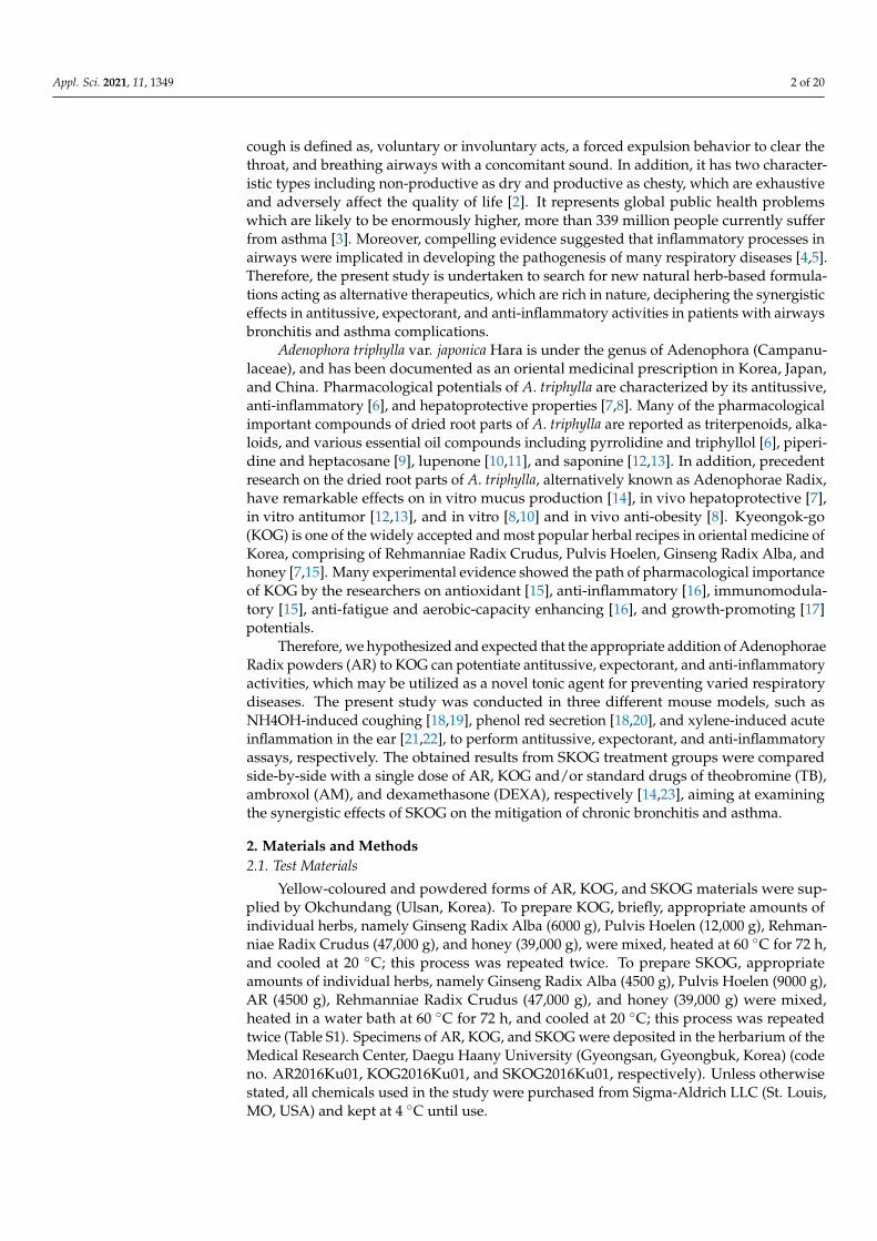
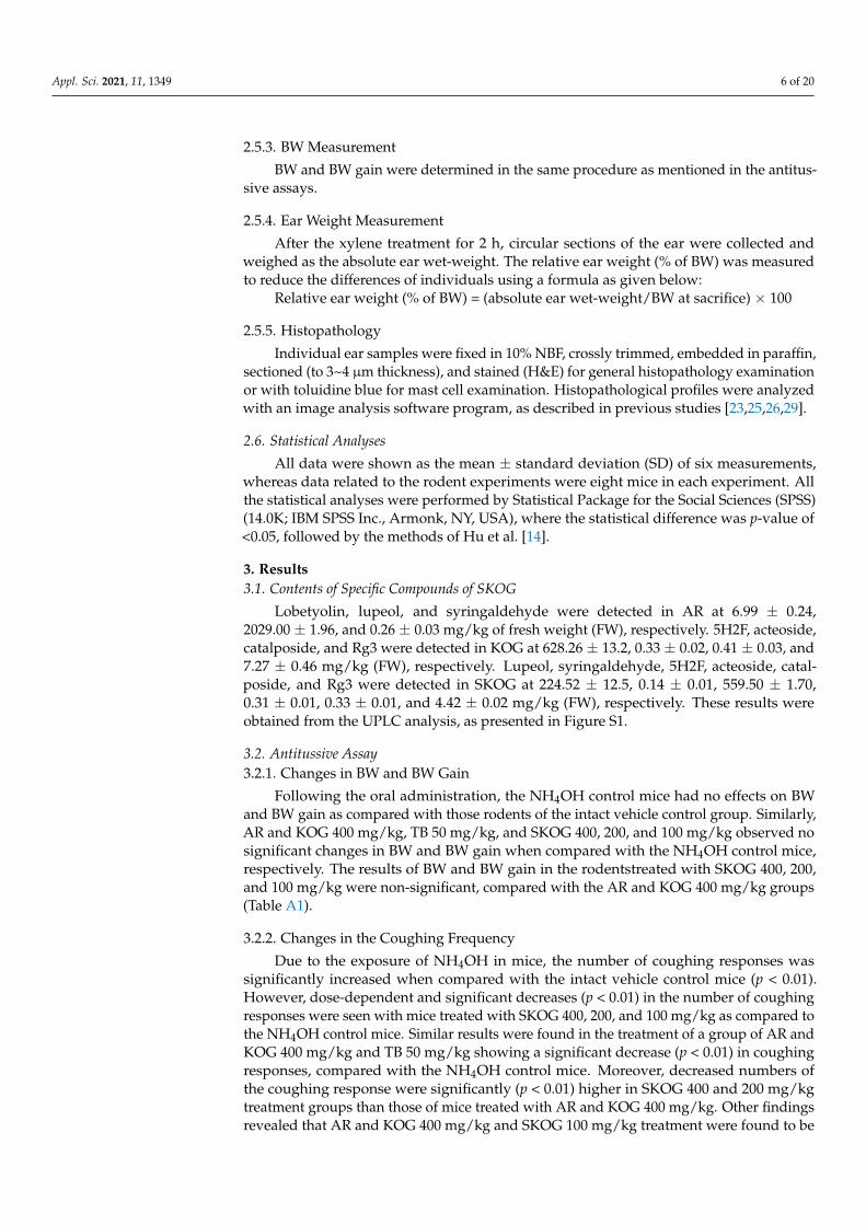
3.2.2. Changes in the Coughing Frequency
Due to the exposure of NH4OH in mice, the number of coughing responses wassignificantly increased when compared with the intact vehicle control mice (p < 0.01).However, dose-dependent and significant decreases (p < 0.01) in the number of coughingresponses were seen with mice treated with SKOG 400, 200, and 100 mg/kg as compared tothe NH4OH control mice. Similar results were found in the treatment of a group of AR andKOG 400 mg/kg and TB 50 mg/kg showing a significant decrease (p < 0.01) in coughingresponses, compared with the NH4OH control mice. Moreover, decreased numbers ofthe coughing response were significantly (p < 0.01) higher in SKOG 400 and 200 mg/kgtreatment groups than those of mice treated with AR and KOG 400 mg/kg. Other findingsrevealed that AR and KOG 400 mg/kg and SKOG 100 mg/kg treatment were found to be
Appl. Sci. 2021, 11, 1349 7 of 20
similar or more favorable to the effects of TB 50 mg/kg against NH4OH-induced coughingresponses (Figure 1).
Figure 1. Effects of adenophorae radix powder (AR), kyeongok-go (KOG), and shashamkyeongok-go (SKOG) on thechanges of coughing frequencies in NH4OH-induced coughing mice. Values are expressed as the mean ± SD of 10 mice.Different letters indicate statistical significance. Treatment groups expressed as a p < 0.01, b p < 0.01, c p < 0.01, and d p < 0.01compared with the intact vehicle control, NH4OH control, AR 400 mg/kg, and KOG 400 mg/kg by the Mann-Whitney U(MW) test, respectively.
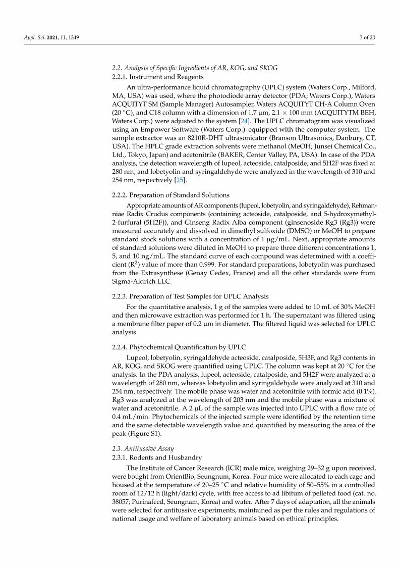
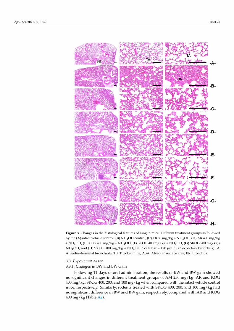
3.2.3. Histopathological Findings of the Trachea and Lung
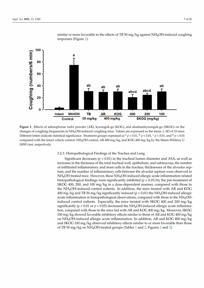
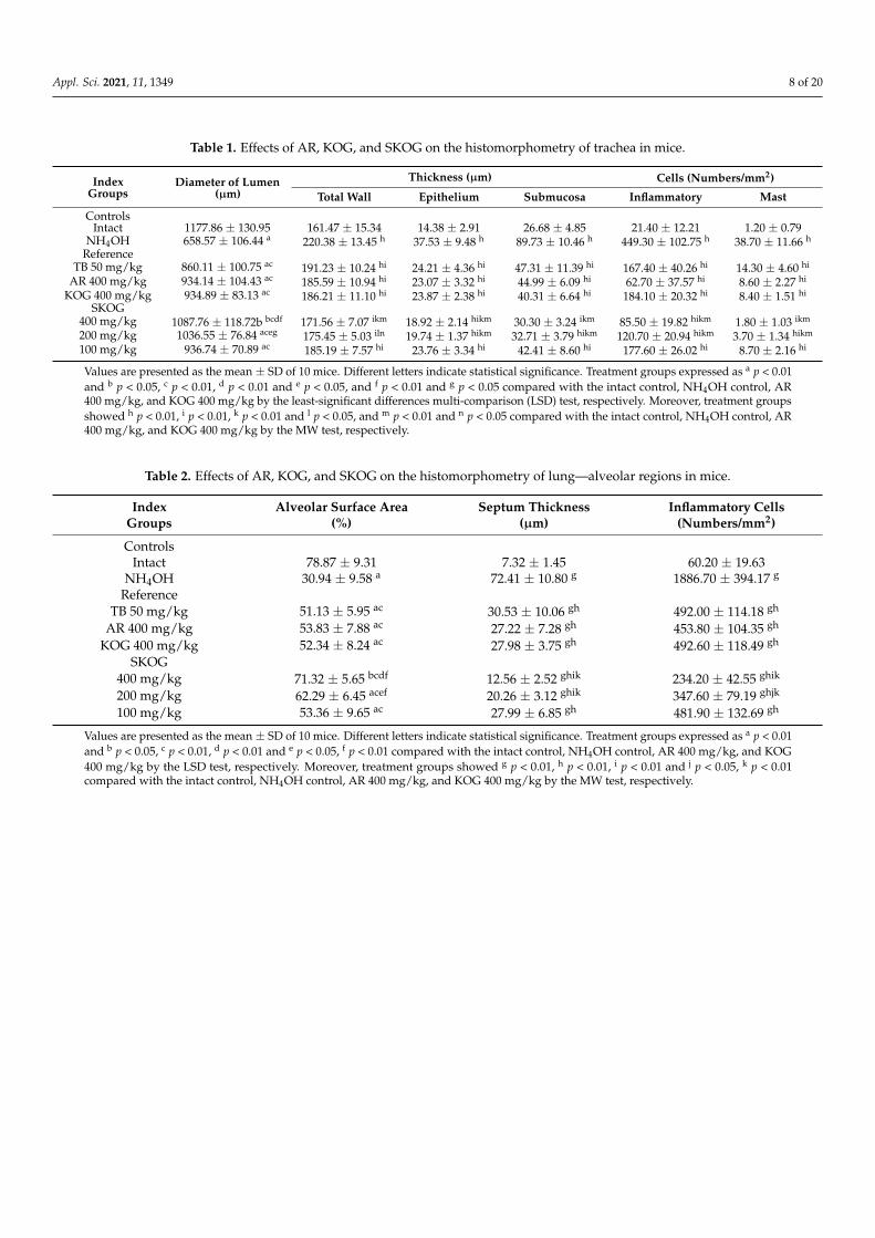
Significant decreases (p < 0.01) in the tracheal lumen diameter and ASA, as well asincreases in the thickness of the total tracheal wall, epithelium, and submucosa; the numberof infiltrated inflammatory and mast cells in the trachea; thicknesses of the alveolar sep-tum; and the number of inflammatory cells between the alveolar septum were observed inNH4OH treated mice. However, these NH4OH-induced allergic acute inflammation-relatedhistopathological findings were significantly inhibited (p < 0.01) by the pre-treatment ofSKOG 400, 200, and 100 mg/kg in a dose-dependent manner, compared with those inthe NH4OH-induced control rodents. In addition, the mice treated with AR and KOG400 mg/kg and TB 50 mg/kg significantly reduced (p < 0.01) the NH4OH-induced allergicacute inflammation in histopathological observations, compared with those in the NH4OH-induced control rodents. Especially, the mice treated with SKOG 400 and 200 mg/kgsignificantly (p < 0.01 or p < 0.05) decreased the NH4OH-induced allergic acute inflamma-tion, compared with those in the mice fed with AR and KOG 400 mg/kg. Moreover, SKOG100 mg/kg showed favorable inhibitory effects similar to those of AR and KOG 400 mg/kgon NH4OH-induced allergic acute inflammation. In addition, AR and KOG 400 mg/kgand SKOG 100 mg/kg observed inhibitory effects similar to or more favorable than thoseof TB 50 mg/kg on NH4OH-treated groups (Tables 1 and 2, Figures 2 and 3).
Appl. Sci. 2021, 11, 1349 8 of 20
Table 1. Effects of AR, KOG, and SKOG on the histomorphometry of trachea in mice.
IndexGroups
Diameter of Lumen(µm)
Thickness (µm) Cells (Numbers/mm2)
Total Wall Epithelium Submucosa Inflammatory Mast
ControlsIntact 1177.86 ± 130.95 161.47 ± 15.34 14.38 ± 2.91 26.68 ± 4.85 21.40 ± 12.21 1.20 ± 0.79
NH4OH 658.57 ± 106.44 a 220.38 ± 13.45 h 37.53 ± 9.48 h 89.73 ± 10.46 h 449.30 ± 102.75 h 38.70 ± 11.66 h
ReferenceTB 50 mg/kg 860.11 ± 100.75 ac 191.23 ± 10.24 hi 24.21 ± 4.36 hi 47.31 ± 11.39 hi 167.40 ± 40.26 hi 14.30 ± 4.60 hi
AR 400 mg/kg 934.14 ± 104.43 ac 185.59 ± 10.94 hi 23.07 ± 3.32 hi 44.99 ± 6.09 hi 62.70 ± 37.57 hi 8.60 ± 2.27 hi
KOG 400 mg/kg 934.89 ± 83.13 ac 186.21 ± 11.10 hi 23.87 ± 2.38 hi 40.31 ± 6.64 hi 184.10 ± 20.32 hi 8.40 ± 1.51 hi
SKOG400 mg/kg 1087.76 ± 118.72b bcdf 171.56 ± 7.07 ikm 18.92 ± 2.14 hikm 30.30 ± 3.24 ikm 85.50 ± 19.82 hikm 1.80 ± 1.03 ikm
200 mg/kg 1036.55 ± 76.84 aceg 175.45 ± 5.03 iln 19.74 ± 1.37 hikm 32.71 ± 3.79 hikm 120.70 ± 20.94 hikm 3.70 ± 1.34 hikm
100 mg/kg 936.74 ± 70.89 ac 185.19 ± 7.57 hi 23.76 ± 3.34 hi 42.41 ± 8.60 hi 177.60 ± 26.02 hi 8.70 ± 2.16 hi
Values are presented as the mean ± SD of 10 mice. Different letters indicate statistical significance. Treatment groups expressed as a p < 0.01and b p < 0.05, c p < 0.01, d p < 0.01 and e p < 0.05, and f p < 0.01 and g p < 0.05 compared with the intact control, NH4OH control, AR400 mg/kg, and KOG 400 mg/kg by the least-significant differences multi-comparison (LSD) test, respectively. Moreover, treatment groupsshowed h p < 0.01, i p < 0.01, k p < 0.01 and l p < 0.05, and m p < 0.01 and n p < 0.05 compared with the intact control, NH4OH control, AR400 mg/kg, and KOG 400 mg/kg by the MW test, respectively.
Table 2. Effects of AR, KOG, and SKOG on the histomorphometry of lung—alveolar regions in mice.
IndexGroups
Alveolar Surface Area(%)
Septum Thickness(µm)
Inflammatory Cells(Numbers/mm2)
ControlsIntact 78.87 ± 9.31 7.32 ± 1.45 60.20 ± 19.63
NH4OH 30.94 ± 9.58 a 72.41 ± 10.80 g 1886.70 ± 394.17 g
ReferenceTB 50 mg/kg 51.13 ± 5.95 ac 30.53 ± 10.06 gh 492.00 ± 114.18 gh
AR 400 mg/kg 53.83 ± 7.88 ac 27.22 ± 7.28 gh 453.80 ± 104.35 gh
KOG 400 mg/kg 52.34 ± 8.24 ac 27.98 ± 3.75 gh 492.60 ± 118.49 gh
SKOG400 mg/kg 71.32 ± 5.65 bcdf 12.56 ± 2.52 ghik 234.20 ± 42.55 ghik
200 mg/kg 62.29 ± 6.45 acef 20.26 ± 3.12 ghik 347.60 ± 79.19 ghjk
100 mg/kg 53.36 ± 9.65 ac 27.99 ± 6.85 gh 481.90 ± 132.69 gh
Values are presented as the mean ± SD of 10 mice. Different letters indicate statistical significance. Treatment groups expressed as a p < 0.01and b p < 0.05, c p < 0.01, d p < 0.01 and e p < 0.05, f p < 0.01 compared with the intact control, NH4OH control, AR 400 mg/kg, and KOG400 mg/kg by the LSD test, respectively. Moreover, treatment groups showed g p < 0.01, h p < 0.01, i p < 0.01 and j p < 0.05, k p < 0.01compared with the intact control, NH4OH control, AR 400 mg/kg, and KOG 400 mg/kg by the MW test, respectively.
Appl. Sci. 2021, 11, 1349 9 of 20
Figure 2. Changes in the histological features of trachea in mice. Different treatment groups asfollowed by the (A) intact vehicle control, (B) NH4OH control, (C) TB 50 mg/kg + NH4OH, (D) AR400 mg/kg + NH4OH, (E) KOG 400 mg/kg + NH4OH, (F) SKOG 400 mg/kg + NH4OH, (G) SKOG200 mg/kg + NH4OH, and (H) SKOG 100 mg/kg + NH4OH. Scale bar = 120 µm. LU: Lumen; CA:Cartilages; SM: Submucosa; EP: Epithelium.
Appl. Sci. 2021, 11, 1349 10 of 20
Figure 3. Changes in the histological features of lung in mice. Different treatment groups as followedby the (A) intact vehicle control, (B) NH4OH control, (C) TB 50 mg/kg + NH4OH, (D) AR 400 mg/kg+ NH4OH, (E) KOG 400 mg/kg + NH4OH, (F) SKOG 400 mg/kg + NH4OH, (G) SKOG 200 mg/kg +NH4OH, and (H) SKOG 100 mg/kg + NH4OH. Scale bar = 120 µm. SB: Secondary bronchus; TA:Alveolus-terminal bronchiole; TB: Theobromine; ASA: Alveolar surface area; BR: Bronchus.
3.3. Expectorant Assay3.3.1. Changes in BW and BW Gain
Following 11 days of oral administration, the results of BW and BW gain showedno significant changes in different treatment groups of AM 250 mg/kg, AR and KOG400 mg/kg, SKOG 400, 200, and 100 mg/kg when compared with the intact vehicle controlmice, respectively. Similarly, rodents treated with SKOG 400, 200, and 100 mg/kg hadno significant difference in BW and BW gain, respectively, compared with AR and KOG400 mg/kg (Table A2).
Appl. Sci. 2021, 11, 1349 11 of 20
3.3.2. Body-Surface Gross Findings
An experiment was set to elucidate the expectorant effect of SKOG 400, 200, and100 mg/kg that showed noticeable and dose-dependent increases of body surface redness,compared with the intact vehicle control rodents, which indicates an increased uptakeand secretion of intraperitoneally injected phenol red. Moreover, AR and KOG mg/kgand AM 250 mg/kg dramatically increased the secretion of phenol red after 30 min ofintraperitoneal injection as compared with the intact vehicle control. Of note, SKOG 400and 200 mg/kg had obvious increases of phenol red secretion with a gross sign of bodysurface redness, while the treatment with AR and KOG 400 mg/kg, and the results weremore similar to or favorable than those of AM 250 mg/kg fed rodents (Figure S2).
3.3.3. Changes in Mucous Secretion
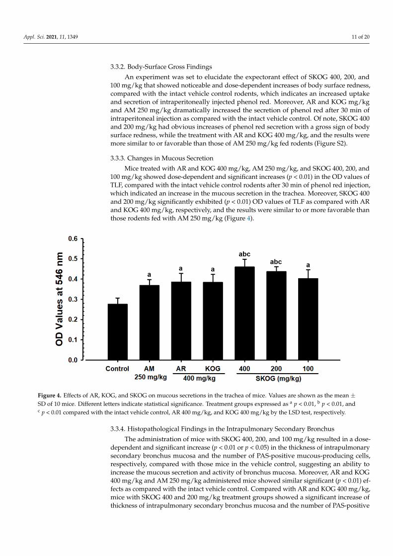
Mice treated with AR and KOG 400 mg/kg, AM 250 mg/kg, and SKOG 400, 200, and100 mg/kg showed dose-dependent and significant increases (p < 0.01) in the OD values ofTLF, compared with the intact vehicle control rodents after 30 min of phenol red injection,which indicated an increase in the mucous secretion in the trachea. Moreover, SKOG 400and 200 mg/kg significantly exhibited (p < 0.01) OD values of TLF as compared with ARand KOG 400 mg/kg, respectively, and the results were similar to or more favorable thanthose rodents fed with AM 250 mg/kg (Figure 4).
Figure 4. Effects of AR, KOG, and SKOG on mucous secretions in the trachea of mice. Values are shown as the mean ±SD of 10 mice. Different letters indicate statistical significance. Treatment groups expressed as a p < 0.01, b p < 0.01, andc p < 0.01 compared with the intact vehicle control, AR 400 mg/kg, and KOG 400 mg/kg by the LSD test, respectively.
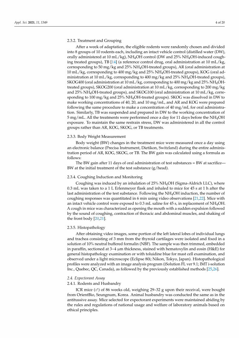
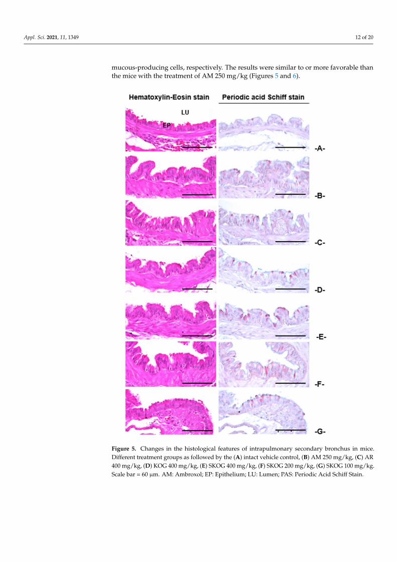
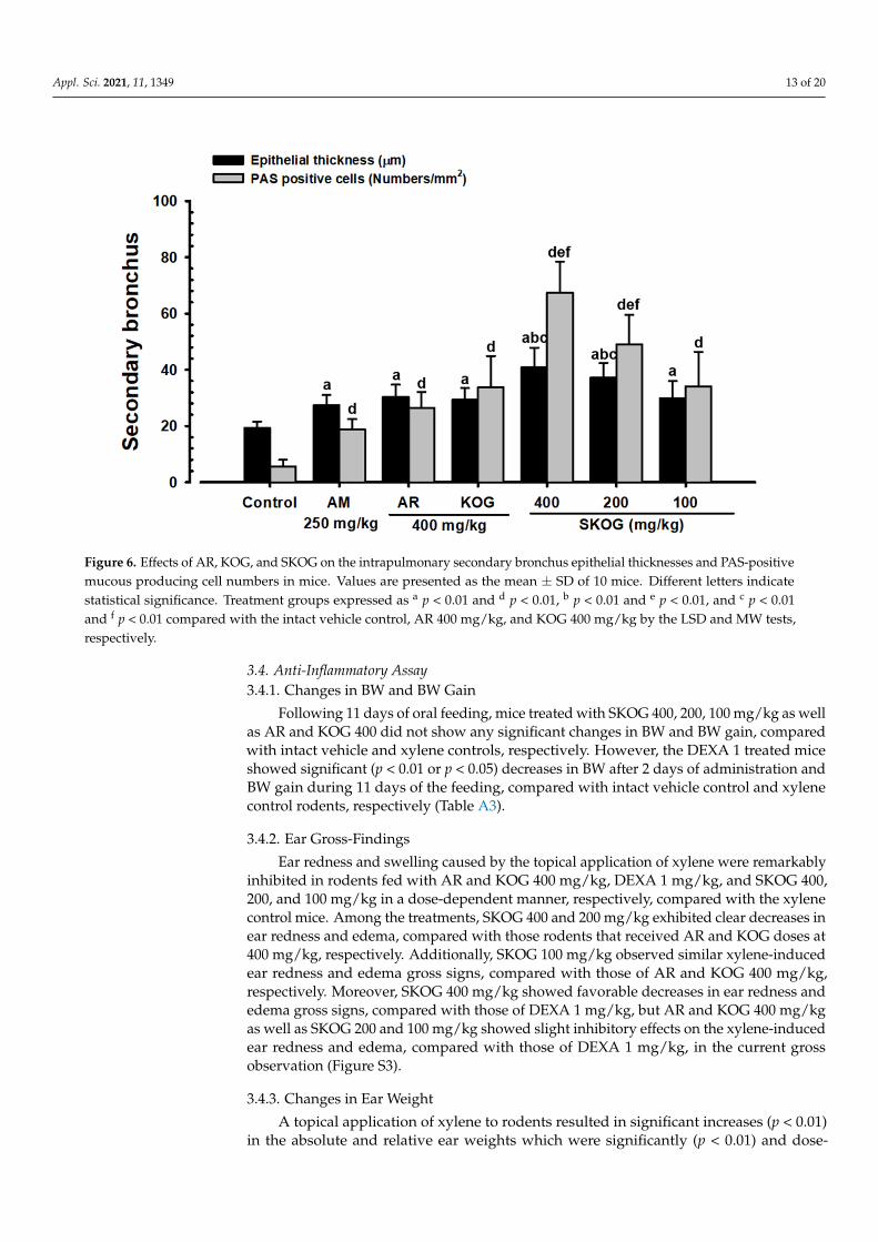
3.3.4. Histopathological Findings in the Intrapulmonary Secondary Bronchus
The administration of mice with SKOG 400, 200, and 100 mg/kg resulted in a dose-dependent and significant increase (p < 0.01 or p < 0.05) in the thickness of intrapulmonarysecondary bronchus mucosa and the number of PAS-positive mucous-producing cells,respectively, compared with those mice in the vehicle control, suggesting an ability toincrease the mucous secretion and activity of bronchus mucosa. Moreover, AR and KOG400 mg/kg and AM 250 mg/kg administered mice showed similar significant (p < 0.01) ef-fects as compared with the intact vehicle control. Compared with AR and KOG 400 mg/kg,mice with SKOG 400 and 200 mg/kg treatment groups showed a significant increase ofthickness of intrapulmonary secondary bronchus mucosa and the number of PAS-positive
Appl. Sci. 2021, 11, 1349 12 of 20
mucous-producing cells, respectively. The results were similar to or more favorable thanthe mice with the treatment of AM 250 mg/kg (Figures 5 and 6).
Figure 5. Changes in the histological features of intrapulmonary secondary bronchus in mice.Different treatment groups as followed by the (A) intact vehicle control, (B) AM 250 mg/kg, (C) AR400 mg/kg, (D) KOG 400 mg/kg, (E) SKOG 400 mg/kg, (F) SKOG 200 mg/kg, (G) SKOG 100 mg/kg.Scale bar = 60 µm. AM: Ambroxol; EP: Epithelium; LU: Lumen; PAS: Periodic Acid Schiff Stain.
Appl. Sci. 2021, 11, 1349 13 of 20
Figure 6. Effects of AR, KOG, and SKOG on the intrapulmonary secondary bronchus epithelial thicknesses and PAS-positivemucous producing cell numbers in mice. Values are presented as the mean ± SD of 10 mice. Different letters indicatestatistical significance. Treatment groups expressed as a p < 0.01 and d p < 0.01, b p < 0.01 and e p < 0.01, and c p < 0.01and f p < 0.01 compared with the intact vehicle control, AR 400 mg/kg, and KOG 400 mg/kg by the LSD and MW tests,respectively.
3.4. Anti-Inflammatory Assay3.4.1. Changes in BW and BW Gain
Following 11 days of oral feeding, mice treated with SKOG 400, 200, 100 mg/kg as wellas AR and KOG 400 did not show any significant changes in BW and BW gain, comparedwith intact vehicle and xylene controls, respectively. However, the DEXA 1 treated miceshowed significant (p < 0.01 or p < 0.05) decreases in BW after 2 days of administration andBW gain during 11 days of the feeding, compared with intact vehicle control and xylenecontrol rodents, respectively (Table A3).
3.4.2. Ear Gross-Findings
Ear redness and swelling caused by the topical application of xylene were remarkablyinhibited in rodents fed with AR and KOG 400 mg/kg, DEXA 1 mg/kg, and SKOG 400,200, and 100 mg/kg in a dose-dependent manner, respectively, compared with the xylenecontrol mice. Among the treatments, SKOG 400 and 200 mg/kg exhibited clear decreases inear redness and edema, compared with those rodents that received AR and KOG doses at400 mg/kg, respectively. Additionally, SKOG 100 mg/kg observed similar xylene-inducedear redness and edema gross signs, compared with those of AR and KOG 400 mg/kg,respectively. Moreover, SKOG 400 mg/kg showed favorable decreases in ear redness andedema gross signs, compared with those of DEXA 1 mg/kg, but AR and KOG 400 mg/kgas well as SKOG 200 and 100 mg/kg showed slight inhibitory effects on the xylene-inducedear redness and edema, compared with those of DEXA 1 mg/kg, in the current grossobservation (Figure S3).
3.4.3. Changes in Ear Weight
A topical application of xylene to rodents resulted in significant increases (p < 0.01)in the absolute and relative ear weights which were significantly (p < 0.01) and dose-
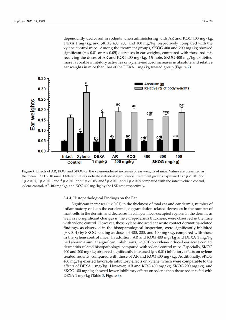
Appl. Sci. 2021, 11, 1349 14 of 20
dependently decreased in rodents when administering with AR and KOG 400 mg/kg,DEXA 1 mg/kg, and SKOG 400, 200, and 100 mg/kg, respectively, compared with thexylene control mice. Among the treatment groups, SKOG 400 and 200 mg/kg showedsignificant (p < 0.01 or p < 0.05) decreases in ear weights, compared with those rodentsreceiving the doses of AR and KOG 400 mg/kg. Of note, SKOG 400 mg/kg exhibitedmore favorable inhibitory activities on xylene-induced increases in absolute and relativeear weights in mice than that of the DEXA 1 mg/kg treated group (Figure 7).
Figure 7. Effects of AR, KOG, and SKOG on the xylene-induced increases of ear weights of mice. Values are presented asthe mean ± SD of 10 mice. Different letters indicate statistical significance. Treatment groups expressed as a p < 0.01 andb p < 0.05, c p < 0.01, and d p < 0.01 and e p < 0.05, and f p < 0.01 and g p < 0.05 compared with the intact vehicle control,xylene control, AR 400 mg/kg, and KOG 400 mg/kg by the LSD test, respectively.
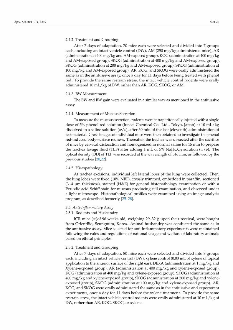
3.4.4. Histopathological Findings on the Ear
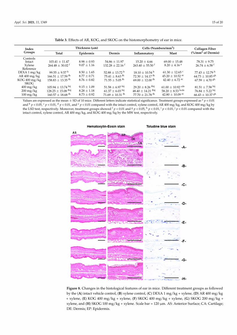
Significant increases (p < 0.01) in the thickness of total ear and ear dermis, number ofinflammatory cells on the ear dermis, degranulation-related decreases in the number ofmast cells in the dermis, and decreases in collagen fiber-occupied regions in the dermis, aswell as no significant changes in the ear epidermis thickness, were observed in the micewith xylene control. However, these xylene-induced ear acute contact dermatitis-relatedfindings, as observed in the histopathological inspection, were significantly inhibited(p < 0.01) by SKOG feeding at doses of 400, 200, and 100 mg/kg, compared with thosein the xylene control mice. In addition, AR and KOG 400 mg/kg and DEXA 1 mg/kghad shown a similar significant inhibition (p < 0.01) on xylene-induced ear acute contactdermatitis-related histopathology, compared with xylene control mice. Especially, SKOG400 and 200 mg/kg observed significantly increased (p < 0.01) inhibitory effects on xylene-treated rodents, compared with those of AR and KOG 400 mg/kg. Additionally, SKOG400 mg/kg exerted favorable inhibitory effects on xylene, which were comparable to theeffects of DEXA 1 mg/kg. However, AR and KOG 400 mg/kg, SKOG 200 mg/kg, andSKOG 100 mg/kg showed lower inhibitory effects on xylene than those rodents fed withDEXA 1 mg/kg (Table 3, Figure 8).
Appl. Sci. 2021, 11, 1349 15 of 20
Table 3. Effects of AR, KOG, and SKOG on the histomorphometry of ear in mice.
IndexGroups
Thickness (µm) Cells (Numbers/mm2) Collagen Fiber(%/mm2 of Dermis)Total Epidermis Dermis Inflammatory Mast
ControlsIntact 103.41 ± 11.47 8.98 ± 0.93 54.86 ± 11.97 15.20 ± 4.66 69.00 ± 15.48 78.31 ± 9.75
Xylene 264.48 ± 30.02 f 9.07 ± 1.16 132.28 ± 22.16 f 263.40 ± 55.50 f 8.20 ± 4.16 a 26.74 ± 6.58 f
ReferenceDEXA 1 mg/kg 99.55 ± 9.57 h 8.50 ± 1.65 52.88 ± 13.72 h 18.10 ± 10.54 h 61.30 ± 12.65 c 77.43 ± 12.79 h
AR 400 mg/kg 166.51 ± 17.59 fh 8.77 ± 0.71 75.41 ± 8.63 fh 72.30 ± 14.17 fh 45.20 ± 10.52 ac 64.73 ± 10.85 gh
KOG 400 mg/kg 158.83 ± 13.35 fh 8.76 ± 0.82 71.55 ± 5.05 fh 69.00 ± 12.00 fh 42.40 ± 6.72 ac 67.59 ± 4.70 gh
SKOG400 mg/kg 105.94 ± 13.74 hij 9.15 ± 1.09 51.58 ± 6.97 hij 29.20 ± 8.26 fhij 61.00 ± 10.92 cde 81.51 ± 7.58 hij
200 mg/kg 128.25 ± 15.08 fhij 8.28 ± 1.18 61.37 ± 6.03 hij 48.40 ± 14.21 fhij 58.20 ± 8.53 bcde 76.84 ± 5.22 hij
100 mg/kg 160.57 ± 18.68 fh 8.73 ± 0.92 71.69 ± 10.31 fh 77.70 ± 21.78 fh 42.80 ± 10.09 ac 66.43 ± 10.37 gh
Values are expressed as the mean ± SD of 10 mice. Different letters indicate statistical significance. Treatment groups expressed as a p < 0.01and b p < 0.05, c p < 0.01, d p < 0.01, and e p < 0.01 compared with the intact control, xylene control, AR 400 mg/kg, and KOG 400 mg/kg bythe LSD test, respectively. Moreover, treatment groups showed f p < 0.01 and g p < 0.05, h p < 0.01, i p < 0.01, j p < 0.01 compared with theintact control, xylene control, AR 400 mg/kg, and KOG 400 mg/kg by the MW test, respectively.
Figure 8. Changes in the histological features of ear in mice. Different treatment groups as followedby the (A) intact vehicle control, (B) xylene control, (C) DEXA 1 mg/kg + xylene, (D) AR 400 mg/kg+ xylene, (E) KOG 400 mg/kg + xylene, (F) SKOG 400 mg/kg + xylene, (G) SKOG 200 mg/kg +xylene, and (H) SKOG 100 mg/kg + xylene. Scale bar = 120 µm. AS: Anterior Surface; CA: Cartilage;DE: Dermis; EP: Epidermis.
Appl. Sci. 2021, 11, 1349 16 of 20
4. Discussion
The phytochemical observation on SKOG signified its medicinal value by the presenceof various pharmacological important bioactive compounds, which was identified andquantified by the UPLC analysis. SKOG was mainly composed of lupeol, syringaldehyde,5H2F, acteoside, catalposide, and Rg3 compounds, which were a direct or indirect linkto the current activities of SKOG. It has been extensively documented that the followingidentified compounds are believed to be a potential source of bioactivities such as ananti-inflammatory and anti-cancer activities of lupeol [30]; antioxidant and antimicrobialactivities of syringaldehyde [31]; antioxidative, anti-hypoxic, anti-allergic, anti-sickling,anti-inflammatory, and anti-hyperuricemic effects of 5H2F [32]; antioxidant and antihyper-tensive activities of acteoside [33]; preventing mucosal inflammation of catalposide [34];ameliorating cancer, lung injury, depression, and diabetes of Rg3 [35].
During the 11-day treatment with different test substances, all the mice in threedifferent models such as antitussive, expectorant, and anti-inflammatory assays showed anormal BW and BW gain that fell in the range of age-matched, normal reference mice [36],except for the xylene-induced acute inflammation rodents fed with DEXA 1 mg/kg. Inaddition, compared with those in the rodents fed with AR and KOG 400 mg/kg, nosignificant changes in BW and BW gain were shown in the rodents fed with SKOG 400, 200,and 100 mg/kg in all the three different assays. The xylene-induced acute inflammationmice treated with DEXA 1 mg/kg showed significant decreases in BW from 2 days after theinitial feeding and in BW gain during the treatment period, when compared with the intactvehicle and xylene control animals. A decrease in BW after the treatment with DEXA hasbeen detected as a major side effect of DEXA in other animal studies of anti-inflammatoryeffects [26,37].
The NH4OH has been a well-known antitussive agent to induce the coughing fre-quency with characteristic acute inflammation in airways [25,26]. The mouse model ofNH4OH exposure-induced coughing was used to confirm whether the antitussive effectsof KOG were potentiated by the appropriate addition of AR. ASA is directly relatedto the gas exchange capacity of the lung; a higher ASA means a higher gas exchangecapacity [26,27,38]. In the antitussive experiment, the NH4OH-induced allergic acuteinflammation with coughing responses and decreased ASA were dose dependently andsignificantly inhibited by the feeding of SKOG at doses of 400, 200, and 100 mg/kg. Thesefindings are considered a piece of direct evidence that the appropriate addition of AR toKOG synergistically increased the antitussive activities, at least in the conditions of thecurrent study.
Expectorant agents have the ability to increase the hydration of secretions so that theirritating respiratory tract becomes lubricated [14]. Therefore, the ability to increase themuscus secrtion can be an effective approach to search for a potential drug. Here, thephenol red solution was used to identify whether the expectorant effects of KOG in micewere synergistically potentiated by the appropriate addition of AR. Later, the PAS stainingmethod was performed to evaluate the mucous intensity and number of mucous-producingcells [28,39,40]. The result of the study was found to be effective in the effects of SKOG,which showed a significant and dose-dependent expectorant ability in oral administeredmice, through enhanced mucous secretion and promoted functional activities of tracheaand bronchus mucosa [21–23]. These findings are considered obvious evidence that theappropriate addition of AR synergistically increased the expectorant activities of KOG.
An investigatoin on the anti-inflammatory efficacy of the test substances can easily beachieved following the application of xylene on the anterior surface of the mouse ear toinduce acute inflammation in the mouse ear model [14]. In the present study, KOG wasinvestigated by whether the appropriate addition of AR could subsequently promote anti-inflammatory potentials, in addition to antitussive and expectorant activities. The xylenecontrol mice are reported to have noticeable changes in the gross sign and weight of theear with characteristic inflammatory-related histopathological features [25,26]. Followingthe administration, SKOG at doses of 400 and 200 mg/kg significantly increased the anti-
Appl. Sci. 2021, 11, 1349 17 of 20
inflammatory effects, compared to those in the mice administered with AR and KOG400 mg/kg on xylene-induced acute inflammation in the rodent ear. In addition, SKOG400 mg/kg showed anti-inflammatory effects comparable to those of DEXA 1 mg/kgon xylene-induced acute inflammation in the rodent ear. These findings are considereddirect and clear evidence that the appropriate addition of AR synergistically increasedthe anti-inflammatory activities of KOG, at least in the conditions of the current study.However, further research is warranted as to whether SKOG has some other effectivebioactive compound(s) and whether their molecular mechanism of actions is responsiblefor the synergistic effects in the current study. Similarly, changes in the relative lungweight could have been measured to ensure that there are no significant changes due tothe treatments and that also needs to be addressed in further studies.
5. Conclusions
In conclusion, our study suggested that the appropriate addition of AR to KOG inorder to prepare the SKOG recipe exhibited significant and synergistic effects in antitus-sive, expectorant, and anti-inflammatory activities by potentiating the modulation of theactivities of respiratory mucous-producing cells and mast cells in animal models. The mosteffective doses of SKOG were found to be 400 and 200 mg/kg, compared to those in themice fed with AR and KOG 400 mg/kg, suggesting that an effective portion of bioactivecompounds is present at these doses. These findings were considered as pharmacologicalshreds of evidence for the traditional use of AR and KOG as antitussive, expectorant,and anti-inflammatory remedies. Therefore, it is expected that SKOG may serve as apreventive or therapeutic agent in varied respiratory diseases, particularly those caused byenvironmental toxicants.
Supplementary Materials: The following are available online at https://www.mdpi.com/2076-3417/11/3/1349/s1, Figure S1: UPLC analysis of identification and quantification of lupeol, syringalde-hyde, 5H2F, acteoside, catalposide, and Rg3 ingredients in SKOG, a mixture of AR and KOG. (A)standard for AR, (B) Test AR, (C) standard for KOG, (D) Test KOG, (E) standard for SKOG, and (F)Test SKOG., Table S1: Composition of KOG and SKOG used in this study.
Author Contributions: Conceptualization, S.-K.K. and J.-S.C.; methodology, J.-R.H. and S.-K.K.; vali-dation, D.-H.J., M.M., and S.-K.K.; formal analysis, S.-M.K., S.-K.K. and H.-J.L.; investigation, S.-M.K.,D.-H.J. and S.-K.K.; resources, C.-J.J. and S.-K.K.; data curation, S.-K.K. and D.-H.J.; writing—originaldraft preparation, S.-K.K. and H.-J.L.; writing—review and editing, M.M. and J.-S.C.; visualization,J.-R.H. and S.-K.K.; supervision, S.-K.K., H.-J.L., and J.-S.C.; project administration, D.-H.J. andS.-K.K.; funding acquisition, S.-K.K. All authors have read and agreed to the published version of themanuscript.
Funding: This work was supported by the National Research Foundation of Korea grant, funded bythe Korea government (MSIT) (no. 2018R1A5A2025272).
Institutional Review Board Statement: Antitussive, expectorant and anti-inflammatory experi-ments in this study were approved by the Institutional Animal Care and Use Committee of DaeguHaany University. The approval numbers were DHU2016-034, DHU2016-035 and DHU2016-036,respectively.
Data Availability Statement: Data supporting reported results can be made available on demand.
Conflicts of Interest: The authors declare no conflict of interest.
Appl. Sci. 2021, 11, 1349 18 of 20
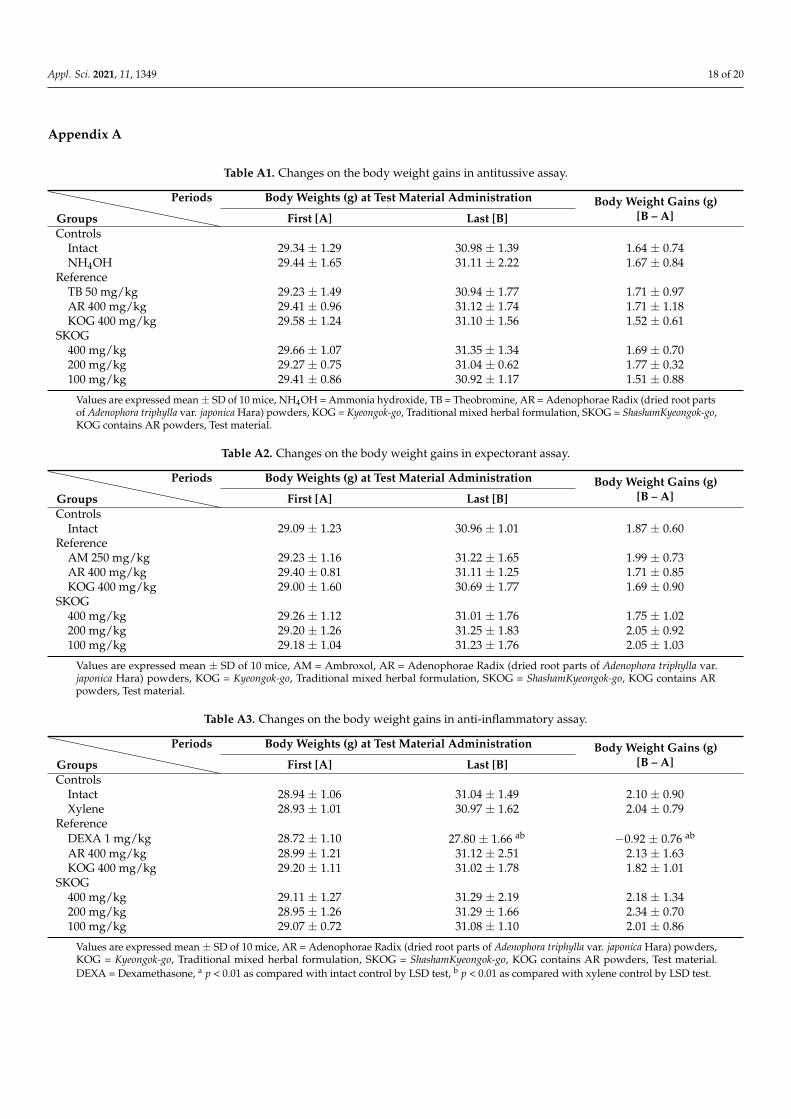
Appendix A
Table A1. Changes on the body weight gains in antitussive assay.
Groups
Periods Body Weights (g) at Test Material Administration Body Weight Gains (g)[B – A]First [A] Last [B]
ControlsIntact 29.34 ± 1.29 30.98 ± 1.39 1.64 ± 0.74NH4OH 29.44 ± 1.65 31.11 ± 2.22 1.67 ± 0.84
ReferenceTB 50 mg/kg 29.23 ± 1.49 30.94 ± 1.77 1.71 ± 0.97AR 400 mg/kg 29.41 ± 0.96 31.12 ± 1.74 1.71 ± 1.18KOG 400 mg/kg 29.58 ± 1.24 31.10 ± 1.56 1.52 ± 0.61
SKOG400 mg/kg 29.66 ± 1.07 31.35 ± 1.34 1.69 ± 0.70200 mg/kg 29.27 ± 0.75 31.04 ± 0.62 1.77 ± 0.32100 mg/kg 29.41 ± 0.86 30.92 ± 1.17 1.51 ± 0.88
Values are expressed mean ± SD of 10 mice, NH4OH = Ammonia hydroxide, TB = Theobromine, AR = Adenophorae Radix (dried root partsof Adenophora triphylla var. japonica Hara) powders, KOG = Kyeongok-go, Traditional mixed herbal formulation, SKOG = ShashamKyeongok-go,KOG contains AR powders, Test material.
Table A2. Changes on the body weight gains in expectorant assay.
Groups
Periods Body Weights (g) at Test Material Administration Body Weight Gains (g)[B – A]First [A] Last [B]
ControlsIntact 29.09 ± 1.23 30.96 ± 1.01 1.87 ± 0.60
ReferenceAM 250 mg/kg 29.23 ± 1.16 31.22 ± 1.65 1.99 ± 0.73AR 400 mg/kg 29.40 ± 0.81 31.11 ± 1.25 1.71 ± 0.85KOG 400 mg/kg 29.00 ± 1.60 30.69 ± 1.77 1.69 ± 0.90
SKOG400 mg/kg 29.26 ± 1.12 31.01 ± 1.76 1.75 ± 1.02200 mg/kg 29.20 ± 1.26 31.25 ± 1.83 2.05 ± 0.92100 mg/kg 29.18 ± 1.04 31.23 ± 1.76 2.05 ± 1.03
Values are expressed mean ± SD of 10 mice, AM = Ambroxol, AR = Adenophorae Radix (dried root parts of Adenophora triphylla var.japonica Hara) powders, KOG = Kyeongok-go, Traditional mixed herbal formulation, SKOG = ShashamKyeongok-go, KOG contains ARpowders, Test material.
Table A3. Changes on the body weight gains in anti-inflammatory assay.
Groups
Periods Body Weights (g) at Test Material Administration Body Weight Gains (g)[B – A]First [A] Last [B]
ControlsIntact 28.94 ± 1.06 31.04 ± 1.49 2.10 ± 0.90Xylene 28.93 ± 1.01 30.97 ± 1.62 2.04 ± 0.79
ReferenceDEXA 1 mg/kg 28.72 ± 1.10 27.80 ± 1.66 ab −0.92 ± 0.76 ab
AR 400 mg/kg 28.99 ± 1.21 31.12 ± 2.51 2.13 ± 1.63KOG 400 mg/kg 29.20 ± 1.11 31.02 ± 1.78 1.82 ± 1.01
SKOG400 mg/kg 29.11 ± 1.27 31.29 ± 2.19 2.18 ± 1.34200 mg/kg 28.95 ± 1.26 31.29 ± 1.66 2.34 ± 0.70100 mg/kg 29.07 ± 0.72 31.08 ± 1.10 2.01 ± 0.86
Values are expressed mean ± SD of 10 mice, AR = Adenophorae Radix (dried root parts of Adenophora triphylla var. japonica Hara) powders,KOG = Kyeongok-go, Traditional mixed herbal formulation, SKOG = ShashamKyeongok-go, KOG contains AR powders, Test material.DEXA = Dexamethasone, a p < 0.01 as compared with intact control by LSD test, b p < 0.01 as compared with xylene control by LSD test.
Appl. Sci. 2021, 11, 1349 19 of 20
References1. Pavord, I.D. Cough and asthma. Pulm. Pharmacol. Ther. 2004, 17, 399–402. [CrossRef] [PubMed]2. Dapaah, G.; Koffuor, G.A.; Mante, P.K.; Ben, I.O. Antitussive, expectorant and analgesic effects of the ethanol seed extract of
Picralima nitida (Stapf) Th. & H. Durand. Res. Pharm. Sci. 2016, 11, 100. [PubMed]3. GBD 2016 Disease and Injury Incidence and Prevalence Collaborators. Global, regional, and national incidence, prevalence, and
years lived with disability for 328 diseases and injuries for 195 countries, 1990–2016: A systematic analysis for the Global Burdenof Disease Study 2016. Lancet 2017, 390, 1211–1259. [CrossRef]
4. Sanak, M. Eicosanoid mediators in the airway inflammation of asthmatic patients: What is new? Allergy Asthma Immunol. Res.2016, 8, 481–490. [CrossRef] [PubMed]
5. Woloski, J.R.; Heston, S.; Calderon, S.P.E. Respiratory Allergic Disorders. Prim. Care Clin. Off. Pract. 2016, 43, 401–415. [CrossRef][PubMed]
6. Konno, C.; Saito, T.; Oshima, Y.; Hikino, H.; Kabuto, C. Structure of methyl adenophorate and triphyllol, triterpenoids ofAdenophora triphylla var. japonica roots. Planta Med. 1981, 42, 268–274. [CrossRef]
7. Kim, J.B.; Song, H.N. Effects of Kyeongok-go and its two added precriptions on hyperlipidemic rats induced by high-fat diet. J.Physiol. Pathol. Korean Med. 2014, 28, 371–378. [CrossRef]
8. Lee, D.-R.; Lee, Y.-S.; Choi, B.-K.; Lee, H.J.; Park, S.-B.; Kim, T.-M.; Oh, H.J.; Yang, S.H.; Suh, J.-W. Roots extracts of Adenophoratriphylla var. japonica improve obesity in 3T3-L1 adipocytes and high-fat diet-induced obese mice. Asian Pac. J. Trop. Med. 2015,8, 898–906. [CrossRef]
9. Asano, N.; Nishida, M.; Miyauchi, M.; Ikeda, K.; Yamamoto, M.; Kizu, H.; Kameda, Y.; Watson, A.A.; Nash, R.J.; Fleet, G.W.J.Polyhydroxylated pyrrolidine and piperidine alkaloids from Adenophora triphylla var. japonica (Campanulaceae). Phytochemistry2000, 53, 379–382. [CrossRef]
10. Ahn, E.K.; Oh, J.S. Lupenone isolated from Adenophora triphylla var. japonica extract inhibits adipogenic differentiation throughthe downregulation of PPARγ in 3T3-L1 cells. Phytother. Res. 2013, 27, 761–766. [CrossRef]
11. Yoon, Y.P.; Lee, H.J.; Lee, D.-U.; Lee, S.K.; Hong, J.-H.; Lee, C.J. Effects of lupenone, lupeol, and taraxerol derived from Adenophoratriphylla on the gene expression and production of airway MUC5AC mucin. Tuberc. Respir. Dis. 2015, 78, 210–217. [CrossRef][PubMed]
12. Kang, M.; Ha, I.J.; Chun, J.; Kang, S.S.; Kim, Y.S. Separation of two cytotoxic saponins from the roots of Adenophora triphylla var.japonica by High-speed Counter-current Chromatography. Phytochem. Anal. 2013, 24, 148–154. [CrossRef]
13. Chun, J.; Kang, M.; Kim, Y.S. A triterpenoid saponin from Adenophora triphylla var. japonica suppresses the growth of humangastric cancer cells via regulation of apoptosis and autophagy. Tumor Biol. 2014, 35, 12021–12030. [CrossRef] [PubMed]
14. Hu, J.R.; Jung, C.J.; Ku, S.M.; Jung, D.H.; Ku, S.K.; Choi, J.S. Antitussive, expectorant, and anti-inflammatory effects ofAdenophorae Radix powder in ICR mice. J. Ethnopharmacol. 2019, 239, 111915. [CrossRef] [PubMed]
15. Na, C.-S.; Shin, W.; Lee, Y.-M.; Moon, Y.-S.; Noh, H.-k.; Seo, S.-H.; Son, H.-S. Effect of original kyungokgo & iksuyongjingo plusSparassis crispa on antioxidant, immunity improvement and sensory evaluation. Korea J. Herbol. 2016, 31, 43–51.
16. Lee, K.-S.; Kim, G.-H.; Kim, H.-H.; Seong, B.-J.; Kim, S.-I.; Han, S.-H.; Kang, E.J.; Yoo, Y.C. Qualities and anti-inflammatory activityof Kyungokgos sold in local markets. J. Korean Soc. Food Sci. Nutr. 2013, 42, 335–341. [CrossRef]
17. Kim, J.-H.; Lee, J.-H.; Oh, J.-M.; Kim, Y.-K. Inhibitory effects on bone resorption and osteoblast proliferation of Kyungok-go. Herb.Formula Sci. 2011, 19, 61–71.
18. Cha, Y.-Y. A Comparative Study on Effects of Kyungohkgo and Kyungohkgo Ga Nokyong on growth in growth deficiency ratwith insufficient nutrition diet. J. Korean Med. Obes. Res. 2009, 9, 59–69.
19. Zhang, J.-L.; Wang, H.; Pi, H.-F.; Ruan, H.-L.; Zhang, P.; Wu, J.-Z. Structural analysis and antitussive evaluation of five novelesters of verticinone and bile acids. Steroids 2009, 74, 424–434. [CrossRef]
20. Wanga, D.; Wanga, S.; Chena, X.; Xua, X.; Zhub, J.; Nieb, L.; Longa, X. Antitussive, expectorant and anti-inflammatory activitiesof four alkaloids isolated from Bulbus of Fritillaria wabuensis. J. Ethnopharmacol. 2012, 139, 189–193. [CrossRef]
21. Engler, H.; Szelenyi, I. Tracheal phenol red secretion, a new method for screening mucosecretolytic compounds. J. Pharmacol.Methods 1984, 11, 151–157. [CrossRef]
22. Cho, K.-H.; Kim, H.-D.; Lee, B.-W.; Lim, M.-K.; Ku, S.K. Effects of magnetic infrared laser on xylene-induced acute inflammationin mice. J. Phys. Ther. Sci. 2008, 20, 255–259. [CrossRef]
23. Lee, H.-S.; Ku, S.-K. Effects of Picrorrhiza Rhizoma on acute inflammation in mice. Biomol. Ther. 2008, 16, 137–140. [CrossRef]24. Ministry of Food and Drug Safety. The Korean Pharmacopoeia (The Korea Food and Drug Administration Notification 2012-9); Ministry
of Food and Drug Safety: Cheongju, Korea, 2012.25. Lebargy, F.; Lenormand, E.; Pariente, R.; Fournier, M. Morphological changes in rat tracheal mucosa immediately after antigen
challenge. Bull. Eur. Physiopathol. Respir. 1987, 23, 417–421. [PubMed]26. Ku, S.K.; Kim, J.W.; Cho, H.R.; Kim, K.Y.; Min, Y.H.; Park, J.H.; Kim, J.S.; Park, J.H.; Seo, B.I.; Roh, S.S. Effect of β-glucan originated
from Aureobasidium pullulans on asthma induced by ovalbumin in mouse. Arch. Pharmacal Res. 2012, 35, 1073–1081. [CrossRef]27. Choi, H.-Y.; Jung, T.-Y.; Ku, S.-K.; Yang, H.-B.; Lee, H.-S. Toxico-pathological study p, p-DDE after experimental aerosol exposed
to ICR mouse. Toxicol. Res. 2005, 21, 151–160.
Appl. Sci. 2021, 11, 1349 20 of 20
28. Honda, H.; Fujimoto, M.; Miyamoto, S.; Ishikawa, N.; Serada, S.; Hattori, N.; Nomura, S.; Kohno, N.; Yokoyama, A.; Naka,T. Sputum leucine-rich alpha-2 glycoprotein as a marker of airway inflammation in asthma. PLoS ONE 2016, 11, e0162672.[CrossRef]
29. Lee, C.W.; Park, S.M.; Kim, Y.S.; Jegal, K.H.; Lee, J.R.; Cho, I.J.; Ku, S.K.; Lee, J.Y.; Ahn, Y.-T.; Son, Y. Biomolecular evidence ofanti-inflammatory effects by Clematis mandshurica Ruprecht root extract in rodent cells. J. Ethnopharmacol. 2014, 155, 1141–1155.[CrossRef]
30. Saleem, M. Lupeol, a novel anti-inflammatory and anti-cancer dietary triterpene. Cancer Lett. 2009, 285, 109–115. [CrossRef]31. Ibrahim, M.N.M.; Balakrishnan, R.B.S.; Shamsudeen, S.; Bahwani, S.A.; Adam, F. A concise review of the natural existance,
synthesis, properties, and applications of syringaldehyde. Bioresources 2012, 7, 4377–4399.32. Shapla, U.M.; Solayman, M.; Alam, N.; Khalil, M.I.; Gan, S.H. 5-Hydroxymethyl furfural (HMF) levels in honey and other food
products: Effects on bees and human health. Chem. Cent. J. 2018, 12, 35. [CrossRef] [PubMed]33. Chao-Hsiang, C.; Yin-Shiou, L.I.N.; Chien, M.-Y.; Wen-Chi, H.O.U.; Miao-Lin, H.U. Antioxidant and antihypertensive activities of
acteoside and its analogs. Bot. Stud. 2012, 53, 421–429.34. Kim, S.-W.; Choi, S.-C.; Choi, E.-Y.; Kim, K.-S.; Oh, J.-M.; Lee, H.-J.; Oh, H.-M.; Kim, S.; Oh, B.-S.; Kimm, K.-C. Catalposide,
a compound isolated from Catalpa ovata, attenuates induction of intestinal epithelial proinflammatory gene expression andreduces the severity of trinitrobenzene sulfonic acid-induced colitis in mice. Inflamm. Bowel Dis. 2004, 10, 564–572. [CrossRef][PubMed]
35. Zhang, H.; Zhou, Z.; Chen, Z.; Zhong, Z.; Li, Z. Ginsenoside Rg3 exerts anti-depressive effect on an NMDA-treated cell modeland a chronic mild stress animal model. J. Pharmacol. Sci. 2017, 134, 45–54. [CrossRef] [PubMed]
36. Horiuchi, S.; Koshimizu, K. Biological Reference Data Book on Experimental Animals; Soft Science: Tokyo, Japan, 1989; p. 158.37. Kim, H.-D.; Cho, K.-H.; Lee, B.-W.; Kwon, Y.-S.; Lee, H.-S.; Choi, S.-H.; Ku, S.-K. Effects of magnetic infrared laser irradiation on
formalin-induced chronic paw inflammation of mice. J. Phys. Ther. Sci. 2010, 22, 395–404. [CrossRef]38. Davey, M.G.; Hedrick, H.L.; Mendoza, J.M.; Kanai, M.; Adzick, N.S.; Flake, A.W. Pulmonary epithelial liquid absorption,
expressed in relation to alveolar surface area, is reduced in fetal lambs following in utero tracheal occlusion. Pediatric Pulmonol.2002, 34, 278–286. [CrossRef] [PubMed]
39. Tam, A.; Wadsworth, S.; Dorscheid, D.; Man, S.-F.P.; Sin, D.D. Estradiol increases. mucus synthesis in bronchial epithelial cells.PLoS ONE 2014, 9, e100633. [CrossRef] [PubMed]
40. Kim, C.G.; Kang, M.; Lee, Y.-H.; Min, W.G.; Kim, Y.H.; Kang, S.J.; Song, C.H.; Park, S.J.; Park, J.H.; Han, C.H. Bathing effectsof various seawaters on allergic (atopic) dermatitis-like skin lesions induced by 2, 4-dinitrochlorobenzene in hairless mice.Evid.-Based Complement. Altern. Med. 2015, 2015, 179185. [CrossRef]