Embed Size (px)
Citation preview
f u n g a l b i o l o g y 1 2 1 ( 2 0 1 7 ) 1 4 5e1 5 7
journa l homepage : www.e lsev ier . com/ loca te / funb io
Deception Island, Antarctica, harbors a diverseassemblage of wood decay fungi
Benjamin W. HELD*, Robert A. BLANCHETTE
Department of Plant Pathology, University of Minnesota, 495 Borlaug Hall, 1991 Upper Buford Circle, St. Paul,
MN 55108, USA
a r t i c l e i n f o
Article history:
Received 4 October 2016
Received in revised form
21 November 2016
Accepted 22 November 2016
Available online 8 December 2016
Corresponding Editor:
John Dighton
Keywords:
Ascomycota
Basidiomycota
Biodeterioration
Fungal diversity
Historic preservation
Polar biology
* Corresponding author. Tel.: þ1 612 625 6231E-mail address: [email protected] (B.W. He
http://dx.doi.org/10.1016/j.funbio.2016.11.0091878-6146/ª 2016 British Mycological Society
a b s t r a c t
Very little is known about fungal diversity in Antarctica as compared to other biomes and
how these important organisms function in this unusual ecosystem. Perhaps one of the
most unusual ecosystems is that of Deception Island; an active volcanic island part of
the South Shetland Islands of the Antarctic Peninsula. Here we describe the fungal diver-
sity associated with historic wood from structures on the island, which reveals a diverse
fungal assemblage of known wood decay fungi as well as the discovery of undescribed spe-
cies. The major group of wood decay fungi identified were species of Cadophora and as
shown in previous studies in other geographic regions of Antarctica, they caused a soft-
rot type of decay in the introduced woods. Additionally, unlike other areas of Antarctica
that have been studied, filamentous basidiomycetes (Hypochniciellum spp. and Pholiota
spp.) were also identified that have different modes of degradation including brown and
white rot. Matches of fungal sequences to known species in temperate regions likely intro-
duced on building materials indicates human influences and volcanic activity have greatly
impacted fungal diversity. Lahars (mudslides from volcanic activity) have partially buried
many of the structures and the buried environment as well as the moist, warm soils pro-
vided conditions conducive for fungal growth that are not found in other regions of Antarc-
tica. The diverse assemblage of decay fungi and different forms of wood decomposition
add to the difficulty of conserving wooden structures at these important polar heritage
sites.
ª 2016 British Mycological Society. Published by Elsevier Ltd. All rights reserved.
Introduction structures still exist on the island today and are listed as His-
Deception Island, part of the South Shetlands, is a small Ant-
arctic island with unique ecological characteristics, unusual
geological features and a rich historical past. The island is
an active volcano that has a flooded caldera with narrow en-
trance to the interior (Fig 1). Early sealers and whalers utilized
this geologic feature for protection from the open ocean when
they visited the island as early as 1820. Historic wooden
.ld).
. Published by Elsevier L
toric Sites and Monuments. Hektor whaling station (Norwe-
gian) on Whalers Bay was established in 1911 as a land
based operation and numerous factory whaling ships used
the harbour in subsequent years. Later, in 1944 following the
crash of the whale oil market, the British used the site and
added a wooden building called Base B. Following that, the
British Antarctic Survey (BAS) used the site as a base for aerial
surveys of the Peninsula, at which time a runway was made
td. All rights reserved.
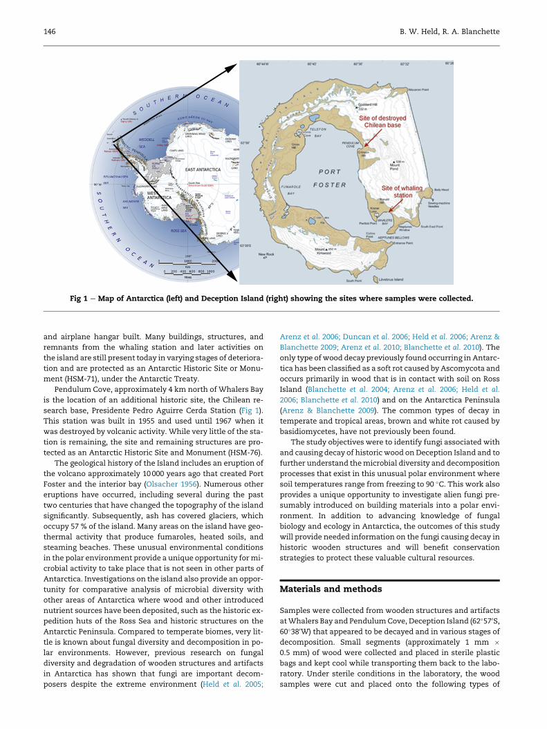
Fig 1 e Map of Antarctica (left) and Deception Island (right) showing the sites where samples were collected.
146 B. W. Held, R. A. Blanchette
and airplane hangar built. Many buildings, structures, and
remnants from the whaling station and later activities on
the island are still present today in varying stages of deteriora-
tion and are protected as an Antarctic Historic Site or Monu-
ment (HSM-71), under the Antarctic Treaty.
Pendulum Cove, approximately 4 km north of Whalers Bay
is the location of an additional historic site, the Chilean re-
search base, Presidente Pedro Aguirre Cerda Station (Fig 1).
This station was built in 1955 and used until 1967 when it
was destroyed by volcanic activity. While very little of the sta-
tion is remaining, the site and remaining structures are pro-
tected as an Antarctic Historic Site and Monument (HSM-76).
The geological history of the Island includes an eruption of
the volcano approximately 10000 years ago that created Port
Foster and the interior bay (Olsacher 1956). Numerous other
eruptions have occurred, including several during the past
two centuries that have changed the topography of the island
significantly. Subsequently, ash has covered glaciers, which
occupy 57 % of the island. Many areas on the island have geo-
thermal activity that produce fumaroles, heated soils, and
steaming beaches. These unusual environmental conditions
in the polar environment provide a unique opportunity formi-
crobial activity to take place that is not seen in other parts of
Antarctica. Investigations on the island also provide an oppor-
tunity for comparative analysis of microbial diversity with
other areas of Antarctica where wood and other introduced
nutrient sources have been deposited, such as the historic ex-
pedition huts of the Ross Sea and historic structures on the
Antarctic Peninsula. Compared to temperate biomes, very lit-
tle is known about fungal diversity and decomposition in po-
lar environments. However, previous research on fungal
diversity and degradation of wooden structures and artifacts
in Antarctica has shown that fungi are important decom-
posers despite the extreme environment (Held et al. 2005;
Arenz et al. 2006; Duncan et al. 2006; Held et al. 2006; Arenz &
Blanchette 2009; Arenz et al. 2010; Blanchette et al. 2010). The
only type of wood decay previously found occurring in Antarc-
tica has been classified as a soft rot caused by Ascomycota and
occurs primarily in wood that is in contact with soil on Ross
Island (Blanchette et al. 2004; Arenz et al. 2006; Held et al.
2006; Blanchette et al. 2010) and on the Antarctica Peninsula
(Arenz & Blanchette 2009). The common types of decay in
temperate and tropical areas, brown and white rot caused by
basidiomycetes, have not previously been found.
The study objectives were to identify fungi associated with
and causing decay of historic wood onDeception Island and to
further understand themicrobial diversity and decomposition
processes that exist in this unusual polar environment where
soil temperatures range from freezing to 90 �C. This work also
provides a unique opportunity to investigate alien fungi pre-
sumably introduced on building materials into a polar envi-
ronment. In addition to advancing knowledge of fungal
biology and ecology in Antarctica, the outcomes of this study
will provide needed information on the fungi causing decay in
historic wooden structures and will benefit conservation
strategies to protect these valuable cultural resources.
Materials and methods
Samples were collected from wooden structures and artifacts
atWhalers Bay and PendulumCove, Deception Island (62�570S,60�380W) that appeared to be decayed and in various stages of
decomposition. Small segments (approximately 1 mm �0.5 mm) of wood were collected and placed in sterile plastic
bags and kept cool while transporting them back to the labo-
ratory. Under sterile conditions in the laboratory, the wood
samples were cut and placed onto the following types of
Diverse wood decay fungi in Antarctica 147
growth media: malt extract agar (1.5 % Difco malt extract),
malt extract agar (1.5 %) amended with 0.5 g of streptomycin
sulphate and a semi-selective medium used to culture basid-
iomycetes (2.0 % Difco malt extract, 0.2 % yeast extract,
0.006% benlatewith 0.2 % lactic acid and 0.001% streptomycin
sulphate added after autoclaving) (Worrall 1991). Isolations
weremade from 188 samples fromWhalers bay (Hektor whal-
ing station, Base B, and BAS hangar) and 30 from the Chilean
base. Isolates were cultured at 8 and 20 �C for several weeks
after which transfers were made to another plate to obtain
pure cultures. DNA was extracted from fungal cultures using
an adapted chloroform procedure (Arenz & Blanchette 2011).
The internal transcribed spacer (ITS) region of ribosomal
DNA was targeted for PCR amplification with the primers
ITS1 þ ITS4 and LROR þ LR5 for large subunit amplification
(White et al. 1990). PCR amplifications were done using Ampli-
taq Gold PCRMaster-mix (Applied Biosystems, Foster City, CA)
and 1 ml of template DNA using the following parameters:
94 �C for 5 min, 35 cycles of 94 �C for 1 min, 50 �C for 1 min,
72 �C for 1 min, and a final extension step of 5 min at 72 �C.PCR amplicons were visualized on a 1 % agarose gel using
SYBR green 1 (Life Technologies, Grand Island, NY) prestain
and a Dark Reader DR45 transilluminator (Clare Chemical Re-
search, Denver, CO). Primers used for PCR were used for se-
quencing reactions on a ABI Prism 377 automated DNA
sequencer using a ABI PRISM Dye Terminator Cycle Sequenc-
ing Ready reaction kit (Applied Biosystems).
Consensus sequences were assembled using Geneious 9.0
(Kearse et al. 2012) and compared to those in GenBank using
BLASTn for identification. Multiple sequence alignments
were done using the MAFFT v7.222 (Katoh & Standley 2013)
alignment plugin in Geneious R9. Phylogenetic treeswere con-
structed using the MrBayes 3.2.6 plugin (Huelsenbeck &
Ronquist 2001) in Geneious R9 where 1 100000 MCMC genera-
tions were used with a sampling frequency every 200 genera-
tions and burn-in length of 100 000. The appropriate
substitution model was selected using jModelTest 2.1.10
(Darriba et al. 2012) according to Corrected Akaike Information
Criterion (AICc).
Decay studies using fungal cultures isolated from sam-
ples were carried out in microcosms over a 16 week period.
Isolates were selected after sequencing and identification
from BLAST searches. Glass filters (55 mm) were sterilized
by autoclaving and placed in 100 mm petri plates containing
media. A malt yeast agar (2 % malt extract agar, 0.2 % yeast
extract) was used for the basidiomycete cultures and a mini-
mal selective media for soft rot fungi (Worrall et al. 1991) was
used for the Ascomycota cultures. Wafers measuring
1.5 � 1.5 � 0.3 cm were cut from sound birch (Betula sp.)
and pine (Pinus sp.) wood blocks and dried at 105 �C for
24 h and weighed to determine dry weight. Wafers were
then hydrated and sterilized by autoclaving before placing
on glass filters in decay microcosms. Plugs (0.4 mm) were
transferred from growing fungal cultures and placed on the
medium surface adjacent to the filter. After a 16 week incu-
bation period, 10 wafers from each treatment were removed
and oven dried to determine biomass loss. Two wafers from
each treatment were not oven dried and used for micromor-
phological study.
The methods for the decay study using Ascomycota were
the same as the Basidiomycota study but a different medium
was used. Instead of malt yeast agar, a minimal media con-
sisting of 1.5 g NH4NO3, 2.5 g KH2PO4, 2 g K2HPO4, 1 g
MgSO4e7H2O, 2.5 g glucose and 0.1 g thiamine per litre was
used (Abrams 1948; Worrall et al. 1991). In addition, instead
of using water to hydrate wafers, a solution of same ingredi-
ents as the medium exclusive of the agar was used. The incu-
bation period was also 16 weeks.
Wood samples for fungal decay observations were pre-
pared for scanning electron microscopy (SEM) by infiltrating
with a 25 % TBS� Tissue FreezingMedium� (Triangle Biomed-
ical Sciences, Durham, NC, U.S.A.) under vacuum followed by
mounting on brass stubs, freezing at �20 �C and sectioning in
a cryostat freezing microtome. Samples were cut transversely
to prepare a clean surface for examination. Cut samples were
thawed and rinsed several times in water, air dried before
mounting on aluminium stubs with carbon tape and coated
with gold using a sputter coater. Samples were viewed using
a Hitachi S3500N scanning electron microscope to determine
characteristics of decay and signatures of fungal colonization
in the cell wall structure.
Results
Fungal isolation attempts from samples collected at Decep-
tion Island yielded 326 isolates from 218 samples. The major-
ity (79 %) of the isolates belonged to the Ascomycota and were
comprised of 53 taxa, followed by 11 taxa (24 %) in the Basidio-
mycota and four Zygomycota taxa (6 %). Some of themost rel-
atively abundant Ascomycota were Cadophora (29 %),
Phialocephala (10 %), LecythophoraeConiochaeta (9 %), Cosmospora
(8 %), and Phoma (8 %). Within the Cadophora group, several
species were found including Cadophora fastigiata/melinii
(55 %), Cadophora malorum (22 %), Cadophora luteo-olivaceae
(9 %), and undescribed species C. sp. NH1-2 (13 %) and C. sp.
5R24-1 (2 %). Among the most relatively abundant genera in
the Basidiomycota were Hypochniciellum (55 %), Pholiota
(18 %), Cerinosterus (11 %), and Tulasnella (8 %). In addition,
there were 15 taxa (ten Ascomycota, four Basidiomycota,
and one Zygomycota) that matched at or below 97 % sequence
identity with described taxa. LSU sequences were used in ad-
dition to ITS sequences for Hypochniciellum isolates because
the nearest percent sequence identity for ITS BLAST searches
was a 85 %match toAmyloathelia crassiuscula. However, BLAST
searches using LSU sequence data showed a 99 % sequence
identity to Hypochniciellum molle for which ITS sequence is
not in GenBank. Due to the fact that the LSU region does not
resolve species effectively and that reference sequences of
the ITS region for this or other Hypochniciellum species were
unavailable, the species of the isolatewe cultured remains un-
certain. However, phylogenetic analysis of LSU sequences re-
veal the isolates most closely resemble H. molle in this gene
region (Fig 2). Analysis of the ITS gene region from Pholiota
spp. sequences form several clades that are different from
other known species. One clade is comprised of isolates exclu-
sively from the Chilean base that group with Pholiota multicin-
gulata. The second clade has both an isolate from the Chilean
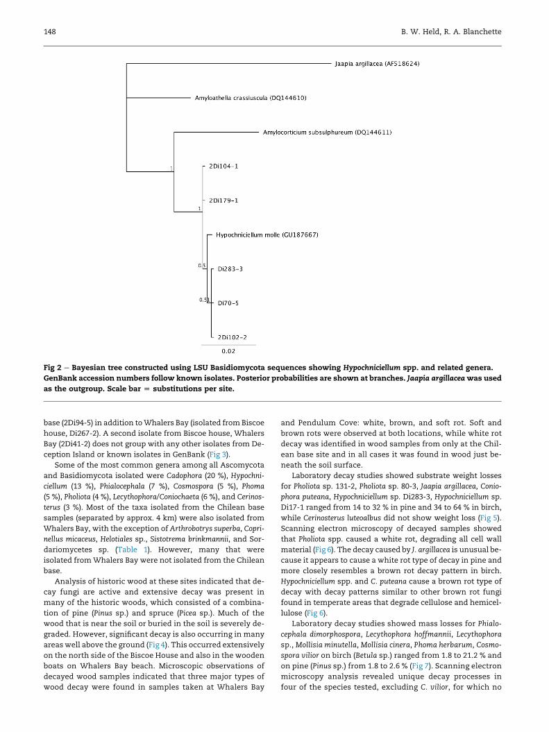
Fig 2 e Bayesian tree constructed using LSU Basidiomycota sequences showing Hypochniciellum spp. and related genera.
GenBank accession numbers follow known isolates. Posterior probabilities are shown at branches. Jaapia argillaceawas used
as the outgroup. Scale bar [ substitutions per site.
148 B. W. Held, R. A. Blanchette
base (2Di94-5) in addition toWhalers Bay (isolated from Biscoe
house, Di267-2). A second isolate from Biscoe house, Whalers
Bay (2Di41-2) does not group with any other isolates from De-
ception Island or known isolates in GenBank (Fig 3).
Some of the most common genera among all Ascomycota
and Basidiomycota isolated were Cadophora (20 %), Hypochni-
ciellum (13 %), Phialocephala (7 %), Cosmospora (5 %), Phoma
(5 %), Pholiota (4 %), Lecythophora/Coniochaeta (6 %), and Cerinos-
terus (3 %). Most of the taxa isolated from the Chilean base
samples (separated by approx. 4 km) were also isolated from
Whalers Bay, with the exception of Arthrobotrys superba, Copri-
nellus micaceus, Helotiales sp., Sistotrema brinkmannii, and Sor-
dariomycetes sp. (Table 1). However, many that were
isolated fromWhalers Bay were not isolated from the Chilean
base.
Analysis of historic wood at these sites indicated that de-
cay fungi are active and extensive decay was present in
many of the historic woods, which consisted of a combina-
tion of pine (Pinus sp.) and spruce (Picea sp.). Much of the
wood that is near the soil or buried in the soil is severely de-
graded. However, significant decay is also occurring in many
areas well above the ground (Fig 4). This occurred extensively
on the north side of the Biscoe House and also in the wooden
boats on Whalers Bay beach. Microscopic observations of
decayed wood samples indicated that three major types of
wood decay were found in samples taken at Whalers Bay
and Pendulum Cove: white, brown, and soft rot. Soft and
brown rots were observed at both locations, while white rot
decay was identified in wood samples from only at the Chil-
ean base site and in all cases it was found in wood just be-
neath the soil surface.
Laboratory decay studies showed substrate weight losses
for Pholiota sp. 131-2, Pholiota sp. 80-3, Jaapia argillacea, Conio-
phora puteana, Hypochniciellum sp. Di283-3, Hypochniciellum sp.
Di17-1 ranged from 14 to 32 % in pine and 34 to 64 % in birch,
while Cerinosterus luteoalbus did not show weight loss (Fig 5).
Scanning electron microscopy of decayed samples showed
that Pholiota spp. caused a white rot, degrading all cell wall
material (Fig 6). The decay caused by J. argillacea is unusual be-
cause it appears to cause a white rot type of decay in pine and
more closely resembles a brown rot decay pattern in birch.
Hypochniciellum spp. and C. puteana cause a brown rot type of
decay with decay patterns similar to other brown rot fungi
found in temperate areas that degrade cellulose and hemicel-
lulose (Fig 6).
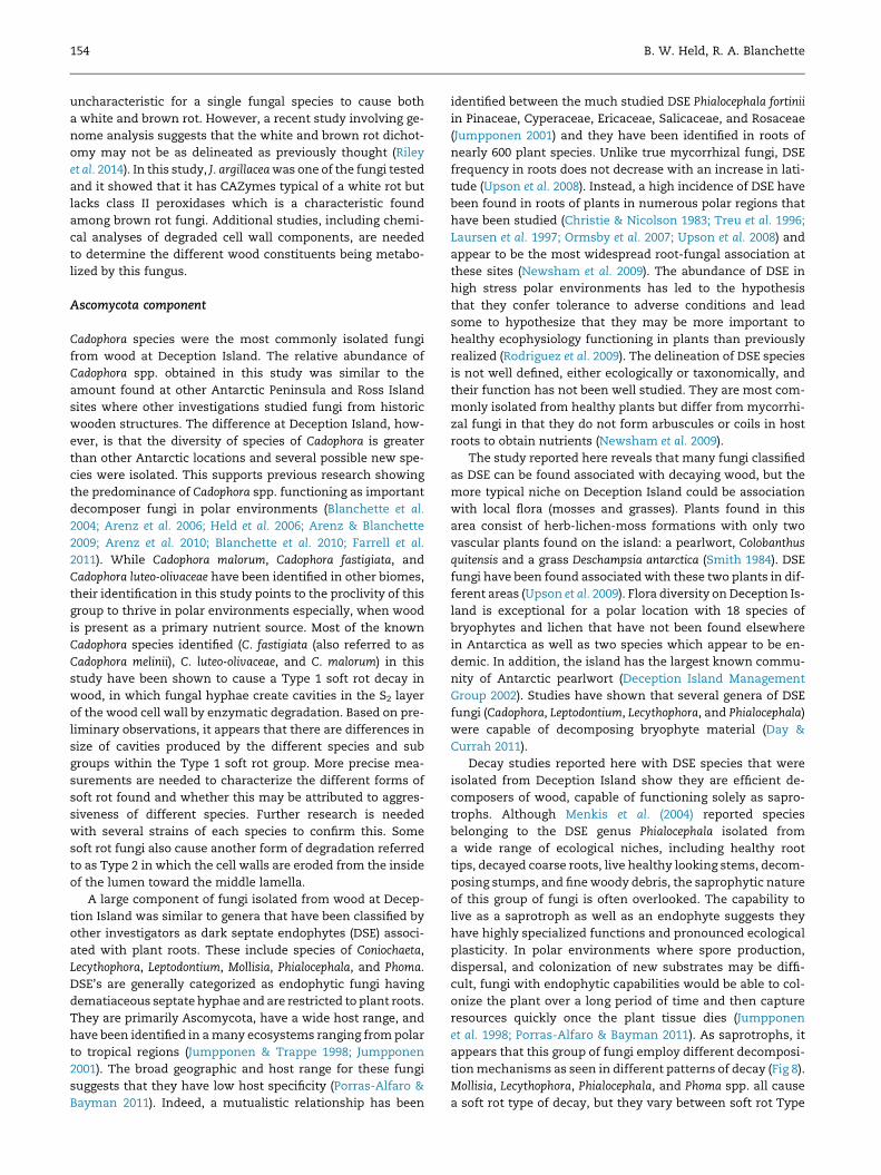
Laboratory decay studies showed mass losses for Phialo-
cephala dimorphospora, Lecythophora hoffmannii, Lecythophora
sp., Mollisia minutella, Mollisia cinera, Phoma herbarum, Cosmo-
spora vilior on birch (Betula sp.) ranged from 1.8 to 21.2 % and
on pine (Pinus sp.) from 1.8 to 2.6 % (Fig 7). Scanning electron
microscopy analysis revealed unique decay processes in
four of the species tested, excluding C. vilior, for which no
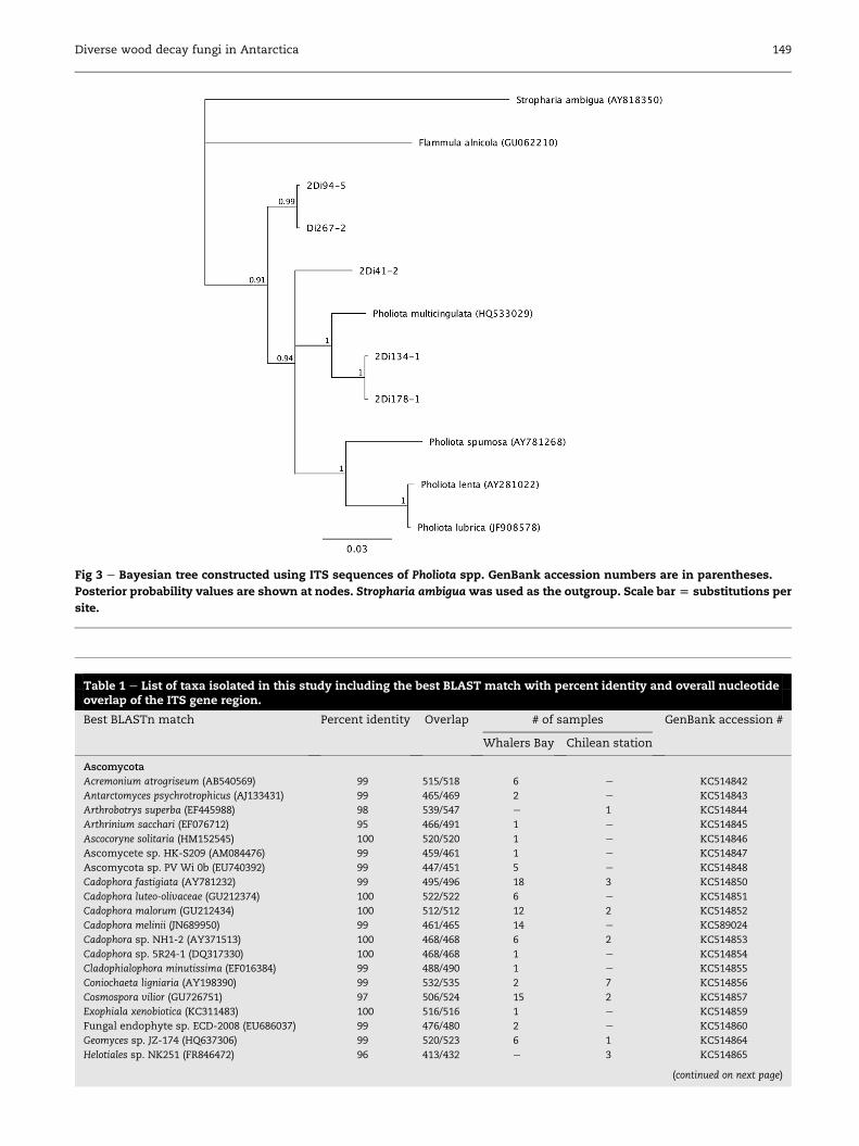
Table 1 e List of taxa isolated in this study including the best BLAST match with percent identity and overall nucleotideoverlap of the ITS gene region.
Best BLASTn match Percent identity Overlap # of samples GenBank accession #
Whalers Bay Chilean station
Ascomycota
Acremonium atrogriseum (AB540569) 99 515/518 6 e KC514842
Antarctomyces psychrotrophicus (AJ133431) 99 465/469 2 e KC514843
Arthrobotrys superba (EF445988) 98 539/547 e 1 KC514844
Arthrinium sacchari (EF076712) 95 466/491 1 e KC514845
Ascocoryne solitaria (HM152545) 100 520/520 1 e KC514846
Ascomycete sp. HK-S209 (AM084476) 99 459/461 1 e KC514847
Ascomycota sp. PV Wi 0b (EU740392) 99 447/451 5 e KC514848
Cadophora fastigiata (AY781232) 99 495/496 18 3 KC514850
Cadophora luteo-olivaceae (GU212374) 100 522/522 6 e KC514851
Cadophora malorum (GU212434) 100 512/512 12 2 KC514852
Cadophora melinii (JN689950) 99 461/465 14 e KC589024
Cadophora sp. NH1-2 (AY371513) 100 468/468 6 2 KC514853
Cadophora sp. 5R24-1 (DQ317330) 100 468/468 1 e KC514854
Cladophialophora minutissima (EF016384) 99 488/490 1 e KC514855
Coniochaeta ligniaria (AY198390) 99 532/535 2 7 KC514856
Cosmospora vilior (GU726751) 97 506/524 15 2 KC514857
Exophiala xenobiotica (KC311483) 100 516/516 1 e KC514859
Fungal endophyte sp. ECD-2008 (EU686037) 99 476/480 2 e KC514860
Geomyces sp. JZ-174 (HQ637306) 99 520/523 6 1 KC514864
Helotiales sp. NK251 (FR846472) 96 413/432 e 3 KC514865
(continued on next page)
Fig 3 e Bayesian tree constructed using ITS sequences of Pholiota spp. GenBank accession numbers are in parentheses.
Posterior probability values are shown at nodes. Stropharia ambigua was used as the outgroup. Scale bar [ substitutions per
site.
Diverse wood decay fungi in Antarctica 149
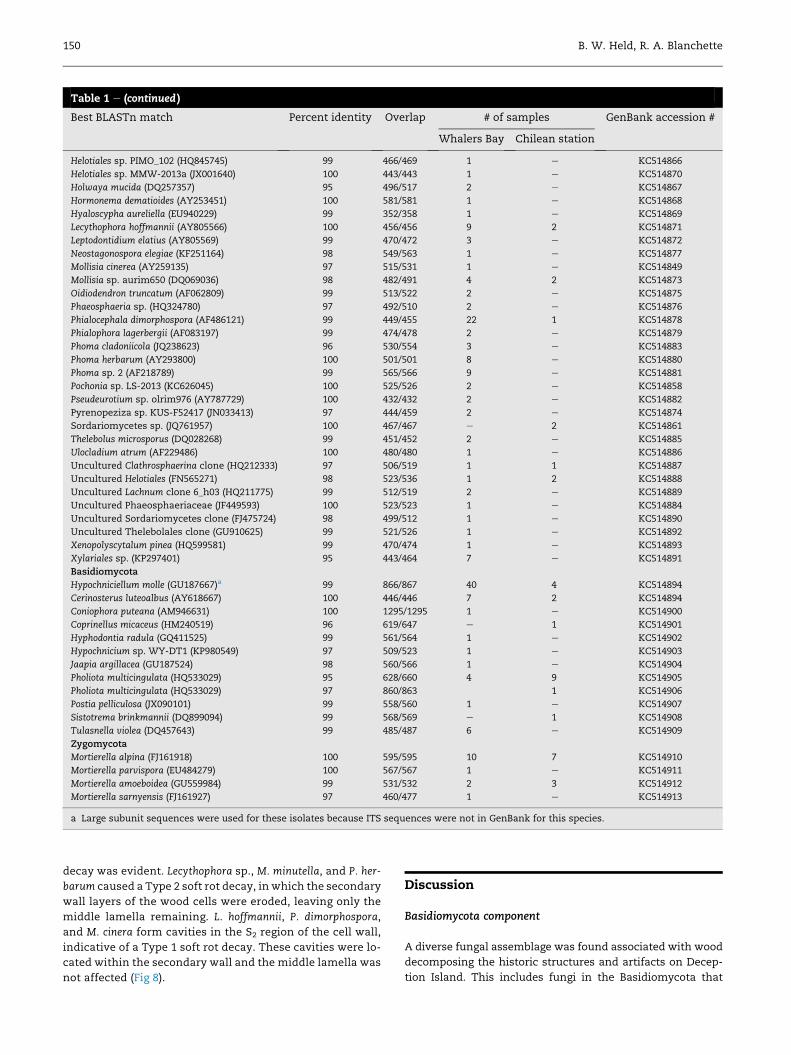
Table 1 e (continued )
Best BLASTn match Percent identity Overlap # of samples GenBank accession #
Whalers Bay Chilean station
Helotiales sp. PIMO_102 (HQ845745) 99 466/469 1 e KC514866
Helotiales sp. MMW-2013a (JX001640) 100 443/443 1 e KC514870
Holwaya mucida (DQ257357) 95 496/517 2 e KC514867
Hormonema dematioides (AY253451) 100 581/581 1 e KC514868
Hyaloscypha aureliella (EU940229) 99 352/358 1 e KC514869
Lecythophora hoffmannii (AY805566) 100 456/456 9 2 KC514871
Leptodontidium elatius (AY805569) 99 470/472 3 e KC514872
Neostagonospora elegiae (KF251164) 98 549/563 1 e KC514877
Mollisia cinerea (AY259135) 97 515/531 1 e KC514849
Mollisia sp. aurim650 (DQ069036) 98 482/491 4 2 KC514873
Oidiodendron truncatum (AF062809) 99 513/522 2 e KC514875
Phaeosphaeria sp. (HQ324780) 97 492/510 2 e KC514876
Phialocephala dimorphospora (AF486121) 99 449/455 22 1 KC514878
Phialophora lagerbergii (AF083197) 99 474/478 2 e KC514879
Phoma cladoniicola (JQ238623) 96 530/554 3 e KC514883
Phoma herbarum (AY293800) 100 501/501 8 e KC514880
Phoma sp. 2 (AF218789) 99 565/566 9 e KC514881
Pochonia sp. LS-2013 (KC626045) 100 525/526 2 e KC514858
Pseudeurotium sp. olrim976 (AY787729) 100 432/432 2 e KC514882
Pyrenopeziza sp. KUS-F52417 (JN033413) 97 444/459 2 e KC514874
Sordariomycetes sp. (JQ761957) 100 467/467 e 2 KC514861
Thelebolus microsporus (DQ028268) 99 451/452 2 e KC514885
Ulocladium atrum (AF229486) 100 480/480 1 e KC514886
Uncultured Clathrosphaerina clone (HQ212333) 97 506/519 1 1 KC514887
Uncultured Helotiales (FN565271) 98 523/536 1 2 KC514888
Uncultured Lachnum clone 6_h03 (HQ211775) 99 512/519 2 e KC514889
Uncultured Phaeosphaeriaceae (JF449593) 100 523/523 1 e KC514884
Uncultured Sordariomycetes clone (FJ475724) 98 499/512 1 e KC514890
Uncultured Thelebolales clone (GU910625) 99 521/526 1 e KC514892
Xenopolyscytalum pinea (HQ599581) 99 470/474 1 e KC514893
Xylariales sp. (KP297401) 95 443/464 7 e KC514891
Basidiomycota
Hypochniciellum molle (GU187667)a 99 866/867 40 4 KC514894
Cerinosterus luteoalbus (AY618667) 100 446/446 7 2 KC514894
Coniophora puteana (AM946631) 100 1295/1295 1 e KC514900
Coprinellus micaceus (HM240519) 96 619/647 e 1 KC514901
Hyphodontia radula (GQ411525) 99 561/564 1 e KC514902
Hypochnicium sp. WY-DT1 (KP980549) 97 509/523 1 e KC514903
Jaapia argillacea (GU187524) 98 560/566 1 e KC514904
Pholiota multicingulata (HQ533029) 95 628/660 4 9 KC514905
Pholiota multicingulata (HQ533029) 97 860/863 1 KC514906
Postia pelliculosa (JX090101) 99 558/560 1 e KC514907
Sistotrema brinkmannii (DQ899094) 99 568/569 e 1 KC514908
Tulasnella violea (DQ457643) 99 485/487 6 e KC514909
Zygomycota
Mortierella alpina (FJ161918) 100 595/595 10 7 KC514910
Mortierella parvispora (EU484279) 100 567/567 1 e KC514911
Mortierella amoeboidea (GU559984) 99 531/532 2 3 KC514912
Mortierella sarnyensis (FJ161927) 97 460/477 1 e KC514913
a Large subunit sequences were used for these isolates because ITS sequences were not in GenBank for this species.
150 B. W. Held, R. A. Blanchette
decay was evident. Lecythophora sp., M. minutella, and P. her-
barum caused a Type 2 soft rot decay, in which the secondary
wall layers of the wood cells were eroded, leaving only the
middle lamella remaining. L. hoffmannii, P. dimorphospora,
and M. cinera form cavities in the S2 region of the cell wall,
indicative of a Type 1 soft rot decay. These cavities were lo-
cated within the secondary wall and the middle lamella was
not affected (Fig 8).
Discussion
Basidiomycota component
A diverse fungal assemblage was found associated with wood
decomposing the historic structures and artifacts on Decep-
tion Island. This includes fungi in the Basidiomycota that
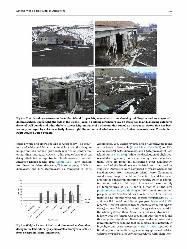
Fig 4 e The historic structures on Deception Island. Upper left; several structures showing buildings in various stages of
decomposition. Upper right; the side of the Biscoe House, a building at Whalers Bay on Deception Island, showing extensive
decay of wall boards and other timbers. Lower left; remnants of a structure that served as a dispensary/store that has been
severely damaged by volcanic activity. Lower right; the remains of what was once the Chilean research base, Presidente
Pedro Aguirre Cerda Station.
Diverse wood decay fungi in Antarctica 151
cause a white and brown rot type of wood decay. The occur-
rence of white and brown rot fungi in Antarctica is quite
unique and has not been previously reported on continental
or maritime Antarctica. However, other studies have reported
decay attributed to saprotrophic Basidiomycota from sub-
Antarctic Islands (Pegler 1980; Smith 1984). Fungi isolated
fromDeception Island sites were, 79 % Ascomycota, 15 % Basi-
diomycota, and 6 % Zygomycota as compared to 82 %
Fig 5 e Weight losses of birch and pine wood wafers after
decay in the laboratory by species of Basidiomycota isolated
from Deception Island, Antarctica.
Ascomycota, 15 % Basidiomycota, and 3 % Zygomycota found
on the Antarctic Peninsula (Arenz & Blanchette 2009) and 75 %
Ascomycota, 21 % Basidiomycota, and 1 % Zygomycota at Ross
Island (Arenz et al. 2006). While the distribution of phyla rep-
resented are generally consistent among these polar loca-
tions, there are important differences. Most significantly,
nearly all of the Basidiomycota isolated from the previous
studies in Antarctica were comprised of yeasts whereas the
Basidiomycota from Deception Island were filamentous
wood decay fungi. In addition, Deception Island lies in an
area that is considered maritime Antarctic, which is charac-
terized as having a cold, moist climate and mean monthly
air temperatures of >0 �C for 3e4 months of the year
(Aleksandrova 1980; Smith 1984) and 560 mm of precipitation
per year. While Ross Island has a colder, drier climate where
there are no months with the average temperature >0 �Cand only 190 mm of precipitation per year. Pegler et al. (1980)
reported Trametes versicolor (which causes a white rot type of
decay) on wood brought to South Georgia that was used in
the whaling station there. Since the wood was introduced, it
is likely that the fungus was brought in with the wood, and
this fungus is not endemic. However, other decomposer basid-
iomycetes have been found that presumably exist naturally in
bryophyte and grass ecosystems. Smith (1994) reported 37
Basidiomycota on South Georgia including species of Collybia,
Galerina, Omphalina, and Coprinus associated with plant litter.
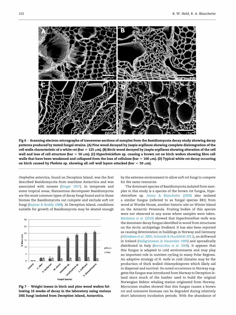
Fig 6 e Scanning electronmicrographs of transverse sections of samples from the Basidiomycota decay study showing decay
patterns produced by tested fungal strains. (A) Pine wood decayed by Jaapia argillacea showing complete disintegration of the
cell walls characteristic of a white rot (bar[ 125 mm). (B) Birch wood decayed by Jaapia argillacea showing alteration of the cell
wall and loss of cell structure (bar [ 50 mm). (C) Hypochniciellum sp. causing a brown rot on birch wafers showing fibre cell
walls that have been weakened and collapsed from the loss of cellulose (bar [ 100 mm). (D) Typical white rot decay occurring
on birch caused by Pholiota sp. showing all cell wall layers attacked (bar [ 50 mm).
152 B. W. Held, R. A. Blanchette
Omphalina antarctica, found on Deception Island, was the first
described Basidiomycota from maritime Antarctica and was
associated with mosses (Singer 1957). In temperate and
some tropical areas, filamentous decomposer Basidiomycota
are themost common types of decay fungi found and in those
biomes the Basidiomycota out compete and exclude soft rot
fungi (Rayner & Boddy 1988). At Deception Island, conditions
suitable for growth of Basidiomycota may be abated enough
Fig 7 e Weight losses in birch and pine wood wafers fol-
lowing 16 weeks of decay in the laboratory using various
DSE fungi isolated from Deception Island, Antarctica.
by the extreme environment to allow soft rot fungi to compete
for the same resources.
The dominant species of Basidiomycota isolated from sam-
ples in this study is a species of the brown rot fungus, Hypo-
chniciellum sp. Arenz & Blanchette (2009) also isolated
a similar fungus (referred to as fungal species BB1) from
wood at Wordie House, another historic site on Winter Island
on the Antarctic Peninsula. Fruiting bodies of this species
were not observed in any areas where samples were taken.
Mattsson et al. (2010) showed that Hypochniciellum molle was
the dominant decay fungus identified in wood from structures
on the Arctic archipelago Svalbard. It has also been reported
as causing deterioration in buildings in Norway and Germany
(Alfredsen et al. 2005; Schmidt & Huckfeldt 2011), on driftwood
in Iceland (Hallgrimsson & Hauerslev 1995) and sporadically
distributed in Italy (Bernicchia et al. 2008). It appears that
this fungus is adapted to cold environments and may play
an important role in nutrient cycling in many Polar Regions.
An adaptive strategy of H. molle in cold climates may be the
production of thick walled chlamydospores which likely aid
in dispersal and survival. Its noted occurrence in Norway sug-
gests the funguswas introduced fromNorway to Deception Is-
land since much of the lumber used to build the original
Norwegian Hektor whaling station originated from Norway.
Microcosm studies showed that this fungus causes a brown
rot and extensive biomass can be degraded during relatively
short laboratory incubation periods. With the abundance of

Fig 8 e Scanning electron micrographs of transverse sections from historic wood showing decay patters of various DSE re-
lated fungi after growing on wood wafers (birch) in a microcosm decay experiment. (A) An example of a Type 2 soft rot decay
where cell walls are eroded leaving the middle lamella intact (arrows) caused by Lecythophora sp. (bar [ 25 mm). (B) Type 1
soft rot cavities formed in the S2 layer of the secondary wall caused by Lecythophora hoffmanii (bar [ 25 mm). (C) Type 2 soft
rot caused by Phoma herbarum (bar [ 25 mm). Arrows indicate areas where the secondary cell has been eroded to the middle
lamella. (D) Phialocephala dimorphospora causing a Type 1 soft rot, showing numerous cavities in cell walls (bar [ 50 mm).
Diverse wood decay fungi in Antarctica 153
brown rot found at Deception Island and from the frequency
with which this fungus was isolated, our study suggests that
this fungus is a major cause of the brown rot decay taking
place on the Island.
Fruiting of several other Basidiomycota (Pholiota spp. and
Omphalina spp.) have been previously reported on Deception
Island (Singer 1967), which indicates that conditions can be
conducive for some Basidiomycota to complete their life cycle.
Singer (1967) described a Pholiota sp. fruiting body growing on
one of the half buried wooden boats at the beach in Whalers
Bay. This area is adjacent to thermally heated soil and water,
which may have contributed to conditions favourable for
fruiting. No fruiting bodies were observed during our field
events to Deception Island for these studies. Based on past ob-
servations of fruiting bodies, it is interesting that Pholiota spp.
was infrequent from samples in Whalers Bay, but was domi-
nant at the Chilean base. Likely, this fungus was also intro-
duced to Deception Island with building materials for the
Chilean base. Its activity strictly below the soil surface indi-
cates that the below ground conditions are conducive on the
island for continued fungal growth. The phylogenetic analysis
reported here suggests three different species of Pholiota are
present which indicates this fungus may have been intro-
duced several different times.
Other Basidiomycota decay fungi that were isolated
matched fungi that are commonly found in temperate re-
gions. Hypochnicium sp. and Hyphodontia radula are known
to cause a white rot type of decay (Gilbertson et al. 1975;
Larsson et al. 2006), Coniophora puteana and Postia pelliculosa
are both brown rot fungi that belong to genera that cause
significant problems in wooden structures and are impor-
tant decomposer fungi in temperate regions. These species
were each isolated from only one sample and do not appear
to be prevalent on Deception Island. Tulasnella violea was
also isolated infrequently and is a cosmopolitan genus
found in many temperate areas that has saprotrophic and
mycorrhizal characteristics (Preussing et al. 2010).
Once thought to be a species complex, a recent study dem-
onstrated that the corticioid genus Jaapia is in fact comprised
of two species; Jaapia argillacea and Jaapia ochroleuca (Telleria
et al. 2015). Jaapia argillacea, the species isolated at Deception
Island, has a wide distribution in Europe but is also considered
a rare species (Nannfeldt & Eriksson 1953). As with the other
Basidiomycota that have been isolated from the wood at De-
ception Island, they possibly were introduced with the
wooden materials brought to the island. However, the rarity
of J. argillacea in Europe suggests that its introduction from
Europe may be unlikely and additional investigation on the
distribution of Jaapia may resolve its origin. The wood decay
produced by J. argillacea in laboratory decay studies reveals
an unusual pattern. Observations from scanning electron mi-
croscopy, shows a decay pattern in pine that resembles the
characteristics of a white rot and in the birch wood the decay
appears different and resembles that of a brown rot. It is
154 B. W. Held, R. A. Blanchette
uncharacteristic for a single fungal species to cause both
a white and brown rot. However, a recent study involving ge-
nome analysis suggests that the white and brown rot dichot-
omy may not be as delineated as previously thought (Riley
et al. 2014). In this study, J. argillaceawas one of the fungi tested
and it showed that it has CAZymes typical of a white rot but
lacks class II peroxidases which is a characteristic found
among brown rot fungi. Additional studies, including chemi-
cal analyses of degraded cell wall components, are needed
to determine the different wood constituents being metabo-
lized by this fungus.
Ascomycota component
Cadophora species were the most commonly isolated fungi
from wood at Deception Island. The relative abundance of
Cadophora spp. obtained in this study was similar to the
amount found at other Antarctic Peninsula and Ross Island
sites where other investigations studied fungi from historic
wooden structures. The difference at Deception Island, how-
ever, is that the diversity of species of Cadophora is greater
than other Antarctic locations and several possible new spe-
cies were isolated. This supports previous research showing
the predominance of Cadophora spp. functioning as important
decomposer fungi in polar environments (Blanchette et al.
2004; Arenz et al. 2006; Held et al. 2006; Arenz & Blanchette
2009; Arenz et al. 2010; Blanchette et al. 2010; Farrell et al.
2011). While Cadophora malorum, Cadophora fastigiata, and
Cadophora luteo-olivaceae have been identified in other biomes,
their identification in this study points to the proclivity of this
group to thrive in polar environments especially, when wood
is present as a primary nutrient source. Most of the known
Cadophora species identified (C. fastigiata (also referred to as
Cadophora melinii), C. luteo-olivaceae, and C. malorum) in this
study have been shown to cause a Type 1 soft rot decay in
wood, in which fungal hyphae create cavities in the S2 layer
of the wood cell wall by enzymatic degradation. Based on pre-
liminary observations, it appears that there are differences in
size of cavities produced by the different species and sub
groups within the Type 1 soft rot group. More precise mea-
surements are needed to characterize the different forms of
soft rot found and whether this may be attributed to aggres-
siveness of different species. Further research is needed
with several strains of each species to confirm this. Some
soft rot fungi also cause another form of degradation referred
to as Type 2 in which the cell walls are eroded from the inside
of the lumen toward the middle lamella.
A large component of fungi isolated from wood at Decep-
tion Island was similar to genera that have been classified by
other investigators as dark septate endophytes (DSE) associ-
ated with plant roots. These include species of Coniochaeta,
Lecythophora, Leptodontium, Mollisia, Phialocephala, and Phoma.
DSE’s are generally categorized as endophytic fungi having
dematiaceous septate hyphae and are restricted to plant roots.
They are primarily Ascomycota, have a wide host range, and
have been identified in amany ecosystems ranging from polar
to tropical regions (Jumpponen & Trappe 1998; Jumpponen
2001). The broad geographic and host range for these fungi
suggests that they have low host specificity (Porras-Alfaro &
Bayman 2011). Indeed, a mutualistic relationship has been
identified between the much studied DSE Phialocephala fortinii
in Pinaceae, Cyperaceae, Ericaceae, Salicaceae, and Rosaceae
(Jumpponen 2001) and they have been identified in roots of
nearly 600 plant species. Unlike true mycorrhizal fungi, DSE
frequency in roots does not decrease with an increase in lati-
tude (Upson et al. 2008). Instead, a high incidence of DSE have
been found in roots of plants in numerous polar regions that
have been studied (Christie & Nicolson 1983; Treu et al. 1996;
Laursen et al. 1997; Ormsby et al. 2007; Upson et al. 2008) and
appear to be the most widespread root-fungal association at
these sites (Newsham et al. 2009). The abundance of DSE in
high stress polar environments has led to the hypothesis
that they confer tolerance to adverse conditions and lead
some to hypothesize that they may be more important to
healthy ecophysiology functioning in plants than previously
realized (Rodriguez et al. 2009). The delineation of DSE species
is not well defined, either ecologically or taxonomically, and
their function has not been well studied. They are most com-
monly isolated from healthy plants but differ from mycorrhi-
zal fungi in that they do not form arbuscules or coils in host
roots to obtain nutrients (Newsham et al. 2009).
The study reported here reveals that many fungi classified
as DSE can be found associated with decaying wood, but the
more typical niche on Deception Island could be association
with local flora (mosses and grasses). Plants found in this
area consist of herb-lichen-moss formations with only two
vascular plants found on the island: a pearlwort, Colobanthus
quitensis and a grass Deschampsia antarctica (Smith 1984). DSE
fungi have been found associated with these two plants in dif-
ferent areas (Upson et al. 2009). Flora diversity on Deception Is-
land is exceptional for a polar location with 18 species of
bryophytes and lichen that have not been found elsewhere
in Antarctica as well as two species which appear to be en-
demic. In addition, the island has the largest known commu-
nity of Antarctic pearlwort (Deception Island Management
Group 2002). Studies have shown that several genera of DSE
fungi (Cadophora, Leptodontium, Lecythophora, and Phialocephala)
were capable of decomposing bryophyte material (Day &
Currah 2011).
Decay studies reported here with DSE species that were
isolated from Deception Island show they are efficient de-
composers of wood, capable of functioning solely as sapro-
trophs. Although Menkis et al. (2004) reported species
belonging to the DSE genus Phialocephala isolated from
a wide range of ecological niches, including healthy root
tips, decayed coarse roots, live healthy looking stems, decom-
posing stumps, and finewoody debris, the saprophytic nature
of this group of fungi is often overlooked. The capability to
live as a saprotroph as well as an endophyte suggests they
have highly specialized functions and pronounced ecological
plasticity. In polar environments where spore production,
dispersal, and colonization of new substrates may be diffi-
cult, fungi with endophytic capabilities would be able to col-
onize the plant over a long period of time and then capture
resources quickly once the plant tissue dies (Jumpponen
et al. 1998; Porras-Alfaro & Bayman 2011). As saprotrophs, it
appears that this group of fungi employ different decomposi-
tionmechanisms as seen in different patterns of decay (Fig 8).
Mollisia, Lecythophora, Phialocephala, and Phoma spp. all cause
a soft rot type of decay, but they vary between soft rot Type

Diverse wood decay fungi in Antarctica 155
1 and Type 2 in birch wood. Very little biomass loss occurred
on pine, suggesting that these fungi have specific require-
ments for certain woods (preference for hardwood vs. coni-
fer) for decay to occur.
Anthropogenic effects
For nearly two centuries, there have beenmany opportunities
for alien fungi to be introduced to Deception Island. The
strong anthropogenic effects over the past decades and those
continuing today with tourists visiting the sites, has undoubt-
edly impacted the fungal diversity and ecology on the island.
The likely avenue for many of the introductions of wood
degrading fungi was the timber and wood used for buildings
and for the wooden boats, barrels, and other items that
came from Europe, South America, and other countries. The
presence of both brown and white rot types of wood decay
at this location and not at other Antarctic locations suggests
the environment at Deception Island influenceswhat alien or-
ganisms survive. The introduction of Basidiomycota decom-
poser fungi is also confirmed by the fact that these fungi are
considered forest fungi and found decaying woody substrates,
which did not exist on Deception Island before human activ-
ity. Additionally, previous research has shown that fungal
abundance of Antarctic soils is most positively correlated
with the percent of organic carbon compared to other soil
characteristics (Arenz & Blanchette 2011). The high degree of
fungal diversity associatedwith historic wood at Deception Is-
land indicates that the large organic carbon input on the is-
land from whaling and other activities is likely a driving
factor for fungal abundance.
In addition to cellulosic nutrient sources brought to the is-
land for buildings and other materials associated with the
whaling activities, there are also many reports of live domes-
tic animals that were housed on the island (Smith 1996). In the
early 1900’s pigswere kept at thewhaling station (Hacquebord
1992) as well as an occasional sheep or cow for a food source
(Scott Polar Research Institute Archives, unpublished data).
The Chilean Base had anywhere from 30 to 60 sheep brought
to the station every year in addition to hens and an occasional
pig or cow (Smith 1996). Hay and corn was also brought to feed
the animals (Smith 1996). Whale processing byproducts
(Hacquebord 1992) and various animal populations provided
a large input of nutrients that would greatly aid decomposi-
tion by fungi, in an otherwise nutrient lacking volcanic soil.
Conclusions
These findings show that all three known types of wood decay
(white, brown, and soft rot) are active and causing extensive
decay in the historic wooden structures and other wooden ar-
tifacts at Deception Island, Antarctica. This is in contrast to
only soft rot fungi identified in wood at other locations in Ant-
arctica. It also appears that brown and white rot Basidiomy-
cota were brought in with the wood and building materials
and have flourished. The dynamic nature of the ecosystem
of Deception Island with soils that range in temperature
from freezing to 90 �C and the large amount of wood present
at the site providing a carbon source apparently allows many
of the introduced fungi to survive. The input of wood also ap-
pears to have influenced the indigenous population of fungi
such as the DSE types found in native plants to expand their
saprophytic existence and colonize the introduced wood.
The fungi colonizing the historic woods are causing extensive
decay that will gradually result in the loss of the historic
structures.
There are many fungal isolates from this study which re-
main unidentified or with poor matches to sequences in Gen-
Bank, which suggests that some of these isolates are new
species and may be indigenous to Antarctica. Cadophora spp.
were the dominant group isolated from Deception Island as
well as other previously studied sites in Antarctica, which fur-
ther suggests this group of fungi plays an important role in de-
composition and nutrient cycling in cold ecosystems.
Additional studies focussing on fungal soil communities
and plant-associated fungi would aid in understanding how
alien fungi brought to Deception Island has affected fungal
populations and ecosystem functioning.
Acknowledgements
The authors would like to thank Brett Arenz and Andy Graves
for assistance in collecting samples, the staff and crew of the
LawrenceM. Gould research vessel. This research is supported
by National Science Foundation grant #0537143.
r e f e r e n c e s
Abrams E, 1948. Microbiological Deterioration of Organic Materials. ItsPrevention and Methods of Test, vol. 188, National Bureau ofStandards, Washington, DC.
Aleksandrova VD, 1980. The Arctic and Antarctic: their division intogeobotanical areas. Cambridge Univeristy Press, Cambridge, UK.
Alfredsen G, Solheim H, Jenssen KM, 2005. Evaluation of decayfungi in Norwegian buildings. In: International Research Groupon Wood Protection IRG/WP, p. 12.
Arenz BE, Blanchette RA, 2009. Investigations of fungal diversityin wooden structures and soils at historic sites on the Ant-arctic Peninsula. Canadian Journal of Microbiology 55: 46e56.
Arenz BE, Blanchette RA, 2011. Distribution and abundance of soilfungi in Antarctica at sites on the Peninsula. Soil Biology andBiochemistry 43: 308e315.
Arenz BE, Held BW, Jurgens JA, Blanchette RA, 2010. Fungal col-onization of exotic substrates in Antarctica. Fungal Diversity49: 13e22.
Arenz BE, Held BW, Jurgens JA, Farrell RL, Blanchette RA, 2006.Fungal diversity in soils and historic wood from the Ross SeaRegion of Antarctica. Soil Biology and Biochemistry 38:3057e3064.
Bernicchia A, Benni A, Venturella G, Gargano ML, Saitta A,Gorjon SP, 2008. Aphyllophoraceous wood-inhabiting fungi onQuercus spp. in Italy. Mycotaxon 104: 445e448.
Blanchette RA, Held BW, Arenz BE, Jurgens JA, Baltes NJ,Duncan SM, Farrell RL, 2010. An Antarctic hot spot for fungi atShackleton’s historic hut on Cape Royds. Microbial Ecology 60:29e38.
Blanchette RA, Held BW, Jurgens JA, McNew DL, Harrington TC,Duncan SM, Farrell RL, 2004. Wood destroying soft rot fungi inthe historic expedition huts of Antarctica. Applied and Envi-ronmental Microbiology 70: 1328e1335.
156 B. W. Held, R. A. Blanchette
Christie P, Nicolson TH, 1983. Are mycorrhizas absent from theAntarctic? Transactions of the British Mycological Society 80:557e560.
Darriba D, Taboada GL, Doallo R, Posada D, 2012. jModelTest 2:more models, new heuristics and parallel computing. NatureMethods 9: 772.
Day MJ, Currah RS, 2011. Role of selected dark septate endophytespecies and other hyphomycetes as sapropes on moss game-tophytes. Botany 89: 349e359.
Deception Island Management Group, 2002. Deception Island Man-agement Package.
Duncan SM, Farrell RL, Thwaites JM, Held BW, Arenz BE,Jurgens JA, Blanchette RA, 2006. Endoglucanase-producingfungi isolated from Cape Evans historic expedition hut on RossIsland, Antarctica. Environmental Microbiology 8: 1212e1219.
Farrell RL, Arenz BE, Duncan SM, Held BW, Jurgens JA,Blanchette RA, 2011. Introduced and indigenous fungi of theRoss Island historic huts and pristine areas of Antarctica. PolarBiology 34: 1669e1677.
Gilbertson RL, Burdsall HH, Larsen MJ, 1975. Notes on wood-rotting hymenomyces in New Mexico. The Southwestern Natu-ralist 19: 347e360.
Hacquebord L, 1992. Hector Station on Deception Island(South Shetland Islands, Antarctica) an environmentalassessment study of a whaling station. Circumpolar Journal1e2: 72e97.
Hallgrimsson H, Hauerslev K, 1995. Lignicolous jelly fungi andaphyllophorales in Iceland. Acta Botanica Islandica 12: 35e52.
Held BW, Jurgens JA, Arenz BE, Duncan SM, Farrell RL,Blanchette RA, 2005. Environmental factors influencing mi-crobial growth inside the historic huts of Ross Island, Ant-arctica. International Biodeterioration and Biodegradation 55:45e53.
Held BW, Jurgens JA, Duncan SM, Farrell RL, Blanchette RA, 2006.Assessment of fungal diversity and deterioration in a woodenstructure at New Harbor, Antarctica. Polar Biology 29: 526e531.
Huelsenbeck JP, Ronquist F, 2001. MRBAYES: bayesian inferenceof phylogeny. Bioinformatics 17: 754e755.
Jumpponen A, 2001. Dark septate endophytes e are they mycor-rhizal? Mycorrhiza 11: 207e211.
Jumpponen A, Mattson KG, Trappe JM, 1998. Mycorrhizal func-tioning of Phialocephala fortinii with Pinus contorta on glacierforefront soil: interactions with soil nitrogen and organicmatter. Mycorrhiza 7: 261e265.
Jumpponen A, Trappe JM, 1998. Dark septate endophytes: a re-view of facultative biotrophic root-colonizing fungi. New Phy-tologist 140: 295e310.
Katoh K, Standley DM, 2013. MAFFT multiple sequence alignmentsoftware version 7: improvements in performance and us-ability. Molecular Biology and Evolution 30: 772e780.
Kearse M, Moir R, Wilson A, Stones-Havas S, Cheung M,Sturrock S, Buxton S, Cooper A, Markowitz S, Duran C,Thierer T, Ashton B, Mentijies P, Drummond A, 2012. Geneiousbasic: an integrated and extendable desktop software plat-form for the organization and analysis of sequence data. Bio-informatics 28: 1647e1649.
Larsson KH, Parmasto E, Fischer M, Langer E, Nakasone KK,Redhead SA, 2006. Hymenochaetales a molecular phylogenyfor the hymenochaetoid clade. Mycologia 98: 926e936.
Laursen GA, Treu R, Seppelt RD, Stephenson SL, 1997. Mycorrhi-zal. Arctic, Antarctic, and Alpine Research 29: 483e491.
Mattsson J, Flyen AC, Nunez M, 2010. Wood-decaying fungi inprotected buildings and structures on Svalbard. Agarica 29:5e14.
Menkis A, Allmer J, Vasiliauskas R, Lygis V, Stenlid J, Finlay R,2004. Ecology and molecular characterization of dark septatefungi from roots, living stems, coarse and fine woody debris.Mycological Research 108: 965e973.
Nannfeldt JA, Erkiksson J, 1953. On the Hymenomycetous genusJaapia Bres. and its taxonomical position. Svensk Botanisk Tid-skrift 47: 177e189.
Newsham KK, Upson R, Read DJ, 2009. Mycorrhizas and darkseptate root endophytes in polar regions. Fungal Ecology 2:10e20.
Olsacher J, 1956. Contribuci�on a la geolog�ıa de la Ant�artidaOccidental: I. Contribuci�on al conocimiento geol�ogico dela Isla Decepti�on. Contribuci�on del Instituto Ant�artico Argentino 2:1e76.
Ormsby A, Hodson E, Li Y, Basinger A, Kaminsky S, 2007. Quan-titation of endorhizal fungi in high Arctic tundra ecosystemsthrough space and time: the value of herbarium archives.Canadian Journal of Botany 85: 599e606.
Pegler DN, Spooner BN, Lewis Smith RI, 1980. Higher fungi ofAntarctica, the subantarctic zone and Falkland Islands. KewBulletin 35: 499e562.
Porras-Alfaro A, Bayman P, 2011. Hidden fungi, emergent prop-erties: endophytes and microbiomes. Annual Review of Phyto-pathology 49: 291e315.
Preussing M, Nebel M, Oberwinkler F, Weiss M, 2010. Divergingdiversity patterns in the Tulasnella (Basidiomycota, Tulasnel-lales) mycobionts of Aneura pinguis (Marchantiophyta, Metz-geriales) from Europe and Ecuador. Mycorrhiza 20: 147e159.
Rayner ADM, Boddy L, 1988. Fungal Decomposition of Wood: its bi-ology and ecology. Wiley, New York.
Riley R, Salamov AA, Brown DW, Nagy LG, Floudas F, Held BW,Levasseur A, Lombard V, Morin E, Otillar R, Lindquist EA,Sun H, LaButti KM, Schmutz J, Jabbour D, Luo H, Baker SE,Pisabarro AG, Walton JD, Blanchette RA, Henrissat B, Martin F,Cullen D, Hibbett DS, Grigoriev IV, 2014. Extensive sampling ofbasidiomycete genomes demonstrates inadequacy of thewhite-rot/brown-rot paradigm for wood decay fungi. Proceed-ings of the National Academy of Sciences 111: 9923e9928.
Rodriguez RJ, White Jr JF, Arnold AE, Redman RS, 2009. Fungalendophytes: diversity and functional roles. New Phytologist182: 314e330.
Schmidt O, Huckfeldt T, 2011. Characterization and identificationof indoor wood-decaying basidiomycetes. In: Adan OCG,Samson RA (eds), Fundamentals of Mold Growth in Indoor Envi-ronments and Strategies for Healthy Living. Wageningen, Wage-ningen, pp. 117e168.
Singer R, 1957. Sydowia1: 16e23.Singer R, 1967. Fungi distribution. In: Bushnell VC (ed.), Terrestrial
Life of Antarctica, vol. 5. American Geographical Society, NewYork.
Smith RIL, 1984. Terrestrial plant biology of the sub-Antarctic andAntarctic. In: Laws RM (ed.), Antarctic Ecology. Academic Press,London, pp. 61e162.
Smith RIL, 1994. Species-diversity and resource relationships ofSouth Georgian fungi. Antarctic Science 6: 45e52.
Smith RIL, 1996. Introduced plants in Antarctica: potential im-pacts and conservation issues. Biological Conservation 76:135e146.
Telleria MT, Due~nas M, Melo I, Salcedo I, Mart�ın M, 2015. Spellingout Jaapia species. Mycological Progress 14: 57.
Treu R, Laursen GA, Stephenson SL, Landolt JC, Densmore R, 1996.Mycorrhizae from Denali National Park and Preserve, Alaska.Mycorrhiza 6: 21e29.
Upson R, Newsham KK, Bridge PD, Pearce DA, Read DJ, 2009.Taxonomic affinities of dark sepatate root endophytesof Colobanthus quitensis and Deschampsia antarctica, the twonative Antarctic vascular plant species. Fungal Ecology 2:184e196.
Upson R, Newsham KK, Read DJ, 2008. Root-fungal associations ofColobanthus quitensis and Deschampsia antarctica in the mari-time and Subantarctic. Arctic, Antarctic, and Alpine Research 40:592e599.

Diverse wood decay fungi in Antarctica 157
White TJ, Bruns T, Taylor J, 1990. Amplification and direct se-quencing of fungal genes for phylogenetics. In: Innis MA,Gelfand GH, Sninsky JJ, White TJ (eds), PCR Protocols: a guide tomethods and applications. Academic Press, New York,pp. 315e322.
Worrall JJ, 1991. Media for selective isolation of hymenomycetes.Mycologia 83: 296e302.
Worrall JJ, Anagnost SE, Wang CJK, 1991. Conditions for soft rot ofwood. Canadian Journal of Microbiology 37: 869e874.