Embed Size (px)
Citation preview
Image Gently and Step Lightly to Minimize Radiation Dose in Pediatric Congenital Imaging
C O N G E N I T A L C A R D I O L O G Y T O D A YTimely News and Information for BC/BE Congenital/Structural Cardiologists and Surgeons
Volume 7 / Issue 12December 2009North American Edition
Introduction
The use of x-ray imaging modalities to diagnose and treat disease continues to increase in medical care in general, and the clinical specialty of congenital cardiology is no exception. X-ray imaging modalities, including computed tomography (CT) and x-ray fluoroscopy, nuclear medicine imaging, and chest radiography, have the potential to provide great benefit to the overall care provided to patients. However, the radiation dose associated with x-ray imaging also carries with it the potential for adverse effects. For young patients, the expected risk is that of a slight increase in the probability of developing cancer sometime later in life. For the relatively low radiation dose associated with medical imaging procedures, cancer risk is assumed to be directly proportional to the magnitude of the radiation dose received by a patient. The likelihood that a patient will develop a radiation-induced cancer later in life can be reduced by minimizing the patient’s radiation dose.
Recently, several professional organizations recognized an opportunity to educate medical professionals and pediatric patient families regarding the benefits and potential risks of x-ray imaging procedures. The Alliance for Radiation Safety in Pediatric Imaging (The Alliance) was formed in 2006 and launched the I m a g e G e n t l y c a m p a i g n i n 2 0 0 8
(www.imagegently.com).1 The purpose of the Image Gently campaign is to share information regarding radiation dose for pediatric CT procedures and to assist providers with optimizing CT systems to minimize radiation dose. In 2009, The Alliance launched the Step Lightly campaign to educate and help minimize radiation dose associated with interventional radiology procedures. While there are some practical differences between general CT versus cardiac CT or interventional radiology versus interventional cardiology, there are enough similarities that both the Image Gently and Step Lightly campaigns are applicable to the specialty practice of pediatric congenital cardiology. This report will provide a general overview of radiation dose and radiation-induced cancer risks associated with cardiac CT and x-ray interventional procedures and will incorporate the foundations of radiation awareness and safety established by the Image Gently and Step Lightly campaigns. Suggestions for minimizing the radiation dose associated with pediatric cardiac CT and interventional fluoroscopy are included.
Radiation Dose and Patient Risk
The potential for biologic injury occurs when an x-ray photon(s) interacts with individual molecules in the nucleus of a cell.2 The energy imparted by the x-ray photon(s) has the potential to break molecular bonds, potentially compromising critical sub-cellular structures, particularly DNA. Minor damage may be automatically repaired, but severe DNA damage may lead to cellular mutation,
By Kenneth A. Fetterly, PhD; James M. Kofler, PhD; Donald J. Hagler, MD
IN THIS ISSUE
Image Gently and Step Lightly to Minimize Radiation Dose in Pediatric Congenital Imagingby Kenneth A. Fetterly, PhD; James M. Kofler, PhD; Donald J. Hagler, MD ~Page 1
Case Report: Fatal Thrombo-embolism in a Child with Noncompaction of Ventricular Myocardiumby Sulafa KM Ali, MD, FACC, FRCPCH~Page 11
CONGENITAL CARDIOLOGY TODAYEditorial and Subscription Offices16 Cove Rd, Ste. 200Westerly, RI 02891 USAwww.CongenitalCardiologyToday.comAddress Service Requested
© 2009 by Congenital Cardiology Today ISSN: 1544-7787 (print); 1544-0499 (online). Published monthly. All rights reserved.
Statements or opinions expressed in Congenital Cardiology Today reflect the views of the authors and sponsors, and are not necessarily the views of Congenital Cardiology Today.
UPCOMING MEDICAL MEETINGS
Evolving Concepts in the Management of Complex Congenital Heart Disease II
Jan. 14-16, 2010; San Diego, CA USAwww.chsd.org/body.cfm?id=1753
Cardiology 2010Feb. 10-14, 2010; Lake Buena Vista, FL USA
www.chop.edu/cardiology2010
8th Utah Conference on Congenital Cardiovascular Disease
Feb. 21-24, 2010; Snow Bird, UT USAwww.primarychildrens.com/
pedscardiodisease
Masterclasess in Cardiac Morphology Mar. 5-7, 2010; Charleston, SC USAwww.musckids.com/heart/conferences/
ACC.10 & i2 Summit 2010Mar. 14-16, 2010; Atlanta, GA USA
acc10.acc.org
Recruitment AdsPages: 3, 4, 7, 10, 12, 13, 14,15
Do you or your colleagues have interesting research results, observations, human interest stories, reports of meetings,
etc. that you would like to share with the congenital cardiology community?
Submit a summary of your proposed article to Congenital Cardiology Today at: [email protected]
Learn more at www.somanetics.com | 800.359.7662
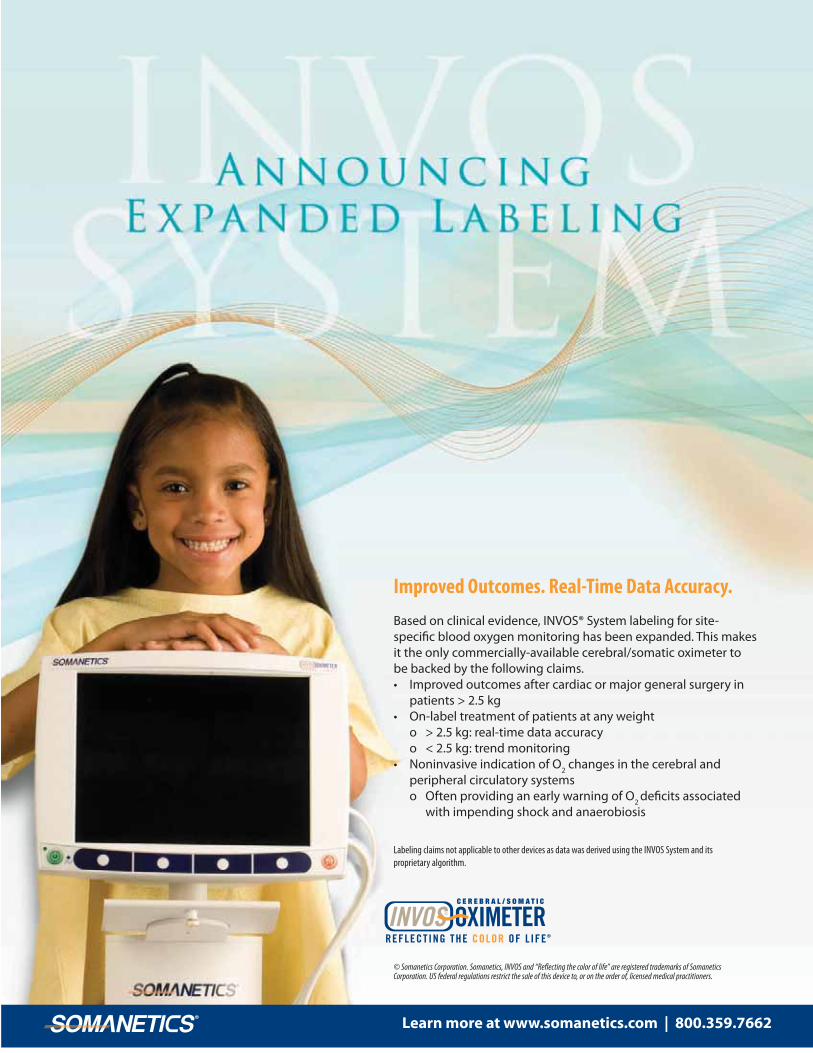
Improved Outcomes. Real-Time Data Accuracy.Based on clinical evidence, INVOS® System labeling for site-
specific blood oxygen monitoring has been expanded. This makes
it the only commercially-available cerebral/somatic oximeter to
be backed by the following claims.
�� ������� ������������������������������������ ���������
patients > 2.5 kg
�� ��������������������������������������������
� � ���!"�#�$������������������� ����
o < 2.5 kg: trend monitoring
�� %���������������������2 changes in the cerebral and
��������������� �������������
o Often providing an early warning of O2 deficits associated
with impending shock and anaerobiosis
Labeling claims not applicable to other devices as data was derived using the INVOS System and its proprietary algorithm.
© Somanetics Corporation. Somanetics, INVOS and “Reflecting the color of life” are registered trademarks of Somanetics Corporation. US federal regulations restrict the sale of this device to, or on the order of, licensed medical practitioners.
Working Together to Develop a Better Tomorrow
potentially resulting in cancer. Because radiation-induced cancers have a latent period of at least several years, it is not possible to establish a direct link between a cancer and a previous exposure to radiation.
Radiation dose to a specific tissue or organ describes the amount of radiation energy absorbed per mass of tissue. It is given the special unit Gray (Gy), or more commonly, milliGray (mGy). Estimation of future radiation-induced cancer risk requires some knowledge or estimate of organ dose. Once known, individual organ dose values are multiplied by a numeric scaling factor representing the relative sensitivity of each organ. The weighted, organ-specific dose values are then summed over all organs to obtain an estimate of effective ‘whole body’ dose (E) with special unit Sievert (Sv).3,4 Given that medical radiation is applied to only a portion of the body and therefore affects only a portion of the organs, the effective dose represents the whole body dose which would have resulted in the same overall cancer risk to the patient. This metric, therefore, allows comparison, in terms of risk for all types of medical radiation exposures, regardless of what part of the body was exposed. Note that most interventional x-ray systems report a cumulative entrance skin exposure (ESE, units gray (Gy)) value that is the product of the radiation exposure time (min) and rate (mGy/min) for the entire procedure. This value has some merit in that it can be used to avoid high skin dose levels for complex interventional procedures performed on adult patients. However, the ESE to pediatric patients is generally far below the threshold for skin injury and is not an effective surrogate for actual organ dose. Therefore, ESE is not useful for estimating risk to pediatric patients.
Generally, excess cancer risk from medical x-ray procedures is too small to be directly measured, even with large cohort patient studies. Notable exceptions include an increased risk of breast cancer in girls who were treated for scoliosis and children who were exposed to radiation in utero.5 Most of the epidemiologic data from which cancer risk from medical procedures is estimated originates from the Japanese atomic bomb survivors from Hiroshima and Nagasaki. A comprehensive review of this data is included in the recent BEIR VII report, Health Risks from Exposure to Low Levels of Ionizing Radiation (NRC 2006).6 This report concluded that, for low radiation doses (<100 mSv), the risk of cancer from radiation is most reasonably modeled as being directly proportional to the dose. This linear, non-threshold dose response model forms the basis for discussions of relative risk and justification of efforts to minimize radiation dose.
CONGENITAL CARDIOLOGY TODAY www.CongenitalCardiologyToday.com December 2009 3
“Recently, several professional organizations recognized an opportunity to educate medical professionals and pediatric patient families regarding the benefits and potential risks of x-ray imaging procedures.” The University of Maryland Hospital for Children is developing
a comprehensive Children’s Heart Center to meet the cardiovascular healthcare needs of the children of Maryland. We are currently recruiting for a variety of faculty positions. Sub-specialty board certification or equivalent work experience is required for each position. Clinical duties will focus primarily in the respective field of each position and participate to varying degrees in the general pediatric cardiology and outpatient practices. The Children’s Heart Center supports integrated quality enhancement and clinical research practices to improve patient outcomes and patient/family experience.
The successful candidates will have faculty appointments in the Department of Pediatrics of the University of Maryland School of Medicine at academic levels to be determined by experience. The University of Maryland Medical Center is a major academic tertiary care center serving Baltimore, the state of Maryland, and the mid-Atlantic region. As the oldest public medical school in the United States, the University of Maryland School of Medicine has an established tradition of outstanding clinical care, education, and research. The Department of Pediatrics is deeply committed to promoting children’s health in the community and across the state, while supporting innovative clinical programs and expanding research initiatives.
Located on the modern and urban campus of the University of Maryland at Baltimore, The School of Medicine is one of seven professional schools within the University of Maryland system. The campus is ideally located within walking distance to the Baltimore Inner Harbor, National Aquarium, Baltimore Convention Center, Hippodrome Theatre, Orioles Park at Camden Yards and Baltimore Ravens M & T Bank Stadium. The University of Maryland Hospital for Children is also close to Historic Annapolis, the Chesapeake Bay, Washington DC, and many residential communities with outstanding public and private schools. The area offers rich cultural fabric and many unique recreational opportunities. The University of Maryland is an EOE/AA/ADA and encourages minorities to apply.
Interested applicants should send CV to:
Dr. Geoffrey L. RosenthalDirector, Pediatric & Congenital Heart Program
University of Maryland Hospital for Children22 S. Greene Street, N5W68
Baltimore, MD [email protected]
WWW.UMM.EDU/PEDIATRICS
4 CONGENITAL CARDIOLOGY TODAY www.CongenitalCardiologyToday.com December 2009
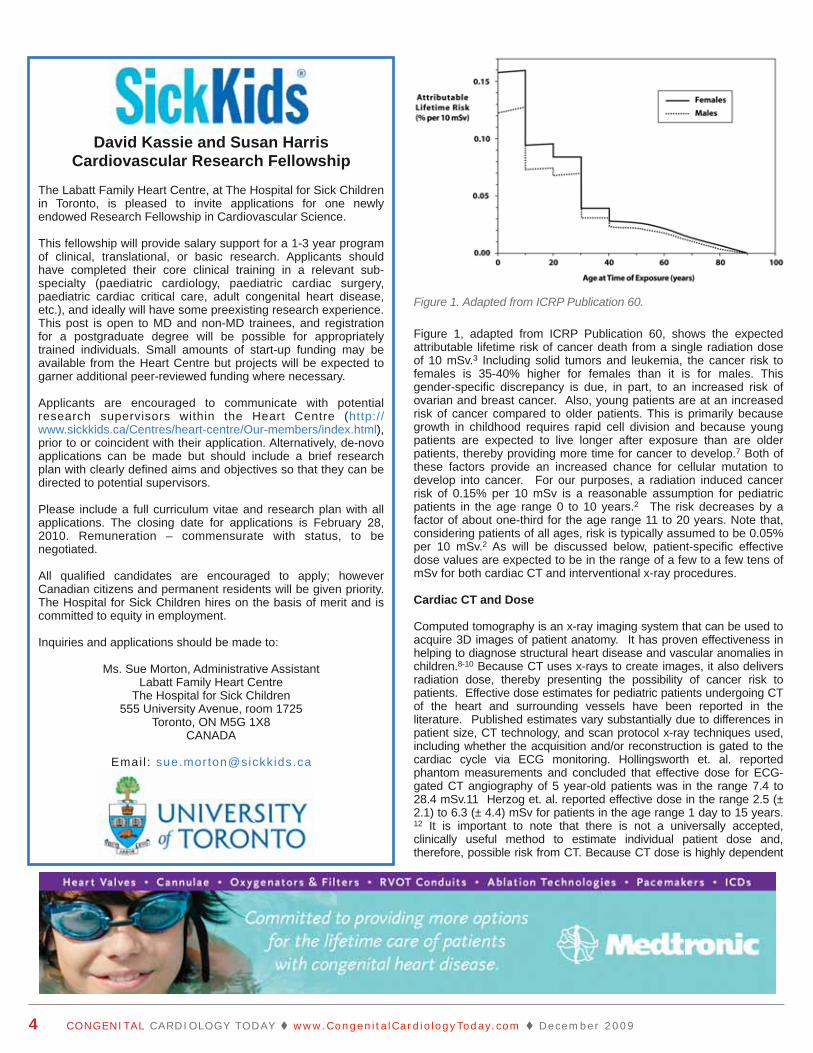
Figure 1, adapted from ICRP Publication 60, shows the expected attributable lifetime risk of cancer death from a single radiation dose of 10 mSv.3 Including solid tumors and leukemia, the cancer risk to females is 35-40% higher for females than it is for males. This gender-specific discrepancy is due, in part, to an increased risk of ovarian and breast cancer. Also, young patients are at an increased risk of cancer compared to older patients. This is primarily because growth in childhood requires rapid cell division and because young patients are expected to live longer after exposure than are older patients, thereby providing more time for cancer to develop.7 Both of these factors provide an increased chance for cellular mutation to develop into cancer. For our purposes, a radiation induced cancer risk of 0.15% per 10 mSv is a reasonable assumption for pediatric patients in the age range 0 to 10 years.2 The risk decreases by a factor of about one-third for the age range 11 to 20 years. Note that, considering patients of all ages, risk is typically assumed to be 0.05% per 10 mSv.2 As will be discussed below, patient-specific effective dose values are expected to be in the range of a few to a few tens of mSv for both cardiac CT and interventional x-ray procedures.
Cardiac CT and Dose
Computed tomography is an x-ray imaging system that can be used to acquire 3D images of patient anatomy. It has proven effectiveness in helping to diagnose structural heart disease and vascular anomalies in children.8-10 Because CT uses x-rays to create images, it also delivers radiation dose, thereby presenting the possibility of cancer risk to patients. Effective dose estimates for pediatric patients undergoing CT of the heart and surrounding vessels have been reported in the literature. Published estimates vary substantially due to differences in patient size, CT technology, and scan protocol x-ray techniques used, including whether the acquisition and/or reconstruction is gated to the cardiac cycle via ECG monitoring. Hollingsworth et. al. reported phantom measurements and concluded that effective dose for ECG-gated CT angiography of 5 year-old patients was in the range 7.4 to 28.4 mSv.11 Herzog et. al. reported effective dose in the range 2.5 (± 2.1) to 6.3 (± 4.4) mSv for patients in the age range 1 day to 15 years.12 It is important to note that there is not a universally accepted, clinically useful method to estimate individual patient dose and, therefore, possible risk from CT. Because CT dose is highly dependent
Figure 1. Adapted from ICRP Publication 60.
David Kassie and Susan Harris Cardiovascular Research Fellowship
The Labatt Family Heart Centre, at The Hospital for Sick Children in Toronto, is pleased to invite applications for one newly endowed Research Fellowship in Cardiovascular Science.
This fellowship will provide salary support for a 1-3 year program of clinical, translational, or basic research. Applicants should have completed their core clinical training in a relevant sub-specialty (paediatric cardiology, paediatric cardiac surgery, paediatric cardiac critical care, adult congenital heart disease, etc.), and ideally will have some preexisting research experience. This post is open to MD and non-MD trainees, and registration for a postgraduate degree will be possible for appropriately trained individuals. Small amounts of start-up funding may be available from the Heart Centre but projects will be expected to garner additional peer-reviewed funding where necessary.
Applicants are encouraged to communicate with potential research supervisors within the Heart Centre (http://www.sickkids.ca/Centres/heart-centre/Our-members/index.html), prior to or coincident with their application. Alternatively, de-novo applications can be made but should include a brief research plan with clearly defined aims and objectives so that they can be directed to potential supervisors.
Please include a full curriculum vitae and research plan with all applications. The closing date for applications is February 28, 2010. Remuneration – commensurate with status, to be negotiated.
All qualified candidates are encouraged to apply; however Canadian citizens and permanent residents will be given priority. The Hospital for Sick Children hires on the basis of merit and is committed to equity in employment.
Inquiries and applications should be made to:
Ms. Sue Morton, Administrative AssistantLabatt Family Heart Centre
The Hospital for Sick Children555 University Avenue, room 1725
Toronto, ON M5G 1X8CANADA
Emai l : [email protected]
CONGENITAL CARDIOLOGY TODAY www.CongenitalCardiologyToday.com December 2009 5
on equipment capabilities (or limitations) and site specific configuration, it is important that pediatric cardiologists work closely with radiologist and medical physicist colleagues to understand and minimize radiation dose for pediatric patients.
Interventional Fluoroscopy & Radiation Dose
Interventional x-ray systems use x-rays to produce real-time images of coronary and related vasculature to diagnose and treat disease. Similar to CT, there is not a standard method to estimate patient-specific effective dose from interventional x-ray in the clinical care setting. Also similar to CT dose estimates, published dose estimates for pediatric cardiac interventional procedures vary greatly and are, in part, influenced by x-ray system design and site-specif ic configuration. For interventional procedures, the complexity of each individual case can lead to substantial differences in radiation dose for a given patient. Rassow et. al. reported effective dose values in the range 2 to 18 mSv for infants undergoing cardiac catheterization procedures.13 Bacher et. al. reported median effective dose values of 4.6 mSv (range 0.6 to 23.2 mSv) for diagnostic cardiac catheterization procedures and 6.0 mSv (range 1.0 to 37.0 mSv) for therapeutic procedures performed on patients in the age range 0.1 to 9.2 years .14
Image Gently for Cardiac CT
The Alliance for Radiation Safety in Pediatric Imaging launched the Image Gently campaign to raise awareness and to help practicing physicians to reduce radiation dose associated wi th pediatr ic computed tomography procedures. This campaign has been successful in helping to reduce radiation dose associated with pediatric CT in general, including cardiac CT. The Image Gently campaign encourages care providers to first consider whether the imaging task could be adequately accomplished without using ionizing radiation, such as with ultrasound or magnetic resonance imaging (MRI). Certainly, this is a very effective method to avoid radiation dose. If an alternative to CT is not appropriate or possible, then Image Gently suggests that “child-size” x-ray techniques be used for the CT scan. This ensures that the pediatric CT is performed with the lowest dose required to achieve adequate diagnostic images. Most importantly, Image Gently provides suggested protocols to reduce dose for pediatric patients of various age and size.15
Dose reduction for pediatric patients is most commonly accomplished by decreasing the current-time product (mAs) for the pediatric exams relative to a standard adult technique. Image Gently provides a table of mAs reduction factors based on the patient thickness in the posterior/anterior direction or based on approximate age. For example,
suggested mAs reduction factors (RF) range from 0.42 for newborns to 0.73 for 15 year-old patients undergoing thoracic CT.14 These RF values represent suggested initial starting points for dose reduction and specific scan protocols should be further optimized based on the imaging task (i.e., low contrast soft tissue visualization compared to high contrast b o n e i m a g i n g , e t c ) . T h o u g h t f u l implementation of RF values for pediatric procedures helps to ensure that radiation dose is minimized while still maintaining clinically appropriate image quality for pediatric patients. Some CT scanners are able to achieve a similar effect automatically by using a technology cal led dose modulation, which is conceptually similar to automatic exposure control in radiography or automatic brightness control (ABC) in x-ray fluoroscopy. To understand dose modulation, one must first realize that a CT image is reconstructed from x-ray projection image data acquired from many different projection angles around the patient. If the x-ray technique is fixed for all angles, then projection images acquired for relatively thin anatomy (PA projection) would be of substantially higher quality than those acquired for relatively thick anatomy (lateral projection). The independent projection images are mathematically reconstructed to produce CT slice images. Ultimately, the quality of the resultant CT images is limited by the lowest quality angular projections. Therefore, the x-ray tube current (mA) used for the thin anatomy can be lowered (and dose decreased) without substantially affecting image quality. Hence, the goal of dose modulation is to equalize the quality of each projection, thereby maximizing the contribution of each x-ray photon to image quality. Proper modulation of the tube current is determined by the patient attenuation data obtained from the 2D scout view image that is acquired prior to acquisition of the CT projection images. This same scheme applies not only to projections through a single patient, but can also apply to patients of different sizes, with smaller patients automatically receiving less radiation than larger-sized patients. Compared to a fixed tube current, tube current modulation can result in radiation dose reduction without
noticeable loss of image quality.7 It may also be appropriate, with some guidance from the manufacturer or a physicist, to reduce the x-ray energies (kVp) used for a given CT scan protocol. Lower kVp settings provide improved contrast between organ boundaries or between tissues and contrast agents, and the improved contrast may allow for a subsequent reduction in x-ray tube current and patient dose.16 However, the lower energy x-rays produced using lower kVp settings do not have the same penetrating capabilities as higher energy x-rays and therefore may not be appropriate for imaging larger children or adults. Additionally, the x-ray production changes non-linearly when the kVp setting is changed, therefore the appropriate mAs value will need to be determined for each kVp setting to be used. Another important consideration is to assure that the CT scanner has been properly calibrated at all kVps that are to be used clinically.
Other radiation-conscious practices include reducing the scan field of view to cover the minimum possible patient volume and minimizing the number of unique CT scan passes (contrast vs. non-contrast, for example) performed on patients. Every healthcare facility that performs CT on pediatric patients should have body part and patient size-specific protocols to help minimize the radiation dose inherent to these procedures. It is important to note that CT continues to realize rapid advances in technology that affects both image quality and patient dose. State-of-the-art CT systems with wide x-ray detector arrays and exceptionally fast rotation speeds are especially useful for cardiac imaging due to the short acquisition times and the large coverage per rotation. One such technology uses two x-ray tubes and a very high pitch helical, or spiral, acquisition to scan the entire heart within a single cycle (assuming the heart rate is not too high) at a very low dose. This is also unique in that axial, or sequential, acquisitions are typically considered superior with respect to both image quality and dose for cardiac imaging. The pediatric cardiologist should be familiar with the technical capabilities (and limitations) of their CT equipment and approximate radiation dose values associated with the cardiac scan protocols used at their
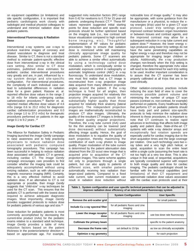
Table 1. System configurationimprove radiation do
and user specific technical paraose efficiency of an interventional
meters that can be adjusted to fluoroscopy system
System Configuration User Specific
Remove the anti-scatter grid for small patients
Include Cu x-ray spectral filter for all pediatric fluoro and cine imaging
Lower the image receptor dose
for all pediatric fluoro and cine imaging use low dose rate fluoroscopy
Collimate the primary beam specific to the patient anatomy
Decrease the frame rate set default to 15 fps as low as clinically acceptable
Optimize x-ray geometry for each projection
site. The pediatric cardiologist should also consult with a medical physicist and the scanner manufacturer to ensure that appropriate steps have been taken to optimize the radiation dose to the imaging task.
Step Lightly for Interventional Cardiology
The Alliance for Radiation Safety in Pediatric Imaging launched the Step Lightly campaign to elevate radiation dose awareness and help practicing physicians reduce radiation dose associated with interventional radiology procedures. Given the similarities between the interventional radiology and cardiology practices, many of the concepts and principles suggested by Step Lightly are equally applicable to interventional cardiology. Generally, Step Lightly encourages practitioners to ensure: that non-radiation imaging alternatives are considered and that the x-ray procedure is appropriate, that the patient dose is monitored and recorded in the patient record, that the x-ray fluoroscopic system is properly configured to ensure clinically useful images using the least possible radiation dose, that all staff receive regular radiation safety training and use sound radiation safety practices for every procedure, and that patient care providers are prepared to speak to families about risks and benefits of radiation used in imaging.
There are several x-ray technical factors that contribute to dose-efficient use of the x-ray system. First, it must be ensured that the x-ray system is in good working condition and that it is set up to specifications required by state and federal regulations. It will likely be necessary to customize an interventional x-ray system to optimize it for pediatric imaging. Such customization should be managed by a qualified medical physicist working in conjunction with physicians and equipment manufacturer image quality experts. When changes are made, measurements should be acquired to determine the effects on both patient dose and image quality. Interventional x-ray systems require a fixed x-ray exposure to the image receptor (image intensifier or digital flat panel). The x-ray fluence from the x-ray tube is automatically adjusted by the automatic brightness control system to account for variations in x-ray absorption by the patient (or transmission through the patient). The ABC automatically adjusts both the x-ray beam energy (via kVp) and the x-ray tube current-time product (mAs) to ensure that the image receptor realizes the pre-set detector dose rate. As patient thickness increases, both the beam energy and current-time product are increased to compensate for increased x-ray attenuation by the patient. The opposite is true for small patients through whom a greater fraction of the incident x-ray photons transmit. As patient thickness decreases, the x-ray tube output and therefore patient skin dose rate is automatically lowered by the imaging system. In this manner, the ABC system automatically reduces the radiation dose rate delivered to small patients. It is of great importance to recognize the radiation safety implications of x-ray fluoroscopy versus cine acquisition mode imaging. X-ray fluoroscopy utilizes a relatively low dose rate x-ray beam to create low-quality images that are appropriate for verifying anatomic landmarks or navigating intravascular catheters. Fluoroscopy images are not automatically saved, however, many systems provide opportunity for users to manually store such images. Cine or acquisition mode imaging utilizes a relatively high dose rate that is typically 10x that of fluoroscopy and produces high-quality images that are appropriate for diagnostic purposes. For both fluoroscopy and cine mode imaging, there are several technical parameters that can and should be properly set to minimize dose to pediatric patients. These factors are listed in Table 1. Some parameters can be adjusted by individual users and some of them are controlled by the system configuration.
The purpose of the anti-scatter grid is to preferentially absorb x-ray photons that interact with patient tissue and are subsequently scattered in direction of the x-ray detector while transmitting primary (unscattered) x-ray photons. This reduction in the number of scattered x-rays reaching the x-ray receptor results in improved x-ray image contrast. However, the grid also reduces the overall photon fluence at the image receptor, requiring compensation by the ABC via increased x-ray tube output and higher radiation dose rate. For small patients, there is very little x-ray scatter and so the grid can be removed from the system without substantial loss of image quality. Our experience is that for small patients (<20 kg), removing the grid reduces patient radiation dose rate by about 35%. Dose rate savings for larger patients will be somewhat greater, and it may be reasonable to remove the grid for patients as large at 50 kg provided that the image quality is adequate. Use of the antiscatter grid may be necessary for larger patients or when additional image quality is required to properly view instruments or anatomy. Modern x-ray fluoroscopy systems allow for the use of a copper x-ray beam spectral filter for both fluoroscopic and cine mode imaging. The Cu filter preferentially removes low energy x-ray photons from the x-ray beam, thereby increasing the average x-ray energy of the beam. Because low energy x-ray photons preferentially contribute to tissue dose relative to image quality, removing the low energy photons from the beam serves to substantially reduce radiation dose. For pediatric imaging, ensure that the x-ray control configuration is set to include a copper x-ray beam spectral filter for cine mode imaging. Our experience is that introduction of a 0.1 mm Cu filter reduces x-ray dose rate by 40% while decreasing image contrast by 10%. Another x-ray system parameter that can be changed to decrease patient dose is the target image receptor dose. Most x-ray systems allow customization of this parameter in 15% or 20% increments. Decreasing the image receptor dose will result in a decrease in patient dose rate. However, it will also result in decreased image quality. For both fluoroscopy and cine acquisition modes, the image receptor dose should be set as low as possible while still maintaining clinically useful image quality. For a typical interventional fluoroscopy system, the radiation dose rate (mGy/min) increases when the x-ray magnification mode is increased (primary field of view decreased). Some modern systems provide for configuration of the change in x-ray dose rate for the various geometric magnification (field of view) modes. The relationship between dose rate and geometric magnification should be understood by the physician operator. Regardless of the field of view selected, the secondary x-ray field collimator blades should be used to confine the x-ray beam to the anatomy of direct relevance, thereby minimizing the patient surface area and tissue volume irradiated. The image acquisition frame rate can be customized to reduce patient dose. For interventional cardiac procedures, the frame rate default is typically set to 15 frames per second. For patients with a rapid heart rate, the frame rate may have to be increased to 30 fps to achieve appropriate temporal resolution. For other patients and procedures (or portions of a procedure) a lower frame rate of 10 fps or 7.5 fps, with proportionately lower x-ray dose rate, may be appropriate. Finally, the geometry of the x-ray system with respect to the patient should be optimized to reduce patient dose. The x-ray dose rate at any point in space decreases at a rate that is inversely proportional to the square of the distance from the x-ray source. Therefore, the relative distance of both the patient and the x-ray detector from the x-ray tube can substantially affect patient radiation dose rate. Optimization of x-ray geometry to minimize patient dose requires only that the patient be moved as far away from the x-ray source as reasonably possible and that the x-ray detector is then moved as close as reasonably possible to the patient. Due to the strong influence of
6 CONGENITAL CARDIOLOGY TODAY www.CongenitalCardiologyToday.com December 2009
For information on PFO detection go to: www.spencertechnologies.com
distance from the x-ray source on dose rate, seemingly modest changes in these distances can have a substantial impact on patient dose rate.
It should be recognized that, after the technical parameters controlling the fluoroscopic system are set, the performing physician can have a great deal of influence on the amount of radiation used during an interventional fluoroscopy procedure. To help minimize radiation dose, each procedure should start with a plan that is discussed with all members of the patient care team. The plan should include relevant details of anticipated x-ray system use. When available, previous imaging procedures should be reviewed. Reducing the duration of time that the x-ray beam is on is a fundamental ideal in the Step Lightly campaign. To this end, ensure that every second of beam on time provides clinically useful information. Though seemingly obvious, never activate the fluoroscopy foot switch if your eyes are not on the image display monitor. Also, step off of the radiation footswitch as soon as the relevant clinical information is acquired. As much as possible, navigate catheters using low dose rate fluoroscopy.
Managing Radiation Dose
X-ray based images, including CT, chest radiography, and x-ray fluoroscopy can be valuable tools for diagnosing, monitoring, and treating pediatric patients with congenital heart and/or vascular defects. However, with the possibility of multiple such procedures contributing to a lifetime cumulative radiation dose, the risk of these patients developing a radiation-induced cancer later in life should always be considered. As medical professionals, we should strive to ensure that x-ray modalities are used appropriately. First, consider whether an x-ray procedure is necessary or whether the information which it will provide could be obtained by another means. Consider the use of ultrasound or MRI, for example. Consider the frequency at which routine x-ray procedures, such as chest radiographs, are acquired and reduce the frequency to the minimum which is clinically appropriate. Once the need for an x-ray based procedure is confirmed, ensure that the radiation dose associated with that procedure is minimized. This requires some planning and work well before the patient reaches the procedure room. For both CT and fluoroscopic imaging, minimizing radiation dose includes using x-ray techniques that are appropriate for the patient size, minimizing the volume of tissue irradiated, and minimizing the duration of time that x-ray beam is on. For every patient, a comprehensive record of all x-ray based procedures should be maintained. This information should be integrated into the patient record and be immediately accessible to all care providers. A clear and concise radiation history in the patient record will help raise the radiation awareness of everyone involved in patient care. The Alliance website provides suggestions for discussing x-ray radiation and related issues with families, and it can be expected that the x-ray procedure history may be of interest to family members. When discussing radiation risks with families, it is important that the risks and benefits of performing versus not performing a procedure which uses ionizing radiation are well appreciated.
Conclusion
Imaging systems which use x-rays are an integral part of the modern medical practice and will remain so for the foreseeable future. Both the potential benefit and risk associated with x-ray imaging need to be well understood to make sound decisions that ensure both short-term and long-term patient health. Based on the linear non-threshold cancer
CONGENITAL CARDIOLOGY TODAY www.CongenitalCardiologyToday.com December 2009 7
Supervisor Pediatric and Fetal Echocardiography Laboratory
The Scott and Laura Eller Congenital Heart Center at St. Joseph’s Hospital and Medical Center in Phoenix, Arizona is recruiting for a SUPERVISOR for its PEDIATRIC AND FETAL ECHOCARDIOGRAPHY LABORATORY. The successful candidate will be joining a new rapidly expanding group of full-time academically-driven pediatric cardiologists, pediatric cardiac intensivists, and pediatric cardiac surgeons. A state-of-the-art brand-new 24-bed Pediatric Cardiac Intensive Care Unit, unique to the state, was recently opened. The echocardiography laboratory, under the direction of Ernerio T. Alboliras, MD, FACC, FAAP, FASE, currently has six sonographers and performs more than 7,000 studies per year, including 650 fetals. All images are digitally archived. There is great opportunity to experience a broad spectrum of imaging congenital and acquired heart defects, from simple to complex; to include echocardiographic involvement during interventional catheterization, Hybrid operative procedures and open heart surgery. All aspects of ultrasound imaging – transthoracic, transesophageal, fetal, intracardiac, and stress – are performed. Tele-echocardiography transmission from other hospitals is routine. Ample opportunity for participation in echocardiography research and education is available. REQUIREMENTS: Five years’ experience in pediatric echocardiography, BCLS, RDMS or RDCS certification, ability to follow a consistent Pediatric Echo Lab imaging protocol, efficiently manage a busy Lab and work and communicate effectively with physicians, sonographers and other staff required. Strong congenital heart and previous supervisory experience in a Pediatric and Fetal Echocardiography Laboratory highly preferred. HOSPITAL SUMMARY: St. Joseph's Hospital and Medical Center has been a symbol of quality healthcare in the Valley of the Sun for more than 110 years. With more than 740-beds and 5,200 employees, we are extremely proud to announce that we are the only Arizona hospital to be voted a "Best Place to Work in the Valley" six years in a row. We are also proud to be named a Top 25 Workplaces for Women, a top hospital by Ranking Arizona Magazine and routinely recognized as one of the country's best neuroscience centers by U.S. News & World Report. The hospital is part of Catholic Healthcare West, which has more than 40 hospitals in Arizona, California and Nevada.
For questions regarding The Scott and Laura Eller Congenital Heart Center p lease v is i t h t tp: / /www.st josephs-phx.org/Medical_Serv ices/Congenital_Heart_Center/index.htm or email [email protected]
Equal Opportunity Employer. Apply on-line to Req ID: 69315 please visit www.stjosephs-phx.org/careers or via email to [email protected]. Personalized relocation and competitive salary offered. Look for us on Facebook, and follow us on Twitter at: http://twitter.com/stjosephsjobs
Working Together to Develop a Better Tomorrow
Rady Childrenʼs Heart Institute has organized and supported symposia dedicated to expanding our knowledge about the diagnosis and treatment of complex forms of congenital heart disease since 1984. The symposia are designed to educate attendees by combining formal presentations with lively discussions of controversial areas in the rapidly evolving science and clinical management of the neonate, child and adult born with congenital cardiovascular disease. For each program, the planners carefully assess prior attendeesʼ comments and reviews,and invite renowned faculty from across the United States to present new data and ideas.
The three-day symposium on Pediatric and Congenital Cardiology and Cardiac Surgery will include lectures, roundtables and case discussions. We will focus on the latest techniques, including cutting-edge technologies such as the hybrid procedure and percutaneous heart valves. We expect to attract an enthusiastic audience of pediatric cardiologists, pediatric cardiology fellows, cardiac surgeons, adult cardiologists with interest in congenital heart disease and cardiac nurse practitioners, as well as some cardiac administrators.
We have pondered what quality measures are appropriate in 2010. We can assess the quality gap by observing adult patient outcomes and comparing those outcomes to the surgical procedures performed earlier in life. The title for this symposium emphasizes the ever-changing diagnostic and treatment modalities in the management of congenital heart defects. The analyses presented will benefit attendees in their medical and surgical practices by outlining the procedures most likely to produceoptimal long-term patient outcomes. The planners hope that the Symposium will provide answers to the perennial question: “What is the right thing to do for this patient?” Faculty panel and question-and-answer sessions will allow audience interaction as we jointly pursue the ideal treatment plan for each complex patient.
We hope you will come warm yourself in beautiful San Diego in January!
Visit our website: www.rchsd.org/cme and click on Conferences & Seminars for more information or for registration and credit card payment form. We accept Visa and MasterCard
The three-day symposium on Pediatric and Congenital Cardiology and Cardiac Surgery will include lectures, roundtables and case discussions. We will focus on the latest techniques, including cutting-edge technologies such as the hybrid procedure and percutaneous heart valves. We expect to attract an enthusiastic audience of pediatric cardiologists, pediatric cardiology fellows, cardiac surgeons, adult cardiologists with interest in congenital heart disease and cardiac nurse practitioners, as well as some
We have pondered what quality measures are appropriate in 2010. We can assess the quality gap by observing adult patient outcomes and comparing those outcomes to the surgical procedures performed earlier in life. The title for this symposium emphasizes the ever-changing diagnostic and treatment modalities in the management of congenital heart defects. The analyses presented will benefit attendees in their medical and surgical
Planning Committee: John J. Lamberti, MD; John W.Moore, MD, MPH; R.R. Skoglund, MD, Immediate Past CME Director; Keith Vaux, MD, CME Director
Educational Design: Didactic presentations, with audio-visual support, are followed by panel discussion and audience question and answer sessions. A syllabus will include abstracts from faculty on their topics, with key references.
Objectives: At the completion of this activity, participants should be able to: • Determine optimal management strategies for common
diagnostic and treatment problems encountered in an outpatient office practice.
• Understand and manage the diagnosis and treatment of cardiomyopathy and pulmonary hypertension in infants, children and young adults.
• Optimize imaging modality selection to obtain a diagnosis in the most efficient and cost-effective manner.
• Diagnose and treat common and complex arrhythmias in patients of all ages.
• Utilize modern catheterization techniques as part of an integrated invasive treatment program for congenital heart disease.
• Integrate the Hybrid approach with modern variations of the Norwood and Fontan procedure to optimize care of infants born with HLHS.
• Utilize modern medical and surgical strategies to treat complex Congenital Heart Disease.
• Understand the role of “benchmark” data in assessing outcomes after treatment of pediatric and adult CongenitalHeart Disease.
Faculty: Zahid Amin, MD; Anjan S. Batra, MD; Daniel Bernstein, MD; ; Jane C. Burns, MD; John P. Cheatham, MD, FAAP, FACC, FSCAI; John S. Child, MD; Joseph A. Dearani, MD; Howaida G. EL-Said, MD; Frank L. Hanley, MD; Dunbar Ivy, MD; Jeffrey P. Jacobs, MD, FACS, FACC,FCCP; Joel Kirsh, MD; Steven E. Lipshultz, MD, FAHA, FAAP; James Lock, MD; Audrey C. Marshall, MD; Gerald Ross Marx, MD; Peter Pastuszko, MD; James C. Perry, MD, FAAP, FACC, FHRS; Beth Feller Printz, MD, PhD; Mohan Reddy, MD; Kevin Shannon, MD; Thomas L. Spray, MD; Vaughn A. Starnes, MD; Lloyd Y. Tani, MD; James S. Tweddell, MD; George F. Van Hare, MD; Victoria L. Vetter, MD; Gary Webb, MD; Gil Wernovsky, MD, FACC, FAAP
response model and considering that pediatric congenital cardiac patients may undergo several CT or interventional fluoroscopy procedures during their childhood, these patients are expected to have a slightly increased lifetime risk of cancer. Minimizing the radiation dose for each procedure helps to minimize this risk. Through the Image Gently and Step Lightly campaigns, The Alliance for Radiation Safety in Pediatric Imaging continues to have a positive influence on the use of ionizing radiation in the care of pediatric patients. As promoted by these campaigns, pediatric radiation dose reduction can be achieved through decreased use of x-ray imaging procedures and through dose-efficient use of x-rays when such a procedure is indicated. Most importantly, these reductions can be realized without compromising image quality or patient care.
References
1. Sidhu, M., B. D. Coley, M. J, Goske, B. Connolly, J. Racadio, T. T. Yoshizumi, T. Utley, and K. J. Strauss, Image Gently, Step Lightly: increasing radiation dose awareness in pediatric interventional r a d i o l o g y, P e d i a t r. R a d . 2 0 0 9 ; 39:1135-1138.
2. Hall, E. J., Radiation biology for pediatric radiologists. Pediatr. Rad. 2009; 39:S57-S64.
3. I C R P P u b l i c a t i o n 6 0 , 1 9 9 0 Recommendations of the International Commission on Radiologic Protection, Ann. ICRP. 1990; 21.
4. ICRP Publication 103, The 2007 Recommendations of the International Commission on Radiologic Protection, Ann. ICRP. 2008; 37.
5. Linet, M. S., K. Kim, P. Rajaraman, Children's exposure to diagnostic medical radiation and cancer risk: epidemiologic and dosimetric considerations. Pediatr. Rad. 2009; 39:S4-S26.
6. National Research Council, 'Health risks from exposure to low levels of ionizing radiation: BEIR VII - Phase 2,' 2006, Washington, DC.
7. Rice, H. E., D. P. Frush, D. Farmer, J. H. Wa l d h a u s e n , A P S A E d u c a t i o n Committee, Review of radiation risks from computed tomography: essentials for the pediatric surgeon. J. Ped. Surg. 2007; 42:603-607.
8. Goo, H. W., D. S. Suh, Tube current reduction in pediatric non-ECG-gated heart CT by combined tube current modulat ion. Pediatr. Rad. 2006; 36:344-351.
9. Greil, G. F., M. Schoebinger, A. Kuettner, J. F. Schaefer, F. Dammann, C. D. Claussen, M. Hofbeck, H. P. Meinzer, and L. Sieverding, Imaging of aortopulmonary collateral arteries with high-resolution multidetector CT. Pediatr. Rad. 2006; 36:502-509.
10. Lerner, C. B., D. P. Frush, D. T. Boll, Evaluation of a coronary-cameral fistula: benefits of coronary dual-source MDCT angiography in children. Pediatr. Rad. 2008; 38:874-878.
11. Hollingsworth, C. L., T. T. Yoshizumi, D. P. Frush, F. P. Chan, G. Toncheva, G. Nguyen, C. R. Lowry, and L. M. Hurwitz, Pediatric cardiac-gated CT angiography: Assessment of radiation dose. Am. J. Roent. 2007; 189:12-18.
12. Herzog, C., D. M. Mulvihill, S. A. Nguyen, G. Savino, B. Schmidt, P. Costello, T. J. Vogl, and U. J. Schoepf, Pediatric cardiovascular CT angiography: radiation dose reduction using automatic tube current modulation. Am. J. Roent. 2008 190:1232-1240.
13. Rassow, J., A. A. Schmaltz, F. Hentrich, and C. Streffer, Effective doses to patients from paediatric cardiac catheterization. Brit. J. Rad. 2000; 73:172-183.
14. Bacher, K., E. Bogaert, R. Lapere, D. De Wolf, H. Thierens, Patient-specific dose and radiation risk estimation in pediatric cardiac catheterization. Circulation 2005; 111:83-89.
15. 15. The Alliance for Radiation Safety in Pediatric Imaging. 2009; Retrieved October 15, 2009, from American College o f R a d i o l o g y w e b s i t e h t t p : / /www.pedrad.org/associations/5364/files/Protocols.pdf
16. Kalender, W. A., P. Deak, M. Kellermeier, M. van Straten, and S. B. Vollmar, Application- and patient size-specific optimization of x-ray spectra for CT. Med. Phys. 2009; 36:993-1007.
CCT
James M. Kofler, PhDDepartment of RadiologyMayo Clinic200 1st St. SWRochester, MN USA
Donald J. Hagler, MDDivision of Pediatric CardiologyMayo Clinic200 1st St. SWRochester, MN USA
Corresponding Author
Kenneth A. Fetterly, PhDAssistant Professor of RadiologyDivision of Cardiovascular DiseasesCollege of MedicineMayo Clinic200 1st St. SWRochester, MN 55905 USAPhone: 507-266-4384; fax 507-255-7688
10 CONGENITAL CARDIOLOGY TODAY www.CongenitalCardiologyToday.com December 2009
Do you or your colleagues have an iPhone?
We have two iPhone professional medical apps: "The Merck Manual Professional Edition"
and “OsiriX” (for display and analysis of medical images), and are looking for
physicians in the field interested to review them. If interested, send an email to:
The University of ChicagoDepartment of Pediatrics
The Department of Pediatrics at the University of Chicago is expanding its section of Pediatric Cardiology. We are recruiting cardiologists to a rapidly growing academic cardiology section. The successful candidate will have an M.D., or M.D./ Ph.D., and hold or be eligible for medical licensure in the states of Illinois and Indiana. The successful candidate must also be BC/BE in pediatric cardiology. Candidates should be able to demonstrate excellent teaching skills. Academic appointment and salary will be commensurate with experience. Research opportunities are available for appropriately qualified candidates.
Candidates with strong clinical skills, particularly in fetal echo are strongly preferred. Additional training or expertise in electrophysiology, post-operative care, and/or echocardiography preferred. Research interests in any of these areas a plus.
This position includes responsibilities for teaching students, residents and fellows. Outpatient and inpatient venues are included.
Interested candidates should submit a curriculum vitae, letter of interest and names of three references to our academic website: academiccareers.uchicago.edu/applicants/Central?quickFind=50715
Introduction
N o n c o m p a c t i o n o f t h e v e n t r i c u l a r myocardium (NCVM) is a cardiomyopathy that is character ized by prominent myocardial t rabeculat ions and deep intertrabecular recesses with variable degrees of myocardial dysfunction. It has variable clinical presentations and in children h a d b e e n a s s o c i a t e d w i t h b e n i g n asymptomatic course, as well as severe myocardial dysfunction.1 In adults, NCVM is well known to be associated with thrombo-embolism.2 In the pediatric age group, we o b s e r v e d t h r o m b o - e m b o l i s m w i t h noncompaction in 9% of patients with the isolated form.3 In this report we describe a further case of fatal thrombo-embolism in a child with NCVM.
Case History
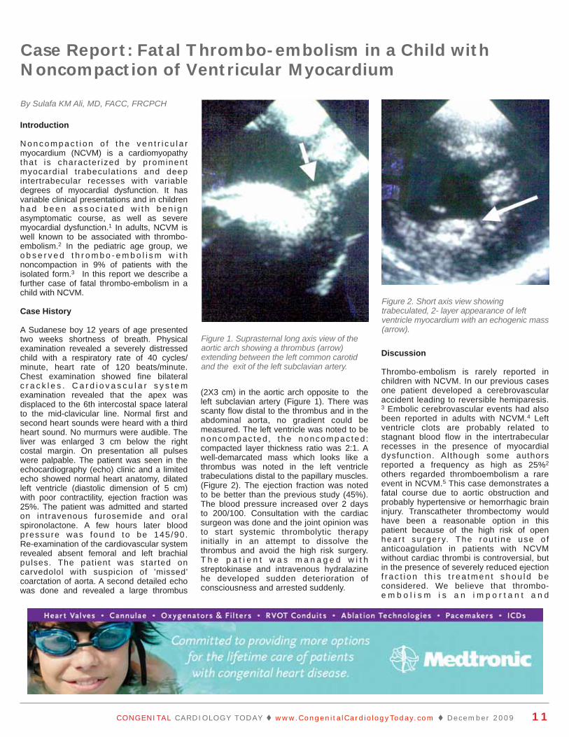
A Sudanese boy 12 years of age presented two weeks shortness of breath. Physical examination revealed a severely distressed child with a respiratory rate of 40 cycles/minute, heart rate of 120 beats/minute. Chest examination showed fine bilateral c r a c k l e s . C a r d i o v a s c u l a r s y s t e m examination revealed that the apex was displaced to the 6th intercostal space lateral to the mid-clavicular line. Normal first and second heart sounds were heard with a third heart sound. No murmurs were audible. The liver was enlarged 3 cm below the right costal margin. On presentation all pulses were palpable. The patient was seen in the echocardiography (echo) clinic and a limited echo showed normal heart anatomy, dilated left ventricle (diastolic dimension of 5 cm) with poor contractility, ejection fraction was 25%. The patient was admitted and started on intravenous furosemide and oral spironolactone. A few hours later blood pressure was found to be 145/90. Re-examination of the cardiovascular system revealed absent femoral and left brachial pulses. The patient was started on carvedolol with suspicion of 'missed' coarctation of aorta. A second detailed echo was done and revealed a large thrombus
(2X3 cm) in the aortic arch opposite to the left subclavian artery (Figure 1). There was scanty flow distal to the thrombus and in the abdominal aorta, no gradient could be measured. The left ventricle was noted to be noncompac ted , the noncompac ted : compacted layer thickness ratio was 2:1. A well-demarcated mass which looks like a thrombus was noted in the left ventricle trabeculations distal to the papillary muscles. (Figure 2). The ejection fraction was noted to be better than the previous study (45%). The blood pressure increased over 2 days to 200/100. Consultation with the cardiac surgeon was done and the joint opinion was to start systemic thrombolytic therapy initially in an attempt to dissolve the thrombus and avoid the high risk surgery. T h e p a t i e n t w a s m a n a g e d w i t h streptokinase and intravenous hydralazine he developed sudden deterioration of consciousness and arrested suddenly.
Discussion
Thrombo-embolism is rarely reported in children with NCVM. In our previous cases one patient developed a cerebrovascular accident leading to reversible hemiparesis.3 Embolic cerebrovascular events had also been reported in adults with NCVM.4 Left ventricle clots are probably related to stagnant blood flow in the intertrabecular recesses in the presence of myocardial dysfunction. Although some authors reported a frequency as high as 25%2 others regarded thromboembolism a rare event in NCVM.5 This case demonstrates a fatal course due to aortic obstruction and probably hypertensive or hemorrhagic brain injury. Transcatheter thrombectomy would have been a reasonable option in this patient because of the high risk of open hear t su rgery. The rou t ine use o f anticoagulation in patients with NCVM without cardiac thrombi is controversial, but in the presence of severely reduced ejection f r a c t i o n t h i s t r e a t m e n t s h o u l d b e considered. We believe that thrombo-e m b o l i s m i s a n i m p o r t a n t a n d
Case Report: Fatal Thrombo-embolism in a Child with Noncompaction of Ventricular Myocardium
By Sulafa KM Ali, MD, FACC, FRCPCH
Figure 1. Suprasternal long axis view of the aortic arch showing a thrombus (arrow) extending between the left common carotid and the exit of the left subclavian artery.
Figure 2. Short axis view showing trabeculated, 2- layer appearance of left ventricle myocardium with an echogenic mass (arrow).
CONGENITAL CARDIOLOGY TODAY www.CongenitalCardiologyToday.com December 2009 11
life-threatening complication of NCVM that should be promptly diagnosed and managed.
References
1. Sulafa K. M. Ali, Michael J. Godman. The variable clinical presentation of, and outcome for, noncompaction of the ventricular myocardium in infants and children, an under-diagnosed cardiomyopathy. Cardiol Young 2004; 14: 409–416.
2. Oechslin EN, Attenhofer Jost CH, Rojas JR, Kaufmann PA, Jenni R. Long-term follow-up of 34 adults with isolated left ventricular noncompaction: a distinct cardiomyopathy with poor prognosis. J Am Coll Cardiol. 2000;36:493-500.
3. Sulafa KM Ali. Unique features of non-compaction of the ventricular myocardium in Arab and African patients. Cardiovasc J Afr 2008; 19: 195–199.
4. Sahin S, Karsidag S. An unusual cause of cardioembolic stroke: isolated left ventricular noncompaction. Neurologist. 2009;15:51.
5. Stöllberger C, Finsterer J. Thrombi in left ventricular hypertrabeculation/noncompaction--review of the literature. Acta Cardiol. 2004 ;59:341-4.
CCT
EP CARDIOLOGISTPEDIATRIC CARDIOLOGY
PEDIATRIC CARDIAC CRITICAL CAREPEDIATRIC CV SURGEON
HCA is the largest healthcare company in the US, with over 160 hospitals in 20 states. Whether you are looking for a place to start, or somewhere to complete a successful career in pediatric cardiac care, chances are we can help you find it. We have opportunities in big cities and rural areas, near sandy beaches and skyward reaching mountains. Wherever your internal compass points, it’s likely that we can help guide you to a destination that will match your personal preferences while fulfilling your career dreams.
As our co-founder, Dr. Thomas Frist, Sr. said, “Bettering the human condition is the greatest good any individual can achieve.” You are doing your part by taking care of our most precious commodity, the children who will be our future.
Now let us do our part by helping you find your dream job.
For more information on available positionscontact Kathleen L. Kyer, Manager,
National Pediatric Subspecialty Recruitment at:937-235-5890 or
Call or e-mail me todayfor moreinformationon opportunitiesin your sub-specialty.
If you are a fellow in training, please be sure toask about our excellent stipend program!
Sulafa KM Ali, MD, FACC, FRCPCHUniversity of KhartoumFaculty of Medicine, Department of Paediatrics and Child Health PO Box 102 Khartoum Erkaweit, Sudan
AND
Department of Pediatric CardiologySudan Heart CenterPO Box 11917Khartoum Erkaweit, Sudan
Tel: +24 (918) 3232133/3232137; Fax: +24 (918) 3232135
sulafakhal [email protected]
“This case demonstrates a fatal course due to aortic obstruction and probably hypertensive or hemorrhagic brain injury. Transcatheter thrombectomy would have been a reasonable option in this patient because of the high risk of open heart surgery.”
12 CONGENITAL CARDIOLOGY TODAY www.CongenitalCardiologyToday.com December 2009
l010
13th Annual Update on Pediatric Cardiovascular DiseaseFebruary 10 – 14, 2010 � Disney’s Contemporary Resort, Florida
www.chop.edu/cardiology2010 ©Disney
CONGENITAL CARDIOLOGY TODAY www.CongenitalCardiologyToday.com December 2009 13
Yale University Yale New Haven
Children’s Hospital
PEDIATRIC CARDIOLOGIST
The Section of Pediatric Cardiology at the Yale University School of Medicine and the Yale New Haven Children’s Hospital is recruiting a Pediatric Cardiologist to join our busy academic practice. Interested candidates should have particular skill in cardiovascular imaging or in general pediatric cardiology. Our section provides the broad range of cardiovascular services including advanced imaging, catheter intervention and electrophysiology to patients throughout the State of Connecticut and our busy congenital heart surgery program performs all types of advanced cardiac surgery for neonates, infants, adolescents and adults with congenital heart disease.
Candidates should be BC/BE in pediatric cardiology and have demonstrated interest in teaching as we have a thriving fellowship that has been supported through an NIH training grant for many years. Appointment at the rank of Assistant or Associate Professor is anticipated, based on experience. The Yale New Haven Children’s Hospital is the primary referral center for southern Connecticut, a state with nearly 3.5 million people. New Haven and the shoreline area of Connecticut offer tremendous cultural, entertainment and recreational opportunities. With great schools, access to New York City (90 minutes) and Boston (2 hours), the quality of life is hard to beat!
Yale University is an equal opportunity, affirmative action employer. Women and minorities are encouraged to apply.
For more information please contact:
Alan Friedman, MDProfessor, Pediatric Cardiology
Yale University School of Medicine333 Cedar Street
New Haven, CT 06520-8064
Deadline for contact is December 31, 2009.
Need to Recruit a Pediatric Cardiologist?
Advertise in Congenital Cardiology Today, the only monthly newsletter dedicated to pediatric and congenital cardiologists.
Reach the most Board Certified or Board Eligible pediatric cardiologists throughout the U.S. and Canada.
Recruitment advertising includes full color in either the North American print edition or the electronic PDF International edition.
Available in 1/3 and 1/2 page vertical Recruitment ad sizes. We can even create the ad for you at no extra charge!
For more information contact:
Tony Carlson, FounderCONGENITAL CARDIOLOGY TODAY
Tel: [email protected]
CONGENITAL CARDIOLOGY TODAY
© 2009 by Congenital Cardiology Today (ISSN 1554-7787-print; ISSN 1554-0499-online). Published monthly. All rights reserved.
Headquarters 9008 Copenhaver Dr. Ste. M Potomac, MD 20854 USA
Publishing Management Tony Carlson, Founder & Senior Editor [email protected] Richard Koulbanis, Publisher & Editor-in-Chief [email protected] John W. Moore, MD, MPH, Medical Editor/Editorial Board [email protected]
Editorial Board Teiji Akagi, MD Zohair Al Halees, MD Mazeni Alwi, MD Felix Berger, MD Fadi Bitar, MD Jacek Bialkowski, MD Philipp Bonhoeffer, MD Mario Carminati, MD Anthony C. Chang, MD, MBA John P. Cheatham, MD Bharat Dalvi, MD, MBBS, DM Horacio Faella, MD Yun-Ching Fu, MD Felipe Heusser, MD Ziyad M. Hijazi, MD, MPH Ralf Holzer, MD Marshall Jacobs, MD R. Krishna Kumar, MD, DM, MBBS Gerald Ross Marx, MD Tarek S. Momenah, MBBS, DCH Toshio Nakanishi, MD, PhD Carlos A. C. Pedra, MD Daniel Penny, MD James C. Perry, MD P. Syamasundar Rao, MD Shakeel A. Qureshi, MD Andrew Redington, MD Carlos E. Ruiz, MD, PhD Girish S. Shirali, MD Horst Sievert, MD Hideshi Tomita, MD Gil Wernovsky, MD Zhuoming Xu, MD, PhD William C. L. Yip, MD Carlos Zabal, MD FREE Subscription Congenital Cardiology Today is available free to qualified professionals worldwide in pediatric and congenital cardiology. International editions available in electronic PDF file only; North American edition available in print. Send an email to [email protected]. Include your name, title, organization, address, phone and email.
Contacts and Other Information For detailed information on author submission, sponsorships, editorial, production and sales contact, current and back issues, see website or send an email to: [email protected].
14 CONGENITAL CARDIOLOGY TODAY www.CongenitalCardiologyToday.com December 2009
Do you or your colleagues have interesting research results, observations, human interest stories, reports of meetings, etc. that you would like to share
with the congenital cardiology community?
Submit a summary of your proposed article to Congenital Cardiology Today at: [email protected]
PEDIATRIC CARDIOLOGIST
Madigan Army Medical Center, Fort Lewis, Washington is seeking a full time civilian pediatric cardiologist to join an Army pediatric cardiologist in practice. Cand ida tes mus t be BC/BE and demonstrate a broad range of clinical experience in pediatric cardiology. Cardiology service provides direct patient care in both inpatient and outpatient settings and consultative expertise to a neonatal intensive care unit and a busy high risk obstetrics clinic. Clinical educator experience desired as the service supports a pediatric residency and a perinatology fellowship.
Madigan Army Medical Center is a 243-bed teaching medical center. It supports a primary care population of greater than 100k. The surrounding greater Puget Sound area offers excellent schools. Proximity to the Cascades and Puget Sound assures year round recreational opportunities.
To learn more about this excellent opportunity contact Medical Provider Recruiter @ (253) 968-4994 or send CV to [email protected].
PICS-AICS Pediatric and Adult Interventional
Cardiac Symposium
www.PICSymposium.com
SAVE THE DATE JULY 18–21, 2010
CHICAGO

Pediatrix Cardiology is a national provider of outpatient andinpatient cardiology care of the fetus, infant, child and adolescent, as well as adults with congenital heart disease.
We have an opportunity for you, whether you’re a recent graduate looking to gain experience or a seasoned pediatriccardiologist seeking a leadership role. With a wide variety ofservices and a presence in several states, you can choose thepractice environment and location that fits you best.
We offer competitive salaries and excellent benefits includinghealth (PPO), life, vision, dental and disability insurance;401(k) with potential company percentage match; annual CMEallowance; potential for relocation assistance; employee stockpurchase plan; opportunities to participate in clinical qualityimprovement initiatives and clinical research; professional liability insurance; and assistance with mandatory hospital credentialing and state licensing, and reimbursement of associated fees.
More than 90 pediatric cardiologists have chosen to join ourteam to pursue their personal and professional goals. Now it’syour turn.
Please contact us to learn more about pediatric cardiology positions in:
Tucson, Arizona Dallas, TexasAlbuquerque, New Mexico San Antonio, Texas
Fairfax, Virginia
800.243.3839, ext. 6511 www.pediatrix.com/cardiopenings
Your Search Ends Here.
1301 Concord TerraceSunrise, Florida 33323
CCT.Dec.09 10/15/09 9:39 AM Page 1

TINY HEARTSINSPIREDHYBRID LABSWITH ACCESS FOR BIG TEAMS.Fixing a heart from birth through adulthood takes big
teams working together. So we examined the needs of
leading clinicians when designing our hybrid solutions.
The result: our InfinixTM
-i with 5-axis positioners and low
profile detectors, stays out of the way, but right where
needed, providing the best possible access to patients.
To lead, you must first listen.
medical.toshiba.com