Embed Size (px)
Citation preview
APPLICATION TO THE ACADEMY OF VETERINARY DENTISTRY
Small Animal
Please read the Application completely before attempting to complete any of the requirements. Also ensure that you have read the “Introduction 2011” available from the “Become A Fellow” page of www.avdonline.org, which outlines requirements for having a mentor and submitting your letter of intent among other things.
In 2011, there are two options for submission: Applicants can submit their entire application package to the Academy Secretary, including signed copies of completed forms (under number 1 below) and one compact disc containing all the required files. The Academy Secretary will transfer the files to the Academy Document Management System so that the Credentials Committee Chair and Members can down-load the files for review.
OR
Applicants can submit the required files directly to DMS. Log in to the Academy DMS site using the Academy User Name and Password assigned to you (the User Name is your firstnamelastname with no spaces or dashes – e.g. CindyCharlier). Your password, unless you have already changed it in DMS, is your last name in lower case letters. Remember that you can change your password once you are logged in to DMS. On the Welcome screen, click Begin a New Document to Submit to the Academy. Click Credentials Application from the Type of Document drop-down menu. Click Attach Multiple Files, to identify and upload the files (check that they have all uploaded successfully), then click Submit and Save Changes. If you elect to submit files directly to DMS, the signed Application, Mentor Accountability and Agreement forms and check are to be mailed separately to the Academy Secretary.
Note: All application materials remain the property of the Academy of Veterinary Dentistry and will not be returned unless the application was rejected as improper, inadequate or incomplete.
The completed Application Package will contain the following items:
1.Items to be mailed directly to the Academy Secretary, Dr. Cindy Charlier, Fox Valley Veterinary Dentistry and Surgery, 37W748 Stratford Lane, Elgin, IL 60124, USA.Completed forms (as printed and signed copies):
- Academy of Veterinary Dentistry Application Form - Applicant/mentor Accountability form- Agreement, signed and notarized
- Credentials Application check listEnclose a check for $300 U.S. made out to the Academy of Veterinary Dentistry in a separate envelope. Resubmission fee is $100.
2. Items submitted either directly to the Academy DMS or on a CD to the Academy Secretary, using the file names shown below:
A. Veterinary Diploma - Reproduction of your veterinary diploma (scanned or photographed).
B. Veterinary License - Reproduction of your current veterinary license (scanned or photographed).
C. Dental Record Forms - Reproduction of your blank dental chart and anesthesia record, with your name and other identifying informed not visible. Submit as high quality scanned or photographed images, to ensure legibility.
D. Equipment: A list categorized by discipline and with photographs of your dental operatory and equipment. Include all instrumentation, materials, and equipment, from the most basic instrument to the most complex materials. Organize the contents under the following categories: dental operatory, anesthesia/monitoring, power handpieces, dental radiograph equipment, periodontal surgery, endodontic, restorative, oral surgery, and orthodontics, as listed in the AVD Application Checklist.
E. Continuing Education and Informal Dental Education. Three Excel spreadsheets, as listed below, using the excel spreadsheet formats available on the “Become a Fellow” page of www.avdonline.org. Do not include your name anywhere in the spreadsheets.a. Lecture Continuing Education Hours. A list the continuing education programs you have
attended in veterinary and human dentistry during the past three (3) years. Include dates, sponsoring organizations, names of speakers and topics covered. The date of lecture, speaker and number of hours are required. Minimum requirement: 40 hours of lecture, with at least 30 hrs. attended In person and a maximum of 10 hrs. of RACE approved online C.E.
b. Wet Lab and In Person Instruction Hours: Documentation that you have attended a minimum of 40 hours of approved wet-labs. NEW REQUIREMENT AS OF JULY 1, 2010: In addition, at least 40 hrs must be spent working with the mentor or receiving in-person instruction by a Fellow of the Academy or a Diplomate of the American Veterinary Dental College. An example of in-person instruction would be time spent with your mentor where either the applicant or mentor are performing dental cases, with active instruction and discussion. NEW REQUIREMENT AS OF JULY 1, 2010: The applicant is also required to attend at least 2 Veterinary Dental Forums in the past 3 years.
c. Informal Veterinary Dental Education. Examples: informal conversations (either in person, by phone or by e-mail) with dentists, veterinary dentists, or other qualified professionals regarding dental techniques or theory, and practicing of procedures on cadavers. Include dates, participants, and topics discussed, or dates of cadaver procedures performed. When practicing cadaver procedures, take radiographs and/or pictures to document your work. If an applicant has nearly achieved but is still lacking the minimum case log requirements near the time of submission, performing needed procedures on cadavers with appropriate documentation may allow an almost complete package to be evaluated by the committee (see “Case Log” below).
F. Personal Library. List the human and veterinary dental texts and journals available in your personal library, including journals and texts with publication dates and edition numbers. Your personal library should include or you should otherwise have access to the textbooks and journals in the ‘Suggested Reading List’.
G. Case Logs: The purpose of the log is to demonstrate to the Credentials Committee the depth and breadth of your dental experience during the required time frame. Use the Microsoft Excel Spreadsheet Template available on the “Become a Fellow” page of www.avdonline.org. The searching and sorting functions of the template make it the most efficient way of tracking and calculating the information. If there are case log deficiencies present 60 days prior to the July 15 submission date, the applicant is to send an appeal letter to the Secretary 60 days prior to the July 15 submission date., describing the case log deficiency and (when practical) provide an
2
explanation for the deficiency. Once received, the Credentials Committee chair will decide if the deficiency is too significant to accept an application during that cycle.
H. Case Log. Use the format in the ‘Sample’ Excel case log available on the “Become a Fellow” web page, by downloading the Excel file and inserting the data. Use separate work-sheets in the Excel file for:
1. Chronological Case Log. List your veterinary dental cases chronologically in an Excel worksheet labeled “Chrono” for a 24 month period in the past 3 years. Utilize the attached abbreviation list for appropriate abbreviation in the diagnosis and treatment columns of the case logs.
2. Categorical Case Log: Cases categorized by discipline on separate Excel worksheets labeled DP, PE, EN, RE, RAD, OR, OS. Utilize the attached abbreviation list for appropriate abbreviation in the diagnosis and treatment columns of the case logs. Total the cases in each discipline at the end of each discipline’s log. A maximum of 3 ‘category’ cases per patient visit is allowed. Dentinal bonding does not count toward the restoration requirement. Consil™ in an extraction site is not considered periodontal surgery. Crown shortening and vital pulpotomy for lingually displaced canine teeth are considered an endodontic case.
3. Case Log Summary: On a new worksheet, enter the categories in one column and the number of cases logged in each category in the next column in the Excel log.
MINIMUM CASE REQUIREMENTS: (24 months must be submitted even if cases exceed minimum requirements)
Dental Cleaning (scale and polish) including closed root planing 300Periodontal Surgery (see note below) ………………………………………………………. 15Endodontic Procedures ……………………………………………………………………... 25Restorative Procedures ……………………………………………………………………...(Includes fracture defect restoration, enamel hypoplasia, crowns or enamel bulge reconstruction. Does not include fracture or access site restorations of endodontic cases)
10
Oral Radiographic Procedures (can include cases in any procedure; one radiographic procedure per patient in which radiographs were taken) ……………………………………..
100
Orthodontic (orthodontic consult, interceptive and appliance orthodontics. -Two of these cases must involve the use of an orthodontic appliance or device.
15
Oral Surgery (see below) ………………………………………………………………………….-five of these may be major extractions (including canine teeth, maxillary fourth premolars or mandibular first molars, full mouth extractions in a cat)-two must be fracture repairs or symphyseal wiring-one oronasal (ONF) fistula repair (pre-existing defect, defined as a communication between the oral and nasal cavities due to developmental or traumatic reasons or associated with loss of a tooth. Surgical extraction of a tooth with a communicating pocket does not count as an ONF repair) -one maxillectomy or mandibulectomy
15
If you have difficulty deciding where a procedure belongs in a discipline, please ask your mentor for advice.
Periodontal surgical procedures include open root planing, flap surgeries, lateral sliding flaps, reverse bevel flaps, envelope flaps, gingivoplasty, apical repositioning, coronal repositioning, free gingival grafts. Do not include flaps made for extractions.
3
A procedure is considered “oral surgery” if it deals with diagnosis or surgical treatment of pathological structures arising from or adversely affecting the normal function of the oral cavity.
Radiographs are REQUIRED in all disciplines and all cases where clinically indicated. This includes endodontic procedures, FORL cases, restorations, orthodontic cases, crown amputation procedures, extractions, and in other cases as deemed appropriate. Radiographs taken for endodontic procedures, etc., may be noted on that case log and simply summarized on the radiographic log by case number.
Collaborative Cases: In the column labeled “P, PA, S” designate those procedures performed in collaboration with another veterinarian or dentist including the name of the individual. You must designate whether you were primary or secondary operator for those procedures that were done with another doctor.
P means you were the primary and were not assisted by a diplomatePA means that you were the primary operator for the case and were assisted by a fellow, diplomate or human dentist. S means that you were the secondary operator assisting a fellow, diplomate or human dentist. Fifty (50) percent of cases in each subcategory are expected to be either P or PA: if this is not true in a specific category, provide an explanation to account for the discrepancy.
In summary: List all cases chronologically for a consecutive 24 month period in the past 3 years. Categorize cases by discipline under separate worksheets (DP, PE, EN, RE, RAD, OR, OS). Complete the ‘Case Log Summary’ table.
I. Case Reports : Four case reports are required. If you have not already submitted your case reports for Pre-Approval, submit each case report either via DMS or on the CD, naming the files as Case report and category, e.g. Case Report 1 (OS), Case Report 2 (EN). Each case report should contain:
-the case report (in Microsoft Word) with photographs and radiographs contained in a separate file. The figures should be referred to within the text and labeled.
-legible, anonymous copies of the medical and dental records of that patient. It is required that medical and dental records are submitted for each visit of the case report patient.
A sample case report is at the end of the Application Package. All FOUR case reports must PASS credential review to have your Application approved.
NEW FOR 2011:
The case reports (Item I) that were not submitted for Pre-Approval will only be reviewed if items 1 and 2 A-H are determined to be satisfactory. If the application fails due to any of the items in 1 and 2, A-H above, the case reports will be returned unreviewed. If this occurs, the unreviewed case reports may be submitted for pre-approval during the pre-approval window of the following year (November 1 –April 15). Alternatively, because they are unreviewed, the same case reports can be submitted again when the entire application is resubmitted for the next credentials cycle.
Therefore, let us consider for example, a candidate submits a credentials application in July of 2011. If the case logs are considered insufficient based on the criteria listed in number 10 above, the application
4
will be denied. The case reports will therefore not be reviewed, but they may be submitted for pre-approval starting November 2011, or they may be resubmitted with the new application in July 2012.
If a candidate is suspected of dishonesty in the credentials application or the case reports, a notice will be sent to the candidate asking for an explanation for the apparent discrepancy. The candidate will have ten (10) days to respond to the request for clarification. If the explanation is satisfactory, the credentials application will be reviewed as submitted. If the explanation is not determined to be satisfactory, the credentials committee has the right to deny the application and recommend that the candidate not be allowed to submit future applications.
All candidates will be required to attend at least one AVD Credentials Information Meeting at the Annual Veterinary Dental Forum during their training period. This meeting is for informational purposes for the candidates in order to make the credentials application process smoother and more successful for all applicants.
REQUIREMENTS FOR CASE REPORTS The candidate must be the primary person performing the case The case reports and their medical record must be submitted anonymously The four required case reports must be in four different disciplines (endodontics, oral surgery,
orthodontics, periodontics, or restorative). You may NOT use the same patient for 2 separate case reports.
Photographs. Photographic documentation of all cases is REQUIRED. The photographs must be of good quality so that the reviewer can easily evaluate your work. Photographs of the procedure should show a ‘step by step’ of the procedure. Photographs should be included as figures within the word document and can be placed either within the text or after the text. Figures should be referred to in the text (for example, “Figure 1” or “Radiograph 1”) and labeled appropriately with a brief description of the photo or radiograph.
Radiographs. Dental radiographs are REQUIRED. Failure to provide diagnostic quality radiographs in appropriate cases will be grounds for rejection of the case.
Medical records. A copy of your medical, dental and anesthesia records shall be included with each case report. All medical records must be written or translated in English. Be sure to include a completed Dental Chart for each anesthetic procedure.
Follow-up. A 6 month follow-up is MANDATORY in all cases. Any case with less than a 6 month follow-up will be rejected.
Conclusion. The final summation in each case report should be the author’s own evaluation of the data, not a paragraph that has been constructed by cutting and pasting other sources’ work.
Original work. You must perform the cases you select for the case reports, and you must write the case reports. If another doctor is involved with the case, this person’s contributions to the case shall be reported. Plagiarism or allowing another person to significantly rewrite your case reports will result in expulsion from the program.
A grade of 80% for each case is required to successfully complete the case reports requirement. The text is to be no more than ten double spaced pages long (not including a title page or pages
containing only foot-notes and references). Photos and radiographs are to be submitted in a separate file.
SUGGESTIONS: Pick a case that exemplifies your best work. Cases need not be complicated or advanced to meet the
passing criteria. Remember, we are using the case reports to determine your ability and knowledge. Before you start……. choose a case with adequate photographic and radiographic documentation and
submit it to your mentor for review before you begin writing.
5
Write the case report as if for publication in a peer-reviewed journal, such as JVD. Describe the treatment in a way that would allow the reader to be able to perform this procedure. Discussion should be used to exhibit your knowledge of the subject and address controversial choices
Criteria for Evaluation of Case Reports1. Attention to patient as a whole
a. Patient Historyb. Problem assessmentc. Physical examination inclusive of oral evaluation (tableside or anesthetized)d. Preoperative laboratory evaluation (i.e. bloodwork, urinalysis, radiographs, histopath)e. Perioperative pain management (i.e. preoperative opioids, NSAIDS, local anesthesia, postoperative medications)f. Anesthetic protocol and monitoring (pulse oximetry, blood pressure, capnography, electrocardiogram, body temperature)g. Intraoperative fluid therapy
2. Appropriate diagnostic and treatment plana. Differential diagnosisb. Tentative/definitive diagnosisc. Treatment options and prognosesd. Logical stepwise description of the treatment plan
3. Radiographs and radiographic interpretationa. Appropriate views to facilitate evaluation of the caseb. Diagnostic quality radiographsc. Proper interpretation of radiographsd. Pre and post procedure radiographse. Adequate follow up radiographs
4. Use of generally accepted technique/ materials that are referenceda. Proper technique to achieve desired resultsb. Logical stepwise description of the chosen technique- procedures, materials and medications
(include drugs, dosages (mg/kg) and routes of administration)c. Description of the actual clinical results
5. Photographic documentation (high quality photographs, lighting, and composition)a. Adequate pre-procedure photographic documentationb. Adequate intraoperative photographic documentation (step-by-step)c. Adequate postoperative photographic documentationd. Adequate follow up photographic documentation
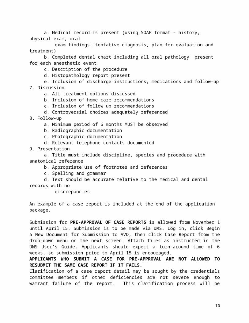
6. Complete & adequate medical record/dental charta. Medical record is present (using SOAP format – history, physical exam, oral exam findings, tentative diagnosis, plan for evaluation and treatment)b. Completed dental chart including all oral pathology present for each anesthetic eventc. Description of the procedured. Histopathology report presente. Inclusion of discharge instructions, medications and follow-up
7. Discussiona. All treatment options discussedb. Inclusion of home care recommendationsc. Inclusion of follow up recommendationsd. Controversial choices adequately referenced
8. Follow-upa. Minimum period of 6 months MUST be observedb. Radiographic documentation
6
c. Photographic documentationd. Relevant telephone contacts documented
9. Presentationa. Title must include discipline, species and procedure with anatomical referenceb. Appropriate use of footnotes and referencesc. Spelling and grammard. Text should be accurate relative to the medical and dental records with no discrepancies
An example of a case report is included at the end of the application package.
Submission for PRE-APPROVAL OF CASE REPORTS is allowed from November 1 until April 15. Submission is to be made via DMS. Log in, click Begin a New Document for Submission to AVD, then click Case Report from the drop-down menu on the next screen. Attach files as instructed in the DMS User’s Guide. Applicants should expect a turn-around time of 6 weeks, so submission prior to April 15 is encouraged. APPLICANTS WHO SUBMIT A CASE FOR PRE-APPROVAL ARE NOT ALLOWED TO RESUBMIT THE SAME CASE REPORT IF IT FAILS. Clarification of a case report detail may be sought by the credentials committee members if other deficiencies are not severe enough to warrant failure of the report. This clarification process will be mediated by the credentials chair or the secretary to maintain anonymity.
Letters of Evaluation:
Letters of evaluation AND the completed evaluation form are required from three (3) colleagues and shall be mailed directly by these individuals to:
Cindy Charlier, DVM, FAVD, Dip AVDC Phone 847-525-8642Secretary of the Academy of Veterinary Dentistry Fax 847-488-0705Fox Valley Veterinary Dentistry and Surgery Email [email protected] Stratford Lane, Elgin, IL 60124
Evaluators shall use the attached evaluation form. Evaluators are also REQUIRED to write a letter of evaluation. Evaluations should come from qualified professionals that are very familiar with veterinary dental techniques and procedures. Academy or College members who have personally observed your work are preferred and highly recommended. A dentist who has observed your work on several occasions could be acceptable. A general practitioner, who has referred multiple cases to you and has seen and followed the referred cases, could also be acceptable, but not as desirable. More weight is given to reference letters from dental experts than from other individuals.
7
APPLICANT/MENTOR ACCOUNTABILITY FORM
Anonymous submissions:Please white out all hospital name headings and references to the hospital or you in all of the documents in your application package. The chairperson of the credentials committee will hold the reference forms and letters of evaluation, the diploma, the state veterinary license and the agreement form. Please submit this signed letter from yourself and your mentor (see attached) stating that the submitted information is the candidate’s own work.
The chairperson will assign each application package a number and the packages will be evaluated anonymously by each committee member.
I hereby certify that the enclosed application package is my own work.
____________________________________Date______________________________Signed Candidate
I hereby certify that I have worked with this candidate in his/her application process and I certify that to the best of my knowledge the information contained in his/her application is correct, true, and his/her own work.
_____________________________________Date_____________________________Signed Mentor
Case report, case logs, and continuing education:
I hereby certify that I have reviewed the candidate’s case reports, case logs, continuing education, equipment list, and other requirements and I certify that to the best of my knowledge the information contained in his/her application is complete according to the current requirements.
_____________________________________Date_____________________________Signed Mentor
8
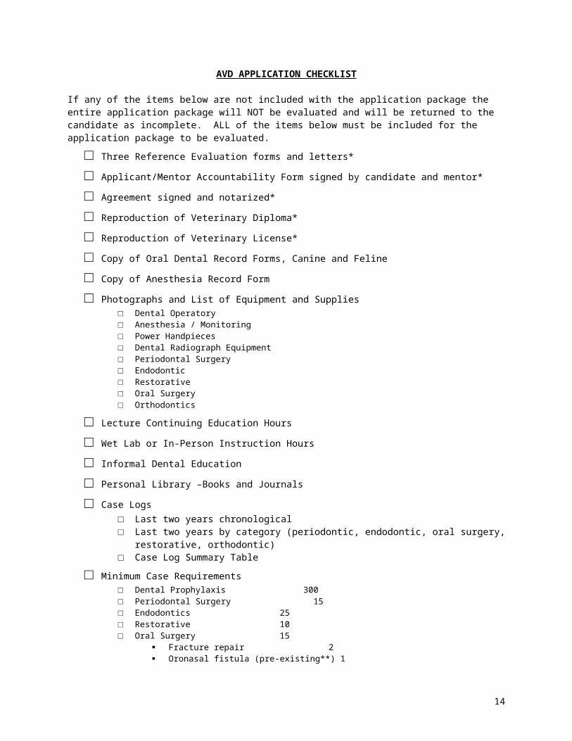
AVD APPLICATION CHECKLIST
If any of the items below are not included with the application package the entire application package will NOT be evaluated and will be returned to the candidate as incomplete. ALL of the items below must be included for the application package to be evaluated.□ Three Reference Evaluation forms and letters*
□ Applicant/Mentor Accountability Form signed by candidate and mentor*
□ Agreement signed and notarized*
□ Reproduction of Veterinary Diploma*
□ Reproduction of Veterinary License*
□ Copy of Oral Dental Record Forms, Canine and Feline
□ Copy of Anesthesia Record Form
□ Photographs and List of Equipment and Supplies□ Dental Operatory□ Anesthesia / Monitoring□ Power Handpieces□ Dental Radiograph Equipment□ Periodontal Surgery□ Endodontic□ Restorative□ Oral Surgery□ Orthodontics
□ Lecture Continuing Education Hours
□ Wet Lab or In-Person Instruction Hours
□ Informal Dental Education
□ Personal Library –Books and Journals
□ Case Logs□ Last two years chronological□ Last two years by category (periodontic, endodontic, oral surgery, restorative, orthodontic)□ Case Log Summary Table
□ Minimum Case Requirements□ Dental Prophylaxis 300□ Periodontal Surgery 15□ Endodontics 25□ Restorative 10□ Oral Surgery 15
Fracture repair 2 Oronasal fistula (pre-existing**) 1 Mxectomy/mnectomy 1 Major extractions 5
□ Orthodontics 15 Appliances cases 2
□ Radiology 100
□ Four Case Reports medical, dental and anesthesia records included (without clinic and applicant names) four reports in separate disciplines: no more than 10 pages of text author is the primary person performing the case pre-, intra- and post-procedure radiographs as indicated
9
requirements for follow-up are met photographic documentation pre-, intra-, post-procedure and follow-up: figures labeled and
captioned*documents held by committee chairperson to insure anonymous evaluation of application packages ** Pre-existing oronasal fistula is a a communication between the oral and nasal cavities due to developmental or traumatic reasons or associated with loss of a tooth. Surgical extraction of a tooth with a communicating pocket does not count as an ONF repair
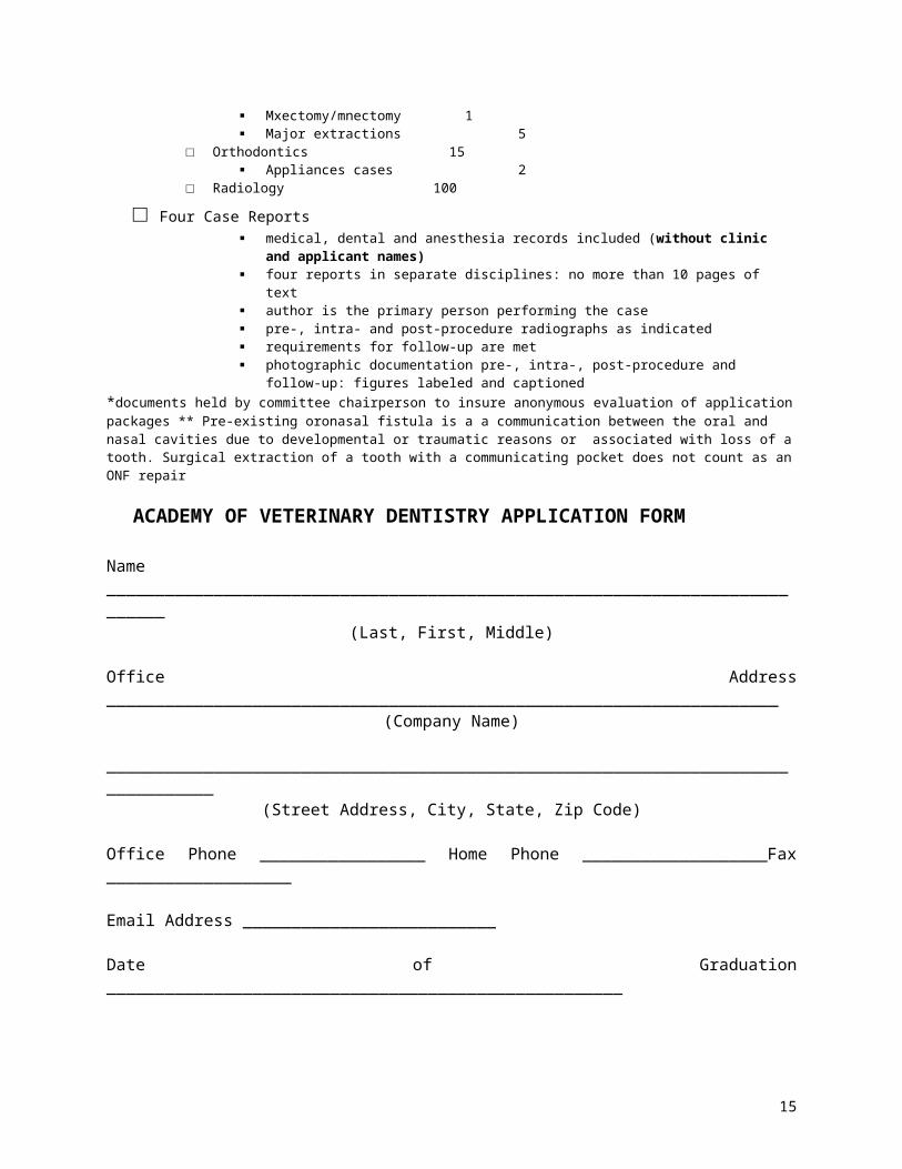
ACADEMY OF VETERINARY DENTISTRY APPLICATION FORM
Name ____________________________________________________________________________(Last, First, Middle)
Office Address _____________________________________________________________________(Company Name)
_________________________________________________________________________________(Street Address, City, State, Zip Code)
Office Phone _________________ Home Phone ___________________Fax ___________________
Email Address __________________________
Date of Graduation _____________________________________________________
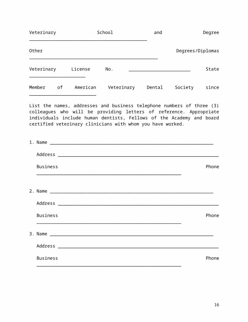
Veterinary School and Degree ____________________________________________
Other Degrees/Diplomas ________________________________________________
Veterinary License No. _______________________ State _____________________
Member of American Veterinary Dental Society since _________________________
List the names, addresses and business telephone numbers of three (3) colleagues who will be providing letters of reference. Appropriate individuals include human dentists, Fellows of the Academy and board certified veterinary clinicians with whom you have worked.
1. Name _____________________________________________________________
Address ____________________________________________________________
Business Phone ______________________________________________________
2. Name _____________________________________________________________
Address ____________________________________________________________
Business Phone ______________________________________________________
3. Name _____________________________________________________________
10
Address ____________________________________________________________
Business Phone ______________________________________________________
11
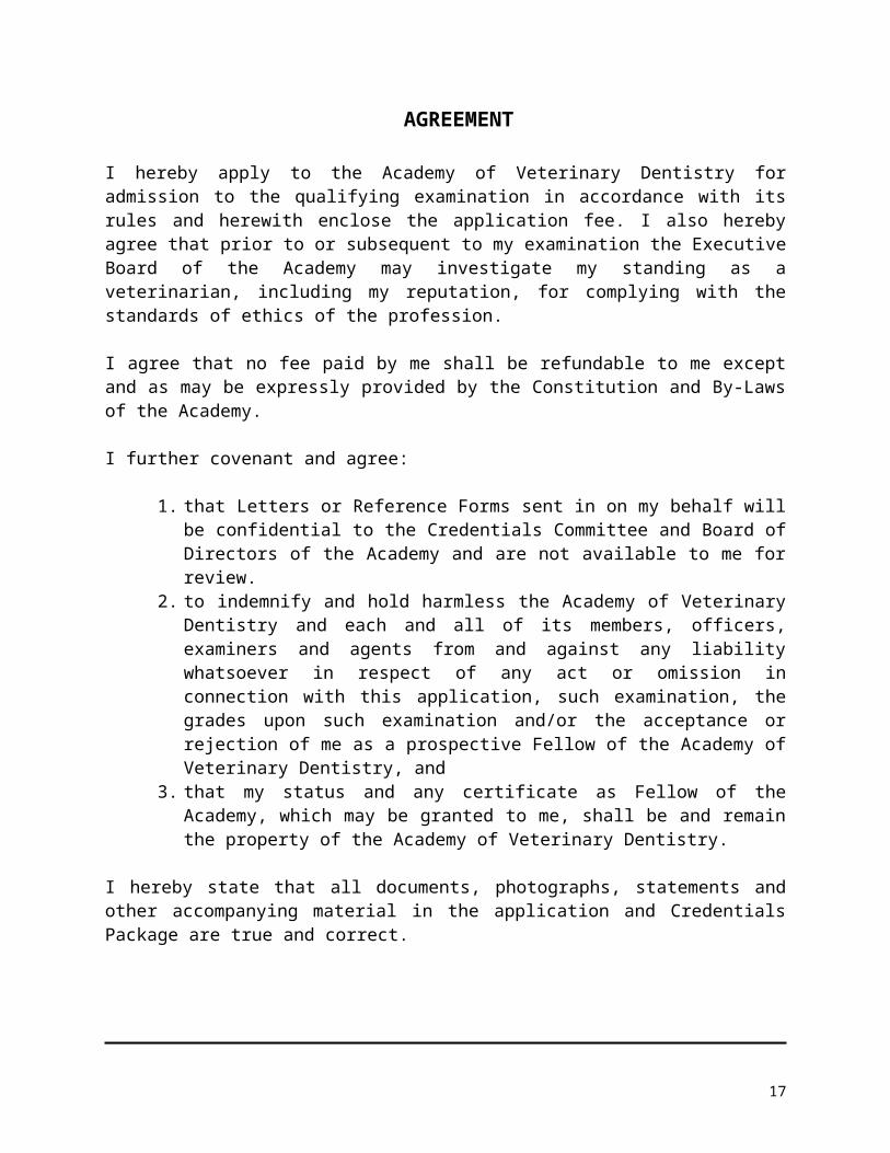
AGREEMENT
I hereby apply to the Academy of Veterinary Dentistry for admission to the qualifying examination in accordance with its rules and herewith enclose the application fee. I also hereby agree that prior to or subsequent to my examination the Executive Board of the Academy may investigate my standing as a veterinarian, including my reputation, for complying with the standards of ethics of the profession.
I agree that no fee paid by me shall be refundable to me except and as may be expressly provided by the Constitution and By-Laws of the Academy.
I further covenant and agree:
1. that Letters or Reference Forms sent in on my behalf will be confidential to the Credentials Committee and Board of Directors of the Academy and are not available to me for review.
2. to indemnify and hold harmless the Academy of Veterinary Dentistry and each and all of its members, officers, examiners and agents from and against any liability whatsoever in respect of any act or omission in connection with this application, such examination, the grades upon such examination and/or the acceptance or rejection of me as a prospective Fellow of the Academy of Veterinary Dentistry, and
3. that my status and any certificate as Fellow of the Academy, which may be granted to me, shall be and remain the property of the Academy of Veterinary Dentistry.
I hereby state that all documents, photographs, statements and other accompanying material in the application and Credentials Package are true and correct.
Signature
12

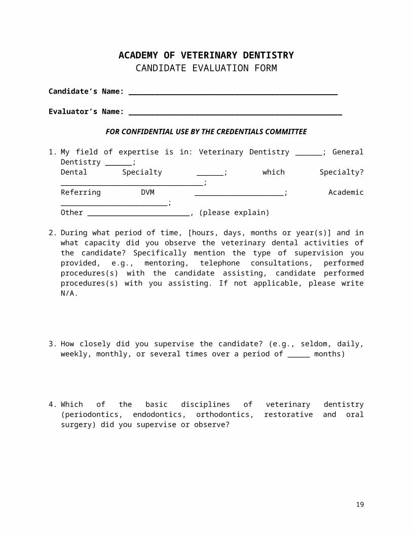
ACADEMY OF VETERINARY DENTISTRYCANDIDATE EVALUATION FORM
Candidate’s Name: _______________________________________________
Evaluator’s Name: ________________________________________________
FOR CONFIDENTIAL USE BY THE CREDENTIALS COMMITTEE
1. My field of expertise is in: Veterinary Dentistry ______; General Dentistry ______; Dental Specialty ______; which Specialty? ________________________________;Referring DVM ____________________; Academic ________________________; Other _______________________, (please explain)
2. During what period of time, [hours, days, months or year(s)] and in what capacity did you observe the veterinary dental activities of the candidate? Specifically mention the type of supervision you provided, e.g., mentoring, telephone consultations, performed procedures(s) with the candidate assisting, candidate performed procedures(s) with you assisting. If not applicable, please write N/A.
3. How closely did you supervise the candidate? (e.g., seldom, daily, weekly, monthly, or several times over a period of _____ months)
4. Which of the basic disciplines of veterinary dentistry (periodontics, endodontics, orthodontics, restorative and oral surgery) did you supervise or observe?
5. In terms of primary patient care responsibility, approximately how many cases were under the exclusive control of the candidate during your period of supervision or observation?
Not applicable ______ 6-10 cases ______
Zero cases ______ 11-25 cases ______
1-5 cases ______ Over 25 cases ______
13
6. Candidate’s knowledge and skills in veterinary dentistry – Please state: N/A, unknown, excellent, very good, satisfactory, needs improvement or unsatisfactory.
Attention to the patient as a whole _______
Knowledge of dental radiographic technique and interpretation _______
Proper management of veterinary dental cases _______
Proper use of techniques and materials which are generally accepted _______
Complete and adequate dental charting _______
Awareness of current literature _______
Ability to make independent decisions _______
7. Candidate’s characteristics. Please state: N/A, unknown, excellent, very good, satisfactory, needs improvement or unsatisfactory.
Reliability _______
Motivation _______
Attention to detail (follows manufacturers instructions exactly) _______
Client control and attitude _______
Professional ethical standards _______
8. Do you believe that the candidate has any characteristics of professional performance that would detract from the candidate’s fitness for membership in the Academy of Veterinary Dentistry? If so, please describe.
14
Date: ______________ Signed __________________________________
Print Name _______________________________
Address: _________________________________
City, State, Zip ____________________________
Telephone: _______________________________
FAX: ___________________________________
Please attach a letter of recommendation to support the candidate’s application for membership in the Academy. The Academy greatly appreciates your time and effort in writing this evaluation.
This form must be sent directly to and received at the Secretary’s office no later than midnight, July 15, 2011. If the postmark is prior to July 8, the form will be accepted even if delayed in transit.
Mail to:Cindy Charlier, DVM, FAVD, Dip AVDCSecretary of the Academy of Veterinary DentistryFox Valley Veterinary Dentistry and Surgery37W748 Stratford LaneElgin, IL 60124Phone 847-525-8642Fax 847-488-0705Email [email protected]
15
ACADEMY OF VETERINARY DENTISTRYSuggested Reading Material
The examination is not limited to the listed readings.1. All issues of The Journal of Veterinary Dentistry.2. Equine Dentistry, 2nd ed., Philadelphia. Elsevier Saunders, 2005.3. Bojrab MJ, Tholen M. Small Animal Oral Medicine and Surgery. Philadelphia: Lea and Febiger,
1990.4. Carranza FA. Glickman’s Clinical Periodontology, 7th ed. Philadelphia: WB Saunders, 1990.5. Cohen S, Burns RC. Pathways of the Pulp, 6th ed. St. Louis: Mosby-Year Book, 1994.6. Conference Proceedings of the AVDC/AVD annual meetings.7. Emily P, Penman S. Handbook of Small Animal Dentistry, 2nd ed. Oxford: Pergamon Press, 1994.8. Ettinger SJ, ed. Veterinary Internal Medicine, 4th ed. Philadelphia: WB Saunders, 1995.9. Hartsfield SM. Anesthetic problems of the geriatric dental patient. In: Manfra Marretta S, ed.
Problems in Veterinary Medicine: Dentistry. Philadelphia: JB Lippincott, March 1990.10. Harvey CE, Emily PP. Small Animal Dentistry. St. Louis: Mosby -Year Book, 1993.11. Harvey CE. Treatment planning for periodontal disease in dogs. JAAHA 1991;27(6):592-596.12. Harvey CE. Veterinary Dentistry. Philadelphia: WB Saunders, 1985. (out of print but very useful if
can get a copy)13. Haws IJ. Local dental anesthesia and pain management. CVDS Proc July 1999: 55-70.14. Holmstrom SE, Frost P, Eisner ER. Veterinary Dental Techniques for the Small Animal Practitioner,
3rd ed. Philadelphia: WB Saunders, 2004.15. Manfra Marretta S, ed. Problems in Veterinary Medicine: Dentistry. Philadelphia: JB Lippincott, Mar
1990.16. Miles AEW, Grigson C. Colyer’s Variations and Diseases of the Teeth of Animals. Cambridge:
Cambridge University Press, 1990.17. Mulligan TW, Aller MS, Williams CA. Atlas of Canine and Feline Dental Radiography, Trenton:
Veterinary Learning Systems, 1998.18. Paddleford RR, ed. Manual of Small Animal Anaesthesia. Philadelphia: WB Saunders, 1999.19. Plumb DC. Veterinary Drug Handbook, 3rd ed. White Bear Lake, MN: Pharma Vet, 1999.20. Proffit WR. Contemporary Orthodontics, 2nd ed. St. Louis: Mosby-Year Book, 1993.21. Wolf HF, Rateitschak EM, et al. Color Atlas of Dental Medicine: Periodontology. Stuttgart: Thieme,
2005.22. Schroeder HE. Oral Structural Biology. New York: Thieme, 1991.23. Schwartz R, Summit J, and Robbins J. Fundamentals of Operative Dentistry: A Contemporary
Approach. Chicago: Quintessence Books, 1996.24. Seymour C, Gleed R, eds. Manual of Small Animal Anaesthesia and Analgesia. Cheltenham:
BSAVA, 1999. 25. Slatter DH, ed. Textbook of Small Animal Surgery, 2nd ed. Philadelphia: WB Saunders, 1993. 26. Ten Cate AR, Oral Histology: Development, Structure, and Function, 4th ed. St. Louis: Mosby-Year
Book, 1994.27. Veterinary Clinics of North America: Equine Practice. Dentistry. 1988 Aug; 14(2).28. Veterinary Clinics of North America: Exotic Animal Practice. Oral Biology, Dental and Beak
Disorders. 2003 Sep; 6(3).29. Veterinary Clinics of North America: Small Animal Practice. Dentistry. 1986 Sep; 16(5).30. Veterinary Clinics of North America: Small Animal Practice. Dentistry. 1992 Nov; 22(6).31. Veterinary Clinics of North America: Small Animal Practice. Dentistry. 2005 Jul; 35(4).32. Wiggs RB, Lobprise HB. Veterinary Dentistry: Principles and Practice, Philadelphia: Lippincott-
Raven, 1997.
16
PARTIAL MAXILLECTOMY FOR TREATMENT OF A PAPILLARY SQUAMOUS CELL CARCINOMA IN A DOG
INTRODUCTION
Oral tumors are the fourth most common neoplasm in dogs representing approximately 6% of all
malignant tumors.1 Oral neoplasms have been treated with various modalities including surgical excision,
cryosurgery, radiotherapy, immunotherapy, or a combination of the above.2 Partial maxillectomy
techniques have been described which permit resection of tumors involving the upper palate while
maintaining function and acceptable cosmetic results.3 Thorough evaluation of the patient including
physical exam, bloodwork, thoracic and oral radiographs, and biopsy assist in determining the treatment
protocol for each individual patient.
SIGNALMENT AND HISTORY:
A 5-month-old male mixed breed dog, was referred on 11/7/75 for evaluation and treatment of an
oral neoplasm. On 10/13/75 the patient presented to the referring veterinarian for evaluation of an oral
mass. According to the medical records, the owner had first noticed the mass the day before initial
presentation. The patient was sent home on clindamycin hydrochloridea 75mg one capsule twice daily.
On 10/16/75 the patient returned to the referring veterinarian for reevaluation. At that time, right lateral
and ventrodorsal thoracic radiographs were taken which showed no evidence of metastatic disease.
Skull radiographs obtained at the same time were described by the referring veterinarian as having a
‘locular appearance to the right maxilla’. Although the right mandibular lymph node was not palpably
enlarged, a fine needle aspirate was obtained. The cytology results reported no evidence of atypical
cells. On 10/25/75 the referring veterinarian took intraoral radiographs and obtained a punch biopsy of the
mass. The histopathology report stated the mass was morphologically consistent with a well-
differentiated squamous cell carcinoma. The histopathology report described this biopsy as an example
of a syndrome of well-differentiated squamous cell carcinomas in very young dogs referred to as papillary
squamous cell carcinomas. According to the report, usually papillary squamous cell carcinomas are of
low-grade malignancy and if completely removed, the dogs can do fairly well. As they are well
differentiated, the chance for metastasis is low.
17

PHYSICAL EXAMINATION
On presentation the patient was bright, alert, responsive, and normally hydrated. The patient
weighed 21.4 kg. General physical examination was unremarkable. Oral examination confirmed the
presence of a 3 cm by 5 cm by 1 cm raised smooth mass in the right rostral maxilla. The mass had a 1
cm area of ulcerated surface surrounding the maxillary right intermediate incisor. The mass extended
from the mesial and palatal surface of the maxillary right central incisor to the distal side of the maxillary
right canine. A widened interproximal space was present between the maxillary right central incisor and
the maxillary right intermediate incisor with displacement of the right intermediate incisor laterally. A
widened interproximal space also existed between the maxillary right intermediate and maxillary right
lateral incisor displacing the maxillary right lateral incisor and canine tooth caudally and buccally. Grade 2
mobility of the right intermediate incisor was present. The mass extended palatally approximately 8 mm
caudally from the right maxillary incisor teeth and apically onto the gingival surface approximately 1 cm
(Pictures 1, 2). All other oral anatomy was within normal limits.
Oral radiographs from the referral veterinarian dated 10/25/75 revealed a large expansile
radiolucent lesion with well-demarcated borders. The lesion involved the supporting alveolar bone at the
roots of the maxillary right incisors and extended caudally to the level of the maxillary right canine tooth
(Radiographs 1, 2).
DIAGNOSIS
Based on history, physical examination, radiographs and biopsy results, tentative diagnosis of a
maxillary well-differentiated squamous cell carcinoma was made. This diagnosis is consistent with
papillary squamous cell carcinomas found in young dogs.
THERAPEUTIC PLAN
This malignancy had a histologically low grade, therefore, a good prognosis could be expected
with complete excision. A recommendation for a partial maxillectomy was made to the owners. Further
diagnostic evaluation was necessary which included bloodwork, a left lateral thoracic radiograph, and
current intraoral radiographs, prior to devising a plan for the maxillectomy. Anesthetic protocol, the
18
surgical procedure, postoperative care and potential complications were discussed with the owner.
Potential complications which were discussed included: dehiscence of the surgical site; hemorrhage
intraoperatively; inadequate resection of the mass; and impingement of the lower right canine tooth on the
upper lip. Because of the involvement of a significant amount of gingival tissue apical to the teeth within
the mass, resection with adequate margins was a concern in this case.
PROCEDURE
To complete the thoracic radiograph series taken by the referring veterinarian, prior to anesthesia
a left lateral thoracic radiograph was taken. There was no radiographic evidence of metastatic disease.
Preoperative complete blood count and serum biochemistry profile were completed and values were
within normal limits.
The patient was premedicated with medetomidine hydrochlorideb 0.009mg/kg, morphine sulfatec
0.55 mg/kg and atropine sulfated 0.04 mg/kg given intramuscularly. An 18 gauge intravenous catheter
was placed in the left cephalic vein. Cefazoline 22 mg/kg was administered intravenously. The patient
was induced with valiumf 0.15 mg/kg and ketamineg 2.8 mg/kg given intravenously. The patient was
intubated with a 10 mm cuffed endotracheal tube. Anesthesia was maintained with isoflurane (1.5 –
2.0%) and oxygen (0.6 liters/min). Intravenous Lactated Ringer’s Solutionh was administered throughout
the procedure at a rate of 10 ml/kg/hour. The patient was monitored intraoperatively with a continuous
electrocardiogram, continuous pulse oximetry, and indirect blood pressure readings every five minutes i.
Intraoral radiographs were obtained (Radiographs 3, 4). There was evidence of an expansile
bone lesion of the right rostral maxilla. It was mixed in opacity with areas of bone lysis. The lesion
appeared to approach but not cross the midline. Based on radiographs taken 10/25/75 and 11/7/75,
gross appearance of the tumor, and palpation of the tumor margins4 a resection was planned to extend
from the mesial side of the maxillary left lateral incisor to the mesial side of maxillary right third premolar
through the palate. The goal was to obtain a minimum of 1 cm of clinical and radiographic tumor free
margin. The planned excision would extend apically approximately 1 cm above the margin of the mass,
preserving enough buccal mucosa to close the oronasal defect.
19
To provide analgesia to the surgical area intraoperatively and postoperatively, right and left
maxillary nerve blocks were performed with marcaine 0.5% with epinephrine 1:200,000 j using a 27 gauge
1” disposable dental needle on an aspirating syringek. Approximately 0.3 cc of the marcaine was injected
at each maxillary site. The maxillary nerve block completely desensitizes the soft tissue, dentition and
bone in one maxillary quadrant.5,6 The patient was positioned in dorsal recumbancy with the head
supported and the mandible retracted caudally.7 The oral cavity was rinsed with 0.12% chlorhexidene
gluconate solutionl. The oral cavity was then isolated with sterile drapes. The palatal mucosa was
incised down to the incisive bone and palatine process of the maxillary bone with a #10 scalpel blade in a
line which extended from the mesial surface of the maxillary left lateral incisor to the mesial surface of
maxillary right third premolar at least 1 cm from the grossly visible tumor margins. In the area of the right
palatine artery the incision did not penetrate the palatal mucosa to the bone. The right major palatine
artery was identified, isolated and ligated with 3-0 polydioxanonem and then transected. The buccal
mucosa was incised approximately 1 cm apical to the margin of the tumor. A Freer periosteal elevatorn
was used to elevate the mucosa and underlying tissues from their attachment on the hard palate,
maxillary and incisive bones. The right infraorbital vessels were identified, isolated and ligated with 3-0
polydioxanone suturem and then transected. The soft tissue of the palate was dissected approximately 2-
3 mm beyond the planned resection border of the bone. An oscillating saw was utilized to transect the
bone from the mesial surface of the maxillary left lateral incisor to the mesial surface of the right maxillary
third premolar. A dorsal osteotomy was performed dorsal to all involved tooth roots through the maxillary
and incisive bones using the oscillating saw. The tumor and adjacent tissue including a small portion of
the nasal turbinates were then removed en bloc. Gelfoamo was placed in the right caudal nasal area to
control hemorrhage. All bone edges were rounded and smoothed with a 4 mm round burr in a Hall air
drill. A .045 k wire in a Jacob’s hand chuck was utilized to drill several holes in the palatine bone in a line
parallel to the incised bone edge 2-3 mm from the incised edge. The labial mucosa and submucosa was
separated from the remainder of the lip using Metzenbaum scissorsp for blunt and sharp dissection. The
lip margin based labial flap was created to allow for tension free closure of the oronasal defect. The
maxillary right third premolar and maxillary left lateral incisor were carefully inspected for any damage.
There was no visible damage to the teeth or tooth root structure and the tooth roots were visibly covered
20
by alveolar bone. The surgical area was copiously lavaged with warm sterile saline solutionq. 3-0
polydioxanonem simple interrupted sutures were placed from the buccal submucosal tissue to the holes
predrilled in the bony hard palate. The labial mucosa and palatine mucosa were apposed with 3-0
polydioxanonem sutures in a simple interrupted pattern (Picture 3). Occlusion was evaluated. The
mandibular right canine tooth was lateral to the upper lip and did not impact the incision (Picture 4). The
resected section of maxilla was submitted for histopathology to Colorado State University to confirm the
histologic diagnosis and assess for the presence of tumor free margins.3 Postoperative radiographs of
the maxilla showed normal anatomy at the resected margins (Radiographs 5, 6).
Morphine sulfatec 0.55mg/kg was administered intramuscularly approximately 15 minutes prior to
the cessation of anesthesia. Recovery from anesthesia was uneventful. The patient was placed on a
continuous morphine drip postoperatively (0.22 mg/kg/hour) and Lactated Ringer’s Solutionh was
continued at a maintenance rate of 2.75 ml/kg/hour postoperatively. An Elizabethan collar was placed on
the patient after anesthetic recovery. Postoperative PCV was 31 and total protein was 6.5 gms/100ml.
The PCV was to be reevaluated in 4 hours.
Four and a half hours postoperatively the patient appeared restless. He was given morphine
sulfatec 0.7 mg/kg intramuscularly and acepromazine maleater 0.02mg/kg intravenously. Five hours
postoperatively the PCV was 41 and the total protein was 5.5 gms/100ml. Six hours postoperatively the
patient received cefazoline 22 mg/kg intravenously and then it was discontinued.
The following morning, 11/8/75, the patient was bright, alert, very responsive and normally
hydrated. Physical examination was unremarkable. The incision appeared unchanged. The continuous
morphine drip was discontinued and oral carprofens 2.2 mg/kg twice daily was started. Twenty-four hours
postoperatively the patient was offered a slurry of Canine p/d t and water, which he ate readily. He was
given access to free choice water, which he was drinking. The intravenous Lactated Ringer’s Solutionh
was discontinued. The patient continued to eat a p/d t slurry every 6 – 8 hours. The patient remained
comfortable throughout the day and night.
The second day postoperatively, 11/9/75, the patient was very bright, alert, and responsive.
Physical examination was within normal limits and the incision appeared unchanged. The patient was
discharged to the client on 11/9/75 with the following instructions:
21
Wear the Elizabethan collar at all times
Continue to soften his food to a slurry consistency
Do not allow him to chew on anything; remove all toys from his environment.
Continue oral carprofens as directed for 5 days postoperatively
Return for reevaluation in 10 days
Preliminary histopathology results received on 11/10/75 reported ‘squamous cell carcinoma
extending into the nasal/sinus cavity but other margins are free of tumor’. Final written histopathology
results were received on 11/15/75. The histopathological diagnosis was ‘squamous cell carcinoma, well
differentiated’. Dr. Powers stated that it was ‘consistent with a papillary squamous cell carcinoma reported
in young dogs, however this tumor is more invasive than is usually seen with papillary squamous cell
carcinoma as there is extensive bone invasion. This tumor appears to be completely removed, although
the tumor does extend into the nasal and sinus cavity where there are no tissue margins, rather only air.
The caudal bone margin is free of tumor’.
FOLLOW UP
The patient returned 9 days later on 11/18/75. He was very happy and energetic. (Picture 5)
Physical examination was within normal limits. Oral examination showed the incision to be healing with
no areas of dehiscence. (Picture 6) There was no impingement of the mandibular right canine tooth on
the upper lip. The clients were very pleased with the cosmetic results of surgery. The Elizabethan collar
was removed. The owner was instructed to continue softened food and no chew toys for an additional
two weeks.
On 12/2/75, approximately 24 days postoperatively, the patient returned for reevaluation. He
weighed 22.7 kg. The owners reported that he was doing very well at home. He was eating his slurry
readily and was showing interest in playing with his stuffed toys. Physical examination was unremarkable.
Oral examination showed no evidence of dehiscence. The incision was healed and there was no visible
evidence of regrowth of the tumor. The clients were instructed to feed the patient’s normal diet of hard
food and recheck in 4 weeks for sedation, intraoral radiographs, and removal of any remaining sutures.
22
On 1/10/76, approximately two months postoperatively, the patient returned for reevaluation. His
owners reported a normal dog at home. He weighed 24 kg. Physical examination was unremarkable.
Oral examination showed no visual evidence of any tumor regrowth. A few sutures remained visible. The
patient was given atropine sulfated .04 mg/kg intramuscularly followed by medetomidine
hydrochlorideb .01 mg/kg and butorphanolu 0.1 mg/kg given intramuscularly twenty minutes later. A
thorough oral examination confirmed no gross evidence of tumor regrowth (Pictures 7, 8, 9). The
remaining sutures were removed and intraoral radiographs were taken. Radiographs showed normal
bony margins with no evidence of tumor regrowth (Radiograph 7). Atipamezole v 0.05 mg/kg was
administered intramuscularly. Recovery from sedation was uneventful. The owner was instructed to
return in 4 months for another follow up evaluation.
On 6/6/76, approximately seven months postoperatively, the patient returned for reevaluation. He
weighed 25 kg. His owners reported a happy normal dog. Physical examination was unremarkable. The
haired surface of his right upper lip had some brown discoloration present, likely due to saliva staining.
Oral examination showed no visible evidence of tumor regrowth. Thoracic radiographs (3 views) were
taken prior to sedation. There was no radiographic evidence of metastatic disease. Utilizing the above
protocol for sedation, thorough oral examination confirmed no visible evidence of tumor regrowth
(Pictures 10, 11, 12, 13). Intraoral maxillary radiographs were taken as well as radiographs of the
maxillary left lateral incisor and maxillary right third premolar. Intraoral radiographs, compared with prior
radiographs showed further remodeling of the bone margins and no areas of abnormal bone. Lateral
oblique skull radiographs showed remodeling of the osteotomy site with no abnormal bone lysis or
production visible. Radiographs of the maxillary left lateral incisor and maxillary right third premolar
showed normal tooth crown and root structure as well as normal surrounding alveolar bone (Radiographs
8, 9, 10, 11). The client was instructed to return in three months (10 months postoperatively) for
reevaluation and radiographs. Another reevaluation would be scheduled for one year postoperatively and
then rechecks were recommended yearly thereafter.
23
DISCUSSION
Oral tumors are the fourth most common neoplasm in dogs, representing approximately 6% of all
malignant tumors.1 The most common types of malignant oral neoplasms in dogs include melanoma,
squamous cell carcinoma, and fibrosarcoma.8 Squamous cell carcinoma is the second most common
oral malignancy in dogs after malignant melanoma.8 Squamous cell carcinomas usually occur in older
dogs (the average age is nine years).8 They are locally invasive but have a low rate of distant
metastatsis.1 Young age, rostral location and maxillary site carry a better prognosis for survival.8
Oral papillary squamous cell carcinomas have been reported in dogs as young as two months of
age.9 Papillary squamous cell carcinomas are essentially squamous cell carcinomas which are well
differentiated, sharply delineated and locally invasive.9 Papillary squamous cell carcinoma is a
progressive disease with a high rate of bone lysis.8 Although papillary squamous cell carcinomas are
locally invasive, they do not tend to metastasize.10 An association between papillary squamous cell
carcinoma and papilloma virus infection has not been determined.9
The initial approach to the management of an oral tumor should include histologic diagnosis via
biopsy and clinical tumor staging.1 Preanesthetic blood work should be obtained to assess the general
health of the patient. After a histologic diagnosis of malignancy has been established, clinical staging
should include three thoracic radiographic views to detect distant metastasis.1 Any local
lymphadenopathy should be further investigated by fine needle aspiration.8 The extent of bone
involvement or local aggressiveness of the tumor can be determined by imaging with conventional skull
radiographs.1 The intraoral view is often the most informative and dental radiographs provide valuable
informaton.1 If possible, more precise tumor evaluation can be accomplished using advanced imaging
techniques (computed tomography, magnetic resonance imaging) which may facilitate surgical and
radiation treatment plannning.1
With oral tumors, the first surgical excision is the most likely to result in tumor control. 8 The tumor
should not be scraped or peeled from the underlying bone, as recurrence is certain and the tumor bed will
be enlarged.8 A definitive first surgery, such as a maxillectomy or mandibulectomy should be performed.8
Partial maxillectomy involves excision of portions of the maxilla, incisive bone or palatine bone.11 Partial
maxillectomy is indicated for excision of malignant oral tumors and benign oral tumors that involve bone
24
or periosteum, such as the epulides and ameloblastoma.11 Other indications for partial maxillectomy
include chronic osteomyelitis, oronasal fistula, and maxillary fractures with severe bone injury or loss. 12
Application of this technique is limited by tumor extension into the labial or buccal mucosa or across the
midline of the central or hard palate.11 Sufficient normal labial or buccal mucosa and hard palate
mucoperiosteum must be available to allow closure of the oronasal defect that results.11 Adherence to the
following principles is important in any type of maxillectomy:
Use of sharp dissection when incising labial, buccal and palatal mucosa Maintenance of adequate blood supply to the mucosal flap used to cover the oronasal defect
resulting from surgery Use of a two layer closure when possible Avoidance of excessive tension across the incision line Establishment of at least a 1 cm border of normal healthy tissue between the tumor and the
line of resection.7
Careful preoperative planning is important to determine if adequate surgical margins can be achieved
and to ensure that the resulting oronasal defect can be closed primarily.11 The limits for surgical
resection of a malignancy should be determined by preoperative imaging, gross visualization of the
tumor and palpation of the tumor at the time of surgery.3
The goal of any partial maxillectomy in the treatment of oral neoplasia should be to obtain a
minimum of 1 cm of clinical and radiographic tumor free margin.12 Perioperative antibiotics are
recommended.12 Antibiotic therapy for more than 24 hours is not indicated unless dictated by the
situation.12 The antibiotic chosen should be effective against the bacterial flora normally found in the oral
cavity. The first generation cephalosporins, penicillins, and synthetic penicillins are generally considered
effective prophylactic oral antibiotics.12
During the procedure ligation of the infraorbital and major palatine vessels to control hemorrhage
does not have any adverse effects. With ligation of the infraorbital artery collateral circulation is
maintained to the labial mucosa via the facial artery and contralateral infraorbital artery. The left and right
major palatine arteries have extensive anastamoses so mucosal circulation can be maintained adequately
by the contralateral vessel.1
Polydioxanone is one of the sutures recommended for wound closure after maxillectomy.12
Polydioxanone is a relatively nonreactive suture that minimizes oral mucosal irritation and maintains
adequate strength during the critical early period of healing.12 It is also monofilament and absorbable. Its
25
absorption is slow and the sutures may result in irritation of the oral mucosa after healing. 12 In this case
all sutures remaining after healing were removed 2 months postoperatively.
Because of the aggressiveness of maxillectomy procedures, the animal should be supported for
the first 24 hours with parenteral fluids and analgesics.12 Intravenous fluid therapy is continued until the
animal is eating and drinking well enough to maintain its hydration.11 The patient is offered soft food and
water the day after surgery.11 An Elizabethan collar is often necessary to prevent self-induced trauma to
the surgical site.12 With a partial maxillectomy, the animal is usually discharged from the hospital when it
is eating well. Postoperative care includes the feeding of softened food for one month and preventing the
pet from chewing on hard objects for that same time period.11
A major postoperative complication of any maxillectomy is partial suture line dehiscence.13 Major
causes of dehiscence include: suture line tension, tumor cells in the edges of the incision, ischemic
necrosis of the mucosal flap and excessive movement of the flap.4 Anemia is also a potential
complication of any type of maxillectomy.4 Intraoperative hemorrhage in this case was controlled by
careful isolation and ligation of the infraorbital and major palatine vessels. The preoperative packed cell
volume which was 41 dropped to 31 immediately postoperatively. This drop in hematocrit was not
unexpected and may have been due to hemodilution due to intravenous fluids intraoperatively in
combination with intraoperative blood loss. The hematocrit was monitored postoperatively and returned
to 41 before the patient was released.
Another potential complication is damage to teeth adjacent to the osteotomy site. If the teeth are
close together, osteotomy may be difficult to perform without entering the alveolus of the adjacent tooth.11
Careful inspection of the teeth at the time of surgery is important. Intraoral radiographs are necessary to
detect iatrogenic trauma to adjacent teeth. Deformity of the muzzle contour can occur after partial
maxillectomy and repair with a labial mucosal-submucosal flap.12 Such indentation usually results from
an insufficient amount of normal labial tissues to create the flap and the problems that may cause. This
indentation was present in our patient, but he was unaffected by it.
Preemptive analgesia refers to the application of analgesic techniques before exposing the
patient to noxious stimuli.5 Multimodal analgesia is accomplished by preemptive administration of a
combination of different classes of drugs that inhibit nociceptive processes at two or more sites.5 The use
26
of an opioid (morphine) and alpha2 agonist (medetomidine) in addition to local nerve blocks allowed
multimodal preemptive analgesia to be achieved in this case. Pain management was continued with an
injection of morphine before anesthetic recovery followed by continuous morphine infusion for the first 24
hours postoperatively and additional analgesics as needed based on patient evaluation. Oral carprofenn
was prescribed for postoperative inflammation and discomfort.
Papillary squamous cell carcinoma is a type of squamous cell carcinoma that occurs in young
dogs. Rostrally located squamous cell carcinomas of the mandible and maxilla are usually locally
aggressive but have a low metastatic potential.14 Therefore, radical surgery, radiation therapy or a
combination of surgery and radiation therapy is considered the most appropriate form of treatment with a
generally good prognosis for long term survival in these dogs.14 Ogilvie reported on three dogs with
papillary squamous cell carcinomas with disease free intervals of 39 months, 32 months, and 10 months
after surgery and radiotherapy.9 To date there have not been any studies or case reports on long-term
survival or prognosis with surgical resection as the sole treatment for papillary squamous cell carcinomas.
Further work is necessary to correlate treatment and survival times in young dogs with papillary
squamous cell carcinomas.
27
aAntirobe, Pharmacia and Upjohn Company, Kalamazoo, MIbDomitor, Pfizer Animal Health, Exton, PAcMorphine, Elkins-Sinn, Inc., Cherry Hill, NJdAtropine Sulfate, Phoenix Pharmaceutical, Inc., St. Joseph, MOeCefazolin, Schein Pharmaceutical, Inc., Florham, NJfDiazepam, Abbott Laboratories, North Chicago, ILgKetaset, Fort Dodge Animal Health, Fort Dodge, IAhLactated Ringer’s Solution, Abbott Laboratories, North Chicago, ILiDRE ASM 5000, DRE Inc., Louisville, KYjMarcaine 0.5%, Abbott Laboratories, North Chicago, ILkAspirating Syringe,Henry Schein, Port Washington, NYlCHX guard, VRx Pharmaceuticals, Harbor City, CAmPDS II, Ethicon, Summerville, NJnFreer periosteal elevator, Spectrum Surgical Instruments, Stow, OHoGelfoam, Pharmacia and Upjohn Company, Kalamazoo, MIpMetzenbaum scissors, Spectrum Surgical Instruments, Stow, OHq0.9% sterile saline, Abbott Laboratories, North Chicago, ILrAcepromazine, Boehringer Ingelhelm Vetmedica, Inc., St. Joseph, MOsRimadyl, Pfizer Animal Health, Exton, PAtCanine p/d, Hill’s Pet Nutrition, Inc., Topeka, KSuTorbugesic, Fort Dodge Animal Health, Fort Dodge, IAvAntisedan, Pfizer Animal Health, Exton, PA
28

REFERENCES
1Dhaliwal RS, Kitchell BE, Marretta SM: Oral tumors in dogs and cats. Part 1. Diagnosis and clinical signs. Compend Contin Educ Pract Vet 20(9);1011-1022, 1998.
2 Schwartz PD, Withrow SJ, Curtis CR, Powers BE, Straw RC: Mandibular resection as a treatment for oral cancer in 81 dogs. J Am Anim Hosp Assoc 27; 601-610, 1991.
3 Schwartz, PD, Withrow SJ, Curtis CR, Powers BE, Straw RC: Partial maxillary resection as a treatment for oral cancer in 61 dogs. J Am Anim Hosp Assoc 27; 617-24, 1991.
4 Wallace J, Matthiesen D, Patnaik A: Hemimaxillectomy for the treatment of oral tumors in 69 dogs. Vet Surg 21(5); 337-341, 1992.
5Thurmon JC, Tranquilli WJ, Benson GJ: Essentials of Small Animal Anesthesia and Analgesia. Baltimore: Lippincott Williams and Wilkins, 28-60, 206, 1999
6Haws, IJ; Local Dental Anesthesia. AVDF Conference Procedings 392-395, 2000.
7Marretta SM: Maxillofacial Surgery. In Vet Clin N Am Small Anim Pract, 28(5); 1285-1296, 1998.
8Ogilvie GK, Moore AS: Managing the Veterinary Cancer Patient: A Practice Manual. Trenton: Veterinary Learning Systems Co., Inc. 327-328, 336-339,479, 1998.
9Ogilvie GK, Sundberg JP, O’Barion K, Badertscher RR, Wheaton LG, Reichmann ME: Papillary squamous cell carcinoma in three young dogs. J Am Vet Med Assoc 192(7); 933-936, 1998. 10Stapleton BL, Barrus JM: Papillary Squamous Cell Carcinoma in a Young Dog. J Vet Dent 13(2):65-68, 1996.
11Salisbury SK: Maxillectomy and Mandibulectomy. In Textbook of Small Animal Surgery (Ed. DH Slatter), 2nd ed. Philadelphia: W.B. Saunders Company, 521-524, 1993.
12Dernell WS, Schwarz PD, Withrow SJ: Maxillectomy and Premaxillectomy. In Current Techniques in Small Animal Surgery (Ed. MJ Bojrab), 4th ed. Baltimore: Williams and Williams, 124-131, 1998.
13Dhaliwal RS, Kitchell BE, Marretta SM: Oral tumors in dogs and cats. Part 2. Prognosis and treatment. Compend Contin Educ Pract Vet 20(10):1109-1119, 1998.
14Schmidt BR, Glickman NW, DeNicola DB, Gortari AE, Knapp DW: Evaluation of piroxicam for the treatment of oral squamous cell carcinoma. J Am Vet Med Assoc 218(11): 1783-1786, 2001.
29
Dental Abbreviations
3D Tertiary Dentin GH Gingival Hyperplasia/ Hypertrophy PLQ PlaqueAB Abrasion GI Gingivitis Index PG Periodontal Pocket, Gingival/PseudoACY Acrylic GLS Glossitis PP Periodontal PocketADD Polylactic Acid Implant GM Gingival Margin PRO Complete Dental ProphylaxisAL Attachment Loss GP Gutta Percha PS Periodontal SurgeryAP Alveoloplasty GP/GV Gingivectomy/ Gingivoplasty PSB Periodontal Pocket, SuprabonyAPG Apexogenesis GR Gum Recession PTD Palatal Trauma DefectAPX Apexification GTR Guided Tissue Regeneration PXB Posterior CrossbiteAS Apical Sealer/ Cement IDW Interdental Wiring R/A Restoration, AmalgamAT Attrition IFA Inferior Alveolar Local Nerve Block R/C Restoration, CompositeAXB Anterior Crossbite HT Hairy Tongue RAD RadiographBE Biopsy, Excisional IFO Infraorbital Local Nerve Block RC Root CanalBFR Buccal Fold Removal IL Inlay R/I Restoration, IonomerBG Bone Graft IMP Implant RCS Root Canal, SurgicalBI Biopsy, Incisional IM Impression RD Retained DeciduousBKT Bracket INT Intrusion RL Resorptive LesionBL Bone Loss/ Recession IO Interceptive Orthodontics RE Root ExposureBP Bridge Pontic IOD Interceptive Orthodontics, Deciduous RP Root PlaningBR Bridge IOP Interceptive Orthodontics, Permanent RPC Root Planing, ClosedBRC Bridge, Cantilever LFD Lip Fold Dermatitis RPO Root Planing, OpenBRM Bridge, Maryland LIP Local Infiltration of Palate ROT Rotated ToothBUC Buccal Local Nerve Block LPS Lymphocytic-Plasmacytic stomatitis RR Root ResorptionCA Cavity, Fracture, Defect ( 1-8 ) M Mobile Tooth RRT Retained Root TipCAL Calculus MAL Malocclusion RRX Root Resection ( Hemisection )CAM Crown Amputation MAX Maxillary Local Nerve Block S SuturingCBU Core Build-Up MEN Mental Local Nerve Block SAL Salivary Gland ( S, M, P, Z, Mo )CFL Cleft Lip MGM Mucogingival Margin SBI Sulcular Bleeding IndexCFP Cleft Palate MM Mucous Membrane SC Subgingival CurettageCFP/R Cleft Palate Repair MN/FX Mandibular Fracture SE Stain, ExtrinsicCFW Circumferential Wiring MX/FX Maxillary Fracture SI Stain, IntrinsicCM Crown Metal NE Near Exposure SL SublingualCMG Crown Metal, Gold NV Non-Vital Tooth SLE Systemic Lupus ErythematosusCMO Craniomandibular Osteopathy O Missing Tooth SM Surgery, MandibulectomyCR Crown OA Orthodontic Appliance SN SupernumeraryCS Culture and Sensitivity OAI Orthodontic Appliance, Install SP Surgery, PalateCT Citric Acid Treatment OAA Orthodontic Appliance, Adjust SPL SplintCU Contact Ulcer OAR Orthodontic Appliance, Remove STM StomatitisCUL Culture OAF Oroantral Fistula SUL SulcusCWD Crowded Tooth OC Orthodontic Consultation SX Surgery, MaxillectomyDB Dentinal Bonding OI Osseous Implant SYM SymphysisDC Dilacerated Crown OL Onlay SYM/S Symphysis/ SeparationDCT Dentigerous Cyst OM Oral Mass TA Tooth AvulsedEC Elastic Chain OM/ADC OM/ Adenocarcinoma TIP TippingED Enamel Defect OM/FS OM/ Fibrosarcoma TL Tooth LuxatedEG Eosinophilic Granuloma OM/LS OM/ Lymphosarcoma TMJ/ DP TMJ DysplasiaEH Enamel Hypocalcification OM/MM OM/ Malignant Melanoma TMJ/ DL TMJ DislocationEP Epulis OM/SCC OM/ Squamous Cell Carcinoma TMJ/L TMJ LuxationEP/A Acanthomatous Epulis ONF Oronasal Fistula TMJ/FX TMJ FractureEP/F Fibrous Epulis ONF/R Oronasal Fistula Repair TN Treatment NeededEP/G Giant Cell Epulis OP Odontoplasty TP Treatment PlanningEP/O Ossifying Epulis OR Orthodontic Recheck TRANS Translocation ( Bodily Movement )EXT Extrusion OST Osteomyelitis TRX Tooth Resection ( Hemisection )FAR Flap, Apically Repositioned OSW Osseous Wiring VER VeneerFB Foreign Body PAP Papillomatosis VP Vital PulpotomyFCR Flap, Coronally Repositioned PCD Pulp Capping, Direct VT Vital ToothFE Furcation Exposed PCI Pulp Capping, Indirect VWD Von Willebrand's DiseaseFEN Flap, Envelope PCT Perioceutic Therapy W1 One Walled Bony PocketFFR Flap, Full Releasing PD Palatal Defect, or Periodontal
disease index when followed by #1-4W2 Two Walled Bony Pocket
FG Fluoride Gel PDL Periodontal Ligament W3 Three Walled Bony PocketFGG Free Gingival Graft PE Pulp Exposure W4 Four Walled Bony Pocket (cup)FLS Flap, Lateral Sliding PEM Pemphigus WIR WireFRB Flap, Reverse Bevel P&FS Pit and Fissure Sealant WRY Wry biteFRE Frenectomy PFM Porcelain Fused to Metal X Extraction, ElevationFRN Frenotomy PH Pulp Hemorrhage XS Extraction, SectionedFV Fluoride Varnish PI Plaque Index XSS Extraction, SurgicalFX Fracture ( Tooth, Jaw... ) PIB Periodontal Pocket, Infrabony ZOE Zinc Oxide EugenolGCF Gingival Crevicular Fluid PLT Palate