Embed Size (px)
Citation preview

de novo Design of Non-Steroidal Oestrogen Receptor Modulating Molecules using Maltanedienol as a Lead Molecule
Maria Cassar1*, Claire Shoemake1, Lilian M Azzopardi1, Charles Saliba2 and Gilles Gutierrez2 1Pharmacy Department, University of Malta, Msida MSD 2080. 2Instituteof Cellular Pharmacology LTD, F24, Mosta Technopark, Mosta, MST 3000 Malta.
ABSTRACT: Padina pavonica (L). is a brown alga endemic to the Mediterranean Sea. One of its components- maltanedienol, has been shown to be capbale of in vivo calcium fixation. Despite having a non-steroidal scaffold, maltanedienol still bears a marked resemblance to 17-β oestradiol, the endogenous ligand for the Oestrogen Receptor (ER). The ER is an important target for the management of conditions particularly osteoporosis and breast cancer. This study focuses on examining the ability of maltanedienol to modulate the oestrogen receptor ligand binding domain, and to consequently design novel structures which could have potential in management of osteoporosis and breast cancer.
Introduction Padina pavonica (L.) is an alga indigenous to the Atlantic Ocean and Mediterranean Sea1. It has attracted interest owing to the fact that despite belonging to a family of brown alga2 (Phaephycea)3,4 its appearance is white due to the deposition of calcium chloride on its surface in the form of aragonite crystals5. It is known however that aragonite could not be formed spontaneously in the Mediterranean owing to the fact that aragonite formation requires the presence of specific biological agents under stringent conditions of temperature and pressure5. This led to the hypothesis that Padina pavonica (L.) was capable of synthesising and emitting a substance that had an effect on calcium fixation5.
The main extract of Padina pavonica (L.) (EPP) has been resolved and registered as Maltanedienol5, (refer to figure 1). Its empirical formula is C28H44O2
5. Its structure is symmetrical with two 6 carbon rings creating a plane of attachment terminating in a 5 carbon structure at each end, creating an overall 3D orientation similar to that of a primitive steroidal molecule. This characteristic central symmetry is typical of a number of primitive compounds such as the tetraterpenes and the carotenoids5.
In vivo studies support the hypothesis that Maltanedienol was capable of calcium fixation. Specifically, it was observed that the molecule increased the fixation of calcium by osteoblasts also in the presence of a calcium channel inhibitor or Interleukin-1 (IL-1), implying that calcium fixation does not occur through the oestrogen receptors (ER) α and β 2, 5. Further competitive
in vivo assays confirmed the non-competitive relationship between Maltanedienol and 17-β oestradiol5.
Fig. 1 The chemical structure of Maltanedienol. The dotted line indicates the symmetrical axis of the
molecule. Figure drawn using Symyx® Draw v3.2b.
This study considers maltanedienol from a different perspective. Maltanedienol is a non-steroidal molecule that still bears a marked similarity to17-β oestradiol, the endogenous ligand for the ER. The ER remains a valid target for the management of conditions that include osteoporosis and breast cancer. Furthermore, maltanedienol is known to possess 4 isomers5, which increases interest in this molecule from a drug design point of view. This in silico study consequently aimed to identify whether or not maltanedienol binds with high affinity to the Oestrogen Receptor Ligand Binding Pocket (ER_LBP). Subsequently, multiple high affinity conformations of this molecule for the ER_LBP were generated computationally and finally, de novo design of maltanedienol-based structures with potential clinical activity in the management of osteoporosis and breast cancer was performed.
International Journal of Drug Design and Discovery
Volume 4 • Issue 1 • January – March 2013. 965-977
* For correspondence: Maria Cassar, Tel: (+365)79323189 Email: [email protected]
965
966 International Journal of Drug Design and Discovery Volume 4 • Issue 1 • Januart – March 2013
Methodology Creation of baseline in silico predicted binding affinity (pKd) data for ligands of known affinity for the ER
Three PDB crystallographic depositions describing the holo –ER (bound to 17-β oestradiol (1A526), raloxifene (1ERR7) and 4-hydroxytamoxifen (3ERT8) resolved to 2.80 Å, 2.60 Å and 1.9 Å respectively, were selected and the in silico predicted Ligand Binding Affinity (LBA) (pKd) of the endogenous agonist ligand 17-β oestradiol and of the established Selective Oestrogen Receptor Modulators (SERMs) raloxifene and 4-hydroxytamoxifen, for the ER was calculated.
All molecular modelling was carried out using Sybyl®-X version 1.19. The selected crystallographic depositions were edited such that an entire holo- monomer was removed in the cases of the dimeric depositions 1A526 and 1ERR7 followed by the removal of water molecules at a distance ≥ 5Å from the ER_LBP for each deposition. In the case of deposition 1A526 6 co-crystallised gold atoms were also eliminated. For deposition 1ERR7 the co-crystallised carboxymethylated cysteine was removed.
This editing process was followed by extraction of the bound ligands from their cognate receptors and estimation of the binding affinity (pKd) for their respective ER_LBP conformation according to the generic algorithm embedded in X-Score version 1.310. The predicted in silico LBA (pKd) data (refer to table 1) obtained set a baseline against which the predicted in silico predicted LBA (pKd) of the various conformations of maltanedienol and of the de novo designed ligands based on its structure could be compared.
In silico estimation of the LBA (pKd) of maltanedienol for the 17-β oestradiol, raloxifene and 4-hydroxy-tamoxifen bound conformations of the ER
In this part of the study, the predicted in silico binding affinity of maltanedienol for 3 distinct conformations of the ER, specifically those delineated by 17-β oestradiol, raloxifene and 4-hydroxytamoxifen was calculated. This was done in order to identify which ER conformation represented the best three- dimension environment with which maltanedienol could interact most successfully.
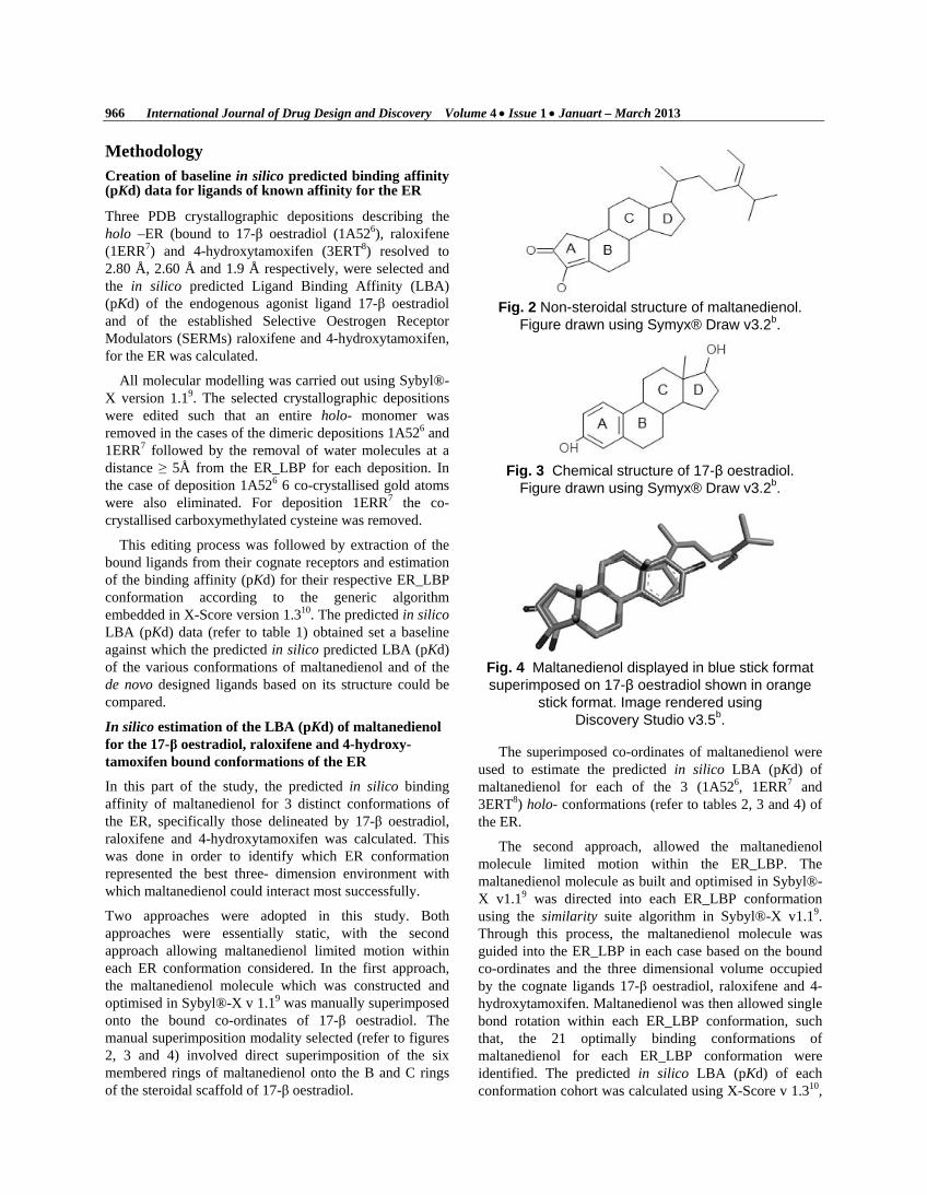
Two approaches were adopted in this study. Both approaches were essentially static, with the second approach allowing maltanedienol limited motion within each ER conformation considered. In the first approach, the maltanedienol molecule which was constructed and optimised in Sybyl®-X v 1.19 was manually superimposed onto the bound co-ordinates of 17-β oestradiol. The manual superimposition modality selected (refer to figures 2, 3 and 4) involved direct superimposition of the six membered rings of maltanedienol onto the B and C rings of the steroidal scaffold of 17-β oestradiol.
Fig. 2 Non-steroidal structure of maltanedienol.
Figure drawn using Symyx® Draw v3.2b.
Fig. 3 Chemical structure of 17-β oestradiol.
Figure drawn using Symyx® Draw v3.2b.
Fig. 4 Maltanedienol displayed in blue stick format superimposed on 17-β oestradiol shown in orange
stick format. Image rendered using Discovery Studio v3.5b.
The superimposed co-ordinates of maltanedienol were used to estimate the predicted in silico LBA (pKd) of maltanedienol for each of the 3 (1A526, 1ERR7 and 3ERT8) holo- conformations (refer to tables 2, 3 and 4) of the ER.
The second approach, allowed the maltanedienol molecule limited motion within the ER_LBP. The maltanedienol molecule as built and optimised in Sybyl®-X v1.19 was directed into each ER_LBP conformation using the similarity suite algorithm in Sybyl®-X v1.19. Through this process, the maltanedienol molecule was guided into the ER_LBP in each case based on the bound co-ordinates and the three dimensional volume occupied by the cognate ligands 17-β oestradiol, raloxifene and 4-hydroxytamoxifen. Maltanedienol was then allowed single bond rotation within each ER_LBP conformation, such that, the 21 optimally binding conformations of maltanedienol for each ER_LBP conformation were identified. The predicted in silico LBA (pKd) of each conformation cohort was calculated using X-Score v 1.310,

Maria Cassar et al : de novo Design of Non-Steroidal Oestrogen Receptor Modulating Molecules using… 967
while Ligand Binding Energy (LBE) (kcal mol-1) calculations were performed in Sybyl®-X v1.19(refer to tables 2,3 and 4).
Graphs of LBA (pKd) and LBE (kcal mol-1) for each conformer were plotted (refer to graphs 1, 2 and 3) and the conformation with the highest LBA (pKd) and lowest LBE (kcal mol-1) was identified (refer to figure 5). In the case of the 21 highest affinity conformations of maltanedienol for the 17-β oestradiol bound conformation of the ER_LBP, conformer No. 14 was selected as the ideal template for further study. Conformer selection was carried out through identification of greatest peak distance between LBA (pKd) and LBE (kcal mol-1) with high affinity and low binding energy being considered as desirable properties. This principle was also applied in the case of the 21 highest affinity conformations of maltanedienol for the raloxifene and 4-hydroxytamoxifen bound conformations of the ER_LBP.
Fig. 5 Superimposition of the 3 highest affinity
conformations of maltanedienol for the 17-β oestradiol, raloxifene and 4-hydroxytamoxifen bound
conformation of the ER_LBP, shown in pink, blue and green stick format representation respectively.
Image rendered by Discovery Studio v 3.5c.
The maltanedienol conformation with the highest predicted in silico LBA (pKd) and lowest LBE (kcal mol-1) combination for the raloxifene bound conformation of the ER_LBP was selected as a template. Selection of this optimally binding conformation, as opposed to its counterparts which were based on the bound co-ordinates of 17-β oestradiol and 4-hydroxytamoxifen was made on the premise that 17-β oestradiol had agonist activity and that while both 4-hydroxtamoxifen and raloxifene are classified pharmacologically as SERMs311, maltanedienol was more structurally similar to raloxifene than to 4-hydroxytamoxifen.
Consequently, this conformation of maltanedienol was used in order to create seed fragments for the de novo design phase of the study. Seed creation was carried out in Sybyl®-X v 1.19. Two seed structures were designed. In the case, of the first seed structure, (seed A), the A and B rings of maltanedienol together with 2 sp3 carbon linkers to the C ring were maintained (refer to figure 6).
Fig. 6 Seed A drawn using Symyx® Draw v3.2b.
The second seed structure (seed B) was made up of two distinct fragments A and B. Fragment A was identical to seed A , while fragment B was essentially the side chain that was attached to the 16 position of the D ring of maltanedienol (refer to figure 7).
Fig. 7 Seed B drawn using Symyx® Draw v3.2b.
de novo design was carried out in LigBuilder® version 1.212. The raloxifene molecule as described in crystallographic deposition 1ERR7 was used to probe its cognate LBP according to the POCKET algorithm of LigBuilder® v1.212. As a result of this process, a three dimensional map of the ER_LBP colour coded according to the nature of the amino acid side chains (hydrophobic, and hydrogen bond donating and accepting) (refer to figure 8) together with a general pharmacophoric structure (refer to figure 9) for ligands to successfully interact with the LBP three dimensional volume were generated.
Fig. 8 The three dimensional map of the ER_LBP colour coded according to the nature of the amino acid side chains. By convention hydrophobic areas are depicted in grey, H-bond donating interaction
sites are coloured in blue and H-bond accepting sites are coloured in red. Image generated using
Discovery Studio v 3.5c.

968 International Journal of Drug Design and Discovery Volume 4 • Issue 1 • Januart – March 2013
Fig. 9 General pharmacophoric structure displaying hydrophobic area in grey and hydrogen-donating area in blue in CPK format. Image generated by
Discovery Studio v 3.5c.
This data was used by the GROW and LINK modules of LigBuilder® v1.212 through which de novo design was actually performed. The two seed structures were planted into the ER_LBP following their superimposition onto their counterpart moieties on maltanedienol (refer to figures 10 and 11), consequently ensuring that they would occupy identical spatial orientations within the ER_LBP.
Fig. 10 Seed A represented in pink superimposed
on maltanedienol shown in cyan both in stick format. Image generated using Discovery Studio v3.5c.
Fig. 11 Seed B (two fragments) represented in purple superimposed on maltanedienol shown in cyan both in stick format. Image generated using
Discovery Studio v 3.5c.
Molecular growth directionally was user driven and guided in the same direction as the space occupied by the maltanedienol molecule. This was done by changing the
atom types on the seed structures at the junctures at which molecular growth was desired. Consequently, the sp3 carbon atoms which formed the bases of the C ring in maltanedienol were changed to H.spc- the atom type recognised as atom attachment points by LigBuilder® v1.212 (refer to figures 4 and 5). For seed B, a further change in atom type was carried out at the sp3 carbon linking the 16 side chain to the pentacyclic D ring of maltanedienol.
Two separate modules of LigBuilder® v1.212 were used for the de novo design phase of the study. For seed A, where only one seed fragment was created, the GROW module guided unidirectional molecular growth towards the space in the ER_LBP occupied by the C and D rings of maltanedienol. For seed B the LINK module of LigBuilder® v1.212 guided molecular growth such that the space between the two fragments A and B, would be rationally occupied by moieties that could interact successfully with the delineating amino acids side chains on the ER, according to the pharmacophoric structure generated by the previously created POCKET module of LigBuilder® v1.212.
Results
Table 1 The predicted in silico LBA (pKd) of 17-β oestradiol, raloxifene, 4-hydroxytamoxifen and
maltanedienol (following manual superimposition) for the ER.
Ligand LBA for the ER in pKd
17-β oestradiol 7.22 Raloxifene 8.41 4-hydroxytamoxifen 7.76 Maltanedienol (superimposed ) 7.46
Table 2 in silico predicted LBA (pKd) and LBE (kcal mol-1) of the 21 highest affinity conformations of maltanedienol for the 17-β oestradiol bound conformation of the ER_LBP. The predicted in silico LBA (pKd) of 17-β oestradiol, raloxifene and 4-hydroxytamoxifen are included for reference in red font. 17 out of the 21 high affinity conformations of maltanedienol had a higher LBA (pKd) than 17-β oestradiol.
Conformer number
Binding energies
kcals mol-1
In silico predicted Ligand Binding Affinity (pKd)
010 1143.436 7.03 012 1178.693 7.08 005 1143.366 7.15 019 1184.461 7.16
Table 2 Contd…

Maria Cassar et al : de novo Design of Non-Steroidal Oestrogen Receptor Modulating Molecules using… 969
17-β oestradiol 35.316 7.22 014 1165.562 7.29 009 1124.401 7.34 002 1125.143 7.36 015 1165.229 7.42
Maltanedienol (superimposed)
213.684 7.46
003 1125.112 7.47 016 1178.962 7.47 000 1142.641 7.50 020 1033.094 7.50 006 1144.356 7.55 018 1144.618 7.56 004 1159.726 7.60 008 1144.038 7.61 001 1178.710 7.62 007 1177.975 7.65 011 1144.114 7.66 013 1131.771 7.70 017 1178.292 7.71
4-hydroxytamoxifen 56.307 7.76 Raloxifene 70.724 8.41
Table 3 in silico predicted LBA (pKd) and LBE (kcal mol-1) of the 21 highest affinity conformations of maltanedienol for the raloxifene bound conformation of the ER_LBP. The predicted in silico LBA (pKd) of 17-β oestradiol, raloxifene and 4-hydroxytamoxifen are included for reference in red font. Only 4 out of the 21 high affinity conformations of maltanedienol had a higher LBA (pKd) than 17-β oestradiol.
Conformer number
Binding energies kcal
mol-1
In silico predicted Ligand Binding Affinity (pKd)
016 1143.593 6.52
013 1143.166 6.58
007 1144.760 6.63
010 1143.225 6.65
004 1166.235 6.80
009 1101.997 6.84
003 1164.813 6.85
002 1164.336 6.86
014 1086.534 6.86
018 1144.295 6.86
005 1134.906 6.87
006 1165.732 6.89
008 1104.059 6.99
012 1164.522 7.00
000 1164.289 7.07 020 1164.289 7.07 011 1178.778 7.20
17-β oestradiol 35.316 7.22 015 1086.908 7.23 017 1088.686 7.23 001 1163.506 7.24 019 1178.759 7.44
Maltanedienol (superimposed)
213.684 7.46
4-hydroxytamoxifen 56.307 7.76 Raloxifene 70.724 8.41
Table 4 in silico predicted LBA (pKd) and LBE (kcal mol-1) of the 21 highest affinity conformations of maltanedienol for the 4-hydroxytamoxifen bound conformation of the ER_LBP. The predicted in silico LBA (pKd) of 17-β oestradiol, raloxifene and 4-hydroxytamoxifen are included for reference in red font. 18 out of the 21 high affinity conformations of maltanedienol had a higher LBA (pKd) than 17-β oestradiol.
Conformer number
Binding energies
kcals mol-1
In silico predicted Ligand Binding Affinity (pKd)
019 1143.855 6.83 018 1160.668 6.97 017 1160.347 7.16
17-β oestradiol 35.316 7.22 002 1166.339 7.24 008 1164.774 7.31 004 1160.708 7.36 005 1172.539 7.36 006 1164.220 7.38 013 1165.627 7.38 016 1126.956 7.41 012 1102.222 7.42 003 1160.999 7.45 014 1161.908 7.46
Maltanedienol (superimposed)
213.684 7.46
000 1160.750 7.47 020 1160.750 7.47 011 1166.294 7.48 001 1165.128 7.56 010 1164.733 7.58 009 1164.668 7.63 015 1117.154 7.63 007 1164.632 7.72
4-hydroxytamoxifen 56.307 7.76 Raloxifene 70.724 8.41

970 International Journal of Drug Design and Discovery Volume 4 • Issue 1 • Januart – March 2013
Graph 1 in silico predicted LBA (pKd) and LBE (kcal mol-1), of the 21 highest affinity conformations of maltanedienol for the 17-β oestradiol bound conformation of the ER_LBP,
highlighting conformer number 14 as the chosen conformer for further analysis.
Graph 2 in silico predicted LBA (pKd) and LBE (kcal mol-1) on the x-axis, of the 21 highest affinity conformations of maltanedienol for raloxifene bound conformation of the ER_LBP,
highlighting conformer number 16 as the chosen conformer for further analysis.

Maria Cassar et al : de novo Design of Non-Steroidal Oestrogen Receptor Modulating Molecules using… 971
Graph 3 in silico predicted LBA (pKd) and LBE (kcal mol-1) on the x-axis, of the 21 highest affinity
conformations of maltanedienol for 4-hydroxytamoxifen bound conformation of the ER_LBP,
highlighting conformer number 16 as the chosen conformer for further analysis
Table 5 Highest affinity (pKd) non-steroidal molecular structures identified through the
de novo design based on seed A (refer to figure 6) using Lipinski’s rule of five as criteria for selection.
Family of
molecule
Chemical
formula
Molecular
weight
Calculated
LogP LBA (pKd) Chemical structure
1 C28H24NO3 422 4.95 8.48
1 C23H21NO2 343 4.50 8.44

972 International Journal of Drug Design and Discovery Volume 4 • Issue 1 • Januart – March 2013
Family of molecule
Chemical formula
Molecular weight
Calculated LogP LBA (pKd) Chemical structure
1 C24H21O3 357 4.80 8.28
1 C25H20NO3 386 3.17 8.19
2 C31H39NO3 473 4.80 9.30
2 C28H31O2 399 5.91 8.67
3 C24H27N2O2 375 4.92 8.59

Maria Cassar et al : de novo Design of Non-Steroidal Oestrogen Receptor Modulating Molecules using… 973
Family of
molecule
Chemical
formula
Molecular
weight
Calculated
LogP LBA (pKd) Chemical structure
3 C25H28O4 392 4.76 8.34
3 C25H29O2 361 5.73 8.22
4 C26H26NO2 382 5.14 8.18
4 C28H28NO3 426 4.59 8.00
6 C24H26NO3 376 4.30 7.91
6 C24H26NO3 376 4.30 7.91

974 International Journal of Drug Design and Discovery Volume 4 • Issue 1 • Januart – March 2013
Family of molecule
Chemical formula
Molecular weight
Calculated LogP
LBA (pKd) Chemical structure
7 C23H25O2 333 4.78 8.50
7 C27H33N2O3 433 4.46 8.72
7 C23H25O2 333 4.78 8.13
Table 6 Highest affinity (pKd) non-steroidal molecular structures identified through de novo design based on seed B (refer to figure 7) using Lipinski’s rule of five as criteria for selection.
Family of molecule
Chemical formula
Molecular weight
Calculated LogP LBA (pKd) Chemical structure
2 C30H24O4 448 4.99 6.14
2 C32H28O4 476 4.94 6.13

Maria Cassar et al : de novo Design of Non-Steroidal Oestrogen Receptor Modulating Molecules using… 975
Family of molecule
Chemical formula
Molecular weight
Calculated LogP LBA (pKd) Chemical structure
4 C29H24NO3 434 4.23 6.33
6 C31H30NO3 464 5.58 7.11
6 C31H26O4 462 5.31 7.11
6 C29H24O4 436 4.58 5.90
8 C28H22NO4 436 3.28 5.37
Discussion The LBAs (pKd) of 17-β oestradiol, raloxifene and 4-hydroxytamoxifen for their cognate receptors according to the co-ordinates described in PDB crystallographic depositions 1A526, 1ERR7 and 3ERT8 were measured owing to the fact that these small molecules are known to have a high (in silico and in vivo quantified) affinity for the ER_LBP7,8,9. Consequently, measurement of the LBAs (pKd) of these molecules was useful in the setting up of a base line against which maltanedienol and the ligands designed de novo in this study could be categorised as high affinity ligands or otherwise.
The docking modality selected in this study for maltanedienol was based on the use of static high speed algorithms. Manual superimposition of the six membered rings of maltanedienol onto the B and C ring of the steroidal scaffold of 17-β oestradiol was the more rigid of the 2 approaches adopted in this study and made an arbitrary assumption that these moieties would necessarily adopt identical spatial orientations within the ER_LBP. The second approach was also essentially rigid, in that, in this scenario no motion was allowed to the amino acid side chains forming the perimeter of the ER_LBP. It was exclusively the maltanedienol molecule that was allowed

976 International Journal of Drug Design and Discovery Volume 4 • Issue 1 • Januart – March 2013
single bond rotation with the confines of a rigid LBP. Both these approaches established maltanedienol as a high affinity ligand (pKd 7.46 for maltanedienol versus pKd 7.22 for 17-β oestradiol and pKd range 7.03- 7.71 of 21 highest affinity conformers of maltanedienol versus 7.22 for 17-β oestradiol).
Analogous measurements of in silico LBA (pKd) of maltanedienol for the raloxifene (1ERR7) and 4-hydroxytamoxifen (3ERT8) bound conformations of the ER were carried out, allowing the maltanedienol molecule single bond rotations within the respective ER_LBP conformations. Once again, maltanedienol bound to these ER_LBP conformations with high affinity (pKd range 6.63-7.44 for the 21 highest conformers of maltanedienol versus pKd 8.41 for raloxifene for its cognate receptor conformation and pKd range 6.83- 7.72 for the 21 highest conformers of maltanedienol versus pKd 7.76 for 4-hydroxytamoxifen for its cognate receptor conformation).
The assumptions of rigidity inherent to both these approaches require further dynamic validation. However, the results obtained were considered sufficiently robust in order to utilise the maltanedienol molecule as a lead in the de novo design of novel high affinity maltanedienol based molecules capable of modulating ER function.
Ligand Binding Energy (LBE) (kcal mol-1) calculations were performed for each conformer in Sybyl® -X v1.19 and the conformer with the highest LBA (pKd) and lowest LBE (kcal mol-1) were identified in each case.
Seed creation was carried out utilising the optimum conformation of maltanedienol for the raloxifene bound conformation of the ER as a template. This was done in an attempt to preserve the SERM co-ordinates of raloxifene during the de novo design of novel non-steroidal ER modulating molecules based on the maltanedienol structure.
The seed molecules selected represented two different approaches- seed molecule one (A and B rings of maltanedienol and two sp3 carbon liners linkers to C ring) allowed a greater degree of growth freedom than did seed two (seed one and 16 substituted side chains). Application of varying degrees of restriction ensured that significant structural variation would be obtained in the de novo structures resulting from seed one utilisation, while that a greater degree of adherence to the maltanedienol structure would be attained as a result of seed two utilisation.
The de novo design process yielded a number of families containing the same basic pharmacophore (n=9 when seed one was utilised and n=13 when seed two was utilised). These families contained varying numbers of de novo structures whose predicted in silico LBA (pKd) varied between 9.67 and 7.90 for seed one and 7.90 and
5.02 for seed two. Further analysis of these structures indicated that 16 molecules derived from seed one and 7 molecules derived from seed two were Lipinski rule of 513 compliant.
Conclusion In conclusion, this study is valuable in having identified the maltanedienol scaffold as a suitable lead for the development of novel structures capable of modulating the ER, and in having proposed a number of novel structures with high in silico predicted LBA for the ER which structures have inbuilt SERM conformations. These molecules are suitable for inclusion into molecular libraries that contain ER modulating properties.
References [1] Pace Debono M. Evaluating the use of the sea weed Padina
pavonica (L.) as a diet for the mass production of the rotifer Brachionus plicatilis [dissertation]. Malta: University of Malta, 1998.
[2] Ladhari A, Omezzine SA, Rinez A, Zakhama M, Haouala R. antagonism and synergy between extracts of Ulva lactuca L., Padina pavonica L., and Corallina officinalis L. Herbiologia 2011; 12(3): 41-54.
[3] Tzouveleki MM, Dokos C, Dokou K. Effects of extracts of marine algae on osteoporosis. Aristotle University Medical Journal 2008; 35(1): 7-12.
[4] Ertukü O, Taş B. Antibacterial and antifungal effects of some marine algae. Kafkas Univ Vet Fak Derg 2011; 17 Suppl A: 121-124.
[5] Galea R. The effect of a marine alga Padina pavonica on Maltese menopausal women [dissertation]. Malta: University of Malta, 2009.
[6] Tanenbaum DM, Wang Y,Williams SP, Sigler PB. Crytallographic comparison of the oestrogen and progesterone receptor’s ligand binding domains. Proc Natl Acad Sci USA 1998; 95 (11): 5998-6003.
[7] Brzozowski AM, Pike AC, Dauter Z, Hubbard RE, Bonn T, Engstrom O, Ohman L, Greene GL, Gustafsson JA, Carlquist M. Molecular basis of agonism and antagonism in the oestrogen receptor. Nature 1997; 389(6652):753-8.
[8] Shiau AK, Barstad D, Loria PM, Cheng L, Kushner PJ, Agard DA, Greene GL. The structural basis of estrogen receptor/coactivator recognition and the antogonism of this interaction by tamoxifen. Cell 1998; 95: 927-937.
[9] Tripos International. Sybyl®-X (version 1.1) [computer program]. Available at http://tripos.com/index.php? family=modules,SimplePage,,,&page=SYBYL-X

Maria Cassar et al : de novo Design of Non-Steroidal Oestrogen Receptor Modulating Molecules using… 977
[10] Wang R, Lai L, Wang S. Further development and validation of empirical scoring functions for structure based binding affinity prediction. J Compt-Aided Mol. Des 2002; 16: 11-26.
[11] Nath A, Sitruk-Ware R. Pharmacology and clinical applications of selective oestrogen receptor modulators. Climacteric 2009; 12:188-205.
[12] Wang R , Gao Y, Lai L. LigBuilder: a multi purpose progam for structure-based drug design. J. Mol. Modeling 2002; 6:498-516.
[13] Lipinski CA, Lombardo F, Dominy BW, Feeney PJ. Experimental and computational approaches to estimate solubility and permeability in drug discovery and development settings. Adv Drug Deliv Rev 2001; 46:3-26.