Embed Size (px)
Citation preview
Cerebral Ischemia
after
Transcatheter Aortic Valve Implantation
Raimund Erbel, H Eggebrecht, P Kahlert for the
Department of Cardiology, Neurology, Radiology, Cardiac Surgery
West-German Heart Center Essen
University Duisburg-Essen
www.wdhz.de
Davos 2011
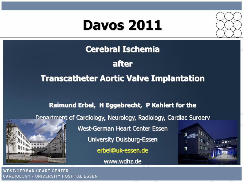
Background
TAVI is increasingly embraced as a viable treatment option for high-risk patients with aortic stenosis
TAVI seems prone to embolic stroke - direct manipulation of the calcified aortic valve - guiding of large-bore catheters- passage of the stiff aorta, aortic arch - prior balloon valvuloplasty- prosthesis induced crushing of calcified leaflets
periprocedural stroke rates: 2.9 - 10%
clinically apparent versus silent cerebral ischemia
Omran et al. Lancet 2003
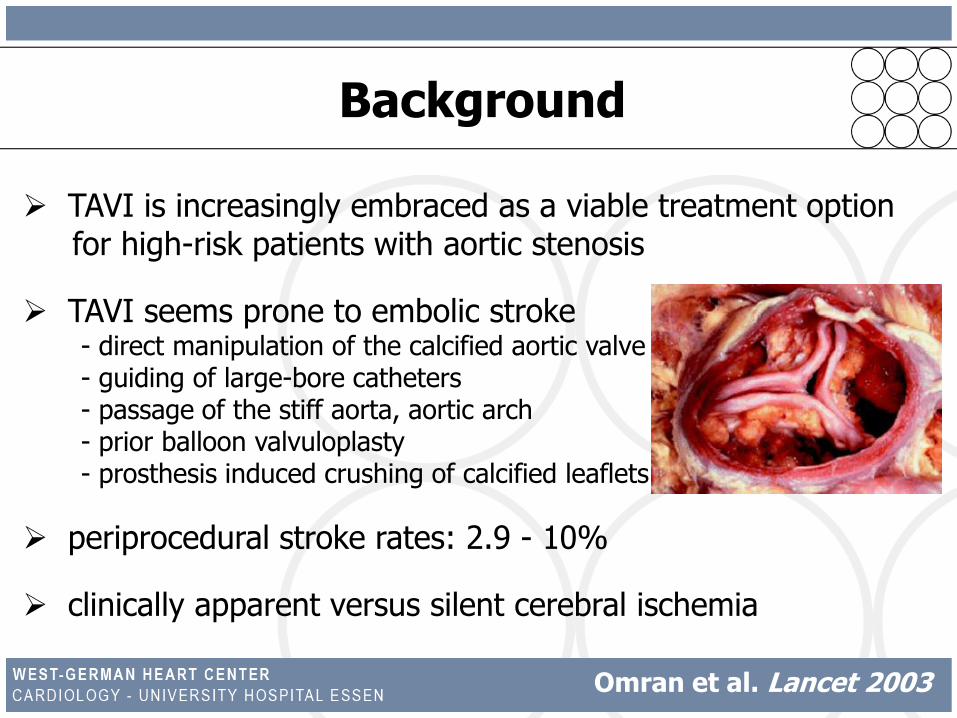
Potential Sources of Embolism
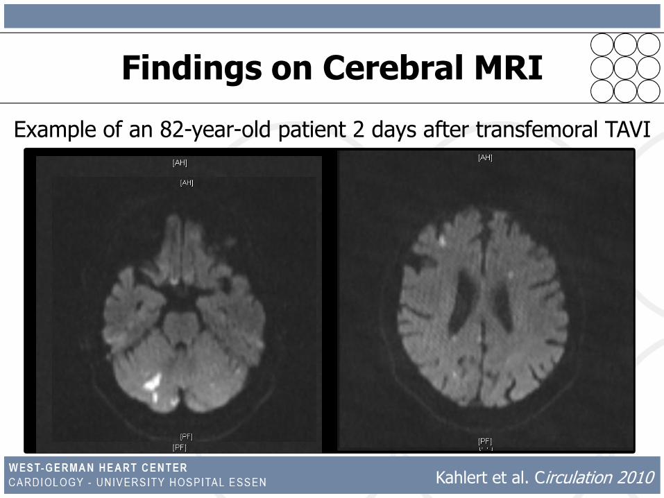
Findings on Cerebral MRI
Example of an 82-year-old patient 2 days after transfemoral TAVI
Kahlert et al. Circulation 2010
Kahlert et al. Circulation 2010
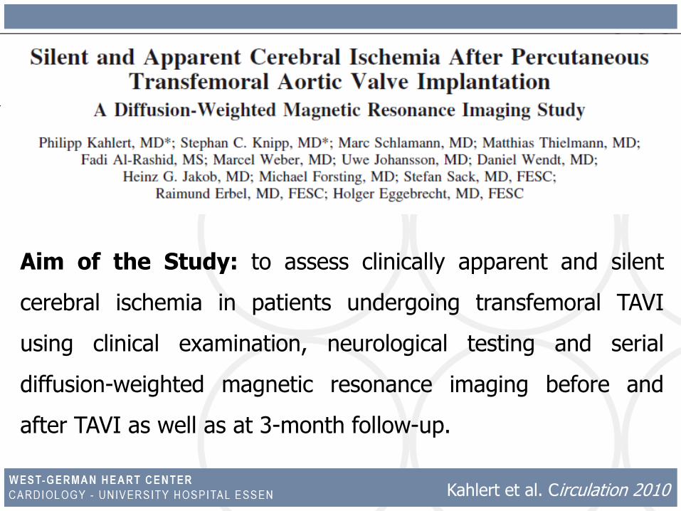
Aim of the Study: to assess clinically apparent and silent
cerebral ischemia in patients undergoing transfemoral TAVI
using clinical examination, neurological testing and serial
diffusion-weighted magnetic resonance imaging before and
after TAVI as well as at 3-month follow-up.
Methods
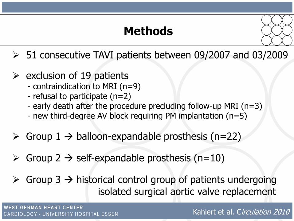
51 consecutive TAVI patients between 09/2007 and 03/2009
exclusion of 19 patients- contraindication to MRI (n=9)- refusal to participate (n=2)- early death after the procedure precluding follow-up MRI (n=3)- new third-degree AV block requiring PM implantation (n=5)
Group 1 balloon-expandable prosthesis (n=22)
Group 2 self-expandable prosthesis (n=10)
Group 3 historical control group of patients undergoing
isolated surgical aortic valve replacement
Kahlert et al. Circulation 2010
Methods
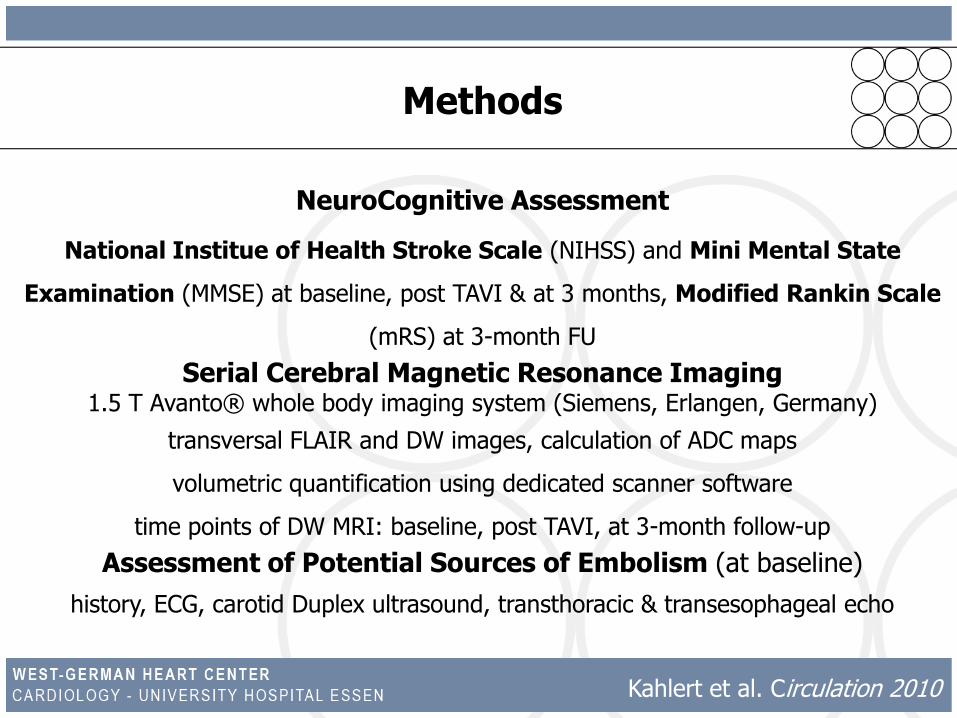
NeuroCognitive Assessment
National Institue of Health Stroke Scale (NIHSS) and Mini Mental State
Examination (MMSE) at baseline, post TAVI & at 3 months, Modified Rankin Scale
(mRS) at 3-month FU
Serial Cerebral Magnetic Resonance Imaging1.5 T Avanto® whole body imaging system (Siemens, Erlangen, Germany)
transversal FLAIR and DW images, calculation of ADC maps
volumetric quantification using dedicated scanner software
time points of DW MRI: baseline, post TAVI, at 3-month follow-up
Assessment of Potential Sources of Embolism (at baseline)
history, ECG, carotid Duplex ultrasound, transthoracic & transesophageal echo
Kahlert et al. Circulation 2010
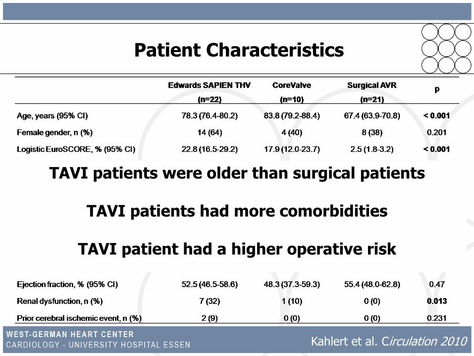
Patient Characteristics
TAVI patients were older than surgical patients
TAVI patients had more comorbidities
TAVI patient had a higher operative risk
Kahlert et al. Circulation 2010
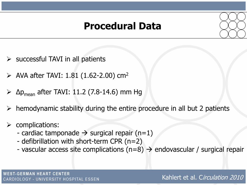
Procedural Data
successful TAVI in all patients
AVA after TAVI: 1.81 (1.62-2.00) cm2
Δpmean after TAVI: 11.2 (7.8-14.6) mm Hg
hemodynamic stability during the entire procedure in all but 2 patients
complications:- cardiac tamponade surgical repair (n=1)
- defibrillation with short-term CPR (n=2)- vascular access site complications (n=8) endovascular / surgical repair
Kahlert et al. Circulation 2010
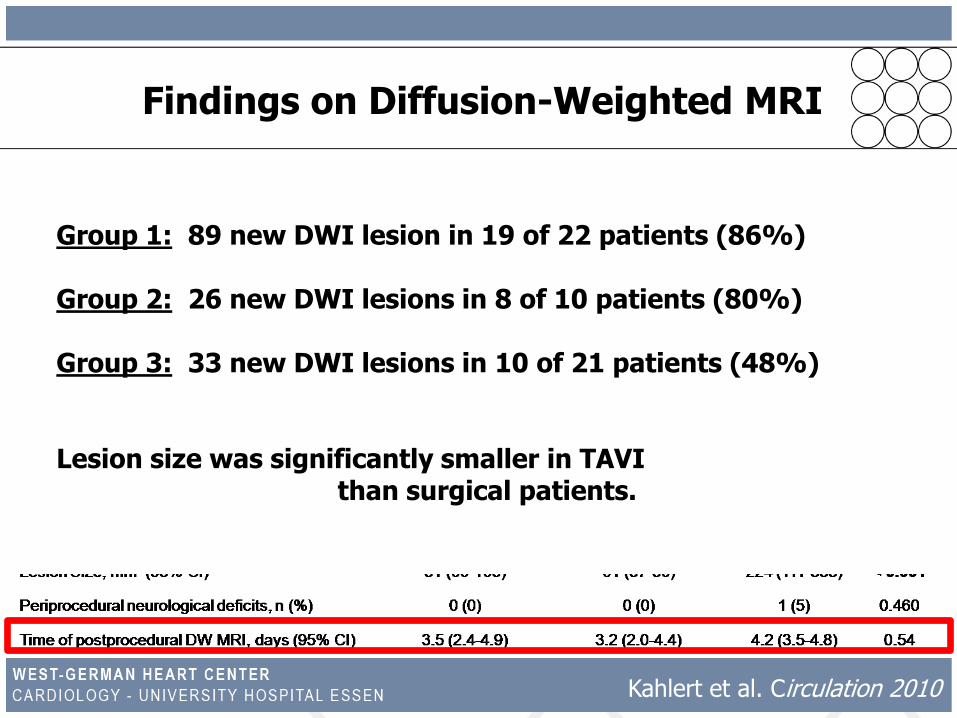
Findings on Diffusion-Weighted MRI
Group 1: 89 new DWI lesion in 19 of 22 patients (86%)
Group 2: 26 new DWI lesions in 8 of 10 patients (80%)
Group 3: 33 new DWI lesions in 10 of 21 patients (48%)
Lesion size was significantly smaller in TAVI than surgical patients.
Kahlert et al. Circulation 2010
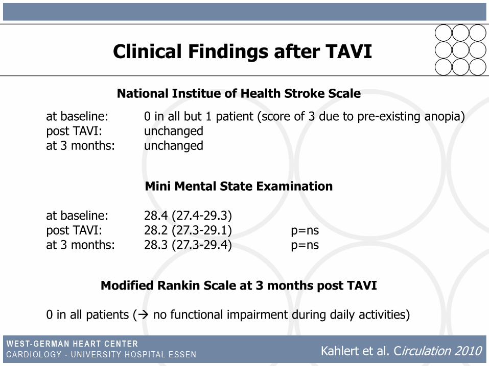
Clinical Findings after TAVI
National Institue of Health Stroke Scale
at baseline: 0 in all but 1 patient (score of 3 due to pre-existing anopia)post TAVI: unchangedat 3 months: unchanged
Mini Mental State Examination
at baseline: 28.4 (27.4-29.3)post TAVI: 28.2 (27.3-29.1) p=nsat 3 months: 28.3 (27.3-29.4) p=ns
Modified Rankin Scale at 3 months post TAVI
0 in all patients ( no functional impairment during daily activities)
Kahlert et al. Circulation 2010
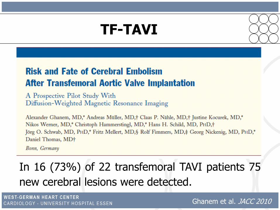
TF-TAVI
Ghanem et al. JACC 2010
In 16 (73%) of 22 transfemoral TAVI patients 75
new cerebral lesions were detected.
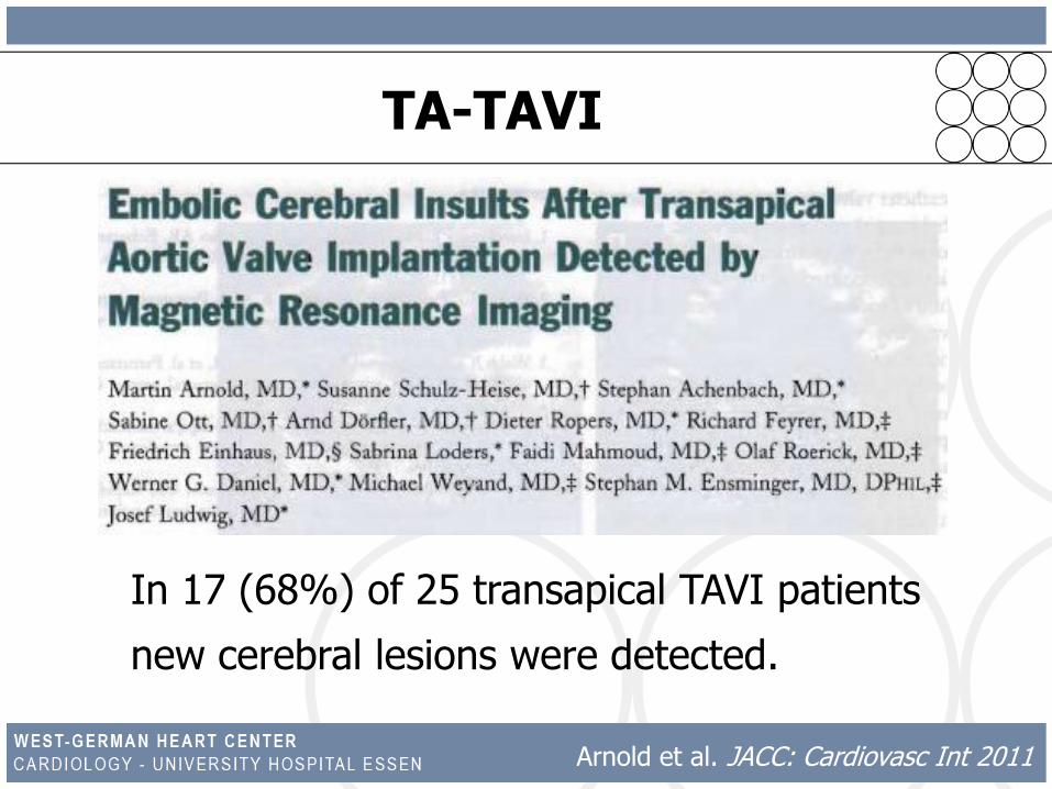
TA-TAVI
Arnold et al. JACC: Cardiovasc Int 2011
In 17 (68%) of 25 transapical TAVI patients
new cerebral lesions were detected.
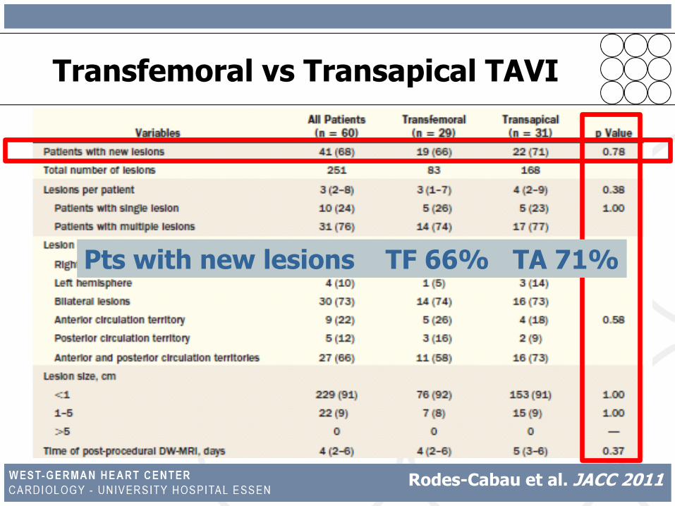
Transfemoral vs Transapical TAVI
Rodes-Cabau et al. JACC 2011
Pts with new lesions TF 66% TA 71%
Cerebral Ischemia
after
Transcatheter Aortic Valve Implantation
Detection of Source of Emboli
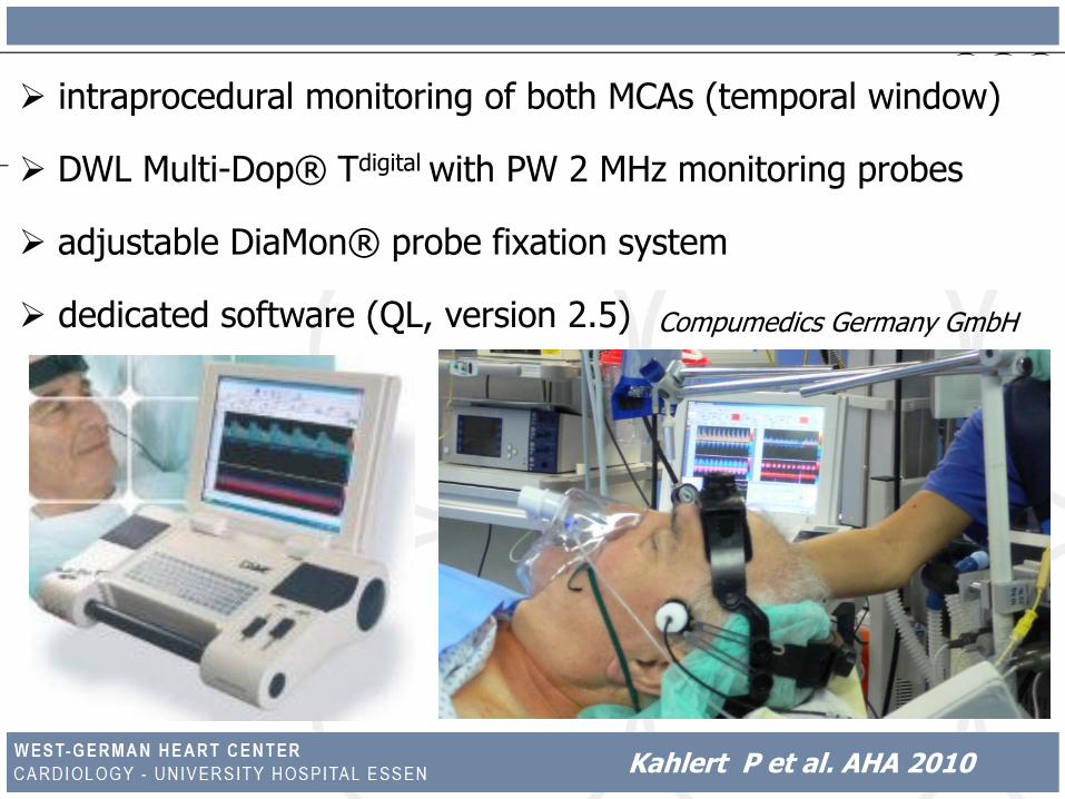
intraprocedural monitoring of both MCAs (temporal window)
DWL Multi-Dop® Tdigital with PW 2 MHz monitoring probes
adjustable DiaMon® probe fixation system
dedicated software (QL, version (2.5 Compumedics Germany GmbH
Kahlert P et al. AHA 2010
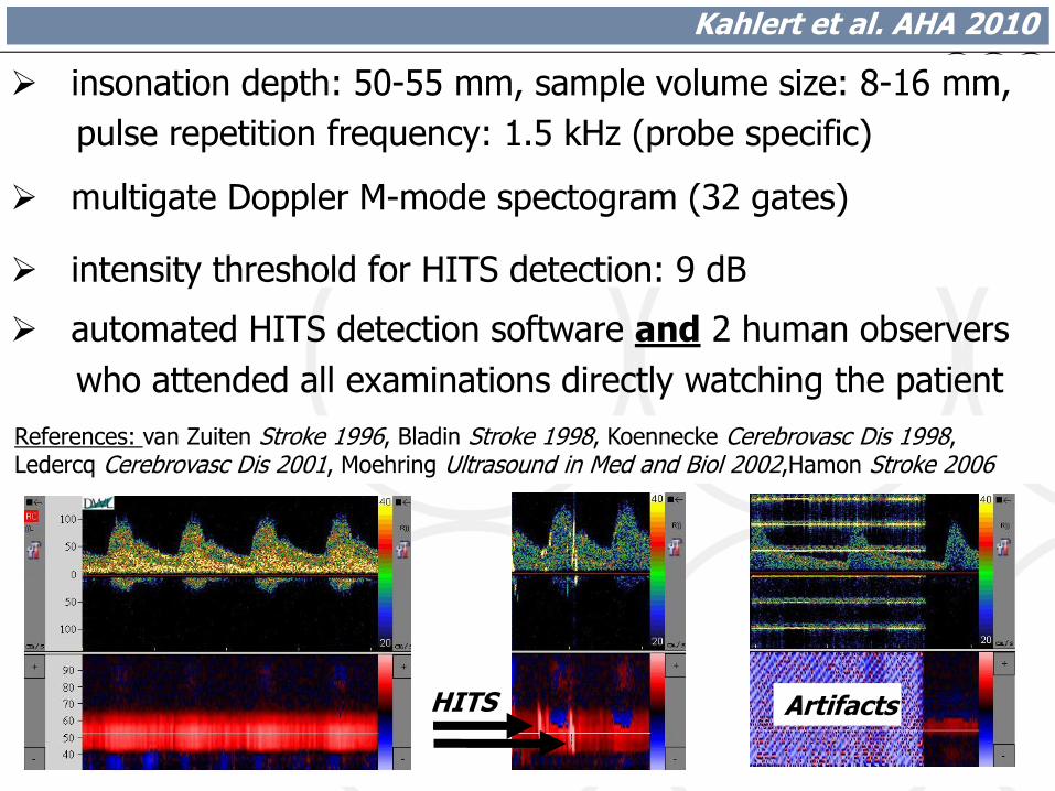
insonation depth: 50-55 mm, sample volume size: 8-16 mm,
pulse repetition frequency: 1.5 kHz (probe specific)
multigate Doppler M-mode spectogram (32 gates)
intensity threshold for HITS detection: 9 dB
automated HITS detection software and 2 human observers
who attended all examinations directly watching the patient
HITS Artifacts
References: van Zuiten Stroke 1996, Bladin Stroke 1998, Koennecke Cerebrovasc Dis 1998, Ledercq Cerebrovasc Dis 2001, Moehring Ultrasound in Med and Biol 2002,Hamon Stroke 2006
Kahlert et al. AHA 2010
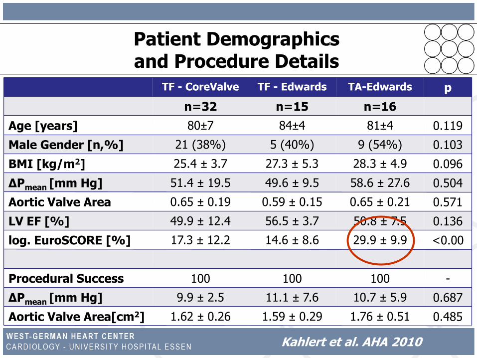
Patient Demographicsand Procedure Details
TF - CoreValve TF - Edwards TA-Edwards p
n=32 n=15 n=16
Age [years] 80±7 84±4 81±4 0.119
Male Gender [n,%] (38%) 21 (40%) 5 (54%) 9 0.103
BMI [kg/m2] 25.4 ± 3.7 27.3 ± 5.3 28.3 ± 4.9 0.096
ΔPmean [mm Hg] 51.4 ± 19.5 49.6 ± 9.5 58.6 ± 27.6 0.504
Aortic Valve Area [cm2]
0.65 ± 0.19 0.59 ± 0.15 0.65 ± 0.21 0.571
LV EF [%] 49.9 ± 12.4 56.5 ± 3.7 50.8 ± 7.5 0.136
log. EuroSCORE [%] 17.3 ± 12.2 14.6 ± 8.6 29.9 ± 9.9 <0.001
Procedural Success [%]
100 100 100 -
ΔPmean [mm Hg] 9.9 ± 2.5 11.1 ± 7.6 10.7 ± 5.9 0.687
Aortic Valve Area[cm2] 1.62 ± 0.26 1.59 ± 0.29 1.76 ± 0.51 0.485
Kahlert et al. AHA 2010
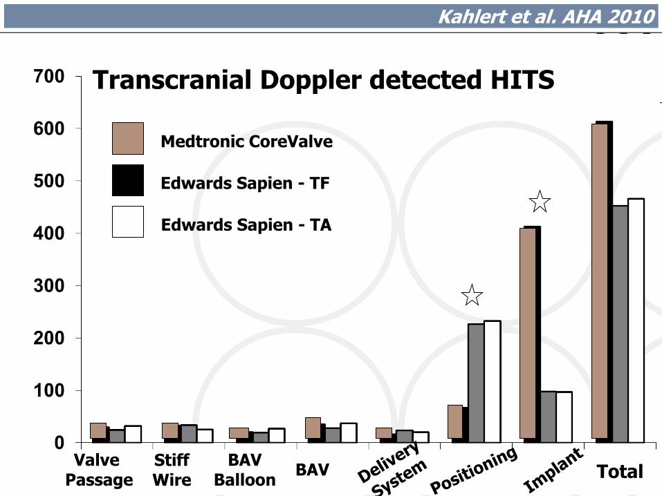
ValvePassage
StiffWire
BAVBalloon
BAV Total
Transcranial Doppler detected HITS
Medtronic CoreValve
Edwards Sapien - TF
Edwards Sapien - TA
Kahlert et al. AHA 2010
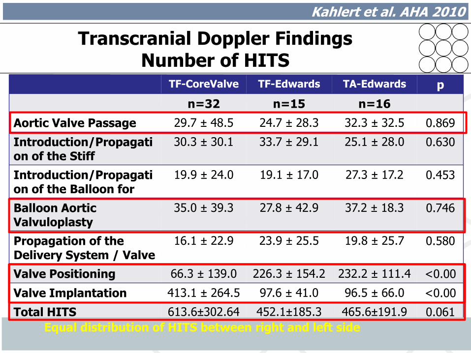
TF-CoreValve TF-Edwards TA-Edwards p
n=32 n=15 n=16
Aortic Valve Passage 29.7 ± 48.5 24.7 ± 28.3 32.3 ± 32.5 0.869
Introduction/Propagation of the Stiff Guidewire
30.3 ± 30.1 33.7 ± 29.1 25.1 ± 28.0 0.630
Introduction/Propagation of the Balloon for BAV
19.9 ± 24.0 19.1 ± 17.0 27.3 ± 17.2 0.453
Balloon Aortic Valvuloplasty
35.0 ± 39.3 27.8 ± 42.9 37.2 ± 18.3 0.746
Propagation of the Delivery System / Valve
16.1 ± 22.9 23.9 ± 25.5 19.8 ± 25.7 0.580
Valve Positioning 66.3 ± 139.0 226.3 ± 154.2 232.2 ± 111.4 <0.001Valve Implantation 413.1 ± 264.5 97.6 ± 41.0 96.5 ± 66.0 <0.001Total HITS 613.6±302.64 452.1±185.3 465.6±191.9 0.061
Equal distribution of HITS between right and left side
Transcranial Doppler FindingsNumber of HITS
Kahlert et al. AHA 2010
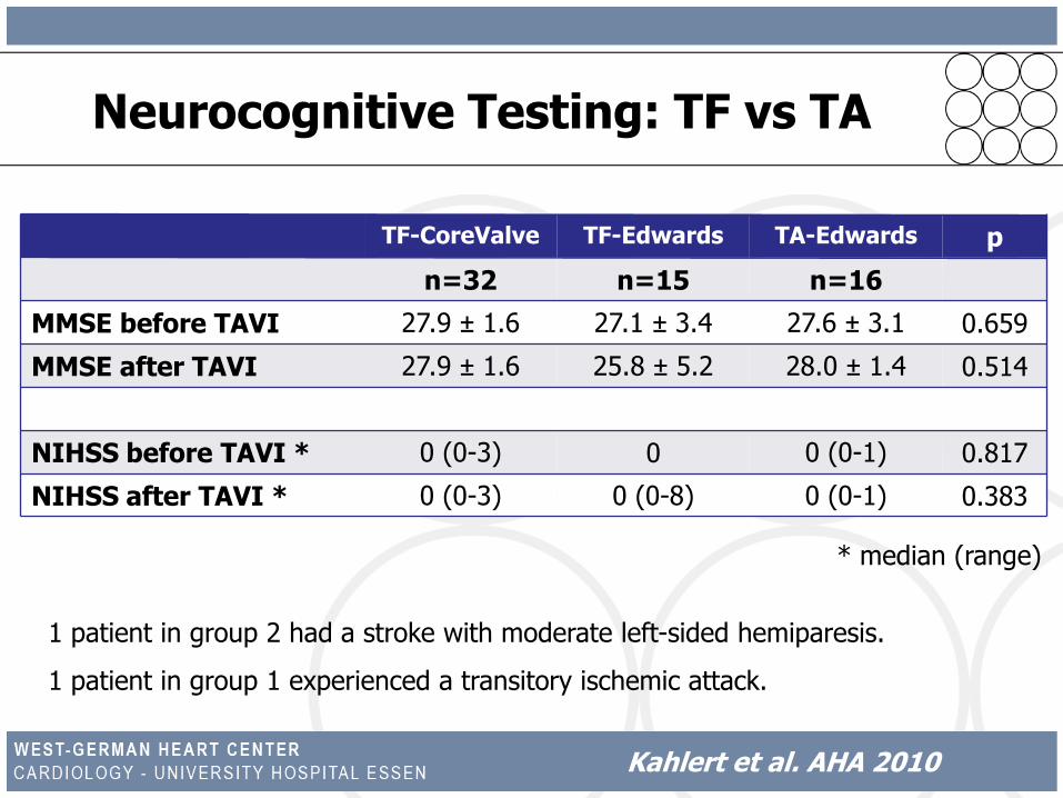
TF-CoreValve TF-Edwards TA-Edwards p
n=32 n=15 n=16
MMSE before TAVI 27.9 ± 1.6 27.1 ± 3.4 27.6 ± 3.1 0.659
MMSE after TAVI 27.9 ± 1.6 25.8 ± 5.2 28.0 ± 1.4 0.514
NIHSS before TAVI * (0-3) 0 0 (0-1) 0 0.817
NIHSS after TAVI * (0-3) 0 (0-8) 0 (0-1) 0 0.383
* median (range)
1 patient in group 2 had a stroke with moderate left-sided hemiparesis.
1 patient in group 1 experienced a transitory ischemic attack.
Neurocognitive Testing: TF vs TA
Kahlert et al. AHA 2010
What can we do?
What should we do?
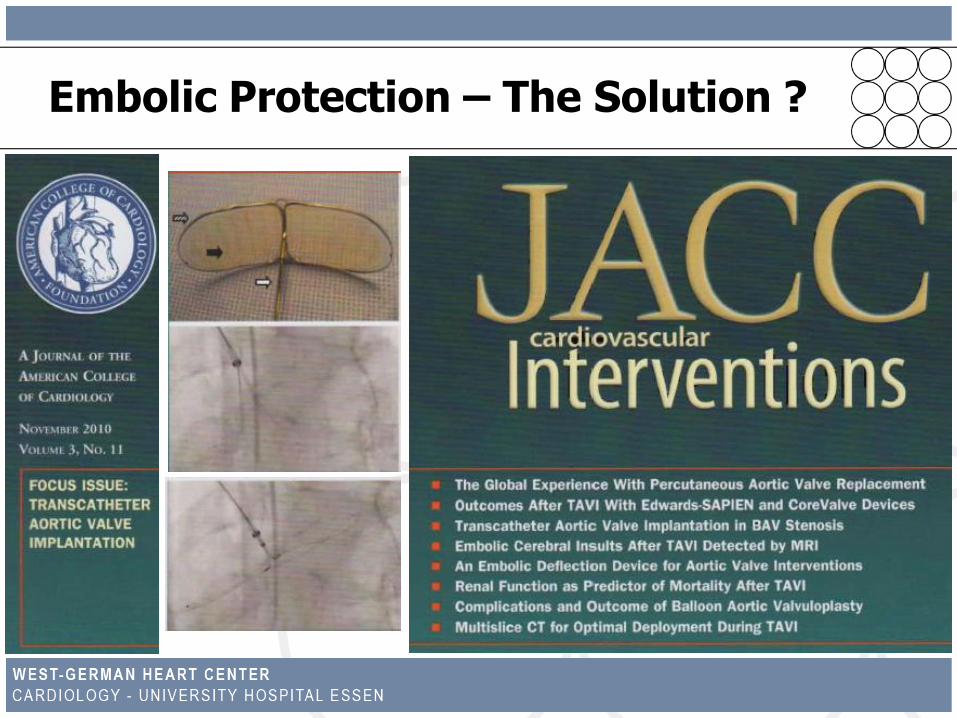
Embolic Protection – The Solution ?
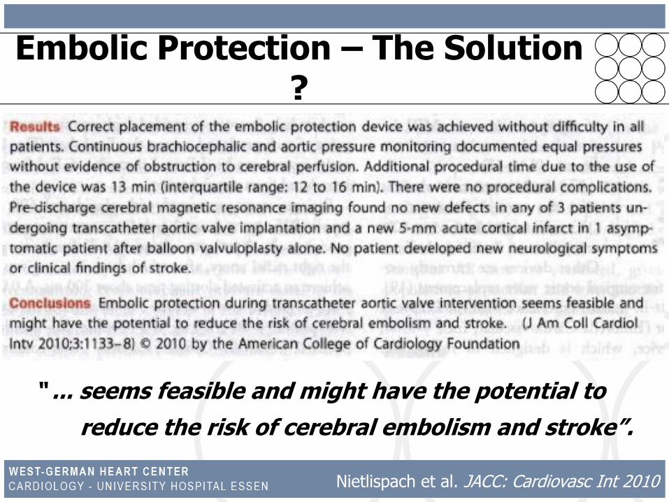
Embolic Protection – The Solution ?
Embolic Protection – The Solution ?
Nietlispach et al. JACC: Cardiovasc Int 2010
“ ... seems feasible and might have the potential to
reduce the risk of cerebral embolism and stroke”.
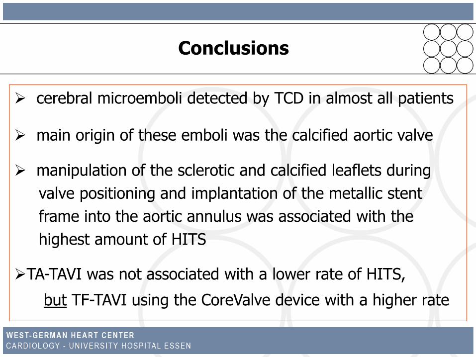
Conclusions
cerebral microemboli detected by TCD in almost all patients
main origin of these emboli was the calcified aortic valve
manipulation of the sclerotic and calcified leaflets during
valve positioning and implantation of the metallic stent
frame into the aortic annulus was associated with the
highest amount of HITS
TA-TAVI was not associated with a lower rate of HITS,
but TF-TAVI using the CoreValve device with a higher rate

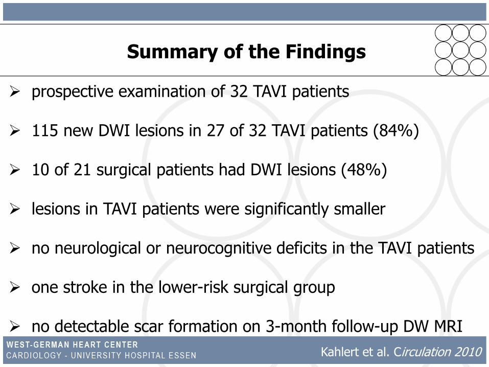
Summary of the Findings
prospective examination of 32 TAVI patients
115 new DWI lesions in 27 of 32 TAVI patients (84%)
10 of 21 surgical patients had DWI lesions (48%)
lesions in TAVI patients were significantly smaller
no neurological or neurocognitive deficits in the TAVI patients
one stroke in the lower-risk surgical group
no detectable scar formation on 3-month follow-up DW MRI
Kahlert et al. Circulation 2010
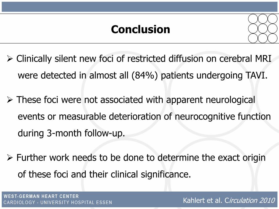
Conclusion
Clinically silent new foci of restricted diffusion on cerebral MRI
were detected in almost all (84%) patients undergoing TAVI.
These foci were not associated with apparent neurological
events or measurable deterioration of neurocognitive function
during 3-month follow-up.
Further work needs to be done to determine the exact origin
of these foci and their clinical significance.
Kahlert et al. Circulation 2010
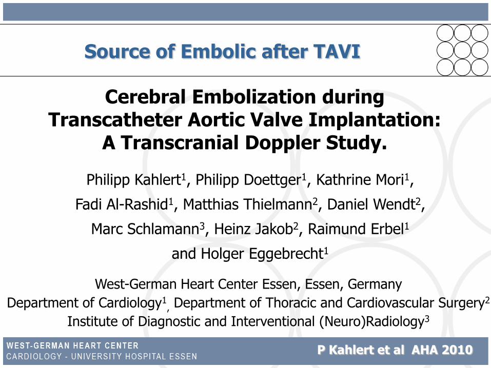
Cerebral Embolization during Transcatheter Aortic Valve Implantation:
A Transcranial Doppler Study.
Philipp Kahlert1, Philipp Doettger1, Kathrine Mori1,
Fadi Al-Rashid1, Matthias Thielmann2, Daniel Wendt2,
Marc Schlamann3, Heinz Jakob2, Raimund Erbel1
and Holger Eggebrecht1
West-German Heart Center Essen, Essen, Germany
Department of Cardiology1, Department of Thoracic and Cardiovascular Surgery2
Institute of Diagnostic and Interventional (Neuro)Radiology3
Source of Embolic after TAVI
P Kahlert et al AHA 2010
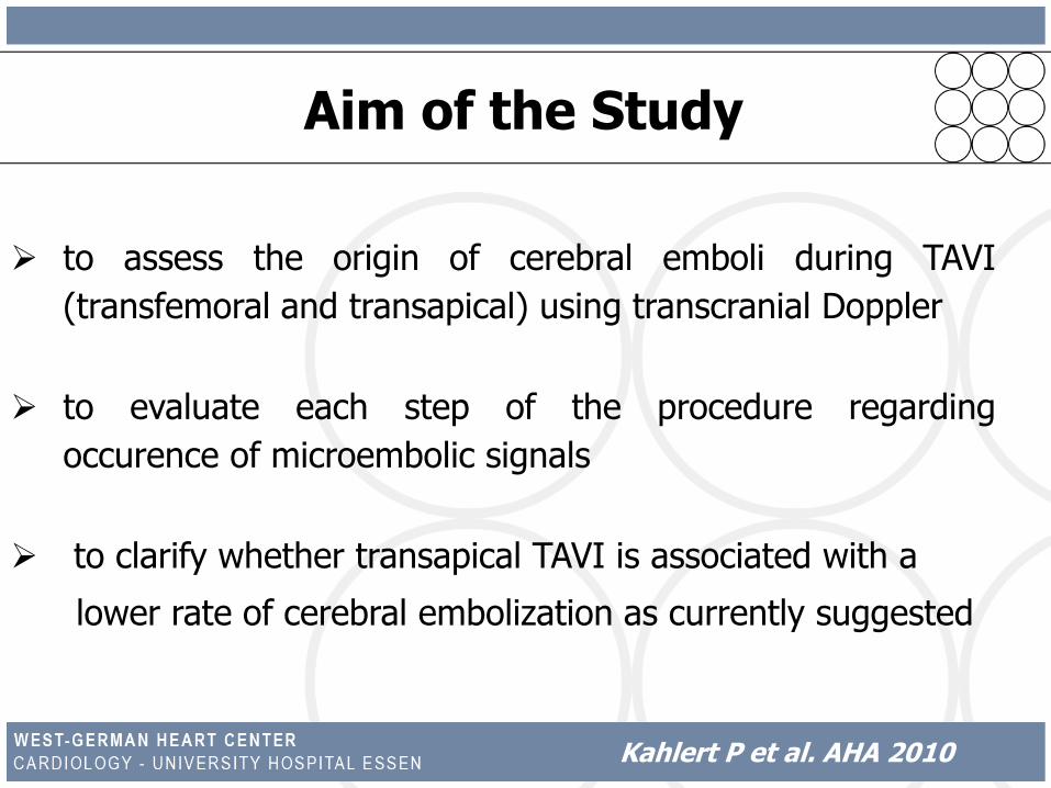
Aim of the Study
to assess the origin of cerebral emboli during TAVI
(transfemoral and transapical) using transcranial Doppler
to evaluate each step of the procedure regarding
occurence of microembolic signals
to clarify whether transapical TAVI is associated with a
lower rate of cerebral embolization as currently suggested
Kahlert P et al. AHA 2010
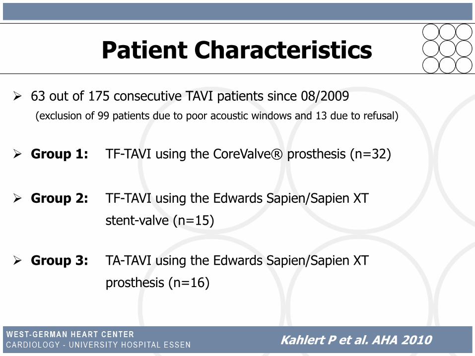
63 out of 175 consecutive TAVI patients since 08/2009
(exclusion of 99 patients due to poor acoustic windows and 13 due to refusal)
Group 1: TF-TAVI using the CoreValve® prosthesis (n=32)
Group 2: TF-TAVI using the Edwards Sapien/Sapien XT
stent-valve (n=15)
Group 3: TA-TAVI using the Edwards Sapien/Sapien XT
prosthesis (n=16)
Patient Characteristics
Kahlert P et al. AHA 2010