Embed Size (px)
Citation preview
Contents lists available at ScienceDirect
Data in Brief
Data in Brief 6 (2016) 542–549
S
http://d2352-34(http://c
n CorrE-m
Pleascanc(201
journal homepage: www.elsevier.com/locate/dib
Data Article
Data on in vivo selection of SK-OV-3 Luc ovariancancer cells and intraperitoneal tumor formationwith low inoculation numbers
Elly De Vlieghere a, Charlotte Carlier b, Wim Ceelen b,Marc Bracke a,n, Olivier De Wever a
a Laboratory of Experimental Cancer Research, Ghent University, Belgiumb Laboratory of Experimental Surgery, Ghent University hospital, Belgium
a r t i c l e i n f o
Article history:Received 10 November 2015Received in revised form15 December 2015Accepted 28 December 2015Available online 6 January 2016
Keywords:in vivo selectionOvarian cancerMouse modelGrowth curvesSurvival curves
x.doi.org/10.1016/j.dib.2015.12.03709/& 2016 The Authors. Published by Elsereativecommons.org/licenses/by/4.0/).
esponding author.ail addresses: [email protected] (M.
e cite this article as: E. De Vliegheer cells and intraperitoneal tumor6), http://dx.doi.org/10.1016/j.dib.2
a b s t r a c t
This data paper contains information about the in vivo model forperitoneal implants used in the paper “Tumor-environment biomi-metics delay peritoneal metastasis formation by deceiving and redir-ecting disseminated cancer cells” (De Vlieghere et al., 2015) [1]. Adouble in vivo selection of SK-OV-3 Luc human ovarian cancer cell linewas used to create SK-OV-3 Luc IP1 and SK-OV-3 Luc IP2 cell lines. Thisdata paper shows functional activities of the three cell lines in vitro andin vivo. Phase-contrast images show the morphology of these cells,metabolic and luciferase activity has been determined. Survival data ofmice peritoneally injected with SK-OV-3 Luc or SK-OV-3 Luc IP2 isavailable with H&E histology of the peritoneal implants. Tumor growthcurves and bioluminescent images of mice inoculated with a differentnumber of SK-OV-3 Luc IP2 cells are also included.& 2016 The Authors. Published by Elsevier Inc. This is an open access
article under the CC BY license(http://creativecommons.org/licenses/by/4.0/).
Specifications table
ubject area
BiologyCancer biologyvier Inc. This is an open access article under the CC BY license
Bracke), [email protected] (O. De Wever).
re, et al., Data on in vivo selection of SK-OV-3 Luc ovarianformation with low inoculation numbers, Data in Brief
015.12.037i
M
TH
DE
E
D
Pc(2
E. De Vlieghere et al. / Data in Brief 6 (2016) 542–549 543
ore specific sub-ject areaype of data
lease cite this articlancer cells and intr016), http://dx.doi.
Phase contrast images, graphs, histology, bioluminescence images
ow data wasacquired� Phase contrast microscope ( DMI 3000B,Leica, Wetzlar, Germany)� Plate reader (Paradigm, Molecular Devices, Sunnyvale, CA, USA)� Light microscope (DM750,Leica, Wetzlar, Germany)� Bioluminescent imager (IVIS, PerklinElmer, Waltham, MA, USA)
ata format
Analyzed xperimentalfactorsSK-OV-3-Luc cells were inoculated intraperitoneally in immune deficientfemale mice Swiss/nu to select for a population that more efficiently formsperitoneal implants.
xperimentalfeatures
The created cell line SK-OV-3 Luc IP1 and SK-OV-3 Luc IP2 are compared withthe parental cell line SK-OV-3 Luc
ata sourcelocation
Ghent, Belgium
ata accessibility
The data is available with this article DValue of the data
� This data shows intraperitoneal tumor formation with low cell inoculation after in vivo selection ofSK-OV-3.
� This method can be applied to other cancer cell lines to increase metastasis take rate even withlower inoculation numbers.
� These data provides growth curves, survival data and histology about the SK-OV-3 Luc (IP2) in vivomodel for peritoneal implants, providing researchers with a references for their in vivo studies.
1. Data
Intraperitoneal injection (IP) of SK-OV-3 (Luc) cells is an established in vivo model for the devel-opment of peritoneal ovarian tumor implants. Usually inoculation numbers of 1–2�106 SK-OV-3(Luc) are used [2–4]. The IP injection of 1�106 SK-OV-3 Luc cells gives a 100% tumor take with anaverage survival time of over 2 months (Fig. 3B) [3]. To better mimic the patient situation where aninitial low number of aggressive disseminated cells can form extensive peritoneal metastasis axenograft mouse model with inoculation of low numbers of cancer cells is needed. in vivo selectionhas been successfully used to increase the metastatic potential of MDA-MB-231 [5]. These datadescribe the successive in vivo selection of the SK-OV-3 Luc cell line. Tumor implants of SK-OV-3 Lucbearing mice are re-cultured in vitro to increase in vivo peritoneal implant formation. After successivein vivo selection, IP inoculation of 10-to-20X lower number (1�105) of SK-OV-3 Luc IP2 is sufficient toform peritoneal implants this model has been applied successfully [1].
2. Experimental design, materials and methods
2.1. Cell culture conditions
SK-OV-3 is a human ovarian cancer cell line (ATCC number: HTB-77). SK-OV-3 Luc (Luciferasepositive SK-OV-3 cells) were prepared by pFL4.76 plasmid transfection and selection (Promega, Lei-den, The Nederlands). SK-OV-3 Luc (IP1/2) were maintained in Dulbecco’s modified Eagle’s medium
e as: E. De Vlieghere, et al., Data on in vivo selection of SK-OV-3 Luc ovarianaperitoneal tumor formation with low inoculation numbers, Data in Brieforg/10.1016/j.dib.2015.12.037i
E. De Vlieghere et al. / Data in Brief 6 (2016) 542–549544
(DMEM) supplemented with 10% fetal bovine serum (FBS) and antibiotics (penicillin/streptomycin),and incubated at 37 °C with 10% CO2 in air.
2.2. in vivo selection
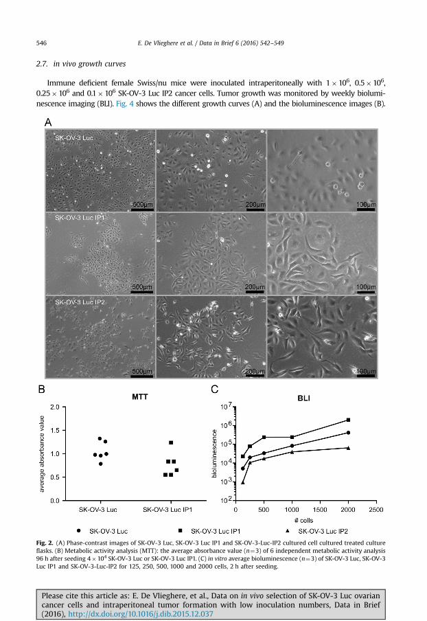
Animal experiments were conducted accordance with the local ethics committee (Ghent Uni-versity Hospital). 1�106 SK-OV-3-Luc cells were inoculated intraperitoneally in immunodeficientfemale Swiss/nu mice (Charles River, Chatillon-sur-Chalaronne, France) to isolate populations thatform peritoneal implants. Tumor implants were collected in saline. The implants were cut into smallpieces (1 mm) and dissociated by de gentleMACS Dissociator (Miltenyi Biotec, Teterow, Germany) inthe presences of 1 mg/ml collagenase from Clostridium histolyticum (Sigma-Aldrich) in PBSDþ . Themixture was cleared through a cell strainer (70 mm) and the homogenized single cell suspension wasexpanded in culture by seeding in DMEM 10% FBS. After 24 h, non-adhered cells were removed andthe medium was refreshed; the resulting culture was maintained as the parental cell line. Eachsubsequent intraperitoneal metastatic generation is designated IP1, IP2. Fig. 1 shows a schematic withthe different stages of the in vivo selection. Fig. 2A shows phase-contrast images of the three cell linescultured on plastic
2.3. Sort tandem repeat (SRT) profiling
SRT-profiling was conducted on the SK-OV-3, SK-OV-3 Luc and SK-OV-3 Luc IP2 cell lines toconfirm the identity of the cell lines. 9 DNA sites were profiled and the alleles were identical betweenthe three cell lines and to the profile published by ATCC (Amelogenin: X; CSF1PO: 11; D13S317: 8,11;D16S539: 12; D5S818: 11; D7S820: 13,14; THO1: 9,9.3; TPOX: 8,11; vWA: 17,18) [6].
2.4. Metabolic activity analysis: 3-4,5-Dimethylthiazolyl-2-2,5-Diphenyltetrazolium Bromide (MTT)
A single cell suspension of 4�104 SK-OV-3 Luc or SK-OV-3 Luc IP1 cells per well, were seeded in a96-well plate. After 96 h, MTT analysis was performed. Briefly, the culture medium was replaced by100 μl culture medium containing 1 mg/ml MTT. Following 2 h incubation at 37 °C, MTT-containingmedium was removed and 150 μl of dimethylsulfoxide (DMSO) was added to dissolve formazancrystals. Absorbance was measured at 570 nm and background measured at 650 nm, with a platereader (Paradigm, Molecular Devices). Fig. 2B shows the average absorbance value (n¼3) of 6 inde-pendent metabolic activity analysis 96 h after seeding 4�104 SK-OV-3 Luc or SK-OV-3 Luc IP1.
2.5. in vitro bioluminescence
A single cell suspension of SK-OV-3 Luc, SK-OV-3 Luc IP1 and SK-OV-3-Luc-IP2 cells were seeded ina 96-well plate in different cell numbers (125, 250, 500, 1000 and 2000 cells per well). Cells wereallowed to adhere for 2 h, just before luciferase activity was measured with IVIS (PerkinElmer),150 mg/ml D-Luciferin, firefly (Perkin-Elmer) was added. Fig. 2C shows the average bioluminescencevalue of three replicates.
2.6. in vivo survival analysis
Animal experiments were conducted in accordance with the local ethics committee (Ghent Uni-versity Hospital). Immune deficient female Swiss/nu mice were inoculated intraperitoneally with1�106 SK-OV-3-Luc or SK-OV-3-Luc-IP2 cells. Mice were monitored and from the first visual signs ofadvanced carcinomatosis (decrease in weight or increase in abdominal circumference) the mice hadreached their end-point and were sacrificed. Peritoneal organs were embedded in paraffin beforestandard hematoxylin and eosin (H&E) staining was conducted. Fig. 3A shows H&E staining of theperitoneal tumor implants, B shows comparative survival curves.
Please cite this article as: E. De Vlieghere, et al., Data on in vivo selection of SK-OV-3 Luc ovariancancer cells and intraperitoneal tumor formation with low inoculation numbers, Data in Brief(2016), http://dx.doi.org/10.1016/j.dib.2015.12.037i
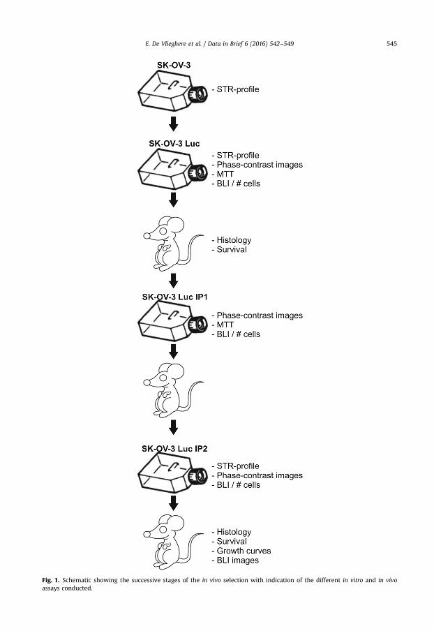
Fig. 1. Schematic showing the successive stages of the in vivo selection with indication of the different in vitro and in vivoassays conducted.
E. De Vlieghere et al. / Data in Brief 6 (2016) 542–549 545
E. De Vlieghere et al. / Data in Brief 6 (2016) 542–549546
2.7. in vivo growth curves
Immune deficient female Swiss/nu mice were inoculated intraperitoneally with 1�106, 0.5�106,0.25�106 and 0.1�106 SK-OV-3 Luc IP2 cancer cells. Tumor growth was monitored by weekly biolumi-nescence imaging (BLI). Fig. 4 shows the different growth curves (A) and the bioluminescence images (B).
Fig. 2. (A) Phase-contrast images of SK-OV-3 Luc, SK-OV-3 Luc IP1 and SK-OV-3-Luc-IP2 cultured cell cultured treated cultureflasks. (B) Metabolic activity analysis (MTT): the average absorbance value (n¼3) of 6 independent metabolic activity analysis96 h after seeding 4�104 SK-OV-3 Luc or SK-OV-3 Luc IP1. (C) in vitro average bioluminescence (n¼3) of SK-OV-3 Luc, SK-OV-3Luc IP1 and SK-OV-3-Luc-IP2 for 125, 250, 500, 1000 and 2000 cells, 2 h after seeding.
Please cite this article as: E. De Vlieghere, et al., Data on in vivo selection of SK-OV-3 Luc ovariancancer cells and intraperitoneal tumor formation with low inoculation numbers, Data in Brief(2016), http://dx.doi.org/10.1016/j.dib.2015.12.037i
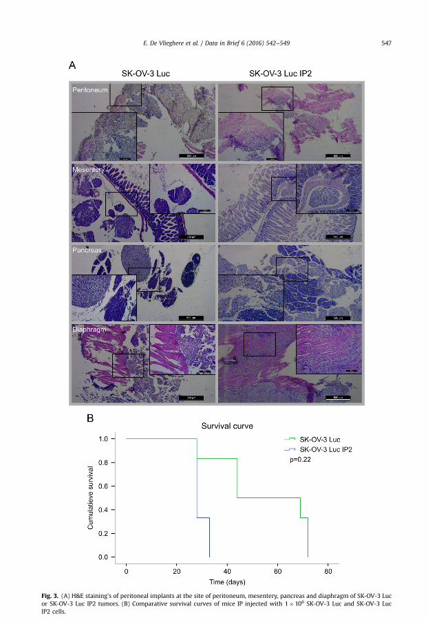
Fig. 3. (A) H&E staining's of peritoneal implants at the site of peritoneum, mesentery, pancreas and diaphragm of SK-OV-3 Lucor SK-OV-3 Luc IP2 tumors. (B) Comparative survival curves of mice IP injected with 1�106 SK-OV-3 Luc and SK-OV-3 LucIP2 cells.
E. De Vlieghere et al. / Data in Brief 6 (2016) 542–549 547
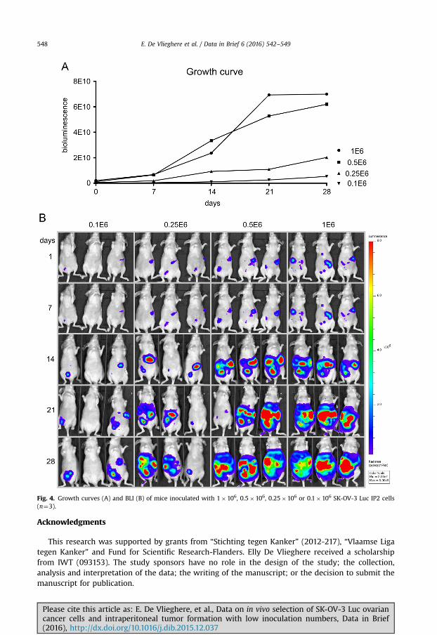
Fig. 4. Growth curves (A) and BLI (B) of mice inoculated with 1�106, 0.5�106, 0.25�106 or 0.1�106 SK-OV-3 Luc IP2 cells(n¼3).
E. De Vlieghere et al. / Data in Brief 6 (2016) 542–549548
Acknowledgments
This research was supported by grants from “Stichting tegen Kanker” (2012-217), “Vlaamse Ligategen Kanker” and Fund for Scientific Research-Flanders. Elly De Vlieghere received a scholarshipfrom IWT (093153). The study sponsors have no role in the design of the study; the collection,analysis and interpretation of the data; the writing of the manuscript; or the decision to submit themanuscript for publication.
Please cite this article as: E. De Vlieghere, et al., Data on in vivo selection of SK-OV-3 Luc ovariancancer cells and intraperitoneal tumor formation with low inoculation numbers, Data in Brief(2016), http://dx.doi.org/10.1016/j.dib.2015.12.037i

E. De Vlieghere et al. / Data in Brief 6 (2016) 542–549 549
Appendix A. Supplementary material
Supplementary data associated with this article can be found in the online version at http://dx.doi.org/10.1016/j.dib.2015.12.037.
References
1 E. De Vlieghere, F. Gremonprez, L. Verset, L. Marien, C.J. Jones, B. De Craene, et al., Tumor-environment biomimetics delayperitoneal metastasis formation by deceiving and redirecting disseminated cancer cells, Biomaterials 54 (2015) 148–157.
2 H. Cho, T.C. Lai, G.S. Kwon, Poly(ethylene glycol)-block-poly(epsilon-caprolactone) micelles for combination drug delivery:evaluation of paclitaxel, cyclopamine and gossypol in intraperitoneal xenograft models of ovarian cancer, J. Control Release166 (1) (2013) 1–9.
3 P.E. Colombo, M. Boustta, S. Poujol, M. Jarlier, F. Bressolle, I. Teulon, et al., Intraperitoneal administration of novel doxorubicinloaded polymeric delivery systems against peritoneal carcinomatosis: experimental study in a murine model of ovariancancer, Gynecol. Oncol. 122 (3) (2011) 632–640.
4 J. Mikula-Pietrasik, P. Sosinska, E. Naumowicz, K. Maksin, H. Piotrowska, A. Wozniak, et al., Senescent peritoneal mesotheliuminduces a pro-angiogenic phenotype in ovarian cancer cells in vitro and in a mouse xenograft model in vivo, Clin. Exp.Metastas (2015).
5 A.J. Minn, G.P. Gupta, P.M. Siegel, P.D. Bos, W. Shu, D.D. Giri, et al., Genes that mediate breast cancer metastasis to lung,Nature. 436 (7050) (2005) 518–524.
6 ATCC: The Global Bioresource Center. ⟨http://www.lgcstandards-atcc.org/Products/All/HTB-77.aspxspecifications⟩.
Please cite this article as: E. De Vlieghere, et al., Data on in vivo selection of SK-OV-3 Luc ovariancancer cells and intraperitoneal tumor formation with low inoculation numbers, Data in Brief(2016), http://dx.doi.org/10.1016/j.dib.2015.12.037i