Embed Size (px)
Citation preview

Daptomycin Tolerance in the Staphylococcus aureus pitA6 Mutant IsDue to Upregulation of the dlt Operon
Lukas Mechler,a Eve-Julie Bonetti,b Sebastian Reichert,a Matthias Flötenmeyer,c Jacques Schrenzel,b Ralph Bertram,a,d
Patrice François,b Friedrich Götza
Microbial Genetics, Interfaculty Institute of Microbiology and Infection Medicine, University of Tuebingen, Tübingen, Germanya; Genomic Research Laboratory, UniversityHospitals of Geneva and University of Geneva, Geneva, Switzerlandb; Max Planck Institute for Developmental Biology, Tübingen, Germanyc; Research Office, ParacelsusMedical University, Nürnberg, Germanyd
Understanding the mechanisms of how bacteria become tolerant toward antibiotics during clinical therapy is a very importantobject. In a previous study, we showed that increased daptomycin (DAP) tolerance of Staphylococcus aureus was due to a pointmutation in pitA (inorganic phosphate transporter) that led to intracellular accumulation of both inorganic phosphate (Pi) andpolyphosphate (polyP). DAP tolerance in the pitA6 mutant differs from classical resistance mechanisms since there is no in-crease in the MIC. In this follow-up study, we demonstrate that DAP tolerance in the pitA6 mutant is not triggered by the accu-mulation of polyP. Transcriptome analysis revealed that 234 genes were at least 2.0-fold differentially expressed in the mutant.Particularly, genes involved in protein biosynthesis, carbohydrate and lipid metabolism, and replication and maintenance ofDNA were downregulated. However, the most important change was the upregulation of the dlt operon, which is induced by theaccumulation of intracellular Pi. The GraXRS system, known as an activator of the dlt operon (D-alanylation of teichoic acids) and ofthe mprF gene (multiple peptide resistance factor), is not involved in DAP tolerance of the pitA6 mutant. In conclusion, DAP toleranceof the pitA6 mutant is due to an upregulation of the dlt operon, triggered directly or indirectly by the accumulation of Pi.
Daptomycin (DAP) is a cyclic lipopeptide antibiotic that wasapproved by the Food and Drug Administration (FDA) for
treatment of complicated skin infections and bacteremia, as wellas endocarditis caused by Gram-positive bacteria (1). DAP ishighly efficient not only against growing cells but also against sta-tionary-phase cultures of Staphylococcus aureus, including meth-icillin-resistant S. aureus (MRSA) strains, and is used as a last-resort antibiotic (2). Interestingly, a MIC creep for DAP has notbeen observed since its approval in 2003 (3); however, the numberof cases with DAP therapy failure due to DAP-resistant (DAP-R)S. aureus strains has increased (1, 4).
Daptomycin is known to bind to the bacterial membranes ofGram-positive bacteria and kills the cell by disrupting the protonmotive force (1). It is known that most of the adaptations inDAP-R strains concern the bacterial cell membrane and cell wall(5); however, the precise mode of action of DAP is still enigmatic.
In addition to antibiotic resistance, antibiotic tolerance canalso be responsible for the failure of antibiotic therapy (4, 6). Incontrast to resistant cells, tolerant strains are unable to grow in thepresence of DAP; however, they survive the bactericidal action ofDAP at least for a restricted period of time (7).
Antibiotic tolerance can be categorized into phenotypic andgenetic tolerance. Phenotypic tolerance is independent from ge-netic alterations and becomes apparent, for instance, in bacterialpersister cells, representing a tolerant subpopulation within iso-genic bacterial populations (8, 9). Persisters are dormant or non-growing, and their tolerance extends to different kind of antibiot-ics (6). Drug indifference is another example of phenotypictolerance which is distributed, however, over the entire popula-tion and is caused by the inhibition of proliferation due to nutrientstarvation (9, 10). Nonproliferating stationary-phase cells that areless sensitive toward treatment with most antibiotics can benamed as a well-known example of drug indifference. In compar-ison, genetic tolerance is caused by genetically manifested and
vertically transmitted mutations (8). All types of tolerance sharethe fact that an increase in the MIC does not occur. Althoughgenetic antibiotic tolerance has been known for several decades,little is known about its generation (11).
In a recent study, we selected a mutant of S. aureus strainHG003 with a 10,000-fold-increased DAP tolerance (7). The tol-erance of the corresponding mutant HG003pitA6 differs fromthat of the wild-type strain as it fulfills all criteria of genetic toler-ance with a rather slow but linear decrease of viability during DAPtreatment (8, 12). In contrast, the wild-type strain shows a bipha-sic killing behavior after the addition of DAP, which is typical ofphenotypic tolerance by persister cells (6, 12). The DAP toleranceof HG003pitA6 was shown to be restricted to the stationarygrowth phase and associated with an increased uptake of inor-ganic phosphate (Pi), which was stored as polyphosphate (polyP)(7). We showed that the genetic basis of this increased DAP toler-ance is represented by an adaptive single point mutation (pitA6) inthe Pi transporter-encoding gene, pitA, which is part of the pitRAoperon.
In this follow-up study, we found that heterologous expressionof exopolyphosphatase (PPX) in the tolerant strain HG003pitA6
Received 17 December 2015 Returned for modification 13 January 2016Accepted 3 February 2016
Accepted manuscript posted online 16 February 2016
Citation Mechler L, Bonetti E-J, Reichert S, Flötenmeyer M, Schrenzel J, Bertram R,François P, Götz F. 2016. Daptomycin tolerance in the Staphylococcus aureus pitA6mutant is due to upregulation of the dlt operon. Antimicrob Agents Chemother60:2684 –2691. doi:10.1128/AAC.03022-15.
Address correspondence to Friedrich Götz, [email protected].
Supplemental material for this article may be found at http://dx.doi.org/10.1128/AAC.03022-15.
Copyright © 2016, American Society for Microbiology. All Rights Reserved.
crossmark
2684 aac.asm.org May 2016 Volume 60 Number 5Antimicrobial Agents and Chemotherapy
on May 14, 2018 by guest
http://aac.asm.org/
Dow
nloaded from

diminishes polyP to a wild-type level; however, the intracellular Pi
level and growth phase-dependent DAP tolerance were not af-fected. Transcriptome analysis followed by quantitative real-timePCR (qRT-PCR) revealed that the dlt operon is upregulated in thepitA6 mutant, which is most likely responsible for the DAP toler-ance. The responsibility of the dlt operon in the pitA6 DAP toler-ance phenotype was confirmed as the deletion of dltA completelyeliminated DAP tolerance caused by the pitA6 allele.
MATERIALS AND METHODSBacterial strains, plasmids, and culture conditions. All bacterial strainsused in this study are listed in Table S2 in the supplemental material.Escherichia coli DC10B (13) was used for molecular cloning of plasmidspMAD (14) and pRAB11 (15). E. coli cells bearing pMAD, pRAB11, ortheir derivatives were grown at 37°C in basic medium (BM; 1% [wt/vol]soy peptone, 0.5% [wt/vol] yeast extract, 0.5% [wt/vol] NaCl, 0.1% [wt/vol] K2HPO4, 0.1% [wt/vol] glucose), supplemented with 100 �g/ml am-picillin (Carl Roth). S. aureus cells with episomal pMAD plasmids wereincubated at 30°C in BM or tryptic soy broth (TSB; 1.7% [wt/vol] peptonefrom casein, pancreatic digest, 0.3% [wt/vol] soy peptone, 0.5% [wt/vol]NaCl, 0.25% [wt/vol] K2HPO4, 0.25% [wt/vol] glucose, pH 7.3 � 0.2)supplemented with 2.5 �g/ml erythromycin, and those carrying pRAB11were incubated at 37°C in medium supplemented with 10 �g/ml chlor-amphenicol (Carl Roth).
Molecular cloning and DNA isolation. Genomic DNA from S. aureuswas isolated as described previously (16). Qiagen kits were used for isola-tion of bacterial plasmids. Successful cloning of plasmid constructs wasverified by colony PCR and sequencing (GATC Biotech). RbCl was usedfor preparation of chemically competent E. coli cells (17), and electrocom-petent cells of S. aureus were prepared as described previously by Mechleret al. (7).
Allelic exchange constructs and homologous recombination. Weused Gibson assembly (18) for cloning of the constructs for chromosomaldeletion of genes dltA, mprF, graR, and graS in S. aureus HG003 wild typeand HG003pitA6. DNA from S. aureus HG003 was used as a template. Theprimer pairs LM_graR_up_f /LM_graR_up_r and LM_graR_down_f/LM_graR_down_r (see Table S3 in the supplemental material for oligo-nucleotides) were used to amplify the upstream and downstream regionsof graR in S. aureus HG003. The deletion region for graR includes 9 bpupstream from the start codon of graR, including parts of the putativeribosomal binding site. graXS fulfills all requirements for functionality inthe corresponding strains. The primer pairs LM_graSup1/LM_graSup2and LM_graSdown1/LM_graSdown1 were used for the deletion constructof graS. Regions consisting of 12 bp encoding the N-terminal part and 21bp encoding the C terminus remained, resulting in functional graR andgraX. For dltA deletion, the primer pairs LM_dlt_up1/LM_dlt_up2 andLM_dlt_down1/LM_dlt_down2 were used for amplification of regionsbracketing dltA, leaving dltX, dltB, dltC, and dltD intact. The deletion partcontains 10 bp upstream of dltA, disrupting the putative Shine-Dal-garno (SD) sequence, but retains 21 bp encoding the N terminus andencompassing the putative SD sequence of dltB. For deletion of mprF,the primer pairs LM_mprFup1/LM_mprFup2 and LM_mprFdown1/LM_mprFdown2 were used. In this construct, 6 bp encoding the Nterminus and 16 bp encoding the C terminus remain, resulting in aframeshift. For cloning of all deletion constructs, the vector pMADwas digested with the enzymes BglII and SalI, and PCR products wereincubated in a 2.5-fold excess of the plasmid backbone for 60 min at50°C, in a final volume of 20 �l of reaction mixture (0.08 U of T5exonuclease, 0.5 U of Phusion DNA polymerase, 80 U of Taq DNAligase [all from New England BioLabs]) and 4 �l of 5� isothermalreaction buffer (25% [wt/vol] polyethylene glycol [PEG] 8000, 500mM Tris-HCl [pH 7.5], 50 mM MgCl2, 50 mM dithiothreitol [DTT], 1mM concentrations of each of the four deoxynucleoside triphosphates[dNTPs], and 5 mM NAD). The procedure of homologous recombi-nation was performed as described previously by Mechler et al. (7).
Inducible expression using plasmid pRAB11. The exopolyphospha-tase (ppx) gene, A284_02120, including its native ribosome-binding se-quence, was PCR amplified from genomic DNA of Staphylococcus warneri(19), using the primer pair ppx(warneri)fw and ppx(warneri)rev. PlasmidpRAB11 was digested with BglII. Again, Gibson assembly gave rise topRAB11-ppx, harboring ppx downstream of the anhydrotetracycline(ATc)-inducible promoter Pxyl-tet. pRAB11-ppx was finally used to trans-form HG003, as well as HG003pitA6. For heterologous expression anddownstream experiments, strains carrying pRAB11-ppx were inoculatedto an optical density at 578 nm (OD578) of 0.07 and were grown for 2 h inTSB at 37°C with agitation before the addition of ATc (0.4 �M). Incuba-tion was continued for a total of 16 h, and then polyP levels, intracellularPi levels, and DAP tolerance were determined.
Determination of MIC and antibiotic tolerance. MIC values weredetermined as described previously (20). The MIC of DAP is 1 �g/ml forboth HG003 and HG003pitA6. For antibiotic tolerance assays, cells weregrown for 16 h at 37°C in TSB with agitation. Afterwards, 2.24 ml of theovernight culture was transferred to 14-ml round-bottom tubes. DAP andCaCl2 were added to final volumes of 100 �g/ml and 50 �g/ml, respec-tively. The number of CFU/milliliter was determined at time points indi-cated in the figures.
Intracellular inorganic phosphate (Pi) and polyphosphate (polyP)determination. Intracellular Pi was quantified as described previously (7)using a commercially available colorimetric approach (ab65622; Abcam).In brief, cells were grown at 37°C in TSB with agitation, harvested at timepoints indicated in the figures, washed with double-distilled H2O(ddH2O), and disrupted using a FastPrep (MP Biomedicals) instrument.Cell debris was pelleted, and supernatant was used to quantify Pi accord-ing to the manual of the colorimetric approach (ab65622; Abcam). Intra-cellular polyP was determined using 4=-6-diamidino-2-phenylindole(DAPI; Sigma) as described previously (7, 21, 22). Again, cells were grownin TSB at 37°C with agitation before being harvested and washed twice inTris-HCl buffer (100 mM Tris; pH 7.5). Samples were adjusted to anOD578 of 0.5, and DAPI was added to a final concentration of 20 �M. ThepolyP fluorescence signal was determined using a microplate reader(TECAN Infinite 200M) with excitation at 415 nm and an emission wave-length of 550 nm.
RNA isolation for microarray and qRT-PCR. RNA for microarrayand qRT-PCR was isolated as described previously (7). In brief, cells wereadjusted to an OD578 of 0.07 in TSB and cultured at 37°C. At the timepoints indicated in the figures, cells were harvested and adjusted to anOD578 of 70 before lysis with 1 ml Trizol (Life Technologies). DNA wasremoved by 2-fold DNase I digestion as described previously (23). A poolof 3 �g of total RNA for each condition was reverse transcribed usingSuperScript II (Invitrogen, Basel, Switzerland). A volume of 5 �l of a1:100-fold dilution was used for quantitative PCRs (qPCRs). Gene-spe-cific primers and 6-carboxyfluorescein (FAM)-coupled probes for hu andmprF (see Table S4 in the supplemental material) were designed usingPrimer Express, version 3.0 (Applied Biosystems), and mixed in qPCRreagents (ABgene). Other genes were quantified by using a Brilliant SYBRGreen master mix (Agilent) with primers described in Table S4 in the supple-mental material. Reactions were performed in a Bio-Rad CFX96 and normal-ized using intensity levels recorded for the hu gene as described previously(24).
Microarray manufacturing and microarray design. The microarraywas manufactured by in situ synthesis of 60-base-long oligonucleotideprobes (Agilent), selected as previously described (25). The microarrayconsists of a glass slide with 15,000 printed oligonucleotides, covering�95% of all open reading frames (ORFs) annotated in strains NCTC 8325(26), UAMS-1 (27), and SA564 (28) as well as Newman (29), includingtheir respective plasmids.
Preparation of labeled nucleic acids for expression microarrays. To-tal RNA was purified from two independent cultures from strain HG003and its pitA6 mutant. After additional DNase treatment, the absence ofDNA traces was confirmed by quantitative PCR (SDS 7700; Applied Bio-
DAP Tolerance of S. aureus HG003pitA6
May 2016 Volume 60 Number 5 aac.asm.org 2685Antimicrobial Agents and Chemotherapy
on May 14, 2018 by guest
http://aac.asm.org/
Dow
nloaded from

systems) with assays specific for 16S rRNA (30). Batches of 5 �g of total S.aureus RNA were labeled by Cy3-dCTP, using SuperScript II (Invitrogen)according to the manufacturer’s instructions. Labeled products were thenpurified onto QiaQuick columns (Qiagen). Purified genomic DNA wasextracted (DNeasy; Qiagen) from the different sequenced strains usedfor the design of the microarray, labeled with Cy5-dCTP using theKlenow fragment of DNA polymerase I (BioPrime), and used for thenormalization process (31). Cy5-labeled DNA (500 ng) and a Cy3-labeled cDNA mixture were diluted in 50 �l of Agilent hybridizationbuffer and hybridized at a temperature of 60°C for 17 h in a hybrid-ization oven (Robbins Scientific). Slides were washed, dried undernitrogen flow, and scanned (Agilent) using 100% photon multipliertube power for both wavelengths.
Microarray analysis. Fluorescence intensities were extracted using Fea-ture Extraction software (version 9; Agilent). Local background-subtractedsignals were corrected for unequal dye incorporation or unequal loading ofthe labeled product. The algorithm consisted of a rank consistency filter and acurve fit using the default LOWESS (locally weighted linear regression)method. Data consisting of three independent biological experiments wereexpressed as log10 ratios and analyzed using GeneSpring, version 8.0 (SiliconGenetics). A filter was applied to select oligonucleotides mapping ORFs in theNewman genome, yielding approximately 95% coverage. Statistical signifi-cance of differentially expressed genes was calculated by analysis of variance(32) using GeneSpring, including the Benjamini-Hochberg false discoveryrate correction of 5% (P value cutoff, 0.05) and an arbitrary cutoff of 2-fold forexpression ratios.
FITC–poly-L-lysine binding assay. To determine the relative surfacecharge, overnight cultures were washed twice with HEPES buffer (20 mM,pH 7.25) and resuspended to an OD578 of 0.1. fluorescein isothiocyanate(FITC)–poly-L-lysine (FPLL) was added to a final concentration of 5 �g/ml, and the percentage of bound FPLL was determined as described pre-viously (33, 34).
Microarray data accession numbers. The complete microarray dataset has been deposited in the Gene Expression Omnibus database (http://www.ncbi.nlm.nih.gov/geo/) under accession numbers GSE77069 andGPL10537 (for platform design).
RESULTSDAP tolerance of HG003pitA6 is independent from polyP. Pre-viously, we selected a mutant of S. aureus HG003 which wasextremely tolerant against DAP. The corresponding strainHG003pitA6 further showed an increased level of intracellularinorganic phosphate (Pi) and inorganic polyphosphate (polyP)(Fig. 1) (7). We heterologously expressed the exopolyphosphatasegene (ppx) from Staphylococcus warneri using the inducible plas-mid pRAB11 to artificially hydrolyze the polyP level in both the
HG003 wild type and mutant HG003pitA6 (Fig. 1A). Overexpres-sion of ppx (Fig. 1, �ppx) in HG003pitA6 decreased polyP to abasal wild-type level (Fig. 1A). The Pi level and DAP tolerance,however, remained high in HG003pitA6 irrespective of ppx ex-pression (Fig. 1B and C). Heterologous expression of ppx inHG003 wild type did not influence the polyP or intracellular Pi
level or DAP tolerance (Fig. 1A to C).Transcriptome analysis disclosed predominant downregula-
tion of genes from several functional categories. We performedtranscriptome analysis using the microarray technique to betterunderstand molecular processes affecting the increase of DAP tol-erance in HG003pitA6.
We showed previously that DAP tolerance and increased Pi
level were restricted to the stationary growth phase (7). We there-fore compared relative gene expression of HG003pitA6 with thatof the wild type after 6 h (early stationary phase) and 8 h (advancedstationary phase) of growth. The complete results are summarizedin Table S1 in the supplemental material. A total of 234 geneswere found to be at least 2.0-fold differentially expressed inHG003pitA6 compared to expression in the HG003 wild-typestrain after 6 h and/or 8 h of growth.
The majority of 170 genes were downregulated, in contrast to65 genes that were upregulated (Fig. 2A). In general, differences ingene expression between the two strains were mostly moderate, ina range of 2- to 5-fold (see Table S1 in the supplemental material);however, downregulation was common in most functional cate-gories (Fig. 2B). Predominantly genes involved in translation(100%), transcription (90%), and amino acid (65%) and nucleo-tide (67%) metabolism, as well as posttranslational modification(82%), were repressed in HG003pitA6 (Fig. 2B; see also Table S1).The percentage reflects the portion of downregulated genes in thecorresponding functional category. Furthermore, a high numberof repressed genes are involved in carbohydrate metabolism(77%), energy production (100%), or lipid transport (100%).
Interestingly, many genes associated with the synthesis of thecell envelope, which was shown before to be affected in DAP-nonsusceptible strains (35–38), are dysregulated, with no cleartendency to up- or downregulation (Fig. 2B). Among them are themurC, murD, and murG genes that are 2- to 4-fold upregulated inHG003pitA6 (see Table S1 in the supplemental material). Thesegenes are involved in the synthesis of peptidoglycan (39). How-ever, murA, which represents the first step in peptidoglycan syn-
FIG 1 DAP tolerance of HG003pitA6 is independent of polyP. Graphs represent the following: quantification of polyP levels in DAPI-stained cells withexcitation at 415 nm and emission at 550 nm (A), intracellular Pi concentration (B), and killing kinetics of stationary-phase cultures treated with 100 �g/ml DAP(C). Strains harboring the empty plasmid are indicated by �ppx, whereas those expressing ppx are indicated by �ppx. All values are averages from at least threeindependent experiments. Error bars indicate the standard deviations, and significance compared to results with HG003 with the empty plasmid pRAB11 wasdetermined using Student’s t test. Asterisks indicate P values of �0.05. wt, wild type; RFU, relative fluorescence units.
Mechler et al.
2686 aac.asm.org May 2016 Volume 60 Number 5Antimicrobial Agents and Chemotherapy
on May 14, 2018 by guest
http://aac.asm.org/
Dow
nloaded from
thesis (39), was found to be 2-fold downregulated in our approach(see Table S1).
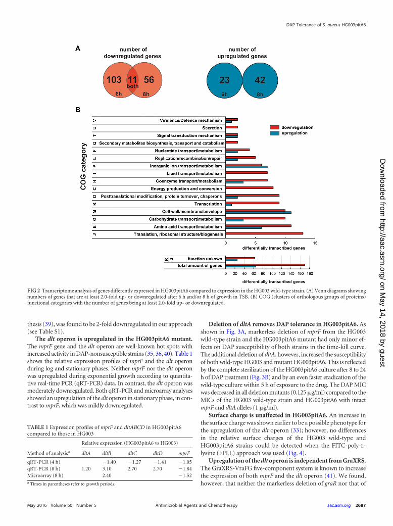
The dlt operon is upregulated in the HG003pitA6 mutant.The mprF gene and the dlt operon are well-known hot spots withincreased activity in DAP-nonsusceptible strains (35, 36, 40). Table 1shows the relative expression profiles of mprF and the dlt operonduring log and stationary phases. Neither mprF nor the dlt operonwas upregulated during exponential growth according to quantita-tive real-time PCR (qRT-PCR) data. In contrast, the dlt operon wasmoderately downregulated. Both qRT-PCR and microarray analysesshowed an upregulation of the dlt operon in stationary phase, in con-trast to mprF, which was mildly downregulated.
Deletion of dltA removes DAP tolerance in HG003pitA6. Asshown in Fig. 3A, markerless deletion of mprF from the HG003wild-type strain and the HG003pitA6 mutant had only minor ef-fects on DAP susceptibility of both strains in the time-kill curve.The additional deletion of dltA, however, increased the susceptibilityof both wild-type HG003 and mutant HG003pitA6. This is reflectedby the complete sterilization of the HG003pitA6 culture after 8 to 24h of DAP treatment (Fig. 3B) and by an even faster eradication of thewild-type culture within 5 h of exposure to the drug. The DAP MICwas decreased in all deletion mutants (0.125 �g/ml) compared to theMICs of the HG003 wild-type strain and HG003pitA6 with intactmprF and dltA alleles (1 �g/ml).
Surface charge is unaffected in HG003pitA6. An increase inthe surface charge was shown earlier to be a possible phenotype forthe upregulation of the dlt operon (33); however, no differencesin the relative surface charges of the HG003 wild-type andHG003pitA6 strains could be detected when the FITC-poly-L-lysine (FPLL) approach was used (Fig. 4).
Upregulation of the dlt operon is independent from GraXRS.The GraXRS-VraFG five-component system is known to increasethe expression of both mprF and the dlt operon (41). We found,however, that neither the markerless deletion of graR nor that of
FIG 2 Transcriptome analysis of genes differently expressed in HG003pitA6 compared to expression in the HG003 wild-type strain. (A) Venn diagrams showingnumbers of genes that are at least 2.0-fold up- or downregulated after 6 h and/or 8 h of growth in TSB. (B) COG (clusters of orthologous groups of proteins)functional categories with the number of genes being at least 2.0-fold up- or downregulated.
TABLE 1 Expression profiles of mprF and dltABCD in HG003pitA6compared to those in HG003
Method of analysisa
Relative expression (HG003pitA6 vs HG003)
dltA dltB dltC dltD mprF
qRT-PCR (4 h) �1.40 �1.27 �1.41 �1.05qRT-PCR (8 h) 1.20 3.10 2.70 2.70 �1.84Microarray (8 h) 2.40 �1.52a Times in parentheses refer to growth periods.
DAP Tolerance of S. aureus HG003pitA6
May 2016 Volume 60 Number 5 aac.asm.org 2687Antimicrobial Agents and Chemotherapy
on May 14, 2018 by guest
http://aac.asm.org/
Dow
nloaded from

graS in either wild-type HG003 or HG003pitA6 was able to alterDAP tolerance (Fig. 5).
DISCUSSION
A fundamental understanding of the mechanisms by which bac-teria are able to escape bactericidal activity of antibiotics is essen-tial for the development of new and possibly better antimicrobialtherapies. Antibiotic tolerance is one aspect explaining the recal-citrance of relapsing and chronic infection by genetically suscep-tible bacteria (6). In an earlier study, we developed a strain show-ing decreased levels of DAP susceptibility in stationary phase (7).This phenotype differed from classical resistance since there wasno increase in the MIC. The only adaptation required to decreaseDAP susceptibility was a nonsynonymous mutation within theinorganic phosphate transporter gene pitA. Additionally, weshowed that this kind of DAP tolerance in the mutant S. aureusHG003pitA6 was restricted to the stationary growth phase andcorrelated with an increased level of intracellular Pi and polyP.Previous studies by Gerdes and Maisonneuve demonstrated thatthe accumulation of polyP increased antibiotic tolerance by per-sister cells in E. coli (42). As polyP levels were also increased in themutant HG003pitA6 (Fig. 1A), we wanted to elucidate a potential
role of increased amounts of polyP in the DAP tolerancephenotype. PolyP is synthesized by polyphosphate kinases(PPKs) and hydrolyzed by exopolyphosphatases (PPXs) (43). S.aureus HG003 contains a putative exopolyphosphatase gene(SAOUHSC_01812) and a putative inorganic polyphosphate/ATP-NAD kinase gene (SAOUHSC_00943), but they are poorlycharacterized and different from the well-known ppk-ppx operonof E. coli (44). For heterologous expression, we decided to use theS. warneri ppx gene (A284_02120), which is a homologue of ppxfrom E. coli but closely related to the S. aureus gene (45). Accord-ing to expectations (43), inducible overexpression of ppx reducedthe amount of polyP in HG003pitA6/pRAB11-ppx to a basal wild-type level (Fig. 1A). Concentrations of intracellular Pi and theincrease in DAP tolerance, however, remained unaffected (Fig. 1Band C), a fact clearly indicating that DAP tolerance mediated bythe pitA6 allele is independent from polyP.
Transcriptome analysis using a microarray technique was ap-plied to unravel the cellular basis of the HG003pitA6 phenotype(Fig. 2A and B; see also Table S1 in the supplemental material). Asthe increase in DAP tolerance is restricted to the stationary phase(7), we checked the expression of HG003pitA6 relative to that ofthe HG003 wild-type strain after 6 h (early stationary phase) and 8h (advanced stationary phase) of growth.
Among a total of 234 genes that were at least 2.0-fold differen-tially expressed after 6 h and/or 8 h (Fig. 2A; see also Table S1 in thesupplemental material), 65 were significantly upregulated, and170 were repressed, in most cases with moderate changes (2- to
FIG 4 Relative surface charge of HG003 and HG003pitA6. The percentage ofFPLL bound to HG003 and HG003pitA6 grown for 16 h in TSB and incubationof cells with FPLL for 10 min at room temperature are depicted. All values areaverages from at least three independent experiments. Error bars indicate thestandard deviations, and significance was determined using Student’s t test(P � 0.05). n.s., not significant.
FIG 5 Time-dependent killing of S. aureus strains with markerless deletion ofgraR or graS by DAP. Stationary-growth-phase cultures of the HG003 graS,HG003 graR, HG003pitA6 graS, and HG003pitA6 graR strains were chal-lenged with 100-fold the MIC of DAP, and CFU counts were recorded. Valuesare the averages from three independent experiments. Error bars representstandard deviations.
FIG 3 Time-dependent killing of mprF and mprF dltA strains by DAP. CFU counts were determined for stationary-growth-phase cultures of the HG003mprF and HG003 mprF dltA strains (A) and for the HG003pitA6 mprF and HG003pitA6 mprF dltA strains (B) upon challenge with 100 �g/ml DAP.Values represent the arithmetic means from three independent experiments. Error bars represent standard deviations.
Mechler et al.
2688 aac.asm.org May 2016 Volume 60 Number 5Antimicrobial Agents and Chemotherapy
on May 14, 2018 by guest
http://aac.asm.org/
Dow
nloaded from

5-fold). A broad range of differentially expressed genes from cen-tral metabolic function was observed, with a predominant down-regulation in most functional categories (Fig. 2B). A prevalentdownshift of expression profiles for genes being involved in trans-lation, transcription, amino acid metabolism, nucleotide metab-olism, or posttranslational modification (Fig. 2B; see also TableS1) indicates that protein biosynthesis is low in HG003pitA6.Likewise, downregulation of genes implicated in carbohydrate orlipid metabolism and especially those responsible for energy gen-eration suggests that a metabolic downshift prevails in the mutantHG003pitA6 at stationary growth phase. This is endorsed by thefact that HG003pitA6 is impaired in growth in comparison toHG003 wild type as soon as it reaches stationary phase (7). Anincrease in the cell wall thickness is assumed to be one reason fordecreased susceptibility to DAP (36�38, 46), and indeed wefound several genes involved in cell wall, membrane, and envelopemaintenance or synthesis to be significantly upregulated. Amongthem were murC, murD, and murG (Fig. 2B; see also Table S1),which are responsible for the synthesis of peptidoglycan, a majorcomponent of the bacterial cell wall (39). However, MurA exe-cutes the first step in peptidoglycan synthesis (39) and is 2-folddownregulated in our data set, which contradicts an increased cellwall thickness phenotype. Accordingly, differences in cell wallthickness of the HG003 wild-type and HG003pitA6 strains werenot detected by transmission electron microscopy (see Fig. S1 inthe supplemental material). Furthermore, the content of wallteichoic acids (WTA) does not differ significantly in these strains(see Fig. S2), indicating that DAP tolerance is independent fromalteration of cell wall thickness.
Beside an increase in thickness, alteration in composition ofthe bacterial cell wall as well as the membrane is a common adap-tation in DAP-R mutants (1, 5). Genes involved in these processesand well-known hot spots for modification in DAP-nonsuscep-tible strains are mprF and the dltXABCD operon (1, 35, 36).
The multipeptide resistance factor gene mprF transfers lysylfrom an L-lysyl-tRNA to membrane-bound phosphatidylglycerol,forming lysyl-phosphatydylglycerol (LPG) (47). The dlt operon isknown to link positively charged D-alanine to both lipid and wallteichoic acids (40). Enhanced expression or activity of either orboth is known to cause DAP nonsusceptibility (35, 36, 40, 48).
We found a time-dependent upregulation of the dlt operon butnot the mprF gene in HG003pitA6 compared to the level in thewild type that could be linked to an increased level of intracellularPi and DAP tolerance (Table 1 and Fig. 1B and C) (7). In accor-dance with previous studies (49), we found that the markerlessdeletion of mprF in both wild-type HG003 and mutantHG003pitA6 increased the general susceptibility for these strains,with a MIC of DAP of 0.125 �g/ml in contrast to a MIC of 1 �g/mlfor HG003 and HG003pitA6 with an intact mprF gene. However,no dramatic increase in DAP susceptibility was detectable in time-kill studies in any of the strains (Fig. 3A). Interestingly, the subse-quent deletion of dltA did not further decrease the MIC of DAPbut completely reversed tolerance for both the HG003 mprFdltA and HG003pitA6 mprF dltA strains, with a completesterilization within 24 h of DAP treatment (Fig. 3A and B).
According to our results, the dlt operon seems to be necessaryfor both the basal tolerance of the wild-type strain and the in-creased DAP tolerance of HG003pitA6. The growth phase-depen-dent upregulation of the dlt operon in the mutant HG003pitA6 istherefore the most plausible explanation for the increase in DAP
tolerance in the HG003pitA6 strain. Nevertheless, it should bementioned that many more genes were dysregulated inHG003pitA6 according to our microarray results, and theirpossible involvement in DAP tolerance was not further inves-tigated and thus cannot be excluded entirely.
It further remains an open question as to how the upregulationof dlt is mediated. The five-component system GraXRS-VraFG isknown to increase the expression of both mprF and the dlt operon(41, 50). We found, however, that the deletion of graR or graS,which will disrupt the function of the system (50), had no effect onDAP tolerance in the HG003 wild-type strain or HG003pitA6(Fig. 5), an observation indicating that the regulation is mediatedby a different mechanism.
The prevailing hypothesis of how dlt decreases DAP susceptibilityis that there is an increase in the relative surface charge due to anincreased D-alanylation of teichoic acids, resulting in repulsion ofcationic peptides like DAP (48). Previous studies suggested that in-creased dlt expression would possibly, but not necessarily, increasethe surface charge (36). Our experiments using HG003pitA6 supportthese findings as higher expression in HG003pitA6 of dlt did notproduce an increase in the net charge (Fig. 4), clearly indicating thatdlt contributes to a DAP-nonsusceptible phenotype by a means otherthan an increase in the cell surface charge.
According to our results, we propose a graphical summary of theDAP tolerance associated with pitA6 in Fig. 6. We conclude that thegrowth phase-dependent increase of intracellular Pi and DAP toler-ance are linked to the upregulation of the dlt operon. As this wasshown before to be causative for DAP nonsusceptibility (35, 36, 48), it
FIG 6 Putative model for DAP tolerance due to pitA6. A mutation in pitAgenerating PitA6 increases intracellular Pi levels and polyP levels. Elevatedconcentrations of intracellular Pi directly or indirectly promote the upregula-tion of the dlt operon, which is responsible for DAP tolerance. PitR is a PhoU-like protein that interacts with itself to most likely form dimers. The precisefunction of PitR is not known; however, it seem to act as a helper protein forphosphate transport and is mandatory for increased intracellular amounts ofPi and DAP tolerance in HG003pitA6.
DAP Tolerance of S. aureus HG003pitA6
May 2016 Volume 60 Number 5 aac.asm.org 2689Antimicrobial Agents and Chemotherapy
on May 14, 2018 by guest
http://aac.asm.org/
Dow
nloaded from

is reasonable to argue that dlt overexpression is causative for DAPtolerance of HG003pitA6, which once more emphasizes the potentialrole of this operon in DAP nonsusceptibility.
ACKNOWLEDGMENTS
We thank Panagiotis Papadopoulos for the excellent technical assistance.
FUNDING INFORMATIONThis work, including the efforts of Patrice Francois, was funded by SwissNational Foundation (31003A_153474). This work, including the effortsof Lukas Mechler, Ralph Bertram, and Friedrich Götz, was funded byDeutsche Forschungsgemeinschaft (DFG) (BE4038/5). This work, in-cluding the efforts of Friedrich Götz, was funded by Deutsche For-schungsgemeinschaft (DFG) (TR-SFB34).
The funders had no role in study design, data collection and interpreta-tion, or the decision to submit the work for publication.
REFERENCES1. Humphries RM, Pollett S, Sakoulas G. 2013. A current perspective on
daptomycin for the clinical microbiologist. Clin Microbiol Rev 26:759 –780. http://dx.doi.org/10.1128/CMR.00030-13.
2. Mascio CT, Alder JD, Silverman JA. 2007. Bactericidal action of dapto-mycin against stationary-phase and nondividing Staphylococcus aureuscells. Antimicrob Agents Chemother 51:4255– 4260. http://dx.doi.org/10.1128/AAC.00824-07.
3. Sancak B, Yagci S, Mirza HC, Hascelik G. 2013. Evaluation of vanco-mycin and daptomycin MIC trends for methicillin-resistant Staphylococ-cus aureus blood isolates over an 11 year period. J Antimicrob Chemother68:2689 –2691. http://dx.doi.org/10.1093/jac/dkt247.
4. Mulcahy LR, Burns JL, Lory S, Lewis K. 2010. Emergence of Pseudomo-nas aeruginosa strains producing high levels of persister cells in patientswith cystic fibrosis. J Bacteriol 192:6191– 6199. http://dx.doi.org/10.1128/JB.01651-09.
5. Bayer AS, Schneider T, Sahl HG. 2013. Mechanisms of daptomycinresistance in Staphylococcus aureus: role of the cell membrane and cell wall.Ann N Y Acad Sci 1277:139 –158. http://dx.doi.org/10.1111/j.1749-6632.2012.06819.x.
6. Lewis K. 2010. Persister cells. Annu Rev Microbiol 64:357–372. http://dx.doi.org/10.1146/annurev.micro.112408.134306.
7. Mechler L, Herbig A, Paprotka K, Fraunholz M, Nieselt K, Bertram R.22 June 2015. A novel point mutation promotes growth phase-dependentdaptomycin tolerance in Staphylococcus aureus. Antimicrob Agents Che-mother http://dx.doi.org/10.1128/AAC.00643-15.
8. Tuomanen E, Durack DT, Tomasz A. 1986. Antibiotic tolerance amongclinical isolates of bacteria. Antimicrob Agents Chemother 30:521–527.http://dx.doi.org/10.1128/AAC.30.4.521.
9. Levin BR, Rozen DE. 2006. Non-inherited antibiotic resistance. Nat RevMicrobiol 4:556 –562. http://dx.doi.org/10.1038/nrmicro1445.
10. Corona F, Martinez JL. 2013. Phenotypic resistance to antibiotics. Anti-biotics 2:237–255. http://dx.doi.org/10.3390/antibiotics2020237.
11. Tomasz A, Albino A, Zanati E. 1970. Multiple antibiotic resistance in abacterium with suppressed autolytic system. Nature 227:138 –140. http://dx.doi.org/10.1038/227138a0.
12. NCCLS. 1999. Methods for determining bactericidal activity of antimi-crobial agents: approved guideline. National Committee for Clinical Lab-oratory Standards, Wayne, PA.
13. Monk IR, Shah IM, Xu M, Tan MW, Foster TJ. 2012. Transforming theuntransformable: application of direct transformation to manipulate ge-netically Staphylococcus aureus and Staphylococcus epidermidis. mBio3:e00277-11. http://dx.doi.org/10.1128/mBio.00277-11.
14. Arnaud M, Chastanet A, Debarbouille M. 2004. New vector for efficientallelic replacement in naturally nontransformable, low-GC-content,gram-positive bacteria. Appl Environ Microbiol 70:6887– 6891. http://dx.doi.org/10.1128/AEM.70.11.6887-6891.2004.
15. Helle L, Kull M, Mayer S, Marincola G, Zelder ME, Goerke C, Wolz C,Bertram R. 2011. Vectors for improved Tet repressor-dependent gradualgene induction or silencing in Staphylococcus aureus. Microbiology 157:3314 –3323. http://dx.doi.org/10.1099/mic.0.052548-0.
16. Nolle N, Schuster CF, Bertram R. 2013. Two paralogous yefM-yoeB loci
from Staphylococcus equorum encode functional toxin-antitoxin systems.Microbiology 159:1575–1585. http://dx.doi.org/10.1099/mic.0.068049-0.
17. Hanahan D. 1983. Studies on transformation of Escherichia coli withplasmids. J Mol Biol 166:557–580. http://dx.doi.org/10.1016/S0022-2836(83)80284-8.
18. Gibson DG, Young L, Chuang RY, Venter JC, Hutchison CA, III, SmithHO. 2009. Enzymatic assembly of DNA molecules up to several hundredkilobases. Nat Methods 6:343–345. http://dx.doi.org/10.1038/nmeth.1318.
19. Wood CA. 1992. Significant infection caused by Staphylococcus warneri. JClin Microbiol 30:2216 –2217.
20. Lechner S, Lewis K, Bertram R. 2012. Staphylococcus aureus persisterstolerant to bactericidal antibiotics. J Mol Microbiol Biotechnol 22:235–244. http://dx.doi.org/10.1159/000342449.
21. Schurig-Briccio LA, Farias RN, Rintoul MR, Rapisarda VA. 2009. Phos-phate-enhanced stationary-phase fitness of Escherichia coli is related toinorganic polyphosphate level. J Bacteriol 191:4478 – 4481. http://dx.doi.org/10.1128/JB.00082-09.
22. Aschar-Sobbi R, Abramov AY, Diao C, Kargacin ME, Kargacin GJ,French RJ, Pavlov E. 2008. High sensitivity, quantitative measurementsof polyphosphate using a new DAPI-based approach. J Fluoresc 18:859 –866. http://dx.doi.org/10.1007/s10895-008-0315-4.
23. Schuster CF, Bertram R. 2014. fluorescence based primer extension tech-nique to determine transcriptional starting points and cleavage sites ofRNases in vivo. J Vis Exp 92:e52134. http://dx.doi.org/10.3791/52134.
24. Garzoni C, Francois P, Huyghe A, Couzinet S, Tapparel C, Charbon-nier Y, Renzoni A, Lucchini S, Lew DP, Vaudaux P, Kelley WL,Schrenzel J. 2007. A global view of Staphylococcus aureus whole genomeexpression upon internalization in human epithelial cells. BMC Genomics8:171. http://dx.doi.org/10.1186/1471-2164-8-171.
25. Charbonnier Y, Gettler B, Francois P, Bento M, Renzoni A, Vaudaux P,Schlegel W, Schrenzel J. 2005. A generic approach for the design ofwhole-genome oligoarrays, validated for genomotyping, deletion map-ping and gene expression analysis on Staphylococcus aureus. BMC Genom-ics 6:95. http://dx.doi.org/10.1186/1471-2164-6-95.
26. Gillaspy A, Worrell V, Orvis J, Roe B, Dyer D, Iandolo J. 2006. TheStaphylococcus aureus NCTC 8325 genome, p 381–212. In Fischetti V,Novick R, Ferretti J, Portnoy D, Rood J (ed), Gram-positive pathogens.ASM Press, Washington, DC.
27. Sassi M, Sharma D, Brinsmade SR, Felden B, Augagneur Y. 2015.Genome sequence of the clinical isolate Staphylococcus aureus subsp. au-reus strain UAMS-1. Genome Announc 3:e01584-14. http://dx.doi.org/10.1128/genomeA.01584-14.
28. Giraud C, Hausmann S, Lemeille S, Prados J, Redder P, Linder P. 2015.The C-terminal region of the RNA helicase CshA is required for the inter-action with the degradosome and turnover of bulk RNA in the opportu-nistic pathogen Staphylococcus aureus. RNA Biol 12:658 – 674. http://dx.doi.org/10.1080/15476286.2015.1035505.
29. Baba T, Bae T, Schneewind O, Takeuchi F, Hiramatsu K. 2008. Genomesequence of Staphylococcus aureus strain Newman and comparative anal-ysis of staphylococcal genomes: polymorphism and evolution of two ma-jor pathogenicity islands. J Bacteriol 190:300 –310. http://dx.doi.org/10.1128/JB.01000-07.
30. Pohl K, Francois P, Stenz L, Schlink F, Geiger T, Herbert S, Goerke C,Schrenzel J, Wolz C. 2009. CodY in Staphylococcus aureus: a regulatorylink between metabolism and virulence gene expression. J Bacteriol 191:2953–2963. http://dx.doi.org/10.1128/JB.01492-08.
31. Talaat AM, Howard ST, Hale Wt Lyons R, Garner H, Johnston SA.2002. Genomic DNA standards for gene expression profiling in Mycobac-terium tuberculosis. Nucleic Acids Res 30:e104. http://dx.doi.org/10.1093/nar/gnf103.
32. Churchill GA. 2004. Using ANOVA to analyze microarray data. Biotech-niques 37:173–175, 177.
33. Taneja NK, Ganguly T, Bakaletz LO, Nelson KJ, Dubey P, Poole LB,Deora R. 2013. D-Alanine modification of a protease-susceptible outermembrane component by the Bordetella pertussis dra locus promotes re-sistance to antimicrobial peptides and polymorphonuclear leukocyte-mediated killing. J Bacteriol 195:5102–5111. http://dx.doi.org/10.1128/JB.00510-13.
34. Mukhopadhyay K, Whitmire W, Xiong YQ, Molden J, Jones T, PeschelA, Staubitz P, Adler-Moore J, McNamara PJ, Proctor RA, Yeaman MR,Bayer AS. 2007. In vitro susceptibility of Staphylococcus aureus to throm-bin-induced platelet microbicidal protein-1 (tPMP-1) is influenced by cell
Mechler et al.
2690 aac.asm.org May 2016 Volume 60 Number 5Antimicrobial Agents and Chemotherapy
on May 14, 2018 by guest
http://aac.asm.org/
Dow
nloaded from

membrane phospholipid composition and asymmetry. Microbiology 153:1187–1197. http://dx.doi.org/10.1099/mic.0.2006/003111-0.
35. Cafiso V, Bertuccio T, Purrello S, Campanile F, Mammina C, Sartor A,Raglio A, Stefani S. 2014. dltA overexpression: a strain-independentkeystone of daptomycin resistance in methicillin-resistant Staphylococcusaureus. Int J Antimicrob Agents 43:26 –31. http://dx.doi.org/10.1016/j.ijantimicag.2013.10.001.
36. Mishra NN, Bayer AS, Weidenmaier C, Grau T, Wanner S, Stefani S,Cafiso V, Bertuccio T, Yeaman MR, Nast CC, Yang SJ. 2014. Phenotypicand genotypic characterization of daptomycin-resistant methicillin-resistant Staphylococcus aureus strains: relative roles of mprF and dlt oper-ons. PLoS One 9:e107426. http://dx.doi.org/10.1371/journal.pone.0107426.
37. Bertsche U, Yang SJ, Kuehner D, Wanner S, Mishra NN, Roth T, NegaM, Schneider A, Mayer C, Grau T, Bayer AS, Weidenmaier C. 2013.Increased cell wall teichoic acid production and D-alanylation are com-mon phenotypes among daptomycin-resistant methicillin-resistantStaphylococcus aureus (MRSA) clinical isolates. PLoS One 8:e67398. http://dx.doi.org/10.1371/journal.pone.0067398.
38. Bertsche U, Weidenmaier C, Kuehner D, Yang SJ, Baur S, Wanner S,Francois P, Schrenzel J, Yeaman MR, Bayer AS. 2011. Correlation ofdaptomycin resistance in a clinical Staphylococcus aureus strain with in-creased cell wall teichoic acid production and D-alanylation. AntimicrobAgents Chemother 55:3922–3928. http://dx.doi.org/10.1128/AAC.01226-10.
39. Typas A, Banzhaf M, Gross CA, Vollmer W. 2012. From the regulationof peptidoglycan synthesis to bacterial growth and morphology. Nat RevMicrobiol 10:123–136. http://dx.doi.org/10.1038/nrmicro2677.
40. Weidenmaier C, Peschel A, Kempf VA, Lucindo N, Yeaman MR, BayerAS. 2005. DltABCD- and MprF-mediated cell envelope modifications ofStaphylococcus aureus confer resistance to platelet microbicidal proteinsand contribute to virulence in a rabbit endocarditis model. Infect Immun73:8033– 8038. http://dx.doi.org/10.1128/IAI.73.12.8033-8038.2005.
41. Falord M, Karimova G, Hiron A, Msadek T. 2012. GraXSR proteinsinteract with the VraFG ABC transporter to form a five-component sys-tem required for cationic antimicrobial peptide sensing and resistance inStaphylococcus aureus. Antimicrob Agents Chemother 56:1047–1058.http://dx.doi.org/10.1128/AAC.05054-11.
42. Gerdes K, Maisonneuve E. 2012. Bacterial persistence and toxin-antitoxin loci. Annu Rev Microbiol 66:103–123. http://dx.doi.org/10.1146/annurev-micro-092611-150159.
43. Rao NN, Gomez-Garcia MR, Kornberg A. 2009. Inorganic polyphos-phate: essential for growth and survival. Annu Rev Biochem 78:605– 647.http://dx.doi.org/10.1146/annurev.biochem.77.083007.093039.
44. Akiyama M, Crooke E, Kornberg A. 1993. An exopolyphosphatase ofEscherichia coli. The enzyme and its ppx gene in a polyphosphate operon.J Biol Chem 268:633– 639.
45. Lamers RP, Muthukrishnan G, Castoe TA, Tafur S, Cole AM, Parkin-son CL. 2012. Phylogenetic relationships among Staphylococcus speciesand refinement of cluster groups based on multilocus data. BMC Evol Biol12:171. http://dx.doi.org/10.1186/1471-2148-12-171.
46. Yang SJ, Nast CC, Mishra NN, Yeaman MR, Fey PD, Bayer AS. 2010.Cell wall thickening is not a universal accompaniment of the daptomycinnonsusceptibility phenotype in Staphylococcus aureus: evidence for multi-ple resistance mechanisms. Antimicrob Agents Chemother 54:3079 –3085. http://dx.doi.org/10.1128/AAC.00122-10.
47. Ernst CM, Peschel A. 2011. Broad-spectrum antimicrobial peptide resis-tance by MprF-mediated aminoacylation and flipping of phospholipids.Mol Microbiol 80:290 –299. http://dx.doi.org/10.1111/j.1365-2958.2011.07576.x.
48. Yang SJ, Kreiswirth BN, Sakoulas G, Yeaman MR, Xiong YQ, Sawa A,Bayer AS. 2009. Enhanced expression of dltABCD is associated with thedevelopment of daptomycin nonsusceptibility in a clinical endocarditisisolate of Staphylococcus aureus. J Infect Dis 200:1916 –1920. http://dx.doi.org/10.1086/648473.
49. Cameron DR, Mortin LI, Rubio A, Mylonakis E, Moellering RC, Jr,Eliopoulos GM, Peleg AY. 2015. Impact of daptomycin resistance onStaphylococcus aureus virulence. Virulence 6:127–131. http://dx.doi.org/10.1080/21505594.2015.1011532.
50. Yang SJ, Bayer AS, Mishra NN, Meehl M, Ledala N, Yeaman MR, XiongYQ, Cheung AL. 2012. The Staphylococcus aureus two-component regu-latory system, GraRS, senses and confers resistance to selected cationicantimicrobial peptides. Infect Immun 80:74 – 81. http://dx.doi.org/10.1128/IAI.05669-11.
DAP Tolerance of S. aureus HG003pitA6
May 2016 Volume 60 Number 5 aac.asm.org 2691Antimicrobial Agents and Chemotherapy
on May 14, 2018 by guest
http://aac.asm.org/
Dow
nloaded from