Embed Size (px)
Citation preview

Page 1/40
Functional Characterization of Cardiac ActinMutants Causing Hypertrophic (p.A295S) andDilative Cardiomyopathy (p.R312H and p.E361G)Constanze Erdmann
Department of Anatomy and Molecular Embryology, Ruhr-UniversityRoua Hassoun
Molecular and Experimental Cardiology,Bergmannsheil and St. Josef Hopsital, Ruhr-University,Sebastian Schmitt
Institute for Structural Biology; University of BonnSetsuko Fujita-Becker
University of Heidelberg BioquantAntonina J. Mazur
University Hospital of Wales Cellular Pathology DepartmentAndreas Mügge
Molecular and Experimental Cardiology, Bergmannsheil and St. Josef Hospital, Ruhr-UniversityRasmus R. Schröder
University of Heidelberg BioquantMatthias Geyer
Institute of Structural Biology, University of BonnKornelia Jaquet
Molecular and Experimental Cardiology, Bergmannsheil and St. Josef Hospital, Ruhr-UniversityHans Georg Mannherz ( [email protected] )
Department of Anatomy and Molecular Embryology https://orcid.org/0000-0001-8158-5722
Original Article
Keywords: ATPase, cardiac actin, calcium, cardiomyopathies, myosin subfragment 1, myosin bindingprotein C
Posted Date: February 2nd, 2021
DOI: https://doi.org/10.21203/rs.3.rs-159358/v1
License: This work is licensed under a Creative Commons Attribution 4.0 International License. Read Full License

Page 2/40
AbstractThe human mutant cardiac α-actins p.A295S or p.R312H (plus p.R312K) and p.E361G correlated withhypertrophic or dilative cardiomyopathy, respectively, were expressed by using the baculovirus/Sf21insect cell system. After puri�cation their biochemical and cell biological properties were analysed andcompared to wild type (wt) cardiac actin identically obtained or conventionally isolated from bovinehearts. DNase I inhibition and their polymerization behaviour indicated that all c-α-actins had maintainedtheir native state. Cardiomyopathy type speci�c differences were observed except for the p.R312Kmutant, which behaved like wt c-α-actin. The extent of myosin-S1 ATPase stimulation by the c-actinvariants and its Ca2+-sensitivity after decoration with tropomyosin (cTm) and troponin complex (cTn)varied being highest for the HCM p.A295S and lower for both DCM mutants. Similar Ca2+-sensitivitydifferences were observed by recording the �uorescence increase of pyrene-cTm in the absence orpresence of myosin-S1 and/or the actin-binding N-terminal fragment of cardiac myosin binding protein C(N-cMyBP-C). Transfection experiments showed the incorporation of the c-actin variants into existingcytoskeletal elements of non-muscle cells. Wt and p.A295S c-α-actin preferably incorporated into themicro�lament system and p.R312H and p.E361G into the submembranous actin network of MDCK cells.Transduction of neonatal rat cardiomyocytes with adenoviral constructs coding for HA-tagged c-α-actinsshowed their incorporation into thin �laments of nascent sarcomeric structures at their plus ends (Z-lines)except the p.E361G mutant, which preferably incorporated at the minus ends. Our data indicatefunctional differences of the c-α-actins that may be causative for the different cardiomyopathyphenotypes.
IntroductionCardiomyopathies (CM) are generally characterized by electric dysfunction, hypertrophy or dilation.Hypertrophic cardiomyopathy (HCM) is characterized by an eccentric left ventricular wall thicknessaccompanied by an increased stiffness of the cardiac muscle resulting in reduced ventricular �llingduring diastole. Dilated cardiomyopathy (DCM) is characterized by �brosis and enlarged left and/or rightventricle with a reduced ejection volume during systole. Whereas HCM has a genetic background in about90% of all cases with a prevalence of 1:500 in the general population, DCM is in only 20% geneticallybased with a prevalence of 1: 2,500. In the remaining 80% of all cases DCM develops as a complicationof bacterial or viral infections, adverse life style, alcoholism, drug abuse or chemotherapy [53,54,66].
The genetically caused (familial) cardiomyopathies (CM) are caused in the vast majority by mutations ingenes encoding sarcomeric [3,53] or cytoskeletal proteins [15,36], i.e. actin binding proteins. About 1,400different mutations causative for HCM or DCM have so far been described. In HCM the most frequentlyaffected sarcomeric proteins are the ß-myosin heavy chain and the myosin binding protein C (MyBP-C)[9,53,54]. It has been observed that mutations in genes of cytoskeletal proteins occur more frequently infamilial DCM, in particular of components of the intercalated disc [15]. In about 20% of the acquired DCMcases, titin gene truncations are observed as de novo mutations [23]. But mutations leading to HCM orDCM have been identi�ed also within the same gene like for instance in the ACTC gene coding for cardiac

Page 3/40
α-actin (c-α-actin). Typically, the CM mutations act in a dominant negative manner and are therefore inmost cases single-allelic. Consequently, it is supposed that the mutated protein overrides in a poisonousmanner the counterpart of the healthy allele [3,39]. The expression dosage of the mutant and healthyallele are usually equal, but deviations will contribute to the resulting phenotype [36,56].
Mutations of the c-α-actin gene are with an incidence of 4–6% an infrequent �nding in patients withfamilial HCM, the incidence may be even lower in patients with familial DCM [11,17,18,39,44]. This lowincidence is in contrast to myopathies of skeletal muscle, which are caused to about 30% by mutations ofthe skeletal muscle actin gene (ACTA1) [36,46].
The �rst mutations of cardiac α-actin, which were found in DCM patients, were the p.R312H and p.E361Gmutations located in subdomain 4 and 1 of actin, respectively [44]. The �rst actin mutants causing HCMwere p.H90Y and p.R97C both located in subdomain 1 [45]. Subsequently, a number of further mutationsin c-α-actin were described that lead to HCM [46]. Presently 40 mutations have been identi�ed [36], ofwhich 14 different HCM and two DCM causing mutations are known [16], most of which are locatedwithin subdomains 1 and 4. Figure 1 shows the 3D structural model of G-actin [27] indicating thepositions of the c-α-actin point mutations correlated to HCM and DCM.
Dysfunctions connected to the different mutations largely depend on the type of amino acid exchange,their localisation within the actin molecule, and the effect on the interaction with speci�c binding partners[45,46,63]. Thus for example, mutations in subdomain 4 affect the stability of the actin �lament, thoughsubdomain localization appears not decisive for HCM or DCM development [6,9,11,18,39]. Thedevelopment of a certain cardiomyopathy phenotype does not follow a single pathway. It is complicatedby the fact that missense mutations in many different sarcomeric proteins can lead to the same diseasephenotype and conversely point mutations at different locations in the same protein can induce diverseoutcomes [66,56]. Therefore for the development of a speci�c disease phenotype it may be decisive howa mutation affects the overall architecture and/or functionality of the sarcomere or the wholecardiomyocyte by for instance its modi�ed interactions with different binding partners. On the way tosuch an understanding it is however necessary to analyse the altered properties of every mutation in asingle protein.
In a previous communication we described the expression of two mutants of c-actin causing HCM(p.Y166C and p.M305L) by the Sf21/baculovirus system and their puri�cation in native state thatsubsequently allowed their biochemical and cell biological characterization [39]. In the current study weinvestigated the functional properties of further mutants of cardiac α-actin puri�ed after recombinantexpression as native and tag-free proteins with the aim to analyse the mechanisms, by which thesemutants might induce the development of the HCM or DCM phenotype. Detailed knowledge of theseprocesses is still lacking. The mutant p.A295S located in subdomain 3 has been reported to cause HCMwith high penetrance [17] and was shown to perturb the interaction of actin �laments with tropomyosin[64]. The c-α-actin mutations p.R312H located in subdomain 4 and p.E361G in subdomain 1 (Fig. 1) arereported to cause DCM [2,11,55] with a benign outcome [11].

Page 4/40
Here we compared wild-type cardiac actin with the listed mutants in a number of different assays. Insummary, we observed subtle differences in the biochemical and cell biological properties of the isolatedc-α-actin mutants In comparison to wt c-α-actin. Our data indicate that after puri�cation the c-α-actinvariants were in native state and able to polymerize albeit with different kinetics and form normallyappearing �laments of varying lengths. Further detected altered properties concern the Ca2+-sensitivityafter decoration with tropomyosin (Tm) and troponin (Tn) complex or their interactions with other isolatedsarcomeric and cytoskeletal proteins. These alterations might however have a larger impact wheninteracting with other regulatory proteins during cardiomyocyte development or within the intactsarcomere.
Materials And Methods
Materials
AntibodiesMonoclonal mouse anti-skeletal α-actin (clone AC-1-20.4), anti-ß-actin (clone AC15), mouse anti-sarcomeric α-actinin, rabbit anti-all actins (clone C11) antibodies were obtained from Sigma-Aldrich(Munich, Germany). Donkey anti-mouse Alexa Fluor® 488 or 568 and anti-rabbit Alexa Fluor® 488 or 568antibodies were from Molecular Probes (Eugene, Oregon; USA). Goat anti-HA (clone Y-11) antibody wasobtained from Santa Cruz Biotechnology (Dallas, Texas, USA); mouse anti-cardiac α-actin was purchasedfrom Progen Biotechnik GmbH (Heidelberg, Germany). Monoclonal anti-myomesin (clone B4) antibody(Grove et al., 1984) was a kind gift from Dr. E. Ehler (King’s College London, London, U.K.).
Clones:The pcDNA3.1/NT-GFP-TOPO®-WT-α-cardiac actin and the mutants p.A295S, p.R312K, and p.E361G weredonated from Dr. Cora-Ann Schoenenberger (University Basel, Switzerland). The p.R312H mutant wasgenerated by site-directed mutagenesis from the p.R312K variant. Since the biochemical properties of thep.R312K variant were found to be very similar to wt c-actin, the results of its analysis are included in theSupplementary Information. The c-α-actin containing plasmids served as templates for cloning the c-α-actin variants into p3xHA-C1 plasmid. The p3xHA-C1 plasmid was a kind gift from Dr. T. Engel (Leibniz-Institut für Arteriosklerosis, Münster University, Germany), who deleted cDNA of EGFP from pEGFP-C1plasmid (Clontech) and instead cloned into this plasmid the cDNA of a three times repeatedhemagglutinin-tag (HA). The primers used for amplifying the actin cDNAs were as follows: 5’-GTTATGTGTGACGACGAGGAGACC-3’ and 5’-ATTGCCCTTTTAGAAGCATTTGCG-3’. PCR inserts werecloned into p3xHA-C1 using XbaI and XhoI sites.
The deletion construct of human gelsolin G4-6 was kindly supplied by Dr. A.G. Weeds (MRC-LMB,Cambidge, UK) and subcloned from shuttle vector pKN172 into the cold-shock expression plasmid

Page 5/40
pCOLD II (Takara Bio Inc., Kusatsu, Japan) using the restriction sites for BamHI and HindIII enzymesobtained from Fermentas (Vilnius, Lithuania). The pCOLD II plasmid provides a His-Tag sequence fora�nity chromatography, which was fused to the N-terminus of G4-6and subsequently used to a�nity-purify the c-α-actins [39,42]. Arp2/3 complex isolated from Acanthameba castellani was kindly suppliedby Prof. M. Barber (San Francisco, California, USA) and mDia3-FH2 by Prof. Alfred Wittinghofer (MPI,Dortmund, Germany).
ReagentsPyrene-maleinimide was obtained from Sigma Aldrich (Munich, Germany). All reagents were of analyticalgrade.
Protein expression and puri�cationRabbit skeletal muscle and bovine c-α-actin were prepared from dried acetone powder and human cardiacmuscle wt α-actin and its mutants were expressed in the baculovirus/Sf21-system [2,39] as detailed in theSupplementary Information. Preprations of myosin subfragment 1 (myosin-S1) from skeletal muscle andbovine cardiac muscle [58], of cardiac tropomyosin (cTm) [4,21] and troponin complex (cTn) [1,10,12], theN-terminal C0C2 fragment of human cardiac MyBP-C [51], and the gelsolin deletion mutant G4-6 [42] wereperformed with modi�cations of published procedures as detailed in the Supplementary Information.
Analytical procedures
ATPase assayStimulation of the ATPase activity of ß-cardiac or skeletal muscle myosin-S1 activated by human wtcardiac α-actin or the cardiomyopathy inducing mutants was performed at 25 °C using a modi�ed versionof NADH-coupled assay according to [35] in a buffer containing 40 mM HEPES, pH 7.4, 25 mM KCl, 2 mMMgCl2, 0.5 mM DTT, 0.2 mM NADH and an ATP regeneration system consisting of 0.05 mg/ml pyruvatekinase, 0.5 mM PEP, and 0.02 mg/ml LDH. The reaction was started by addition of myosin-S1 to a �nalconcentration of 1 µM. NADH oxidation was followed by measuring the decrease in absorption at 340 nm(ε = 6220 M1 cm− 1) [48,61] using a spectrophotometer (DU 800, Beckman Coulter, Krefeld, Germany). TheATPase rates were determined by linear curve �tting and repeated at least three times for each conditionwith at least two different c-actin variant puri�cations.
DNase I inhibition assayThe DNase I inhibition assay was performed as described [34]. The DNase test solution contained50 µg/ml salmon sperm DNA (Sigma-Aldrich D1626) in 10 mM Tris-HCl, pH 8.0, 1 mM MgCl2 and 0.1 mMCaCl2. To determine the endonuclease activity of DNase I, a 10 µl pre-incubation reaction containing3.2 µM DNase I from bovine pancreas (Sigma-Aldrich DN25) and zero to 6.4 µM of G-actin was prepared
Page 6/40
in G-buffer and incubated at room temperature for 20 min. Aliquots of the samples were added to 0.8 mlof 50 µg/ml DNA-solution and the absorbance was immediately monitored at 260 nm for 10 min usingthe Beckman DU 800 spectrophotometer. From the ratio of the initial linear rates of DNase I activity weredetermined and expressed as Kunitz units (KU/min; 1 KU = ΔOD 260 nm of 0.001).
Gel electrophoresisPolyacrylamide gel electrophoresis in the presence of SDS (SDS-Page) was performed as given [30].Native gel electrophoresis was performed on 10% polyacrylamide gels without SDS and run as describedpreviously [47].
Immunoblotting:Cells were lysed 10 mM Tris-HCl, pH 7.4, 100 mM NaCl, 1 mM EDTA, 1 mM EGTA, 1 mM NaF, 20 mMNa4P2O7, 2 mM Na3VO4, 1% Triton X-100, 10% glycerol, 0.1% SDS, 0.5% deoxycholate) and vortexed for30 s and frozen until use. After thawing, the extracts were vortexed again and centrifuged at 20,817x g at4 °C for 5 min. The protein concentration was estimated according to Bradford [7]. 30 µg of proteinextracts were separated on 12.5 % SDS-Page gels. Proteins were transferred to nitrocellulose membraneusing a wet blotter [24,59]. Subsequently the membranes were blocked for 1 h in Tris-buffered saline with1% Tween-20 (TBS-T) containing 5 % non-fat milk powder (blocking solution) and then incubatedovernight at 4 °C with primary antibody diluted in blocking solution (goat anti-HA 1:500, mouse anti-α-cardiac actin 1:200, mouse anti-α-actinin 1:2,000, mouse anti-GFP at 1:2,500 and rabbit anti-actin C11 at1:1,000 dilution). After three washing steps with TBS-T for 15 min at room temperature, the nitrocellulosesheets were incubated with secondary antibodies linked to horse radish peroxidase (HRP) diluted inblocking solution (1:2,000) directed against either mouse or rabbit or goat for 1 h at RT. The nitrocellulosemembranes were developed with the help of enhanced chemiluminescence (ECL) system (GE Amersham,Stepstone, UK). Occasionally membranes were subsequently stripped, re-blotted and immunostained fortotal actin.
Actin polymerization assays:Actin polymerization rates were determined by the increase in �uorescence caused by incorporation ofpyrene-labelled actin into actin �laments [28,39]. Pyrene-labelled actin was pre-cleared by dialysis againstG-buffer (10 mM Tris-HCl, pH 8.0, 0.2 mM CaCl2, 7 mM β-mercaptoethanol, 1 mM ATP) and centrifugationat 100,000xg for 30 min. In these tests we used pyrene-labelled skeletal muscle actin that was added tothe c-actins at a ratio of 20:1 (0.25 to 5 c-α-actin). Since pyrene-labelled skeletal-actin on its own at0.25 µM did not show signi�cant polymerization, i.e. increase in �uorescence. Therefore, we assume thatthe increase in �uorescence observed after mixing it with globular c-α-actin in G-buffer was solely due tothe polymerization of the c-α-actins. Polymerization was induced by addition of 2 mM MgCl2 and 0.1 M

Page 7/40
KCl. Though The increase of pyrene �uorescence with excitation wavelength of 365 nm was monitored at385 nm using a Schimadzu RF5001PC spectro�uorometer.
Critical concentrationTo determine the critical concentration of c-α-actin polymerization the varying concentrations of the c-α-actins supplemented with 5% pyrene-actin were polymerized in the presence of 2 mM MgCl2 and 0.1 MKCl overnight. The actin concentrations varied from 0.1 to 10 µM. The steady-state �uorescence ofpolymerized actin was plotted versus monomeric actin concentration and the critical concentration wascalculated form the intersections with the abscissa.
Determination of the Ca 2+ -dependence of Tm movement on cardiacF-actins:
The Ca2+-dependence of pyrene-labelled cTm movement on polymerized c-actin variants was determinedby the increase of the eximer pyrene-�uorescence at excitation and emission wavelengths of 340 nm and480 nm, respectively, using an In�nite 200 microplate reader (Tecan, Männedorf, Switzerland). Thin�laments were reconstituted from each c-actin variant with pyrene-labelled cTm and reconstituted cTncomplex, myosin-S1, and N-cMyBP-C each added at a 1:6 molar ratio to actin subunits. Distinct free Ca2+-concentrations in the presence of 1 mM ATP were generated in black 96-well plates [10]. Fluorescenceintensities were corrected for background �uorescence and normalized to Fmax = 1 and Fmin = 0. Nineexperiments were performed for each c-α-actin variant and condition. The data were �tted using anormalized Hill equation (Sigma Plot, Systat Software, Erkrath, Germany).
Generation of recombinant adenovirusesFor the generation of recombinant adenoviruses (Ad) the AdEasy™ kit (Qbiogene) was applied [37,39].DNA sequences encoding wt- and the mutants A295S-, R312H-, and E361G- c-α-actins fused at the N-terminus to a HA-tag were ampli�ed by PCR with the primers: 5’ATCATGGATTACCCATACGATGTTC-3’ and5’-ATCGCCCTTTTAGAAGCATTTGCG-3’. As templates served p3xHA-C1 plasmids encoding wt and themutant cardiac α-actins. The EcoRV site was used to clone PCR inserts into pAdTrack-CMV shuttleplasmid. Electro-competent bacteria E.coli BJ5183 were simultaneously transformed with the shuttleplasmid linearized with the help of PmeI and adenoviral AdEasy-1 DNA backbone. Following homologousrecombination in bacteria, clones were screened by restriction with the PacI enzyme that in the case ofpositive clones resulted in two 33 kb and 4.5 kb fragments. Lipofectamine™ (Invitrogen) reagent wasused to transfect HEK293 cells with linearized pAdEasy-1 construct encoding wild type and the mutantcardiac α-actins. Since pAdEasy-1 lacks of E1 and E3 genes critical for successful packaging ofadenoviruses, it was crucial to generate adenoviral particles in HEK293 cells, which contain these twogenes. The adenoviral DNA encoded additionally EGFP enabling tracking the generation of viral particles.After two to three weeks, the cells were lysed liberating viral particles. HEK293 cells were twice re-infected

Page 8/40
with recombinant adenoviruses in order to obtain higher amounts of viral particles. For more detailsconcerning the structure of recombinant adenoviral DNA and the steps of recombinant adenovirusesgeneration see [22]. The correctness of DNA constructs was veri�ed by sequencing.
Cell culture and immunohistological procedures
CellsHeLa, C2C12 were from DSMZ (Deutsche Sammlung von Mikroorganismen und Zellkulturen,Braunschweig, Germany) and MDCK cells (Marvin-Darby canine cells) were kindly supplied by Prof. AnnaStarzinski-Powitz (Frankfurt, Germany). The cells were propagated in DMEM medium containing 0.5%glucose, 1% penicillin/streptomycin, 1% glutamine, 0.5% sodium pyruvate, and 10% fetal calf serum. Cellswere cultured in 25 cm2 �asks (Falcon®, Becton Dickinson GmbH, Heidelberg, Germany) at 37 °C in 5%CO2 and 90% humidi�ed air and split weekly, using 0.25% trypsin/0.05% EDTA solution.
Cardiomyocytes from 1–5 days old rats were isolated following the modi�ed protocol described in [16,37]as detailed in the Supplementary Information.
Cell transfection
The cells were seeded on glass coverslips in 6-well plates (3 x 10⁵ cells/well) and transfected with thehelp of MATra-A reagent (Iba, Munich, Germany) with 3 µg of DNA encoding either for GFP-actins or HA-actins. For Western blot analysis the cells were seeded in 6 cm plastic Petri dishes and transfected with5 µg DNA. 24 h or 48 h after transfection the cells on coverslips were �xed with 4% formaldehyde (FA) orharvested in Petri dishes in lysis buffer using a rubber policeman.
NRCs were infected with 20 µl of adenoviruses added to 2 ml of medium following the proceduredescribed [39]. 72 h after infection the cells were either �xed with warm (37 °C) 4% formaldehyde (FA) forimmunocytochemistry or harvested with the help of rubber policeman for Western blotting. For controls,cells were infected with only EGFP-encoding viruses.
Confocal microscopyControl cells, transfected cells and those infected with adenoviruses were �xed with warm (37 °C) 4% FAfor 20 min at RT and permeabilized with 0.1% Triton X-100 in PBS for 6 min. For staining with anti-cardiacα-actin-antibody, we additionally �xed the cells with ice-cold methanol for 6 min at 4 °C. After �xation thecoverslips or plastic dishes were blocked for 30 min with 3% BSA in PBS. All antibodies were diluted inPBS containing 3% BSA. The cells were immunostained either with goat anti-HA IgGs, or with monoclonalantibodies directed against anti-β-actin, anti-c-α-actin, anti-sarcomeric α-actinin, and anti-myomesin. Thesecondary IgGs were conjugated either with Alexa Fluor® 488 or Alexa Fluor® 568. In the case of doubleimmunostaining, when the goat anti-HA antibody was applied, donkey anti-mouse IgGs were used in

Page 9/40
order to avoid cross-reactivity. F-actin was visualized by staining with TRITC-conjugated phalloidin(Sigma-Aldrich). The nuclei were visualized with the help of Hoechst 33342 (Riedel-de-Haen). Thecoverslips or plastic dishes were washed several times with PBS for 5 min. After all incubations andwashing steps the cells were mounted in DAKO cytomatic �uorescent mounting medium.Immuno�uorescence microscopy was performed using a Zeiss LSM 800 laser-scanning microscope(Jena, Germany). For documentation at least 5 cells were photographed from three independentexperiments and a representative image is presented. Co-localization analysis was performed by usingZEN 2007 software (Carl Zeiss Vision GmbH, Goettingen, Germany) and con�rmed when the Pearson’scorrelation coe�cient was > 0.3.
Control cells, transfected cells and those infected with adenoviruses were �xed with warm (37 °C) 4% FAfor 20 min at RT and permeabilized with 0.1% Triton X-100 in PBS for 6 min. For staining with anti-cardiacα-actin-antibody, we additionally �xed the cells with ice-cold methanol for 6 min at 4 °C. After �xation thecoverslips or plastic dishes were blocked for 30 min with 3% BSA in PBS. All antibodies were diluted inPBS containing 3% BSA. The cells were immunostained either with goat anti-HA IgGs, or with monoclonalantibodies directed against anti-β-actin, anti-c-α-actin, anti-sarcomeric α-actinin, and anti-myomesin. Thesecondary IgGs were conjugated either with Alexa Fluor® 488 or Alexa Fluor® 568. In the case of doubleimmunostaining, when the goat anti-HA antibody was applied, donkey anti-mouse IgGs were used inorder to avoid cross-reactivity. F-actin was visualized by staining with TRITC-conjugated phalloidin(Sigma-Aldrich). The nuclei were visualized with the help of Hoechst 33342 (Riedel-de-Haen). Thecoverslips or plastic dishes were washed several times with PBS for 5 min. After all incubations andwashing steps the cells were mounted in DAKO cytomatic �uorescent mounting medium.Immuno�uorescence microscopy was performed using a Zeiss LSM 800 laser-scanning microscope(Jena, Germany). For documentation at least 5 cells were photographed from three independentexperiments and a representative image is presented. Co-localization analysis was performed by usingZEN 2007 software (Carl Zeiss Vision GmbH, Goettingen, Germany) and con�rmed when the Pearson’scorrelation coe�cient was > 0.3.
Control cells, transfected cells and those infected with adenoviruses were �xed with warm (37 °C) 4% FAfor 20 min at RT and permeabilized with 0.1% Triton X-100 in PBS for 6 min. For staining with anti-cardiacα-actin-antibody, we additionally �xed the cells with ice-cold methanol for 6 min at 4 °C. After �xation thecoverslips or plastic dishes were blocked for 30 min with 3% BSA in PBS. All antibodies were diluted inPBS containing 3% BSA. The cells were immunostained either with goat anti-HA IgGs, or with monoclonalantibodies directed against anti-β-actin, anti-c-α-actin, anti-sarcomeric α-actinin, and anti-myomesin. Thesecondary IgGs were conjugated either with Alexa Fluor® 488 or Alexa Fluor® 568. In the case of doubleimmunostaining, when the goat anti-HA antibody was applied, donkey anti-mouse IgGs were used inorder to avoid cross-reactivity. F-actin was visualized by staining with TRITC-conjugated phalloidin(Sigma-Aldrich). The nuclei were visualized with the help of Hoechst 33342 (Riedel-de-Haen). Thecoverslips or plastic dishes were washed several times with PBS for 5 min. After all incubations andwashing steps the cells were mounted in DAKO cytomatic �uorescent mounting medium.Immuno�uorescence microscopy was performed using a Zeiss LSM 800 laser-scanning microscope
Page 10/40
(Jena, Germany). For documentation at least 5 cells were photographed from three independentexperiments and a representative image is presented. Co-localization analysis was performed by usingZEN 2007 software (Carl Zeiss Vision GmbH, Goettingen, Germany) and con�rmed when the Pearson’scorrelation coe�cient was > 0.3.
Electron microscopyFor negative staining F-actin samples were diluted to 0.1 mg/ml and adsorbed to freshly glow-dischargedcarbon-coated copper grids (200 mesh) for 45 sec. Negative staining with 1.0% uranyl acetate wasperformed as described [26,48]. The samples were examined in a Zeiss electron microscope EM923(SESAM) run at 150 kV �tted with a TemCamF416 camera (Tietz Video and Image Processing Systems,Gauting, Germany).
Data evaluationDNA sequences were analysed in DNAstar Lasergene software (DNASTAR Inc., Madison, Wisconsin,USA). Densitometric analysis of bands was performed with the help of the Ultra Quant 6.0 software(Thermo Fisher Scienti�c, Schwerte, Germany). Graphs were plotted in Excel 2007 (Microsoft®) or inOrigin 8.5 (OriginLab). In both the myosin-S1 ATPase activity and Ca2+-dependence of cTm movementassays the data are given as mean values (± SEM, standard error of the mean). In addition, thesigni�cance of the Ca2+-dependency data were analysed by the Student´s t-test and the R-square (R2)analysis using Sigma Plot Software (Systat, Erkrath, Germany).
Results
Expression and puri�cation of the cardiac α-actin variantsExpression of the c-α-actins (wt plus the mutants) was achieved in Sf21 insect cells by using thebaculovirus system and puri�ed after a�nity binding to gelsolin G4-6 as reported previously [39] (shownfor wt and the p.E361G mutant in the Online Supplementary Information; Fig. S1A,B). The isolatedproteins showed only one main band after SDS-Page (Fig. 2A) and were shown to be cardiac α-actin byimmunoblotting using an anti-cardiac α-actin antibody (dot blots Fig. 2B and Western blots in Fig. 2C).Since it was suspected that this puri�cation procedure does not discriminate between the expressedcardiac and endogenous insect cell actin, we performed also immunoblots with an anti-ß-actin antibody[37,39] assuming that this antibody recognized the endogenous actin of the insect Sf21-cells. Indeed,after expression in Sf21-cells the puri�ed cardiac actins possessed ß-actin immune-reactivity (Fig. 2C).Cardiac α-actin was also conventionally puri�ed from acetone powder obtained from bovine hearts. AfterSDS-Page its migration behaviour and reactivity in Western blots against anti-cardiac α-actin was foundidentical to recombinant wt c-α-actin. In contrast, prominent anti-β-actin reactivity was observed forpuri�ed bovine c-α-actin (Fig. 2C) probably due to the presence of β-actin in either �broblastic cells or sub-membranously within the cardiomyocytes themselves.

Page 11/40
Initially we had aimed to selectively extract the expressed cardiac α-actins using a N-terminal His-tag.This procedure, however, led to a considerable lower expression and furthermore the puri�ed cardiac α-actins were suspected not to adopt a fully native con�guration as they failed to polymerize. We generatedalso c-α-actin constructs containing a C-terminal thymosin β4-His-tag [6], however, in our hands also thisapproach led to only reduced expression and only small amounts could be puri�ed that showed alsoreduced polymerizability. It was because of these failures that we switched to the previously usedprocedure using His-tagged gelsolin G4-6 as means to effectively a�nity purify the Sf21-cell expressedcardiac actins.
Tests for native con�guration of the cardiac actin variantsAfter their puri�cation the native state of the α-actins was veri�ed by their ability to inhibit DNase I. All thepuri�ed c-α-actin variants inhibited deoxyribonuclease I (DNase I) activity. Wt c-α-actin inhibited with alower e�ciency than skeletal muscle actin (Fig. 3A). The p.R312H mutant inhibited with a similar activityas wt c-α-actin, whereas the p.E361G and the p.A295S mutants showed a higher or lower inhibitorycapacity, respectively (Fig. 3A). These results suggested that the c-α-actin mutants might have attainedslightly different conformations though none of the mutations is located within the DNase I-binding looplocated in subdomain 2 [27]. Native gel electrophoresis used as additional test for their nativeconformation con�rmed that all c-α-actin variants were able to bind thymosin β4 and gelsolin-segment 1(G1) as described previously for other c-α-actin mutants (not shown; [39]).
Polymerization behaviour of recombinant c-actinsThe ability of the isolated cardiac actins to polymerize was analysed by measuring the �uorescenceincrease of added pyrenyl-actin [28]. Conventionally puri�ed bovine cardiac actin showed almost identicalpolymerization behaviour as expressed wt c-α-actin (Fig. 3B) giving additional con�rmation that theexpression of c-α-actins by the baculovirus/Sf21 system did not cause an impairment of its functionality.Clear differences, however, were observed between the mutant c-α-actin variants. The p.E361G DCMmutant showed the fastest polymerization kinetics (Figure 3F). Wt c-α-actin and the p.A295S (HCM)mutant polymerized with almost identical speed, whereas the DCM p.R312H mutant showed reducedkinetics and extent of polymerization (Fig. 3E) as also veri�ed by the calculation of the half times of theirpolymerization (Table 1A). The critical concentrations of their polymerization (Cc) were determinedseparately (see Table 1B). The data obtained indicated that the Ccs did not differ signi�cantly between wtc-α-actin and the mutants except the p.R312H mutant, which exhibited a higher Cc (Table 1) in agreementwith previous data showing a higher thermal instability [67]. Table 1 contains also the data obtained forthe p.R312K variant, which has been supposed to cause also DCM (personal communication (Dr.Schoenenberger, Basel, Switzerland). Since no clinical reports exist demonstrating a correlation ofp.R312K to CM and its polymerization properties closely resembled wt c-α-actin (see SupplementaryInformation), we did include this variant in further studies.

Page 12/40
Table 1Polymerization parameters c-actins.
(A) Half times of polymerization determined in 5 independent experiments (see also Fig. 3B-F) in minutes
Half time of polymerization (t 1/2) in min.
Bovine c-actin recomb. wt c-actin p.A295S p.R312H p.R312K p.E361G
6.7 ± 0.6 6.8 ± 0.3 5.5 ± 0.6 10.8 ± 0.9 2.5 ± 0.2 1.6 ± 0.2
Table 1Polymerization parameters c-actins.
(B) independently determined critical concentrations of polymerization (Cc) of the cardiac actinvariants (determined as detailed in Materials and methods). The experiments were repeated three
times with two different protein preparations. Variations are given as SEM.Critical concentration of polymerization (Cc) in μM
Bovine c-actin recomb. wt c-actin p.A295S p.R312H p.R312K p.E361G
0.21 ± 0.1 0.2 ± 0.09 0.4. ± 0.3 0.7 ± 0.12 0.21 ± 0.12 0.2 ± 0.11
Polymerization behaviour of recombinant c-α-actins in thepresence of nucleatorsDuring cardiomyocyte differentiation and sarcomerogenesis a number of different actin-binding proteinsare involved in the processes of micro�lament transformation into sarcomeres [8]. Indeed recent datahave shown the presence of actin polymerization nucleating factors (nucleators) in cardiac muscle [52].Therefore, we analysed the polymerization behaviour of the c-α-actin variants in the presence of thenucleators Arp2/3 complex and the active FH2-domain of the formin mDia3, which induce either actin�lament networks or straight �laments, respectively [29]. In these experiments the nucleators were addedto actin at a 100:1 ratio before initiating polymerization. The data showed that both nucleatorsstimulated the polymerization rate of wt c-α-actin (Fig. 3D), whereas the polymerization of the mutantswas affected differently: The HCM p.A295S mutant was clearly stimulated by mDia3-FH2, but onlyminimally by Arp2/3 complex (Fig. 3E). In contrast, the DCM mutant p.R312H was stimulated by bothnucleators albeit more strongly by mDia3-FH2 (Fig. 3G). In contrast, Arp2/3 complex had only a smallstimulating effect on the rate of polymerization of the p.E316G mutant, whereas mDia3-FH2 appearednot to affect its polymerization behaviour (Fig. 3H). The data (compiled in Table 2) show that thepolymerization of most c-α-actin variants was stimulated by mDia3-FH2, except the p.E316G mutant. Insummary, these data show signi�cant differences in the polymerization behaviour of the analysed c-α-

Page 13/40
actin variants and its modulation by these nucleating actin-binding proteins that might have functionalconsequences during cardiomyocyte development.
Table 2Effect of polymerisation nucleators on cardiac actin variants. Polymerisation parameters of 5 µM cardiac
actin variants in the absence or presence of 50 nM Arp2/3 complex or 50 nM mDia3-FH2 (data takenfrom Fig. 3D-G). The experiments were repeated three times with two different protein preparations.
Half times of polymerization (t1/2) in minutes in the presence of nucleators (data taken from Fig. 3D-G).
recomb. wt c-actin p.A295S p.R312H p.E361G
control 8.6
12.2 13.8 4.0
Arp2/3 complex 2.1
1.2 1.0 1.8
mDia3-FH2 6.9
3.5
4.1 2.9
Electron microscopy of �laments formed by the c-actinvariantsFilament formation was further veri�ed by electron microscopy after negative staining (EM) before andafter decoration with cardiac tropomyosin (cTm) and troponin complex (cTn) at molar ratio of 7:1:1 (Fig.4). The data indicated formation of normally appearing actin �laments at this resolution for bovine andwt recombinant c-α-actin (Fig. 4C). Decoration of with cTm and cTn led to straighter and apparentlylonger �laments (Fig. 4). EM showed that the mutants p.A295S and p.R312H formed more curved�laments and frequently exhibited strand breaks (Fig. 4D,E). The p.E361G mutant formed curved andoften only short �laments (Fig. 4F). After decoration with cTm/cTn, all mutants formed more regular andlonger �laments though the p.R312H mutant appeared to retain the wavy appearance andfragmentations were still occasionally detectable (Fig. 4E´).
Ca2+-dependence of the myosin-S1 ATPase stimulation byc-α-actin variantsThe main function of �lamentous actin is the stimulation of the myosin ATPase activity during its cyclicinteraction with myosin-heads leading to muscle contraction. Therefore we determined the stimulation ofthe myosin-S1 ATPase by the c-actin variants under two different conditions. The c-actin concentrationdependency is given in the Supplementary Information and showed that decoration of the c-actin variants

Page 14/40
with cTm and cTn increased their stimulatory activity except for the p.R312H mutant (Fig. S3 and TableS1).
Next we determined the Ca2+-concentration dependence of the myosin-S1 ATPase stimulation afterdecoration of the �lamentous c-actins with cTm/cTn (see Materials and methods). The absolute ATPaserates (shown in Figure 5A,B and D-F) indicate a rather similar extent of stimulation albeit the maximalATPase stimulation was highest for bovine c-α-actin and p.A295S (Table 3). The superposition of thenormalized rates indicate that bovine and recombinant wt c-α-actin stimulate the myosin-S1 ATPase witha similar Ca2+-dependence (Fig. 5C and Table 3). The superposition of the Ca2+-dependences of thestimulation of the myosin-S1 ATPase by wt and the three c-α-actin mutants shown in Figure 5G indicatedthat in comparison to wt c-α-actin the p.A295S mutant showed a slightly higher and the two DCMmutants (p.R312H and p.E361G) a rather similar Ca2+-sensitivity (Fig. 6G). The pCa50 values anddifferences in Hill coe�cients are compiled in Table 3.
Table 3Parameters from Ca2+ -ion dependence of myosin-S1 ATPase stimulation. Parameters are derived from
experiments depicted in Figure 5. All measurements were performedfor each c-actin variant in�lamentous form after decortion with cTm and cTn at a ratio of 6:1:1. Hill coe�cients, pCa50-values andthe R-squared (R2) values were calculated by the Sigma Plot Systat Software (Erkrath, Germany). N gives
the number of experiments. Variations are given as SEM. The fold stimulation was calculated from theratio of the maximal (max.)/minimal (min.) ATPase activity.
actin
variant
pCa50
Hill
coe�cient
min. ATPase
µM ATP
hyd/µMS1/s
max.
ATPase
µMATP
hyd/µMS1/s
fold
stimulation
(pCamax./min.)
Bovine N = 3 R2 = 0.891
6.933 ±0.204
1.182 ±0.54
0.086 0.44 5.625
WT rec
N = 4 R2 = 0.894
6.844 ± 0.093
1.68 ±0.559
0.093 0.379 4.0
p.A295S N = 3 R2
= 0.9337.060 ± 0.097
1.497±0.452
0.078 0.387 4.96
p.R312H
N = 3 R2= 0.789
6.923 ±0.196
1.342 ±0.824
0.054 0.346 5.83
p.E361G N = 3 R2 =0.927
6.844 ±0.116
1.021 ±0.256
0.057 0.301 5.0

Page 15/40
The data additionally show that the HCM p.A295S mutant possesses in comparison to cTm/cTndecorated wt c-α-actin and the two DCM mutants a higher Ca2+-sensitivity (p.R312H and p.E361G), but alower Hill coe�cient (Table 3), i.e. cooperative propagation ability of the myosin-S1 binding (Table 3). Thereduced Hill coe�cient of the DCM mutants might be due to the fact that the p.R312H and p.E361Gmutations are located within the cTm and the myosin head binding regions, respectively (see alsoDiscussion).
Student t-distribution tests and R square (R2) analysis indicated that the measurements were signi�cantfor each special condition albeit not of high signi�cance when comparing different mutants with eachother or different conditions (varying compositions of actin-binding proteins, see also Table 3).
Ca2+-dependence of tropomyosin movement on c-α-actinvariantsBinding of one Ca2+-ion to the cardiac troponin subunit TnC induces a conformational alteration of the Tncomplex leading to an azimuthal movement of Tm along the actin �lament and the exposure of bindingsites for myosin heads [13]. Functionally three different Tm positions have been identi�ed: (i) in relaxedmuscle the blocked (B-) state, that inhibits myosin head attachment, (ii) upon cytosolic Ca2+-ion increaseand binding to TnC the Tm moves to the closed (C-) state, that allows binding of myosin heads with lowa�nity, (iii) and �nally induced by myosin head binding the open (M-) state allowing their binding withhigh a�nity [5,19,20,32,62]. The extent of Tm movement is largest (azimuthal angle of about 20°) whengoing from the B- to the C-state, whereas the transition to the M-state requires only a smaller azimuthalmovement (angle change about 5°). Due to a lower energy barrier the transition from C- to M-state occursmuch more readily in cardiac than skeletal muscle [32].
Cardiac Tm is a homodimer composed of two α-chains. Labelling with pyrenyl- maleinimide occurs atCys190 of both chains producing a �uorescent eximer [21] that allows to monitor the Ca2+-dependency ofcTm movement on F-actin from the closed (C-) to the open (M-) state [25]. Using this assay we analysedwhether the observed Ca2+-sensitivities of the ATPase stimulation were paralleled by a similar behaviourof the Tm movement, i.e., a �uorescence increase of pyrene-cTm. Therefore, polymerized c-α-actins weredecorated with pyrene-labelled cTm and cTn at a 6:1:1 molar ratio (see Materials and methods). Due tothe low energy barrier of the C- to M-state transition of cardiac muscle, we expected to observe only asingle step in �uorescence increase of pyrene-cTm.
The measurements were performed for four different conditions: in the absence and presence of cardiacmyosin-S1 plus 1 mM ATP and for both cases with the additional presence of the F-actin binding N-terminal fragment of cardiac myosin binding protein C comprising the C0, C1, M, and C2 domains(subsequently named N-cMyBP-C; for details see Discussion). The C1-domain has been shown to bind tosubdomain 1 of actin and cTm, thereby increasing the Ca2+-sensitivity of native cardiac thin �laments[43,51,57]. In native muscle the M-domain is phosphorylated in response to external signals, however, in

Page 16/40
our experiments we used recombinantly expressed and therefore non-phosphorylated N-cMyPB-C. LikecTm and cTn cardiac N-MyBP-C and cardiac myosin-S1 (where indicated) were added at a 6fold lowermolar concentration to actin subunits.
In a �rst series of experiments we compared recombinant wt with conventionally puri�ed bovine c-α-actin(Fig. 6). Surprisingly, the Ca2+-sensitivity of the cTm movement in the presence of only cTm/cTn waslower than that of the stimulation of the myosin-S1 ATPase (Fig. 6; compare Table 3 and 4). The data,however, showed that the addition of myosin-S1 did not increase the Ca2+-sensitivity of bovine but of thec-α-actins (Fig. 6; Table 4). However, in the presence of myosin-S1 both bovine and wt c-α-actin exhibitedalmost identical Ca2+-dependency of cTm movement (Fig. 6). Addition of N-cMyBP-C, however, resulted inan increase in Ca2+-sensitivity of both bovine and recombinant wt c-α-actin in the absence and presencemyosin-S1 (Fig. 6; Table 4). These data indicate that also under conditions more closely resembling thenatural situation both bovine and recombinant c-α-actin behaved identically.
Next we tested the Ca2+-sensitivity of the mutant c-α-actins after decoration with cTm and cTn underidentical conditions. The p.A295S mutant (correlated with HCM) showed an increased Ca2+-sensitivitywhen compared to the p.R312H and p.E361G mutants (correlated to DCM). All mutants showed adecreased Ca2+-sensitivity when compared to wt c-α-actin. Differences were observed between the mutantc-α-actins in their Ca2+-sensitivity and the effects of addition of myosin-S1 and N-cMyBP-C (Fig. 6; Table4). In the presence of myosin-S1 the p.A295S mutant showed the highest Ca2+-sensitivity in comparisonto wt and both DCM mutants (Fig. 6E, Table 4). Addition of N-cMyBP-C had a slightly "�atting" effect onthe Ca2+-sensitivity, i.e. a decrease of the Hill coe�cient from 1.7 to 0.98 (Table 4).

Page 17/40
Table 4Parameters of the Ca2+-ion dependence of �uorescence increase of pyrene-cTm; i.e. cTmmovement The c-actin variants were decorated with pyrene-labelled cTm at a ratio of 6:1and the dependence of the increase of pyrene-�uorescence on Ca2+-concentration was
determined (see Materials and methods) in the absence or additional presence of myosin-S1 alone and/or plus N-cMyBP-C. Hill coe�cients, pCa50-values and the R2-coe�cient were
calculated by the Sigma Plot Systat Software (Erkrath, Germany). N gives the number ofexperiments. Variations are given as SEM.
bovine c-actin recomb. wt c-actin
pCa50 Hill pCa50 Hill
+ cTm/cTn 7.327 ± 0.147
(N = 9;
R2 = 0.724)
1.032 ± 0.27 7.555 ± 0.215
(N = 4;
R2 = 0.797)
0.888 ± 0.317
+ S1 7.53 ± 0.128
(N = 6;
R2 = 0.8128)
1.224 ± 0.31 7.45 ± 0 .13
(N = 4;
R2 = 0,8713)
1.31 ± 0.36
+ N-cMyBP-C 7.15 ± 0.14
(N = 2;
R2 = 0.901)
1.52 ± 0.67 7.48 ± 0.17
(N = 3;
R2 = 0.913)
1.74 ± 0.63
+ S1 + N-cMyBP-C 7.47 ± 0.2
(N = 3;
R2 = 0.789)
1.1 ± 0.42 7.32 ± 0.06
(N = 3;
R2 = 0.977)
1.58 ± 0.26

Page 18/40
p.A295S p.R312H p.E361G
pCa50 Hill pCa50 Hill pCa50 Hill
+ cTm/cTn 7.32 ± 0.1
(N = 9;
R2 =0.873)
1.55 ±0.43
7.1 ± 0.03
(N = 3;
R2 = 0.950)
3.9 ± 2.4 7.6 ± 0.11
(N = 6;
R2 =0.90)
1.05 ±0.2
+ S1 7.26 ±0.61
(N = 4;
R2 =0,8638)
1.74 ±0.61
6.76 ± 0.21
(N = 5;
R2 = 0.817)
1.28 ±0.26
7.07 ±0.07
(N = 6;
R2
=0.7614)
2.77 ±1.56
+N-cMyBP-C 7.37 ± 0.1
(N = 5;
R2 =0.936)
0.85 ±0.13
7.1 ± 0.09
(N = 3; R2 =0.936
1.42 ±0.37
7.38 ± 0.2
(N = 3;
R2 =0.879)
0.86 ±0.27
+S1 + N-cMyBP-C
7.22 ±0.16
(N = 5;
R2
=0.842)
0.96 ±0.25
7.5 ± 0.17
(N = 3;
R2 = 0.893)
1.69 ±0.65
7.13 ±0.14
(N = 3;
R2 = 0.878
1.33 ±0.49
The p.R312H mutant showed a different behaviour. In the absence of myosin-S1 and N-cMyBP-C thismutant showed the steepest increase Ca2+-sensitivity, i.e. the highest Hill coe�cient (Table 4), which wasreduced by about 50% after addition of myosin-S1 and/or N-cMyBP-C. Its Ca2+-sensitivity was notaffected by N-cMyBP-C, but further decreased by myosin-S1 (Fig. 6E).
In contrast, for the p.E361G mutant we observed in the absence of myosin-S1 and N-cMyBP-C the highestCa2+-sensitivity that was, however, reduced after addition of myosin-S1 and/or N-cMyBP-C (Fig. 6; Table4).
Student-t test distribution and R-square (R2) analysis indicated that the measurements were signi�cantfor each special condition albeit not of high signi�cance when comparing different mutants with eachother (see Table 3 and 4, which gives the R2-values for each condition). Nevertheless, the data suggestedthat in the presence of only myosin-S1 the HCM p.A295S mutant showed a higher and the DCM R312H

Page 19/40
and E361G mutants a lower Ca2+-sensitivity than wt c-actin corresponding to a higher and lowercontractility supposedly typical for HCM and DCM, respectively.
Transfection of established cell lines with cardiac actinvariantsThe physiological or cell biological effects of these cardiac actin variants were tested also by transfectionexperiments into established cell lines like HeLa, C2C12 (not shown) and MDCK cells (Fig. 7). For theimmunohistochemical localisation the c-α-actins were N-terminally tagged with a HA- (hemagglutinin)extension by standard procedures (see Materials and methods). After transfection (see Materials andmethods), the cells were �xed, immunostained with anti-HA, TRITC-phalloidin and Hoechst 33342, andexamined by confocal microscopy. The results obtained indicated that all c-α-actin variants incorporatedinto the cellular cytoskeleton of the three cell lines. The data collected for the MDCK cells are shown inFigure 7A-D and demonstrate that wt and p.A295S (HCM) mutant c-α-actins are preferentiallyincorporated into stress �bres forming either �lamentous structures composed of apparently only thetransfected c-α-actins as indicated by anti-HA staining (green; arrow in Fig. 7A,C) or copolymerizing withthe endogenous actin (yellow arrow heads) in the merged images (Fig. 7A´´). In contrast, the p.R312H andp.E361G mutants did not signi�cantly form stress �bres, but appeared to incorporate into apicalstructures of the polarized MDCK cells like the sub-membranous actin belt underneath presumedjunctional contacts (arrows; Fig. 7B,D) and microvillar extensions (arrows in Fig. 7C,D).
Infection of rat neonatal cardiomyocytes with the cardiacactin variantsIn order to observe speci�c effects of the cardiac α-actin variants in their natural cell type, adenoviralconstructs of the c-α-actin variants were generated as described [37,39] (see also Materials and methods)and used to transduce neonatal rat cardiomyocytes (NRCs) isolated from one to three days old newbornrats. For these experiments the c-α-actins were N-terminally HA-tagged for selective immunostaining.Subsequent TRITC-phalloidin staining allowed to observe their incorporation into the intracellular �lamentsystem, in particular into nascent sarcomeres (Figure 7E-I). The data indicate that the exogenous c-α-actins preferentially incorporated into or formed centrally located (around the nuclei) �lamentousstructures, whereas the TRITC-phalloidin staining appeared to be concentrated in peripheral �laments(Fig. 7E-I). Higher magni�cation of NRCs transfected with wt HA-c-α-actin showed a gradual increase inthe concentration of the exogenous c-α-actins towards the cell centre (Fig. 7E), whereas the TRITC-phalloidin staining became weaker towards the cell centre (Fig. 7E) as indicated by a shift in the stainingcolour from yellow (overlap region) to green (Fig. 7E´). Sarcomeric structures were apparent in theperiphery and within the yellow overlap region, whereas they appeared to vanish in the perinuclear regionat the cell centre (Fig. 7E) suggesting that the transfected c-α-actin were generated within the perinuclearregion and initially formed micro�laments before being transformed into sarcomeric structures.

Page 20/40
Only the p.R312H mutant showed an additional peripheral localisation when HA-tagged (Fig. 7G). Thefact that the centrally located �lamentous c-α-actins were only weakly stained by TRITC-phalloidin maysuggest that the HA-tag of the exogenous c- α-actins impaired its binding.
Since the N-terminal HA-tag blocks also the binding of the monoclonal anti-cardiac α-actin antibody [39],it became possible to differentiate by immunostaining the localisation of endogenous and exogenous c-α-actins. Indeed the obtained data supported the assumption of a distinct distribution of endogenous andexogenous c-α-actins in infected NRCs. Figure 8 shows a clear concentration of the anti-c-α-actin mABimmunostaining for endogenous actin (red; Fig. 8A and merge A´´) at the cell periphery or in peripheralextensions of NRCs expressing wt HA-c-α-actin (anti-HA immunostained in green; Fig. 8A). Similar resultswere obtained for NRCs infected with the p.A295S mutant (Fig. 8B; only merged image), the p.R312Hmutant (Fig. 8C), or the p.E361G mutant (Fig. 8D). In NRCs transduced with vectors coding for thep.A295S or p.E361G mutant, the endogenous c-α-actin was concentrated in peripheral extensions but alsopresent underneath the plasma membrane along the whole cell periphery where it appeared to partiallyco-localise with the exogenous mutant α-actins (Fig. 8B,D). Nevertheless, the exogenous c-α-actins wereclearly concentrated in the middle of the infected NRCs.
Finally, transduced NRCs were counterstained with anti-α-actinin or -myomesin to identify the site ofincorporation of the exogenous c-α-actins. Three days after isolation the NRCs had formed intracellularsarcomeric structures, which in some instances spanned the whole cytoplasm. Immunostaining with anti-α-actinin (Fig. 8E-I) or -myomesin (Fig. 8J-M) identi�ed the Z- or M-lines, to which the thin �laments areattached by their plus ends or mark the minus ends, respectively. The data indicated that wt, p.A295S andp.R312H incorporated preferentially at the plus ends, i.e. at the region of the Z-lines (co-localisation withanti-α-actinin staining in Fig. 8E´,F,G), whereas the p.E361G c-actin did not colocalize with the anti-α-actinin staining (Fig. 8I). Only occasionally was a colocalisation of the p.E361G staining with anti-α-actinin detected (see below). The anti-myomesin staining revealed that wt, p.A295S and p.R312H did notcolocalize with myomesin (Fig. 8I,K,L), but a signi�cant colocalisation of p-E361G with the anti-myomesinstaining (Fig. 8M). Figure 8M also indicated the additional presence of a faint anti-HA-band between thestronger anti-myomesin band colocalising with the anti-HA-p.E361G staining (arrow heads in Fig. 8M)indicating that a small fraction of HA-p.E61G had incorporated at the region of the Z-line. The preferentialincorporation of the p.E361G actin at the minus end might be due to the fact that the mutated site iswithin the binding region of α-actinin, a Z-line component. These data suggest that in case of a highexpression level of the p.E361G mutant the thin �lament attachment to Z-line might be weakened leadingto a reduced stability of the sarcomere.
DiscussionHypertrophic and dilative cardiomyopathies (CM) are the most frequent genetic diseases of the heart andcaused to a high percentage by point mutations of genes encoding sarcomeric proteins. Here weanalysed the biochemical and physiological properties of three mutations of cardiac α-actin correlated tocardiomyopathies. For this study wild type cardiac α-actin, the p.A295S mutant causing HCM, and the

Page 21/40
p.R312H and p.E361G mutants correlated to DCM were expressed using the Sf21/baculovirus system,isolated in native state and analysed. It was hoped that analysing actin mutants causative for either ofthese two CM forms might reveal distinctly different properties that could be speci�cally attributed to aparticular CM disease phenotype.
After puri�cation the cardiac α-actins were pure as judged by SDS-Page, but appeared to contain about10% cytoplasmic ß-actin as revealed by Western blotting with an anti-ß-actin speci�c antibody. Weassumed that the source of ß-actin was the Sf21 insect cells. Bovine cardiac α-actin puri�ed fromacetone powder contained about the same amount of ß-actin presumably present underneath thecardiomyocyte sarcolemma or in vessel walls. Unfortunately it was not possible to purify the c-α-actinsafter N-terminal tagging. Nevertheless, we are con�dent that the properties and activities of the puri�ed α-actins are attributable to the cardiac α-actins, since the observed deviations from the wild type behaviourcan only be due to alterations of the properties of the mutant forms.
The puri�ed c-α-actins appeared to be in native state as veri�ed by their ability to inhibit DNase I activity.Bovine, wt and the p.E361G mutant c-α-actin showed identical polymerization behaviour, whereas bothp.A295S and p.R312H mutants polymerized much slower though the critical concentrations ofpolymerization (Cc) were similar and showed only a small difference from the Cc of bovine c-α-actin.Electron microscopy indicated that all α-actin variants formed typical �laments. The mutants, however,appeared to show �lament breaks that became less frequent after decoration with cTm and cTn, atreatment that also led to more and longer �laments.
In muscle the function of F-actin is to stimulate the myosin ATPase activity. Myosin-S1 of skeletal orcardiac muscle represents the globular N-terminal domain of myosin that contains both the actin bindingand the separate ATPase site. Our data show that every c-α-actin isoform was able to stimulate themyosin-S1 ATPase in a concentration dependent manner (see Fig. S3). Decoration with cTm/cTn led toan increase of the myosin-S1 ATPase stimulatory activity of wt, p.A295S and p.E361G c-α-actin probablydue to stabilisation of the �lament structure as indicated by EM, except for the p.R312H mutant (see Fig.S3) that also after cTm/cTn decoration retained a wavy structure and whose myosin-S1 stimulatoryactivity did not increase after cTm/Tn decoration (Fig. 4). Previously, a similar behaviour of the p.R312Hmutant has been reported that was releated to a reduced stability [41,67]. These effects are re�ected bythe kinetic parameters (see Table S1), which in particular indicate a higher myosin-S1 ATPase stimulatorycapacity (Vmax) of p.A295S after decoration with cTm/cTn than wt and the DCM c-α-actin variants (TableS1).
Decoration of all c-α-actin variants with cTm/cTn conferred Ca2+-sensitivity of their myosin-S1 ATPasestimulatory activity underlining their full functionality. Slight differences in the pCa50 were observed. The
p.A295S HCM mutant showed an at least equal or slightly higher Ca2+-sensitivity than wt c-α-actin, whichwas clearly higher than that of the two DCM variants (see Tables 3 and 4). The data are in line with theassumption that HCM is correlated with a high and DCM with decreased Ca2+-sensitivity leading to ahigher myosin-ATPase activity already at low Ca2+-concentration [62].

Page 22/40
The pCa50 differences between the c-α-actins were principally maintained when determining the�uorescence increase of pyrene-cTm presumably paralleling the cTm movement from the C- to M-statethough it occurred at lower Ca2+-ion concentrations (Fig. 6 and Table 4). The reasons for this differenceare presently not clear. It is possible that an internal conformational change of pyrene-cTm leads to anearlier response of the �uorescent eximer [25]. Using this procedure the p.A295S mutant showed thehighest Ca2+-sensitivity in the presence of only myosin-S1 (Fig. 6E), whereas that of the p.R312H andp.E361G mutants was lower than of recombinant wt c-α-actin (Fig. 6E and Table 4). Thus these data arein agreement with the notion that HCM or DCM are correlated with and increased or decreased myosinactivity, respectively [56, ].
In these measurements we also included the N-terminal fragment C0C2 of cMyBP-C. MyBP-C was �rstdiscovered in skeletal muscle bound to the thick �laments, where it is restricted to the overlap region ofthick and thin �laments [43]. MyBP-C is able to attach also to actin and titin thereby connecting thin andthick �laments. Meanwhile three isoforms of MyBP-C were identi�ed: skeletal, smooth and cardiacmuscle speci�c isoforms [51,57]. Cardiac MyBP-C is composed of eight immunoglobin-like and three�bronectin-like domains, which are numbered from C0 (N-terminus) to C10 [51]. C0 and C1 are linked by a�exible proline-alanine rich linker and between C1 and C2 is placed the M-domain, which isphosphorylated by a number of signalling protein kinases [51,57]. In our experiments we used the N-terminal and actin-binding fragment C0-C2 of cardiac MyBP-C, which has been shown to broaden theCa2+-response (decrease of the Hill coe�cient) and thereby to increase the Ca2+-sensitivity of native thin�laments at low Ca2+-concentrations [51]. Due to its recombinant expression the C0C2-fragment was non-phosphorylated and probably unable to bind myosin-S1.
Our data show N-cMyBP-C was able to increase the Ca2+-sensitivity of bovine and wt c-α-actin both in theabsence and presence of myosin-S1, whereas its effect on the mutant c-α-actins varied from none to aslight increase in Ca2+-sensitivity of p.R312H and p.E361G in the absence of myosin-S1 (Table 4). Thesedifferent responses might be due to alterations of the interaction of N-cMyBP-C with the mutated actins.Indeed, previous data have shown that the a�nity of the C0C2 fragment is reduced to mutated c-actinsespecially to the p.E361G mutant [9], because this mutation in subdomain 1 might be located close to thepresumed binding region of N-cMyBP-C [51]. Nevertheless, it appeared that in the presence of myosin-S1N-cMyBP-C shifted the Ca2+-sensitivity of p.A295S below that of wt (compare Fig. 6E with 6F) suggestingthat it might reduce deleterious effects of c-actin mutants.
Finally, we tested the behaviour of the cardiac actins when transfected into established cell lines or ratneonatal cardiomyocytes. The transfection of non-muscle cell lines indicated incorporation of all c-α-actins into the cellular cytoskeleton though we noted a preference of wt and p.A295S c-α-actins for themicro�lament system, whereas the p.R312H and E361G mutants incorporated into the submembranouscortical F-actin network, a behaviour more typical of cytoplasmic actins. This might appear surprising,since the six mammalian actins contain identical residues at positions 295, 312, and 361 (A, R and E,respectively). Therefore the mutations at the positions 312 and 361 might have reduced the strength of

Page 23/40
their interactions with �lament forming actin binding proteins, especially of the p.E361G mutant, whoserate of polymerization was not stimulated by mDia3-FH2 (Fig. 3).
In contrast, after transduction of neonatal rat cardiomyocytes (NRCs) the c-α-actin variants were found toform or incorporate into sarcomeric structures (Fig. 7,8). A clear separation, however, of the intracellularlocalisation of exogenous and endogenous c-actins was found in one to two days old NRCs. These cellsshowed a more central localisation of the exogenous actins, irrespective of the variant, in sarcomeric andmicro�lamentous organisations, and a peripheral of the endogenous c-α-actin in sarcomeric form(Fig. 7A). Sarcomerogenesis has been shown to start at the cell periphery at sites of attachment to theextracellular matrix and to subsequently extend centripetally [8]. These sites (also termed proto-costameres) might have been initiated and developed before translation of the adenovirally introducedexogenous c-α-actin variants. It is also possible that the mRNA of endogenous c-α-actin containedadditional localisation signals that directed its translation to the cell periphery and therefore attractedpreferentially newly formed endogenous c-α-actins [31]. Such signals were absent in the adenoviralconstructs.
Finally, we determined the site of incorporation of the exogenous c-α-actins into the sarcomeric thin�laments by co-staining with anti-α-actinin and -myomesin, markers of the Z- and M-line, respectively. Allc-α-actins appeared to incorporate at the Z-line, i.e. the thin �lament plus end, except the p.E361G mutant,which preferentially incorporated in the region of the M-line. The p.E361G residue is localised insubdomain 1 of actin and has been implicated in binding to α-actinin [65]. This mutation has been shownto reduce its a�nity α-actinin [65] and thereby impairs �lament plus end incorporation. However, theimmunostaining showed a weak HA-p.E361G immunostaining between the myomesin bands indicatingalbeit reduced plus-end incorporation.
ConclusionsIn summary, our data show that recombinant wt c-α-actin behaved very similar to conventionally puri�edbovine c-α-actin. This concordance strengthens the assumption that the isolated mutant c-α-actins werealso in native state and exhibited their intrinsic functionality. Our results describe a number of differencesof the c-actin mutants in their biochemical and cell biological behaviour. It can be stated that each variantshowed individual differences:
The HCM p.A295S mutant showed an EM appearance and polymerizability similar to wt c-actin, but wasless e�ciently stimulated by Arp2/3 complex but stronger by mDia3-FH2 than wt c-actin, a behaviour thatmight lead to its preferable incorporation into stress �bre systems. In agreement with previous reports wealso observed that p.A295S possessed a slightly higher Ca2+-sensitivity and stimulatory activity of themyosin-S1 ATPase than the other c-actins [64]. It has been proposed that its higher Ca2+-sensitivity wasdue to a charge disturbance of the A-triad, since residue 295 is located on a helix opposite the A-triadcontaining loop [27,32,36]. The A-triad is a sequence of positvely charged residues (K326, K328, andR147) that is implicated in stabilising tropomysin binding in the closed-blocked (C,B) position. Since
Page 24/40
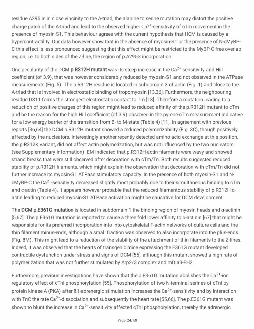
residue A295 is in close vincinity to the A-triad, the alanine to serine mutation may distort the positivecharge patch of the A-triad and lead to the observed higher Ca2+-sensitivity of cTm movement in thepresence of myosin-S1. This behaviour agrees with the current hypothesis that HCM is caused by ahypercontractility. Our data however show that in the absence of myosin-S1 or the presence of N-cMyBP-C this effect is less pronounced suggesting that this effect might be restricted to the MyBP-C free overlapregion, i.e. to both sides of the Z-line, the region of p.A295S incorporation.
One pecularity of the DCM p.R312H mutant was its steep increase in the Ca2+-sensitivity and Hillcoe�cient (of 3.9), that was however considerably reduced by myosin-S1 and not observed in the ATPasemeasurements (Fig. 5). The p.R312H residue is located in subdomain 3 of actin (Fig. 1) and close to theA-triad that is involved in electrostatic binding of tropomyosin [13,36]. Furthermore, the neighbouringresidue D311 forms the strongest electrostatic contact to Tm [13]. Therefore a mutation leading to areduction of positive charges of this region might lead to reduced a�nity of the p.R312H mutant to cTmand be the reason for the high Hill coe�cient (of 3.9) observed in the pyrene-cTm measurement indicativefor a low energy barrier of the transition from B- to M-state (Table 4) [11]. In agreement with previousreports [36,64] the DCM p.R312H mutant showed a reduced polymerizability (Fig. 3C), though positivelyaffected by the nucleators. Interestingly another recently detected amino acid exchange at this position,the p.R312K variant, did not affect actin polymerization, but was not in�uenced by the two nucleators(see Supplementary Information). EM indicated that p.R312H-actin �laments were wavy and showedstrand breaks that were still observed after decoration with cTm/Tn. Both results suggested reducedstability of p.R312H �laments, which might explain the observation that decoration with cTm/Tn did notfurther increase its myosin-S1 ATPase stimulatory capacity. In the presence of both myosin-S1 and N-cMyBP-C the Ca2+-sensitivity decreased slightly most probably due to their simultaneous binding to cTmand c-actin (Table 4). It appears however probable that the reduced �lamentous stability of p.R312H c-actin leading to reduced myosin-S1 ATPase activation might be causative for DCM development.
The DCM p.E361G mutation is located in subdomain 1 the binding region of myosin heads and α-actinin[5,67]. The p.E361G mutation is reported to cause a three fold lower a�nity to α-actinin [67] that might beresponsible for its preferred incorporation into into cytoskeletal F-actin networks of culture cells and thethin �lament minus-ends, although a small fraction was observed to also incorporate into the plus-ends(Fig. 8M). This might lead to a reduction of the stability of the attachment of thin �laments to the Z-lines.Indeed, it was observed that the hearts of transgenic mice expressing the E361G mutant developedcontractile dysfunction under stress and signs of DCM [55], although this mutant showed a high rate ofpolymerization that was not further stimulated by Arp2/3 complex and mDia3-FH2.
Furthermore, previous investigations have shown that the p.E361G mutation abolishes the Ca2+-ionregulatory effect of cTnI phosphorylation [55]. Phosphorylation of two N-terminal serines of cTnI byprotein kinase A (PKA) after ß1-adrenergic stimulation increases the Ca2+-sensitivity and by interactionwith TnC the rate Ca2+-dissociation and subsequently the heart rate [55,66]. The p.E361G mutant wasshown to blunt the increase in Ca2+-sensitivity affected cTnI phosphorylation, thereby the adrenergic

Page 25/40
responsiveness of cardiac muscle is lost leading to a decrease of its Ca2+-sensitivity further supportingthe development of DCM [55].
In summary, our data indicate that each c-actin mutation possesses its own speci�c alterations in themode of interactions with particular sarcomeric proteins. Apart from the general accepted notion thatalterations in the Ca2+-sensitivity affecting the contractlity can lead to either HCM or DCM, our datademonstrate that modi�es interactions of mutant c-actins with other sarcomeric proteins duringdevelopment and in adult cardiac muscle may also play an essential role for CM establishment.
Declarations
Acknowledgement:The �nancial support by the Deutsche Forschungsgemeinschaft (DFG: Ma807/19-1), and the GermanAcademic Exchange Service (DAAD), Bad-Godesberg, Germany, for RH are gratefully acknowledged.
Con�icts of interest/Competing interests:The authors declair no con�icts of interests.
Availability of data and material
Code availability(software application or custom code)
Authors' contributions:All authors contributed to the study conception and design. Material preparation, data collection andanalysis were performed by C. Erdmann, R. Hassoun, S. Schmitt, S. Fujita-Becker, A.J. Mazur, and H.G.Mannherz. The �rst draft of the manuscript was written by H.G. Mannherz and all authors commented onprevious versions of the manuscript. All authors read and approved the �nal manuscript.
References1. Babu A, Scordilis SP, Sonnenblick EH & Gulati J (1987) The control of myocardial contraction with
skeletal fast muscle troponin. J Biol Chem 262: 5815–5822.
2. Bai F, Caster HM, Rubenstein PA, Dawson JF, and Kawai M (2014) Using baculovirus/insect cellexpressed recombinant actin to study the molecular pathogenesis of HCM caused by actin mutation

Page 26/40
A331P. J Mol Cell Cardiol 74: 64-75.
3. Barry SP, Davidson SM, Townsend PA (2008) Molecular regulation of cardiac hypertrophy. TheInternational J Biochem Cell Biol 40: 2023–2039.
4. Bailey K (1948) Tropomyosin: a new asymmetric protein component of the muscle �bril. BiochemicalJournal 43(2): 271-279
5. Behrmann E, Müller M, Penczek PA, Mannherz H G, Manstein DJ, Raunser S (2012) Structure of therigor actin-tropomyosin-myosin complex. Cell 150: 327–338.
�. Bookwater CS, Trybus KM (2006) Functional consequences of a mutation in an expressed humanalpha-cardiac actin at a site implicated in familial hypertrophic cardiomyopathy. J Biol Chem 28:16777-84.
7. Bradford MM (1976) A rapid and sensitive method for the quantitation of microgram quantities ofprotein utilizing the principle of protein-dye binding. Anal Biochem 72: 248–254.
�. Chobra A, Kutys ML, Zhang K, Polacheck WJ, Sheng CC, Luu RJ, Eyckmans J, Hinson JT, SeidmanJG, Seidman CE, Chen CS (2018) Force generation via β-cardiac myosin, titin, and α-actinin drivescardiac sarcomere assembly from cell-matrix adhesions. Dev Cell 44: 87-96.
9. Chow ML, Shaffer JF, Harris SP, Dawson JF (2014) Altered interactions between cardiac myosinbinding protein-C and α-cardiac actin variants associated with cardiomyopathies. Arch BiochemBiophys 15: 28-32.
10. Cimiotti D, Fujita-Becker S, Möhner D, Smolina N, Budde H, Wies A, Morgenstern L, Gudkova A,Sejersen T, Sjöberg G, Mügge A, Nowaczyk MM, Reusch P, P�tzer G, Stehle R, Schröder RR, MannherzHG, Kostareva A, Jaquet K (2020) Infantile restrictive cardiomyopathy: CtnI-R170G/W impair theinterplay of sarcomeric proteins and the integrity of thin �laments. PLoS One. 15(3): e0229227,doi: 10.1371/journal.pone.0229227.
11. Debold EP, Saber W, Cheema Y, Bookwalter CS, Trybus KM, Warshaw DM, Vanburen P (2010) Humanactin mutations associated with hypertrophic and dilated cardiomyopathies demonstrate distinctthin filament regulatory properties in vitro. J Mol Cell Cardiology 48: 286–292.
12. Deng Y, Schmidtmann A, Redlich A, Westerdorf B, Jaquet K, Thieleczek R (2001) Effects ofPhosphorylation and Mutation R145G on Human Cardiac Troponin I Function. Biochemistry 40:14593-14602.
13. Ebashi S, Endo M, Otsuki I (1969) Control of muscle contraction. Rev Biophys 2:351–384.
14. Ehler E, Moore-Morris T, Lange S (2013) Isolation and culture of neonatal mouse cardiomyocytes. JVis Exp 50154.
15. Ehler E (2018) Actin-associated proteins and cardiomyopathy-the ‘unknown’ beyond troponin andtropomyosin. Biophys Rev 10(4): 1121–1128.
1�. Erdmann C (2017) Charakterisierung von HCM und DCM auslösenden Mutationen in humanemkardialem α-Aktin. PhD-thesis, Ruhr-University Bochum.

Page 27/40
17. Fananapazir L (2000) Inherited and de novo mutations in the cardiac actin gene cause hypertrophiccardiomyopathy. J Mol Cell Cardiology 32: 1687–1694.
1�. Feng JJ, Marston S (2009) Genotype–phenotype correlations in ACTA1 mutations that causecongenital myopathies. Neuromus Disorders 19: 6–16.
19. Geeves MA, Lehrer SS (1984) Dynamics of the muscle thin �lament regulatory switch: the size of thecooperative unit. Biophys J 67(1): 273–282.
20. Geeves MA, Lehrer SS, Lehman W (2019) The mechanism of thin �lament regulation: Models incon�ict. J Gen Physiol 151: 1265- 1271.
21. Graceffa P, Lehrer SS (1980) The eximer �uorescence of pyrene-labeled tropomyosin: A probe forconformational dynamics. J Biol Chem 255: 11296-11300.
22. He T-C, Zhou S, da Costa LT, Yu J, Kinzler KW, Vogelstein B (1998) A simpli�ed system for generatingrecombinant adenoviruses. Proc Natl Acad USA 95(5): 2509–2514.
23. Herman DS, Lam L, Taylor MRG, Wang L, Teekakirikul P, Christodoulou Conner DL, Steven R.McDonough DB, Sparks E, et al (2012) Truncations of Titin causing Dilated Cardiomyopathy. NewEngl J Medicine 366: 619-628.
24. Hesterkamp T, Weeds AG, Mannherz HG (1993) The two actins of the gelsolin:2 actin complex are inantiparallel orientation. Eur J Biochem 318: 507-513.
25. Ishii Y, Lehrer SS (1990) Excimer �uorescence of pyrenyliodoacetamide- labeled tropomyosin.Biochemistry 29: 1160-1166.
2�. Jahn W (1995) Easily prepared holey films for use in cryo-electron microscopy. J Microscopy 179:333–334.
27. Kabsch W, Mannherz HG, Pai E, Suck D, Holmes KC (1990) The atomic structure of actin:DNase Icomplex. Nature 347: 37-44.
2�. Kouyama T, Mihashi K (1981) Fluorimetry study of N-(1-pyrenyl) Iodoacetamide-labelled F-actin.Local structural change of actin protomer both on polymerization and on binding of heavymeromyosin. Eur J Biochem 114: 33–38.
29. Kühn S, Geyer M (2014) Formins as effector proteins of Rho GTPases. Small GTPases 5: e29513.
30. Laemmli UK (1970) Cleavage of structural proteins during the assembly of the head ofbacteriophage T4. Nature 227: 680-685.
31. Lawrence JB, Singer RH (1986) Intracellular localization of messenger RNAs for cytoskeletalproteins. Cell 45:407–415.
32. Lehman W, Rynkiewicz MJ, Moore JR (2020) A new twist on tropomyosin binding to actin �laments:perspectives on thin �lament function, assembly and biomechanics J Muscle Res Cell Motility 41:23-38
33. Lynn ML, Lehman SJ, Tadiff JC (2018) Biophysical Derangements in Genetic Cardiomyopathies.Heart Failure Clin. 14: 147-159.

Page 28/40
34. Mannherz HG, Brehme H, Lamp U (1975) Depolymerization of F-actin and its repolymerization in thepresence of analogs of adenosine triphosphate. Eur J Biochem 60: 109-116.
35. Mannherz HG, Goody, R.S., Konrad, M., and Nowak, E. (1980) The interaction of pancreaticdeoxyribonuclease I and skeletal muscle actin. Eur J Biochem 104: 367-379.
3�. Marston SB (2011) How do mutations in contractile proteins cause the primary familialcardiomyopathies? J Cardiovascular Translational Res 4: 245–255.
37. Mazur AJ (2008) Expression of constructs of WT-alpha-cardiac actin and its mutants in different celllines and primary rat cardiac myocytes. PhD-thesis, Ruhr-University Bochum.
3�. Moreno Gonzales A, Drapala P, Kruetziger K, Regnier M (2003) Decreased Ca2+ binding by troponin Cisoforms enhances crossbridge contribution to thin �lament activation. Biophys J 84: 449a.
39. Müller M, Mazur JA, Behrmann E, Diensthuber RP, Radke MP, Qu Z, Littwitz C, Raunser S,Schoenenberger C-A, Manstein DJ, Mannherz HG (2012) Functional characterization of the human α-cardiac actin mutations Y166C and M305L involved in hypertrophic cardiomyopathy. Cellul MolecLife Sci 69; 3457-3479.
40. Mogensen J, Perrot A, Andersen PS, Havndrup O, Klausen IC, Christiansen M, Bross P, Egeblad H,Bundgaard H, Osterziel KJ, Haltern G, Lapp H, Reineke P, Gregersen N, Borglum AD (2004) Clinicaland genetic characteristics of alpha cardiac actin gene mutations in hypertrophic cardiomyopathy. JMed Genet 41: e10, doi 10.1136/jmg 2003.010447.
41. Mundia MM, Demers RW, Chow ML, Perieteanu AA, Dawson JF (2012) Subdomain location ofmutations in cardiac actin correlate with type of functional change. Plos ONE 7: e3621.
42. Ohki T, Ohno C, Oyama K, Mikhailenko SV, Ishiwata S (2009) Puri�cation of cytoplasmic actin bya�nity chromatography using the C-terminal half of gelsolin. Biochem Biophys Res Commun 383:146–150.
43. Offer G (2015) Myosin-binding protein-C: bridging the gap. J Mol Biol 427: 231- 235.
44. Olson TM, Michels VV, Thibodeau SN, Tai Y-S, Keating MT (1998) Actin mutations in DilatedCardiomyopathy, a heritable Form of Heart failure. Science 280: 750-752.
45. Olson TM, Doan TP, Kishimoto NY, Whitby FG, Ackerman MJ, Fananapazir L (2000) Inherited and denovo Mutations in the cardiac actin gene cause hypertrophic cardiomyopathy. J Mol Cell Cardiol 32:1687- 1694.
4�. Parker F, Baboolal TG, Peckham M (2020) Actin mutations and their role in disease. Int. J Sciences21: 3371-3387.
47. Qu Z, Silvan U, Jockusch BM, Aebi U, Schoenenberger CA, Mannherz HG (2015) Distinct oligomersmodulate differently the activity of actin nucleators. FEBS J 282(19): 3824-3840.
4�. Qu Z, Fujita-Becker S, Ballweber E, Ince S, Herrmann C, Schröder RR, Mannherz HG (2018) Interactionof isolated cross-linked short actin oligomers with the skeletal muscle myosin motor domain. FEBS J285(9):1715-1729. doi: 10.1111/febs.14442.

Page 29/40
49. Reiffert S, et al., Characterization of the cardiac holotroponin complex reconstituted from nativecardiac troponin T and recombinant I and C. The FEBS Journal, 1999. 261(1): p. 40-47
50. Reinecke P, Gregersen N, Børglum AD (2004) Clinical and genetic characteristics of alpha cardiacactin gene mutations in hypertrophic cardiomyopathy. J Medical Genetics 41: e10.
51. Risi C, Belknap B, Forgacs-Lonart E, Harris SP, Schröder GF, White HD, Galkin VE (2018) N-terminaldomains of cardiac myosin binding protein C cooperatively activate the thin �lament. Structure 26: 1-8.
52. Rosado M, Barber CF, Berciu C, Feldman S, Birren SJ, Nicastro D, Good BL (2014) Critical roles formultiple formins during cardiac myo�bril development and repair. Mol Biol Cell 25(6): 811–827.
53. Sabater-Molina M, Pérez-Sánchez I, Hernández Del Rincón JP, Gimeno JR (2017) Genetics ofhypertrophic cardiomyopathy: a review of current state. Clinical Genetics 93: 3-14.
54. Seidman JG, Seidman C (2001) The genetic basis for cardiomyopathy: from mutation identificationto mechanistic paradigms. Cell 104: 557–567.
55. Song W, Dyer E, Stuckey D, Leung M-C, Memo M, Mansfeld C, Ferenci M, Liu K, Redwood C, Nowak K,Harding S, Clarke K, Wells D, Marston S (2010) Investigation of a transgenic mouse model of familialdilated cardiomyopathy. J Mol Cell Cardiol 49: 380-389.
5�. Spudich JA (2015) The myosin mesa and a possible unifying hypothesis fort he molecular basis ofhuman hypertrophic cardiomyopathy. Biochem Soc Trans 43: 64-72.
57. Starr R, Offer G (1978) The interaction of C-protein with heavy meromyosin and subfragment 2.Biochem J 171: 813-816.
5�. Tanaka H, Sekine T, Kawanishi T, Nakamura R, Shigenobu K (1998) Intrasarcomere [Ca2+] gradientsand their spatio-temporal relation to Ca2+ sparks in rat cardiomyocytes. J Physiol 508: 145-152.
59. Tayler RS, Weeds AG (1976) The magnesium-ion-dependent adenosine triphosphatase of bovinecardiac Myosin and its subfragment-1. Biochem J 159: 301-315.
�0. Towbin H, Staehelin T, Gordon J (1979) Electrophoretic transfer of proteins from polyacrylamide gelsto nitrocellulose sheets: procedure and some applications. Proc Nat Acad Sci USA 76: 4350–4354.
�1. Trentham DR, Bardsley RG, Eccleston JF, Weeds AG (1972) Elementary processes of the magnesiumion-dependent adenosine triphosphatase activity of heavy meromyosin. A transient kinetic approachto the study of kinases and adenosine triphosphatases and a colorimetric inorganic phosphateassay in situ. The Biochem J 126: 635–644.
�2. Trivedi DV, Adhikari AS, Sarkar SS, Ruppel KM, Spudich JA (2018) Hypertrophic cardiomyopathy andthe myosin mesa: viewing an old disease in a new light. Biophys Rev 10: 27–48.
�3. von der Ecken J, Müller M, Lehman W, Manstein DJ, Penczek PA, Raunser S (2015) Structure of the F-actin-tropomyosin complex. Nature 519: 114–117.
�4. Viswanathan MC, Schmidt W, Rynkiewivz MJ, Agarwal K, Gao J, Katz J, Lehman W, Cammarato A(2017) Distortion of the actin A-triad results in contractile disinhibition and cardiomyopathy. CellReports 20: 2612-2625.

Page 30/40
�5. Vikhorev PG, Song W, Wilkinson R, Copeland O, Messer AE, Ferenczi MA, and Marston SB (2014) TheDilated Cardiomyopathy-Causing Mutation ACTC E361G in Cardiac Muscle Myo�brils Speci�callyAbolishes Modulation of Ca2+ Regulation by Troponin I. Biophys J 107: 2369–2380.
��. Weintraub RG, Semsarian C, Macdonald P (2017) Dilated cardiomyopathy. Lancet (London, England)390: 400-414.
�7. Wong WW, Doyle TC, Cheung P, Olson TM, Reisler E (2001) Functional studies of yeast actin mutantscorresponding to human cardiomyopathy mutations. J Muscle Res Cell Motil 22: 665-674.
Figures
Figure 1
3D structural model of G-actin derived from the skeletal muscle actin:DNase I complex [27] indicating thepositions of the mutated residues of the c-actin mutants investigated. In addition subdomains (SD) aswell as the position of ATP and Ca2+ are indicated.
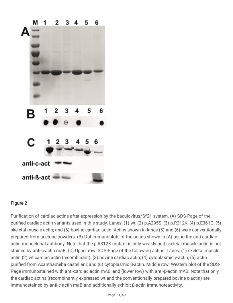
Page 31/40
Figure 2
Puri�cation of cardiac actins after expression by the baculovirus/Sf21 system. (A) SDS-Page of thepuri�ed cardiac actin variants used in this study. Lanes: (1) wt; (2) p.A295S; (3) p.R312K; (4) p.E361G; (5)skeletal muscle actin; and (6) bovine cardiac actin. Actins shown in lanes (5) and (6) were conventionallyprepared from acetone powders. (B) Dot immunoblots of the actins shown in (A) using the anti-cardiacactin monoclonal antibody. Note that the p.R312K mutant is only weakly and skeletal muscle actin is notstained by anti-c-actin maB. (C) Upper row: SDS-Page of the following actins: Lanes: (1) skeletal muscleactin (2) wt cardiac actin (recombinant); (3) bovine cardiac actin; (4) cytoplasmic γ-actin; (5) actinpuri�ed from Acanthameba castellani; and (6) cytoplasmic β-actin. Middle row: Western blot of the SDS-Page immunostained with anti-cardiac actin mAB; and (lower row) with anti-β-actin mAB. Note that onlythe cardiac actins (recombinantly expressed wt and the conventionally prepared bovine c-actin) areimmunostained by anti-c-actin maB and additionally exhibit β-actin immunoreactivity.
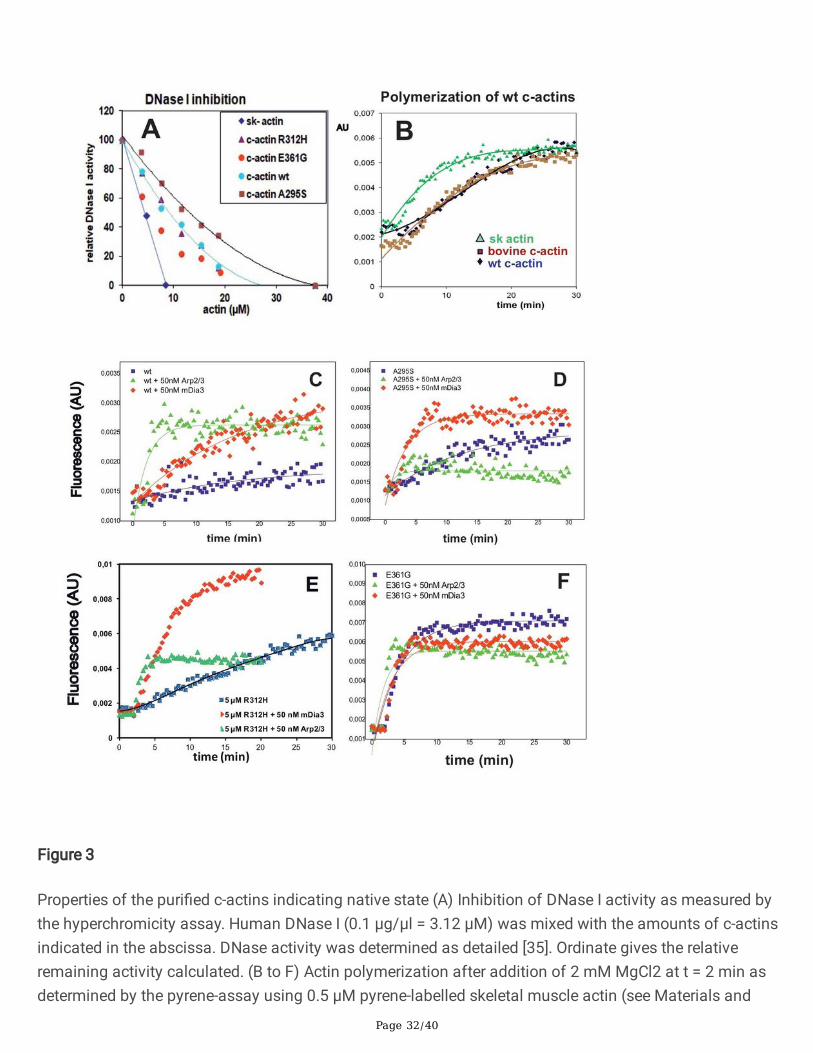
Page 32/40
Figure 3
Properties of the puri�ed c-actins indicating native state (A) Inhibition of DNase I activity as measured bythe hyperchromicity assay. Human DNase I (0.1 μg/μl = 3.12 μM) was mixed with the amounts of c-actinsindicated in the abscissa. DNase activity was determined as detailed [35]. Ordinate gives the relativeremaining activity calculated. (B to F) Actin polymerization after addition of 2 mM MgCl2 at t = 2 min asdetermined by the pyrene-assay using 0.5 μM pyrene-labelled skeletal muscle actin (see Materials and

Page 33/40
methods). (B) Comparison of puri�ed sk-actin and bovine c-actin puri�ed from acetone powders withrecombinant wt c-actin, each at 5 μM Note the faster polymerization rate of sk-actin and the equal ratesof c-actins. (C to F) In�uence of nucleators on the rates of polymerization of c-actins. (C) Recombinantwt-c-actin, (D) p.A295S, (E) p.R312H, and (F) p.E361G. All c-actins at 5 μM and in the absence of presenceof either 50 nM Arp2/3 complex or 50 nM mDia3-FH2 (for details see text).
Figure 4

Page 34/40
EM analysis after negative staining of the polymerized c-actins before and after decoration with cTm andcTn at a 7:1:1 molar ratio. (A,A´) Wt recombinant c-actin, (B,B´) bovine c-actin, (C,C´) p.A295S, (D,D´)p.R312H (arrow heads point to strand breaks and wavy �laments), and (E,E´) p.E361G. Scale bar in (E´) =100 nm (applicable to all images).
Figure 5

Page 35/40
Ca2+-concentration dependence of the stimulation of the myosin-S1 ATPase by c-actins decorated withcTm and cTn at 6:1:1 molar ratio. Skeletal muscle myosin-S1 ATPase activity was determined by theenzyme-linked assay. ATPase stimulation by (A) recombinant wt c-actin, (B) bovine c-actin, and (C)comparison of (A) and (B). ATPase stimulation by (D) p.A295S, (E) p.R312H, and (F) p.E361G. (G)Comparison of the mutant forms of c-actin. Final F-c-actin concentrations in assay were 1.5 μM and ofrabbit skeletal muscle myosin-S1 0.5 μM. ATPase activity (ordinate) is given as μM ATP hydrolysed/sec/μM myosin-S1 in (A;B;D-F) and normalized in (C and G) setting the minimal ATPase activity (generally atpCa 9.3) as zero and the maximal at 1 (generally at pCa 4.8). Abscissa gives pCa-values (-log molarCa2+-concentration). All measurements were performed in triplicate.

Page 36/40
Figure 6
Comparison of the Ca2+-concentration dependence of the �uorescence increase (movement) of pyrene-labelled cTm on bovine and recombinant c-actin variants. Pyrene-labelled cTm was used to determine itsmovement along �lamentous bovine and recombinant wt c-actin (for details see text). The c-actins (�nalconcentration in microtiter plate 0.4 μM) were decorated with pyrene-labelled cTm and cTn at a molarratio of 6:1:1 (see Materials and methods) and the Ca2+-dependence of the �uorescence increase of the

Page 37/40
pyrene-labelled cTm corresponding to Tm-movement was determined using a Tecan Elisa-reader asdetailed in Materials and methods [10]. (A) Comparison of bovine c-actin and recombinant wt c-actindecorated with pyrene-cTm and cTn in the absence and (B) the presence of N-cMyBP-C. (C,D) Identicalexperiment to (A) and (B) using the recombinant c-actins. (E,F) Identical experiment to (C) and (D) usingthe recombinant c-actins in the additional presence of myosin-S1. The ordinates give the �uorescenceintensities (F) normalized to Fmax = 1, Fmin = 0. and abscissa the pCa (for compilation of data see alsoTable 4). The colour coding for the c-actins is given in the Figure.

Page 38/40
Figure 7
Transfection of MDCK cells with HA-tagged c-actin variants. The MDCK cells were transfected withvectors containing HA-tagged c-actin variants (for details see Materials and Methods). Subsequently thecells were immunostained with anti-HA and counterstained with TRITC-phalloidin. (A) wt c-actin stainedwith anti-HA (green); (A´) TRITC-phalloidin staining; (A´´) merge. (B-B´´) Identical staining for the p.R312Hmutant. (C) Merged image of identical staining of p.A295S and (D) of the p.E361G mutant. Note for wtand the p.A295S mutant c-actins preferential incorporation in the stress �bre system, whereas thep.R312H and p.E361G mutants showed a more circumferential anti-HA staining suggesting preferredincorporation into short cortical actin �laments. Bars: 50 μm. (E-E´) Transduction of NRCs with adenoviralconstructs encoding wt c-actin. (E) Immunostaining with anti-HA antibody (green), (E´) with TRITC-phalloidin (red), and (E´´) merged image also including nuclear staining with Hoechst 33342 (blue). (F,G,I)Merged images of NRCs expressing after transduction the p.A295S, R312H, and E361G mutants,respectively, stained by the identical procedure. Bars: 20 μm.

Page 39/40
Figure 8
NRCs expressing HA-tagged c-actin variants counterstained with anti-c-actin mAB. (A) NRCs transducedwith adenoviral vector containing HA-tagged wt c-actin stained with anti-HA (green = exogenous) and (A´)with anti-c-actin mAB (red = endogenous), (A´´) merge. Note localisation of endogenous c-actin in cellperiphery and extensions. (B) Merged images of identical immunostaining of NRCs infected with HA-tagged p.A295S; (C) p.R312H, and (D) p.E361G. Note partial peripheral co-localisation of endo- and

Page 40/40
exogenous c-actin in (B) and (D), whereas in (C) the endogenous c-actin (red) appears more peripherallyconcentrated without signi�cant co-localising exogenous c-actin. Bars: 20 μm. (E-M) NRCs expressingHA-tagged c-actin variants counterstained with anti-α-actinin or anti-myomesin. (E) NRC transduced withvectors coding wt HA-tagged c-actin stained with anti-HA (green) were counterstained with (E) anti-α-actinin (red) showing cross-striations (arrows). (E´) gives merged image. Note that cross-striation appearin many cases yellowish stained indicating co-localisation (arrows). (F,G,I) gives merged images ofidentical staining protocol for p.R312H (F), p.A295S (G), and p.E361G (I). Note that in (F) and (C) thecross-striations are yellowish stained indicating also for HA-p.R312H and HA-p.A295S co-localisationwith α-actinin suggesting preferred incorporation in the region of the Z-line, i.e. at the plus-ends. Incontrast, HA-p.E361G (I) transduced NRCs show a clear separation of the anti-HA (green) and anti-α-actinin immunostaing (red) indicating incorporation at the minus ends in the region of the M-line. (J-J´)Similar immunostaining of wt HA-tagged c-actin (green) and anti-myomesin (red). Note separation ofanti-HA and -myomesin in merged image (J´´). (K) merged image for HA-p.A295S and (L) for HA-p.R312Hc-actins. Note separation of anti-HA and –myomesin staining in (K and L). In contrast, (M) NRCtransduced with HA-p.E361G indicates co-localisation of this c-actin mutant with myomesin supportingthe notion that its incorporation into sarcomeric structures occurs at the minus ends (arrow) of existingactin �laments in the region of the M-line. In addition weakly stained anti-HA-immunoreactivity is visiblein the middle between the anti-myomesin bands (arrow head). Bars give 20 μm.
Supplementary Files
This is a list of supplementary �les associated with this preprint. Click to download.
SupplementaryInformation.docx