Embed Size (px)
Citation preview


Czech Anatomical Society Czech Society for Histochemistry and Cytochemistry
Charles University First Faculty of Medicine
MORPHOLOGY 2021 52nd International Congress on Anatomy 57th Lojda Symposium on Histochemistry
Under the Auspices of
Prof. MUDr. Tomáš Zima, DrSc., MBA Rector of the Charles University
Prof. MUDr. Martin Vokurka, CSc.
Dean of the First Faculty of Medicine, Charles University
Honorary Committee O. Naňka, J. Mokrý, K. Smetana, T. Kučera, E. Mechírová
Organizing and Scientific Committee
J. Bartoníček, D. Kachlík, I. Klepáček, H. Kolesová, A. Kvasilová, T. Kučera, O. Naňka, V. Nemravová, D. Sedmera, A. Shbat, K. Smetana, K. Strnadová, P.
Szabo, Z. Vačkářová, Ortopedické centrum s. r. o.
Topics
Oncology, Early Morphogenesis, Morphogenesis, Neurosciences, Clinical Anatomy, Teaching of Anatomy
Prague, September 9 – 11, 2021

2
General Information
Venue Institute of Anatomy, First Faculty of Medicine, U Nemocnice 3, CZ-128 00 Prague 2, phone: +420 224 965 780, fax. +420 224 965 770 e-mail: [email protected], web page: cas.lf1.cuni.cz, morphology.lf1.cuni.cz Registration and Information Desk Ground level of the Institute of Anatomy, First Faculty of Medicine, Charles University U Nemocnice 3, Prague 2, phone: 224 96 5 718 Office Hours: Thursday, September 9th, 2021 15:00 - 17:00 Friday, September 10th, 2021 08:00 - 17:00 Saturday, September 11th, 2021 08:00 - 17:00 Languages English, Czech, Slovak Oral Presentations Plenary lectures - 20 min, lectures - 10 min, discussion - 5 min. Windows PC with USB port are installed in lecture halls. Presentation software is PowerPoint 365. Videos can be presented only through PowerPoint presentation. Speakers are kindly asked to check their presentations at the lecture hall before beginning of each session. Poster Presentation The size of poster panel is 115 cm (height) and 90 cm (width). All posters will be displayed from Friday to Saturday in dissection rooms (ground level). Posters will be presented on Friday, September 10. Pushpins will be available. Authors are asked to be present at posters during the poster presentation. Industrial Exhibition The exhibitions are located at the ground level and open throughout the meeting. Exhibitors: Animalab, s.r.o. Asklepion, s.r.o. Bamed, s.r.o. Baria, s.r.o. Elsevier EXBIO Praha, a.s. Grada Publishing, a.s. HELAGO-CZ s.r.o. Cheirón, a.s.
Jacek Lewinson Kuba Libri, s.r.o. Olympus Czech Group, s.r.o. P-Lab, a.s. Schoeller Instruments, s.r.o. Thieme Medical Publishers TRIGON PLUS, s.r.o. Wolters Kluwer
Welcome Reception will be held immediately after the Opening ceremony on Thursday, September 9th, in the Institute of Anatomy building. It will include buffet and drinks. It is free of charge.

3
Social Evening Congress dinner will take place in the backyard of the Institute of Anatomy, U Nemocnice 3, 120 00 Praha 2 on Friday, September 10th from 19:00 until 22:00 (price 500,- CZK). Coffee breaks Refreshments will be served in the rooms of the Institute of Anatomy, free of charge. Lunch Lunches will be served in the rooms of the Institute of Anatomy, pre-paid by attendant (price 120,- CZK). Transportation See city maps in this booklet, pp. 120. How to reach the Institute of Anatomy – U Nemocnice 3, Praha 2 By tram No. 4, 6, 10, 16, 22, station Štěpánská; by tram No. 2, 3, 4, 10, 16, 18, 24 station Moráň or Karlovo náměstí By Metro: line C (red) - station I. P. Pavlova; line B (yellow) - station Karlovo náměstí, exit to Karlovo náměstí. By car: parking in streets of Prague 2 is paid-parking The blue zone is intended for resident parking. Other motorists may park in the blue zone for a limited period of time only after paying the parking fee via the Virtual Parking Meter (mpla.cz/Prague) web app. The purple zone is intended for so-called mixed parking. Only people with a valid parking permit can park in the purple zone without restrictions. This can be issued on the basis of permanent residence in the given area and proof of legal relationship to the vehicle. Other motorists can park in the purple zone for a maximum of 24 hours, and only after payment in a parking meter or via the Virtual Parking Clock web application (mpla.cz/Prague). The orange zone is intended for short-term parking. Time restrictions apply to parking here. Payment can be made via a parking meter or through the Virtual Parking Meter web app (mpla.cz/Prague). Czech Anatomical Society – member of the Council of Scientific Societies of the Czech Republic Secretary office: Institute of Anatomy, First Faculty of Medicine, Charles University, U Nemocnice 3, CZ 128 00 Prague 2 phone: 00 420 224 965 780, fax. 00 420 224 965 770, e-mail: [email protected] http://cas.lf1.cuni.cz Czech Society for Histo- and Cytochemistry – member of the Council of Scientific Societies of the Czech Republic Secretary office: Šimkova 870, 500 38 Hradec Králové. phone: +420-495 816 294, fax: +420-495 816 376, e-mail: [email protected] http://www.cshc.cz

4
Transit tariffs
Transit ticket vending machines Ticket vending machines are installed in all metro stations and at selected surface transit stops. They are intended for the purchase of individual tickets. SMS ticket Single transfer tickets can be purchased via SMS. Send SMS DPT42 to purchase regular 90 minute ticket for 42 CZK DPT31 to purchase reduced 30 minute ticket for 31 CZK DPT120 to purchase 24hour ticket for 120 CZK* DPT330 to purchase 72hour ticket for 330 CZK* to number 902 06. You will receive SMS ticket within approximately 2 minutes. * Then confirm by replying „Yes“. Advance ticket sales Sales locations in the metro offer the entire range of tickets, and are intended primarily for the sale of transit passes, both for fixed and sliding validity periods. Info centres Information centres sell individual tickets and short-term (tourist) passes. Tobacconists and wholesalers Selected tobacconists and wholesalers sell individual tickets.
5
Czech Anatomical Society Czech Society for Histochemistry and Cytochemistry
Charles University in Prague First Faculty of Medicine
Programme
MORPHOLOGY 2021 52nd International Congress on Anatomy 57th Lojda Symposium on Histochemistry
September 9 – 11, 2021 Prague, Czech Republic
6
Thursday, September 9, 2021 17:00 - 19:00 Opening Ceremony Chairs: Ondřej Naňka, Eva Mechírová, Jaroslav Mokrý Words of Welcome Jan Royt “The Valley of Dry Bones“ – about iconography of skeleton in art Hans J. ten Donkelaar Recent Developments in Neuroanatomical Terminology: An Introduction to the TNA and Beyond 2021 Czech Anatomical Society & Olympus Award - Winner Karolína Strnadová, Jiří Novotný Biological models to study melanoma behaviour and tumour microenvironment: using chicken chorioallantoic membrane and single-cell RNA sequencing 2021 Czech Anatomical Society & Olympus Award – Runner-Up Peter Solár Subarachnoid Hemorrhage Induces Dynamic Immune Cell Reactions in the Choroid Plexus Music performance (M. Španko, T. Bartošová) 19:00 – 20:30 Welcome drink

7
Friday, September 10, 2021 8:30 - 17:30 Plenary Lectures, Sessions in Section A and B, Poster Session 8:30 - 9:20 Plenary Lectures: Oncology + Morphogenesis (Lecture Hall A) Chairs: David Sedmera, Eva Mechírová 8:30 - 8:55 1 – Smetana K Jr Interleukin-6: Molecule in the intersection of cancer, ageing and COVID-19 8:55 - 9:20 2 – Černý R The Old Secrets of the New Head of Vertebrates
9:20 - 9:35 Coffee Break 9:35 - 11:05 Oncology (Lecture Hall A) Chairs: Karel Smetana Jr., Marian Adamkov 9:35 – 9:50 3 – Adamkov M, Krajňáková B, Csizmárová S, Mešťanová V, Škuciová V Is surviving level identical between adenomas of proximal and distal colon? 9:50 – 10:05 4 – Csizmárová S, Mešťanová V, Krajňáková B, Adamkov M Immunohistochemical analysis of fascin and its function in EMT in cervical lesions 10:05 – 10:20 5 – Hurník P, Štembírek J, Ševčíková T, Chyrá Z, Putnová B, Čermáková Zděblová Z, Buchtová M
Morphological study of HNSCC focused on perineural invasion - a single institutional study with five year follow up
10:20 – 10:35 6 – Mešťanová V, Csizmárová S, Krajňáková B, Adamkov M EMT and galectin expression in uterine cervix lesions 10:35 – 10:50 7 – Pavliuk-Karachevtseva A, Mihalik J, Benetinová Z, Flešárová S, Rybárová S, Hodorová I Detection of selected glutathione peroxidases in human colorectal carcinoma 10:50 – 11:05 8 – Štembírek J, Hurník P, Putnová B, Chyrá Z, Ševčíková T, Čermáková Zděblová Z, Blažek T, Buchtová M "Molecular" resection margins in squamous cell carcinoma of the orofacial region

8
9:35 – 11:05 Morphogenesis (Lecture Hall B) Chairs: Robert Černý, Marcela Buchtová 9:35 – 9:50 9 – Steklíková K, Dalecká L, Pavlíková Z, Hovořáková M Composite development of the mouse first molar and supernumerary tooth formation 9:50 – 10:05 10 – Moldovan Putnová B, Putnová I, Hrubá E, Hurník P, Štembírek J, Daněk Z, Buchtová M Wnt Signalling in Ameloblastoma 10:05 – 10:20 11 – Gregorovičová M, Šaňková B, Bartoš M, Sedmera D
Reptiles as a model in myocardial regeneration
10:20 – 10:35 12 – Kolesová H, Lapierre-Landry M, Kvasilová A, Bartoš M, Watanabe M, Sedmera D Imaging of coronary microvasculature development 10:35 – 10:50 13 – Jandová N, Kohoutek J, Procházka J, Kavková M, Zikmund T, Hampl M, Buchtová M Role of CDK13 in limb morphogenesis 10:50 – 11:05 14 – Olbertová K, Hrčkulák D, Kříž V, Hrubá E, Kořínek V, Buchtová M Fate of mesenchymal LGR5-positive cells during craniofacial development 11:05 – 11:30 Coffee Break + Congress photo 11:30 - 12:30 Neurosciences 1 (Lecture Hall A) Chairs: Petr Dubový, Marek Joukal 11:30 - 11:45 15 – Dubový P, Bretová K, Svobodová V, Bagó A, Boadas-Vaello P Fractalkine/CX3CL1 and its receptor CX3CR1 in the anterior cingulate cortex of the experimental model of neuropathic pain 11:45 - 12:00 16 – Bretová K, Svobodová V, Bagó A, Boadas-Vaello P, Dubový P Activation of astrocytes in the glia limitans superficial of the anterior cingulate cortex in experimental neuropathic pain models

9
12:00 – 12:15 17 – Joukal M, Vulchanova L, Huffman C, Dubový P, Honda CN Isolated skin-nerve model for neuropathic pain drug testing 12:15 – 12:30 18 – Kubíčková L, Dubový P A role of chemokine CCL2 and CX3CL1 in the induction of orofacial mechanical hyperalgesia 11:30 - 12:30 Early Morphogenesis (Lecture Hall B) Chairs: Tomáš Kučera, Jaroslav Mokrý 11:30 - 11:45 19 – Jirkovská M, Korabečná M, Mikešová M How to reach the fetal cells non-invasively 11:45 - 12:00 20 – Čížková K, Koubová K, Foltýnková T, Tauber Z Soluble epoxide hydrolase as an important player in intestinal cell differentiation 12:00 – 12:15 21 – Krehelová A, Kovaříková V, Fabián D, Hodorová I, Mihalik J The presence of glutathione peroxidase (GPx) 1, 2 and 3 in mouse oocytes and preimplantation embryos (O/PE) 12:15 – 12:30 22 – Kovalská M, Hnilicová P, Tatarková Z, Kalenská D, Adamkov M, Lehotský J The effect of methionine diet on neurodegeneration in animal model 12:30 - 13:30 Lunch 13:30 - 15:00 Neurosciences 2 (Lecture Hall A) Chairs: Ingrid Hodorová, Petr Zach 13:30 - 13:45 23 – Zamani A, Kubíčková L, Lakatosová K, Dubový P, Joukal M Molecular and Cellular Respons of Choroid plexus to Paclitaxel Treatment 13:45 - 14:00 24 – Bareš M, Solár P, Zamani A, Joukal M Expression of chemotactic molecules in the choroid plexus following subarachnoid hemorrhage 14:00 - 14:15 25 – Bálentová S, Hnilicová P, Kalenská D, Muríň P, Hajtmanová E, Adamkov M
Relationship between metabolic, volumetric and histopathological changes in the rat brain after fractionated whole-brain irradiation

10
14:15 - 14:30 26 – Al-Redouan A, Holding K, Kachlík D "Suprascapular canal": Reporting an anatomical topography in correlation to its clinical implication in entrapment syndrome 14:30 - 14:45 27 – Beneš M, Kachlík D, Belbl M, Kunc V, Havlíková S, Whitley A, Kunc V Variability of the roots, trunks, divisions and cords forming the brachial plexus: the meta-analytic results 14:45 - 15:00 28 – Pisal RV, Mokrý J Efficient protocol for differentiating mouse embryonic stem cells into neural stem cells that express Sox1 and Oct4 genes 13:30 - 15:00 Clinical anatomy (Lecture Hall B) Chairs: Václav Báča, Jiří Uhlík, Zdeněk Tauber
13:30 - 13:45 Al-Redouan A, Kachlík D – Cheirón a.s. Our experience with the Anatomage virtual dissection table 13:45 - 14:00 29 – Felsöová A, Sloboda T, Hudec L, Pohunek P, Martinů V, Kadlecová S, Varényiová Ž, Uhlík J Quantitative assessment of primary ciliary dyskinesia with use of automatic analysis
14:00 - 14:15 30 – Tauber Z, Čížková K
Morphometric analysis of Hofbauer cells in normal placenta and chorioamnionitis in humans 14:15 - 14:30 31 – Ševčíková Z, Vištejnová L, Danešová M, Vrlíková L, Klein P, Chaloupková R, Buchtová M The effect of stabilized FGF2 on wound healing in diabetic rat model
11
14:30 - 14:45 32 – Malečková A, Kochová P, Pálek R, Liška V, Mik P, Bońkowski T, Horák M, Tonar Z Blunt injury of liver and spleen: mechanical response of porcine abdominal organs in experimental impact test 14:45 - 15:00 33 – Fedosieieva O Aberrant expression of antibodies to thyroglobulin and Fox-1 as a marker of morphogenetic processes in the thyroid gland after prenatal immunostimulation 15:00 - 17:30 Plenary meeting of CAS and CSHC + Poster session 15:00 - 15:30 Plenary meeting of CAS and CSHC (Lecture Hall B) 15:30 - 16:00 Coffee Break 16:00 - 17:30 Poster session (Dissection rooms) 19:00 Social Evening

12
Saturday, September 11, 2021 8:30 - 12:15 Section A 8:30 – 10:00 Clinical anatomy (Lecture Hall A) Chairs: Josef Stingl, David Kachlík 8:30 - 8:45 35 – Grajciarová M, Malečková A, Tonar Z Quantitative histological study of porcine and ovine carotid arteries – to be used for rating of the vascular grafts for coronary artery bypass 8:45 - 9:00 36 – Kučera T, Jedličková K, Šramko M, Peichl P, Cvek J, Knybell L, Neuwirth R, Jiravský O, Voska L, Kautzner J Histological changes in ventricular myocardium after stereotactic radiosurgery for recurrent ventricular tachycardia 9:00 - 9:15 37 – Naňka O, Salavcová L, Geri G, Barna M, Fojtík P, Štulík J Pediatric dens anatomy and its implications for fracture treatment: An anatomical and radiological study 9:15 – 9:30 38 – Novotný T, Uhlik J, Eckhardt A, Doubková M, Knitlová J, Ošťádal M New histological findings in the field concerned with clubfoot deformity tissue – Increase in the level of vascularity in the contracted side of the relapsed clubfoot. 9:30 – 9:45 39 – Olson CVL, Itani MD, Al-Redouan A, Kachlík D Calculating Curvature Through Gradient Descent and Nonlinear Regression: A Novel Mathematical Approach to Digital Anatomical Morphometry 9:45 – 10:00 40 – Salavová Š, Al-Redouan A, Belb M, Kachlik D A new anatomical structure on the radius 10:00 - 10:30 Coffee break

13
10:30 - 12:15 Clinical Anatomy + Teaching (Lecture Hall A) Chairs: Zbyněk Tonar, Milena Králíčková, Eliška Kubíková 10:30 – 10:45 Małgorzata Warmińska-Marczak – Elsevier New concepts in teaching of anatomy 10:45 – 11:00 41 – Korim F, Karamanová M, Kuricová M, Lipták T The most often bone preparation techniques in veterinary medicine 11:00 - 11:15 42 – Šedý J, Kachlík D, Žižka R Masticatory muscles – old muscles, new findings 11:15 - 11:30 43 – Tonar Z, Malečková A, Králíčková M Using learning outcomes and other evidence-based practices in Histology and Embryology classes 11:30 – 11:45 44 – Tauber Z, Čížková K Modernization of didactic technique as one of the important factors in teaching histology using virtual microscopy 11:45 - 12:00 45 – Eberlová L, Ferda J Our experience with the transformation of anatomy teaching at our faculty 12:00 - 12:15 46 – Kachlík D, Musil V, Báča V Second version of Terminologia Anatomica 12:15 Closing ceremony (Lecture Hall A)
14
Posters Friday, September 10, 16:00 - 17:30 Poster session (dissection rooms) 1. Al-Redouan A, Busch A, Salaj M, Kubová H, Druga R Degenerative neuronal changes in the rat dorsal striatum of 18 days variant intervals induced status epilepticus 2. Al-Redouan A, Kubová H, Druga R Neuronal Degeneration Induced by Status Epilepticus in the the Zona Incerta of Immature Rats 3. Al-Redouan A, Lehto C, Oliveira I, Kachlík D Vertebrobasilar complex anatomy and clinical implication: Preliminary study 4. Al-Redouan A, Račanská M, Oliveira I, Vaňatková V, Joukal M, Kachlík D The jugular foramen based on its morphometric analysis is rather a canal 5. Al-Redouan A, Sadat M, Theodorakioglou A, Holding K, Belbl M, Naňka O, Kachlík D Mapping the suprascapular notch topographical variations as a guidance to ultrasound imaging 6. Al-Redouan A, Salavová Š, Theodorakioglou A, Cvrček J, Velemínská J, Velemínský P, Kachlík D Suprascapular osseous canal enclosing the passage between the suprascapular and spinoglenoid notches would hinder suprascapular nerve block and posterior surgical approach: Case report series 7. Belbl M, Kachlík D, Whitley A Variant origins of the middle colic artery from the coeliac trunk and its branches 8. Berger I, Schwartzman A, Kučera T Mast Cells in the Right and Left Ventricular Myocardium of Patients with Heart Failure and Right Ventricular Dysfunction
9. Břežná V, Cimlerová M, Dalecká L, Pavlíková Z, Steklíková K, Kolesová H, Hovořáková M Sonic Hedgehog expression in the heart development in the mouse 10. Cimlerová M, Břežná V, Dalecká L, Pavlíková Z, Steklíková K, Bartoš M, Tucker AS, Hovořáková M
Morphogenesis of murine limbs is disrupted by changes in the dosages of Sprouty2 and 4 genes 11. Dalecká L, Steklíková K, Hovořáková M Odontogenic potential of the epitheliums in oral cavity

15
12. Demcisakova Z, Luptakova L, Kvasilova A, Petrovova E Morphological analysis of the avian chorioallantoic membrane with focus on developing stages used for biomaterials testing 13. Dodevski A, Zhivadinovik J, Papazova M, Lazareska M, Stojovska E, Jakimovska M, Kostov M
Origin of the vertebral artery examined with CTA
14. Dubaic M, Hampl M, Barta T, Shylo NA, Kavkova M, Zikmund T, Weatherbee SD, Buchtova M The Role of Ciliophaty Protein Tmem107 in the Vertebrate Eye Development 15. Filušová J, Hurník P, Horanský M, Štembírek J, Ševčíková T, Buchtová M, Putnová B Role of primary cilia in oral tumors 16. Flešárová S, Hodorová I, Pavliuk-Karachevtseva A The Human body – entrance gate into the Museum of Anatomy 17. Foltýnková T, Čížková K, Tauber Z Soluble epoxide hydrolase inhibitor affects intestinal cell differentiation via expression of AKT and PTEN 18. Hamouzová P, Čížek P, Goździewska-Harłajczuk K, Klećkowska-Nawrot J Morphology of the rete ovarii and the development of cysts in the guinea pig (Cavia porcellus) 19. Hodorová I, Lovásová K, Mihalik J, Rybárová S Introduction to Ultrasound Anatomy
20. Horák O, Pyszko M, Páral V, Maláč M Two unusual autopsy findings discovered during practical trainings of Comparative anatomy of vertebrates 21. Horanský M, Hurník P, Filušová J, Štembírek J, Ševčíková T, Buchtová M, Putnová B Molecular Regulation of Perineural Invasion in Oral Squamous Cell Carcinoma 22. Hryntsová N Participation of heat shock proteins (Hsp90α) in the pineal gland's adaptive rearrangements of rats after long-term exposure to heavy metal salts 23. Hutečková B, Šulcová M, Hovořáková M, Tucker AS, Buchtová M
Early molecular determination of the dental and vestibular lamina
24. Jedličková A, Dumková J, Kristeková D, Smutná T, Vrlíková L, Hampl A, Mikuška P, Buchtová M
Immune response and reparation of target organs after soluble lead nanoparticles inhalation
16
25. Jeřábek A, Kučera T The number of CD68+ cells is decreased in the right ventricular myocardium of patients with heart failure 26. Korim F, Lecová M, Karamanová M
The study of blood vessels of the equine tarsal joint 27. Kuraieva A, Savosko SI Spinal cord neuronal cell reactions after intracerebral hemorrhage in rats 28. Kvasilová A, Olejníčková V, Kolesová H, Sedmera D Development of the cardiac conduction system in different bird species: Electrophysiological and immunohistochemical study 29. Lakatosová K, Zamani A, Joukal M A study of the effect of DAMPs on choroid plexus using an in-vitro model 30. Lovásová K, Borza B, Hodorová I Radiological imaging methods and their using in morphology – A rare case of ameloblastoma 31. Mazura M, Kachlík D, Blanková A, Malíková H, Whitley A, Landor I, Dzupa V A morphological analysis of the pubic symphysis using computed tomography and magnetic resonance imaging 32. Melnyk OP, Guminskii YJ, Tkach GF, Grytsenko SI, Stelmashchuk PO, Zalevsky LL, Frišhons J, Melnyk OO, Maksymova OS
Care of the embalmed body of Professor N. I. Pirogov 140 years after his death 33. Mihalik J, Domoráková I, Rybárová S, Hodorová I Immunohistochemical detection of GPx8 in rat male genital organs 34. Moráň L, Sedláková V, Krejčí L, Veselá T, Hampl T, Vaňhara P Stress of endoplasmic reticulum alters morphology and cellular functions of surface epithelium of cultured ovarian explants 35. Musil V, Vaněk T, Šach J, Zolmanová N, Patzelt M, Kudrna V, Patloka R, Stingl J
Double injection technique for visualisation of vasa nervorum of pig heart 36. Novotný T, Novotná E, Černý J, Kraus J, Vaňásková E, Tichá K, Hrach K The Teaching of the Subject of Anatomy for the Students Attending the Educational Programme General Nursing Care before and during the COVID-19 Pandemic 37. Papazova M, Zhivadinovik J, Dodevski A Anatomical characteristics of the anterior communicating complex 38. Pastornická A, Rybárová S, Mihalik J, Hodorová I Expression of CA IX and drug resistance in rat mammary tumor cells

17
39. Pavlíková Z, Dalecká L, Hovořáková M Potential rescue effect of elevated incubating temperature on chicken embryos during formation of developmental defects 40. Pecháčková A, Miklošová M, Kovářová M
Adductor minimus muscle 41. Pyszko M, Frgelecová L, Drábková Z, Horák O A unique congenital heart defect of Hafling horse – an anatomical study 42. Richter D, Shbat A, Klepáček I The experimental vascular blockade resulting in the skeletal deformities of the embryonic limbs at the avian model. 43. Rybárová S, Pastornická A, Hodorová I, Mihalik J Expression of βIII tubulin and survivin in rat mammary cancer after doxorubicintreatment 44. Stetsuk Ye, Shepitko V, Yakushko O, Boruta N, Vilkhova O, Skotarenko T, Rud M Morphological and biochemical changes in rat testes under prolonged central deprivation of testosterone synthesis 45. Svobodová K, Pierzynová A, Šrámek J, Kučera T Changes of myocardial capillary network associated with type 2 diabetes 46. Szotkowská T, Killinger M, Bryja V, Buchtová M Effect of porcupine inhibition on chondrogenesis 47. Šimaiová V, Krešáková L, Vdoviaková K, Petrovová E, Danko J, Medvecký Ľ Regeneration of articular cartilage defects using a calcium phosphate based scaffold 48. Šrámek J, Pierzynová A, Kučera T Semianatomical model of the post-mortem decay of red blood cells and its application the post-mortem level of potassium in serum 49. Vaňhara P, Pečinka L, Moráň L, Porokh V, Pelková V, Kotasová H, Havel J, Hampl A
Bioanalytical tools for safe applications of clinical-grade embryonic stem cells and progenitors
50. Vymazalová K, Vargová L, Horáčková L, Kala J, Přichystal M, Brzobohatá K, Fialová D, Skoupý R, Drozdová E, Vaníčková E
Complex analysis of skeletons from a mass grave in Brno 51. Zach P, Spaniel F, Mrzilková J, Brabec M, Musil V, Lagutina A
Anterior cingulate cortex folding pattern in schizophrenia and healthy controls

18

19

20
Czech Anatomical Society Czech Society for Histochemistry and Cytochemistry
Charles University in Prague First Faculty of Medicine
Abstracts
MORPHOLOGY 2021 52nd International Congress on Anatomy 57th Lojda Symposium on Histochemistry
September 9 – 11, 2021 Prague, Czech Republic

21
Is survivin level identical between adenomas of proximal and distal colon? Adamkov, M.1, Krajňáková, B.1, Csizmárová, S.1, Mešťanová, V.1, Škuciová, V.1
1Comenius University in Bratislava, Jessenius Faculty of Medicine in Martin [email protected]
There are considerable differences between proximal and distal colon in anatomical, histological, biochemical, and physiological characteristics. These include blood supply, innervation, crypt histomorphology, capillary network of mucosa, fat and bile metabolites, apoptotic activity, differences in bacterial flora and variations of luminal content. Above mentioned distinctions between the proximal and distal parts of colon may influence the development of various clinico-morphological conditions with specific features, such as inflammatory processes, benign, premalignant, and malignant lesions, as well. Survivin is a member of IAP (inhibitor of apoptosis) protein family. It is a unique multifunctional protein, that is involved in regulation of cell division, suppresses apoptotic cell death, and also enhances angiogenesis. Survivin is expressed in wide spectrum of cancers, but it is usually absent in adult tissues. This protein is known by its cell compartmentalization, it may be present in cytoplasm, nucleus or in both. Due to significant quantitative differences in the level of survivin expression and its intracellular pattern between cancers and corresponding normal tissues, this protein may represents promising tumor biomarker. Its molecular features are associated with increased aggressiveness of cancers and poor radiotherapy and chemotherapy. Considering salient features of protein in question and distinction between right and left colon in all above mentioned aspects, we hypothesize that survivin expression level and its subcellular location may contribute to higher proliferative phenotype of proximally sided adenomas along with antiapoptotic function. We discuss the antiapoptotic role of protein in question and its fundamental role in mitotic cell division in respect to signaling pathways involved in initiation of colorectal lesions.
The study was supported by the VEGA 1/0129/16 and UK/21/2020.

22
Degenerative neuronal changes in the rat dorsal striatum of 18 days variant intervals induced status epilepticus Azzat Al-Redouan1, Aaron Busch1, Martin Salaj1, Hana Kubova2, Rastislav Druga1
1Department of Anatomy, Second Faculty of Medicine, Charles University, and 2Department of developmental Epileptology, Institute of Physiology, The Czech Academy of Sciences. Prague, Czech Republic [email protected]
Objectives: To obtain new data on the extent of neuronal degeneration in Dorsal striatum (DS) during development after status epilepticus (SE).
Methods: Lithium pilocarpine model of SE: Wistar pups 18 days old. 3mmol/kg, i.p. LiCl were injected 24 h before 40 mg/kg,i.p. pilocarpine. Survival intervals: 4, 8, 12, 24, 48 h, 1 week post SE. 3-4 rats per age and interval group. Anaesthesia: 2.5 g/kg, i.p urethane and perfused with PBS followed by 4% paraformaldehyde in 0.1 M phosphate buffer, pH 7.4. Brains were sectioned into 50 μm. FJB-labeled degenerated neurons were plotted to standard stereotaxic sections.
Results: Degeneration of DS neurons was observed in the 18 days rats. Severity of damage reached a peak at 24 and 48 h post SE. At intervals up to 24 h post SE, FJB-positive neurons exhibited intense staining of cell body. At 48 h some of positive neurons were shrunken and less intensely stained and surrounded by background of disintegrated fibers.
Conclusions: Neuronal degeneration within DS could be explained by the hyperactivity in afferent systems. In the rostral DS overlap with amygdalostriatal projections and projections from anterior cerebral cortex and from the thalamus. In the caudal DS overlap with corticostriatal projections from posterior cerebral cortex and visual was evident. SE induced hyperactivity of corticostriatal, thalamostriatal and amygdalostriatal glutamatergic projection together with postnatal development of striatal synapses may result in excessive glutamate release and in development of excitotoxic damage of striatal neurons.
The study was supported by the Grants No. 309/01/0285 and 304/07/1137 of Grant Agency of Czech Republic.

23
"Suprascapular canal": Reporting an anatomical topography in correlation to its clinical implication in entrapment syndrome Azzat Al-Redouan1, Keiv Holding1, David Kachlik1
1Department of Anatomy, Second Faculty of Medicine, Charles University, Prague, Czech Republic [email protected]
Background: Suprascapular nerve (SN) passes through the suprascapular notch (SSN) running on the dorsal surface of the scapula via an osteofibrous canal to exit from the spinoglenoid notch (SGN). This topographical space has not yet been described in a complete form. This study defined this topography as the suprascapular canal (SSC) and illustrated its role in SN entrapment.
Methods: Observational study on 30 free limb formaldehyde fixed cadaveric dissections. The SN and vessels were traced as they passed through the canal. The SSC boundaries were observed. The SSC was exposed by reflecting away the bordering muscles. The dimensions of the SSC were measured using digital capiler. A thorough literature review was made to survey the SN entrapment incidence by site.
Results: The SSC is suited in the spinoglenoid fossa of a 13.48 ±2.37 mm in width and runs underneath the supraspinatus muscle in a distance of a 25.10 ±3.42 mm between the SSN and SGN sloping in an inferio-dorso-lateral direction. The first segment represents the SSC entrance site composed of two spaces, an osteofibrous space and a musculofibrous space. The second segment is bordered by the supraspinatus muscle fascia, lateral margin of the supraspinous fossa, glenohumeral joint capsule, and bony surface of spinoglenoid fossa. The third segment emerges around the spinoachromial arch exiting through the SGN enclosed by the spinoglenoid ligament.
Conclusions: The distal SN passes through the SSC via five intervals, corresponding to five SSC potential anatomical entrapment sites: at the pre-entrance site, entrance site, passage site, exit site, and post-exit site. Each of those sites were found to be associated with specific causes of entrapment.
The study was supported by the Grant Agency of Charles University: GAUK No. 1720119
24
Our experience with the Anatomage virtual dissection table Azzat Al-Redouan1, David Kachlik1
1Department of Anatomy, Second Faculty of Medicine, Charles University, Prague, Czech Republic [email protected]
New modern technologies are emerging as teaching tools in anatomy. There are variety of tools and modalities aiming to deliver visual interactive experience. Anatomage Table was among the tools we had adopted and experienced for 3 years by now. We had used it as a supplementary tool for teaching. Hereby we share our personal positive experience with the Anatomage table and elaborate on its useful use as well as on its limitations. The most benefit we gained was demonstrating the cross-sectional anatomy interactively for the whole body “head-to-toe” navigating forth and back in the three standards planes (tranverse, sagittal and frontal). The cross-sectional anatomy is known to be one of the most challenging skills medical students face and yet is a vital foundation to the clinical practice. Students joined an optional course to specifically learn the cross-sectional anatomy and to better correlate what they observed with the basic anatomy on radiological imaging of CT/MRI and ultrasound. The virtual dissecting feature was not of any cadaveric dissection substitute, but was of a good use as an introductory medium. Students as well as demonstrators had the chance to interactively be better prepared for the cadaveric dissection course. The Anatomage Table was available for students to utilize for revision in small groups before each test. We also had upper years students visiting on occasions for a quick review, mainly for urology. Concerning the limitations, the Anatomage Table requires some time of practice to adopt smoother transition between its features. Therefore, we had better experience after having few students to practice and participate as virtual anatomy tutors. We found it more practical to limit the number of students around the table to ideally 8 and no more than 10. Also, students may not be aware of the existing pathologies such as the swelling lymph nodes. In general, those pathologies were actually beneficial to demonstrate, for example the cadaver with the brain hernia.

25
Neuronal Degeneration Induced by Status Epilepticus in the the Zona Incerta of Immature Rats Azzat Al-Redouan1, Hana Kubova2, Rastislav Druga1
1Department of Anatomy, Second Faculty of Medicine, Charles University, and 2Department of developmental Epileptology, Institute of Physiology, The Czech Academy of Sciences. Prague, Czech Republic [email protected]
Background: The zona incerta (ZI) is described as a heterogenous structure containing sectors rostral, dorsal, ventral and caudal sectors (ZIr, ZId, ZIv, ZIc). The ZI principal connections are with the cerebral cortex, basal ganglia, thalamus, hypothalamus, brain stem and spinal cord. ZI contain significant proportion of the inhibitory GABAergic neurons.
The present study was designed to obtain more data about the distribution and dynamics of neuronal degeneration in the ZI during development.
Methods: Lithium pilocarpine model of SE: Wistar pups 15, 18 and 21 days old. 3mmol/kg, i.p. LiCl were injected 24 h before 40 mg/kg,i.p. pilocarpine. 2 h post SE, motor seizures were suppressed with 0.3-0.6 ml/kg i.p. paraldehyde. Anaesthesia: 2.5 g/kg, i.p urethane and perfused with PBS followed by 4% paraformaldehyde. Brains were sectioned into 50 μm. FJB-labeled degenerated neurons were plotted to standard stereotaxic sections.
Results: Negative finding in the P15. In P18 and P21 the ZI contained a moderate to large number of degenerated neurons. Damage was extensively evident only in the ZIr. The Zld and ZIv contained only isolated degenerated neurons. In the Zlc it was negative finding.
Conclusions: Severity of damage was age and survival interval dependent. Neuronal degeneration in the ZI was restricted to the ZIr which is reciprocally connected with the limbic cortex and receives substantial input from the brainstem, amygdala and basal forebrain. Hyperactivities in these systems may thus contribute to hyperactivity and consecutive degenerations in the ZIr.
The study was supported by the Grants No. 309/01/0285 and 304/07/1137 of Grant Agency of Czech Republic.

26
Vertebrobasilar complex anatomy and clinical implication: Preliminary study Azzat Al-Redouan1, Cristina Lehto1, Isabela Oliveira1, David Kachlik1
1Department of Anatomy, Second Faculty of Medicine, Charles University, Prague, Czech Republic [email protected]
Background: The vertebrobasilar complex (VBC) consists of paired vertebral (VA) and unpaired basilar artery (BA) arranged in a rather variable geometric configuration. Its spatial arrangement would probably influence the physiological properties of its blood flow. The anatomical knowledge of the VBC features some gaps.
Methods: Scoping review was conducted to survey the VBC configuration. The suggested configuration found in the literature was used as a reference to evaluate 6 retrospective VBC angiography and 8 cadaveric brain VBC, beside 96 VBC samples found in the literature. The collective samples of the VBC in this study was 110. The VBC was classified into 3 categories based on its shape as follows: (1) Tuning fork – two equal VA forming a symmetrical confluence at the BA origin; (2) Walking – two equal VA bend in same direction before the confluence at the BA origin; (3) Lambda –one dominant VA and the other VA is smaller and forming a pseudo T-junction. In addition, a comprehensive list of reported anomalies concerning the VBC and its branches was constructed and their incidence were analyzed.
Results: The configuration types of the VBC was found to be: Tuning fork (23/110), Walking (22/110), Lambda (65/110). The VBC exhibited distinctive pattern of anomalies and variations.
Conclusions: Majority of the VBC (Lambda type – 59.09%) showed asymmetrical form of VA accompanied by a curved BA. In the Walking type, the BA took a counter-curved direction to that of the VA. Only 20.91% (Tuning fork type) showed symmetrical non-curved vessels. The remaining 79.09% exhibited configurations that seem to be a consequence of vascular remodeling changes associated with the blood flow dynamics. The anatomical-physiological relationship of this varying geometric configuration needs to be investigated further.
27
The jugular foramen based on its morphometric analysis is rather a canal Azzat Al-Redouan1, Michaela Račanská2, Isabela Oliveira1, Veronika Vaňatková2, Marek Joukal2, David Kachlik1
1Department of Anatomy, Second Faculty of Medicine, Charles University, Prague 2Department of Anatomy, Faculty of Medicine, Masaryk University, Brno, Czech Republic [email protected]
Background: The jugular foramen (JF) is split by a fibrous bridge into the anteromedial portion conveying the glossopharyngeal (IX) nerve and the posterolateral portion carrying the vagus (X) and accessory (XI) nerves as well as the internal jugular vein and posterior meningeal artery. Jugular foramen syndrome (JFS) is characterized by neurological symptoms of the passing cranial nerves associated with some localized etiology at the JF. Whether the morphology of the JF plays a role in the JFS is not well elaborated.
Methods: The JF was bilaterally measured by a digital caliper in 302 dry skulls with an opened cranial cavity. The length of JF between the external plane and the internal plane of the JF (Ext-Int), and the depth of JF between the external plane of the JF and the jugular fossa (Ext-Fossa) were measured. The shape of the JF was assessed externally and internally: the maximum length (L) in an anteroposterior dimension versus the maximum width (W) in a mediolateral dimension.
Results: The JF length (Ext-Int) was 11.55 ± 2.89 mm and the JF depth (Ext-Fossa) was 11.31 ± 3.42 mm. The external aperture of the JF was found to be slightly larger than the internal one. The shapes of the JF showed disparity with oval being the dominate presentation.
Conclusions: The JF can be thought of as a canal between its external aperture at the skull base and its internal aperture in the posterior cranial fossa and varies in alignment from a straight to a sloped line in direction. The JF fossa represents an internal interval of the JF canal. The presence of a complete or incomplete bridge could mean a calcified fibrous bridge or could mean an actual osteofibrous morphological variation.

28
Mapping the suprascapular notch topographical variations as a guidance to ultrasound imaging Azzat Al-Redouan1, Mehdi Sadat1, Aimilia Theodorakioglou1, Keiv Holding1, Miroslav Belbl1, Ondřej Naňka1,2, David Kachlik1
1Department of Anatomy, Second Faculty of Medicine, Charles University, Prague, Czech Republic. 2Institute of Anatomy, First Faculty of Medicine, Charles University, Prague, Czech Republic. [email protected]
Background: Vascular variations around the suprascapular notch (SSN) have been reported throughout the literature; however, a specific map has not yet been established and controversies on the extent of variability remain. The suprascapular artery (SA) may travel under the suprascapular ligament (SSL) within the SSN. The suprascapular vein (SV) has also been observed to be highly variable as on occasion there may even be more numerous veins traveling with different proximities to the suprascapular nerve (SN) and SA. This variation forms obstacles during ultrasound assessment of the SSN.
Methods: The SSN was observed on 77 formaldehyde fixed cadaveric prossections (30 bilateral on full body, 24 right free limbs, 23 left free limbs). Variations were observed and parametric measurements were recorded.
Results: A single SN of a 2-3 mm in diameter was constantly passing inside the SSN in all cases with no variation. In the 30 bilateral SSN (15 pairs), 6 had a symmetrical morphology while 9 SSN had an asymmetrical morphology. The topography of the SA and SV were highly variable.
Conclusions: Nine variants of vascular topography at the SSN were observed. It has been proposed to name the vessels passing inside the SSN as “suprascapular notch” artery and vein, respectively, to eliminate ambiguity. The six cases of absent SA could mean that the supraspinatus muscle may receive varying blood supply from the subscapular artery since it is known that it anastomoses with the SA. The documented variant combination throughout the literature does not accurately represent this disparity.
The study was supported by the Grant Agency of Charles University: GAUK No. 1720119

29
Suprascapular osseous canal enclosing the passage between the suprascapular and spinoglenoid notches would hinder suprascapular nerve block and posterior surgical approach: Case report series Azzat Al-Redouan1, Šarka Salavova1, Aimilia Theodorakioglou1, Jan Cvrček2,3, Jana Velemínská2, Petr Velemínský3, David Kachlik1
1Department of Anatomy, Second Faculty of Medicine, Charles University, Prague, Czech Republic. 2Department of Anthropology and Human Genetics, Faculty of Science, Charles University, Prague, Czech Republic. 3Department of Anthropology, National Museum, Prague, Czech Republic. [email protected]
Background: Suprascapular nerve block (SSNB) is a common anesthetic procedure in suprascapular nerve (SN) entrapment favorably via the posterior SSNB approach under ultrasound guidance. Blinded non-imaging guided techniques had recently emerged. The anatomical variations within the suprascapular canal (SSC) can obscure the direct SN posterior approach.
Methods: Two cases encountered during dry bone observations of 240 paired scapulae of Central European origin belonging to the Pachner’s skeletal documented collection from the early 20th century. The ossified SSC was examined by X-rays and its internal path was exposed by CT sections.
Results: Two left unilateral osseous SSC. 1) 65 years old male, 160cm estimated height. The roof of this SSC was composed of bone tissue with the absence of line traces of soft tissue ossification. 2) 77 years old male, 171cm estimated height. The roof of this SSC was composed of bone tissue exhibiting typical marks of soft tissue ossification with demarcated indented margins.
Conclusions: It is needed to visualize the vicinity of the SN within the SSC by some imaging method in SSNB and posterior surgical approach due to the rare potential existence of an ossified barrier hindering the procedure. Osseous foramen and canals might be inherent and not exclusively ossified ligaments and fascia.
The study was supported by the Grant Agency of Charles University: GAUK No. 1720119. And the Ministry of Culture of the Czech Republic (DKRVO 2019-2023/7.I.c, 00023272).

30
Relationship between metabolic, volumetric and histopathological changes in the rat brain after fractionated whole-brain irradiation
Bálentová S1, Hnilicová P2, Kalenská D3, Muríň P4, Hajtmanová E4, Adamkov M1.
1Dep of Histol and Embryol, Jessenius Fac of Med in Martin, Com Univ in Bratislava, Slovakia 2Biomed Martin, Jessenius Fac of Med in Martin, Com Univ in Bratislava, Slovakia 3Dep of Anat, Jessenius Fac of Med in Martin, Com Univ in Bratislava, Slovakia 4Dep of Radiother and Oncol, Martin Univ Hospital, Martin, Slovakia [email protected] Objective: In the present study we investigated the relationship between radiation-induced metabolic, volumetric and histopathological changes in the brain under experimental conditions. Methods: Adult male Wistar rats received fractionated whole-brain irradiation (fWBI) with a total dose of 32 Gy delivered in 4 fractions (dose 8 Gy per fraction) once a week on the same day for 4 consecutive weeks. Proton magnetic resonance spectroscopy (1H MRS) and imaging (MRI) were used to detect metabolic and volumetric changes in selected brain areas (i.e., dorsal hippocampus; DH, corpus striatum; CS, and olfactory bulb; OB). Histopathological changes were determined by image analysis of immunofluorescent stained sections. Results: Metabolic changes after completion of fWBI showed a significant decrease in the ratio of total N-acetylaspartate to total creatine (tNAA/tCr) in the CS. We found a significant decrease in glutamine+glutamate to tCr (Glx/tCr) and, conversely, an increase in gamma-aminobutyric acid to tCr (GABA/tCr) in OB. The ratio of astrocyte marker myoinositol to tCr (mIns/tCr) significantly increased in the DH and CS. MRI-based volumetry showed a significant increase in volume, and a concomitant increase in the T2-relaxation time in the DH. The histomorphological analysis showed elimination of neuroblasts and increased astrocyte proliferation. Conclusions: Our results reflect early subacute changes 9-11 weeks after fWBI with strong manifestations of brain edema, decrease in neurogenesis and astrogliosis. The study was supported by a project „ Identification of novel markers in the diagnostic panel of neurological diseases“, code: 26220220114, co-financed from EU sources and grant of Scientific grant agency (VEGA) of the Ministry of Education, Slovak Republic No 1/0356/18.
31
Expression of chemotactic molecules in the choroid plexus following subarachnoid hemorrhage
Martin Bareš1, Peter Solár1, Alemeh Zamani1, Marek Joukal1
1Department of Anatomy, Faculty of Medicine, Masaryk University in Brno, Czech Republic [email protected] Subarachnoid hemorrhage (SAH) is a subtype of hemorrhagic stroke. In our previous study we found dynamic immune cell response in the choroid plexus (CP) induced by SAH as well as increased intracranial pressure. The exact source of immune cells is not known. The aim of presented study was to assess the number of C-C chemokine receptor type 2 (CCR2) and C-X3-C motif chemokine receptor 1 (CX3CR1) positive cells, the expression of C-C motif chemokine ligand 2 (CCL2), C-X3-C motif chemokine ligand 1 (CX3CL1) and tumor necrosis factor α (TNFα) in the CP in different time intervals after induction of SAH or application of artificial cerebrospinal fluid (ACSF). Our experiments were performed on 56 Wistar rats (males, 250g). SAH was induced by application of autologous blood or ACSF into the cisterna magna. The animals were then left to survive 1, 3 and 7 days after application. After time of survival, the SAH, ACSF and naive rats were perfused transcardially with Zamboni´s fixative. Coronal cryostat sections through the brains were cut and immunostained for CCR2, CCL2, CX3CR1, CX3CL1 and TNFα. Immunohistochemical staining showed that SAH leads to increased number of CCR2 positive cells 3 and 7 days following SAH as well as 3 days after application of ACSF when compared to naive animals. Increased number of CX3CR1 positive cells was found 3 days after induction of SAH. The amount of CCL2 as well as CX3CL1 did not shown any significant changes. Expression of TNFα was increased 3 and 7 days after induction of SAH or ACSF injection when compared with naive rats. In conclusion, our findings suggest that CCR2 and CX3CR1 positive cells invade the CP mainly 3 days following induction of SAH. TNFα may play an important role in chemotaxis of these cells. The study was supported by the grant MUNI/A/1520/2020.

32
Variant origins of the middle colic artery from the coeliac trunk and its branches
Miroslav Belbl1, David Kachlik1,3, Adam Whitley1,2
1Department of Anatomy, Second Faculty of Medicine, Charles University, Prague, Czech Republic 2Department of Surgery, University Hospital Kralovske Vinohrady, Third Faculty of Medicine, Charles University, Prague, Czech Republic 3Department of Health Care Studies, College of Polytechnics, Jihlava, Czech Republic [email protected]
We report cases of rare variant origins of the middle colic artery. On a full-body multi-detector computed tomography scan and in a cadaver, we identified the middle colic artery arising aberrantly either from the coeliac trunk, common hepatic artery or splenic artery. The vessels passed posteriorly to the body of the pancreas before entering the transverse mesocolon. In some cases, the artery supplied pancreas before it terminated supplying the transverse colon. Knowledge of these variations is important to prevent inadvertent injury in digestive surgery, especially in the hepatopancreatic region.
The study was supported by Grant PROGRES Q37.

33
Variability of the roots, trunks, divisions and cords forming the brachial plexus: the meta-analytic results Beneš M1, Kachlík D1, Belbl M1, Kunc V2, Havlíková S1, Whitley A1, Kunc V1
1Department of Anatomy, Second Faculty of Medicine, Charles University, Prague, Czech Republic 2Department of Computer Science, Czech Technical University, Prague, Czech Republic The knowledge of the brachial plexus (BP) variability is of outmost importance for several medical specialities. Due to the amount of existing literature on this topic, we aimed to make a cumulative review with the use of meta-analytic techniques to summarize the possible variations and to create their pooled prevalence data. A systematic search of major medical databases was conducted and consequently the eligibility was assessed. Only original anatomical studies written in English were deemed eligible. We identified 40 suitable articles (3,055 upper limbs) that were finally included and data on the cohort size, demographic information, variable morphological and morphometric patterns were extracted. For clearer orientation the variations were divided into the following groups: roots forming trunks; divisions forming cords; communicating branches; topographical relationship with the axillary artery (AA) and scalene muscles (SMs); and morphometric measurements of individual components. The usual pattern for the roots forming trunks was calculated to be 84%. Additional analysis revealed the pooled prevalence of the prefixed BP to be 11%, of the postfixed type to be 1% and in less than 0.1% the BP received contributions from both C4 and T2 roots. Textbook arrangement of the divisions forming cords was observed in 96%. Additional communicating branches between the BP components appeared in 5%. The relationship between the BP and AA and SMs was considered regular in 96% and 86%, respectively. Analysis of the morphometric data revealed proportional consistency during aging. With the current meta-analysis we present a highly valuable data for clinical practice and epidemiological purposes. No funding was received for the current study.

34
Mast Cells in the Right and Left Ventricular Myocardium of Patients with Heart Failure and Right Ventricular Dysfunction
Itay Berger, Amarell Schwartzman, Supervisor: doc. MUDr. Tomáš Kučera, Ph.D. Institute of Histology and Embryology, First Faculty of Medicine, Charles University in Prague, Prague, Czech Republic. [email protected], [email protected]
Aim: The study assessed mast cell populations in left and right ventricular myocardial tissue of heart failure patients with severe right ventricular dysfunction (RVD) and milder RVD.
Methods: Tissue samples from the right and left ventricles were obtained from deceased donors. The samples were fixed with formaldehyde and embedded into paraffin. Sections were used to detect mast cells immunohistochemically using anti-mast cell tryptase antibody. Systematic uniform random sampling was performed for quantification of mast cells. Frequency of cells was expressed as the number per square mm.
Results: Mast cells immunoreactive for mast cell tryptase were detected in samples from both patient groups and displayed their typical morphology. They were mostly found either as single cells in the endomysium or in clusters in the perimysial connective tissue of the ventricular myocardium around blood vessels. The quantitative analysis of the frequency of mast cells in the ventricular myocardium led to the following results: the left ventricle of patients with severe RVD 4,57±1,83 vs. 4,74±2,61 in patients with mild RVD, the right ventricle of patients with severe RVD 5,14±2,57 vs. 5,9±2,95 in patients with mild RVD.
Conclusion: The quantitative differences in mast cell frequency were not statistically significant when patients with severe RVD and mild RVD were compared. However, it remains to be determined whether a difference in mast cell activity is found between the two cell populations.
35
Activation of astrocytes in the glia limitans superficial of the anterior cingulate cortex in experimental neuropathic pain models
Bretová K.1, Svobodová V.1, Bagó A.2, Boadas-Vaello P.2, Dubový P.1
1Dept. of Anatomy, Fac. of Med., Masaryk Univ., Czech Rep. 2Dept. de Ciències Mèdiques, UDG, Spain [email protected] The glia limitans superficialis (GLS) in rodents is made up of astrocyte somas located underneath the pia at the cortical surface. Based on our and published results, the activation of protoplasmic and fibrillary astrocytes was found in the anterior cingulate cortex (ACC) of rodent models of neuropathic pain based on damage to the peripheral nerve or spinal cord. The goal of present experiments was to investigate activation of astrocytes in the GLS of the ACC in rats and mice after the sciatic nerve compression (SNC) or spinal cord injury (SCI). The glial fibrillary acidic protein (GFAP) immunoreactive astroglia were investigated in the GLS in the frontal sections through ACC. We found increased intensity of GFAP immunofluorescence indicating activation of astrocytes in the GLS of rats operated on SNC. Besides the increased GFAP intensity, astrocytes of the GLS sent off more cytoplasmic processes into lamina I of ACC 7 days after SNC or sham operation when compared with naive animals. In the mice, we compared the model of SNC and SCI for 21 days of survival. The SNC induced an increased GFAP intensity of the GLS including appearance of abundant cytoplasmic processes whereas in naive and sham operated animals the changes of GFAP immunostaining were not observed. Moreover, the changes of GFAP immunofluorescence intensity and development of cytoplasmic processes were more distinct after SCI. The results indicated that both SNC or SCI induced activation of not only protoplasmic astrocytes present in the cortical laminae, but also astrocytes of the GLS. Supported by MUNI/A/1520/2020 and La MARATÓ de TV3 Foundation (201705.30.31).

36
Sonic Hedgehog expression in the heart development in the mouse
Brezna V1,2, Cimlerova M1,2, Dalecka L1,3, Pavlikova Z1,2, Steklikova K1,3, Kolesova H4, Hovorakova M1
1Laboratory of Experimental Embryology, Institute of Histology and Embryology, First Faculty of Medicine, Charles University in Prague, Czech Republic 2Department of Anthropology and Human Genetics, Faculty of Science, Charles University in Prague, Czech Republic 3Department of Cell Biology, Faculty of Science, Charles University in Prague, Czech Republic 4Institute of Anatomy, First Faculty of Medicine, Charles University in Prague, Czech Republic [email protected]
The Hedgehog signalling pathway plays an essential role in controlling the normal development of numerous organ systems. Although Sonic Hedgehog (Shh) does not appear to be expressed in the early stages of the developing heart, it is expressed in the ventral neural tube and ventral pharyngeal endoderm and could directly affect the development of cells involved in cardiac development. According to our preliminary results the expression of Shh could play a role in the development of myocardium during more advanced stages. Our main goal is to map Shh expression during heart development. In our study, we focus on the expression of Shh directly in cardiac cells. The hearts of CD1/ShhEGFPCre positive and TdTomato/ShhEGFPCre positive specimens were examined prenatally and postnatally and the expression of Shh current (GFP) as well as the descendant cells expressing Shh in the past (RFP) were detected. Using a confocal microscopy, we observed that the myocardial cells in prenatal hearts expressed Shh at E14.5. We observed RFP positivity also in the myocardial cells at postnatal week 8 documenting the descendants of the population expressing Shh prenatally.

37
Morphogenesis of murine limbs is disrupted by changes in the dosages of Sprouty2 and 4 genes
Cimlerová M 1,2, Břežná V 1,2, Dalecká L 1, Pavlíková Z 1,2, Steklíková K 1, Bartoš M 3,4, Tucker AS 1,5, Hovořáková M 1
1Institute of Histology and Embryology, 1st Faculty of Medicine, Charles University in Prague, Czech Republic 2Department of Anthropology and Human Genetics, Faculty of Science, Charles University in Prague, Czech Republic 3Department of Stomatology, General University Hospital in Prague, Czech Republic 4Institute of Anatomy, 1st Faculty of Medicine, Charles University in Prague, Czech Republic 5Centre for Craniofacial and Regenerative Biology, Faculty of Dentistry, Oral and Craniofacial Sciences, King’s College London, UK [email protected] Sprouty proteins play significant roles in the development of a number of body tissues. They act as antagonists of fibroblast growth factor (FGF) pathways through inhibition of FGF-mediation phosphorylation. FGFs are involved in the regulation of embryonic development, cell proliferation and differentiation. Sproutys are known to be essential for normal limb development during embryogenesis. The disruption to limb formation is potentially due to inhibition of Fibroblast growth factor receptor 3 (FGFR3), as FGFR3 has an important role in normal skeletal development and in the regulation of chondrocyte differentiation and proliferation. The present study involves the screening of the morphology of the limbs of both pre- and postnatal Sprouty 2 and/or 4 mutants. Embryonic phenotypes will be analysed in Sprouty2/Sprouty4 mutants from 13,5 ED (embryonic day) to 18,5 ED using microscopy and uCT scanning. The degree of ossification will be assessed at the 17,5 and 18,5 embryonic stages as well as postnatally. The frequency of distinct pathological phenotypes will be evaluated. Our preliminary results show that in mice with deletion of Sprouty2 and/or 4 the development of the limbs is impaired. A variety of morphogenetic changes were detected in the anterior limbs of these mice, such as changes to digit number, size and shape or digit fusions. The study was supported by GACR 21-04178S.
38
Immunohistochemical analysis of fascin and its function in EMT in cervical lesions Csizmárová, S.1, Mešťanová V.1, Krajňáková B.1, Adamkov, M.1, Kajo, K.2
1Department of Histology and Embryology, Jessenius Faculty of Medicine in Martin, Comenius University in Bratislava, Slovak republic 2Oncological Institute of St. Elizabeth, Department of Pathology in Bratislava, Slovak republic [email protected]
Epithelial-mesenchymal transition (EMT) has a crucial role in tumor metastasis. EMT ensures the transition through the individual steps of the metastatic cascade. During EMT epithelial cells lose their typical characteristics and acquire a new phenotype. In our study we focus on the role of actin-bundling protein fascin in EMT in cervical lesions. Fascin is physiologically expressed in brain, ovaries, testicles, muscle tissue and fibroblasts. Several studies have confirmed its increasing expression in various types of malignant lesions. This abnormal expression is thought to lead to EMT and promote tumor cell progression and metastasis. Tissue sections of 3 µm from paraffin-embedded blocks were immunohistochemically processed. We semiquantitavely evaluated the expression of fascin with following parameters: intensity of immunoreaction, percentage of positive cells and subcellular localization of biomarker. Fascin expression was cytoplasmic in all cases. In most LSIL samples, we detected weak positivity of the immunoreaction. Similar to LSIL, HSIL samples showed rather weak expression but it was detected throughout the epithelial thickness not only in basal and parabasal layers. Squamous cell carcinoma in most cases showed moderate intensity of immunoreaction. Fascin has also been detected in the invasive parts of lesions and vascular endothelial cells. These results are preliminary as we are going to identify the expression of fascin in more detail and do a statistical evaluation as well as correlation of clinical and morphological characteristics (age, sex, TNM, grading) with immunohistochemical parameters. We will try to gain a deeper understanding of the context in the process of EMT and cervical cancer and evaluate the possibility of using fascin as a plausible diagnostic, differential-diagnostic or prognostic marker.
The study was supported by the VEGA 1/0129/16 and UK/21/2020.

39
Soluble epoxide hydrolase as an important player in intestinal cell differentiation Čížková K., Koubová K. Foltýnková T., Tauber Z. Department of Histology and Embryology, Faculty of Medicine and Dentistry, Palacky University, 779 00, Olomouc, Czech Republic [email protected]
There is a growing evidence that soluble epoxide hydrolase (sEH) may play a role in cell differentiation. sEH metabolizes biologically highly active and generally cytoprotective epoxyeicosatrienoic acids (EETs), generated from arachidonic acid metabolism by CYP epoxygenases (CYP2C and CYP2J subfamilies), to less active corresponding diols. We investigated the effect of sEH inhibitor (TPPU) on expression of villin, and sEH in undifferentiated and in vitro differentiated HT-29 and Caco2 cell lines. The administration of 10 μM TPPU on differentiated HT-29 and Caco2 cells resulted in a significant decrease in expression of villin, marker of intestinal cell differentiation. It was accompanied by disruption of brush border when microvilli appeared sparse and short in atomic force microscope scans of HT-29 cells. Although inhibition of sEH in differentiated HT-29 and Caco2 cells led to increase in sEH expression in both cell lines. In addition, tissue samples of colorectal carcinoma and adjacent normal tissues from 45 patients were immunostained for sEH and villin. We detected a significant decrease in expression of both proteins in colorectal carcinoma in comparison to adjacent normal tissue, and the decrease in both sEH and villin expression revealed moderate positive association. Taken together, our results proved that sEH is an important player in intestinal cell differentiation.

40
Odontogenic potential of the epitheliums in oral cavity
Dalecka L1,2, Steklikova K1,2, Hovorakova M1
1Laboratory of Experimental Embryology, Institute of Histology and Embryology, First Faculty of Medicine, Charles University, Prague, Czech Republic 2Department of Cell Biology, Faculty of Science, Charles University, Prague, Czech Republic [email protected] Teeth are indispensable organs for mammals and their pathologies can have serious impact on the quality and length of individual´s lives. Due to these attributes it is needed to fully understand their development and to apply this knowledge in searching for the causes of dental pathologies. The tooth development is a process based on the epithelial-mesenchymal interaction. As a model for studies on molecular aspects of tooth development the mouse odontogenesis is typically used. Mice have only one tooth generation. However, during the prenatal development, there are several rudimentary structures in the diastemal part of the upper and lower jaws, which disappear during the later development or become incorporated to a functional tooth. These rudimentary structures also have their own signalling centres and can be a great source for understanding the tooth initiation and early development. It was also shown that in some genetic disorders, rudimentary structures are not correctly incorporated and that can play critical role in formation of dental pathologies. Our main goal is to find markers involved in tooth initiation and in early tooth development and to test their potential to form teeth also in nondental epitheliums in the oral cavity. In vitro cultivations, next generation sequencing, immunohistochemistry, in situ hybridization and classical histology were used in our study. Our preliminary results showed promising markers playing important role in the tooth development which could reawaken their potential in nondental areas and act in the aetiology of pathologies with dental tissue presence. The study was supported by the project GAUK 1312820.
41
Morphological analysis of the avian chorioallantoic membrane with focus on developing stages used for biomaterials testing Demcisakova Z1, Luptakova L2, Kvasilova A3, Petrovova E1 1Department of Morphological Disciplines 2Department of Biology and Physiology, University of Veterinary Medicine and Pharmacy in Kosice, Kosice, Slovak Republic 3Institute of Anatomy, Charles University, Prague, Czech Republic [email protected]
The chorioallantoic membrane (CAM) is an extraembryonic membrane that is commonly used for the study of angiogenesis, and its inhibition, tumor growth and metastasis, as well as drug efficacy. In this study, we evaluated the morphological characteristics and angiogenic features of the quail (Coturnix coturnix japonica) and chicken (Gallus gallus domesticus) CAM regarding to days which are used for implantation of tested materials and the retrieval of samples (quail: ED6q/ED9q, chicken: ED7ch/ED10ch). Morphometric analysis was performed on 6 CAMs from the best preserved specimens for each group. We evaluated the number and diameter of vessels and thickness of CAM layers on standard H-E sections in combination with Alcian blue. For the visualization of the vascular network, we used WGA marker of embryonic endothelium. The morphometric analysis showed the differences between the average number of vessels at the day of implantation (quail 16.93±0.88, chicken 12.83±0.99) compared to the day of sampling when the number of vessels increase in both models (quail 31.39±1.54, chicken 19.56±1.04). The most represented group of vessels was up to 50 μm (ED6q 71.27%, ED9q 73.30%, ED7ch 60.01%, ED10ch 75.55%). The number of vessels up to 100 μm was approximately the same for each group, but the number of vessels above 100 µm was higher at the day of implantation (ED6q 18.46%, ED7ch 24.54%), and decrease at the day of sampling (ED9q 13.14%, ED10ch 12.64%). The thickness of the mesoderm increased depending on the ED from 38.59±5.69 μm to 83.58±6.37 μm in the quail CAM and 48.31±8.20 μm to 63.15±11.19 μm in the chicken CAM. We conclude that there exist morphological differences between CAMs depending on the avian species as well as on ED when the CAM is used for material testing.
The study was realized within the project of Ministry of Education, Science, Research and Sport of the Slovak Republic VEGA 1-0050/19 and APVV-20-0073.

42
Origin of the vertebral artery examined with CTA
Dodevski A1, Zhivadinovik J1, Papazova M1, Lazareska M2, Stojovska E2, Jakimovska M2, Kostov M3
1Institute of Anatomy 2University Clinic for Radiology 3University Clinic for Neurosurgery, Faculty of Medicine, Ss. Cyril and Methodius University, Skopje, RN Macedonia [email protected]
The vertebral artery and its branches are target of arteriographic investigations, ultrasound and Doppler visualization, MRI and CT imaging in many contemporary diagnostic procedures. The aim of this study was to examine vertebral artery origin, as well as its variations, and to emphasize their clinical importance. We examined radiographs of patients who had CT angiography undertaken for a variety of clinical reasons, performed as a part of their medical treatment at the University Clinic for Radiology in Skopje, R. Macedonia. The study population included 103 patients, 58 males and 45 females, age range from 25-82, mean age 58.4 years. The left vertebral artery arose from the left subclavian artery in 94.17% and the right vertebral artery had origin from the right subclavian artery in 99.02%. Variable origin of the left vertebral artery from the aortic arch was noticed in 5.82% of the patients. In one patient (0.97%) we found atypical arisen of the right vertebral artery from the right common carotid artery in combination with an aberrant right subclavian artery. Although anatomically interesting, an awareness of the vertebral artery anatomy and variations is clinically important. A precise understanding of the vertebral artery anatomy is fundamental for planning and performing endovascular procedures and neuro-interventions, as well as for the accurate interpretation of ischemic areas. For the anatomists, the results obtained from this study present valuable teaching material for students and postgraduates. Key words: vertebral artery, anatomy, variations

43
Recent Developments in Neuroanatomical Terminology: An Introduction to the TNA and Beyond
Hans J. ten Donkelaar, M.D., Ph.D. Former Coordinator Working Group Neuroanatomy, FIPAT
Department of Neurology, Radboud University Medical Centre, and Donders Institute for Brain, Cognition and Behaviour, Nijmegen, The Netherlands A recent revision of the terminology of the central and peripheral parts of the nervous system as well as of the sensory organs from the Terminologia Anatomica (1998) and the Terminologia Histologica (TH 2008) has been posted to the open part of the Federative International Programme for Anatomical Terminology (FIPAT) website (http://FIPAT.library.dal.ca) as the official FIPAT terminology for the nervous system and the sensory organs, the Terminologia Neuroanatomica (TNA). August 2019, the TNA was accepted at the 19th World Congress of the IFAA in London as its official terminology for the central nervous system, the peripheral nervous system and the sense organs. In general, the TNA uses a more natural hierarchical and embryologically-based classification of brain structures for the prosencephalon (forebrain), following the influential prosomeric model for the brain. Neuron types are implemented in all of the sections. Given these novelties, involving a framework change in the prevalent neuromorphological descriptive paradigm (that is the current prosomeric model versus Herrick’s columnar model), and their potential impact on the future communication of neuroanatomical research data, the scientific community might profit from a wider discussion of the FIPAT’s decisions. In this lecture, an overview of the TNA, the prosomeric model and its implications for neuroanatomical terminology, the “pons” problem and the increasing importance given to the isthmus today, and a recent new nomenclature for the long association tracts of the cerebral cortex will be discussed. Current developments in the TNA include its data base implementation, and its translation into French, Spanish and Russian.

44
The Role of Ciliophaty Protein Tmem107 in the Vertebrate Eye Development Dubaic M1,2, Hampl M1,2, Barta T3, Shylo NA4, Kavkova M5, Zikmund T5, Weatherbee SD4, Buchtova M1,2 1Institute of Animal Physiology and Genetics, CAS, Brno, Czech Republic 2Department of Experimental Biology, Masaryk University, Brno, Czech Republic 3Department of Histology and Embryology, Faculty of Medicine, Masaryk University, Brno, Czech Republic 4Department of Genetics, Yale University, School of Medicine, New Haven, CT, USA 5CEITEC—Central European Institute of Technology, Brno University of Technology, Brno, Czech Republic [email protected] Primary cilium is a microtubule-based organelle, which performs sensory and mechanical functions and also acts as a main coordinator of a Hedgehog pathway during development with implication of activity in other signaling processes. Disturbance in a ciliary biogenesis or function often causes a broad variety of defects known as ciliopathies, which often include ocular deformities such as retinitis pigmentosa and macular degeneration. In our research, we investigated a role of Tmem107 in eye development using mouse model with a complete knockout of Tmem107. First, we performed gene expression by RNAScope at stages E10-E15 to determine expression pattern of Tmem107 during early embryonic stages of murine eye. Next, we examined expression of selected markers typical for an eye development such as Sox1, Sox2, Pax2 and Pax6 in Tmem107-deficient mice. Tmem107 was found to be expressed in several eye structures during embryogenesis, such as neural retina, retinal pigmented epithelium, lens and optic stalk. The neural retina exhibited the strongest expression of Tmem107 during all developmental stages. Furthermore, mice embryos lacking Tmem107 displayed eye defects such as anophthalmia or microphthalmia. Finally, immunofluorescent analysis of early markers Sox1, Sox2, Pax2 and Pax6 uncovered their impaired expression in all analyzed stages during eye development. Altogether, our data demonstrates the importance of ciliary protein TMEM07 in vertebrate eye development and suggests its role in early processes like pre-patterning of individual stuctures and specification of neural retina and retinal pigmented epithelium.
The study was supported by the Czech Science Foundation (21-05146S).

45
Fractalkine/CX3CL1 and its receptor CX3CR1 in the anterior cingulate cortex of the experimental model of neuropathic pain Dubový P.1, Bretová K.1, Svobodová V.1, Bagó A.2, Boadas-Vaello P.2 1Dept. of Anatomy, Fac. of Med., Masaryk Univ., Czech Rep. 2Dept. de Ciències Mèdiques, UDG, Spain [email protected]
The anterior cingulate cortex (ACC) mediates the affective component of neuropathic pain responses. Given the strategic functions of chemokines in chemoattraction, the study of the functional involvement of CX3CL1/CX3CR1 signaling in the brain was focused on neuron-microglia interactions, neglecting the signaling role in neurons by an autocrine manner. Female adult CD1 Swiss mice were operated on the spinal nerve injury (SCI) by contusion using weight drop apparatus following dorsal laminectomy at T8-T9 (n=16). Naive and sham-operated mice were used as controls. Operated animals were left to survive for 6 or 12 weeks. Coronal cryostat sections through ACC (n=8) were immunostained for cellular detection of CX3CL1/CX3CR1, CathepsinS, and ADAM17 under the same conditions. Immunofluorescence (IF) intensity was measured using our standard protocol. Moreover, CX3CL1, CX3CR1, and CathepsinS protein levels were assessed using western blot analysis. We found that SCI induced increased IF for both CX3CL1 and CX3CR1 in the ACC neurons of the lamina 2/3 when compared with controls. The increased protein levels were verified by western blot analysis. CathepsinS-IF and ADAM17-IF were also observed in the ACC neurons while western blot failed to detect CathepsinS protein levels. The biological activity of CX3CL1 is regulated by the conversion of a membrane integrated into a soluble form. Our results of immunofluorescence staining suggested that neuronal sCX3CL1 in ACC after SCI may act in an autocrine manner to be involved in neuroprotection.
Supported by MUNI/A/1520/2020 and La MARATÓ de TV3 Foundation (201705.30.31).

46
Our experience with the transformation of anatomy teaching at our faculty
Eberlova Lada1, Ferda Jiri2
1Department of Anatomy, Medical Faculty in Pilsen, Czech Republic 2Department of Imaging Methods, University Hospital in Pilsen, Czech Republic In our contribution, we would like to share our experience from the almost three-year transformation of education at our faculty, which affected anatomy in a fundamental way. In coordination with the related theoretical and clinical subjects, the teaching of anatomy was reduced to two semesters in the first year, a whole-semester elective subject Clinically Applied Anatomy was added in the second year curriculum, and the obligatory Neuroscience in the third year was newly introduced. The former curriculum has been completely changed, teaching of the topographic anatomy - the sectional anatomy including - was fundamentally emphasized. Based on the cooperation with the clinicians as well as on the explicitly stated Learning goals and new e-Learning courses, blended learning combining e-learning as well as distance education were introduced to strengthen practical knowledge of anatomy. https://lms.lfp.cuni.cz/course/view.php?id=176 Financial support: Project Zvýšení kvality vzdělávání na UK a jeho relevance pro potřeby trhu práce, CZ.02.2.69/0.0/0.0/16_015/0002362; SVV 260535, IRP 010-23613
47
Aberrant expression of antibodies to thyroglobulin and Fox-1 as a marker of morphogenetic processes in the thyroid gland after prenatal immunostimulation Fedosieieva Olha Zaporizhzhia State Medical University, Zaporizhzhia,Ukraine [email protected]
In the process of morphogenesis and establishment of the synthetic function of the thyroid gland after the prenatal action of staphylococcal toxoid, 3 aberrant intersections in nuclear and cytoplasmic Fox-1 expression were detected among themselves and with indicators of proliferative activity. In the thyroid gland, such aberrant intersections occur at the 3rd-7th day, the 14th-21st and the 30th-45th, which indicates the intensification of the processes of establishment and normalization of structural and functional units of the thyroid gland. These data correlate with the expression of thyroglobulin, both cytoplasmic and colloidal, which is respectively expressed by signs of secretory inversion of thyroglobulin first with morphological signs of hypofunction up to 14 days which then change to hyperfunctional with normalization of immunomorphological parameters by the end of the infantile period. Thus there is an adaptogenic immune-stimulated intracellular and structural reorganization of the thyroid gland which is tuned to normalize and maintain intraorganic homeostasis and compensatory adaptive functional activity of the thyroid gland, which affects the development and condition of the body as a whole. We hypothesize that, changes in the rate and pathways of organ morphogenesis and aberrant morphogenetic crossings, may increase the risk of developing pathological conditions of the thyroid under the action of trigger factors during these periods.
The study was supported by the Zaporizhzhia State Medical University.
48
Quantitative assessment of primary ciliary dyskinesia with use of automatic analysis
Felšöová A1,2, Sloboda T3, Hudec L3, Pohunek P4, Martinů V4, Kadlecová S1, Varényiová Ž4, Uhlík J1
1Department of Histology and Embryology, Second Faculty of Medicine, Charles University, Prague, Czech Republic 2Clinical and Transplant Pathology Centre, Institute for Clinical and Experimental Medicine, Prague, Czech Republic 3Institute of Computer Engineering and Applied Informatics, Faculty of Informatics and Information Technologies, Slovak University of Technology, Bratislava, Slovakia 4Department of Paediatrics, Second Faculty of Medicine, Motol University Hospital, Prague, Czech Republic [email protected]
The changes of ciliary ultrastructure include primary defects found in primary ciliary dyskinesia (PCD) and secondary defects developing in secondary ciliary dyskinesia (SCD). PCD is a genetic disease resulting from impaired ciliary motility causing chronic disease of the respiratory tract. SCD is an acquired condition, which can be caused by respiratory infection or exposure to tobacco smoke. The key diagnostic method is the evaluation of the ciliary ultrastructure by transmission electron microscopy. Our goal was to create a program capable of automatic quantitative analysis of ciliary ultrastructure, determining the ratio of primary and secondary defects as well as analysis of the mutual orientation of cilia in the ciliary border. In the presented work, we compared time consumption and accuracy of our original quantitative evaluation of ciliary ultrastructure by a manual taxonomy and automatic counting with the newly developing program PCD Quant. The results were statistically evaluated showing quite low precision and sensitivity mostly in the category of primary defects. PCD Quant cannot yet be used as a stand-alone method for evaluation, nevertheless, it allows the subsequent saving of annotations and their gradual implementation into the program. Thus, we see a great potential for the future in the automatic analysis of the ciliary ultrastructure.
The study was supported by Ministry of Health of the Czech Republic, grant No. NV19-07-00210, all rights reserved, Charles University Grant Agency, grant No. 670119P, and Motol University Hospital, Prague, Czech Republic, 00064203 (conceptual development of research organization).

49
Role of primary cilia in oral tumors Filušová J1, Hurník P2, Horanský M1, Štembírek J3, Ševčíková T4, Buchtová M1, Putnová B1,5
1Laboratory of Molecular Morphogenesis, Institute of Animal Physiology and Genetics, Czech Academy of Sciences, Brno, Czech Republic 2Department of Pathology, University Hospital Ostrava, Ostrava, Czech Republic 3Department of Maxillofacial Surgery, University Hospital Ostrava, Ostrava, Czech Republic 4Faculty of Medicine, University of Ostrava, Ostrava, Czech Republic 5Department of Pathological Morphology and Parasitology, University of Veterinary Sciences, Brno, Czech Republic [email protected]
The primary cilia (PC) are immobile microtubular structures localized on the surface of most human cells. These organelles are sensory mediators, relaying environmental signals into the regulation of signaling pathways. Cilia-related pathologies are suspected to be associated with many cancers. Our goal is to uncover the function of primary cilia in the pathogenesis of oral squamous cell carcinoma (OSCC) and ameloblastoma. PCs play an essential role in Hedgehog signaling, so we focused on the genes involved in this pathway. We used immunofluorescence confocal microscopy to visualize PCs in OSCC and ameloblastoma tissues. We also performed real-time PCR to assess the primary cilia and Hedgehog signaling-related gene expression in OSCC and ameloblastoma, using healthy gingiva as a control healthy tissue. There is a significant difference between PCs occurrence in both analyzed tumors. While primary cilia are mostly absent on OSCC cells, tumorous cells of ameloblastomas display abundant and elongated primary cilia. Gli1 expression was upregulated in OSCC, while Ptch1 was slightly downregulated in comparison to gingiva. In ameloblastoma, Gli1 and Ptch1 were significantly upregulated. These findings might lead to the identification of causes of aberrant ciliogenesis in OSCC and explain different cell behavior between these tumors. This study was supported by the Ministry of Health CR (NV19-08-00383).
50
The Human body – entrance gate into the Museum of Anatomy
Flešárová S., Hodorová I., Pavliuk-Karachevtseva A.
Department of Anatomy, Faculty of Medicine, Pavol Jozef Šafárik University in Košice, Košice, Slovak republic [email protected]
The Department of Anatomy of the Faculty of Medicine, Pavol Jozef Šafárik University in Košice began its activities in 1949. Since the establishment of the department, the workplace has undergone various changes, from the reconstruction of practical rooms to the restoration of the basement in 2020.
During the years of the existence of the Department of Anatomy, anatomical specimens were made, which still serve as a dissection material or as exhibits at department. Constant use of fixed specimens leads to their wear and subsequent destruction. The aim of our work is to prepare the human skeleton and selected specimens of organs of the human body, which will be exhibited in central hall of the building, which also houses the Department of Anatomy. The anatomical specimens will be freely accessible for inspection by the students of university and in the future also to the public. We also successfully introduced modern educational methods in the form of video – atlas that have become part of teaching anatomy and are becoming increasingly popular among students. Interconnection of new method of teaching made at the Department of Anatomy together with a real body structures will be a perfect educational tool in education of students. Combining of exhibition of exhibits new Museum of Anatomy with modern forms of teaching anatomy in form of video – atlas made at Department of Anatomy will give to students a comprehensive view of complete human body. There will be unique exhibits issued in accordance with the moral principles and the Code of Ethics of UPJŠ in Košice. Supported by the Grants: VVGS-2021-1739 and KEGA 018UPJS-4/2021.

51
Soluble epoxide hydrolase inhibitor affects intestinal cell differentiation via expression of AKT and PTEN
Foltýnková T., Čížková K., Tauber Z.
Department of Histology and Embryology, Faculty of Medicine and Dentistry, Palacky University, 779 00, Olomouc, Czech Republic [email protected]
Differentiation of intestinal cells is the most important prerequisite for the proper functioning of the intestinal mucosa. It is controlled by signaling pathways forming an interconnected network, including the PI3K pathway. Its inhibition is necessary for morphological differentiation and proliferation of intestinal epithelial cells. This signaling pathway includes the Akt and PTEN proteins, with Akt being an agonist and PTEN antagonist of this pathway. Moreover, it is known that dysregulation leads to cancer. Intestinal cell differentiation is accompanied by increase in expression of villin and soluble epoxide hydrolase (sEH). Our previous experiments showed that administration of TTPU on differentiated HT-29 cells decreased villin expression disrupted the formation of brush border and thus differentiation status of the cells. We suppose that this phenomen is mediated via PI3K pathway. We estimated changes in expression of PTEN and phosphorylated Akt (pAkt) in tumor HT-29 cell line in both, undifferentiated and sodium butyrate-differentiated cells. Subsequently, we tested the effect of sEH inhibitor TPPU at the concentrations 1 and 10 µM on expression of proteins of interest. The first protein studied from the PI3K pathway was pAkt, which phosphorylates a large number of targets. His main abilities include survival, migration, angiogenesis and other processes that also take place in oncogenesis. The pAkt protein showed reduced expression during differentiation. Tumor suppressor gene PTEN, in contrast, was increased. In differentiated cells, exposure to the inhibitor of sEH, PTEN expression was downregulated and expression of pAkt was not affected. Thus we conclude that effect of TPPU in diffferentiated cells on decrease of villin could be mediated via PTEN downregulation. This study was supported by Palacky University, LF_2021_005.

52
Quantitative histological study of porcine and ovine carotid arteries – to be used for rating of the vascular grafts for coronary artery bypass Grajciarová M.1, Malečková A. 1, Tonar Z.1 1Department of Histology and Embryology, Faculty of Medicine in Pilsen, Charles University in Prague [email protected]
Coronary artery bypass grafting belongs to common cardiac surgery. Manufacturing of the small-diameter (2-5 mm) vascular grafts are important for patients who are lacking first-choice autologous vascular conduits. Porcine and ovine common carotid arteries (CCAs) are used as large animal models for in vivo testing of newly developed arterial grafts. The diameter (2-5mm) of porcine and ovine CCAs suitably overlaps with the diameter of the substituted parts of the human coronary arteries (CA). The aim of study was to investigate proximodistal differences in CCAs structures of the pigs (n=21), sheep (n=22) and take a critical comparison with the sample of the human CA structure (n=21). Using quantitative histology analysis, we mapped the volume fractions of elastin, collagen, smooth muscle actin, chondroitin sulfate and density of the vasa vasorum and nervi vasorum. So far incomplete data suggest that the fraction of elastin decreased and the fraction of actin increased in the proximodistal direction in the porcine and ovine CCA segments. The intima-media thickness decreased in the proximodistal direction in the porcine and ovine CCA segments. The intima-media of the human CA is thinner than ovine and porcine CCAs. The fraction of actin was greater in the ovine CCAs than in human CAs. On the contrary, human coronary arteries had more total collagen and greater density of vasa vasorum compared to ovine CCAs. The porcine CCAs had more elastin, collagen and actin than the human CA. The study was supported by the Ministry of Education, Youth and Sports under the project FIND No. CZ.02.1.01/0.0/0.0/16_019/0000787; project NPU I Nr. LO1503; the Charles University Research Fund (Progres Q39); Charles University Grant Agency, project No. 1313420 and SVV 260 536.

53
Reptiles as a model of myocardial regeneration Gregorovičová Martina1,2, Šaňková Barbora1, Bartoš Martin1,3, Sedmera David1,2
1Charles University, First Faculty of Medicine, Institute of Anatomy, U nemocnice 3, 128 00 Prague, Czech Republic 2Czech Academy of Science, Institute of Physiology, Department of Developmental Cardiology, Videnska 1083, 142 20 Prague, Czech Republic 3Charles University, First Faculty of Medicine, Institute of Dental Medicine, Katerinska 32, 121 08 Prague, Czech Republic [email protected]
Ischemic heart disease is the most frequent cause of mortality in humans. Therefore, the interest of experimental as well as clinical cardiologists has been steadily increasing to analyze the pathogenetic mechanisms of cardiac ischemic injury. However, little is known about the regenerative process, which could have conserved features among vertebrates. We chose as model species squamate reptiles (Eublepharis macularius, Python regius, and Varanus acanthurus) because of their varied healing abilities and different positions in the phylogenetic tree. We performed cryoinjury at the heart apex in those species and after different healing intervals, we used optical mapping and microCT to evaluate the myocardial regeneration. Our results indicate that in squamate reptiles the myocardium has generally a great regenerative potential, but with species-specific intensity, which could be also linked with the level of heart septation and myocardial compaction. We proposed to study reptilian model species because of their phylogenetically close relation to mammals, with higher blood pressure, and more compact myocardial wall compared to other vertebrate models (such as zebrafish). Therefore, squamate reptiles could provide us novel insight to myocardial healing and could reveal studying myocardial noncompaction in comparison to mammals, including humans. The study was supported by Ministry of Education, Youth and Sports – PROGRES-Q38/LF1, PROGRES-Q29/LF1, Czech Academy of Science RVO: 67985823, and Ministry of Health NU21-02-00402.
54
Cytotoxic effect of lead nanoparticles on mice endothelial cells
Gubar I1,2,3, Yavorovsky O1, Sokurenko L1,3, Kaminsky R1, Mervinskyi T1, Motorna N1, Kantser O2
1Bogomolets National Medical University, Ukraine 2SI “Kundiiev Institute of Occupational Health”, NAMS of Ukraine, Ukraine 3Educational and Scientific Center “Institute of Biology and Medicine” of Taras Shevchenko National University, Ukraine
It is known that endothelium performs many functions, playing an important role in the pathogenesis of toxicity of lead compounds nanoparticles.
The aim of the study was to evaluate the cytotoxic effect on mouse endothelial cells (MAEC line) under the action of lead sulfide nanoparticles depending on the size of the NP (nanoparticles).
Materials and methods. In vitro toxicity of nanoparticals (NP) lead compounds was estimated. PbS NP of average size 26-34 nm (PbS NP26-34) and 50-80 nm (PbS NP50-
80) as well as ionic form (lead nitrate Pb (NO3)2) in IC50 concentration were studied using a micronucleus test on a model of mouse endothelial cells (MAEC line) using Acridine Orange (Sigma, USA) according to the Hayashi method in 1000 cells on an Axiostar plus microscope with a fluorescent prefix.
Results of the study. The obtained results show that PbS NP26-34 and Pb (NO3)2 in IC50 concentrations, namely 0.04 x 10⁻³ mol and 0.16 x 10⁻³ mol do not have a toxic effect on MAEC cells because the value of the micronucleus test significantly does not differ from this indicator of the control group (5.5 ± 0.7 and 6.0 ± 1.4 vs 5.0 ± 0.0). Statistically significant differences were determined only in the samples exposed to PbS NP50-80 - twice higher (11.5 ± 0.7) compared to the control. In addition, analysis of endothelium revealed apoptotic changes (vascularization of cellular structures, DNA out of the nucleus), blebing and deformation of the cell nucleus in the presence of PbS NP50-80 and PbS NP26-34. The agent Pb (NO3)2 did not change the morphology of the cells.
Conclusion. Therefore, lead nanoparticles have a cytotoxic effect on MAEC cells and Pb (NO3)2 has an antiproliferative effect.
Funding sources none declared.
Morphology of the rete ovarii and the development of cysts in the guinea pig (Cavia porcellus)

55
Hamouzová P1, Čížek P2, Goździewska-Harłajczuk K3, Klećkowska-Nawrot J3
1Department of Physiology 2Department of Anatomy, Histology and Embryology, University of Veterinary Sceinces Brno, Czech Republic 3Department of Biostructure and Animal Physiology, Wrocław University of Environmental and Life Sciences, Poland [email protected] The rete ovarii (RO) develops from mesonephric cells. Cysts formed from the RO are common in guinea pigs. A description of the rete, as well as aspects that can affect the development of cysts, was performed. All 21 adult guinea pigs whose organs were used were kept as pets and ovariectomized for preventive or therapeutic reasons. No experimental procedure was performed on them. Ovaries were fixed in 10 % formalin. Paraffin sections were routinely processed for HE staining, PAS staining and CD10 IHC detection. Slides were evaluated under 400x and 1000x magnification. The intraovarian portion of the RO was found in all samples. RO consisted of individual tubules of various sizes, the lumen of which is lined with a simple cuboidal ciliated epithelium. It was found in the hilus and in the medulla, but did not reach the cortex unless a cystic dilation increased its size. The formation of cysts developed from RO was found in 57 % of samples. CD10 expression confirmed their origin from the RO in the cases of large cysts. PAS-positive fluid indicating secretion activity of the lining epithelium of the RO/cysts was not found in the lumen. A possible effect of Kurloff cells on the development of these cysts was also excluded, as they were not found in the proximity of the RO, the cysts, or in the whole ovary. This study describes the microstructure of RO. It confirms the frequent presence of the intraovarian rete in adult guinea pigs and the extremely frequent development of cysts. Certain factors that could cause cyst development were not confirmed. The research was supported by IGA VETUNI (grant 112/2021/FVL) and ITA VFU Brno (grant FVL/Tichý/ITA2020).

56
Introduction to Ultrasound Anatomy
Hodorová I., Lovásová K., Mihalik J., Rybárová S.
Department of Anatomy, Faculty of Medicine, Pavol Jozef Šafárik University in Košice, Slovak republic [email protected]
The university textbook “Ultrasound Anatomy 1” is primarily intended for
students of Medical faculties but also for all those interested in this publication. The publication fills a gap in the medical literary market, where such kind of textbook is not yet present.
The requirements of medical students in higher grades were also considered in the process of preparation of this book. In the textbook, the reader will become acquainted with the correlation of anatomical and ultrasound images of organs and structures, with a focus on topographical anatomy under physiological conditions. This correlation points to a close connection between Anatomy as a theoretical subject and clinical practice, specifically, the ultrasound examination which is now an integral part of medical examination.
The textbook consists of the anatomical part, which is devoted to a basic anatomical description of the abdomen and pelvis, accompanied by original photographs of these organs as a whole, or their sections. The main part of the textbook is the sonographic part, which offers a description and characterization of ultrasound images. The reader can directly compare the appearance and possible differences between organs and structures from two different views. Intraperitoneal organs, located in the abdomen are easily accessible and visible immediately after opening the anterior abdominal wall, were also subject of our description. In addition to these, some specific pelvic organs have also been described.
The chapters of the textbook are complemented by cartoon illustrations. We believe that the university textbook “Ultrasound Anatomy” will meet the expectations of students as well as all those interested in the field of ultrasound imaging. We also hope that the textbook will be a useful aid in the preparation of lectures on anatomy and topographical anatomy, as well as on any other subjects analyzing the methods necessary for a proper patient diagnosis.
We would like to continue in this work writing second part of this book dealing with peripheral nerves and blood vessels. Supported by KEGA 018UPJS-4/2021.
57
Two unusual autopsy findings discovered during practical trainings of Comparative anatomy of vertebrates Horak O.1, Pyszko M.1, Paral V.1, Malac M.2 1Department of Anatomy, histology and embryology, VETUNI Brno, Brno, Czech Republic 2Zoo Jihlava, Jihlava, Czech Republic [email protected]
Comparative anatomy of vertebrates is a required elective subject, which has been part of the new curriculum at FVM VETUNI Brno for over ten years. The subject is focused on the comparative anatomy of cartilaginous fish, fish, amphibians, reptiles, birds and mammals. Unexpected findings are commonly found during autopsies, but in two cases they were even more surprising than usual. The first was a finding from the autopsy of the North Sulawesi babirusa (Babyrousa celebensis) and the second from the autopsy of the Persian leopard (Panthera pardus saxicolor). Autopsies were performed in 2019. Both individuals were from the Jihlava Zoo. In the case of the babirusa, it was a 17.5-year-old female who, despite numerous attempts to produce offspring, never conceived. An autopsy and subsequent histopathological examination revealed the reason for this condition. A neoplasm located in the abdominal cavity, probably arising from the wall of the cervix uteri or vagina. This neoplasm was specified as leiomyoma. Due to its size (32x30 cm), it filled half of the abdominal cavity and greatly oppressed all organs in the mesogastrium and hypogastrium. In the case of the Persian leopard, it was again a female that was 10 years old. She was suspiciously pregnant before she died, which we confirmed during the autopsy. The female died during birth due to dystocia fetalis, specifically an absolutely large fetus. The cause of death was cardiovascular failure due to blood stagnation in the blood vessels, rupture of the uterus and hypovolemic shock. The findings from both autopsies were not only interesting, but also enriched the students with pathologies that they may encounter in their practice.
The study was supported by the VETUNI Brno.

58
Molecular Regulation of Perineural Invasion in Oral Squamous Cell Carcinoma
Horanský M1, Hurník P2, Filušová J1, Štembírek J3, Ševčíková T4, Buchtová M1, Putnová B1,5
1Laboratory of Molecular Morphogenesis, Institute of Animal Physiology and Genetics, Czech Academy of Sciences, Brno, Czech Republic 2 Department of Pathology, University Hospital Ostrava, Ostrava, Czech Republic 3Department of Maxillofacial Surgery, University Hospital Ostrava, Ostrava, Czech Republic 4 Faculty of Medicine, University of Ostrava, Ostrava, Czech Republic 5Department of Pathological Morphology and Parasitology, University of Veterinary Sciences, Brno, Czech Republic [email protected] Perineural invasion (PNI) is the infiltration of the perineural space by tumorous cells, accompanied by nerve growth in proximity of the tumor. PNI is a valuable prognostic marker in oral squamous cell carcinoma (OSCC) associated with poor survival rates and lower quality of life in patients. Here, we aim to correlate the occurrence of PNI in OSCC with a gene expression profile of the tumorous tissue, thus identifying a potential set of diagnostic markers or therapeutic targets. We used immunohistochemical and immunofluorescent staining of neural differentiation-specific molecules to determine the presence of PNI in OSCC samples. We also analysed gene expression of selected samples using RT2 Profiler PCR Arrays while using healthy gingiva as a control tissue and PNI-negative OSCC samples for the comparison. Our analyses uncovered upregulation of BCL2, CRHBP, FRS3 and MYC in tumor samples in comparison compared to controls. Genes IL6 and TGF were significantly downregulated in PNI-positive OSCC samples. We also optimized protocol of dorsal root ganglia co-culture with OSCC cells, which enable us to evaluate the process of neuritogenesis in functional tests. Our goal is to determine possible role of these molecules in this PNI model. This research was supported by the Ministry of Health CR (NV19-08-00383).
59
Participation of heat shock proteins (Hsp90α) in the pineal gland's adaptive rearrangements of rats after long-term exposure to heavy metal salts
Hryntsova N. Sumy State University, Medical Institute, Sumy, Ukraine [email protected]
Introduction: The development of the pathology of individual organs and systems undoubtedly depends on adverse environmental factors. Today, an important environmental problem of some northern regions of Ukraine is the accumulation of heavy metal salts in the soil, water and air, which is observed in various combinations depending on the region and causes adverse effects on population’s health. Epiphyseal hormones play a key role in regulating and maintaining basic body functions.
Material and Methods: The study of immunohistochemical rearrangements (Hsp90α) in the pineal gland of adult male rats under 90-days influence of heavy metal salts complex (zinc, chromium, lead, manganese, copper and iron) on the body and its correction with L-tocopherol (during 30- days).
Results: In pinealocytes of control rats, the expression of the heat shock protein Hsp90α was not established. 90-day exposure to heavy metal salts caused significant immunohistochemical changes in the pinealocytes of experimental animals. Adaptive changes in pineal endocrinocytes after 30 days of L-tocopherol use were characterized by an increase in the level of Hsp90α expression in the cytoplasm of 87-90% of cells. The level of Hsp90α expression in the cytoplasm of pinealocytes varied from low (1 point) and moderate (2 points) on the periphery to a strongly positive (3 points) level in the center of the gland. Conclusions: Increased expression of Hsp90 in the cytoplasm of experimental animals pinealocytes, after 30 days of L-tocopherol, indicates a fairly high level of general restorative mechanisms of cellular and organ antistress protection in this organ in response to long-term action of heavy metal salts. The production of heat shock proteins (HSPs) by pinealocytes makes these cells more resistant to further extreme conditions, develops resistance to stress, increases the protection of cells from stress-induced apoptosis, blocks the activation pathways and stabilizes cell structures. These adjustments, of course, have a positive effect on the secretory activity of the pineal gland.
The work was carried out within the framework of a scholarship of the Government of the Slovak People's Republic (2020) at the Department of Anatomy of the LF UPJS, Kosice, Šrobarova 2, Kosice, SROV.

60
Morphological study of HNSCC focused on perineural invasion - a single institutional study with five year follow up
Hurník P.1,2,3, Štembírek J.4, Ševčíková T.2, Chyrá Z.2, Putnová B.5, Čermáková Z.6, Buchtová M.5
1Institute of Pathology, University Hospital Ostrava, Ostrava, Czech Republic 2Faculty of Medicine, University of Ostrava, Ostrava, Czech Republic 3Department of Histology and Embryology, Faculty of Medicine, Masaryk University, Brno, Czech Republic 4Department of Oral and Maxillofacial Surgery, University Hospital Ostrava, Ostrava, Czech Republic 5Institute of Animal Physiology and Genetics, Czech Academy of Sciences, Brno, Czech Republic 6Department of Oncology, University Hospital Ostrava, Ostrava, Czech Republic [email protected]
Head and neck carcinoma (HNSCC) is a carcinoma with squamous differentiation arising from mucosal epithelium. It affects oral cavity, mobile and fixed tongue and oropharynx. It represents the 6th most common cancer in the world. We retrospectively analyzed 487 patients with HNSCC who underwent curative surgery with bilateral cervical block dissection in period 2006-2016. We focused on evaluation of stage, nodal status, PNI, BVI and LVI. Moreover, we added new parameters such as the worst pattern of invasion, tumor budding and lymphocyte infiltration. Most of our cases exhibited 4th degree of WPOI (212 cases). PNI was present with an increasing frequency of classification WPOI (3:12,9%, 4: 26,9% 5: 55,6%). Tumor budding correlated with incidence of PNI, where 85% of HNSCC with PNI developed HG budding. Brisk (49,5%) and non-brisk (42,9%) immune response manifested by TIL correlated with these morphological signs. The evaluation of PNI morphology was performed, where type B (58,6%) was the most common. Our study revealed an association between PNI and other analyzed common diagnostic factors as well as newly selected morphological features. Next, we plan to focus on cellular and molecular processes accompanying the initiation of PNI with aim to uncover main cancer characteristics and possible involvement of neuronal chemoattractant. The study was supported by the Ministry of Health of Czech Republic, grant nr. AZV NV19-08-00383.
61
Early molecular determination of the dental and vestibular lamina
Huteckova B1,2, Sulcova M1,2, Hovorakova M3, Tucker AS3,4, Buchtova M1,2
1Laboratory of Molecular Morphogenesis, Institute of Animal Physiology and Genetics, Czech Academy of Sciences, Brno, Czech Republic 2Department of Experimental Biology, Faculty of Science, Masaryk University, Brno, Czech Republic 3Institute of Histology and Embryology, First Faculty of Medicine, Charles University, Prague, Czech Republic 4Department of Craniofacial Development and Stem Cell Research, King's College London, London, United Kingdom The vestibular lamina originates from interactions between the oral epithelium and mesenchyme during early stages of development of the oral cavity. In the anterior region, the vestibular and dental laminas develop from a common epithelial protrusion. The main aim of our study is to determine the signals that regulate cell fate decisions in this early epithelial thickening, directing cells towards a dental or vestibular fate. First, we investigated the gene expression differences between the vestibular and dental area by RNA sequencing from mouse embryos. Next, we evaluated those genes with the most significant difference in levels of expression by RNAScope. Expression of Meis2 and Cd44 were located in the vestibular lamina and in the cells connecting the lamina and the dental germ. Meis1 was found in the vestibular lamina and in the dental epithelium growing into the cervical loops. Gas1 and Nr4a2 were specifically expressed in the labial epithelium and mesenchyme that gives rise to the future lip. Moreover, Wnt7b and Otx1 were detected in the cells connected to the basement membrane on the labial side of the vestibular lamina. In conclusion, we found several candidate genes located specifically in the vestibular and/or dental regions of the lower incisor region. We are now testing how these molecular signals are involved in the regulation of development and fate of the oral vestibule and tooth germ. This project was supported by Czech Science Foundation (18-04859S).

62
Role of CDK13 in limb morphogenesis Jandova N1,2, Kohoutek J1,2, Prochazka J3, Kavkova M4, Zikmund T4, Hampl M1,2, Buchtova M1,2
1Institute of Animal Physiology and Genetics, Czech Academy of Sciences, Brno, CR 2Department of Experimental Biology, Faculty of Science, Masaryk University, Brno, CR 3Czech Centre for Phenogenomics, Institute of Molecular Genetics, Czech Academy of Sciences, Prague, CR 4Central European Institute of Technology, Brno University of Technology, Brno, CR [email protected]
Cyclin-dependent kinase 13 (CDK13) is a transcriptional kinase with the ability to regulate transcription via phosphorylation of the C-terminal domain of RNA polymerase II. In humans, the mutation in the CDK13 molecule is associated with Congenital heart defects, mental disorders and dysmorphic facial features syndrome. These patients suffer from developmental delay, facial dysmorphism, speech disorders, structural brain, heart and digit abnormalities. The recently generated Cdk13-deficient mouse model exhibits a similar phenotypic manifestation including the changes in limb development. The purpose of this study is to reveal molecular mechanisms leading to limb defects in the mouse model. Macroscopic and microscopic analysis revealed differences between wild type and mutant embryos at the stage from E12.5 to E14.5. In mutant embryos, developmental delay of limbs and missing digits were observed in comparison to their wild type littermates. The zone of polarizing activity (ZPA) is involved in the formation of the anterior-posterior axis in limbs. The gradient of morphogen Sonic hedgehog (Shh) originating from ZPA is responsible for digit identity. The phenotype observed in Cdk13-deficient mice resembles the SHH pathway malfunction. We propose that changes in mutant mouse embryos are caused by changes in SHH expression or components of this pathway, which will be further tested in future. The study was supported by the Ministry of Education, Youth and Sports of the Czech Republic (CZ.02.1.01/0.0/0.0/15_003/0000460).

63
Immune response and reparation of target organs after soluble lead nanoparticles inhalation Jedličková A1, Dumková J2, Kristeková D1,3, Smutná T1,2, Vrlíková L1, Hampl A2, Mikuška P4, Buchtová M1,3
1LMM, IAPG, CAS, Brno, CR 2Faculty of Medicine, MU, Brno, CR 3Faculty of Science, MU, Brno, CR 4IACH, CAS, Brno, CR [email protected] Lead nanoparticles (PbNPs), which are produced by industrial processes, are a potential risk for all living organisms. Here, we aim to reveal the effect of lead nanoparticles on mice after their inhalation in whole-body chambers. Mice were exposed to lead(II) nitrate nanoparticles [Pb(NO3)2NPs], which represent a highly soluble form of lead and histopathological changes, immune response, and reparation mechanisms were examined with aim to uncover potential ability of tissues to clear the lead during clearance period. We observed significant pathological changes in all target organs. Clearance period induced reparative processes in all organs with some individual variability. Enhance of proliferation activity and increased number of progenitor cells was not sufficient for effective tissue repair, which was verified by the presence of pulmonary and hepatic fibrosis. Immune response was diminished after PbNPs inhalation, however, the clearance period returned investigated parameters close to control values. In conclusion, sustained pathological changes, chronic inflammation, and fibrotic changes confirmed that even long clearance period was not sufficient to reverse all negative effect of lead nanoparticle inhalation.
The study was supported by the CSF (20-02203S).

64
The number of CD68+ cells is decreased in the right ventricular myocardium of patients with heart failure Jeřábek Adam, Kučera Tomáš Institute of Histology and Embryology, First Faculty of Medicine, Charles University, Prague, Czech Republic Introduction: During heart failure (HF), myocardium undergoes structural changes detectable at the microscopic level including inflammatory cell infiltration. The aim of our study was to determine the number of CD68+ cells in the right ventricular myocardium of patients suffering from the terminal HF with and without the right ventricular dysfunction (RVD) compared to controls. We hypothesized an increase in number of CD68+ cells in patients with HF especially in RVD subgroup. Material and methods: The samples of ventricular wall from 28 HF patients undergoing heart transplant and 18 controls were processed into paraffin sections. An immunohistochemical detection of CD68+ cells was performed and the number of cells were quantified using light microscopy and image analysis. Results: The CD68+cells were detected in all samples. There were 82,0±35,6 cells/mm2 in myocardia of HF patients and 157,6±48,8 cells/mm2 in myocardia of donors. In the subgroup of HF patients with RVD there were 76,9±21,8 cells/mm2 and in myocardia of HF patients without RVD there were 93,0±45,9 cells/mm2. Conclusion: The number of CD68+cells was decreased in the myocardium of HF patients compared to controls while no significant difference was found when RVD and non-RVD patients with HF were compared. To evaluate possible pathological significance of macrophages in the RV of failing hearts it will be necessary to analyze different macrophage subgroups. Supported by Project AZV - 17-28784A and PROGRES Q25.

65
How to reach the fetal cells non-invasively
Jirkovská Marie1, Korabečná Marie2, Mikešová Michaela3
1Institute of Histology and Embryology 2Institute of Biology and Medical Genetics 3Department of Obstetrics and Gynaecology, First Faculty of Medicine and General University Hospital, Charles University, Prague, Czech Republic [email protected] The prenatal genetic diagnostics is based on the analysis of fetal DNA. Samples of the fetal tissues may be obtained by invasive methods (namely chorionic villi sampling, amniocentesis and cordocentesis). Except chorionic villi sampling, those methods may be performed late, in the 2nd trimester. Nevertheless, all those methods bring a risk of potential complications to both mother and fetus, therefore the fetal DNA circulating in the maternal blood plasma is nowadays used for prenatal genetic analysis. It is easy to reach it, there is no risk for mother and fetus. Placenta serves as the source of this DNA. The placental syncytiotrophoblast forms syncytial sprouts containing accumulated nuclei. The sprouts detach into intervillous space and enter the maternal vascular system where they are gradually destroyed. The fragments of those syncytial sprouts were described in maternal blood taken by the venous puncture. As shown recently, the cytotrophoblast also migrates throughout the decidua during the development of placenta. It enters arterial, venous, and glandular lumina. Endoglandular cytotrophoblast from glands at the margin of expanding placenta enters the cervical plug between 6 and 20 weeks of pregnancy. Here we present the preliminary results of experiments in which we tried to visualize fetal cells in both maternal blood and cervical plug. We discuss the limitations of recent technologies especially with regards to their potential applications in prenatal diagnostics. The study was supported by the Progress Q 25.
66
Isolated Skin-Nerve Model for Neuropathic Pain Drug Testing
Joukal M.1, Vulchanova L.2, Huffman C.2, Dubový P.1, Honda C.N.2
1Department of Anatomy, Faculty of Medicine, Masaryk University in Brno, Czech Republic 2Department of Neuroscience, University of Minnesota, Minneapolis, United States [email protected]
Neuropathic pain (NP), regardless of the underlying etiology, is a significant medical burden, with a limited response to drug therapy. Alteration in the neurophysiological properties of afferent neurons resulting from nerve damage can eventually lead to the development of NP. Therefore, combining electrophysiology and pharmacology approaches, we propose a novel ex-vivo model based on spared tibial nerve injury to study the properties of individual skin nerves in peripheral drug testing.
We first induced the spared tibial nerve injury on mice. Next, an isolated platar skin flap with spared innervation was prepared and transported to perfusion/recording chamber. After phenotyping the C-fibers, anti-nociceptive drug (e.g. Deltorphin II) was administrated to a chamber containing the individual cutaneous C-fiber nociceptors. The electrophysiological recording was then performed.
A murine model of peripheral neuropathic pain with combination of ex-vivo skin-nerve model allows all possible pharmacological testing including application of skin recoptor agonists, antagonists and building concentration dependent curve.
Using our proposed model, it is possible to receive convincing data for neuropathic pain treatment by peripherally acting drugs. The study was supported by the PHS grants DA09641 (CNH), DE021996 (LV), NS088518 (LV), Proshek-Fulbright Scholarship (MJ), and funds from the Faculty of Medicine, Masaryk University to junior researcher Marek Joukal grant No. ROZV/24/LF18/2018; ROZV/23/LF14/2019 and MUNI/A/1520/2020.

67
Second version of Terminologia Anatomica David Kachlik1,2, Václav Báča2, Vladimír Musil3
1Department of Anatomy, Second Faculty of Medicine, Charles University, Prague, Czech Republic 2Department of Health Care Studies, College of Polytechnics Jihlava, Jihlava, Czech Republic 3Centre of Scientific Information, Third Faculty of Medicine, Charles University, Prague, Czech Republic [email protected]
Terminologia Anatomica is the only official anatomical nomenclature, issued by International Federation of Associations of Anatomists (IFAA). It replaced Nomina Anatomica in 1998 and it was recently revised and the revision was accepted by IFAA. Prior to that, referendum (September 2020) concerning the primary use of Regular Anatomical Terms (RAT) happened and unfortunately, the RAT precede now the Latin terms (although it is acceptable to use Latin terms if preferred). This contribution reviews and presents changes in TA and discrepancies in comparison to Terminologia Neuroanatomica (TNA), approved by IFAA in 2019. All nomenclatures are freely available at: https://fipat.library.dal.ca/.

68
Imaging of coronary microvasculature development
Kolesová H1, Lapierre-Landry M2, Kvasilová A1, Bartoš M1, Watanabe M2, Sedmera D1
1Institute of Anatomy, First Faculty of Medicine, Charles University, Prague, CZ 2Dept. of Biomedical Engineering, Case Western Reserve University, Cleveland, OH, USA [email protected] The coronary microvasculature plays a role in many congenital heart diseases as well as in myocardial infarction (coronary microvascular disease). Development and diseases of coronary microvasculature differ from main coronary vessels development and pathologies; however, little is known about microvasculature development in the embryo. We aim to provide a new imaging toolset to visualize the coronary microvasculature in embryonic hearts in 3D and to quantify the vessel organization with respect to cardiomyocytes. The fluorescent dyes DiI and DAPI are used to stain the coronary vasculature and cardiomyocyte nuclei, respectively, in the quail embryo. Optical clearing followed by confocal microscopy is then used to obtain volumetric images of coronary vessels and cardiomyocytes. Another method of coronary microvasculature visualization was also developed. Vessels are visualized in whole mount using QH1 antibody conjugated with Ag ions and visualized using microCT. A dense, highly aligned system of coronary microvasculature was revealed and quantified. In the mid- as well as in the late fetal stages of development, the microvessels were gradually changing their orientation within the ventricular wall. The change in coronary microvasculature orientation was highly aligned with orientation of the surrounding cardiomyocytes. Vessel density in the left ventricle did not significantly change during and after myocardial compaction. We used a novel combination of imaging and analysis tools to reveal and quantify an extensive network of coronary vessels in the embryo ventricle. Our results demonstrate that the coronary microvasculature is highly organized and strictly follows the cardiomyocyte orientation and its density remains constant during the myocardial compaction.

69
The most often bone preparation techniques in veterinary medicine Korim F1, Karamanová M1, Kuricová M2, Lipták T2
1Equine Clinic, University Veterinary Hospital, The University of Veterinary Medicine and Pharmacy in Košice, Košice, Slovak Republic 2Small Animal Clinic, University Veterinary Hospital, The University of Veterinary Medicine and Pharmacy in Košice, Košice, Slovak Republic [email protected]
Bones play an important role in the teaching of osteology, and their quality is directly reflected in the effectiveness of teaching. The bones must be as realistic as possible, while maintaining all physiological structures. Nowadays exist many preparation techniques of bone specimen. Very popular method is boiling, hot maceration, cold maceration, biological maceration and methods using insects such as larvae of flies and Dermestidae beetles. Every method has advantages, disadvantages and characteristics material equipment and requirements. In our laboratory we perform hot maceration, cold maceration and biological maceration. The most common species of animal the specimen are made of are horses, dogs and cats. We reported the best result with cold maceration technique. The time of preparation is 10 – 30 days using cold maceration, 3 – 72 hours using hot maceration and 20 – 70 days of biological maceration.
The study was supported by the grant of Tatrabanka foundation – „Veterinary atrhrology of horse and dog“ and by the grant KEGA 001UVLF-4/2019 - ,,Sophisticated Clinical Skills Laboratory for Veterinary Medicine Students“

70
The study of blood vessels of the equine tarsal joint Korim F, Lecová M, Karamanová M
Equine Clinic, University Veterinary Hospital, The University of Veterinary Medicine and Pharmacy in Košice, Košice, Slovak Republic [email protected]
Currently, equine surgery of musculoskeletal system reports dynamic development. Arthroscopic surgeries in horses are performed to remove bone fractures fragments, repairing osteochondral defects, foreign bodies and to perform perfusion of joint capsule with antibiotics. The knowledge of vessels and variations of vessels of tarsal joint can reduce complications during operation and wound healing. Cadaveric study was performed on the six equine tarsal regions, cutted approximately 15 cm up and down from tarsal joint. Blood vessels were perfused and silicone was injected. Serial cuts (0.5 – 2.0 cm thin) were performed by specialized band saw. The deep arterial supply of the tarsal region is provided by a. tibialis cranialis and a. tibialis caudalis and superficial arterial supply is provided by a. saphena. We observed numerous venous branches, junctions and anastomoses formations from v. tibialis cranialis (v. dorsalis pedis) on the dorsal side of tarsal joint. The most important superficial vein is v. saphena medialis. The saphenous vein is visible on the medial side of tarsal region and is forming different venous patterns. The saphenous vein can be used to regional perfusion of the tarsal joint during antibiotics treatment of infections, application of drugs and stem cells to the tarsal joint.
The study was supported by the grant of Tatrabanka foundation – „Veterinary atrhrology of horse and dog“.

71
The effect of methionine diet on neurodegeneration in animal model Kovalská M1, Hnilicová P2, Tatarková Z3, Kalenská D4, Adamkov M1, Lehotský J2,3
1Department of Histology and Embryology, Jessenius Faculty of Medicine, Comenius University in Bratislava, Martin, Slovak Republic 2Biomedical Center Martin, Jessenius Faculty of Medicine, Comenius University in Bratislava, Martin, Slovak Republic 3Department of Medical Biochemistry, Jessenius Faculty of Medicine, Comenius University in Bratislava, Martin, Slovak Republic 4Department of Anatomy, Jessenius Faculty of Medicine, Comenius University in Bratislava, Martin, Slovak Republic [email protected] L-methionine, an essential amino acid, plays a critical role in cell physiology. High intake and/or dysregulation in methionine (Met) metabolism results in accumulation of its intermediate(s) or breakdown products in plasma, including homocysteine (Hcy). High level of Hcy in plasma, hyperhomocysteinemia (hHcy), represents a strong risk factor for atherosclerosis-associated diseases, like stroke, dementia or Alzheimer´s disease. The study contributes to the exploration of the impact of Met enriched diet inducing mild hHcy on nervous tissue by detecting the histomorphological, metabolomic and behavioural alterations. Adult male Wistar rats were 4 weeks treated with Met at a dose 2 g/kg of animal weight/day. We explored the changes in rat brain using the proton magnetic resonance spectroscopy (1H MRS), involving 7T MR scanner. After sacrifice, the brains were harvested, frozen, cut and then processed for histomorphological as well as immunofluorescence analyses. We found an oedema of selective vulnerable brain areas, altered plasma metabolomic profile, modified spatial and learning memory acquisition as well as remarkable histomorphological changes such as a decrease in neurons' vitality, alterations in the morphology of neurons in the selective vulnerable brain areas of animals treated with Met enriched diet. Results of these approaches suggest that the mild hHcy alters plasma metabolome and behavioural and histomorphological patterns in rats, likely due to the potential Met induced changes in “methylation index” of brain, which eventually aggravates the noxious effect of high methionine intake. The study was supported by Grant VEGA No. 1/0230/20.
72
The presence of glutathione peroxidase (GPx) 1, 2 and 3 in mouse oocytes and preimplantation embryos (O/PE) Kreheľová A.1, Kovaříková V.2, Fabián D.2, Hodorová I.1, Mihalik J.1 1Department of Anatomy, Faculty of Medicine, Pavol Jozef Šafárik University, Košice, Slovakia 2Institute of Animal Physiology, Centre of Biosciences, Slovak Academy of Sciences, Košice, Slovakia [email protected]
15 female mice were used for GPx1, 2 and 3 detections in O/PE by immunofluorescent analysis. Superovulation was performed using PMSG and hCG administration. Mice were killed and oviducts and uteri were flushed out to obtain O/PE. GPx1 and 3 were visualize by antibody conjugated with FITC, GPx2 by antibody conjugated with CF594. Hoechst 33342 was used for DNA visualization. O/PE were observed under confocal microscope. GPx1 formed clusters mainly around the nuclei in the oocyte, zygote and 2-cell embryo. In 4-cell embryo and blastocyst, GPx1 was situated moreover under the cytoplasmic membrane. In the degenerated blastocyst GPx1 was localized in some blastomeres homogeneously in the whole cytoplasm, but in other blastomeres formed clusters. GPx2 was not detected in O/PE. GPx3 had a similar pattern in the cytoplasm of oocyte, zygote and 2-cell embryo as GPx1. In 4-cell embryo, GPx3 clusters were localized mainly on the periphery of the blastomeres. In blastocyst, GPx3 formed clusters mainly near the nuclei. Moreover, GPx3 formed vesicles in nuclei from zygote to blastocyst. This is the first research describing the presence of GPx1, 2 and 3 in O/PE. Based on our results, GPx1 and GPx3 may play a certain role in the preimplantation period of pregnancy in mice. On the other hand, GPx2 was not presented in O/PE until implantation. This suggests, that GPx2 may be important after implantation, when gastrointestinal tract is formed.
The study was supported by the Scientific Grant Agency of the Ministry of Education, Science, Research, and Sports of the Slovak Republic and the Slovak Academy of Sciences VEGA under number 1/0173/19, and by the Slovak Research and Development Agency project APVV‐18‐0389.

73
A role of chemokine CCL2 and CX3CL1 in the induction of orofacial mechanical hyperalgesia
Kubíčková Lucie, Dubový Petr
Department of Anatomy, Cellular and Molecular Research Group, Faculty of Medicine, Masaryk University, Kamenice 3, Brno, 62500, Czech Republic [email protected] Trigeminal neuropathic pain caused by damage to the trigeminal nerve is very intense and extremely stressful compared to other types of neuropathic pain. Released chemokines during trigeminal nerve injury contribute to induction and maintenance of orofacial neuropathic pain. The role of CCL2 and CX3CL1 was investigated in the induction of orofacial mechanical hyperalgesia in the trigeminal ganglion (TG) following unilateral ligature of the infraorbital nerve (IONL). The IONL- and sham-operated rats survived for 1, 3, 7 and 14 days (n=6 each group). Von Frey monofilaments was used for measurement of mechanical hyperalgesia. The rats were perfused with Zamboni solution and TG was dissected. Longitudinal cryostat sections (12µm) were immunostained under the same conditions to explore immunodetection of CCL2 and CX3CL1. Mechanical hyperalgesia occurred and persisted bilaterally until postoperative day 7 (POD7) in sham-operated rats and POD14 in the rats after the unilateral IONL. The immunodetection of CCL2 and CX3CL1 was found bilaterally in both V1/2 and V3 compartments of TG after unilateral IONL. This indicated a spread of inflammatory conditions throughout the TG compartments with activation of both injured and non-injured neurons. The CCL2 immunofluorescence increased while CX3CL1 decreased from POD1 to POD 14. Elevation of CCL2 was associated with the development of mechanical hypersensitivity in bilateral vibrissal pads. Chemokines CCL2 and CX3CL1 play probably a different role in sensitization of the first order neurons of the trigeminal pathway and, therefore, may become a therapeutic target for the treatment of trigeminal hyperalgesia. Supported by the Project SV MUNI/A/1520/2020.

74
Histological changes in ventricular myocardium after stereotactic radiosurgery for recurrent ventricular tachycardia
Kučera T1, Jedličková K3, Šramko M2, Peichl P2, Cvek J4, Knybel L4, Neuwirth R5,6, Jiravský O5,6, Voska L3 and Kautzner J2 1Institute of Histology and Embryology, First Faculty of Medicine, Charles University, Prague 2Department of Cardiology, Institute for Clinical and Experimental Medicine (IKEM), Prague 3Department of Pathology, IKEM, Prague 4Department of Oncology, University Hospital Ostrava, Ostrava 5Masaryk University, Brno 6Nemocnice Agel Trinec Podlesi a.s., Trinec, Prague, Czech Republic [email protected]
Catheter ablation is therapy of choice for recurrent ventricular tachycardias in structural heart disease. Recently, stereotactic radiosurgery has been introduced as an alternative therapeutical approach for cases with unreachable substrate.
We aimed at histological and immunohistochemical analysis of myocardia from 3 patients who underwent radiosurgery and died for various reasons 13 weeks to 6 months after radiotherapy.
In Case 1 (death 13 weeks after radiotherapy) we observed a sharp transition between relatively intact and irradiated regions. Using immunohistochemistry, we detected number of active-caspase-3 immunoreactive cells indicating apoptosis. Furthermore, we detected numerous inflammatory cells including CD68+/CD11c+ macrophages, CD4+ and CD8+ T-lymphocytes and some scattered CD20+ B-lymphocytes. Mast cells were diminished in contrast to viable myocardium. In Case 2 and Case 3 (death 6 and 9 months after radiotherapy, respectively) we found mostly fibrosis, lipomatosis and foci of calcification. Inflammatory infiltrates were less pronounced and apoptotic cells were scarce.
Our observations are in accordance with animal experimental studies and confirm a progress from myolysis to fibrosis. In addition, we demonstrate a role of pro-inflammatory macrophages and apoptotic cell death in the initial stages of myocardial remodeling after stereotactic radioablation for ventricular tachycardia.
The study was supported by the PROGRES Q25.

75
Spinal cord neuronal cell reactions after intracerebral hemorrhage in rats
Kuraieva A.1, Savosko S.I.1 1Department of Histology and Embryology, Bogomolets National Medical University, Kyiv, Ukraine [email protected] Studies of the structural basis of motor dysfunction after stroke are relevant. The aim: to investigate the spinal cord neurons response in the modeling of intracerebral hemorrhage (ICH) in rats. In our experiment, we simulated unilateral ICH in the right internal capsule in Wistar rats. After 24 hours, rats were evaluated with inverted test, then removed from the experiment, and cross sections of the brain and spinal cord were examined histologically. In rats with ICH, a significant loss of motor function was recorded: limb weakness and grasping reflex during the test. The ventral horns of the spinal cord segments L1-L3 in cross sections (Rexed’s laminae VII – IX) were analyzed. Hallocyanine-chrome alums made it possible to better identify damaged neurons by the intensity of the reaction with RNA in the cytoplasm of neurons. Individual neurons with low staining intensity were found in the ventrolateral group of plate IX neurons and the ventromedial group of plate VIII neurons. The motoneurons in lamina IX guide axons through the ventral roots and are final in the pathway of skeletal muscle innervation. Plate VIII contains motor interneurons with axons that have terminals on contralateral neurons. Neuronal responses were largely unilateral, although bilateral neuronal damage was not ruled out. As a conclusion, we hypothesize that damage of anterior horn neurons may have been the cause of impaired or lost motor function of the hind limb after stroke. The delayed effect of ICH in the brain on spinal cord neurons has yet to be established, but the data obtained may be of functional significance, as different parts of corticospinal projections are associated with different aspects of motor function control. The study was supported by the Ministry of Health of Ukraine, State Registration no. 0120U101376.

76
Development of the cardiac conduction system in different bird species: Electrophysiological and immunohistochemical study Kvasilová A.1, Olejníčková V.1, 2, Kolesová H.1, 2, Sedmera D.1, 2 1Institute of Anatomy, First Faculty of Medicine, Charles University, Prague, Czech Republic 2Institute of Physiology, The Czech Academy of Sciences, Charles University, Prague, Czech Republic [email protected] The chick embryo is an accessible and powerful model system to study development and the forming of the cardiac conduction system (CCS). Could it be really regarded as a representative and universal model for all birds? The hearts of avian species available to us (chick, goose, pigeon, budgerigar) were collected at various stages of embryonic development. We tried to capture both the pre-septation and post-septation stages. At first, we analyzed most hearts using optical mapping, where we compared mainly the ventricular activation patterns. Then we performed staining on alternating serial paraffin sections using histology (Alcian Blue/Hematoxylin&Eosin) and immunohistochemistry (IHC) staining with Human Natural Killer-1 (HNK-1) antibody to visualize the positivity in specialized myocyte populations of the CCS, and fluorescent co-staining with different myocardial markers for better visualization of the atrioventricular (AV) myocardial continuity. In electrophysiological analyzes, we observed the usual activation patterns, similar to those known from the analogous chick developmental stages. IHC study was focused on the myocardium of the sinus venosus, the atrioventricular conduction axis, and the extracellular matrix of the cardiac cushions/valves. Meanwhile, HNK-1 visualization has not shown significant differences in the positivity of the specialized structures belonging to the central CCS (SAN, AV myocardium, His bundle and proximal bundle branches) in our studied group of avian hearts. The HNK-1 was also expressed by the cardiac nerve fibers and ganglia, epicardium, valve mesenchyme, and subendocardium in all examined species. According to our observations, we conclude that the chick embryo could be regarded as an optimal avian representative and it is an appropriate and sufficient model for the study of the CCS development. The study was supported by the GA UK No. 1456217, PROGRES-Q38/LF1, AZV No. NU21-02-00402, and No. NU21J-02-00039.
77
A study of the effect of DAMPs on choroid plexus using an in-vitro model
Lakatosová K., Zamani A., Joukal M.
Department of Anatomy, Faculty of Medicine, Masaryk University in Brno, Czech Republic [email protected] In case of tissue injury, damage-associated molecular patterns (DAMPs) are released into the blood circulation. DAMPs might migrate through the blood-cerebrospinal fluid barrier (BCSF-B) and trigger toll-like receptors (TLRs) in the choroid plexus, which can potentially result in the immune reaction in the central nervous system. To study this mechanism an in-vitro model of choroid plexus (Z310 cells) was used and the proinflammatory reaction of choroidal epithelial cells in response to DAMPs was examined. Z310 cells represent primary choroidal epithelial cell culture established from rat choroid plexus tissue. Z310 cells were incubated with the pattern recognition receptors (PRRs) agonists, such as LPS (lipopolysaccharide), N-Formylated Peptide (fmlp), and CpG oligodeoxynucleotides (CPG ODN) for 24 hours. Cells were then used for immunocytochemistry and western blot analyses of expression of TLR4, TLR9, and cytokines IL-6, IL1-β, TNF-α. The expression of cytokines IL-6, IL1-β, TNF-α were upregulated in Z310 cells, following activation of PRRs via DAMPs. After tissue injury, DAMPs can migrate through BSCF-B and they have the ability to increase the pro-inflammatory profiling of the choroidal epithelial cells. The study was supported by MUNI/A/1520/2020.

78
Radiological imaging methods and their using in morphology – A rare case of ameloblastoma Květuše Lovásová, Branislav Borza, Ingrid Hodorová Department of Anatomy, Faculty of Medicine, Pavol Jozef Šafárik University in Košice, Košice, Slovak Republic [email protected]
Ameloblastoma is a benign odontogenic tumour characteristic for the slow growth causing painless facial swelling. The tumour can behave locally aggressively; it may have direct destructive effects on the surrounding soft and hard tissues. This study aims to report the unique case of patient with giant ameloblastoma of the mandible. Computed tomography examination showed an enormous swelling of the left side of the face, the resorption of the hemi-mandible region, left maxilla, and the tissues of temporal, infratemporal, and pterygopalatine fossae. The tumour pressure resulted in displacement and destruction of the facial skeleton, upper aero-digestive tract structures, and some structures of the neck. A precise knowledge of anatomical structures, their location, topographical relationships, and last but not least, the selection of appropriate radiological modalities are required in the diagnosis and treatment plan for each surgical procedure.
The study was supported by the grant KEGA 018UPJŠ-4/2021.
79
Blunt injury of liver and spleen: mechanical response of the porcine abdominal organs in experimental impact test Malečková A1,2, Kochová P3, Pálek R2,4, Liška V2,4, Mik P5, Bońkowski T6, Horák M7, Tonar Z2
1Department of Histology and Embryology, 2Biomedical Center, 4Department of Surgery and 5Department of Anatomy, Faculty of Medicine in Pilsen, Charles University, Prague, Czech Republic 3European Centre of Excellence NTIS and 7Department of Mechanics, Faculty of Applied Sciences, University of West Bohemia, Pilsen, Czech Republic 6New Technologies – Research Centre, University of West Bohemia, Pilsen, Czech Republic [email protected]
The liver and the spleen are frequently affected during the organ injuries in the abdominal region. The severity of the injuries can be predicted by ex vivo modelling of the impact situations. Such models require input data like morphological parameters of the organ and the organ’s mechanical behavior. The aim of this study was to describe the pressure changes within porcine liver and spleen, the severity of liver and spleen injury, the relation between the porcine liver microstructure and rupture propagation and the relation between porcine spleen microstructure and its mechanical behavior in an experimental impact test. Porcine liver specimens (n = 24) were uniformly compressed using a drop tower technique at four impact heights (200, 300, 400 and 500 mm). The same approach was applied to the spleen specimens (n = 18) with impact heights of 50, 100 and 150 mm. Changes in the intravascular pressure were measured via catheters placed in portal vein, caudate vena cava and splenic vein. The severity of induced injuries was analyzed on the macroscopic level. Volume fraction of the main splenic compartments and rupture propagation with respect to liver microstructure were analyzed using stereological methods. We identified the regions of the liver and the spleen affected by the impact test the most. Higher impact height led to more severe injury. The liver ruptures followed along reticular fibers and interlobular septa rather than tearing them. The microscopic structure of the spleen affected the overall mechanical behavior of the spleen. The obtained mechanical data could be further used for evaluation and development of liver and spleen mechanical models. The study was supported by the Charles University Research Fund Progres Q39, SVV 260 536, GAUK 1098120, the Centre of Clinical and Experimental Liver Surgery UNCE/MED/006; by the University of West Bohemia Project number SGS-2019-002; by European Regional Development Fund-Project No. CZ.02.1.01/0.0/0.0/17_048/0007280.

80
A morphological analysis of the pubic symphysis using computed tomography and magnetic resonance imaging Matej Mazura1,2, David Kachlik1, Alzbeta Blankova1, Hana Malikova1,3, Adam Whitley1, Ivan Landor2, Valer Dzupa4
1Department of Anatomy, Second Faculty of Medicine, Charles University, V Úvalu 84, 150 06, Prague 5, Czech Republic; [email protected]; [email protected]; [email protected]; [email protected]; [email protected] 2First Department of Orthopaedics, First Faculty of Medicine Charles University and Motol University Hospital, V Uvalu 84, 150 06, Prague 5, Czech Republic; [email protected] 3Department of Radiodiagnostics, Third Faculty of Medicine, Charles University and University Hospital Kralovske Vinohrady, Srobarova 50, Prague, 100 00, Czech Republic 4Department of Orthopaedics and Traumatology, Third Faculty of Medicine, Charles University and University Hospital Kralovske Vinohrady, Srobarova 50, Prague, 100 00, Czech Republic; [email protected] Introduction: The pubic symphysis is composed of the interpubic disc and supporting ligamentous structures. Methods: The morphology of the pubic symphysis was investigated in 652 patients (348 women and 304 men), of which 449 CT scans and 203 MR scans were used. The average age of male patients was 48 and female was 39. Investigated parameters included the dimensions of the interpubic disc with protuberances, visibility and width of the ligaments. The results were compared with BMI, age and control group of 20 volunteers. Results: The craniocaudial diameter of the pubic disc was 36–37.7 mm in women and 42–42.3 mm in men, the ventrodorsal diameter was 14.8–15.2 mm in women and 18.6-–19 mm in men and the mediolateral diameter was 2.2–4.2 mm in women and 2.4–4.5 mm in men. Higher age correlated with shorter mediolateral diameter and larger craniocaudal and ventrodorsal diameters. The superior pubic ligament was visible in 93.1% of males (1.44 mm thick in average), and in 100% of females (1.7 mm i thick in average). The inferior pubic ligament was visible in 89.7% of males (1.74 mm thick in average), and in 88% of females (1.95 mm thick in average). The anterior pubic ligament was visible in 96.6% of males (1.5 mm thick in average), and in 82% of females (1.34 mm thick in average). The posterior pubic ligament was visible in 65.5% of males (1.18 mm thick in average), and in 63.7% of females (0.83 mm thick in average). Conclusion: A set of presented parameters can serve for diagnostics of pathological conditions. Further, detailed description of the until now not officially termed anatomical structures – retropubic eminence (eminentia retropubica), anterior pubic ligament (ligamentum pubicum anterius) and posterior pubic ligament (ligamentum pubicum posterius) – is provided.

81
Care of the embalmed body of Professor N. I. Pirogov 140 years after his death Melnyk OP1, Guminskii YJ2, Tkach GF3, Grytsenko SI2, Stelmashchuk PO2, Zalevsky LL2, Frišhons J4, Melnyk OO1, Maksymova OS3 1Department of Animals Anatomy, Histology and Pathomorphology, National University of Life and Environmental Sciences of Ukraine, Kiev, Ukraine 2Department of Human Anatomy, National Pirogov Memorial Medical University, The Ministry of Healthcare of Ukraine, Vinnytsya, Ukraine 3Department of Morphology, Sumy State University, Sumy, Ukraine 4Department of Anatomy, Faculty of Medicine, Masaryk University, Brno, Czech Republic [email protected] In 1881, he died the world-famous anatomist, surgeon and scientist Professor N.I. Pirogov. His body was first embalmed by Professor D.I. Vyvodtsev by arterial injection of a solution of thymol, ethanol, glycerin and distilled water. Visceral organs were not removed. During body rembalming N.I. In 2018, Pirogov's examination of the body and its placement, whole-body CT examination, and microbiological study were performed, Furthermore, macroscopic and ultra microscopic examinations of skin and subcutaneous tissue, muscle and bone. And also chemical analyzes of the original embalming solution and tissues using spectroscopic and spectrophotometric methods. The microbiological study was essentially negative with the finding of inactivated Penicillium citronum at two sites. CT showed the condition of visceral organs, upper airway tamponade and reduced density of some bone structures. Determination of the dynamics of distribution of K, Na, Ca and Mg elements in body tissues with embalming solution showed its good distribution into tissues. Certain destructive changes have been demonstrated in skin and muscle. After chemical analyzes, it was stated that the embalming solution impregnates the tissues well and its new composition was proposed. Ultra microscopic analysis showed, despite a certain degree of destructive changes, relatively good tissue preservation. It was found that the body of N.I. Pirogov can be maintained in good condition for years to come. Methods for non-contact registration of volume, soft tissue relief and skin color have been proposed.
The study was supported by the Vinnytsia National Medical University N.I. Pirogov.

82
EMT and galectin expression in uterine cervix lesions Mešťanová V1, Csizmárová S1, Krajňáková B1, Adamkov M1
1Department of Histology and Embryology, Jessenius Faculty of Medicine in Martin, Comenius University in Bratislava, Slovak republic (institution, city, country) [email protected] Cervical cancer is the second most common malignant tumor among women worldwide. The diagnostics of cervical lesions is still based on histopathological results. To improve the diagnosis accuracy, it is inevitable to know its etiopathological background. Cervical cancer onset and progression is closely associated with the infection of high risk HPV as well as other genetic and molecular factors. Beside the other markers, our study focuses on galectin3, a ubiquitous agent likely to modulate different pro-survival properties necessary for neoplastic cells, recently emerging as the guardian of tumour microenvironment. Its uniqueness lies in the regulatory role being performed in tumor progression and metastasis. Functional properties of galectin-3 push the tumor cells into the cancer stemness via the signaling pathways associated with epithelial-mesenchymal transition (EMT). Presented immunohistochemical analysis indicates more pronounced percentual range of galectin-3-positive cells and immunoreaction intensity together with transitional shift within subcellular positioning in high-grade intraepithelial lesions (HSIL) and invasive squamocellular cancer (SCC) compared to low-grade intraepithelial lesions (LSIL). Variations in galectin-3 expression might indicate increased invasive potential of cervical cancer cells in interaction with other markers in question (fascin, ß-catenin, E-cadherin, etc.), thus targeting EMT cascade. Cervical cancer progression is determined by complex, mutually dependent signaling pathways. Therefore, further diagnostic, differential-diagnostic and prognostic biomarkers and their interactions must be brought to light. The study was supported by the VEGA 1/0129/16 and UK/21/2020.
83
Immunohistochemical detection of GPx8 in rat male genital organs
Mihalik J.1, Domoráková I.2, Rybárová S.1, Hodorová I.1
1Department of Anatomy, Faculty of Medicine, Šafárik University, Košice, Slovakia 2Department of Histology and Embryology, Faculty of Medicine, Šafárik University, Košice, Slovakia [email protected]
Introduction: Oxidative stress, which may contribute to the development of various diseases, such as cancer, diabetes, inflammation, and infertility, is a result of imbalance between reactive oxygen species (ROS) and antioxidant defense system. Glutathione peroxidases (GPxs) are one part of such defense mechanism. The total lack of information as to GPx8 distribution in male genital organs prompted us to start investigation into this area. Material and Methods: 5 sexually matured rat males of Sprague-Dawley (SD) strain were killed by a lethal dose of anesthetics. Testis, epididymis, ductus deferens, seminal vesicle and prostate gland were removed and the enzyme was detected under light microscope by immunohistochemical method employing DAB as chromogen. Results: Besides of prostate, GPx8 was detected in all other male genital organs. In the testis was the enzyme detected in the cytoplasm of residual bodies and interstitial Leydig cells. In the epididymis was the enzyme found in both cytoplasm and nuclei of epididymal epithelium. Similar situation was observed also in the mucous membrane of ductus deferens. in the seminal vesicle was GPx8 detected in the cytoplasm of secretory epithelial cells. Moreover, the enzyme was also present in smooth muscle cells of ductus deferens and seminal vesicles. Conclusions: We found that GPx8, the last discovered antioxidant enzyme from the GPxs family, is present in many different cells of rat male genital organs predominantly in the cytoplasm, but in some specific cell types also in the nuclei. The study was supported by the VEGA grant No. 1/0173/19.

84
Wnt Signalling in Ameloblastoma Moldovan Putnová B1,2, Putnová I1,3, Hrubá E1, Hurník P1,4, Štembírek J1,5, Daněk Z, Buchtová M1 1Laboratory of Molecular Morphogenesis, Institute of Animal Physiology and Genetics, Czech Academy of Sciences, Brno, Czech Republic 2Department of Pathological Morphology and Parasitology, University of Veterinary Sciences, Brno, Czech Republic 3Department of Anatomy, Histology and Embryology, University of Veterinary Sciences, Brno, Czech Republic 4Department of Pathology, University Hospital Ostrava, Ostrava, Czech Republic 5Department of Maxillofacial Surgery, University Hospital Ostrava, Ostrava, Czech Republic [email protected] The wingless-related integration site pathway (WNT) is crucial for tissue homeostasis and its misregulation was described in numerous diseases including cancer. We focused on the expression and possible alteration of WNT signalling pathway in human ameloblastoma - the most common oral tumor with an odontogenic origin. Ameloblastoma is a benign tumor, but its invasive behaviour often makes the treatment complicated. We used immunohistochemistry and immunofluorescence to map the expression of key WNT members and PCR array to analyze gene expression of 84 WNT members in ameloblastoma samples. Our analysis revealed that members of canonical WNT signaling pathway such as EP300 and TCF7 were significantly upregulated. The most up regulated receptors in ameloblastoma were FZD1, FZD7 and co-receptor LRP5. On the other hand, our analysis also revealed significant upregulation of several genes associated with inhibition of WNT signaling such as SFRP1, or NLK in ameloblastoma. A number of WNT signaling target genes were also affected, with PITX2 and AXIN2 significantly upregulated and FOSL1 downregulated. Two planar cell polarity genes, VANGL2 and MAPK8 were upregulated, while DAAM1 was downregulated in ameloblastoma when compared to gingival tissues. In conclusion, our study provided deeper insight into gene expression profile of WNT pathway in ameloblastoma, opening up new avenues which should be tested for potential prediction of future cell behaviour in this neoplasm. This study was supported by the Ministry of Health CR (NU20-08-00205).
85
Stress of endoplasmic reticulum alters morphology and cellular functions of surface epithelium of cultured ovarian explants
Lukáš Moráň, Veronika Sedláková, Lenka Krejčí, Tereza Vesselá, Aleš Hampl, Petr Vaňhara Department of Histology and Embryology, Faculty of Medicine, Masaryk University, Kamenice 3, 625 00 Brno, Czech Republic [email protected] Ovarian surface epithelium (OSE) is a superficial cellular layer lining ovaries. OSE forms a simple layer of cells covering the ovary, actively participates in the ovulatory cycle, and is regularly undergoing physiological cycles of wound and repair. OSE is also constantly exposed to a variety of stress factors coming either from the inside of the ovary or extrinsically from the peritoneal cavity. Therefore, understanding the morphological changes of OSE cells and their response to stress evoked during aging or incessant ovulation can clarify histopathological background of ovarian dysfunction, infertility, and cancer.
The endoplasmic reticulum (ER) is a principal cellular organelle essential for protein synthesis, posttranslational modifications, membrane biosynthesis, and calcium ions management. ER is also a signaling hub integrating various forms of stress and orchestrating a signaling response. The state of ER stress can be induced by various factors and has already been demonstrated to play a role in many pathologies.
In our study, we studied morphological changes of OSE evoked by ER stress. To tackle these research questions, we have thoroughly studied whole mouse ovarian explants by electron (SEM, TEM), confocal and light microscopy (CLARITY®, IF, IHC), and advanced bioanalytical methods, such as intact cell mass spectrometry. We revealed that the OSE cells exposed to ER stress shift the epithelial morphology towards mesenchymal, decrease the number of their microvilli on cell surface, change intracellular levels of major regulators of canonical and non-canonical ER stress response as well as their distribution in ovarian cortical structures. Cells with deregulated ER stress response show the altered proliferation rate, propensity to senescence and also distinct molecular signature revealed by mass spectrometry fingerprinting.
Our findings can help to clarify key aspects of ovarian carcinogenesis, regeneration or functions. In addition, identification of biologically relevant links between molecular machinery of ER and ovarian biology may broaden the portfolio of druggable molecular targets in female reproductive system.
This study was supported by funds of the Masaryk University
(MUNI/A/1689/2020) and by Ministry of Health of the Czech Republic (NV18-08-00299). All rights reserved. Supported by funds from the Faculty of Medicine MU to junior researcher (Lukáš Moráň, ROZV/28/LF/2020).

86
Vasa nervorum prasečího srdce zobrazená dvojí injekční technikou
Musil V1, Vaněk T2, Šach J3, Zolmanová N4, Patzelt M5, Kudrna V6, Patloka R5, Stingl J5
1Středisko vědeckých informací, 3. LF UK Praha 2Kardiochirurgická klinika FN KV a 3. LF UK Praha 3Ústav patologie 3. LF UK a FN KV, Praha 4Státní veterinární správa Praha 5Ústav anatomie 3. LF UK Praha 6Výzkumný ústav živočišné výroby Praha [email protected] Studie byla provedena na 20 srdcích zdravých dospělých prasat. Vasa nervorum subepikardiálních nervů byla zobrazena injekcí tuše ředěné 1:1 destilovanou vodou do levé koronární tepny (10 srdcí) nebo retrográdně cestou sinus coronarius (10 srdcí). Po 10denní fixaci všech srdcí in toto v 10% formalinu byly cévy studovány přes zcela průsvitný epikard pod preparační lupou a dále histologicky zvyklým způsobem (řezy 5 µm, barvení hematoxylin-eosin, van Gieson a modrý trichrom). V obou skupinách byly zobrazeny subepikardiální arteriální a žilní větve v oblasti sulcus interventricularis anterior a na přilehlých částech předních stěn obou komor. Oba způsoby injekcí zajistily i velmi dobrou náplň vasa nervorum subepikardiálních nervů v těchto oblastech. Nejlépe byly patrné postkapilární a žilní segmenty tohoto mikrocirkulačního řečiště, včetně bohaté variability jejich architektoniky. U retrográdních injekcí jsme histologicky prokázali významnou insuficienci chlopní větších i menších žil. Pouze ve třech případech byla retrográdní injekce nápadně blokována chlopněmi ve velkých žilách. Obě naše studie jsou historicky prvními podrobnými zprávami o morfologii vasa nervorum kardiálních nervů (zatímco u periferních nervů byly popsány již v polovině 18. století). Domníváme se proto, že mohou významně přispět jak k lepšímu pochopení trofiky srdečních nervů, tak i k vysvětlení naprosté úspěšnosti metody retrográdní aplikace kardioplegik, celosvětově používané po mnoho dekád v kardiochirurgii. Práce byla podpořena projektem Q41.
87
Pediatric dens anatomy and its implications for fracture treatment: An anatomical and radiological study
O. Naňka1, L. Salavcová1,2, G. Geri2, M. Barna2, P. Fojtík 2, 3, J. Štulík2 1Institute of Anatomy, First Faculty of Medicine, Charles University, Prague, Czech Republic 2Department of Spinal Surgery, First Faculty of Medicine, Charles University and University Hospital Motol, Prague, Czech Republic 3Department of Orthopaedics, First Faculty of Medicine, Charles University and Military University Hospital Prague, Czech Republic Purpose: Separation of C2 growth plates and dens fractures are the most common types of injuries to the axis (C2) in children. Operative treatment of these injuries with the use of direct osteosynthesis requires a profound knowledge of detailed anatomy and dimensions of the axis. The main issue addressed by the study was the age at which the size of the dens is adequate at all levels to accommodate two screws, and the size of the Posterior dens angulation angle (PDAA) in a healthy child in individual age periods. Methods: Dimensions and angles of the dens and C2 in individual age categories in both boys and girls were measured in a series of 203 CT scans of individuals 0-18 years old and on anatomical specimens (42 samples). In addition, 5 histological series of this region from the fetal period were reviewed. Results: Dimensions of the dens gradually increase with age, with a considerable acceleration during growth spurt periods that are different in boys and girls. PDAA is markedly changing with age; in the fetal period the dens shows a slight anterior angulation which gradually transforms into posterior angulation, as early as between 4-6 years of age. The screw insertion angle changes accordingly. Conclusion: During growth there occur changes in PDAA that should be respected in evaluation of transformation of anterior into posterior angulation, as shown by imaging methods. Dens dimensions theoretically allow insertion of two 3.5mm screws as early as from the age of 1 year.

88
The Teaching of the Subject of Anatomy for the Students Attending the Educational Programme General Nursing Care before and during the COVID-19 Pandemic
Tomáš Novotný1,2, Eva Novotná3, Jan Černý1, Jaroslav Kraus1, Eliška Vaňásková1, Kateřina Tichá3, Karel Hrach3
1Department of Orthopaedics, University J.E. Purkyně, Masaryk Hospital, Usti nad Labem, Czech Republic 2Department of Histology and Embryology, Second Faculty of Medicine, Charles University, Prague, Czech Republic 3Department of Music Education, Faculty of Education, University J.E. Purkyně, Usti nad Labem, Czech Republic 4Faculty of Health Studies, University J.E. Purkyně, Usti nad Labem, Czech Republic [email protected] The COVID-19 pandemic has affected the educational system all across the country as never before. At the University of Jan Evangelista Purkyně in Usti nad Labem the teaching of the subject of Anatomy for students from distance studies, took place only by means of no contact form of teaching via the internet platform. The aim of this study was to assess the objective results of this alternative form of teaching within the subject of Anatomy and the satisfaction-rate of Anatomy students compared with the results of students attending the contact teaching. By means of the questionnaire method, via the specialized internet platform Survio, we have examined the opinions of Anatomy students attending the educational programme General Nursing within the distance study form during the academic year 2020/2021, where there was no contact form of teaching implemented, and also in the academic year 2019/2020, where the contact form of teaching was implemented. Furthermore, we have assessed the objective parameters by means of the students’ teaching results. While maintaining the same teaching materials and conditions within study the subject of Anatomy in the distance study form of the educational programme General Nursing Care during the no contact form of teaching resutted in the decline of success rate within the given subject and the above-mentioned education at the university. The students’ satisfaction rate of the teaching and its organization was unaltered. The contact form of the teaching of the subject Anatomy is not able to compare, and even replaceable with the no contact form of teaching. The no contact form of teaching can be understood only as the enforced alternative under the state of emergency, not as the possible standard in the education of non-medical workers. The results were affected by the higher workload and mental exhaustion of the students working as nurses, who are studying at the university in the form of supplementing their education, as opposed to that of the full-time students, for which during their first year of study, the work demand has not been observed.

89
New histological findings in the field concerned with clubfoot deformity tissue – Increase in the level of vascularity in the contracted side of the relapsed clubfoot.
Tomáš Novotný1,2, Jiří Uhlik2, Adam Eckhardt3, Martina Doubková3, Jarmila Knitlová3, Martin Ošťádal4
1Department of Orthopaedics, University J.E. Purkyně, Masaryk Hospital, Ústí nad Labem, Czech Republic 2Department of Histology and Embryology, Second Faculty of Medicine, Charles University, Prague, Czech Republic 3Institute of Physiology of the Czech Academy of Sciences, Prague, Czech Republic 4Department of Orthopaedics, University Hospital Bulovka, Prague, Czech Republic [email protected] Clubfoot deformity belongs to a group of fibroproliferative disorders. Despite the fact that it is one of the most common birth defects, its ethiology is still unknown. This study is aimed at a histological and immunohistochemical comparison of contracted vs. non-contracted relapsed clubfoot tissue vascularity and its regulatory mechanisms. Applying immunohistochemistry, light microscopy and an image analyzer, we compared microvessel density, arteriole density and concentration of angioproliferation related proteins found in-between contracted clubfoot tissue, i.e. the medial side of the foot (M-side), and non-contracted clubfoot tissue, i.e. the lateral side of the foot (L-side). Samples from ten patients were analyzed (n=10 for M-side, n=10 for L-side). We observed a significant increase in the microvessel and arteriole density in M-side of the relapsed clubfoot tissue, which was accompanied by a significantly higher concentration of vascular endothelial growth factor (VEGF) and vascular endothelial growth factor receptor 2 (VEGFR 2) in the contracted clubfoot tissue. Fibroproliferation in the M-side side of clubfoot is accompanied by a higher microvessel and arteriole density. The increased level of vascularity is intermediated by specific proangiogenic pathways which we were able to detect. These findings may ultimately contribute to the clarification of the ethiology and subsequently the development of the disease as such, and thereby further develop the therapeutic strategies for clubfoot deformity.
90
Fate of mesenchymal LGR5-positive cells during craniofacial development Olbertova K1,2, Hrckulak D3, Kriz V3, Hruba E1,2, Korinek V3, Buchtova M1,2 1Laboratory of Molecular Morphogenesis, Institute of Animal Physiology and Genetics, Brno, Czech Republic 2Department of Experimental Biology, Faculty of Science, Masaryk University, Brno, Czech Republic 3Laboratory of Cell and Developmental Biology, Institute of Molecular Genetics, Prague, Czech Republic [email protected]
LGR5 (Leucine-rich repeat containing G-protein-couple receptor 5) is a WNT pathway member, which has been recognized as a stem cell marker in numerous tissues. To analyze the distribution of LGR5-positive cells during craniofacial development, Lgr5-EGFP-CreERT2 mice were used and positive cells were located in the mesenchyme of palatal shelves, areas surrounding lingual groove or vomeronasal organ. We did not observe overlap of LGR5 signal with other stem cell marker such as SOX2 in analyzed craniofacial structures and their expression patterns were rather complementary to each other. To further follow the fate of LGR5-positive cells, we performed their lineage tracing using an inducible Cre knock-in allele-in combination with Rosa26-tdTomato reporter mice. The expansion of LGR5-positive cells was found around vomeronasal organ, in the nasal cavity and around epithelium in the lingual groove, however, the most of LGR5-positive cells remained in their original location. Moreover, LGR5 knockout mice displayed distinct defects in LGR5-positive areas especially nasopharyngeal duct reduction, and palatal shelves shape alteration including the shape of bones forming the palate, disruption of salivary gland development and epithelial protrusions in the lingual groove area. Therefore, LGR5-positive cells represent very specific population of mesenchymal cells adjacent to epithelium undergoing folding or groove formation in adjacent epithelium. Our results indicate possible novel role of LGR5 in morphogenetic processes during complex epithelial structures formation in craniofacial areas not related to stem cell properties as was previously defined for the intestine.
The study was supported by the Czech Science Foundation (19-01205S) and by the MEYS CR (CZ.02.1.01/0.0/0.0/15_003/0000460).

91
Calculating Curvature Through Gradient Descent and Nonlinear Regression: A Novel Mathematical Approach to Digital Anatomical Morphometry
Olson CVL1, Al-Redouan A1, Kachlík D1
1Dept of Anat, Sec Fac of Med, Charles Univ, Praha, Czech Republic [email protected] Angular projection measurements have long been an established approach in anatomical morphometry. However, many described projection angles are in reference to inherently curved structures, often oversimplifying their topologies. Measuring the curvature of a structure allows for a more accurate description of the structure’s course and behavior in a given plane. Our goal was to develop a quick, quantitative method for determining structural curvature from digital images. Projection curvature was modeled on and assessed by the acromions of 50 dry scapulae. Digital images were taken at a known scale, perpendicular to the acromion, and then processed in ImageJ software where 7 markers were placed along the exterior margin. The marker positions were recorded as pixel coordinates and imported into Excel. Utilizing Excel’s Solver function, the coordinate points were passed through a rotation matrix and optimized for second order regression. Solver was instructed to minimize sum of squared error by manipulating angle of point rotation and regression coefficients. Outputted data reported acromion curvature in mm-1 and the model R2. This method was subsequently tested on 15 radiographs and 1 CT reconstruction for in vivo assessment. Mean external acromion curvature was found to be 0.055 ± 0.015 mm-1 at R2 = 0.99 ± 0.01. Sample set was log-normally distributed. Radiograph and CT analyses were successful in proof of concept. Solver allows for both researchers and clinicians to quickly characterize morphometric courses and properties of a given structure, even in vivo. Paired with other scalar measurements, curvature can complete the picture of an anatomical structure’s pattern. Study was supported by the Grant Agency of Charles University: GAUK No. 1720119.
92
Anatomical characteristics of the anterior communicating complex
Papazova M, Zhivadinovik J, Dodevski A
Institute of Anatomy, Faculty of Medicine, Ss. Cyril and Methodius University, Skopje, RN Macedonia [email protected] Cerebral circulation, especially arterial, in recent decades has attracted the interest of anatomists and clinicians. The anterior communicating complex is formed by the anterior cerebral artery and anterior communicating artery and adjacent branches. The aim of this study was to determine normal and variant vascular anatomy of the anterior communicating complex. The investigations of anatomical characteristics of the anterior communicating complex was made on 133 human brains from both sexes at age from 23 to 68. Brains were fixed in a 10% solution of formaldehyde, and the obtained material was analyzed using a stereoscopic light microscope. The length of the anterior communicating artery ranged from 0.6 to 7.6 mm, with mean value of 2.6 mm. The diameter ranged from 0.5 to 5.1 mm, with a mean value of 2.0 mm. In 54% of the cases anterior communicating artery was presented as a single artery connecting the anterior cerebral arteries. The most common variations of the anterior communicating artery were Y or V shaped (frequency 29%), plexular (frequency 8%), duplication (frequency 4%) and common trunk of anterior cerebral arteries with absence of anterior communicating artery (frequency 4%). The length of the A1 segment of ACA was in range from 6.8 to 20.8 mm on the left side and from 7.4 to 21.8 mm on the right side. The mean diameter of A1 segment of ACA was 2.2 mm on the left side and 2.0 mm on the right side. The most common variations of the A1 segment was hypoplasia (frequency 8%) and duplication (frequency 0.5%). Detailed anatomical knowledge of the anterior communicating complex is important when considering vascular surgery in the area of the anterior portion of the circle of Willis, since is the most common site of intracranial aneurysm formation. Key words: anterior communicating artery, anterior cerebral artery, brain, anatomy
93
Expression of CA IX and drug resistance in rat mammary tumor cells
Pastornická A., Rybárová S. , Mihalik J., Hodorová I.
Department of Anatomy, Faculty of Medicine, Pavol Jozef Šafárik University in Košice, Košice, Slovak Republic [email protected]
Expression of CA IX proteins is induced by hypoxia in various tumors. Physiologically, CA IX expression is usually restricted to the gastrointestinal tract to maintain an acidic pH. However, it is very often and strongly expressed in tumors, where it provides resistance to treatment, including chemotherapy, radiotherapy and antiangiogenic therapy. CA IX has been shown to be a clinically relevant biomarker and potential anti-cancer target. The goal of this study was to observe the impact of paclitaxel and doxorubicin therapy on the expression of CA IX in chemically-induced mammary tumors. Forty-four rat females were used in this study. Mammary tumors were induced by 7,12 dimethylbenz(a)anthracene (DMBA). Animals were randomly divided into treatment groups and the control group (non-treated group). According to the tumor stage, they were subdivided into carcinoma in situ (CIS) and invasive carcinoma (IC). To detect the protein expression the immunohistochemical staining (IHC) was used. Statistical analysis of the data was evaluated by Fisher-exact test. A total of 125 mammary gland tumors were analyzed. CA IX was expressed in 38 (77.6%) of 49 samples in treated group by PTX (p < 0.05) and 27 (79.4%) of 34 samples in treated group by DOX (p < 0.05). The statistical evaluation showed that IC treated tumors in paclitaxel and doxorubicin groups differed significantly from IC non-treated tumors (p < 0.05). The difference in CA IX expression between CIS treated vs. CIS non-treated tumors was statistically non-significant (p > 0.05). Overexpression of CA IX in mammary tumors demonstrated in this study is likely to indicate an increase in drug-induced resistance, especially in the aggressive type.
The study was supported by the Grant Science Agency – VEGA No. 1/0173/19, Ministry of Education, Slovak Republic.

94
Potential rescue effect of elevated incubating temperature on chicken embryos during formation of developmental defects
Pavlikova Z1, 2, Dalecka L1, Hovorakova M1
1Institute of Histology and Embryology, First Faculty of Medicine, Charles University, Prague, Czech Republic 2Department of Anthropology and Human Genetics, Faculty of Science, Charles University, Prague, Czech Republic [email protected]
Classical methods of testing embryotoxicity use healthy maternal organisms. However, the present study examined the teratological interaction between hyperthermia, representing the sickness, and hydrocortisone (HC) application, representing usage of medicaments. The chicken embryos were treated with 3 µl of HC or physiological solution at embryonic day 4 (ED4) using the Chick Embryotoxicity Screening Test. Four various schemes of incubation were performed: 1) at 37.5°C, 2) at 39°C, 3) at 37.5°C and at increased temperature of 39°C after application of HC, 4) at 39°C and at decreased temperature of 37.5°C after application of HC. Embryos were sacrificed at ED10. The percentages of normal (N), malformed (M) and dead (D) embryos were determined for each group. The percentages of dead and malformed (D+M) embryos in the groups 1-4 were: 1) 56%, 2) 70%, 3) 79% and 4) 44% in HC embryos; 1) 11%, 2) 47%, 3) 40% and 4) 17% respectively in control embryos (with physiological solution treatment). The main malformations were cleft lip and palate (CLP) and eventration of the abdominal cavity. The most D+M embryos were in group 3. This model could reflect the effect of elevated temperature with added action of HC. The lowest number of D+M embryos was observed in group 4. We suggest that the elevated temperature is likely the factor capable to increase cell proliferation, and this could cause the rescue effect in developing pathologies (e.g., CLP arising due to tissue insufficiency) in embryos, which would be affected if treated by HC in normal temperature. This will be furtherly tested in the mouse model in vitro using the palatal shelves explants cultured in vitro in different conditions.

95
Detection of selected glutathione peroxidases in human colorectal carcinoma Pavliuk-Karachevtseva A.1, Mihalik J.1, Benetinová Z.2, Flešárová S.1, Rybárová S.1, Hodorová I.1
1Department of Anatomy, Medical Faculty, Pavol Jozef Šafárik University in Košice, Košice, Slovak Republic 2Department of Pathology, Pavol Jozef Šafárik University in Košice, Košice, Slovak Republic [email protected]
Colorectal carcinoma (CRC) is the second most common cause of death among females and the third highest cause of death among males with malignant neoplasms worldwide. Oxidative stress (OS), an imbalance between the production of reactive oxygen species (ROS) and the activity of the protective antioxidant system of the organism, is an important factor in the occurrence of the cancer. Glutathione peroxidases (GPxs) are the group of enzymes involving in catalyzing of H2O2 and organic hydroperoxides reduction to water and corresponding alcohols, and protect the organism from OS.
The aim of our study will be to detect the presence of GPx4 and GPx8 in specimens from healthy large intestine and colorectal carcinoma using immunohistochemical methods, and determination of the possible correlation between the presence of antioxidants and the severity of disease.
The expression of GPx4 and GPx8 in 58 specimens of human colorectal cancer tissues and normal tissues was detected by immunohistochemistry using the primary antibodies.
We found a positive immunohistochemical reaction to the anti-GPx4 antibody in 41.4% and the anti-GPx8 antibody in 29.3% of cases of human colorectal adenocarcinoma specimens. High detection of GPx8 was identified in macrophages of the lymphoid follicle, and in plasma inside the cross –section of vessels of healthy colon tissue. We did not find any significant difference between the accumulation of antioxidants GPx4 and GPx8 in the pathologically altered and healthy tissue of the colon.
GPx4 and GPx8, which are known to possess the dual function in carcinogenesis could be involved in the prevention or in the development of many types of cancers. It is important to continue to analyze the impact of GPxs. This can help us to find a possible relationship between colorectal carcinoma and oxidative stress. The new findings will expand the horizons of understanding the etiology and pathogenesis of cancer.
This study was supported by grants VEGA No. 1/0173/19 and KEGA 018UPJS-4/2021

96
Adductor minimus muscle Pecháčková A.1, Miklošová M.1, Kovářová M.1
1Department of Anatomy, Faculty of Medicine, University of Ostrava, Czech Republic [email protected]
The aim of this case study was to describe the adductor minimus and its topographic relationships to the other anatomical structures of the thigh’s posterior compartment. The muscle was found on the lower extremity on a cadaver of male gender. The adductor minimus is one of the accessory muscles of the thigh’s medial group; the muscle usually originates from the inferior pubic ramus and together with the upper part of the adductor magnus is inserted to the proximal part of linea aspera. Knowledge of this variation can be helpful both for physiotherapists, dealing with pain and problems in the posterior compartment of the thigh, as well as surgeons performing surgeries in this area.

97
Efficient protocol for differentiating mouse embryonic stem cells into neural stem cells that express Sox1 and Oct4 genes Rishikaysh V. Pisal, Jaroslav Mokry Department of Histology and Embryology, Charles University, Medical Faculty in Hradec Králové, Hradec Králové, Czech Republic [email protected] Primitive neural stem cells (p-NSCs) are immediate descendants of embryonic stem cells (ESCs) and they appear transiently during neural differentiation of ESCs. These cells provide a model for understanding mechanism that governs early phase of neural commitment. In our study, we have investigated NSCs derived from mouse ESCs. 46C, D3 and R1 murine embryonic stem cell lines were cultured on 0.1% gelatin-coated plate in N2B27 media with 100 U/ml LIF, 3 μM CHIR99021 and 1 μM PD0325901. Cocktail of 0.2 μM LDN193189, 20 μM, SB431542, 3 μM CHIR99021, 2 μM XAV 939, 1000 U/ml LIF in N2B27 media was used for differentiation. Differentiation was carried for 6 days and qRT-PCR was performed to analyze the expression of genes. Cell cycle analyses was performed using flow cytometer. qRT-PCR analysis revealed that p-NSCs retained expression of key pluripotency genes Oct4 and Nanog with simultaneous up-regulation of neural markers Sox1 and Pax6. p-NSCs had slightly different cell cycle profile compared to ESCs. Our p-NSCs retained expression of Sox1 and Oct4 on re-plating as opposed to article published by Tsang et al. (Stem Cell Res. 11, 2013). Moreover, p-NSCs can revert to ESC stage by re-plating in ESC culture condition. Compared to protocol published by Tsang et al. 2013 our protocol is more efficient, refined and quicker. In addition, our p-NSCs retained expression of Sox1 even after 4 passages without any serious impact on proliferation. Furthermore, our protocol was highly robust as we could differentiate three different murine ES cell lines into p-NSCs. This work was supported by Progres Q40/06.

98
A unique congenital heart defect of Hafling horse – an anatomical study Pyszko M.1, Frgelecova L.2, Drabkova Z.3, Horak O1. 1Department of Anatomy, Histology and Embryology, VETUNI Brno, Brno, Czech Republic 2Department of Pathology and Parasitology, VETUNI Brno, Brno, Czech Republic 3Equine clinic, VETUNI Brno, Brno, Czech Republic [email protected]
The fact that the heart of mammals has two atria and two ventricles, is generally known information that students learn in elementary school. However, a horse whose heart resembled more fish than vertebrates at a high stage of development came to our autopsy table. It was a Haflinger horse that lived nine months without showing any significant changes in heart rate. His only problems were stunting, higher fatigue, and occasional shortness of breath. After a series of examinations at the equine clinic of FVM VETUNI, an unfavourable prognosis was made and the horse was euthanasied at the request of the owner. Many unique pathologies were found during the autopsy, but the most interesting were in the heart. The heart of this horse had only one atrium and one ventricule. The interventricular septum contained multiple defects and the interatrial septum did not exist at all. Only few structures of the right half of heart was developed. No ostium atrioventriculare dextrum or tricuspid valve was developed. No "right-sided" papillary muscles or chordae tendineae were developed in the cavity. The pulmonary veins ended in the same place as the vena cava cranialis et caudalis. The aorta and truncus pulmonalis emerged from a single ventricle. The fact that this horse survived more than a day after birth is a miracle that we will probably never meet again at our autopsy room.
The study was supported by the VETUNI Brno
99
The experimental vascular blockade resulting in the skeletal deformities of the embryonic limbs at the avian model.
Richter D1, Shbat A1, Klepáček I1
1Inst Anat, 1st Fac Med, Charles Univ, Prague, Czech Republic [email protected] The short and bowed zeugopodial skeletal primordia surrounded with the spatially disorganized vascular pattern were regular findings in the chicken wings developed on the chorioallantoic membrane (CAM). In congenital fibular deficiency an anomalous tibial nutrient arterial branches, originating from the primitive axial artery are frequently followed by an arch-like deformities in the diaphyseal cartilaginous segment. At this study are shown the changes of the external form and skeletal arrangements in the developing lower limb after targeted blockade of the vascular stems using special metal microclips. The 32 chicken and 19 duck embryos (staged 23-24HH and 29-31HH) were used. The limb compression at level of the supplying and draining vessels was made. The differentiation of the limbs were continuously observed during first five days. Limb shortening and arch-like deformities of the bone primordia, mainly in the zeugopodium, appeared to be similar to those found in limb transplants developed on the CAM. Thus, the local blockade of the vascular stems at level of the both limb girdles or stylopodia can elucidate shortening of the limb segments and skeletal disturbances repeatedly in the same way. The assymetric dysplasia of the zeugopodial bones frequently appears. It can be assumed that vascular disturbance results in asymmetric mineral deposition and thickening of the diaphyseal compact layer. An intimate interplay between the disturbed initial vascularization and differentiation of the cartilage anlage results in anomalous rearrangement of the limb skeletal pattern. The study was supported by the Progres Q28/LF1

100
Expression of βIII tubulin and survivin in rat mammary cancer after doxorubicintreatment
Rybárová S., Pastornická A., Hodorová I., Mihalik J.
Department of Anatomy, P.J. Šafárik University, Faculty of Medicine, Šrobárova 2, 041 80 Košice, Slovak Republic [email protected]
Breast cancer is the most common cancer in females. The aim of this study was to determine the effect of doxorubicin (DOX) therapy on the βIII-tubulin and survivin expression in chemically-induced rat mammary tumors. Animals with induced mammary carcinogenesis were randomly divided into treatment groups and the untreated group. The total proportion of tumors, the proportion of carcinoma in situ (CIS) and invasive carcinoma (IC) were evaluated. Protein expression in tumor tissue was determined by IHC. Statistical analysis of the data was evaluated by Fisher-exact test and unpaired T-test. Significant increase levels of the proteins in the tumor cells were confirmed using the IHC method for all studied proteins. The expression of βIII-tubulin and survivin increased significantly after treatment with cytostatic doxorubicin (DOX). Depending on the type of tumor, only in CIS samples a significant increase of βIII-tubulin and survivin expression was observed after a DOX treatment. The results suggest that βIII-tubulin and survivin may be significant drug resistance marker and clinical regulation of their activity may be an effective means of reversing this resistance. The study was supported by the Grant Science Agency – VEGA No. 1/0173/19, of Ministry of Education and Slovak Academy of Science, Slovak Republic.
101
A new anatomical structure on the radius Salavova Sarka1, Al-Redouan Azzat1, Belb Miroslav1, Kachlik David1
Department of Anatomy, Second Faculty of Medicine, Charles University, Prague, Czech republic1
Even though radius is a well described bone, a recent study of Rougereau’s team discovered a new structure – tuberositas interossea radii – by examining fresh cadavers and radiological material. We followed up on this study and examined the tuberositas interossea radii on dry bones. The tuberositas interossea radii is located on the surface of margo interosseus radii (anterior and posterior). For our study, we only examined healthy and fully ossified bones. We evaluated the presence of the tuberosity, and – utilizing osteometry measurements – we examined 200 right and 230 left dry bones from the collection of all three Prague medical faculties of Charles University, and from Šporkova collection (16th century). The aim of our study is to expand the current knowledge of this new anatomical structure and compare our results with Rougereau’s study.

102
Interleukin-6: Molecule in the intersection of cancer, ageing and COVID-19 Smetana K Jr1,2, Lacina L1,2,3, Brábek J4, Kolář M5, Jakubek M2 1Inst of Anat, 2BIOCEV and 3Dept Dermatovenerol, 1st Fac Med., 4Dept Cell Biol, Fac Sci, BIOCEV, Charles Univ.,5Inst Molec Genetics, Acad Sci, Czech Rep, Prague and Vestec, Czech Republic [email protected]
IL-6 cytokine is produced by numerous cell types including fibroblasts. Although its function is predominantly associated with the initiation of immune response, function of IL-6 is more complex. Serum level of IL-6 is significantly elevated in elderly where it participates in numerous age-related problems. IL-6 is produced also by macrophages and cancer-associated fibroblasts as representatives of non-cancerous cells of cancer ecosystem and significantly influences biological properties of malignant tumors. IL-6 participates in the preparation of premetastatic niche and it supports the formation of lymphatic/distant metastases. Elevated level of IL-6 in serum of cancer patients has an important role in the control of metabolism of hepatocytes, adipocytes and striated muscle fibers that induces cancer-related wasting and cachexia. This cytokine also crosses the blood-brain barrier and via interaction with hypothalamic and hippocampal neurons it is responsible for cancer-related depression and food intake reduction. Significant elevation of IL-6 accompanies severe viral infections including COVID-19, where many evidences show that this cytokine plays a fundamental role in the hyperactivation of immune system known as cytokine storm/releasing syndrome that increases the risk of the death of the patient. Pediatric post-COVID syndrome (PIMS) observed in children after SARS-CoV-2 infection seems to be also induced by IL-6/IL-6 receptor/STAT3 axis activation. Presented data show the IL-6 signalization as very important not only under physiological conditions but also in serious disease including cancer and virus-induced infects. Better understanding of IL-6 and its role can bring the new therapeutic approaches for the treatment of these pathological situations.
For further reading: Int J Mol Sci. 2020, 21: 7937. doi: 10.3390/ijms21217937 Int J Oncol. 2020, 57: 619-630. doi: 10.3892/ijo.2020.5090
The study was supported by the project Center for Tumor Ecology No. CZ.02.1.01/0.0/0.0/16_019/0000785
103
Composite development of the mouse first molar and supernumerary tooth formation
Steklíková K 1,2, Dalecká L 1, 2, Pavlíková Z 1, 3, Hovořáková M 1
1Institute of Histology and Embryology, First Faculty of Medicine, Charles University in Prague, Czech Republic 2Department of Cell Biology, Faculty of Science, Charles University in Prague, Czech Republic 3Department of Anthropology and Human Genetics, Faculty of Science, Charles University in Prague, Czech Republic [email protected]
The mouse embryonic jaws comprise two types of tooth primordia in the cheek region, the progressive tooth primordia of prospective functional teeth and rudimentary tooth primordia in premolar region. A supernumerary tooth can be formed in front of the lower first molar (M1) primordium on the base of rudimentary structures during embryogenesis in mice with different genetic aberrations as for example in mice lacking Sprouty genes. Using wild type mouse embryos, we showed that during the normal development of M1 Shh signalling domain of the rudimentary tooth bud (R2) fused together to form the typical signalling center representing primary enamel knot (pEK) of M1 germ. In contrast to this, in embryos with lower Spry2;Spry4 gene dosages, the fusion of original R2 and M1 Shh signalling domains did not occur or was not completed and thus a supernumerary tooth primordium formed anteriorly to M1. We also showed experimentally in in vitro culture in the mouse that anterior part of M1 is fully capable to form a separate tooth germ if separated from posterior part due to disrupted activation – inhibition playing a role during the development of the tooth row. Our findings contribute not only to the understanding of the origin of M1 in the mouse but might help to elucidate also the supernumerary tooth formation. Sprouty genes and RTK signalling seem to be the key-players in the modulating spatio-temporal relationships of the tooth-germs during the early embryogenesis. The study was supported by the projects: GACR 21-04178S and GAUK 1312820.
104
Morphological and biochemical changes in rat testes under prolonged central deprivation of testosterone synthesis
Stetsuk Ye., Shepitko V., Yakushko O., Boruta N., Vilkhova O., Skotarenko T., Rud M.
Department of Histology, Cytology and Embryology, Ukrainian Medical Stomatological Academy, Poltava, Ukraine [email protected] The testosterone plays an important role in maintaining of intercellular interaction in the testes. Aim of the study was to investigate changes in the production of nitric oxide and superoxide anion radical, morphological changes in the rats’ testes under prolonged (270 and 365 days) central deprivation of testosterone synthesis. An experimental study was performed on 30 mature rats of Wistar line. Animals were divided into 3 groups: group 1 (control); in group 2 and 3 the central deprivation of testosterone synthesis was modeled for 270 and 365 days. The animals received subcutaneous injection of dipherelin at a dose of 0.3 mg/kg. On the 270th day, interstitial endocrinocytes were reduced in size. The desquamation of spermatids was detected in the convoluted seminiferous tubules. The number of seminiferous tubules with a complete absence of spermatogenic cells and supporting cells have increased on the 365th day. The production of nitric oxide was reduced on 270th and 365th days of the experiment by 68.5% and 42.6%, respectively. Conclusions. The prolonged central deprivation of testosterone synthesis leads to fibrosis of the interstitium, disturbance of the structural organization of the convoluted seminiferous tubules, hemodynamic disorders. A reducing of the nitric oxide production by constitutive isoforms of NO synthase leads to an increasing in the production of reactive oxygen species in the rats' testes. The study is a fragment of the research project “Experimental and morphological study of the effect of cryopreserved preparations of cord blood and embryopetoplacental complex, dipherelin, ethanol and 1% methacrylic acid ester on the morphofunctional state of a number of internal organs”, state registration No. 0119U102925.

105
Biological models to study melanoma behaviour and tumour microenvironment: using chicken chorioallantoic membrane and single-cell RNA sequencing
Strnadová K1,4, Novotný J2,3, Dvořánková B1,4, Kolář M2, Lacina L1,4,5, Klepáček I1, Shbat A1, Smetana Jr. K1,4
1Institute of Anatomy, First Faculty of Medicine, Charles University, Prague, Czech Republic. 2Laboratory of Genomics and Bioinformatics, Institute of Molecular Genetics of the Czech Academy of Sciences, Prague, Czech Republic. 3Department of Informatics and Chemistry, Faculty of Chemical Technology, University of Chemistry and Technology, Prague, Czech Republic. 4BIOCEV, First Faculty of Medicine, Charles University, Vestec, Czech Republic. 5Department of Dermatovenereology, First Faculty of Medicine, Charles University and General University Hospital, Prague, Czech Republic. [email protected] Malignant melanoma is one of the most dangerous forms of skin cancer, most often caused by exposure to UV radiation. Standard 2D culture conditions do not allow to confidently mimic complex multilateral interactions occurring in tumour microenvironment and therefore gradually lose their significance in the experiment. Research techniques are increasingly focusing on the 3D modelling or in vivo biological models. The aim of the first presented series of experiments (1) was to define functional heterogeneity in the tumour microenvironment on the 3D spheroid melanoma model. The aim of the second series of experiments (2) was to monitor the behaviour of melanoma cells on the chicken choriolallantoic membrane in vivo. (1) Heterogeneous spheroids were formed from melanoma cell line and variously photodamaged dermal fibroblasts. A key method to distinguish the behaviour of differently sun-exposed fibroblasts in creating a melanoma microenvironment was single-cell RNAseq. Bioinformatic analysis showed that both types of fibroblasts form clusters, defined by (a) the expression of proinflammatory factors, (b) genes for the extracellular matrix, and (c) genes for the TGFbeta signalling cascade. Photodamaged fibroblasts showed higher heterogeneity. Single-cell RNAseq thus reveals the effect of actinic damage on the behaviour of different fibroblasts and their interaction within the melanoma environment. (2) The chicken chorioallantoic membrane represents an interesting and affordable model for studying the behaviour of malignant melanoma in vivo. It enables the study of invasion and migration, or tumour-induced angiogenesis. After the application of melanoma cells to the membrane, we observed remodelling of the bloodstream architecture. On the other hand, invasion of melanoma cells into the stroma of the chorioallantoic membrane was observed generally rare. The reason may be either the short development of the chicken in ovo, and thus the insufficient time for melanoma invasion, or the ability of this embryonic environment to overdrive the tumour cell malignant potential. Both biological models represent a practically useful way to further advanced study of malignant melanoma in the context of the tumour microenvironment. The study was supported by the CZ.02.1.01/0.0/0.0/16_019/0000785, CZ.1.05/2.1.00/19.0400, 19-05048S, MEYS Grant No: LM2018131, Progres Q28.
106
Changes of myocardial capillary network associated with type 2 diabetes
Svobodová K.1, Pierzynová A.1, Šrámek J.1, Kučera T.1
1Institute of Histology and Embryology, First Faculty of Medicine, Charles University, Prague Long lasting type 2 diabetes leads ultimately to damage of different tissues. Both direct and indirect pathological processes can cause changes in the capillary network. Capillary network is a 3D structure, but only 2D histological sections are usually available for analysis. Therefore, the capillary network is analyzed as a point-pattern. The simplest approach is based on assessing the mean number of capillaries per area unit (microvascular density). We have used an approach based on the Voronoi tessellation which takes into account not only mean density of capillaries, but also their distribution on the section. We have used the photos of human samples of myocardium harvested during cardiac surgery. First, we have assessed both microvascular density and Voronoi-based descriptors and we analyzed their relationship in the sense of correlations. Second, we have analyzed how the Voronoi-based descriptors are related to the presence of type 2 diabetes. Voronoi-based descriptors of the microvascular network contain more information about the capillary network in the myocardium. Our result highlights the complexity of pathological remodelation of the myocardium associated with type 2 diabetes. Supported by Project PROGRES Q25.
107
Effect of porcupine inhibition on chondrogenesis Szotkowská T1,2, Killinger M1, Bryja V2, Buchtová M1,2 1Laboratory of Molecular Morphogenesis, Institute of Animal Physiology and Genetics, Czech Academy of Sciences, Brno, Czech Republic 2Department of Experimental Biology, Faculty of Science, Masaryk University, Brno, Czech Republic [email protected]
Porcupine (PORCN) is a membrane-bound endoplasmic reticulum protein belonging to the O-acyl transferase superfamily. PORCN is necessary for the palmitoylation of WNT proteins, which subsequently leads to their secretion and proper signalling. Mutations or loss of function of PORCN lead to embryonic lethality and gastrulation defects in animals, while significant skeletal abnormalities occur in human patients. Recently, powerful PORCN inhibitors C59 and LGK974 have become commercially available and have been successfully used to inhibit WNT pathway in several cancer lines. We analysed the possible effect of these inhibitors on chondrogenesis with a focus on endochondral bone formation. We also tested inhibitor PF670462 of WNT pathway targeting casein kinase 1 to distinguish direct effect of PORCN inhibitors. In vitro cultures of embryonic tibias treated by C59 or LGK974 displayed a massive increase of cartilaginous mass. Histological analysis revealed enlargement of the hypertrophic cartilage zone that was confirmed by RNAScope analysis. We did not observe increased chondrogenesis after PF670462 treatment, concluding that only PORCN inhibitors caused a positive effect on chondrogenesis. However, the molecular effect of PORNC inhibitors on signalling during chondrogenesis has not been determined yet and it will be necessary to uncover in future. The study was supported by the MEYS CR (CZ.02.1.01/0.0/0.0/15_003/0000460).
108
Masticatory muscles – old muscles, new findings Jiří Šedý1,2, David Kachlík1, Radovan Žižka2,3 1Department of Anatomy, Second Faculty of Medicine, Charles University, Prague, Czech Republic 2Institute of Dentistry and Oral Sciences, Faculty of Medicine and Dentistry, Palacky University, Olomouc, Czech Republic 3Czech Educational Dental Research Innovative Group, Brno, Czech Republic [email protected]
Main structure and function of masticatory muscles (masseter, temporalis, medial and lateral pterygoid muscles), also known as “supramandibular chewing muscles”, are generally known to anatomists as well as dentists and manual therapist. However, in recent decades, many important details in their clinical morphology and function appeared. Most of masticatory muscles form more clinically relevant groups with different organization in terms of morphology and function, than is generally taught. Masseter and medial pterygoid muscles together form the clinically important pterygomasseteric sling. Masticatory muscles are also involved with the movement and stabilization of articular disc of temporomandibular joint. Their fasciae are also clinically relevant, although they had been widely underestimated until recently. They have a crucial role in etiopathogenesis of craniomandibular parafunctions, dislocations of mandibular fractures and relapses after orthognathic surgical approaches.
109
Stabilized FGF2 in the Treatment of Chronic Wound Healing in ZDF Rat Model
Ševčíková Z1, Vrlíková L1, Vištejnová L3, Klein P3, Chaloupková R2, Danešová M2, Buchtová M1
1Institute of Animal Physiology and Genetics, Czech Academy of Sciences, Brno, Czech Republic 2Enantis, Brno, Czech Republic 3Biomedical Centre, Plzen, Czech Republic [email protected]
Chronic wounds represent an important challenge for patients suffering by diabetes. Understanding of molecular mechanism of chronic wound healing could help to enhance skin healing and to find possible new cures. Fibroblast growth factor 2 (FGF2) has been proven to demonstrate significant positive effects in wound healing. Here, we tested the effect of stable form of FGF2 (FGF2-STAB) to improve efficiency of wound healing in diabetic male rats. We generated two wounds in the skin of their dorsum and treated them by scaffolds soaked with FGF2-WT or FGF2-STAB. One of the wounds was collected after 14 days of healing, the second wound after 28 days. Histopathological analyses uncovered reduced panniculitis in rats treated by FGF2-STAB. Immunohistochemical analyses revealed decreased amount of myofibroblasts in FGF2-STAB treated samples. Moreover, we found increased cell proliferation in tissues treated by both forms of FGF2 proteins. The number of inflammatory cells was reduced in the animals treated by FGF2-STAB compared to untreated animals. Our study revealed that the speed and even the quality of skin wound healing was enhanced by FGF2-STAB treatment in comparison to controls. Based on our results, we predict FGF2-STAB as possible future treatment for wound healing.
The study was supported by the European Union’s Horizon 2020 Programme SME Instrument GA No. 756471.

110
Regeneration of articular cartilage defects using a calcium phosphate based scaffold
Šimaiová V.1, Krešáková L.1, Vdoviaková K.1, Petrovová E.1, Danko J.1, Medvecký Ľ.2
1Department of morphological disciplines, University of Veterinary Medicine and Pharmacy in Košice, Slovak Republic 2Institute of Materials Research, Slovak Academy of Sciences in Košice, Slovak Republic [email protected] Introduction: Orthopedic surgeons and researchers around the world are constantly facing the challenge of regenerating articular cartilage defects. Limited ability to regenerate articular cartilage after trauma, but also degeneration due to avascularization, absence of lymphatic tissue, innervation and low activity of chondrocytes, is the main impetus for research in this area. In the treatment of osteochondral defects in young patients with localized cartilage defects, the use of biomaterials is increasingly coming to the fore.
Material and methods: 5 months old pigs of the Slovak white noble female breed (n = 8) were used in the procedures. In experimental pigs (n = 6) under general anesthesia, a traumatic joint cartilage defect was performed in the left knee joint on the medial condyle of the femur using a 10 x 10 mm diameter Osteochondral autograft transfer system kit (Arthrex, USA). The site of defects was subsequently filled with calcium phosphate-based biocement in experimental pigs, in control animals (n = 2) the cartilage defect was not filled with biomaterial. At the end of the 3-month monitoring period of the regeneration process, the pigs were humanely killed under anesthesia at the end of the procedure. Subsequently, samples were taken from the implantation site for histological and immunohistochemical analysis. An immunohistochemical reaction was performed to demonstrate the presence of collagen II using the primary Rabbit polyclonal anti-collagen antibody (Abcam) and the secondary DB DET SYS kit, the DB detection kit - rabbit / mouse dual system (Biotech). DAB (3,3'-diaminobenzidine) (DAKO) was used to visualize the reaction. Finally, the cell nuclei were stained with acidic Mayer's hematoxylin.
Results: The control group of pigs showed cartilage of hyaline character with a regular arrangement of layers. Type II collagen was present in the superficial zone. In the experimental group of animals, the production of type II collagen was observed mainly in the superficial zone of cartilage, in the area of territorial (TM) and interritorial matrix (IM) of cartilage, pericellular matrix (PM) of chondrocytes and transition to subchondral bone.
Conclusion: Partial results of the study suggest a positive effect of calcium phosphate biocement on the regeneration of osteochondral defects of articular cartilage in pigs.
The study was supported by the VEGA Grant No. 1/0336/20 and APVV-17-0110.

111
Semianatomical model of the post-mortem decay of red blood cells and its application the post-mortem level of potassium in serum Šrámek J1, Pierzynová A1, Kučera T1 Institute of Histology and Embryology, 1st Faculty of Medicine, Charles University, Prague, Czech Republic [email protected]
There are several techniques of estimation of the time of death. Post-mortem changes of levels of several chemical compounds in body fluids can have biological meaning and seem to be useful. Level of potassium in serum increases rapidly after death, mainly due to disintegration of red blood cells. One can assume straightforwardly that an increase of the potassium can be used as a tool for estimation of the time of death.
We have built a model describing how the decay of red blood cells affects the level of potassium in post-mortem serum. In order to keep number parameters as low as possible, we have used some anatomical simplifications, but the main assumed mechanism of the post-mortem increase of the potassium should be preserved. We have performed numerical experiments with the model using the Octave language (ver. 6.1.1).
Our model shows strong sensitivity to initial conditions and small disturbances in parameters. This result offers a theoretical explanation for previously published observations that the level of potassium is not very useful as a tool for estimation of the time of death. Common anatomical variations cause relatively small variation in the initial conditions, but main nature of the systema enhances these differences among different trajectories of the systema. Taken together, post-mortem level of potassium does not allow good estimation of time of death by its nature.

112
"Molecular" resection margins in squamous cell carcinoma of the orofacial region
Štembírek J1, 2, Chyra Z3, Putnová B2,4, Čermáková Z5, Ševčíková T3, Blažek T5, Hurník P2,6, Res O1, Stránský J1, Buchtová M2
1Department of Oral and Maxillofacial Surgery, University Hospital Ostrava, CR 2Institute of Animal Physiology and Genetics, Czech Academy of Sciences, Brno, CR 3 Faculty of Medicine, University of Ostrava, CR 4Department of Pathological Morphology and Parasitology, University of Veterinary and Pharmaceutical Sciences Brno, CR 5Department of Oncology, University Hospital Ostrava, CR 6Department of Pathology, University Hospital Ostrava, CR [email protected]
Squamous cell carcinoma of the head and neck (SCC) originates from the mucosal lining of the upper aero-digestive tract. Almost half of the newly diagnosed cases are classified as disease stage IV, which makes resection difficult. Despite the careful resection of the tumor including the safety margins, the relapse is a frequent complication. Here, we focused on sensitive mutation analysis of tumors including resection margins to improve the prediction of relapse. DNA was isolated from 20 patients (tumor, peripheral blood and margins from all patients). Sequencing was performed on Illumina platform using a panel of 88 cancer genes. In tumors, we found 21 mutated genes, where most of them (70%) were tumor suppressor genes involved in DNA repair pathway (e.g. TP53, MLHL, BRCA1, BRCA2). Median mutation load was 2 mutated genes per patient; this parameter did not correlate with the presence of relapse of the disease or with the presence of risk factors like perineural invasion or lymphovascular invasion. Tumor margins possessed tumor mutations in 4 cases (20%) and surprisingly not all of them correlated with histopathological assessment. Our analysis of somatic mutations in OSCC tumors and tumor margins represents the biggest cohort of European patients. The range of the described mutations was similar to previous studies with the exception of mutations in BRCA genes, which have not been previously identified. This research was supported by the Institutional support of RVO-FNOs/2018 and AZV NV19-08-00383.

113
Morphometric analysis of Hofbauer cells in normal placenta and chorioamnionitis in humans Tauber Z., Čížková K. Department of Histology and Embryology, Faculty of Medicine and Dentistry, Palacky University, 779 00, Olomouc, Czech Republic [email protected]
Hofbauer cells are macrophages residing in the stroma of placental villi and play a number of roles during normal pregnancy, as well as pathological conditions. A morphometric analysis of Hofbauer cells, in particular to investigate the number of cells, their size and shape in samples of normal human placenta from 1st trimester, term and with chorioamnionitis was performed. Tissue samples were immunostained for CD206 antigen and evaluated using ImageJ software. We detected significant changes in number and morphology of Hofbauer cells between normal placenta and placenta with chorioamnionitis samples. In chorioamnionitis, the cells were unevenly distributed within the villi, generally present in higher numbers, larger and more elongated than those in normal 1st trimester and term placenta. Moreover, we demonstrated a statistically significant strong positive correlation between the circularity of Hofbauer cells and Il-10 expression in chorioamnionitis but not in normal placenta.

114
Modernization of didactic technique as one of the important factors in teaching histology using virtual microscopy Tauber Z., Čížková K. Department of Histology and Embryology, Faculty of Medicine and Dentistry, Palacky University, 779 00, Olomouc, Czech Republic [email protected]
Virtual microscopy is directly dependent on the quality of computer equipment. The aims of the study were to find out whether and how the upgrade of the computer equipment of the Histology classroom was perceived by students and how this action could affect the students´ performance in tests. In this case-control study we distributed a structured researcher-made questionnaire containing 5 questions to 83 students (23 males, 60 females, 20-22 years old) who had scheduled Histology at the of Faculty of Medicine and Dentistry Palacky University Olomouc using census sampling method. The study group received education with old technical equipment for the first semester and modernized ones for the second semester in 2019. Moreover, we compared student´s performance in computer-aided tests. We compared the average test scores (% of correct answers) of the same group of students before and after computer equipment upgrade by Wilcox test. To exclude that observed improvement is caused by different influences as upgrade of computer equipment (i.e. better understanding of Histology in semester 2, approaching of the Histology exam, etc), we also compared the test scores obtained for students attended Histology classes one year ago. We collected 75/83 (90,4 %) evaluable questionnaires. Our results of the questionnaire survey showed that the majority of students 62/75 (82.7 %) perceived a difference in the computer equipment of the classroom. They acknowledged overall comfort of viewing virtual slides using widescreen full HD monitors 64/75 (85.3 %), perception of details 59/75 (78.7 %) and decrease in the incidence of technical difficulties 46/75 (61.3 %). Moreover, the upgrade of computer equipment was reflected by the improvement in students´ performance in tests (p = 0.0002). Investment to the modernization of didactic technique is one of the important factors in e-learning teaching strategy of morphological fields such as Histology.
115
Using learning outcomes and other evidence-based practices in Histology and Embryology classes Tonar Z, Malečková A, Králíčková M Department of Histology and Embryology and Biomedical Center, Faculty of Medicine in Pilsen, Charles University, Czech Republic [email protected]
Evidence-based education is a strategy promoting the use of techniques that have been identified to improve the outcome of learning and teaching in well-controlled studies as well as in daily practice. Our aim is to share a 4 years’ experience with implementing tools for which the evidence is the strongest. This includes using of specific learning outcomes, systematic and regular use of formative assessment, case-based discussion classes when students work together in teams to solve problems, asking conceptual questions representing higher level of the Bloom’s taxonomy, using the minute paper, the start/stop/continue feedback, and other techniques.
Most of the concepts rely on learning outcomes that are supposed to be student-centered, measurable, achievable, realistic, concise, and timely. In our contribution, we would like to share and discuss the time and personnel costs necessary for implementing and revising the evidence-based education techniques. We will demonstrate our ongoing effort to analyze and improve the validity, the reliability, and the education impact of PC-delivered tests. The usefulness of the techniques was tested during both the regular and the distance education period. We will share the benefits provided and the both positive and negative feedback received when trying to balance the pros and cons of evidence-based techniques at the Department level, at the Faculty level, as well as at the level of international cooperation and design learning courses within the 4EU+ European University Alliance.

116
Bioanalytical tools for safe applications of clinical-grade embryonic stem cells and progenitors
Petr Vaňhara1,2, Lukáš Pečinka1,3, Lukáš Moráň2, Volodymyr Porokh2, Vendula Pelková2, Hana Kotasová1,2, Josef Havel1,4, Aleš Hampl1,2
1International Clinical Research Center, St. Anne's University Hospital Brno, Czech Republic 2Faculty of Medicine, Masaryk University, Brno, Czech Republic 3Faculty of Science, Masaryk University, Brno, Czech Republic Human embryonic stem cells (hESCs) represent a unique cell type capable of self-renewal and in parallel of differentiation into cells of all three germ layers. hESCs might offer important cellular source for cell therapy, tissue regeneration and other applications, including disease modelling, drug development or tissue engineering. However, hESCs in long-term cultures suffer from acquisition of unwanted phenotypic traits, genomic alterations or failure of differentiation into functional cell types. These abnormalities, however, may stay unnoticed unless the morphology or expression of key molecular factors are altered. Therefore, feasible, sensitive and cost-effective methods that would provide quality control of cultured hESCs and also indicate anomalies in culture or differentiation protocols are needed. Here we introduced intact cell mass spectrometry to stem cell cultures and use it for monitoring of differentiation of hESCs into expandable lung-like epithelial progenitors (ELEPs). ELEPs may offer a useful model for various biomedical, structural or pharmacological applications, including clinical. We generated ELEPs by endodermal specification and lung differentiation in vitro from hESCs, and established their long-term culture. The hESC-derived ELEPs express thyroid transcription factor 1 (TTF1), a marker of early lung epithelial lineage and display properties of cells in early stages of surfactant production. They have a high proliferative potential in vitro and are capable of differentiation into mature epithelial cells. Under 3D culture conditions, both in vitro and in vivo, ELEP arrange themselves into structures resembling normal lung tissue. The cultures of hESCs, ELEPs and the individual stages on the differentiation trajectory were analysed by intact cell mass spectrometry followed by cluster analysis of the mass spectra or machine learning-based classification. Spectral patterns provided unique and unambiguous fingerprints that assigned the cultured cells to proper stage within the differentiation trajectory and discriminated the mature ELEPs from immature stages, or cancer cells originated in lung tissue. In summary, intact-cell mass spectrometry is a promising tool for quality control in long-term stem cells cultures. This study was supported by funds from Ministry of Health of the Czech Republic (project: NV18-08-00299) and from Faculty of Medicine of Masaryk University (project: MUNI/A/1689/2020). All rights reserved.
References
1. Vanhara, P, et al. Intact Cell Mass Spectrometry as a Quality Control Tool for Revealing Minute Phenotypic Changes of Cultured Human Embryonic Stem Cells. Stem Cells Translational Medicine 7, 109-114, 2018; 2. Vaňhara, P, et al. Intact Cell Mass Spectrometry for Embryonic Stem Cell Biotyping. In Mitulović, Goran (ed.). Mass Spectrometry. London: IntechOpen, 2020;
117
Complex analysis of skeletons from a mass grave in Brno Vymazalová K1, Vargová L1, Horáčková L2, Kala J3, Přichystal M3, Brzobohatá K4, Fialová D4, Skoupý R5, Drozdová E4, Vaníčková E6
1Dept. Of Anat., Fac. of Medicine, Masaryk Univ., Brno, Czech Republic 2Dept. of Kinesiol., Fac. of Sports Studies, Masaryk Univ., Brno, Czech 3Archaeol. Heritage Inst., Brno, Czech Republic 4Dept. of Experimental Biol., Fac. of Science, Masaryk Univ., Brno, Czech Republic 5Inst. of Scientific Instruments, Czech Academy of Sc., Brno, Czech Republic 6Cent. for Cultural Anthropol., Moravian Museum, Brno, Czech Republic [email protected]
During the reconstruction of the cellars of a house in Staňkova Street in Brno (Czech Republic), the human skeletal remains of 12 individuals stored in a common grave pit were discovered. By its arrangement, this grave corresponded to mass burials during war conflicts, epidemics or famines. Using the radiocarbon method, the skeletons were dated to the early 19th century, when the Napoleonic Wars took place in Central Europe. A detailed analysis of the osteological collection revealed that young men aged from 20 to 30 years, with an average body height of 167.6 cm were buried in the grave. On one of the skeletons, a comminuted fracture of the humerus was recorded, probably caused by a gunshot wound; on another, there were visible traces of a surgical procedure – amputation. From the results of the examination, it is possible to assume that these were soldiers who had died in a temporary field hospital. The hypothesis was confirmed by the analysis of dental calculus, in which traces of sulfur and nitrogen were found, which are components of gunpowder. Degenerative changes in the spine and bones of the lower limbs also indicate a huge physical strain when carrying heavy weapons during long marches in heavy military footwear. Genetic determination of the mitochondrial DNA haplogroups revealed three different haplogroups that occur with different frequencies in different parts of Europe. So it is probable that victims of the Battle of the Three Emperors, which took place in nearby Austerlitz (Slavkov) on 2 December 1805, were buried in the mass grave under study.

118
Anterior cingulate cortex folding pattern in schizophrenia and healthy controls. Petr Zacha,b,*, Filip Spanielb, Jana Mrzilkovaa, Marek Brabecc, Vladimír Musild, Anastasiya Lagutinaa aDepartment of Anatomy, Third Faculty of Medicine, Charles University, Ruska 87, Prague 10, 100 00, Czech Republic, bNational Institute of Mental Health, Topolová 748, 250 67 Klecany, Czech Republic, cDepartment of Statistical Modeling, Institute of Computer Science, Academy of Sciences of the Czech Republic, Pod Vodárenskou věží 2, 182 07, Prague 8, Czech Republic, dCenter of Scientific Information, Third Faculty of Medicine, Charles University, Ruska 87, Prague 10, 100 00, Czech Republic * Corresponding author: Petr Zach, Department of Anatomy, Third Faculty of Medicine, Charles University, Ruska 87, Prague 10, 100 00, Czech Republic Abstract Introduction Cortical folding of anterior cingulate cortex (ACC) represents a neurodevelopmental marker since individual patterning of its major anatomical landmarks, cingulate (CS) and paracingulate (PCS) sulci remains fixed upon beginning of third trimester of in utero development. Presumed deviant in utero development in schizophrenia could be therefore traced by use of CS and PCS morphometry. Methods MRI high‐resolution T1 mapping of the brain at 3T allowed us to assess morphology types of CS and PCS in 93 patients with first-episode schizophrenia (patients) and compare them with 42 age/sex matched healthy participants (controls). The length of the CS and PCS without parcellation and also the length of their segments were measured. Frequency of the particular types of CS/PCS morphology were compared between controls and patients and with respect to the left-right asymmetry. Results Distribution of all CS and PCS morphotypes (prominent, present, absent) in patients was significantly different from controls. Parcellated sulcal pattern CS3a in the left hemisphere was significantly longer in patients with schizophrenia (53.8 ± 25.7 mm vs 32.7 ± 19.4 mm in controls, p<0.05) but in sulcal pattern CS3c it was reversed - longer in controls (52.5 ± 22.5 mm) compared to schizophrenia patients (36.2 ± 12.9 mm, n.s.). Non parcellated PCS in the right hemisphere was statistically significantly longer in patients with schizophrenia compared to controls (19.4 ± 10.2 mm vs 12.1 ± 12.4 mm, p<0.001). Conclusion Our study expands previously documented morphological deviations in sulcal pattern within ACC, i in patients with schizophrenia when compared to healthy controls. It consistently points towards deviant ACC cortical folding and thus changes in brain ontogeny as a consequence of presumed developmental insult during early in utero development in the disease.
119
Molecular and Cellular Respons of Choroid plexus to Paclitaxel Treatment
Zamani A., Kubíčková L., Lakatosová K., Dubový P., Joukal M
Department of Anatomy, Faculty of Medicine, Masaryk University in Brno, Czech Republic [email protected]
Introduction: Paclitaxel, a chemotherapeutic agent, causes toxic effects on peripheral nerves, often leading to neuropathic pain, yet with no treatment. Research shows that upregulated proinflammatory cytokines are involved in the development of neuropathic pain. However, the underlying mechanism of the spread of subsequent inflammation into the CNS remains to be explained. We aimed to study whether paclitaxel treatment has any direct or/and indirect effects on the choroid plexus (CP), one of the oldest and most understudied structures of the CNS.
Methods: We examined whether the released damage-associated molecular patterns (DAPMs) can trigger key mediators of the immune system in the CP. To this end, we employed an in-vivo model of paclitaxel-induced neuropathic pain. Brains of Wistar rats treated with paclitaxel or its vehicle were harvested in different time points and were further analyzed using immunohistochemistry and western blot techniques. Moreover, to study the direct effect, Z310 cells an in-vitro model of CP were incubated with paclitaxel or different DAMPs for immunostaining and western blot analysis.
Results: We found a higher expression of TLR9, TLR4, and FPR2 in CP epithelial cells when compared with controls in both in-vivo and in-vitro models. Next, we observed the production of proinflammatory cytokines, namely TNFα, IL6, and IL1ß, in CP and overexpression of regulatory proteins NFκB and STAT3.
Conclusions: Our data show that paclitaxel application causes both indirect and direct molecular changes in the CP. Upregulation of immune mediators results in the release of proinflammatory cytokines and could potentially alter the structure and function of CP. Our data provides the possible molecular mechanism of the neuroinflammation in the CNS caused by paclitaxel. The study was supported by the Grant Postdoc @MUNi CZ.02.2.69/0.0/0.0/16_027/0008360.

120
Maps: Institute of Anatomy, U Nemocnice 3, Praha 2 By tram No. 4, 6, 10, 16, 22, station Štěpánská; by tram No. 2, 3, 4, 10, 16, 18, 24 station Moráň or Karlovo náměstí By Metro: line C (red) - station I. P. Pavlova; line B (yellow) - station Karlovo náměstí, exit to Karlovo náměstí.

121
Notes:

122
Notes:

123
Notes:

Title: Programme and Abstract Book of the 52nd International Congress on Anatomy and 57th Lojda Symposium on Histochemistry: MORPHOLOGY 2021, Prague, Czech Republic, September 9 - 11, 2021 Publisher: Czech Anatomical Society, Czech Society for Histochemistry and Cytochemistry, 2021 Editors: doc. MUDr. Ondřej Naňka, Ph.D., Mgr. Karolína Strnadová
124

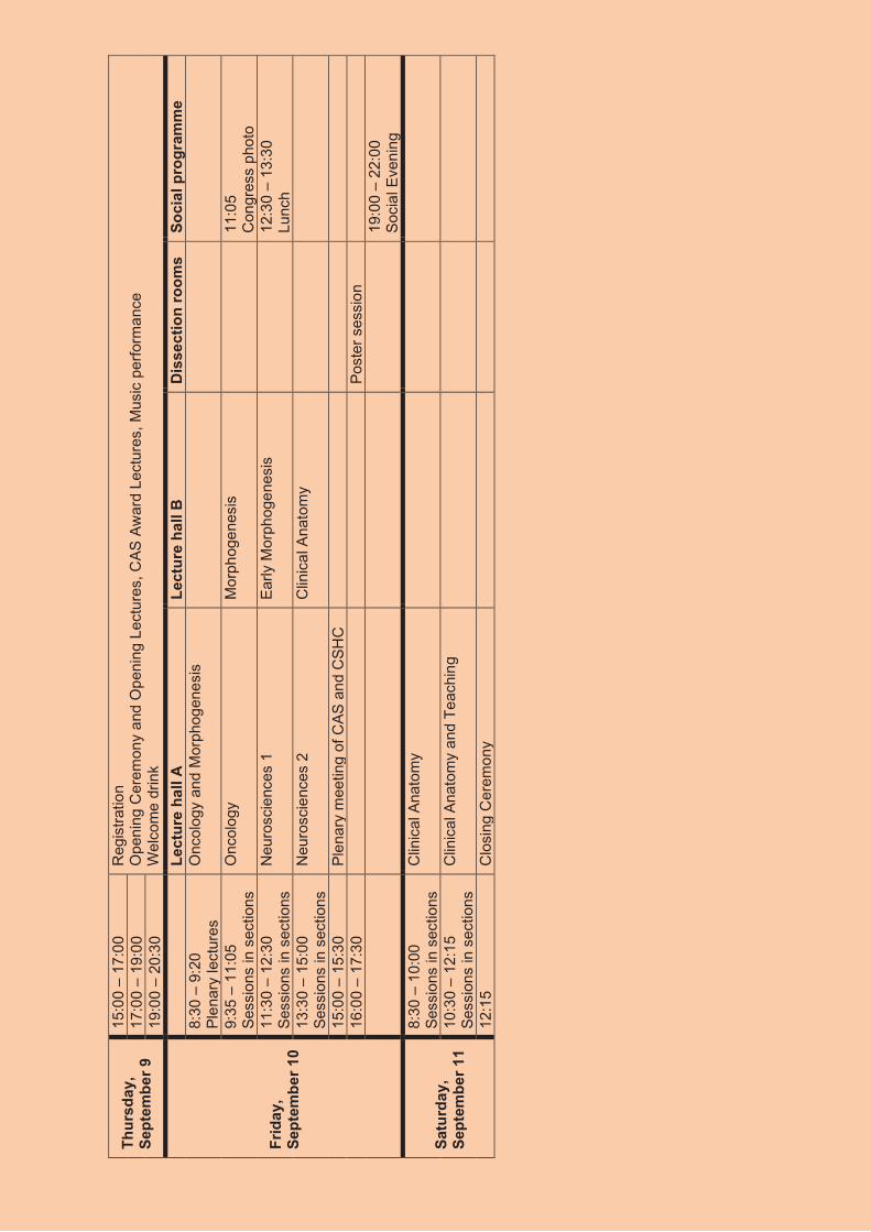
Thur
sday
, Se
ptem
ber 9
15:0
0 –
17:0
0 R
egis
tratio
n O
peni
ng C
erem
ony
and
Ope
ning
Lec
ture
s, C
AS A
war
d Le
ctur
es, M
usic
per
form
ance
W
elco
me
drin
k 17
:00
– 19
:00
19:0
0 –
20:3
0
Frid
ay,
Sept
embe
r 10
Le
ctur
e ha
ll A
Le
ctur
e ha
ll B
D
isse
ctio
n ro
oms
Soci
al p
rogr
amm
e 8:
30 –
9:2
0 Pl
enar
y le
ctur
es
Onc
olog
y an
d M
orph
ogen
esis
9:35
– 1
1:05
Se
ssio
ns in
sec
tions
O
ncol
ogy
Mor
phog
enes
is
11
:05
C
ongr
ess
phot
o 11
:30
– 12
:30
Sess
ions
in s
ectio
ns
Neu
rosc
ienc
es 1
Ea
rly M
orph
ogen
esis
12:3
0 –
13:3
0 Lu
nch
13:3
0 –
15:0
0 Se
ssio
ns in
sec
tions
N
euro
scie
nces
2
Clin
ical
Ana
tom
y
15:0
0 –
15:3
0 Pl
enar
y m
eetin
g of
CAS
and
CSH
C
16
:00
– 17
:30
Post
er s
essi
on
19
:00
– 22
:00
Soci
al E
veni
ng
Satu
rday
, Se
ptem
ber 1
1
8:30
– 1
0:00
Se
ssio
ns in
sec
tions
C
linic
al A
nato
my
10:3
0 –
12:1
5 Se
ssio
ns in
sec
tions
C
linic
al A
nato
my
and
Teac
hing
12:1
5 C
losi
ng C
erem
ony