Embed Size (px)
Citation preview
The aim of this study was to evaluate the cytotoxicity and bioactivity of calcium silicate-based cements combined with niobium oxide (Nb2O5) micro and nanoparticles, comparing the response in different cell lines. This evaluation used four cell lines: two primary cultures (human dental pulp cells - hDPCs and human dental follicle cells - hDFCs) and two immortalized cultures (human osteoblast-like cells - Saos-2 and mouse periodontal ligament cells - mPDL). The tested materials were: White Portland Cement (PC), mineral trioxide aggregate (MTA), white Portland cement combined with microparticles (PC/Nb2O5µ) or nanoparticles (PC/Nb2O5n) of niobium oxide (Nb2O5). Cytotoxicity was evaluated by the methylthiazolyldiphenyl-tetrazolium bromide (MTT) and trypan blue exclusion assays and bioactivity by alkaline phosphatase (ALP) enzyme activity. Results were analyzed by ANOVA and Tukey test (α=0.05). PC/Nb2O5n presented similar or higher cell viability than PC/Nb2O5µ in all cell lines. Moreover, the materials presented similar or higher cell viability than MTA. Saos-2 exhibited high ALP activity, highlighting PC/Nb2O5µ material at 7 days of exposure. In conclusion, calcium silicate cements combined with micro and nanoparticles of Nb2O5 presented cytocompatibility and bioactivity, demonstrating the potential of Nb2O5 as an alternative radiopacifier agent for these cements. The different cell lines had similar response to cytotoxicity evaluation of calcium silicate cements. However, bioactivity was more accurately detected in human osteoblast-like cell line, Saos-2.
Cytotoxicity and Bioactivity of Calcium Silicate Cements Combined with Niobium Oxide in Different Cell Lines
Leticia Boldrin Mestieri, Ana Lívia Gomes-Cornélio, Elisandra Márcia Rodrigues, Gisele Faria, Juliane Maria Guerreiro-Tanomaru, Mário Tanomaru-Filho
Department of Restorative Dentistry, Araraquara Dental School, UNESP – Universidade Estadual Paulista, Araraquara, SP, Brazil
Correspondence: Prof. Dr. Mario Tanomaru-Filho, Rua Humaitá, 1680, 14.801-903 Araraquara, SP, Brasil. Tel: +55-16-3301-6391. e-mail: [email protected]
Key Words: alkaline phosphatase, cytotoxicity, mineral trioxide aggregate, nanoparticles, niobium oxide.
ISSN 0103-6440Brazilian Dental Journal (2017) 28(1): 65-71http://dx.doi.org/10.1590/0103-6440201700525
IntroductionMineral trioxide aggregate (MTA) is indicated for the
treatment of radicular perforations, dental resorptions, dental pulp protection and retrograde filling, due to its biological properties (1). The physicochemical and biological properties of the calcium silicate MTA and Portland cements are similar (2). In the composition of MTA, the radiopacifier agent, bismuth oxide, produces a gradual increase in the material porosity (3). Therefore, new radiopacifying agents, like calcium tungstate (4), zirconium oxide and niobium oxide (5), have been studied as an alternative to replace bismuth oxide.
Niobium oxide (Nb2O5) is a substance studied to enhance the biological properties of materials and used in titanium alloys for endo-osseous implants, due to its biocompatibility, higher degree of mechanical strength and resistance to corrosion and disintegration (6). Viapiana et al. (5) demonstrated that Nb2O5 nanoparticles, used as radiopacifier in a calcium silicate endodontic sealer promoted setting time and flow ability that were adequate for clinical use, satisfactory compressive strength and low solubility. The goal of incorporating nanoparticles into dental materials is to improve the biological properties of the material, such as biocompatibility (7).
Endodontic reparative materials may come into direct contact with dental pulp cells, dental follicle cells, periodontal ligament cells, such as fibroblasts, osteoblasts, undifferentiated mesenchymal cells, among other cell types. Immortalized cell cultures are frequently used in cytotoxicity and bioactivity assays (8-10) and are recommended by international standards for testing new dental materials (11). Immortalized cell lines are more used due to their facility of culture and the higher phenotypical stability compared to primary cells, which become senescent after a limited number of cell passages (12). However, biological processes, cellular components and molecular functions may be changed in immortalized cells (13), which may influence the outcomes of cytotoxicity and bioactivity assays. Therefore, information about the effects of dental materials on primary cells isolated directly from dental tissues may be more reliable (14) compared with immortalized cell lines. However, the isolation of primary cells from tissues can be labor- and time-consuming, the cell number obtained can be very low and, the most important is the limited number of passages that these cells can be cultured because of the senescence process (12).
The comparison between primary and immortalized
Braz Dent J 28(1) 2017
66
L.B.
Mes
tier
i et a
l.
cell lines allows the evaluation of the influence of different cell types on cytotoxicity and bioactivity assays of endodontic cements. Thus, the aim of this study was to evaluate cytotoxicity and bioactivity of calcium silicate cements combined with niobium oxide (Nb2O5) micro and nanoparticles, comparing the responses in different cell lines.
Material and MethodsAfter approval by the Research Ethics Committee of
the Araraquara Dental School – FOAr – UNESP (Protocol 33/10), patients with the indication for extraction of impacted third molars at the incomplete root formation phase and presence of dental follicle were selected. Dental follicles were collected immediately after extraction and separated from the dental crown using a Molt instrument. Dental pulp was collected via apical opening using a Hedströen file (Maillefer, Dentsply, Ballaigues, Switzerland). The tissues were stored in a Petri dish measuring 35x10 mm, containing Alpha Modification Minimum Essential Eagle’s Medium (α-MEM; Sigma-Aldrich, St. Louis, MO, USA) supplemented with 10% fetal bovine serum (FBS; Gibco Life Technologies, Grand Island, NY, USA) and 1% penicillin and streptomycin (PenStrep; Gibco). After that, the tissues were sectioned into fragments of approximately 1 mm3, submitted to enzymatic digestion using a 3 mg/mL collagenase solution (Sigma-Aldrich) for 1 h, centrifuged for 5 min at 4000 rpm to remove the remaining collagenase and seeded in a Petri dish measuring 35x10 mm containing α-MEM culture medium supplemented with 10% FBS and 1% PenStrep. Culture medium was changed 24 h after the collection and then every 48 h, until the establishment of cell culture. Once the cells began forming a monolayer, the tissue fragments in the plate were removed carefully with the use of a pipette tip. Successive passages were performed for cellular expansion and the experiments were performed at the fourth cell passage.
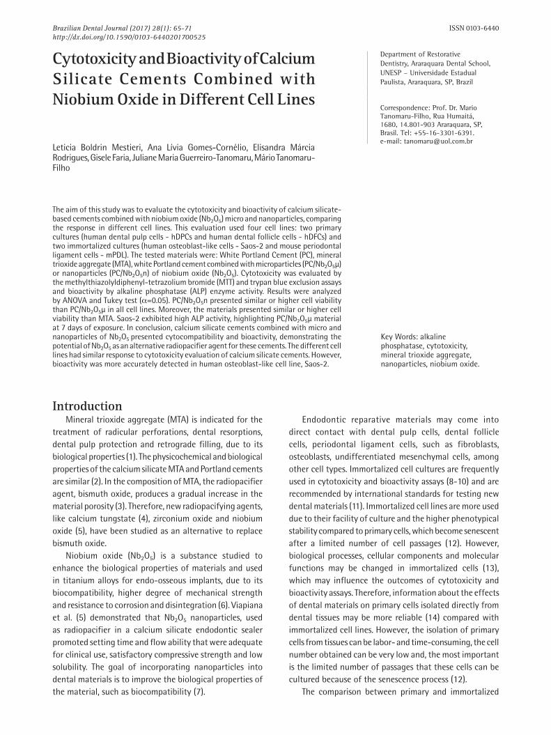
The experimental materials were determined as shown in Table 1. Primary cells obtained from human dental pulp (hDPCs) and human dental follicle (hDFCs) tissues,
and immortalized cells (osteoblast cells derived from human osteosarcoma - Saos-2 and fibroblasts from mouse periodontal ligament - mPDL) were used.
Preparation of MaterialsTo obtain the nanoparticles of Nb2O5, the polymeric
precursor method was used, at the Physics Institute of São Carlos (University of São Paulo, São Carlos, SP, Brazil). An aqueous solution of ammonium niobium oxalate (CBMM, Companhia Brasileira de Metalurgia e Mineração, Araxá, MG, Brazil) was prepared and ammonium hydroxide was added to form a niobium hydroxide precipitate. It was filtered and washed to eliminate the oxalate ions and dissolved in a citric acid solution. The niobium content in the solution was determined by gravimetric analysis and the solution was maintained under agitation at 70 °C for 2 h for reaction. Ethylene glycol was added in the ratio of 60:40 and the solution was maintained heated and under agitation. This solution was heated to 300 °C in an electric oven for 4 h and the resultant mass was ground and calcinated in an electric oven to 700 °C on aluminum plates for 2 h (5).
The cements were prepared according to the proportions mentioned in 1. Test specimens with an internal diameter of 5 mm and 1 mm high were obtained and were kept in an incubator at 37 °C and 100% humidity for 15 h. Next, the specimens were exposed to ultraviolet light (UV) in a laminar flow chamber for 30 min for disinfection and 4 specimens of each material were put into a 6-well plate (TPP; Techno Plastic Products, Trasadingen, Switzerland), filled with 10 mL of culture medium without FBS and incubated at 37 °C, 95% humidity and 5% CO2 for 24 h, in order to obtain the cement extracts.
Cell CultureTo perform the cytotoxicity tests (methyl tetrazolium
- MTT and Trypan Blue), the cells were plated at the concentration of 2x10⁵ cells per well in 96-well plates (TPP) (n=3 for each material). For the primary cultures, α-MEM medium (Sigma-Aldrich) was used supplemented with 5%
Table 1: Experimental materials and mixing ratios
Group MaterialMixing ratios
(material: distilled water)Manufacturer
PC White Portland cement 1 g : 320 µLVotoran, Votorantin cimentos, Rio
Branco do Sul, PR, Brazil
MTA White mineral trioxide aggregate 1 g : 320 µL Angelus, Londrina, PR, Brazil
PC/Nb2O5µPC (70%) + 30% microparticulated
niobium oxide1 g : 400 µL
Institute of Physics, University of São Paulo, USP, São Carlos, SP, Brazil
PC/Nb2O5nPC (70%) + 30% nanoparticulated
niobium oxide1 g : 400 µL
Institute of Physics, University of São Paulo, USP, São Carlos, SP, Brazil
Braz Dent J 28(1) 2017
67
Calc
ium
sili
cate
cem
ents
cyt
otox
icity
FBS (Gibco) and 1% penicillin and streptomycin (Gibco). The immortalized cultures were prepared in Dulbecco’s Modified Eagle Medium (DMEM; Sigma-Aldrich) supplemented with 5% FBS and 1% penicillin and streptomycin (Gibco). The assays were performed in triplicate, using dilutions of 1:2, 1:3, 1:4, 1:6 and 1:8 of the extracts in culture medium without FBS (α-MEM for the primary cultures and DMEM for the immortalized cultures) for 24 h.
The bioactivity evaluated by alkaline phosphatase (ALP) enzyme activity was performed using an intermediate dilution (1:4) of the materials and the cells were plated at a concentration of 2x10⁴ cells per well in 96-well plates (TPP) (n=3 wells for each material). The assay was performed in time intervals of 1, 3 and 7 days, with the culture medium renewed after 3 days.
All assays were performed in triplicate and repeated three independent times. Culture medium was used as negative control and 1 mM hydrogen peroxide (Sigma-Aldrich) was used as positive control.
MTT Assay After the cells were exposed to the cement extracts, they
were replaced by 100 µL of a 5 mg/mL MTT solution (Sigma-Aldrich), followed by incubation at 37 °C for 3 h. Next, the content of the well was removed and the colorimetric product solubilized in 100 µL of acidified isopropanol 0.04 N (Sigma-Aldrich). The absorbance of the solutions was measured in a spectrophotometer (Elx800; Instruments Bio-Tek, Winooski, VT, USA) at 570 nm wavelength. The absorbance readouts were normalized with cells exposed to the α-MEM or DMEM and represented the succianate dehydrogenase activity (cell metabolism).
Trypan Blue AssayAfter removing the cells from the culture plate by the
dissociator agent (Cell Dissociation Buffer Enzyme-Free/ Hank’s-based; Invitrogen, Life Technologies, Grand Island, NY, USA), the cell suspension and the 0.4% Trypan Blue solution (Sigma-Aldrich) were added in equal parts (100 µL) and 10 µL of this content was transferred to a Neubauer chamber for counting the four lateral quadrants under an inverted light microscope. The viability calculation was made according to the following formula: %viable cells=(live cells/total cells) x 100.
ALP Enzyme ActivityALP activity was evaluated as the release of
tymolphtaleine from tymolphtaleine monophosphate using alkaline phosphatase kit (Labtest Diagnóstica, Lagoa Santa, MG, Brazil). The cells were lysed for 30 min at room temperature using sodium lauryl sulfate solution 1:1 (SDS, Sigma-Aldrich) and the assay was performed according to
the manufacturer’s instructions. Absorbance was measured at the wavelength of 590 nm in a spectrophotometer, the results were calculated and data were expressed as ALP activity (µmol tymophtaleine/min/L).
Statistical AnalysisANOVA and Tukey post-hoc tests were performed,
considering a level of significance of 5% (GraphPad Prism 5 software, GraphPad Software Inc., San Diego, CA, USA). All data are presented as mean and standard deviation of the mean.
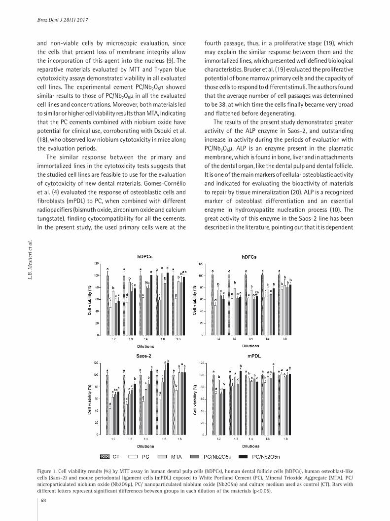
Results The MTT assay demonstrated that PC group had lower
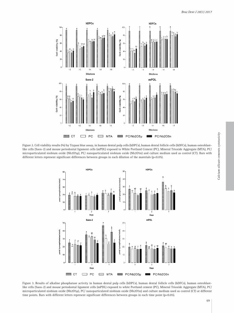
viability for Saos-2 and hDPCs in all evaluated dilutions (p<0.05). Furthermore, there was no statistical difference in the viability of the cells exposed to higher dilutions (1:6 and 1:8) of the materials containing Nb2O5 and those exposed to MTA in all evaluated lines (p>0.05) as shown in Figure 1. This was also observed in the Trypan blue assay (Fig. 2), which shows that the experimental materials PC/Nb2O5µ and PC/Nb2O5n showed similar (p>0.05) or higher (p<0.05) cell viability results than MTA in all the evaluated concentrations and cell lines, except for the dilution 1:2 in the Saos-2 cell line. The experimental cement PC/Nb2O5n had similar (p>0.05) or superior (p<0.05) results to those of PC/Nb2O5µ in all the cell lines and concentrations evaluated on both assays.
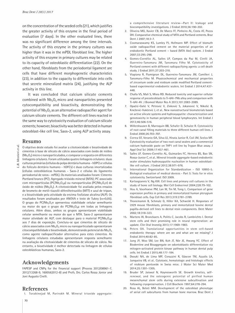
The highest activity for ALP enzyme was observed in Saos-2 (Fig. 3), with an increase in activity according to the evaluated time intervals (p<0.05) and great activity in the final period for PC/Nb2O5µ (p<0.05). The activity of this enzyme in primary cultures was higher than it was in mPDL (p<0.05); however, there was no statistical difference (p>0.05) among the evaluated time intervals in these three cell lines (hDPCs, hDFCs and mPDL).
Discussion Cells that originate from different tissues may respond
in a different manner when used for cytotoxicity tests. In this study, fibroblasts from the periodontal ligament and osteoblast cells were used due their participation in periapical tissue repair (8). Undifferentiated mesenchymal cells from the human dental pulp and follicle have been used due to their potential to differentiate into various cell types (15) and due to its participation in the periapical and pulp reparative processes (16).
The MTT assay is the most used in vitro method for determining cytotoxicity by measuring cell metabolism, using a reaction catalyzed by the succinate dehydrogenase enzyme from mitochondria of viable cells (17). Trypan blue is an assay that allows differentiation between viable
Braz Dent J 28(1) 2017
68
L.B.
Mes
tier
i et a
l.
and non-viable cells by microscopic evaluation, since the cells that present loss of membrane integrity allow the incorporation of this agent into the nucleus (9). The reparative materials evaluated by MTT and Trypan blue cytotoxicity assays demonstrated viability in all evaluated cell lines. The experimental cement PC/Nb2O5n showed similar results to those of PC/Nb2O5µ in all the evaluated cell lines and concentrations. Moreover, both materials led to similar or higher cell viability results than MTA, indicating that the PC cements combined with niobium oxide have potential for clinical use, corroborating with Dsouki et al. (18), who observed low niobium cytotoxicity in mice along the evaluation periods.
The similar response between the primary and immortalized lines in the cytotoxicity tests suggests that the studied cell lines are feasible to use for the evaluation of cytotoxicity of new dental materials. Gomes-Cornélio et al. (4) evaluated the response of osteoblastic cells and fibroblasts (mPDL) to PC, when combined with different radiopacifiers (bismuth oxide, zirconium oxide and calcium tungstate), finding cytocompatibility for all the cements. In the present study, the used primary cells were at the
fourth passage, thus, in a proliferative stage (19), which may explain the similar response between them and the immortalized lines, which presented well defined biological characteristics. Bruder et al. (19) evaluated the proliferative potential of bone marrow primary cells and the capacity of those cells to respond to different stimuli. The authors found that the average number of cell passages was determined to be 38, at which time the cells finally became very broad and flattened before degenerating.
The results of the present study demonstrated greater activity of the ALP enzyme in Saos-2, and outstanding increase in activity during the periods of evaluation with PC/Nb2O5µ. ALP is an enzyme present in the plasmatic membrane, which is found in bone, liver and in attachments of the dental organ, like the dental pulp and dental follicle. It is one of the main markers of cellular osteoblastic activity and indicated for evaluating the bioactivity of materials to repair by tissue mineralization (20). ALP is a recognized marker of osteoblast differentiation and an essential enzyme in hydroxyapatite nucleation process (10). The great activity of this enzyme in the Saos-2 line has been described in the literature, pointing out that it is dependent
Figure 1. Cell viability results (%) by MTT assay in human dental pulp cells (hDPCs), human dental follicle cells (hDFCs), human osteoblast-like cells (Saos-2) and mouse periodontal ligament cells (mPDL) exposed to White Portland Cement (PC), Mineral Trioxide Aggregate (MTA), PC/microparticulated niobium oxide (Nb2O5µ), PC/ nanoparticulated niobium oxide (Nb2O5n) and culture medium used as control (CT). Bars with different letters represent significant differences between groups in each dilution of the materials (p<0.05).
Braz Dent J 28(1) 2017
69
Calc
ium
sili
cate
cem
ents
cyt
otox
icity
Figure 2. Cell viability results (%) by Trypan blue assay, in human dental pulp cells (hDPCs), human dental follicle cells (hDFCs), human osteoblast-like cells (Saos-2) and mouse periodontal ligament cells (mPDL) exposed to White Portland Cement (PC), Mineral Trioxide Aggregate (MTA), PC/ microparticulated niobium oxide (Nb2O5µ), PC/ nanoparticulated niobium oxide (Nb2O5n) and culture medium used as control (CT). Bars with different letters represent significant differences between groups in each dilution of the materials (p<0.05).
Figure 3. Results of alkaline phosphatase activity in human dental pulp cells (hDPCs), human dental follicle cells (hDFCs), human osteoblast-like cells (Saos-2) and mouse periodontal ligament cells (mPDL) exposed to white Portland cement (PC), Mineral Trioxide Aggregate (MTA), PC/ microparticulated niobium oxide (Nb2O5µ), PC/ nanoparticulated niobium oxide (Nb2O5n) and culture medium used as control (CT) at different time points. Bars with different letters represent significant differences between groups in each time point (p<0.05).
Braz Dent J 28(1) 2017
70
L.B.
Mes
tier
i et a
l.
on the concentration of the seeded cells (21), which justifies the greater activity of this enzyme in the final period of evaluation (7 days). In the other evaluated lines, there was no significant difference among the time intervals. The activity of this enzyme in the primary cultures was higher than it was in the mPDL fibroblast line. The higher activity of this enzyme in primary cultures may be related to its capacity of osteoblastic differentiation (22). On the other hand, fibroblasts from the periodontal ligament are cells that have different morphogenetic characteristics (23), in addition to the capacity to differentiate into cells that secrete mineralized matrix (24), justifying the ALP activity in this line.
It was concluded that calcium silicate cements combined with Nb2O5 micro and nanoparticles presented cytocompatibility and bioactivity, demonstrating the potential of Nb2O5 as an alternative radiopacifier agent for calcium silicate cements. The different cell lines reacted in the same way to cytotoxicity evaluation of calcium silicate cements; however, bioactivity was better detected in human osteoblast-like cell line, Saos-2, using ALP activity assay.
ResumoO objetivo deste estudo foi avaliar a citotoxicidade e bioatividade de cimentos à base de silicato de cálcio associados com óxido de nióbio (Nb2O5) micro e nanoparticulados, e comparar a resposta em diferentes linhagens celulares. Foram utilizadas quatro linhagens celulares: duas culturas primárias (células da polpa dentária humana - hDPCs e células do folículo dentário humano - hDFCs) e duas culturas imortalizadas (células osteoblásticas humanas - Saos-2 e células do ligamento periodontal de ratos - mPDL). Os materiais analisados foram: Cimento Portland branco (PC); Agregado trióxido mineral (MTA); PC associado com micropartículas (PC/Nb2O5µ) ou nanopartículas (PC/Nb2O5n) de óxido de nióbio (Nb2O5). A citotoxicidade foi avaliada pelos ensaios de brometo de metil-tiazolil-difeniltetrazólio (MTT) e azul de tripan, e a bioatividade pela atividade da enzima fosfatase alcalina (ALP). Os resultados foram analisados por ANOVA e teste de Tukey (α=0,05). O grupo do PC/Nb2O5n apresentou viabilidade celular semelhante ou maior do que o grupo do PC/Nb2O5μ em todas as linhagens celulares. Além disso, ambos os grupos apresentaram viabilidade celular semelhante ou maior do que o MTA. Saos-2 apresentaram maior atividade de ALP, com destaque para o material PC/Nb2O5μ aos 7 dias de exposição. Concluiu-se que cimentos de silicato de cálcio associados com Nb2O5 micro ou nanoparticulado apresentaram citocompatibilidade e bioatividade, demonstrando potencial do Nb2O5 como agente radiopacificador alternativo para estes cimentos. As linhagens celulares estudadas apresentaram resposta semelhante na avaliação da citotoxicidade de cimentos de silicato de cálcio. No entanto, a bioatividade é melhor detectada na linhagem de células osteoblásticas humanas, Saos-2.
AcknowledgementsFAPESP and CNPq for the financial support (Process 2012/08961-7, 2012/13268-9, 1600562012-8) and Profs. Drs. Carlos Rossa Júnior and Joni Augusto Cirelli.
References 1. Torabinejad M, Parirokh M. Mineral trioxide aggregate:
a comprehensive literature review--Part II: leakage and biocompatibility investigations. J Endod 2010;36:190-202.
2. Oliveira MG, Xavier CB, De Marco FF, Pinheiro AL, Costa AT, Pozza DH. Comparative chemical study of MTA and Portland cements. Braz Dent J 2007;18:3-7.
3. Coomaraswamy KS, Lumley PJ, Hofmann MP. Effect of bismuth oxide radiopacified cement on the material properties of an endodontic Portland cement – based (MTA like) system. J Endod 2007;33:295-298.
4. Gomes-Cornélio AL, Salles LP, Campos da Paz M, Cirelli JA, Guerreiro-Tanomaru JM, Tanomaru Filho M. Cytotoxicity of Portland cement with different radiopacifying agents: a cell death study. J Endod 2011;37:203-210.
5. Viapiana R, Flumignan DL, Guerreiro-Tanomaru JM, Camilleri J, Tanomaru-Filho M. Physicochemical and mechanical properties of zirconium oxide and niobium oxide modified Portland cement-based experimental endodontic sealers. Int Endod J 2014;47:437-448.
6. Challa VS, Mali S, Misra RD. Reduced toxicity and superior cellular response of preosteoblasts to Ti-6Al-7Nb alloy and comparison with Ti-6Al-4V. J Biomed Mater Res A 2013;101:2083-2089.
7. Opačić-Galić V, Petrović V, Zivković S, Jokanović V, Nikolić B, Knežević-Vukčević J, et al.. New nanostructural biomaterials based on active silicate systems and hydroxyapatite: characterization and genotoxicity in human peripheral blood lymphocytes. Int Endod J 2013;46:506-516.
8. Willershausen B, Marroquin BB, Schafer D, Schulze R. Cytotoxicity of root canal filling materials to three different human cell lines. J Endod 2000;26:703-707.
9. Correa GT, Veranio GA, Silva LE, Hirata Junior R, Coil JM, Scelza MF. Cytotoxicity evaluation of two root canal sealers and a commercial calcium hydroxide paste on THP1 cell line by Trypan Blue assay. J Appl Oral Sci 2009;17:457-461.
10. Salles LP, Gomes-Cornélio AL, Guimarães FC, Herrera BS, Bao SN, Rossa-Junior C, et al.. Mineral trioxide aggregate-based endodontic sealer stimulates hydroxyapatite nucleation in human osteoblast-like cell culture. J Endod 2012;38:971-976.
11. International Organization for Standardization - ISO 10993-5. Biological evaluation of medical devices - Part 5: Tests for in vitro cytotoxicity. Switzerland: ISO 2009.
12. Kartsogiannis V, Ng KW. Cell lines and primary cell cultures in the study of bone cell biology. Mol Cell Endocrinol 2004;228:79-102.
13. Hou A, Voorhoeve PM, Lan W, Tin M, Tong L. Comparison of gene expression profiles in primary and immortalized human pterygium fibroblast cells. Exp Cell Res 2013;319:2781-2789.
14. Thonemamm B, Schmalz G, Hiller KA, Schweikl H. Responses of L929 mouse fibroblasts, primary and immortalized bovine dental papilla-derived cell lines to dental resin components. Dent Mater 2002;18:318-323.
15. Martens W, Bronckaers A, Politis C, Jacobs R, Lambrichts I. Dental stem cells and their promising role in neural regeneration: an update. Clin Oral Investig 2013;17:1969-1983.
16. Peters OA. Translational opportunities in stem cell-based endodontic therapy: where are we and what are we missing? J Endod 2014;40:82-85.
17. Jung JY, Woo SM, Lee BN, Koh JT, Nör JE, Hwang YC. Effect of Biodentine and Bioaggregate on odontoblastic differentiation via mitogen-activated protein kinase pathway in human dental pulp cells. Int Endod J 2015;48:177-184.
18. Dsouki NA, de Lima MP, Corazzini R, Gáscon TM, Azzalis LA, Junqueira VB, et al.. Cytotoxic, hematologic and histologic effects of niobium pentoxide in Swiss mice. J Mater Sci Mater Med 2014;25:1301-1305.
19. Bruder SP, Jaiswal N, Haynesworth SE. Growth kinetics, self-renewal, and the osteogenic potential of pirified human mesenchymal stem cells during extensive subcultivation and following cryopreservation. J Cell Biochem 1997;64:278-294.
20. Rosa AL, Beloti MM. Development of the osteoblast phenotype of serial cell subcultures from human bone marrow. Braz Dent J

Braz Dent J 28(1) 2017
71
Calc
ium
sili
cate
cem
ents
cyt
otox
icity
2005;16:225-230.21. Higuita-Castro N, Gallego-Perez D, Pelaez-Vargas A, García Quiroz
F, Posada OM, López LE, et al.. Reinforced Portland cement porous scaffolds for load-bearing bone tissue engineering applications. J Biomed Mater Res B Appl Biomater 2012;100:501-507.
22. Gustavsson J, Ginebra MP, Planell J, Engel E. Osteoblast-like cellular response to dynamic changes in the ionic extracellular environment produced by calcium-deficient hydroxyapatite. J Mater Sci Mater Med 2012;23:2509-2520.
23. Hou LT, Liu CM, Lei JY, Wong MY, Chen JK. Biological effects of cementum and bone extracts on human periodontal fibroblasts. J Periodontol. 2000;71:1100-1109.
24. Murakami Y, Kojima T, Nagasawa T, Kobayashi H, Ishikawa I. Novel isolation of alkaline phosphatase-positive subpopulation from periodontal ligament fibroblasts. J Periodontol. 2003;74:780-786.
Received September 11, 2016Accepted November 28, 2016