Embed Size (px)
Citation preview
CYTOPROTECTIVE AND GENOPROTECTIVE EFFECTS OF AQUEOUS EXTRACTS OF PROCESSED PLEUROTUS
SAJOR-CAJU FRUITING BODIES ON HYDROGEN PEROXIDE-INDUCED DAMAGE OF HUMAN PERIPHERAL
BLOOD MONONUCLEAR CELLS
MOHAMAD FAIRUS BIN ABDUL KADIR
2010

UNIVERSITY OF MALAYA
ORIGINAL LITERARY WORK DECLARATION
Name of Candidate : Mohamad Fairus Bin Abdul Kadir (801016-01-6227)
Registration /Matric No. : SGF060005 Name of Degree : Master of Biotechnology Title of Dissertation (‘’this Work’’) : CYTOPROTECTIVE AND GENOPROTECTIVE EFFECTS OF AQUEOUS EXTRACTS OF PROCESSED PLEUROTUS SAJOR-CAJU FRUITING BODIES ON HYDROGEN PEROXIDE-INDUCED DAMAGE OF HUMAN PERIPHERAL BLOOD MONONUCLEAR CELLS Field of Study : Mycology Biotechnology I do solemnly and sincerely declare that:
(1) I am sole author/writer of this Work; (2) This Work is original; (3) Any use of any work in which copyright exists was done by way of fair
dealing and for permitted purposes and any excerpt from, or reference to or reproduction of any copyright work has been disclosed expressly and sufficiently and the title of the Work and its authorship have been acknowledge in this Work;
(4) I do not have actual knowledge nor do I ought reasonably to know that the making of this Work constitutes an infringement of any copyright work;
(5) I hereby assign all and every rights in the copyright to this Work to the University of Malaya (“UM”), who henceforth shall be owner of the copyright in this Work and that any reproduction of use in any form or by any means whatsoever is prohibited without the written consent of UM having been first had and obtained;
(6) I am fully aware if in the course of making this Work I have infringed any copyright whether intentionally or otherwise, I may be subjected to legal action or any other action as may be determined by UM.
Candidate’s Signature, Date Subscribed and solemnly declared before, Witness’s Signature Name: Designation: Date
Abstract
i
ABSTRACT
Pleurotus sajor-caju authority is one of the mushroom species that is widely grown in
Malaysia. The present study was undertaken to evaluate the antioxidant, cytotoxic,
cytoprotective, genotoxic and genoprotective activities of crude water extracts of this
mushroom. Crude aqueous extracts were prepared from the fruiting bodies that had
been processed at different temperatures such as P1: blanching (95°C ± 2°C), P2: sun
drying (30°C ± 2°C), P3: freeze drying (-46°C ± 3°C) and different temperatures of
oven drying (P4: 45°C, P5: 50°C, P5: 55°C and P6: 60°C ± 2°C respectively).
Antioxidant capacity of all the extracts was determined using ferric reducing power
(FRAP) and DPPH free radical scavenging assays and the total phenolic content (TPC)
was also determined. The TPC per gram of extract in descending order was P2, P4, P7,
P6, P5, P3 and P1. Sun dried samples had the highest antioxidant activity compared to
other processing method. The correlation analysis showed mild to moderate positive
correlation between TPC, FRAP and DPPH. Cytotoxicity and cytoprotective activities
were determined by assessing the viability of human peripheral blood mononuclear
cells (PBMC) viability using MTT (3-(4, 4-dimethylthiazol-2-yl)-2, 5-diphenyl
tetrazolium bromide) assay. None of the processed mushroom extracts were cytotoxic to
the PBMCs. Hydrogen peroxide (H2O2), an oxidant; was used to induce cytotoxicity in
PBMC. At a concentration of 0.3mM, H2O2 showed 25% inhibition of cell viability
compared to untreated cells. At 100µg/ml, the extracts P3 and P4 exhibited the highest
cell viability (10.24±0.36% and 8.20±1.06% increase respectively) compared with
untreated PBMCs, while 20µg/ml of phytohaemagglutinin (PHA) only showed
6.66±0.52% increase in cell viability compared with untreated PBMCs. All the
mushroom extracts exhibited mild protection against H2O2 induced toxicity. The highest
protection of 10.47±0.87% and 15.37±0.99% at 80 and 100µg/ml respectively was by
Abstract
ii
sun dried extracts, whereas at 10µg/ml vitamin C exerted 6.50±1.94% protection.
Genotoxicity in PBMC was assessed using single cell gel electrophoresis (COMET
assay). None of the mushroom extracts tested had genotoxic effects on the cells. The
percentage of head DNA was more than 80% which was similar to control cells treated
with water. The cells incubated with 0.2mM H2O2 caused 50% DNA damage. The
genoprotective effect of the mushroom extracts was determined by assessing the
COMET tail in PBMC, which had been incubated in the presence of H2O2 and the
extracts. The P1 mushroom extract (80µg/ml) exhibited approximately 80% protection
against H2O2 induced damage. The extracts P2, P3 and P4 of the fruiting bodies of P.
sajor-caju showed similar degrees of protection. Thus, it can be concluded that the
processing methods do affect the antioxidant level which in turn may affect the
cytoprotective and genoprotective activities of P. sajor-caju against cellular or DNA
damage induced by H2O2.

Abstract
iii
ABSTRAK
Pleurotus sajor-caju adalah merupakan salah satu spesies cendawan yang diusahakan
secara meluas di Malaysia. Kajian ini dijalankan bagi menilai aktiviti- aktiviti seperti
antioksidan, sitotoksik, sitoprotektif, genotoksik dan genoprotektif (sekiranya ada), di
dalam ekstrak mentah air cendawan ini. Ekstrak mentah air disediakan daripada
cendawan yang telah diproses pada suhu yang berbeza seperti, P1: celuran (95°C ±
2°C), P2: pengeringan di bawah cahaya matahari (30°C ± 2°C), P3: pembekukeringan (-
46°C ± 3°C) dan pengeringan pada suhu – suhu berbeza dalam ketuhar panas (P4: 45°C,
P5: 50°C, P5: 55°C and P6: 60°C ± 2°C). Keupayaan antioksidan bagi semua ekstrak
dikenalpasti menggunakan asei kuasa penurunan ferum (FRAP) dan pelupusan radikal
bebas DPPH dan jumlah kandungan fenol (TPC). Jumlah kandungan fenol (TPC) per
gram ekstrak dalam turutan menurun adalah seperti berikut; P2, P4, P7, P6, P5, P3 dan
P1. Sampel P2 menunjukkan aktiviti antioksidan yang paling tinggi berbanding dengan
kaedah pemprosesan yang lain. Analisa hubungkait antara TPC, FRAP dan DPPH
menunjukkan hubungkait yang sangat positif. Aktiviti sitotoksik dan sitoprotektif
dijalankan dengan mengenalpasti jumlah kehadiran sel darah mononuklear (PBMC)
dengan menggunakan asei MTT (3-(4, 4-dimethylthiazol-2-yl)-2, 5-diphenyl
tetrazolium bromide). Kesemua ekstrak P. sajor-caju menunjukkan ianya tidak toksik
kepada PBMC. Hidrogen peroksida (H2O2), sebagai oksidan, digunakan untuk
mengaruh sitotoksik pada PBMC. Pada kepekatan 0.3mM, H2O2 menunjukkan
perencatan bilangan sel sebanyak 25% berbanding dengan sel yang tidak dirawat
dengan H2O2. Pada 100µg/ml, ekstrak P3 dan P4 menunjukkan kehadiran jumlah
bilangan sel yang tertinggi iaitu peningkatan sebanyak 10.24±0.36% dan 8.20±1.06%
masing – masing, berbanding dengan sel yang dirawat dengan air sahaja. Manakala,

Abstract
iv
20µg/ml fitohemaglutinin (PHA) hanya menunjukkan peningkatan kehadiran jumlah sel
sebanyak 6.66±0.52% berbanding dengan sel yang tidak dirawat (rawatan air suling
sahaja). Kesemua ekstrak P. sajor-caju menunjukkan aktiviti perlindungan terhadap sel
pada tahap yang memuaskan bertentangan dengan toksisiti yang diaruh oleh H2O2.
Tahap perlindugan sel yang tertinggi boleh dilihat pada kepekatan 80µg/ml dan
100µg/ml yang menunjukkan peratusan perlindungan sebanyak 10.47±0.87% dan
15.37±0.99% masing – masing oleh P2, manakala pada kepekatan vitamin C sebanyak
10µg/ml peratusan perlindungan pada sel darah mononuclear adalah sebanyak
6.50±1.94% sahaja. Genotoksik pada PBMC dikaji dengan menggunakan teknik gel
elektroforesis satu sel (asei COMET). Kesemua ekstrak tidak genotoksik pada sel darah
mononuclear normal manusia. Rata – rata sel yang dirawat dengan ekstrak P. sajor-caju
memamerkan peratusan kepala DNA pada komet lebih daripada 80% iaitu lebih kurang
sama dengan set kawalan. Sel yang diinkubasi dengan 0.2mM H2O2 mampu
memusnahkan 50% DNA sel darah mononuclear manusia. Kesan genoprotektif ekstrak
P. sajor-caju dikenalpasti dengan menggunakan kaedah COMET pada PBMC yang
diinkubasikan dengan kehadiran H2O2 dan ekstrak. Ekstrak P1 (80µg/ml) menunjukkan
lebih kurang 80% perlindungan terhadap sel yang diaruh dengan H2O2. Tambahan lagi,
ekstrak P2, P3 dan P4 P. sajor-caju juga menunjukkan tahap perlindungan yang hampir
serupa dengan P1. Maka, boleh disimpulkan di sini bahawa kaedah pemprosesan
memberi kesan kepada tahap antioksidan di mana ia mungkin memberi kesan yang baik
kepada aktiviti sitoprotektif dan genoprotektif pada P. sajor-caju bagi melawan
kemusnahan selular atau DNA yand diaruh oleh H2O2.
Acknowledgements
v
ACKNOWLEDGEMENTS
All praise belongs to Allah.
Humble salutations and deepest gratitude I wish to express towards both my
supervisors namely Professor Dr. S. Vikineswary and Associate Professor Dr. Umah
Rani Kuppusamy who never ceased to give their fullest attention and guidance
throughout this course. Without their constant supervision, constructive advice,
encouragements and moral support, this dream of me attaining Masters could have
never been realized. I’m truly honoured to have had the opportunity of studying and
working under them.
Sincere thanks to all my fellow friends and co-workers from Biochemistry and
Mycology laboratory especially, Mr. Saravanan, Mr. Indran, Ms. Chandra, Mr. Chai,
Ms. Ii Lee, Ms. Dahlia, Ms. Noriza, Ms. Mei Heng and Ms. Izlina for sharing your
knowledge, experience, selflessly lending a hand whenever needed and assisting me
along my entire research work. The friendliness and warm working environment all of
you created has truly touched my heart and made my research period a joyous and
memorable one.
I would also like to extend my special thanks to Mushroom Research Center
(MRC) of University Malaya for funds and facilities provided by University of Malaya
throughout the study. This study has given me further understanding in medicinal or
nutraceutical values of mushrooms as well as an opportunity to enrich my knowledge of
application in biotechnology.
Last but not least, I dedicate this thesis as my offering to my parents Mr. Abdul
Kadir and Mrs. Yatuma Alee for bringing me up with love and care whilst instilling in
me morality, culture, teaching me the importance of education and virtue of prayers.
Such upbringing is the reason for me to be able to complete this thesis with the grace of
god.
vi
TABLE OF CONTENTS Pages ABSTRACT …………………………………….………………………………………… i ABSTRAK ……………………………………………………………………………….. iii ACKNOWLEDGMENTS …………………………………………………………......... v LIST OF TABLES .......………………………………………………………………….. ix LIST OF FIGURES ...………………………………………………………………….... x LIST OF ABBREVIATIONS ………………………………………………………….. xii CHAPTER 1: INTRODUCTION………………………………………………………...1 1.1 Introduction………………………………………………………………………..1 CHAPTER 2: LITERATURE REVIEW………………………………………………...7 2.1 Free radicals …………………………………………………………………...…..7 2.2 Genotoxic effects of DNA damage by ROS/oxyradicals ……………………......9 2.3 Antioxidants …………………………………………………………………...…10 2.4 Mushroom ………………………………………………………………………..11
2.4.1 Nutritional values of mushrooms …………………………………….....12 2.4.2 Antitumor activity of mushrooms …………………………………....... 13 2.4.3 Other bioactivities of mushrooms …………………………………........15
2.5 Pleurotus spp. …………………………………………………………………......19
2.5.1 Pleurotus sajor-caju ……………………………………………………....23 CHAPTER 3: MATERIALS AND METHODS ….……………………………...…….25
3.1 Preparation of mushroom extracts from fruiting bodies of P. cajor-caju …….25
3.2 Antioxidant potential and polyphenol content of P. sajor-caju extracts ….…..26 3.2.1 Total phenolic content (TPC) of crude P. sajor-caju extracts ………....26 3.2.2 Antioxidant activity assay ………………………………...……………...27
3.2.2.1 DPPH free radical scavenging activity assay …………………..27

vii
3.2.2.2 Ferric reducing antioxidant power (FRAP) assay ...………......28
3.3 Cytotoxicity, cytoprotective, genotoxicity and genoprotective activities of processed P. sajor-caju extract .…………………………………………………30
3.3.1 Isolation of peripheral blood mononuclear cells (PBMCs) …………...30
3.3.3 Cytotoxicity and cytoprotective activity of P. sajor-caju extracts on 3.3.2 Determination of cell viability of PBMCs ………………………….......31
PBMCs …………………………………………………...………………32 3.3.3.1 Determination of hydrogen peroxide (H2O2) an oxidant …………………………………...…………………....32
concentration as
3.3.3.2 Effects of P. sajor-caju extract on the viability of PBMCs ….....33 3.3.3.3 The cytoprotective effects of P. sajor-caju extract against H2O2
3.3.4 Genotoxicity and genoprotective activity of P. sajor-caju extracts…….36
induced toxicity on PBMCs …………………………........36
3.3.4.1 Comet assay to assess DNA damage of PBMCs ………………..36 3.3.4.2 Preparation of slides for single cell gel electrophoresis
(SCGE) …………………………………………………………... 38 3.3.4.3 Electrophoresis of microgel slides containing PBMCs in
agarose gel ………………………………………………………..41 3.3.4.4 Effects of H2O23.3.4.5 Effects of P. sajor-caju extract on DNA of PBMC’s………........43
on DNA of PBMC’s ……………………………43
3.3.4.6 Protective effects of P. sajor-caju extract against H2O2 DNA damage in PBMCs ………………………………………....44
induced
3.3.5 DNA staining and visualization ………………………………...……….44
3.4 Statistical analysis …………………………………………………………….….45 CHAPTER 4: RESULTS AND DISCUSSIONS ………………………………………47 4.1 Preparation of aqueous extracts from fruiting bodies of P. sajor-caju …….…47 4.2 Antioxidant potential and polyphenol content of P. sajor-caju extracts ….….49
4.2.1.1 Total phenolic content (TPC) of aqueous extracts of P. sajor- 4.2.1. Antioxidant activity assay …………………………………...…….…….49
caju ………………………………………………………………..49 4.2.1.2 P. sajor-caju
DPPH free radical scavenging activity assay on processed extracts
4.2.1.3 Ferric reducing antioxidant power (FRAP) assay of P. sajor-caju ………………………………………..........51
crude extracts ………………………………………...……….….54 4.2.3 Correlation …………………………………………………………….….56
4.3 Determination of viable cells……………………………………………………..60
4.3.1 Determination of hydrogen peroxide (H2O2) Oxidant …………………………………………………………………....60
concentration as an

viii
4.3.2 Effects of P. sajor-caju extract on the viability of PBMCs ………….....66
4.3.3 Protective effects of P. sajor-caju extract on the viability PBMCs …...74 4.4 The effects P. sajor-caju aqueous extracts on DNA damage of PBMCs ...………81
4.4.1 DNA staining and visualization (Comet Assay) …………………...…...81 4.4.2 Effects of H2O24.4.3 Effects of P. sajor-caju on PBMC’s DNA ……..………………………..86
on PBMC’s DNA ………………………………………82
4.4.4 Protective effects of P. sajor-caju extracts against PBMC’s DNA damage by H2O2
………………………..………………………………...90
4.5 Future Investigation ………………………………………………………………..97 CHAPTER 5: CONCLUSION…………………………………………………………..99 5.1 Conclusion ………………………………………………………………...………….99 References ……………………………………………………………………………….102 Appendices ……………………………………………………………………………....119

ix
LIST OF TABLES Pages Table 2.1: Source, type and bioactivity of some macrofungal polysaccharides …...... 17 Table 2.2: Proximate composition of Pleurotus spp. ……………………………...…... 20 Table 2.3: Amino acid composition of Pleurotus spp. ……………………………...…. 21 Table 3.1: Temperatures at which fruiting bodies of the P. sajor-caju were processed ……………….……………………………………………...…….. 26 Table 4.1: The extraction yields after the processed samples were extracted in
boiling water for three hours …………………………………………......... 48
x
LIST OF FIGURES Pages Figure 3.1: Procedure for MTT assay ……………………………………………......... 34 Figure 3.2: Experimental protocol for the effects of mushroom extracts/PHA/
H2O2
on the viability of PBMCs using MTT assay…………...…....……... 35
Figure 3.3: Experimental protocol to study the effects of pre-incubated P. sajor-caju extracts on the viability of PBMCs which then
challenged with 300 µM of H2O2.
…..………………………………...……. 37
Figure 3.4: Flow chart of the preparation of slides to be used before electrophoresis for Comet assay…………..……………………...………. 40 Figure 3.5: Flow chart of electrophoresis process for comet assay after slides were
prepared as shown in Figure 3.4…....……………………………...……… 42 Figure 4.1: Total phenolic contents of various aqueous extracts of different processed P. sajor-caju………………………………………………...…… 50 Figure 4.2: Scavenging ability of various processing methods of P. sajor-caju aqueous extracts on 1,1-diphenyl-2-picrylhydrazyl radicals (DPPH)….... 52 Figure 4.3: Ferric reducing antioxidant power (FRAP) level of various aqueous extracts of different processed P. sajor-caju…………………………...….. 55 Figure 4.4: Correlation between TPC, DPPH and FRAP of different processed aqueous extracts of P. sajor-caju……………………………………...…… 58 Figure 4.5: Determination of the cell viability of PBMC in the presence of H2O2 RPMI-1640 medium…………………………………………….....……….. 62
in
Figure 4.6: Determination of the cell viability of PBMC in the absence of H2O2 RPMI-1640 medium …………………………………………………...…... 63
in
Figure 4.7: Dose response effects of H2O2
on PBMC viability…………………...…… 64
Figure 4.8: Cytotoxicity of different processed aqueous extracts of P. sajor-caju on PBMCs after two hours incubation …………..……………………...….... 71
Figure 4.9: Cytoprotective effect of different processed aqueous extracts of P. sajor- caju against H2O2
of incubation ....…………………………………...…………………...…… 78 -induced (300µM) damage on PBMCs after two hours
xi
Figure 4.10: Representative comet images showing different levels of damage in visual scoring………………………………………………….......………. 82 Figure 4.11: DNA damage in PBMC exposed to H2O2
for two hours ………...…....... 84
Figure 4.12: Genotoxicity effects of aqueous extracts of different processed P. sajor- caju on DNA damage in PBMCs after 30 minutes of incubation .……... 89 Figure 4.13: Genoprotective effects of aqueous extracts of P. sajor-caju on PBMCs
against H2O2
-induced DNA damage after 30 minutes of incubation ..... 94
Figure 4.14: Fifty percentage (50%) of effective concentration (EC50) on genoprotection of different processed P. sajor-caju extracts against H2O2
-induced DNA damage ...……………………………………...……. 95
xii
LIST OF ABBREVIATIONS
α alpha
AIDS Acquired immune deficiency syndrome
ALS Alkaline labile site
AZT Azidothymidine
BC Before Christ
β beta
CO2
DAPI Diamino-2-phenylindole
Carbon dioxide
DMSO Dimethyl sulfoxide
DNA Deoxyribonucleic acid
DPPH 1, 1-Diphenyl-2-picrylhydrazyl
EC50
EDTA Ethylenediaminetetra-acetic acid, di-sodium salt
Effective concentration-50
ELISA Enzyme-linked immunosorbent assay
EtBr Ethidium bromide
FBS Fetal bovine serum
FDA Food and Drug Administration
FeSO4.7H2
FRAP Ferric reducing antioxidant power
O Iron (II) sulphate heptahydrate
g gram
g Gravity
GAEs Gallic acid equivalents
GC/MS Gas chromatography/Mass spectrometry

xiii
h hour
H2O2
HDL High density lipoprotein
Hydrogen peroxide
HIV Human immunodeficiency virus
HPLC High pressure liquid chromatography
IC50
kg kilogram
Inhibition concentration - 50
LDH lactase dehydrogenase leakage
LDL low density lipoprotein
LEM medium of L. edodes
LMP Low melting point
mg milligram
ml milliliter
mM milimolar
MTT 3 - (4, 4 - dimethylthiazol – 2 – yl) – 2, 5 – diphenyl tetrazolium
Bromide
NaCl Natrium chloride
Na2CO3
NK Natural killer
sodium carbonate
nm Nanometer
NMP Normal melting point
O2
O
Oxygen
2•
OH• Hydroxyl radical
Superoxide anion
ONO2• Peroxynitrate

xiv
˚ C Degree celcius
PBMCs Peripheral blood mononuclear cells
PBS Phosphate buffer saline
PHA Phytohaemaglutinin
PSK Polysaccharide-K
RNA Ribonucleic acid
RO Reverse osmosis
ROS Reactive oxygen species
RPMI Roswell Park Memorial Institute
SSB Single strand break
SEM Standard error mean
SDS Sodium dodecyl sulphate
SPSS Statistical package for social sciences
spp species
ssDNA Single stranded deoxyribonucleic acid
TPC Total phenolic content
TPTZ 2, 4, 6 – Tripyridyl – S – Triazine
UV ultraviolet
VLDL very low density lipoprotein
v/v volume/volume
w/v weight per volume
% percentage
µg Microgram
µL Microliter
µM Micromolar

CHAPTER 1
INTRODUCTION

Chapter 1 Introduction
1
CHAPTER 1
INTRODUCTION
1.1 Introduction
Since the beginning of human civilization, the use of natural products with therapeutic
properties has been common. For ages, mineral, plant, and animal products were the main
sources of drugs (Rates, 2001). The term natural products commonly refers to herbs, herbal
concoctions, dietary supplements, traditional Chinese medicine, or alternative medicine
(Holt and Chandra, 2002). Natural products are generally either of prebiotic origin or
originate from microbes, plants, or animal sources. Components of natural products include
terpenoids, polyketides, amino acids, peptides, proteins, carbohydrates, lipids, nucleic acid
bases, ribonucleic acid (RNA) and deoxyribonucleic acid (DNA) (Croteau et al., 2000).
Nature has provided many remedies for human kind over the years, including
natural products for preventive or therapeutic intervention of diseases. Chinese herb guides
documented the use of herbaceous plants as far back in time as 2000 BC (Holt and
Chandra, 2002). Extracts from the basidiomycetes Mycenapura and Nidula candida may be
useful in the treatment of leukemia. Treatment of diseases with medicine of plant origin is
an integral part of many cultures throughout the world (Bhattaram et al., 2002). Today,
many people are surprised to find natural products still play an important role as sources of
medicine. Many pharmaceutical agents have been discovered by screening natural products
from plants and microorganisms (Rocha et al., 2001).
Chapter 1 Introduction
2
The structural diversity of plants and other natural products makes them a valuable
source of novel lead compounds against newly discovered therapeutic targets (Harvey,
1999). The belief that natural medicines are much safer than synthetic drugs has gained
popularity in recent years and this has lead to the tremendous growth in
phytopharmaceutical industry (Bhattaram et al., 2002). Biological activities of natural
sources have been screened extensively in search of new anticancer, antiviral, and
fertility/antifertility drugs (Phillipson, 2001). The extraction of bioactive agents from
natural sources is one of the most intensive areas of natural product research today.
In ancient times, mushrooms were used in folk medicine throughout the world.
Edible mushrooms have been used to maintain health, increase longevity and consumed as
food (Manzi and Pizzoferrato, 2000). Nowadays, mushrooms are increasingly consumed
for their nutritional and medicinal properties. Attempts have been made in many parts of
the world to explore the use of mushrooms and their metabolites for treatment of a variety
of human ailments (Jose and Janardhanan, 2000).
Kosem et al. (2007) have reported the antioxidant activity and cytoprotective effect
of Garcinia mangostana, may play a vital role as chemopreventive activities via a reducing
mechanism and inhibition of intracellular oxidative stress which may lead to reduced cell
or DNA damage. Recently, studies have been carried out to evaluate the antioxidant
activities of commercial mushrooms (Mau et al., 2001 and Mau et al., 2002). In
mushrooms it has been shown that phenolic compounds are responsible for the radical
scavenging effects (Yang et al., 2002) and these are the major naturally occurring
antioxidant components found in mushrooms (Cheung et al., 2003). Antioxidants are
substances that may protect cells from the damage caused by unstable molecules known as

Chapter 1 Introduction
3
free radicals. Further, it has been reported that the various processing techniques to
preserve the perishable edible mushrooms may affect the antioxidant properties of
mushrooms, too (Vikineswary et al., 2008). Thus, this study was undertaken to investigate
the effects of different processing methods of P. sajor-caju on the antioxidant activity of
the mushroom, if any, and its effects on protecting the cells against damage or DNA
damage.
Manzi et al. (1999a) reported that mushrooms are healthy food, low in calories and
fat, rich in proteins, chitin, vitamins and minerals. This chemical composition along with
many other components render the mushroom as therapeutic foods that are useful in
preventing diseases such as hypertension, hypercholesterolemia and cancer (Manzi et al.,
2001). In addition, Hasler (1998) reported that mushrooms contained high concentrations
of minerals, rich in vitamin D (ergosterol), thiamin (B1), riboflavin (B2), and niacin (B3) as
well as all the essential amino acids.
They have also become one of the most desirable foods in Oriental cuisine because
of their low caloric value, characteristic smell, taste and texture. Thus, mushrooms are
considered as highly valuable bio-engineering resources for development of food material
(functional foods) as well as for use of starting material in the production of drugs (Mizuno,
1999). Functional compounds (polysaccharides) in mushrooms have recently been
highlighted as they are able to lower cholesterolemia, modulate the immune system and
inhibit tumoral growth (Zhang et al., 2001).

Chapter 1 Introduction
4
In Japan, hundreds of mushroom species have been studied for the past 20 years.
Most of the medicinal mushrooms such as Ganoderma lucidum (reishi), Lentinula edodes
(shiitake) and Grifola frondosa (maitake) show a common property of enhancing immune
function by stimulating cell-mediated immunity. These mushrooms seem to stimulate cells
in the immune system, called T-cells, which appear to have significant cancer-fighting
properties. Weng and Chen (1996) had reported that three different anticancer drugs
extracted from mushrooms have been approved by the Japanese equivalent of FDA--the
Health and Welfare Ministry. They are lentinan, derived from L. edodes; schizophyllan,
derived from Schizophyllum commune (suehirotake); and polysaccharides-K (PSK),
derived from Trametes versicolor (kawaratake) (Yang et al., 1992). Polysaccharides-K was
reported as one of the best selling anticancer drug in the world (Yang et al., 1992).
There are approximately 200 species of mushrooms that have been identified to
markedly inhibit the growth of different kind of tumors (Wasser and Weis, 1999).
Pleurotus sajor-caju (oyster mushroom) is a class of “Edible Fungal Food” that has been
discovered to have definite nutritive and medicinal values. As a good source of nonstarchy
carbohydrate, quality protein with presence of most essential amino acids (Bano and
Rajarathnam, 1988), minerals (Breene, 1990) and vitamins (Stamets, 1993), Pleurotus spp.
have been shown to modulate the immune system, have hypoglycemic activity,
antithrombotic effect and reduce blood pressure and blood lipid concentrations and to
inhibit tumor growth, inflammation, and microbial action (Cimerman, 1999). Pleurotus is
becoming popular in many of the European and Southern Asian countries due to its
characteristic texture and pleasant flavor (Bano and Rajarathnam, 1988).

Chapter 1 Introduction
5
Mushrooms are however, highly perishable commodities. Therefore, processing is
essential not only to preserve their nutritional and medicinal properties but also increase
shelf-life (Vikineswary et al., 2008). Food processing is a technique used to transform raw
ingredients into food for consumption by humans and to increase shelf-life or storage
period compared to fresh food. Common food processing techniques include removal of
water by sun drying, oven drying, fermentation, cooking, deep frying and pasteurization
(Rodriguez-Amaya, 1997). Benefits of food processing include toxin removal,
preservation, increase food consistency and ease distribution and marketing tasks.
However, it also has some disadvantages. For example, the content of vitamin C which is a
well known antioxidant, is destroyed by heat and lowers the vitamin C content compared to
fresh ones, as its stability is greatly influenced by temperature, oxygen, and metal ion
content (Ryley and Kajda, 1994; Marcy et al., 1989).
As discussed above, the processing of the fruiting bodies of Pleurotus sajor-caju
either by cooking or drying could possibly cause loss of nutrients and their protective
effects. Wong et al. (2009) and Kho et al. (2009) reported that processing of mushroom can
affect the antioxidant level and medicinal properties such as antimicrobial activity. Kho et
al. (2009) showed that the different methods of processing Auricularia auricula-judae fresh
fruiting bodies such as oven drying and freeze drying could affect the antioxidant potential.
Similarly, Wong et al. (2009) reported that the different processing method employed for
the preparation of extracts from H. erinaceus affected the selected bioactive properties of
the mushroom. In some cases, heat treatment caused no change or even improved the
content and activities of naturally occurring antioxidants. Choi et al., (2006) reported that
heat treatment significantly enhanced the overall antioxidant activities of Shiitake
mushroom.
Chapter 1 Introduction
6
To date, Pleurotus spp. are marketed fresh, canned or dried. There has been no
investigation on the effects of processing, on medicinal properties of Pleurotus spp.
Therefore, the present study was carried out to evaluate antioxidant properties and
cytoprotective and genoprotective activities of processed Pleurotus sajor-caju grown in
Malaysia.
The objectives of the present study were to:-
a) assess the antioxidant and polyphenol levels in the crude water extracts of
Pleurotus sajor-caju subjected to different processing methods.
b) screen for cytoprotective activity of crude aqueous extracts from Pleurotus sajor-
caju subjected to different processing methods using human peripheral blood
mononuclear cellular viability assessment.
c) screen for genoprotective activity of crude aqueous extracts from Pleurotus sajor-
caju subjected to different processing methods in human peripheral blood
mononuclear cells by using single cell gel electrophoresis (Comet assay).
CHAPTER 2
LITERATURE REVIEW

Chapter 2 Literature Review
7
CHAPTER 2
LITERATURE REVIEW
2.1 Free radicals
A major development in carcinogenesis research caught the attention of scientists in the
mid 1980s with the discovery of DNA damage and the mutation induced by reactive
oxygen species (ROS). These ROS can be generated endogenously during normal cellular
metabolism or acquired from exogenous sources such as ultraviolet (UV) radiation, air
pollutants and etc. (Marnett, 2000). Damaged bases or strand breaks can be produced by
excessive levels of oxygen radicals generated during the reduction of O2
which in turn
attacks DNA bases or dexyribose residues (Ward et al., 1987). Attempts in replication of
the altered or damaged DNA leads to mutation or apoptosis (Johnson et al., 1996).
The identity of the oxidants responsible for the production of oxidized DNA bases
is still the focus of numerous studies. The hydroxyl radical (HO•) is an obvious candidate
because it is extremely reactive and adds to DNA bases or abstracts hydrogen atoms to
produce many of the products that occur in the genome (Ward, 1988; Grisham and
McCord, 1986). According to Marnett (2000), the HO• plays a role in the endogenous
oxidation of DNA but this HO• generated does not diffuse into the nucleus of the adjacent
cells. The reactivity of HO• is so great that it does not diffuse more than one or two
molecular diameters before reacting with a cellular component (Marnett, 2000). In order to

Chapter 2 Literature Review
8
oxidize DNA, the HO• must be generated immediately adjacent to the nucleic acid
molecule (Marnett, 2000). It is likely that H2O2
serves as a diffusible, latent form of HO•
that reacts with a metal ion in the vicinity of a DNA molecule to generate the oxidant
(Henle and Linn, 1997 and Cadet et al., 1999).
Another oxidant that can generate many of the products observed with HO• is
peroxynitrite (ONO2-) (Beckman et al., 1990). Peroxynitrite is the coupling product of
nitric oxide and superoxide and is an extremely strong oxidant (Koppenol, 1992). The
pattern of products generated by DNA oxidation by ONO2• is complex and mirrors the
diversity of oxidized DNA detected in tissues (Burney et al., 1999). Interestingly, ONO2• is
more reactive toward 8-oxo-deoxyguanosine (8-oxo-dG) than to unmodified DNA bases
(Burney et al., 1999). 8-oxo-dG has been detected previously in DNA treated with a variety
of oxidants and is easily measured by HPLC and GC/MS (Dizdaroglu and Gajewski, 1990).
ONO2• has the ability to diffuse within cells and may be taken up by some cells via anion
transporters (Radi, 1998). This may provide a link between inflammation and the induction
of mutation by virtue of the ability of ONO2•
to oxidize DNA. If these free radicals are not
neutralized, they can quickly bind to our cells vital proteins, carbohydrates and even our
DNA, potentially altering its structure. This may result in a decrease in immune function or
a change in the genetic makeup of cell, which could affect the cell’s ability to reproduce
normally (Maynard, 2001). These, uncontrolled productions of ROS is also involved in the
onset of many common illnesses such as cancer, rheumatoid arthritis, and cardiovascular as
well as in degenerative processes of aging (Halliwell and Gutteridge, 1985). In order to
preserve genomic stability, a tightly regulated network of intracellular antioxidative and
DNA repair pathways has evolved (Sasaki, et al., 2002).

Chapter 2 Literature Review
9
2.2 Genotoxic effects of DNA damage by ROS/oxyradicals
Genotoxic processes may lead to irreversible changes in the structure of the genetic
material of cells. This effect is considered as an important process in cancer development.
The term ‘genotoxic’ was used for the first time in 1973 by Hermann Druckrey during a
conference on ‘Evaluation of genetic risks of environmental chemicals’ in Sweden. He
stated: ‘In order to describe the components of chemical interaction with genetic material,
the term genotoxic is proposed as a general expression to cover toxic, lethal and heritable
effects to karyotic and extrakaryotic genetic material in germinal and somatic cells’
(Weisberger and Williams 2000).
A variety of techniques for measuring carcinogen interaction with DNA has been
developed since the first discovery of mutation and DNA damages. The techniques
including binding of radio-labelled chemical, one of the reliable and predictive hepatocyte
DNA repair assay (Williams et al., 1998) and the 32P-postlabeling method of Randerath and
Gupta (Reddy et al., 1984). Hence, these procedures can be used to classify carcinogens
that are DNA reactive, i.e., genotoxic. Conversely, a substantial number of carcinogens
were found to lack genotoxicity and this knowledge was embodied in a key distinction
between genotoxic and epigenetic carcinogens
based on the underlying mechanism of
action (Weisberger and Williams, 2000).
Chapter 2 Literature Review
10
2.3 Antioxidants
Antioxidants are substances that delay or prevent the oxidation of cellular oxidizable
substrates (Yuan et al., 2005). A multitude of natural antioxidants have already been
isolated from different kinds of plant materials such as seeds, cereal crops, vegetables,
fruits, leaves, roots, spices and herbs. The body produces a variety of antioxidants to
maintain healthy systems and defray cellular destruction. Because antioxidants stop this
damaging process, thus supplements and foods containing them are often termed "anti-
aging".
Antioxidants have been thought to be important in preventing a variety of disease
progression caused by UV radiation from sunlight, cosmic radiation, chemicals, and
pollutants and internally generated free radicals. Although almost all organisms possess
antioxidant defence and repair systems that have evolved to protect them against oxidative
damage, these systems are insufficient to prevent the damage entirely (Simic, 1988). Jeng
et al. (2002) reported that when the mechanism of antioxidant protection becomes
unbalanced by factors such as aging, deterioration of physiological functions, resulting in
diseases and acceleration of aging.
The damage of DNA (the cell’s blue print) by the free radicals is prevented with the
presence of antioxidant. This could give antioxidants an important role and potential to
reduce the genetic instability of cancer cells and thus may be useful in treatment (Maynard,
2001; and Reddy et al., 2003). The demand for natural antioxidant from botanical sources,
especially edible plants increased because of the question on the long-term safety and
negative consumer perception of synthetic drugs (Amarowicz et al., 2004; Sakanaka et al.,
Chapter 2 Literature Review
11
2004). Ascorbic acid, most commonly known as vitamin C is the most abundant of water
soluble antioxidants available and acts primarily in cellular fluid. Vitamin C is important
to the body's biosynthesis of collagen, catecholamine and carnitine and is integral to proper
formation of proteins, neurotransmitters, hormones, DNA and RNA in the body (Hathcock,
2004).
Kim et al. (1994) and Jeng et al. (2002), reported that some Chinese herbs used in
traditional medicines exhibited significant antioxidant activity and key ingredients in their
medicines also included mushrooms. Some common edible mushrooms, which are widely
consumed in Asian culture, had recently been found to possess antioxidant activity and this
correlated with their total phenolic content (Cheung et al., 2003). Mushrooms accumulate a
variety of secondary metabolites, including phenolic compounds, polyketides, terpenes and
steroids. Phenolic compounds were found to have antioxidant activity which could inhibit
low density lipoprotein (LDL) oxidation (Teissedre and Landrault, 2000). Cheung et al.,
(2003) reported that phenolic antioxidants, such as variegatic acid and diboviquinone were
found in mushrooms.
2.4 Mushroom
Mushrooms represent one of the world's greatest untapped resources of nutritious food. The
number of mushroom species on the earth is estimated to be 140 000 and only 10% are
known. Mushrooms which are primarily basidiomycete fungi are rich in protein, minerals,
and vitamins, and they contain an abundance of essential amino acids (Mattila et al., 2002).
Therefore, mushrooms can be a good food supplement. However, many people are
Chapter 2 Literature Review
12
apprehensive about mushrooms as a food source. Ignorance has led many to become
skeptical about whether food of fungal origin can hold any great nutritional promise. It
seems much education is needed before full advantage can be taken of this readily
available, nutritionally rich food source (Chang and Buswell, 1996).
2.4.1 Nutritional values of mushrooms
The nutritional values and organoleptic components of a number of commercial
mushrooms have been well studied (Yang et al., 2001). Medicinal mushrooms from Asia
may be among the best examples of foods containing cancer-fighting nutrients. It is
estimated that there are 140,000 different kinds of mushrooms, of which 700 are used for
food. Mushrooms are rich sources of minerals, vitamin D (ergosterol), thiamin (B1),
riboflavin (B2), and niacin (B3) as well as all essential amino acids; while still being low in
fat and calories. Only recently have some of the more exotic varieties appeared in markets;
Lentinula edodes (shiitake), Agaricus bisporus (Portabello), Flammulina velutipes (enoki),
Pleurotus spp. (oyster) and Boletus edulis (porcini) are now popular gourmet cuisine
ingredients. The 2006 Urban Rural Interface Conference was a great opportunity for
producers and consumers to learn more about fruits, vegetables, and mushrooms that
provide major benefits to health, as well as new ways to market these products (Hasler,
1998).
Chapter 2 Literature Review
13
Among the edible and medicinal mushrooms Ganoderma lucidum, widely used for
the promotion of health and longevity is reported to have antioxidant and genoprotective
(protects the DNA from oxidative damage) properties (Wachtel-Galor et al., 2004).
Oxidation of DNA can produce a number of molecular alterations, including cleavage,
cross-linkage between DNA and proteins and oxidation of purines. Unless corrected by the
DNA repair machinery, those alterations can lead to mutations, carcinogenesis, and
senescence (Deshpande and Irani, 2002).
In order to protect genomic stability, tightly regulated networks of intracellular
pathways have evolved. The salient intracellular mechanisms responsible for modulating
oxidative stress include the thioredoxin system and antioxidant enzymes, such as catalase,
glutathione peroxidise and superoxide dismutase. Low molecular weight compounds,
including selenium and phytochemicals, such as ascorbic acid (Vitamin C), α-tocopherol
(Vitamin E) and β-carotene have also been found to protect against oxidative stress
(Lindsay and Astley, 2002).
2.4.2 Antitumor activity of mushrooms
Mushroom-derived beta-glucan has been used in Asia for centuries for health
purposes. A review article on mushroom-derived beta glucans showed that different
mushrooms exhibited differing effects so that some mushrooms appear to act more strongly
against some health challenges than others (Brochers et al., 1999). Beta glucans can make
the natural killer (NK) cells work more effectively as they actually bind to the NK cells and
Chapter 2 Literature Review
14
increase the proportion of target-binding lymphocytes and of the damaged target cells in
the conjugates (Renzo et al., 1991).
For almost 40 years, mushrooms have been intensively investigated for medicinal
effect in in vivo and in vitro model systems, and many new antitumor and
immunomodulating polysaccharides have been identified and put into practical use
(Mizuno et al., 1996, 1999; Wasser and Weis 1999; Ikekawa 2001). Mushroom
polysaccharides are known to stimulate natural killer cells, T-cells, B-cells, and
macrophage-dependent immune system responses (Wasser, 2002). G. frondosa, a well
known mushroom in Japan showed clear anti-tumor effect against both MM-46 and IMC
carcinomas in mice (Mayell, 2001). Mushroom polysaccharides are present mostly as
glucans with different types of glycosidic linkages, such as (1-3), (1-6)-beta-glucans and
(1-3)-alpha-glucans, but some are true heteroglycans (Wasser, 2002).
Polysaccharides are the best known and most potent mushroom derived substances
with antitumor and immunomodulating properties as shown in Table 2.1. The proposed
mechanism by which most mushroom polysaccharides exert antitumor effect is via
activation of the immune response of the host organism. They are regarded as biological
response modifiers (Wasser and Weis 1999), which includes a) cancer preventing activity;
b) immune –enhancing activity; and c) direct tumor inhibition activity (Brekhman, 1980).
The natural antitumor polysaccharides isolated from mushrooms include acidic and neutral
ones with different types of glycosidic linkages, while some are bound to protein or peptide
residues such as polysaccharide-protein and peptide complexes (Cun et al., 1994).
Chapter 2 Literature Review
15
2.4.3 Other bioactivities of mushrooms
Medicinal mushrooms have a variety of biological activities such antimicrobial, antiviral,
hepatoprotective, antidiabetic, and many others. For instance, G. lucidum is a medicinal
mushroom that has antidiabetic, antioxidant, immunomodulatory, antitumor and
antimetastatic activities (Kimura et al., 2002).
Mushrooms need antibacterial and antifungal compounds to survive in their natural
environment. It is therefore not surprising that compounds with strong antimicrobial
activities could be isolated from many mushrooms and that they could be of benefit for
human (Lindequist et al., 2005). The purified materials from mycelium extracts of edible
mushroom, Hericium erinaceum, showed antimicrobial effects against Escherichia coli,
Bacillus subtilis, Staphylococcus aureus, Aspergillus niger, Candida albicans and
Microsporum gypseum (Dong et al., 2000). Cochran (1978) reported that edible
mushrooms such as L. edodes, Coprinus comatus and Qudemansiella mucida have
antifungal activity. He also reported that mushroom species such as Boletus frostii,
Calvatia gigantean, Chlorophyllum molybdites and Agaricus campestris protect mice
against poliomyelitis (antiviral).
An antifungal peptide (eryngin) with a molecular mass of 10 kDa was isolated from
fruiting bodies of the mushroom Pleurotus eryngii (Wang and Ng, 2004) was reported to
have the ability to inhibit mycelial growth in Fusarium oxysporum and Mycosphaerella
arachidicola. Japanese researchers reported that lentinan isolated from L. edodes, in
combination with the drug azidothymidine (AZT) was more effective than AZT itself in
suppressing the proliferation of the AIDS virus (Tochikura et al., 1988).

Chapter 2 Literature Review
16
On the other hand, there are some evidences on the hepatoprotective effects of
mushroom extracts such as G. frondosa, Dendropolyporus umbellatus, S.commune, T
versicolor, Tremella fuciformis and Wolfiporia cocos (Wasser and Weis, 1999). There are
other studies which showed that fractions isolated from the fruiting body of A. blazei
exhibit antimutagenic, anticarcinogenic, and immunostimulative activities (Shimura et al.,
1983; Itoh et al., 1994 and Osaki et al., 1994). Various fractions of A. blazei inhibited
benzo-(a)pyrene induced mutagenicity, and that linoleic acid was found to be the main
substance associated with such activity (Osaki et al., 1994). Anti-HIV activities were
reported for mycelial culture medium of L. edodes (LEM) and water-soluble lignin in LEM
(Tochikura et al., 1988 and Suzuki et al., 1989).
Chapter 2 Literature Review
17
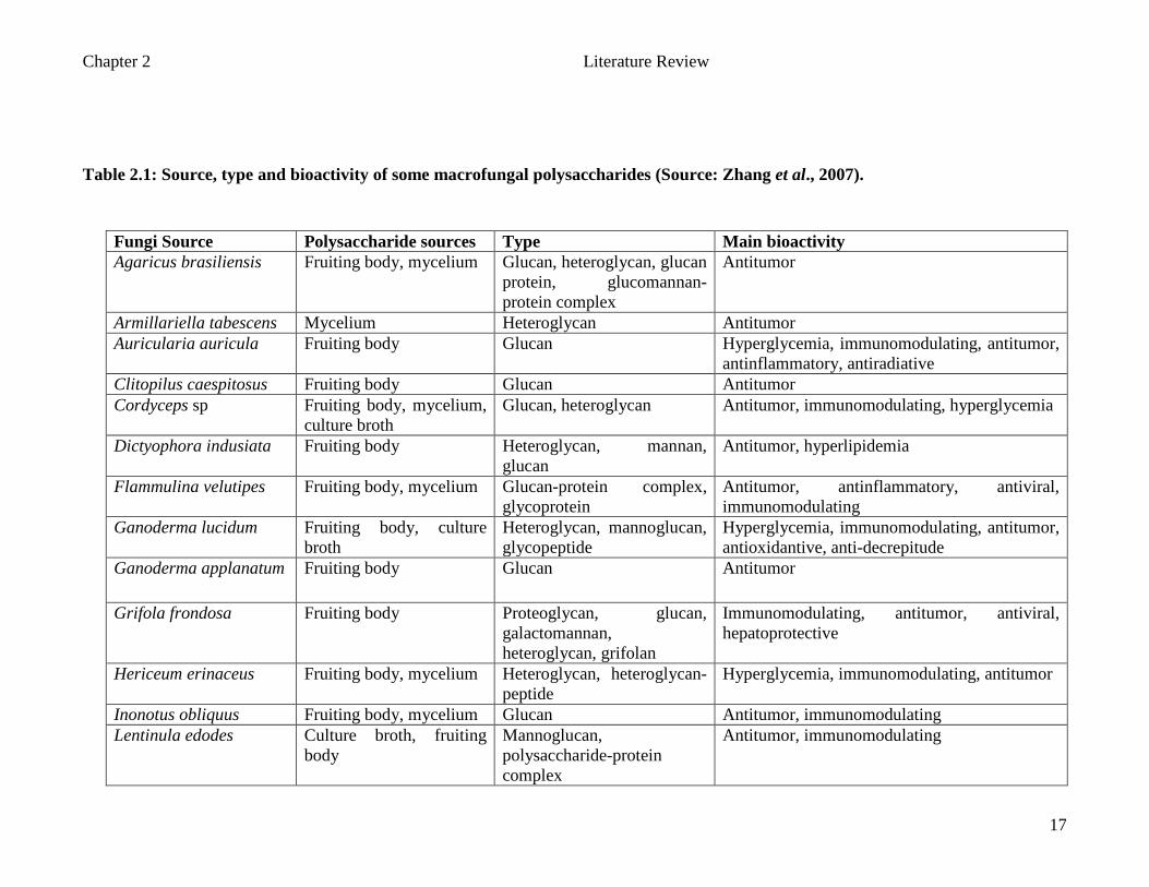
Table 2.1: Source, type and bioactivity of some macrofungal polysaccharides (Source: Zhang et al., 2007).
Fungi Source Polysaccharide sources Type Main bioactivity Agaricus brasiliensis Fruiting body, mycelium Glucan, heteroglycan, glucan
protein, glucomannan-protein complex
Antitumor
Armillariella tabescens Mycelium Heteroglycan Antitumor Auricularia auricula Fruiting body Glucan Hyperglycemia, immunomodulating, antitumor,
antinflammatory, antiradiative Clitopilus caespitosus Fruiting body Glucan Antitumor Cordyceps sp Fruiting body, mycelium,
culture broth Glucan, heteroglycan Antitumor, immunomodulating, hyperglycemia
Dictyophora indusiata Fruiting body Heteroglycan, mannan, glucan
Antitumor, hyperlipidemia
Flammulina velutipes Fruiting body, mycelium Glucan-protein complex, glycoprotein
Antitumor, antinflammatory, antiviral, immunomodulating
Ganoderma lucidum Fruiting body, culture broth
Heteroglycan, mannoglucan, glycopeptide
Hyperglycemia, immunomodulating, antitumor, antioxidantive, anti-decrepitude
Ganoderma applanatum Fruiting body Glucan Antitumor
Grifola frondosa Fruiting body Proteoglycan, glucan, galactomannan, heteroglycan, grifolan
Immunomodulating, antitumor, antiviral, hepatoprotective
Hericeum erinaceus Fruiting body, mycelium Heteroglycan, heteroglycan-peptide
Hyperglycemia, immunomodulating, antitumor
Inonotus obliquus Fruiting body, mycelium Glucan Antitumor, immunomodulating Lentinula edodes Culture broth, fruiting
body Mannoglucan, polysaccharide-protein complex
Antitumor, immunomodulating
Chapter 2 Literature Review
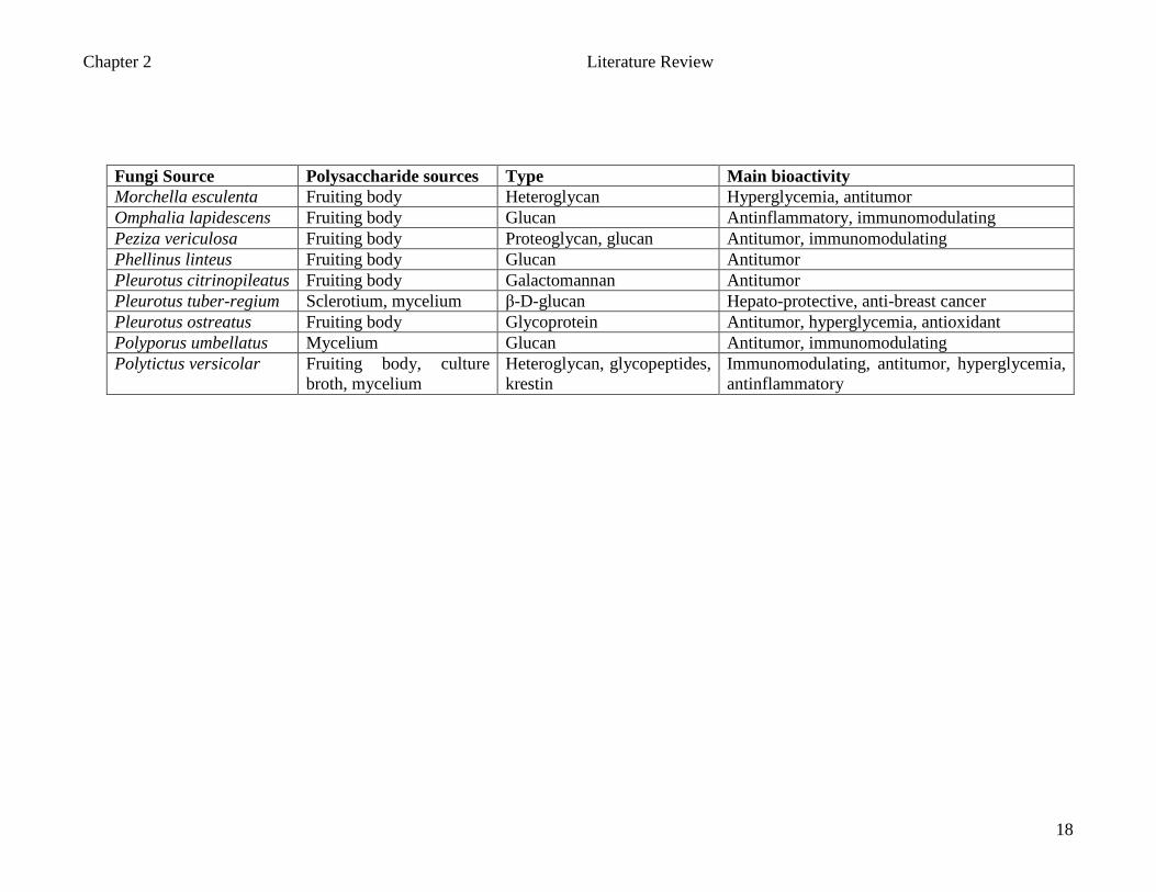
18
Fungi Source Polysaccharide sources Type Main bioactivity Morchella esculenta Fruiting body Heteroglycan Hyperglycemia, antitumor Omphalia lapidescens Fruiting body Glucan Antinflammatory, immunomodulating Peziza vericulosa Fruiting body Proteoglycan, glucan Antitumor, immunomodulating Phellinus linteus Fruiting body Glucan Antitumor Pleurotus citrinopileatus Fruiting body Galactomannan Antitumor Pleurotus tuber-regium Sclerotium, mycelium β-D-glucan Hepato-protective, anti-breast cancer Pleurotus ostreatus Fruiting body Glycoprotein Antitumor, hyperglycemia, antioxidant Polyporus umbellatus Mycelium Glucan Antitumor, immunomodulating Polytictus versicolar Fruiting body, culture
broth, mycelium Heteroglycan, glycopeptides, krestin
Immunomodulating, antitumor, hyperglycemia, antinflammatory
Chapter 2 Literature Review
19
2.5 Pleurotus spp
Mushrooms of genus Pleurotus are the most popular fungal food. Worldwide interest in the
mushrooms has increased because of their relative ease of cultivation and the ability to
grow on a variety of agricultural residues. The first sustained commercial production of
Pleurotus spp. on straw began in Europe in the early 1960s with large-scale industrial
production beginning in the 1970s (Zadril, 1978). This fungus is industrially produced as
human food, and it accounts for nearly a quarter of the world mushroom production
(Chang, 1996) and also occupies the second most important position in the world
mushroom market, led by the button mushroom Agaricus bisporus (Baars et al., 2000;
Royse, 1996). It is also used for the bioconversion of agricultural, industrial, and
lignocellulose wastes (Ballero et al., 1990 and Puniya et al., 1996), as an agent for
bioremediation (Axtell et al., 2000) and as organic fertilizer (Abdellah et al., 2000).
Oyster mushrooms which fall under the genus of Pleurotus, family Pleurotaceae
and the order Agaricales, are one of the most popular edible mushrooms especially in
Oriental region such as China and Japan. The oyster mushrooms were first cultivated in the
United States at the beginning of the last century, before being introduced to Europe and
India (Guzman, 2000). More than 1000 species of Pleurotus have been described
throughout the world; however, only approximately 50 species are recognized as Pleurotus
(Guzman, 2000). The genus can be recognized by their white spores, the stem that attached
at the side of the cap, or at least off center, and the fleshy or tough texture of the cap and
the ability to grow at temperate climates.

Chapter 2 Literature Review
20
At the beginning of 20th
century, the method of Pleurotus cultivation on tree stumps
and logs was first introduced (Purkayastha and Chandra, 1985). Today, Pleurotus spp. can
be grown on various agricultural waste materials especially on lignocellulosic materials
using different technologies. This white rot fungus is capable of degrading both lignin and
cellulose. Pleurotus spp are primary agents of decomposition. They have the ability,
therefore, to directly break down cellulose and lignin bearing materials without chemical or
biological preparation (composting) (Kaul, 2002).
Pleurotus spp. became one of the choice edible mushrooms cultivated in tropics
(Quimio, 2001) and widely consumed as food in the east and increasingly in the west
(Cimerman, 1999). One of the reasons for its success is that oyster mushrooms have good
nutritional (Cimerman, 1999) and medicinal values (Bano and Rajarathnam, 1988). They
are good sources of nonstarchy carbohydrates, dietary fiber (that can help in reducing the
plasma cholesterol), essential amino acid, minerals and vitamins of B group, and folic acid
(Bano and Rajarathnam, 1988). The proximate analysis or composition of four Pleurotus
spp. showing significant difference in the values of crude protein and fat is presented in
Table 2.2.
Table 2.2: Proximate composition of Pleurotus spp.
Pleurotus spp Moisture (%)
Composition (%)* Total Carbohydrates
+
Energy Value
Kcal (%) Crude protein
Fat Ash
P. sajor-caju 88.75 26.94 2.88 11.82 58.36 379 P. florida 91.50 37.19 3.72 10.98 48.11 385 P. sapidus 88.35 35.87 2.41 12.35 49.37 373 P. ostreatus 89.25 27.38 2.27 13.44 56.91 371
*Dry weight basis (Bano and Rajarathnam, 1988) + calculated by difference from *

Chapter 2 Literature Review
21
Considering the essential amino acid index, biological value, in vitro digestibility,
nutritional index and protein source, Pleurotus spp. fall between high grade vegetables and
low grade meats (Bano and Rajarathnam, 1988). The amino acid profile of Pleurotus spp. is
presented in Table 2.3. All the values are expressed as g amino acid/100g crude protein. In
all, 17 amino acids including all the essential amino acids were detected in significant
quantities.
Table 2.3: Amino acid composition of Pleurotus spp.
Amino acids P. sajor-caju
P. florida P. sapidus P. ostreatus Hen’s egg Protein
Alanine 10.237 6.388 9.124 7.775 5.920 Arginine 2.463 1.534 1.694 - 6.096 Aspartic Acid 1.237 1.534 2.032 4.294 9.616 Cystine 0.650 0.554 0.735 0.380 2.432 Glutamic acid 7.83 4.918 3.644 5.975 12.736 Glycine 4.371 4.882 3.130 5.165 5.312 Histadine 1.025 1.134 1.122 4.203 2.432 Isoleucine 3.572 2.416 3.098 2.792 6.288 Leucine 8.665 6.578 4.153 6.433 8.896 Lysine 5.435 3.196 2.152 3.286 6.976 Methionine 2.043 1.840 1.398 1.235 3.360 Phenylalanine 6.035 5.820 5.333 5.992 5.728 Proline 2.375 2.912 2.237 2.720 4.160 Threonine 2.900 5.038 3.201 2.554 5.120 Tyrosine 2.272 1.426 1.580 1.524 4.160 Serine 0.148 0.110 0.322 0.270 7.648 Valine 6.350 3.430 4.731 4.728 6.848 Total Aromatic
8.307 7.246 6.913 7.516 9.888
Sulphur containing amino acid
37.930 30.298 26.381 25.024 51.216
Total amino acid
67.769 53.710 49.685 56.026 103.136
Data presented in gram of amino acid/100g crude protein (Khanna et al., 1992) (-) Not detected
Chapter 2 Literature Review
22
Zhang et al. (2001) reported that the modified fractions of P. tuber-regium exhibited
antitumor activities on solid tumour cell lines in vitro. In addition, other Pleurotus species
such as P. passeckerianus and P. mutilis which contain anticarcinogenic and antiviral agent
pleuromutilin, showed an active antiviral reaction against PR8 influenza virus (Jong and
Donovick, 1989). Antiviral agents have also been obtained from water extracts of P.
ostreatus mycelium (Cimerman, 1999).
Cimerman (1999) reported that antibiotic substances had been isolated from the
water extracts of the fruiting bodies of P. griseus, P. palmatus, P. sapidus and P. ulmarius.
These Pleurotus spp. showed a good antibiotic activity against Staphylococcus aureus and
cholesterol lowering activity in rats. The hypocholesterolemic effect of oyster mushroom is
comparable to the effect of L. edodes. The addition of 5% dried oyster mushrooms to a high
cholesterol diet effectively reduced cholesterol accumulations in the serum and liver of rats.
Cimerman also reported that the distribution of cholesterol was more towards the presence
of high-density lipoprotein (HDL), reduced production of very low-density lipoprotein
(VLDL) and low density lipoprotein (LDL), reduced cholesterol absorption and HMG CoA
reductase activity in liver.
2.5.1 Pleurotus sajor-caju
Known as Houbitake in Japan, this mushroom was first found by an Indian Scholar, at the
foot of Himalayan Mountain. This species was then distributed to China through India and
Australia (Zhuang et al., 1993). P. sajor-caju grows mostly on stumps and trunks of a wide
range of deciduous trees, particularly Populus, Alnus, Salix and Betula (Wasser and Weis,
Chapter 2 Literature Review
23
1999). P. sajor-caju is tolerant of a tropical temperature of 28 – 30ºC, although it fruits
faster and produces larger mushrooms at 25ºC during the cooler month of the year or in the
highlands of the tropics (Quimio, 2001).
P. sajor-caju is cultivated using cotton waste as substrate (Chang, 1999). Currently,
it is cultivated in China using bark and trunks of banana trees, and rice straw (Zhuang et al.,
1993). This species is now popularly grown in the tropical South East Asian countries,
including Malaysia and India (Tan and Wahab, 1997; Quimio, 2001). P. sajor-caju is
considered as a delicacy and treasured for its flavours and taste (Mizuno and Zhuang, 1995;
Zhuang et al., 1993 and Chang, 1999). The fresh fruiting bodies of P. sajor-caju contain
85% - 90% of moisture, low lipid levels and practically no starch but alpha glucan is
present (Mizuno and Zhuang, 1995).
P. sajor-caju contains about 21% protein (dry weight) and eight kinds of amino
acids essential to humans where lysine and threonine concentrations are high as compared
to other types of fungi. Pleurotus sajor-caju contains various vitamins such as vitamin C
(33mg), vitamin B1 (0.2 – 0.3 mg), vitamin B2
(1.1 - .14 mg) and niacin (18.2 – 21.3 mg)
per 100 g, respectively, in dry matter (Mizuno and Zhuang, 1995; Zhuang et al., 1993).
They also contain various minerals such as zinc (9.31 mg), ferum (7.94 mg), phosphorus
(716.31 mg), calcium (23.66), magnesium (157.67), potassium (2687 mg) and sodium
(750.77 mg) respectively per kg wet weight (Caglarirmak, 2007).

Chapter 2 Literature Review
24
It has been demonstrated that this mushroom is not only a gustatory delight but also
may be useful for the development of antitumor drugs and other therapeutic agents (Zhuang
et al., 1993). Its frequent intake may reduce the cholesterol level in blood; prevent cancers
and exert carcinostatic activity based on immunoactivation (Mizuno and Zhuang, 1995;
Zhuang et al., 1993). Zhuang et al. (1993) also demonstrated that P. sajor-caju exhibited
high antitumor activity in mice with Sarcoma 180 that were injected with P. sajor caju
intraperitoneally. An extract containing mannogalactan, a polysaccharide comprising
xylose, mannose and galactose was extracted from the fruiting bodies of P. sajor-caju
which have high antitumour activity (Zhuang et al., 1993). The methanolic extract of
P.sajor-caju possessed significant hydroxyl radical scavenging, lipid peroxidation
inhibiting activities and significant inhibitory effect on solid tumour induced by EAC cells
(Jose et al., 2002). The IC 50 value for hydroxyl-radical scavenging and lipid peroxidation
inhibition was 476µg/ml and 960µg/ml respectively (Lindequist et al., 2005).
CHAPTER 3
MATERIALS &
METHODS
Chapter 3 Materials and Methods
25
CHAPTER 3
MATERIALS AND METHODS
3.1 Preparation of mushroom extracts from fruiting bodies of P. cajor-caju
Approximately 7 kg of the mature fruiting body of P. sajor-caju was obtained from a local
farmer in Malaysia. The fruiting bodies were divided into seven, one kilogram portions and
each portion was sliced. The portions were subjected to the following processing
temperatures: blanching for 10 minutes at 95-100ºC (P1), oven dried at 45ºC (P2), 50ºC
(P3), 55ºC (P4) and 60ºC (P5) for 16 hours, sun dried for two days (P6) and freeze dried
(P7). The temperatures of processing are shown in Table 3.1 below. The weights after each
processing technique were recorded.
The processed samples were extracted with hot water. Three volumes of water were
added into a container together with the processed samples. Then, the containers were
placed into boiling (100ºC) water bath for three hours. The coarse residues were filtered
using cheesecloth. Then, the samples were centrifuged at 15, 000 x g for 30 minutes at 4 ±
2ºC. The supernatant was freeze-dried and the weights of the samples were determined
once again. The freeze-dried samples were kept in -20 ºC for further use.
Chapter 3 Materials and Methods
26
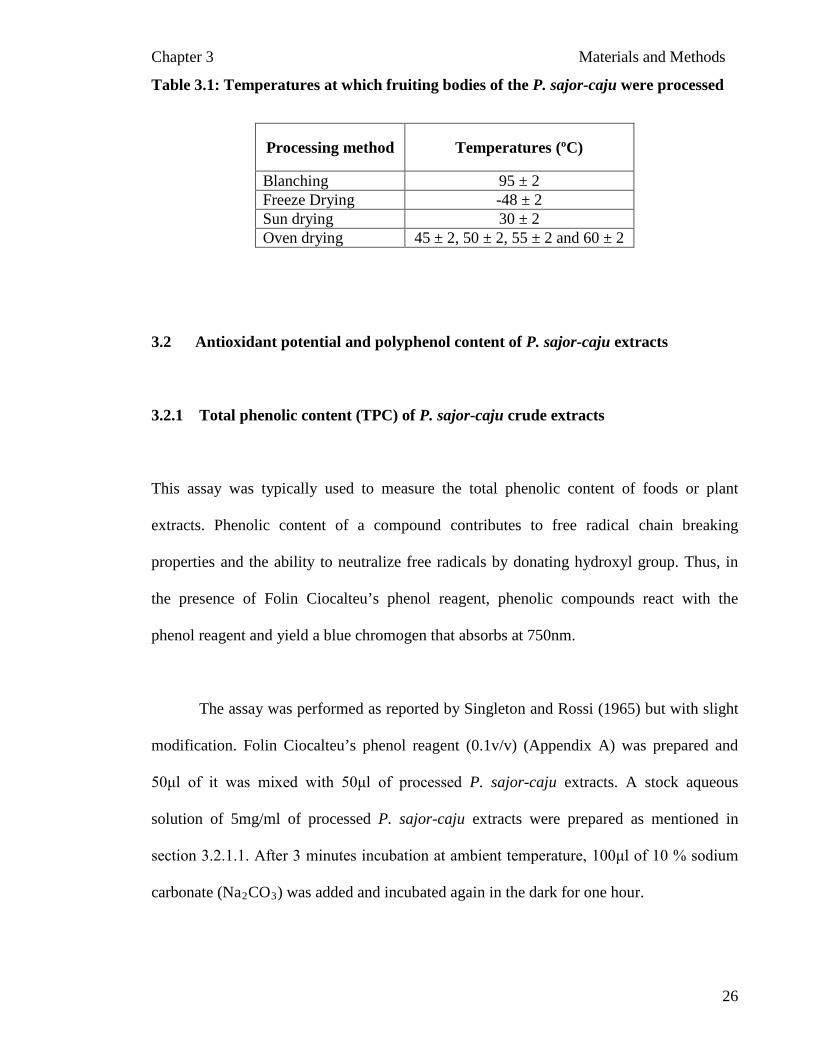
Table 3.1: Temperatures at which fruiting bodies of the P. sajor-caju were processed
3.2 Antioxidant potential and polyphenol content of P. sajor-caju extracts
3.2.1 Total phenolic content (TPC) of P. sajor-caju
crude extracts
This assay was typically used to measure the total phenolic content of foods or plant
extracts. Phenolic content of a compound contributes to free radical chain breaking
properties and the ability to neutralize free radicals by donating hydroxyl group. Thus, in
the presence of Folin Ciocalteu’s phenol reagent, phenolic compounds react with the
phenol reagent and yield a blue chromogen that absorbs at 750nm.
The assay was performed as reported by Singleton and Rossi (1965) but with slight
modification. Folin Ciocalteu’s phenol reagent (0.1v/v) (Appendix A) was prepared and
50μl of it was mixed with 50μl of processed P. sajor-caju extracts. A stock aqueous
solution of 5mg/ml of processed P. sajor-caju extracts were prepared as mentioned in
section 3.2.1.1. After 3 minutes incubation at ambient temperature, 100μl of 10 % sodium
carbonate (Na2CO3
) was added and incubated again in the dark for one hour.
Processing method Temperatures (ºC)
Blanching 95 ± 2 Freeze Drying -48 ± 2 Sun drying 30 ± 2 Oven drying 45 ± 2, 50 ± 2, 55 ± 2 and 60 ± 2
Chapter 3 Materials and Methods
27
The colour change was measured spectrophotometrically at 750 nm. Gallic acid was
used as standard and the phenolic content was expressed as mg GAE / 100 g of mushroom
extracts (Appendix B):-
Total phenolic content (mg GAE / 100g sample) = mg/ml sample
μg/ml GAE
=
mg sample μg GAE
= 100 x 100g sample
mg GAE
3.2.2
Antioxidant activity assay
3.2.2.1
DPPH free radical scavenging activity assay
The DPPH assay is a fast method to determine the free radical scavenging activity of the
mushroom extracts based on the reduction of 1, 1-diphenyl-2-picrylhydrazyl hydrate
(DPPH) radical. In the presence of antioxidant, the DPPH free radical will donate its
hydrogen which subsequently be reduced. The quenching of the DPPH radical was
measured spectrophotometrically at 515 nm. This assay was carried out according to
Gerhauser et al. (2003). Ascorbic acid was used as the positive reference standard in the
DPPH assay (Appendix C). Reaction mixtures of ascorbic acid, DPPH and methanol for the
assay were prepared freshly (Appendix A). A stock aqueous solution of 5mg/ml of
processed P. sajor-caju extracts were prepared (the solubility of the processed P. sajor -
caju was very low at concentration more than 5mg/ml). One hundred and ninety five
microlitres of various concentrations of DPPH were added in a 96 well plate. Then, five
Chapter 3 Materials and Methods
28
microlitre of various concentrations of ascorbic acid ranging from 0 – 1000 µg/ml and
extracts of P. sajor-caju (5mg/ml) were added with the DPPH reagent. The final
concentration of the diluents was 40x. The absorbance of the incubation mixture was
measured every 20 minutes at a wavelength of 515nm.
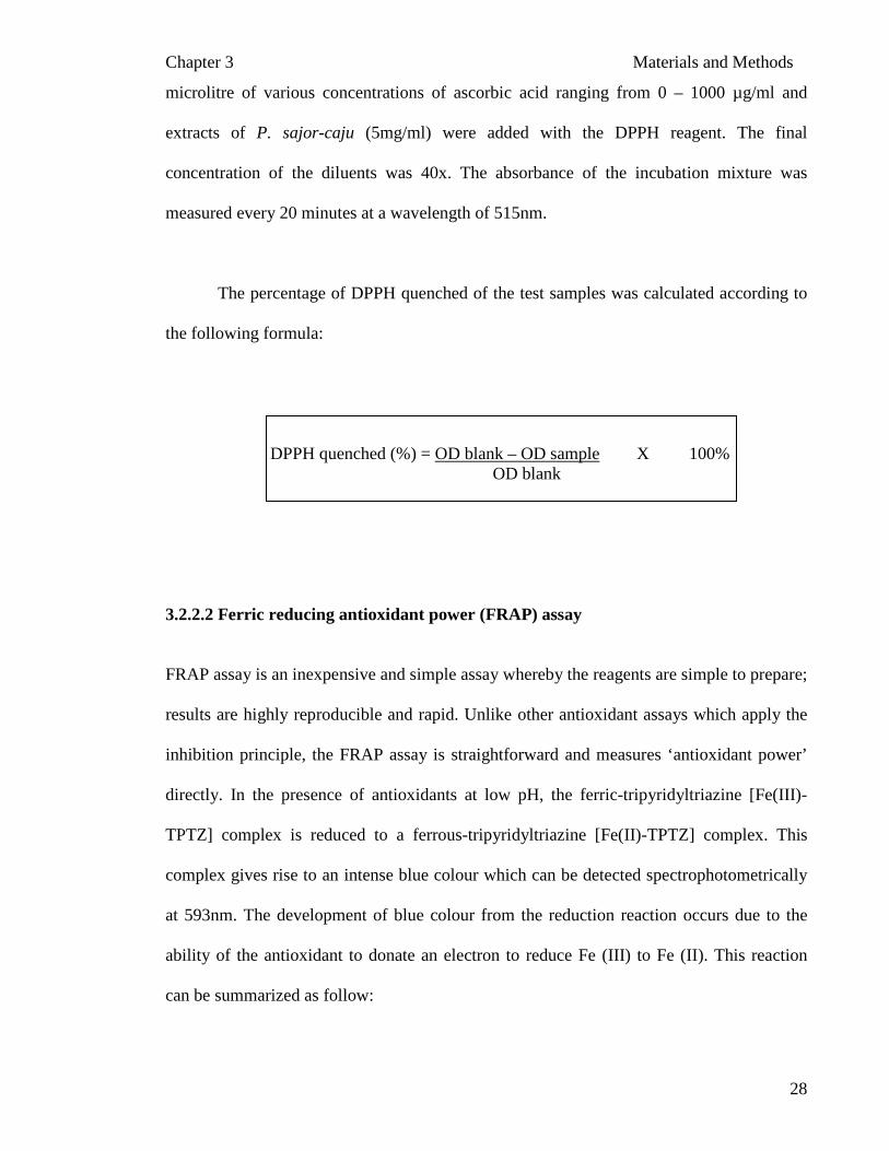
The percentage of DPPH quenched of the test samples was calculated according to
the following formula:
DPPH quenched (%) = OD blank – OD sample OD blank
X 100%
3.2.2.2
Ferric reducing antioxidant power (FRAP) assay
FRAP assay is an inexpensive and simple assay whereby the reagents are simple to prepare;
results are highly reproducible and rapid. Unlike other antioxidant assays which apply the
inhibition principle, the FRAP assay is straightforward and measures ‘antioxidant power’
directly. In the presence of antioxidants at low pH, the ferric-tripyridyltriazine [Fe(III)-
TPTZ] complex is reduced to a ferrous-tripyridyltriazine [Fe(II)-TPTZ] complex. This
complex gives rise to an intense blue colour which can be detected spectrophotometrically
at 593nm. The development of blue colour from the reduction reaction occurs due to the
ability of the antioxidant to donate an electron to reduce Fe (III) to Fe (II). This reaction
can be summarized as follow:
Chapter 3 Materials and Methods
29
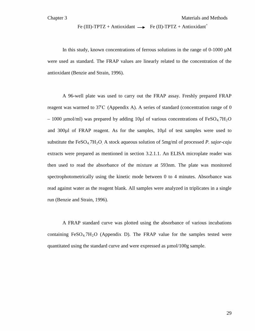
Fe (III)-TPTZ + Antioxidant Fe (II)-TPTZ + Antioxidant
+
In this study, known concentrations of ferrous solutions in the range of 0-1000 µM
were used as standard. The FRAP values are linearly related to the concentration of the
antioxidant (Benzie and Strain, 1996).
A 96-well plate was used to carry out the FRAP assay. Freshly prepared FRAP
reagent was warmed to 37̊ C (Appendix A). A series of standard (concentration range of 0
– 1000 µmol/ml) was prepared by adding 10µl of various concentrations of FeSO4.7H2O
and 300µl of FRAP reagent. As for the samples, 10µl of test samples were used to
substitute the FeSO4.7H2O.
A stock aqueous solution of 5mg/ml of processed P. sajor-caju
extracts were prepared as mentioned in section 3.2.1.1. An ELISA microplate reader was
then used to read the absorbance of the mixture at 593nm. The plate was monitored
spectrophotometrically using the kinetic mode between 0 to 4 minutes. Absorbance was
read against water as the reagent blank. All samples were analyzed in triplicates in a single
run (Benzie and Strain, 1996).
A FRAP standard curve was plotted using the absorbance of various incubations
containing FeSO4.7H2
O (Appendix D). The FRAP value for the samples tested were
quantitated using the standard curve and were expressed as µmol/100g sample.

Chapter 3 Materials and Methods
30
3.3 Cytotoxicity, cytoprotective, genotoxicity and genoprotective assays on
processed P. sajor-caju extract
3.3.1 Isolation of peripheral blood mononuclear cells (PBMCs)
The lymphocytes were isolated according to the method of Boyum (1968). Fresh whole
blood was collected by venipunture from healthy donors in sterile heparinized vacutainer
tubes. The blood was centrifuged at 900 x g for 30 minutes and a sterile pipette was used to
transfer the plasma into a Falcon tube. The packed cells were resuspended in an equal
volume of phosphate buffer saline (PBS). This mixture was layered carefully on to an equal
volume of Histopaque-1077 solution in a 50ml Falcon tube. The solution was centrifuged at
900 x g for 30 minutes.
After centrifugation, the upper layer of PBS was discarded to within 0.5cm of the
opaque interface containing the lymphocytes. The lymphocytes were aspirated using sterile
pipette and transferred into a sterile Falcon tube. Ten milliliters of PBS solution was added
and centrifuged at 600 x g for ten minutes. The supernatant was discarded and the pellet
was resuspended in 7ml of ammonium chloride solution to lyse the contaminating red cells
for ten minutes. Then, the contents were centrifuged again for ten minutes at 600 x g. The
supernatant was discarded and the pellet was resuspended again with PBS solution and
centrifuged for another ten minutes. The packed lymphocytes obtained at the end of the
centrifugation were resuspended with one to two milliliters of Roswell Park Memorial
Institute -1640 (RPMI 1640) solution that contained 10% fetal bovine serum (FBS) and 1%
L-Glutathione.
Chapter 3 Materials and Methods
31
The haemocytometer grid system was used to estimate the number of lymphocytes.
A sample of the PBMCs was diluted 1:1 with tryphan blue dye to a final volume of 20µl.
Cell number was estimated according to the following formula:
Cell concentration (cells/ml) = mean cell count x dilution factor x 10
= mean cell count x 2 x 10
4
4
The cells were resuspended in RPMI 1640 medium to the required concentration
and kept in the incubator with 5% CO2
at 37ºC until further use. The maximum incubation
period for the cells was two to three days.
3.3.2
Determination of cell viability of PBMCs
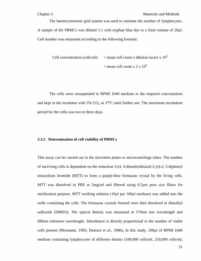
This assay can be carried out in the microtitre plates or microcentrifuge tubes. The number
of surviving cells is dependent on the reduction 3-(4, 4-dimethylthiazol-2-yl)-2, 5-diphenyl
tetrazolium bromide (MTT) to form a purple-blue formazan crystal by the living cells.
MTT was dissolved in PBS at 5mg/ml and filtered using 0.2μm pore size filters for
sterilization purpose. MTT working solution (10μl per 100μl medium) was added into the
wells containing the cells. The formazan crystals formed were then dissolved in dimethyl
sulfoxide (DMSO). The optical density was measured at 570nm test wavelength and
690nm reference wavelength. Absorbance is directly proportional to the number of viable
cells present (Mosmann, 1983; Denizot et al., 1986). In this study, 100μl of RPMI 1640
medium containing lymphocytes of different density (100,000 cells/ml, 250,000 cells/ml,

Chapter 3 Materials and Methods
32
500,000 cells/ml and 1,000,000 cells/ml) were pipetted into microtitre wells in triplicates.
This was done under sterile condition. Then, 10μl of MTT solution was added into each
well and the plates were incubated at 37ºC, 5% CO2
for four hours for the reaction to take
place. Then, 100µl of 100% DMSO was added to solubilize the insoluble purple formazan
product to form a colored solution. The absorbance of this colored solution was measured
using Powerwave 340 Microtitre-Plate ELISA Reader at wavelength of 570nm. The plates
have to be read within an hour after the addition of DMSO (Mosmann, 1983). Appendix E
shows the curve for the absorbance versus the number of cells. Figure 3.1 shows the flow
chart of MTT assay. The percentage of cell viability was calculated as follows:-
% of Cell viability = Abs Test Sample – Abs Test Control Abs
x 100%
Test Control
3.3.3 Cytotoxicity and cytoprotective activity of P. sajor-caju extracts on PBMCs
3.3.3.1 Determination of hydrogen peroxide (H2O2)
concentration as an oxidant
Hydrogen peroxide (H2O2) is poorly reactive, freely miscible with water and able to cross
cell membranes readily (Halliwell et al., 2000). It can act as a mild oxidizing or as a mild
reducing agent, but it does not oxidize most biological molecules readily, including lipids,
DNA and proteins. The LD50 values and the mode of cell death induced (apoptosis or
necrosis) by H2O2 depends on the cell type used, its physiological state, length of exposure
to H2O2, the H2O2 concentration used, and the cell culture media employed (Halliwell et
al., 2000). A number of reports have described high (≥ 50 μM) levels of H2O2 as being
cytotoxic to a wide range of animal, plant and bacterial cells in culture (Halliwell et al.,

Chapter 3 Materials and Methods
33
2000). The PBMCs were incubated with various concentrations of H2O2
(0mM to 800mM)
to determine the appropriate concentration to induce cytotoxicty (MTT).
3.3.3.2 Effects of P. sajor-caju extract on the viability of PBMCs
Hundred microlitre of freshly isolated cells were seeded into the 96-well plate at a density
of 5 x 10⁴ cells/ well. Ten microlitre of various concentrations 0µg/ml to 200µg/ml (final
concentration) of extracts or phytohemaglutinin (PHA) or H2O2 (final concentration of
300µM) were introduced to the cells and the cells were then incubated for two hours
(Figure 3.2). Phytohemaglutinin (PHA) is known as a mutagen. Thus, in this experiment,
three different concentrations of PHA were used to compare the viability of PBMCs mixed
with mushroom extract alone. The concentration of PHA used was 10µg/ml (M1), 20µg/ml
(M2) and 40µg/ml (M3). Water was used to blank the reading for spectrometer. Percentage
of cell viability was calculated as shown in section 3.3.2. All the extracts and controls were
diluted in water as the extraction of the processed mushroom was carried out using water.
Phytohemaglutinin was used as positive control to maintain the cell viability whereas H2O2
was used as a negative control to induce toxicity to the PBMCs. The PBMC viability was
assessed using the MTT assay (see section 3.3.2).
Chapter 3 Materials and Methods
34
Figure 3.1: Procedure for MTT assay.
Add 10 µl MTT solutions Incubate for 4 hours
Add 100 µl DMSO (100%)
Abs at 570nm test wavelength and 690nm reference wavelength
100µl Cell + RPMI-1640
Cell + RPMI-1640 + MTT solution

Chapter 3 Materials and Methods
35
Figure 3.2: Experimental protocol for the effects of mushroom extracts/PHA/H2O2
on the viability of PBMCs using MTT assay.
Freshly isolated cells
Various concentrations of extracts/ PHA/H2O2 and
2 hours, 37oC, 5% CO2 incubator
MTT assay
4 hours, 37oC, 5% CO2 incubator

Chapter 3 Materials and Methods
36
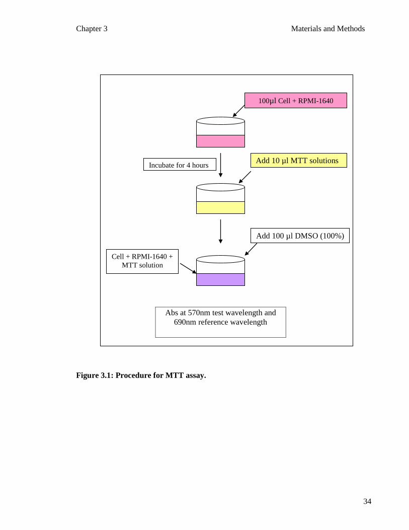
3.3.3.3 The cytoprotective effects of P. sajor-caju extract against H2O2
induced
toxicity on PBMCs
Hundred microlitres of PBMCs in RPMI-1640 medium were seeded in 96 microtitre plate
and were incubated with various concentrations of mushroom extracts for two hours at
37ºC, 5% CO2. Subsequently, ten microlitres of H2O2 (final concentration of 300 µM) was
added to the mixture of PBMCs and mushroom extracts (without washing off the
mushroom extracts). Then, the mixtures were incubated for another two hours at 37ºC, 5%
CO2
. Then, ten microlitre of MTT solution was added and the mixtures were incubated for
another four hours under the same conditions (Figure 3.3). The viability of PBMCs was
determined according to the procedure mentioned in section 3.3.2.
3.3.4 Genotoxicity and genoprotective activity of P. sajor-caju extracts on PBMCs
3.3.4.1 Comet assay to access DNA damage on PBMCs
The study of DNA damage in individual cells popularly known as comet assay was first
described by Ostling and Johanson (1984). In the comet assay, nucleated cells are
embedded in low melting point agarose on a microscope slide, and the membrane and
histones are removed by high salt solution (Tice et al., 2000). Generally, the DNA is
organized in a tightly supercoiled form (Cook et al., 1976), and alkaline solution is used to
unwind this supercoiled DNA for the migration during electrophoresis. The electrophoresis
at neutral, mildly alkaline or strongly alkaline conditions is applied to continue the
unwinding step (Angelis et al, 1999).
Chapter 3 Materials and Methods
37
Figure 3.3: Experimental protocol to study the effects of preincubated P. sajor-caju extracts on the viability of PBMCs which then challenged with 300 µM of H2O
2.
Add H2O2 (300 µM)
MTT assay
2 hours at 37ºC, 5% CO2
Cells + Extracts in RMPI-1640 medium
2 hours at 37ºC, 5% CO2
4 hours at 37ºC, 5% CO2
Chapter 3 Materials and Methods
38
At alkaline pH the phosphate groups in DNA will be negatively charged and the
relaxed loops of damaged DNA containing breaks will be pulled towards the anode during
electrophoresis, forming a comet ‘tail’, while the DNA remaining coiled within the
nucleoid forms the comet ‘head’ (Tice et al., 2000). The comet is visualized by a DNA
staining fluorescent dye or silver stain (Clingen et al., 2000). The most frequently used
stains are ethidium bromide (Tice et al., 2000), propidium iodide (Olive, 2002), 4,6-
diamidino- 2-phenylindole (DAPI) (Panayiotidis and Collins, 1997) and YOYO-1 (Singh,
1996). The damaged DNA is scored using either visual or computerized image analysis.
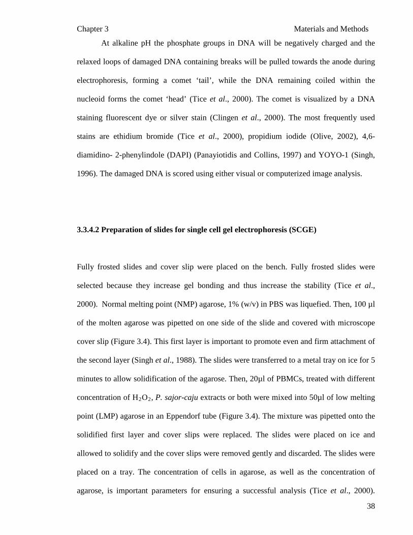
3.3.4.2 Preparation of slides for single cell gel electrophoresis (SCGE)
Fully frosted slides and cover slip were placed on the bench. Fully frosted slides were
selected because they increase gel bonding and thus increase the stability (Tice et al.,
2000). Normal melting point (NMP) agarose, 1% (w/v) in PBS was liquefied. Then, 100 µl
of the molten agarose was pipetted on one side of the slide and covered with microscope
cover slip (Figure 3.4). This first layer is important to promote even and firm attachment of
the second layer (Singh et al., 1988). The slides were transferred to a metal tray on ice for 5
minutes to allow solidification of the agarose. Then, 20µl of PBMCs, treated with different
concentration of H2O2, P. sajor-caju extracts or both were mixed into 50µl of low melting
point (LMP) agarose in an Eppendorf tube (Figure 3.4). The mixture was pipetted onto the
solidified first layer and cover slips were replaced. The slides were placed on ice and
allowed to solidify and the cover slips were removed gently and discarded. The slides were
placed on a tray. The concentration of cells in agarose, as well as the concentration of
agarose, is important parameters for ensuring a successful analysis (Tice et al., 2000).

Chapter 3 Materials and Methods
39
Overlapping comets were obtained if the density of the cells were too high with high levels
of DNA migration. The extent of DNA migration was also affected by higher
concentrations of agarose.
After the slides were transferred into a tray, it was submerged in lysing solution
containing high salts and detergents overnight at 4 ± 2ºC (Figure 3.4) to break the cells.
When treated with alkaline solution the DNA unwinds and this enables the migration of the
DNA during electrophoresis. The lysis solution contained EDTA, Tris Base, sodium
chloride, sodium dodecyl sulphate (SDS) and Triton-X. The lysing solution was chilled 30
minutes prior to use, primarily to maintain the stability of the agarose gel (Tice et al.,
2000). At the end of lysing period, the slides were rinsed carefully dipped thrice in TAE
buffer in a Coplin jar to remove residual detergents and salts. The slides were drained on
paper towel and were ready for electrophoresis.

Chapter 3 Materials and Methods
40
Figure 3.4: Flow chart of the preparation of slides to be used before electrophoresis for Comet assay.
On Ice for 5 minutes
1% Low Melting Point Agarose + Cells/Treated
cells
On Ice for 5 minutes
Lysis solution for overnight at 4ºC
Rinse with TAE buffer (Coplin jar) at 4ºC
Ready for electrophoresis
100μl of 1% Normal Melting Point Agarose
Chapter 3 Materials and Methods
41
3.3.4.3 Electrophoresis of microgel slides containing PBMCs in agarose gel
The slides were placed side by side on the horizontal electrophoresis tank near the anode
end, sliding them as close together as possible. The buffer reservoir was filled with freshly
prepared electrophoresis buffer (which was chilled at least 30 minutes prior to use) until the
slides were submerged in the solution. Prior to electrophoresis, the slides were incubated
for 40 minutes in alkaline (pH > 13) electrophoresis buffer at four degree Celsius to
produce single stranded DNA (ssDNA) and to express the alkaline labile sites (ALS) as
single strand breaks (SSB) (Tice et al., 2000). The power supply was turned on to 25V after
40 minutes and the current adjusted to 300mA. The duration of the electrophoresis was 20
minutes. Throughout the electrophoresis, the buffer temperature was maintained at four
degree Celsius to prevent melting of the LMP layer of the microgel by placing the tank on
bed of ice. After the electrophoresis, the power was switched off and the slides were lifted
gently from the buffer. The slides were drained on paper towel.
After electrophoresis, the alkaline pH of the gels was neutralized by rinsing the
slides with neutralization buffer pH 7.5. Singh et al. (1988) proposed three washes of
trizma buffer for 5 minutes each. However, increased rinsing may be useful in situations
where a high background was seen during scoring (Rojas et al., 1999). After,
neutralization, the slides were fixed in absolute ethanol for two hours to dehydrate the
agarose gel and this enabled longer storage period when compared with non-dehydrated
agarose gel (Tice et al., 2000).
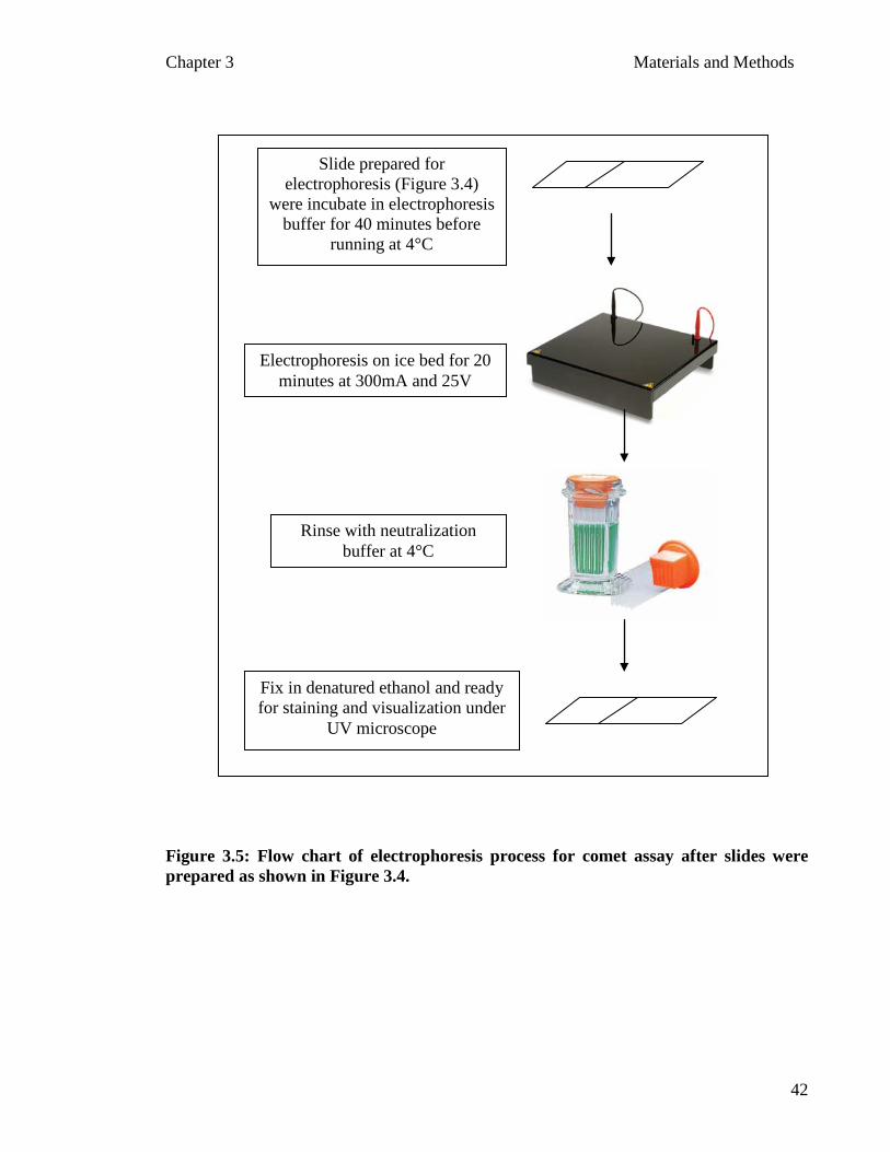
Chapter 3 Materials and Methods
42
Figure 3.5: Flow chart of electrophoresis process for comet assay after slides were prepared as shown in Figure 3.4.
Slide prepared for electrophoresis (Figure 3.4)
were incubate in electrophoresis buffer for 40 minutes before
running at 4°C
Electrophoresis on ice bed for 20 minutes at 300mA and 25V
Rinse with neutralization buffer at 4°C
Fix in denatured ethanol and ready for staining and visualization under
UV microscope
Chapter 3 Materials and Methods
43
3.3.4.4 Effects of H2O2
on DNA damage of PBMC’s
Peripheral blood mononuclear cells were prepared as described in section 3.3.1. Various
concentrations of H2O2 (0mM to 0.3mM) were used to induce DNA damage. After the
incubation of PBMCs at 37ºC and 5% CO2 for 30 minutes, the cells were immediately
placed in ice for 5 minutes and then slides were prepared and the electrophoresis was
carried out as mentioned in section 3.3.4.2 and 3.3.4.3. The electrophoresed slides were
stained with ethidium bromide as described in section 3.3.5. The stained slides were viewed
under florescent microscope and the percentage of tail and head DNA were recorded. The
concentration of H2O2
that caused 50% DNA damage was selected for the subsequent
experiments that required DNA damage to be induced.
3.3.4.5 Effects of P. sajor-caju extract on DNA of PBMC’s
Peripheral blood mononuclear cells (prepared as mentioned in 3.3.1) were incubated with
various concentrations (0µg/ml to 200µg/ml) of different processed mushroom extracts for
30 minutes prior to electrophoresis. The concentration of H2O2 that caused 50% damage on
DNA in PBMC’s (IC50
) was used as positive control. After the incubation, the treated cells
were incubated in ice for 5 minutes and then slides were prepared and the electrophoresis
was carried out as described in sections 3.3.4.2 and 3.3.4.3. The electrophoresed slides
were stained with ethidium bromide as described in section 3.3.5. The stained slides were
viewed under florescent microscope and the percentage of tail and head DNA were
recorded.
Chapter 3 Materials and Methods
44
3.3.4.6 Protective effects of P. sajor-caju extract on H2O2
PBMCs
induced DNA damage in
Peripheral blood mononuclear cells were incubated with various concentrations of extracts
ranging from 0µg/ml to 200µg/ml for 30 minutes. Subsequently, the concentration of H2O2
that caused 50% inhibition was added to the mixture and was incubated for another 30
minutes. After the incubation, the treated cells were incubated in ice for 5 minutes and then
slides were prepared and the electrophoresis was carried out as described in sections 3.3.4.2
and 3.3.4.3. The electrophoresed slides were stained with ethidium bromide as described in
section 3.3.5. The stained slides were viewed under florescent microscope and the
percentage of tail and head DNA were recorded.
3.3.5 DNA Staining and Visualization
The slides were stained with 20µg/ml ethidium bromide (EtBr). The DNA specific dye and
the magnification used for comet visualization depend largely on investigator-specific
needs and presumably have little effect on assay sensitivity or reliability (Tice et al., 2000).
Comet images were visualized and scored under 200x magnification on a fluorescent
microscope. The selection of magnification is dependent on the type of cells, the range of
migration to be measured and constraint of the microscope or imaging system (Tice et al.,
2000).
Chapter 3 Materials and Methods
45
All the slides, including the positive and negative controls were scored using an
image analyzer which is fully automated comet analysis imaging system, where the extent
of the migration of DNA was scored individually. The proportional level of SSB and ALS
related directly to the DNA fragment size and generally the migration length (Tice et al.,
2000). The average of head and tail DNA of a certain number of scored cells was
calculated using SPSS software or Excel worksheet. Tables and bar graphs were generated
from the data collected.
3.4 Statistical Analysis
Three replicates of each sample were used for statistical analysis. All the results obtained
were expressed as mean ± standard error mean (S.E.M). The SPSS program version 13.0
and GraphPad Prism version 4.1 was used to calculate the significance of the results.
Differences at p < 0.05 were considered to be significant.

CHAPTER 4
RESULTS &
DISCUSSION

Chapter 4 Results and Discussion
46
CHAPTER 4
RESULTS AND DISCUSSION
4.1 Preparation of aqueous extracts from fruiting bodies of P. sajor-caju
Pleurotus sajor-caju was processed and extracted using hot water. The fruiting bodies were
divided into seven groups of one kilogram each that were processed at different
temperatures based on the cooking or industrial processing methods of this edible oyster
mushroom. The temperature for each processing method used is given in Table 3.1 (see
section 3.1).
The weights of the various P. sajor-caju extracts were measured and the values are
depicted in Table 4.1. The hot water extract for blanched P. sajor-caju contained high
content of water (approximately 89%) and this was possibly due to the high water content
in the freshly boiled mushroom. The moisture content of the mushroom observed in the
present study was similar to that reported by Breene (1990). The dried P. sajor-caju lost
approximately 90% (w/w) of its water content. The highest loss of water content was
observed in freeze dried P. sajor-caju. Freeze-drying or lyophilisation is a usual method
used in scientific work to remove water from any substance fast and efficiently.

Chapter 4 Results and Discussion
47
The yield from the final extract of blanched P. sajor-caju was approximately eight
grams. The total loss of water was more than 99% of the initial weight. This was probably
due to the fact that blanching cause’s water absorption and that contributed to the initial
weight. The yield of the extracts was determined by comparing the mass of the final and
initial weight of the P. sajor-caju. The highest percentage of yield was obtained from 45 ±
2ºC oven dried followed by sun dried, 50 ± 2ºC oven dried, 60 ± 2ºC oven dried, 55 ± 2ºC
oven dried and freeze dried mushrooms. The crude hot water extracts of each processed P.
sajor-caju were further tested for their cytotoxic and genotoxic activities using MTT assay
and Comet assay respectively.
Mushroom polysaccharides exist as a structural component of fungal cell wall
(Zhang et al., 2007). Their cell wall is composed of two major types of polysaccharides: -
1) A rigid fibrillar chitin (or cellulose) and 2) A matrix–like beta glucan, alpha glucan, and
glycoproteins (Zhang et al., 2007). Selection of an extraction method depends on the cell
wall structure of the mushroom. The hot water extraction has been a popular approach for
mushroom extraction, especially to have a higher yield of polysaccharides’ that are water
soluble (Zhang et al., 2007). In addition, Lee et al. (2007) reported that the yields of water
extracts of P. citrinopileatus were significantly higher than those of ethanolic extracts for
three types of samples; fruiting bodies, mycelia and culture filtrate. The difference of the
yield might be due to the fact that water extracts contained certain amount of soluble
polysaccharides that could be precipitated from aqueous suspension of fruiting bodies or
mycelia (Lee et al., 2007). Hence, in this study, the similar finding on the yield of aqueous
extracts was expected.

Chapter 4 Results and Discussion
48
Table 4.1: The extraction yields after the processed samples were extracted in boiling water for three hours
The P. sajor-caju obtained from the farmer was weight and processed accordingly. The weight before and after processing was recorded.
Then, the different processed P. sajor-caju was extracted using aqueous extraction in 95ºC hot water bath. The weights of the lyophilized
extracts were determined using weighing balance. The percentage of extraction (%) was calculated using this formula:-
(Lyophilized extract (g) ÷ Weight after processing (g) x 100%).
Pleurotus sajor-caju Weight before (g) Weight after
processing (g)
Extracted
with boiling
water (L)
Lyophilized
extract (g)
Percentage of
Extraction (%)
Blanching (P1) 1000 882.5 2.5 8.4 1.0
Sun dry (P2) 1000 95.0 1.0 27.8 29.3
Freeze dry (P3) 1000 103.1 0.8 18.5 17.9
45º oven dry (P4) 1000 96.7 1.0 29.4 30.4
50º oven dry (P5) 1000 105.0 1.2 23.8 22.7
55º oven dry (P6 1000 104.4 1.1 22.7 21.7
60º oven dry (P7) 1000 103.6 1.1 23.0 22.2

Chapter 4 Results and Discussion
49
4. 2 Antioxidant potential and polyphenol content of P. sajor-caju extracts
4.2.1 Antioxidant activity assay
4.2.1.1 Total phenolic content (TPC) of aqueous extracts of P. sajor-caju
In this study, the phenol content of different processed P. sajor-caju was investigated. The
total phenolic content was expressed as mg of GAEs/g of dry mushroom (Figure 4.1). The
amount of phenolic compounds in extracts P2 (254.63±4.39 mg GAEs/g of extract) and P4
(257.56±3.62 mg GAEs/g of extract) were among the highest with p<0.05, followed by P5,
P6 and P7 (approximately 230 mg GAEs/g of extract). The blanched (P1) mushroom
extract contained the least amount of phenols (approximately 185 mg GAEs/g of extract).
Based on the earlier results (Table 4.1), P2 and P4 had comparatively higher phenolic
content could be due to their different processing methods. Meanwhile, the lowest
extraction yield was in P1.
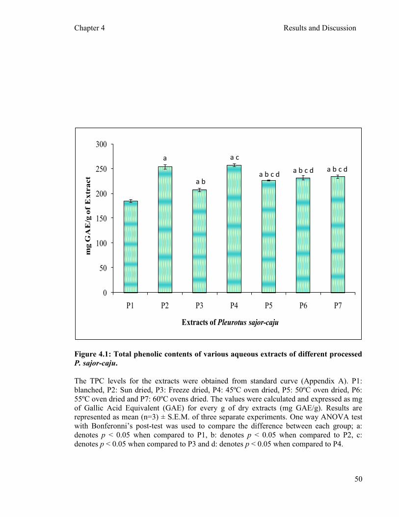
Chapter 4 Results and Discussion
50
0
50
100
150
200
250
300
P1 P2 P3 P4 P5 P6 P7
mg
GA
E/g
of
Ex
tra
ct
Extracts of Pleurotus sajor-caju
Figure 4.1: Total phenolic contents of various aqueous extracts of different processed
P. sajor-caju.
The TPC levels for the extracts were obtained from standard curve (Appendix A). P1:
blanched, P2: Sun dried, P3: Freeze dried, P4: 45ºC oven dried, P5: 50ºC oven dried, P6:
55ºC oven dried and P7: 60ºC ovens dried. The values were calculated and expressed as mg
of Gallic Acid Equivalent (GAE) for every g of dry extracts (mg GAE/g). Results are
represented as mean (n=3) ± S.E.M. of three separate experiments. One way ANOVA test
with Bonferonni’s post-test was used to compare the difference between each group; a:
denotes p < 0.05 when compared to P1, b: denotes p < 0.05 when compared to P2, c:
denotes p < 0.05 when compared to P3 and d: denotes p < 0.05 when compared to P4.
a
a b a b c d
a c
a b c d a b c d

Chapter 4 Results and Discussion
51
Phenolics are one of the major groups of non-essential dietary components that have
been associated with the inhibition of atherosclerosis and cancer (Teissedre et al., 1996).
The bioactivity of phenolics may be related to their ability to chelate metals, inhibit
lipoxygenase activity and scavenge free radicals (Decker, 1997). Mushrooms accumulate a
variety of secondary metabolites, including phenolic compounds, polyketides, terpenes and
steroids. Velioglu et al., (1998) reported that there is a correlation between the antioxidant
activities of plant extracts with the content of their phenolic compounds. In addition,
Cheung et al. (2003) reported that the phenolic compound found in edible mushrooms such
as L. edodes and V. volvacea could make a significant contribution to the antioxidant
activity.
4.2.1.2 DPPH free radical scavenging activity assay on processed mushroom extracts
Figure 4.2, depicts DPPH radical scavenging activity by different processed P. sajor-caju
extracts. The aqueous extracts of P. sajor-caju were shown to scavenge the stable DPPH
radical directly to different extents at a concentration of 5mg/ml (initial concentration) with
the minimum percentage of inhibition exerted by extracts P1 and P3. In general, all the
extracts depicted low scavenging activity. However, from this result it can be assumed that
the extracts are a free radical inhibitor or scavenger, acting possibly as primary
antioxidants.
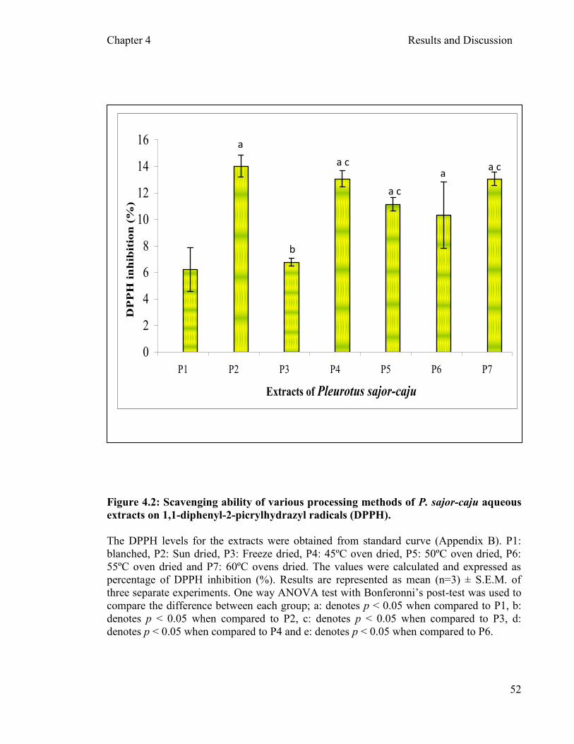
Chapter 4 Results and Discussion
52
Figure 4.2: Scavenging ability of various processing methods of P. sajor-caju aqueous
extracts on 1,1-diphenyl-2-picrylhydrazyl radicals (DPPH).
The DPPH levels for the extracts were obtained from standard curve (Appendix B). P1:
blanched, P2: Sun dried, P3: Freeze dried, P4: 45ºC oven dried, P5: 50ºC oven dried, P6:
55ºC oven dried and P7: 60ºC ovens dried. The values were calculated and expressed as
percentage of DPPH inhibition (%). Results are represented as mean (n=3) ± S.E.M. of
three separate experiments. One way ANOVA test with Bonferonni’s post-test was used to
compare the difference between each group; a: denotes p < 0.05 when compared to P1, b:
denotes p < 0.05 when compared to P2, c: denotes p < 0.05 when compared to P3, d:
denotes p < 0.05 when compared to P4 and e: denotes p < 0.05 when compared to P6.
0
2
4
6
8
10
12
14
16
P1 P2 P3 P4 P5 P6 P7
Extracts of Pleurotus sajor-caju
DP
PH
in
hib
itio
n (
%)
a
a c a
b
a c
a c

Chapter 4 Results and Discussion
53
Free radical scavenging is one of the known mechanisms by which antioxidants
inhibit lipid oxidation. The method of scavenging DPPH free radicals can be used to
evaluate the antioxidant activity of specific compounds or extracts in a short time. The
scavenging abilities of hot water extracts of G. tsugae, A. cylendracea, H. marmorues and
P. citrinopleatus fruiting bodies at 20 mg/ml were 73.8-80.1%, 66.2%, 77.2% and 52.3%
respectively (Lee et al., 2007). Apparently, the scavenging ability of P. sajor-caju hot
water extracts at 5mg/ml was less effective (Figure 4.2) than those mentioned above. This
could be caused by the concentration of the extract or the extraction method might play an
important role in the free radical scavenging activity. Lee et al. (2008) reported that
ethanolic extracts showed higher scavenging activities on DPPH radical than hot water
extracts. The scavenging activities of the fruiting bodies of the white mutant of H.
marmoreus at 5mg/ml were 75.5% for ethanolic extracts and 36.8% for hot water extracts,
respectively (Lee et al., 2008). At 10 mg/ml, scavenging abilities were 94.8% for ethanolic
extracts and 40.3% for hot water extracts from fruiting bodies of this mushroom,
respectively. This shows that at two different concentrations the mushrooms extracts
conferred different level of scavenging activities regardless of the method of extraction
employed.
Generally, the aqueous/solvent extracts contained antioxidant components
regardless of their processing method, which could react rapidly with DPPH radicals, and
reduce most DPPH radical molecules (Brand-Williams et al., 1995). Hence, this result
discloses that the P. sajor-caju extracts are free radical inhibitors or scavengers, acting
possibly as primary antioxidants. The scavenging activity of the extracts might be due to
the presence of hydrogen-donating components in the mushroom extracts (Mau et al.,
Chapter 4 Results and Discussion
54
2002). Although the different processed P. sajor-caju extracts showed lower than 20%
scavenging activity compared to other reported mushrooms as mentioned above, one
should not rule out the possibility that the P. sajor-caju aqueous extracts might react with
free radicals, particularly peroxy radicals, which are the major propagator of autooxidation
chain of fat, thereby terminating the chain reaction (Mau et al., 2002). Antioxidant activity
of natural antioxidants has been shown to be involved in termination of free radical reaction
(Shimada et al., 1992; Tanaka et al., 1988).
4.2.1.3 Ferric reducing antioxidant power (FRAP) assay of P. sajor-caju crude
extracts
In this experiment the reducing power of seven different processed mushrooms was
assessed. Figure 4.3 depicted that P2 and P7 had the highest reducing power with
49.66±0.94 µmol FRAP/ g extract and 48.33 µmol FRAP/ g extract, respectively. P1 and
P3 showed almost the same amount of reducing power (38 µmol FRAP/ g extracts). Tsai et
al., (2007) reported that P. citrinopileatus showed a high reducing power of 1.03 at 5
mg/ml.
The reducing capacity of various extracts might be due to its hydrogen-donating
ability, as described by Shimada et al. (1992). Therefore, the extracts might contain
reductones (strong reducing agents commonly derived from saccharides by oxidation at the
carbon atom alpha to the carbonyl function), which could react with free radicals to
stabilize and terminate radical chain reactions.
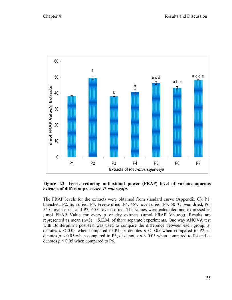
Chapter 4 Results and Discussion
55
0
10
20
30
40
50
60
P1 P2 P3 P4 P5 P6 P7
Extracts of Pleurotus sajor-caju
µm
ol
FR
AP
Va
lue
/g E
xtr
ac
ts
Figure 4.3: Ferric reducing antioxidant power (FRAP) level of various aqueous
extracts of different processed P. sajor-caju.
The FRAP levels for the extracts were obtained from standard curve (Appendix C). P1:
blanched, P2: Sun dried, P3: Freeze dried, P4: 45ºC oven dried, P5: 50 ºC oven dried, P6:
55ºC oven dried and P7: 60ºC ovens dried. The values were calculated and expressed as
µmol FRAP Value for every g of dry extracts (µmol FRAP Value/g). Results are
represented as mean (n=3) ± S.E.M. of three separate experiments. One way ANOVA test
with Bonferonni’s post-test was used to compare the difference between each group; a:
denotes p < 0.05 when compared to P1, b: denotes p < 0.05 when compared to P2, c:
denotes p < 0.05 when compared to P3, d: denotes p < 0.05 when compared to P4 and e:
denotes p < 0.05 when compared to P6.
a
b
a c d e
b
a b c a c d

Chapter 4 Results and Discussion
56
4.2.3 Correlation
The correlation between total phenol content, FRAP level and DPPH scavenging activity
was carried out. . Figure 4.4a-c showed the correlation (linear regression curve) for TPC
and FRAP, TPC and DPPH as well as FRAP and DPPH respectively. The strongest
correlation coefficient and regression was observed between scavenging activity of DPPH
and phenolic content in processed mushroom (r2
= 0.867, R2
= 0.752 with p<0.0001)
(Figure 4.4b). The lowest correlation coefficient and regression (r2
= 0.606, R2
= 0.368 with
p<0.005) was present between phenolic content and FRAP in the processed mushroom
(Figure 4.4a). Thus, the phenolic compounds present in the P. sajor-caju extracts contribute
to free radical scavenging activity and less of the reducing activity.
Effective antioxidants can be found in compound with phenol group such as BHT
and gallate (Madhavi et al., 1996). Phenolic compounds are well known for their ability to
scavenge free radicals and chelating ferrous ions which make them the best candidate as
antioxidant, antimutagen and anticancer (Ahmad and Mukhtar, 1999). Thus, the higher
content of phenols in the fruiting bodies of P. sajor-caju extracts was responsible for their
better antioxidant properties. In general, all the different extract of processed P. sajor-caju,
contained antioxidant properties which were moderate to high at 5mg/ml. The P2 and P4
had the highest content of phenols; FRAP level and free radical scavenging activity, where
as the P1 showed the lowest antioxidant and phenolic content amongst the extracts tested.
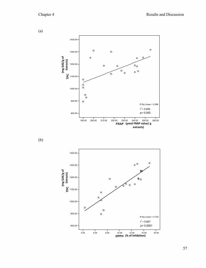
Chapter 4 Results and Discussion
57
(a)
190.00 200.00 210.00 220.00 230.00 240.00 250.00 260.00
FRAP
800.00
900.00
1000.00
1100.00
1200.00
1300.00
1400.00
TP
C
R Sq Linear = 0.368
(b)
4.00 6.00 8.00 10.00 12.00 14.00 16.00
DPPH
800.00
900.00
1000.00
1100.00
1200.00
1300.00
1400.00
TP
C
R Sq Linear = 0.752
r2 = 0.606
r2 = 0.867
p< 0.005
p< 0.0001
(mg
GA
E/g
of
Extr
acts
)
(mg
GA
E/g
of
Extr
acts
)
(µmol FRAP value/ g extracts)
(% of Inhibition)

Chapter 4 Results and Discussion
58
(c)
4.00 6.00 8.00 10.00 12.00 14.00 16.00
DPPH
190.00
200.00
210.00
220.00
230.00
240.00
250.00
260.00
FR
AP
R Sq Linear = 0.601
Figure 4.4: Correlation between TPC, DPPH and FRAP of different processed
aqueous extracts of P. sajor-caju.
Correlation of TPC levels, DPPH scavenging activities and FRAP levels of seven
processed P. sajor-caju extracts were assessed using Pearson correlation analysis. Panels
(a) depicts the correlation between TPC and FRAP levels, panel (b) depicts correlation
between TPC levels and DPPH scavenging activities and panel (c) shows the correlation
between FRAP levels and DPPH scavenging activities.
r2 = 0.775
p< 0.0001
(% of Inhibition)
(µm
ol F
RA
P v
alu
e/
g
ext
ract
s)
Chapter 4 Results and Discussion
59
Generally, the P. sajor-caju extracts showed moderate polyphenolic content, RAP
value and DPPH scavenging activities regardless of their processing methods. Lee et al.
(2007) reported the correlation of TPC– FRAP, TPC-DPPH and FRAP-DPPH with the
correlation coefficients (r2) of 0.527, 0.556 and 0.425, respectively. Alternatively, in this
study the correlation coefficient (r2) that was established was higher (Figure 4.4a-c)
compared to correlation coefficients reported by Lee et al. (2007). Hence, it shows that
processed P. sajor-caju extracts can still be a reliable source of antioxidants and phenols
for health benefits.
During food processing, considerable changes in level and nature of antioxidant
activity can frequently occur as a result of various reactions such as hydrolysis, oxidation
and the Maillard reaction (Konecny et al., 2009). In future, a detailed study of antioxidant
properties of the different processed P. sajor-caju must be carried out to identify the
mechanism and the specific phenolic component/s that is/are present in the extracts. The
fractionation of these processed mushrooms will help us to discover the specific phenolic
component mentioned above.
Chapter 4 Results and Discussion
60
4.3 Determination of viable cells
4.3.1 Determination of hydrogen peroxide (H2O2) concentration as an oxidant
The cells were incubated with H2O2 concentrations ranging from zero to 800mM (final
concentration). However, at concentrations above 10mM, H2O2 showed an apparent
increase in cell viability (Figure 4.5). In order to rule out the possible interference of the
RPMI medium in the MTT assay, the assay was carried out using cell-free medium. Figure
4.6 shows an increase in absorbance with increasing concentration of H2O2 (5mM to
800mM). It is possible that the media, MTT solution or the H2O2 concentrations have
reactions between each other that showed false positive reactions. Hence, the identification
of IC50 was not possible and the range of H2O2 that was used in this experiment was limited
to a maximum of 1mM. The cell viability of the PBMCs exposed to H2O2 concentrations
ranging from 0 – 1mM is depicted in Figure 4.7. Hydrogen peroxide concentration of
0.3mM was used as the baseline or control to induce a mild cytotoxic effect (25% inhibition
of cell viability). Kuppusamy et al. (2009) reported that 5µM of H2O2 can cause 50%
inhibition on PBMCs; however, some other reports have shown a higher concentration of
H2O2 can be employed to induce required toxicity on PBMCs as there are variations in
susceptibility of PBMC from different individual to H2O2 (Kuppusamy et al., 2009).
Hydrogen peroxide appears to be a ubiquitous molecule and poorly reactive. It is
relatively stable and is known for its capacity to diffuse across cellular membrane
(Knaapen, 2006). They can be a mild oxidizing or reducing agent but it does not oxidize
most biological molecules such as lipids, DNA and proteins unless the latter have hyper-
Chapter 4 Results and Discussion
61
reactive thiol groups or methionine residues (Halliwell et al., 2000). Various report has
been published on the levels of H2O2 as being cytotoxic (usually > 50µM) to a wide range
of animal, plant and bacterial cells in culture (Halliwell et al., 2000). However, the LD50
values and the mode of cell death (either apoptosis or necrosis) induced by H2O2 depend
on the cell type used, its physiological state, length of exposure to H2O2, the H2O2
concentration used, and the cell culture media in use (Halliwell and Gutridge, 2003).
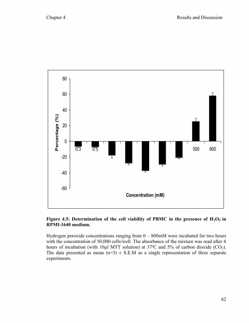
Chapter 4 Results and Discussion
62
-60
-40
-20
0
20
40
60
80
0.3 0.5 1 5 10 50 100 500 800
Concentration (mM)
Percen
tag
e (
%)
Figure 4.5: Determination of the cell viability of PBMC in the presence of H2O2 in
RPMI-1640 medium.
Hydrogen peroxide concentrations ranging from 0 – 800mM were incubated for two hours
with the concentration of 50,000 cells/well. The absorbance of the mixture was read after 4
hours of incubation (with 10µl MTT solution) at 37ºC and 5% of carbon dioxide (CO2).
The data presented as mean (n=3) ± S.E.M as a single representation of three separate
experiments.
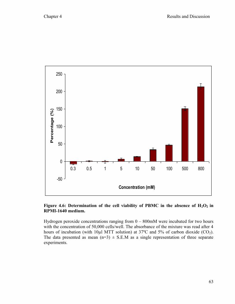
Chapter 4 Results and Discussion
63
-50
0
50
100
150
200
250
0.3 0.5 1 5 10 50 100 500 800
Concentration (mM)
Percen
tag
e (
%)
Figure 4.6: Determination of the cell viability of PBMC in the absence of H2O2 in
RPMI-1640 medium.
Hydrogen peroxide concentrations ranging from 0 – 800mM were incubated for two hours
with the concentration of 50,000 cells/well. The absorbance of the mixture was read after 4
hours of incubation (with 10µl MTT solution) at 37ºC and 5% of carbon dioxide (CO2).
The data presented as mean (n=3) ± S.E.M as a single representation of three separate
experiments.
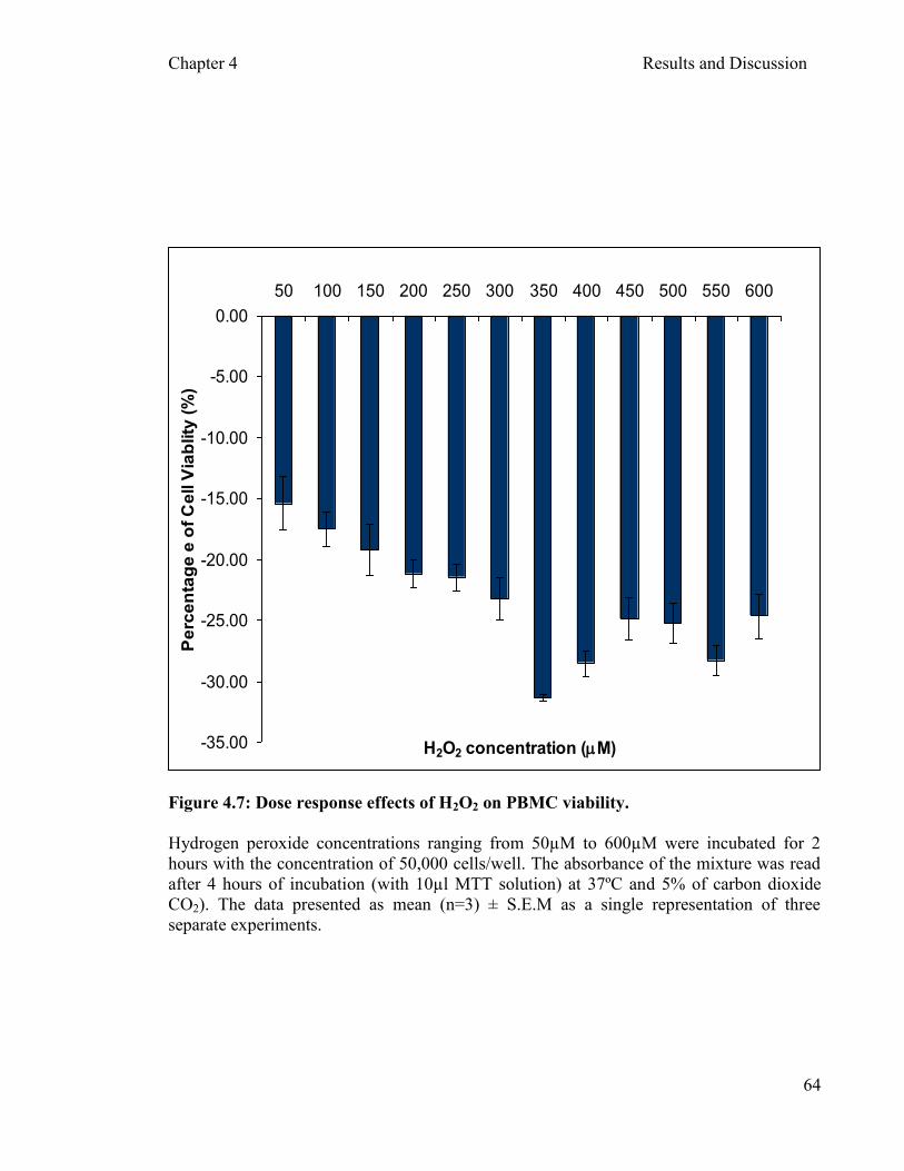
Chapter 4 Results and Discussion
64
-35.00
-30.00
-25.00
-20.00
-15.00
-10.00
-5.00
0.00
50 100 150 200 250 300 350 400 450 500 550 600
H2O2 concentration (mM)
Pe
rce
nta
ge
e o
f C
ell V
iab
lity
(%
)
Figure 4.7: Dose response effects of H2O2 on PBMC viability.
Hydrogen peroxide concentrations ranging from 50µM to 600µM were incubated for 2
hours with the concentration of 50,000 cells/well. The absorbance of the mixture was read
after 4 hours of incubation (with 10µl MTT solution) at 37ºC and 5% of carbon dioxide
CO2). The data presented as mean (n=3) ± S.E.M as a single representation of three
separate experiments.
Chapter 4 Results and Discussion
65
Higher concentration of H2O2 will be more susceptible to diffuse into the cell
membranes. Hydrogen peroxide is hazardous to human tissues when it is either exposed to
ultraviolet light or interacts with a range of transition metal ions which will convert them
into reactive hydroxyl radical (•OH) (Ueda et al., 1996). On the other hand, H2O2 can
contribute to Fenton chemistry not only by being one of the substrates but also by liberating
iron from heme proteins (Halliwell et al., 2000). Addition of H2O2 to cells in culture can
lead to transition metal ion-dependent •OH
mediated oxidative DNA damage, although this
damage appears to be rapidly repaired provided that the cells have not turned non-viable by
an excess of H2O2 (Spencer et al., 1996).
In this study, H2O2 showed a significant interference or false positive cell viability
(p<0.05) at concentrations higher than 1mM (Figure 4.5 and 4.6). Thus, the results were
considered acceptable for the concentration of H2O2 used in this experiment (0.3mM)
which showed no significant interference at lower concentration of H2O2 (Figure 4.5 and
4.6). Halliwell (2003) have reported that resistance of cells to damage by ROS varies
widely, depending on the extent to which the cell has adapted to the oxidative stress of the
cell culture environment. Low level of H2O2 can have the paradoxical effects of
accelerating proliferation in some cell type as depicted in Figure 4.5 and 4.6 (Halliwell,
2003). Most culture media maintained the survival of cells in the culture with the addition
of foetal bovine serum which contains some antioxidant enzymes including low level of
catalase; superoxide dismutase and glutathione peroxidase (Halliwell, 1990). Hence, the
presence of these enzymes could be the possible effect of protection on the PBMCs against
high concentration of H2O2.
Chapter 4 Results and Discussion
66
However, in this study, the cytotoxic effects of H2O2 are the main concern in
understanding the in vitro mechanism of P. sajor-caju extracts as potential antioxidant that
could protect the normal cells from known oxidant (H2O2). Therefore, the 25% inhibition
shown by H2O2 on PBMCs is considered acceptable to be employed throughout this study.
The percentage of inhibition showed by H2O2 are significant compared to the cell with no
H2O2 treatment i.e. blank (Figure 4.9). Hence, MTT assay was used to determine the cell
survival of PBMCs with exposure to various extracts (processed P. sajor-caju) and H2O2.
4.3.2 Effects of P. sajor-caju extract on the viability of PBMCs
In the present study, a simple colorimetric assay (MTT assay) was used to estimate
the cell viability in PBMC in the presence of H2O2 and or seven different processed P.
sajor-caju (Section 3.3.3). Figures 4.10a to Figure 4.10g show the effects of crude water
extracts of seven processed P. sajor-caju on PBMC viability. None of the processed P.
sajor-caju extracts showed any cytotoxic effect (Figure 4.8a – g). Thus it is pertinent to
suggest that the mushroom (P. sajor-caju) is non toxic to human PBMC regardless of the
temperature at which the mushroom was processed.
All the P. sajor-caju extracts showed higher cell viability (10%) compared to
positive control cells incubated with PHA for two hours of incubation with PBMCs. The
incubation period of two hours was used to have a quick look at the effect of P. sajor-caju
extracts on the viability of PBMCs. This could give an idea of rapid effect of P. sajor-caju
extracts on cells. In addition, in order to perform comet assay, a cytotoxicity test should be

Chapter 4 Results and Discussion
67
performed earlier to determine the non-cytotoxic concentration of the extracts to better
evaluate cytoprotective effects and induced DNA damage (Hartmann and Speit, 1997).
Hence, a quick cytotoxic assay with the extracts indicates that the extracts are non toxic to
the normal cells.
Drug development programs for the identification of new cancer chemotherapeutic
agent involve extensive pre-clinical evaluation of vast number of chemicals (Wilson,
1986). Since secondary metabolites from natural sources have been elaborated within living
systems, they are often perceived as showing more “drug-likeness and biological
friendliness than totally synthetic molecules,” (Koehn and Carter, 2005) making them good
candidates for further drug development (Balunas and Kinghorn, 2005; Jones et al., 2006).
The secondary metabolites from plants can be toxic to human cells, especially in killing the
cancer cells. Thus, they are referred to as cytotoxic agents.
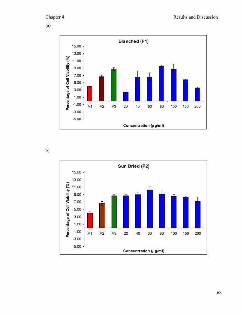
Chapter 4 Results and Discussion
68
(a)
Blanched (P1)
-5.00
-3.00
-1.00
1.00
3.00
5.00
7.00
9.00
11.00
13.00
15.00
M1 M2 M3 20 40 60 80 100 150 200
Concentration (mg/ml)
Perc
en
tag
e o
f C
ell V
iab
ilit
y (
%)
b)
Sun Dried (P2)
-5.00
-3.00
-1.00
1.00
3.00
5.00
7.00
9.00
11.00
13.00
15.00
M1 M2 M3 20 40 60 80 100 150 200
Concentration (mg/ml)
Perc
en
tag
e o
f C
ell V
iab
ilit
y (
%)

Chapter 4 Results and Discussion
69
(c)
Freeze Dried (P3)
-5.00
-3.00
-1.00
1.00
3.00
5.00
7.00
9.00
11.00
13.00
15.00
M1 M2 M3 20 40 60 80 100 150 200
Concentration (mg/ml)
Perc
en
tag
e o
f C
ell V
iab
ilit
y (
%)
(d)
45°C Oven Dried (P4)
-5.00
-3.00
-1.00
1.00
3.00
5.00
7.00
9.00
11.00
13.00
15.00
M1 M2 M3 20 40 60 80 100 150 200
Concentration (mg/ml)
Perc
en
tag
e o
f C
ell V
iab
ilit
y (
%)
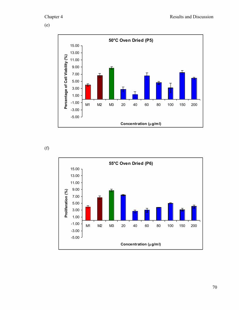
Chapter 4 Results and Discussion
70
(e)
50°C Oven Dried (P5)
-5.00
-3.00
-1.00
1.00
3.00
5.00
7.00
9.00
11.00
13.00
15.00
M1 M2 M3 20 40 60 80 100 150 200
Concentration (mg/ml)
Perc
en
tag
e o
f C
ell V
iab
ilit
y (
%)
(f)
55°C Oven Dried (P6)
-5.00
-3.00
-1.00
1.00
3.00
5.00
7.00
9.00
11.00
13.00
15.00
M1 M2 M3 20 40 60 80 100 150 200
Concentration (mg/ml)
Pro
life
rati
on
(%
)
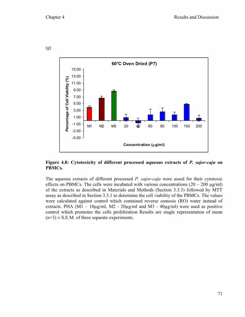
Chapter 4 Results and Discussion
71
(g)
60°C Oven Dried (P7)
-5.00
-3.00
-1.00
1.00
3.00
5.00
7.00
9.00
11.00
13.00
15.00
M1 M2 M3 20 40 60 80 100 150 200
Concentration (mg/ml)
Perc
en
tag
e o
f C
ell V
iab
ilit
y (
%)
Figure 4.8: Cytotoxicity of different processed aqueous extracts of P. sajor-caju on
PBMCs.
The aqueous extracts of different processed P. sajor-caju were assed for their cytotoxic
effects on PBMCs. The cells were incubated with various concentrations (20 – 200 µg/ml)
of the extracts as described in Materials and Methods (Section 3.3.3) followed by MTT
assay as described in Section 3.3.1 to determine the cell viability of the PBMCs. The values
were calculated against control which contained reverse osmosis (RO) water instead of
extracts. PHA (M1 – 10µg/ml, M2 - 20µg/ml and M3 - 40µg/ml) were used as positive
control which promotes the cells proliferation Results are single representation of mean
(n=3) ± S.E.M. of three separate experiments.

Chapter 4 Results and Discussion
72
Cytotoxicity assays are widely used in in vitro toxicology studies and serve as one
of the chemotherapeutic targets of antitumor activity (Suffness and Pezzuto, 1991). Most of
the clinically used antitumor agents possess significant cytotoxic activity in cell culture
systems (Ajith and Janardhanan, 2003). It is a rapid standardized, sensitive and inexpensive
method to measure drug-induced alterations in metabolic pathways or structural integrity
which may or may not be related directly to cell death. The methyl tetrazolium (MTT)
assay, the neutral red (NR) assay, the lactate dehydrogenase leakage (LDH) assay and a
protein assay are the most common methods employed for the determination of cytotoxicity
or cell viability following the exposure to toxic substances. Weyermann et al. (2005) have
previously reported that different cytotoxic assays can give variable results depending on
the test agent. The NR assay and MTT assay are the most sensitive cytotoxic assays that
show statistically significant difference between the treated cells and the controls (Fotakis
and Timbrell, 2005).
On the other hand, in the development of drugs from natural products, the potential
chemopreventive agents selected for testing in people at high risk of developing cancer
must have low toxicity as compared to the drugs used to treat existing cancer (Greenwald,
2002). Most cancer chemotherapeutants severely affect the host normal cells (Mascarenhas,
1994). The discovery of new protective substances with chemopreventive characteristics
has been the target of various studies (Machado et al., 2005). Polysaccharides from
mushrooms are among the various naturally occurring substances that may prove to be one
of the useful candidates in the search for non-toxic, effective substances with antioxidant
activity (Tsai et al., 2007).
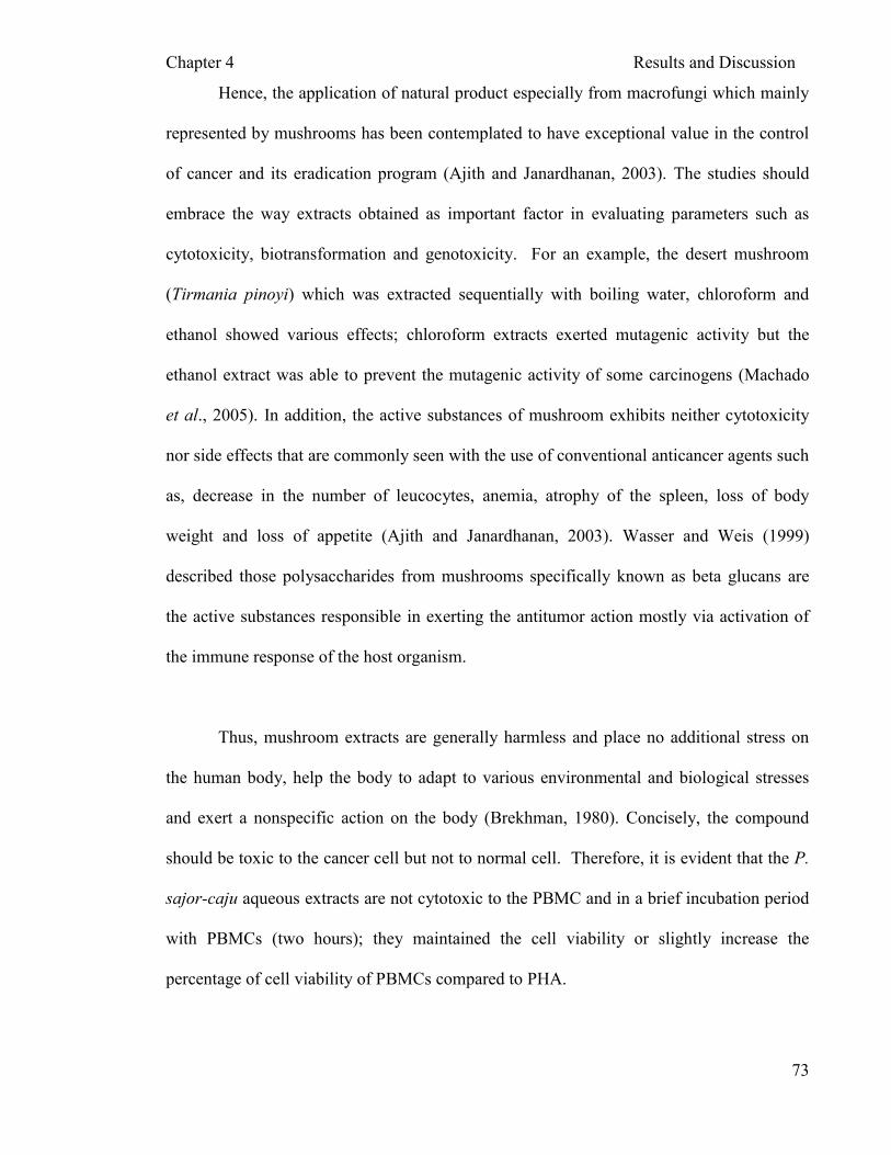
Chapter 4 Results and Discussion
73
Hence, the application of natural product especially from macrofungi which mainly
represented by mushrooms has been contemplated to have exceptional value in the control
of cancer and its eradication program (Ajith and Janardhanan, 2003). The studies should
embrace the way extracts obtained as important factor in evaluating parameters such as
cytotoxicity, biotransformation and genotoxicity. For an example, the desert mushroom
(Tirmania pinoyi) which was extracted sequentially with boiling water, chloroform and
ethanol showed various effects; chloroform extracts exerted mutagenic activity but the
ethanol extract was able to prevent the mutagenic activity of some carcinogens (Machado
et al., 2005). In addition, the active substances of mushroom exhibits neither cytotoxicity
nor side effects that are commonly seen with the use of conventional anticancer agents such
as, decrease in the number of leucocytes, anemia, atrophy of the spleen, loss of body
weight and loss of appetite (Ajith and Janardhanan, 2003). Wasser and Weis (1999)
described those polysaccharides from mushrooms specifically known as beta glucans are
the active substances responsible in exerting the antitumor action mostly via activation of
the immune response of the host organism.
Thus, mushroom extracts are generally harmless and place no additional stress on
the human body, help the body to adapt to various environmental and biological stresses
and exert a nonspecific action on the body (Brekhman, 1980). Concisely, the compound
should be toxic to the cancer cell but not to normal cell. Therefore, it is evident that the P.
sajor-caju aqueous extracts are not cytotoxic to the PBMC and in a brief incubation period
with PBMCs (two hours); they maintained the cell viability or slightly increase the
percentage of cell viability of PBMCs compared to PHA.

Chapter 4 Results and Discussion
74
4.3.3 Protective effects of P. sajor-caju extract on the viability PBMCs
The mushroom extracts obtained via different processing methods were not toxic to
human peripheral blood mononuclear cells. The cells were preincubated with the
mushroom extracts at various concentrations for two hours. The cells were then challenged
with known concentrations of H2O2 (0.3mM) for two hours. The concentration of H2O2
(0.3mM) was previously shown to cause approximately 25% inhibition of cell viability
(Figure 4.7). The MTT assay to estimate cell viability was carried out according to the
procedure described in Material and Methods (Section 3.3.2). Figures 4.9a-g illustrate the
protective effects of the seven different processed (P1-P7) crude water extracts of P. sajor-
caju. Vitamin C was used as the positive control for this test as it is a well known water-
soluble antioxidant.
All the mushroom extracts demonstrated a mild protective effect against H2O2-
induced toxicity. The protection level in PBMCs for 10µg/ml of vitamin C was
6.50±1.94% cell viability. In brief, the processed mushroom extract (P1 to P7) showed cell
viability in the range between 2%-15%. The P. sajor-caju extracts, regardless of their
concentration showed a mild protective effect against H2O2 which was comparable to
vitamin C (10µg/ml).
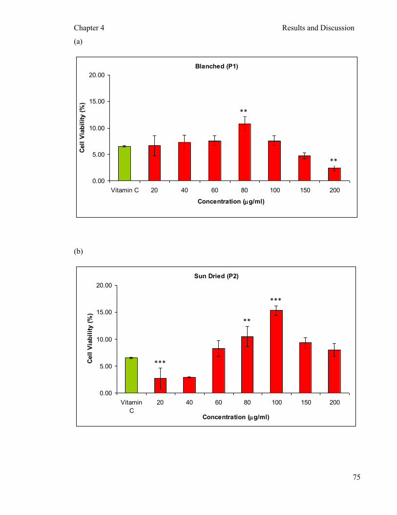
Chapter 4 Results and Discussion
75
(a)
Blanched (P1)
0.00
5.00
10.00
15.00
20.00
Vitamin C 20 40 60 80 100 150 200
Concentration (mg/ml)
Cell
Via
bil
ity (
%)
(b)
Sun Dried (P2)
0.00
5.00
10.00
15.00
20.00
Vitamin
C
20 40 60 80 100 150 200
Concentration (mg/ml)
Cell
Via
bil
ity (
%)
**
**
***
**
***

Chapter 4 Results and Discussion
76
(c)
Freeze Dried (P3)
-10.00
-5.00
0.00
5.00
10.00
15.00
20.00
Vitamin C 20 40 60 80 100 150 200
Concentration (mg/ml)
Cell
Via
bil
ity (
%)
(d)
45°C Oven Dried (P4)
0.00
5.00
10.00
15.00
20.00
Vitamin
C
20 40 60 80 100 150 200
Concentration (mg/ml)
Cell
Via
bil
ity (
%)
***
**
* **
**
*
*

Chapter 4 Results and Discussion
77
(e)
50°C Oven Dried (P5)
0.00
5.00
10.00
15.00
20.00
Vitamin C 20 40 60 80 100 150 200
Concentration (mg/ml)
Cell
Via
bil
ity (
%)
(f)
55°C Oven Dried (P6)
-20.00
-15.00
-10.00
-5.00
0.00
5.00
10.00
15.00
20.00
Vitamin C 20 40 60 80 100 150 200
Concentration (mg/ml)
Ce
ll V
iab
ilit
y (
%)
***
*
**
* *
*** * *
**
*
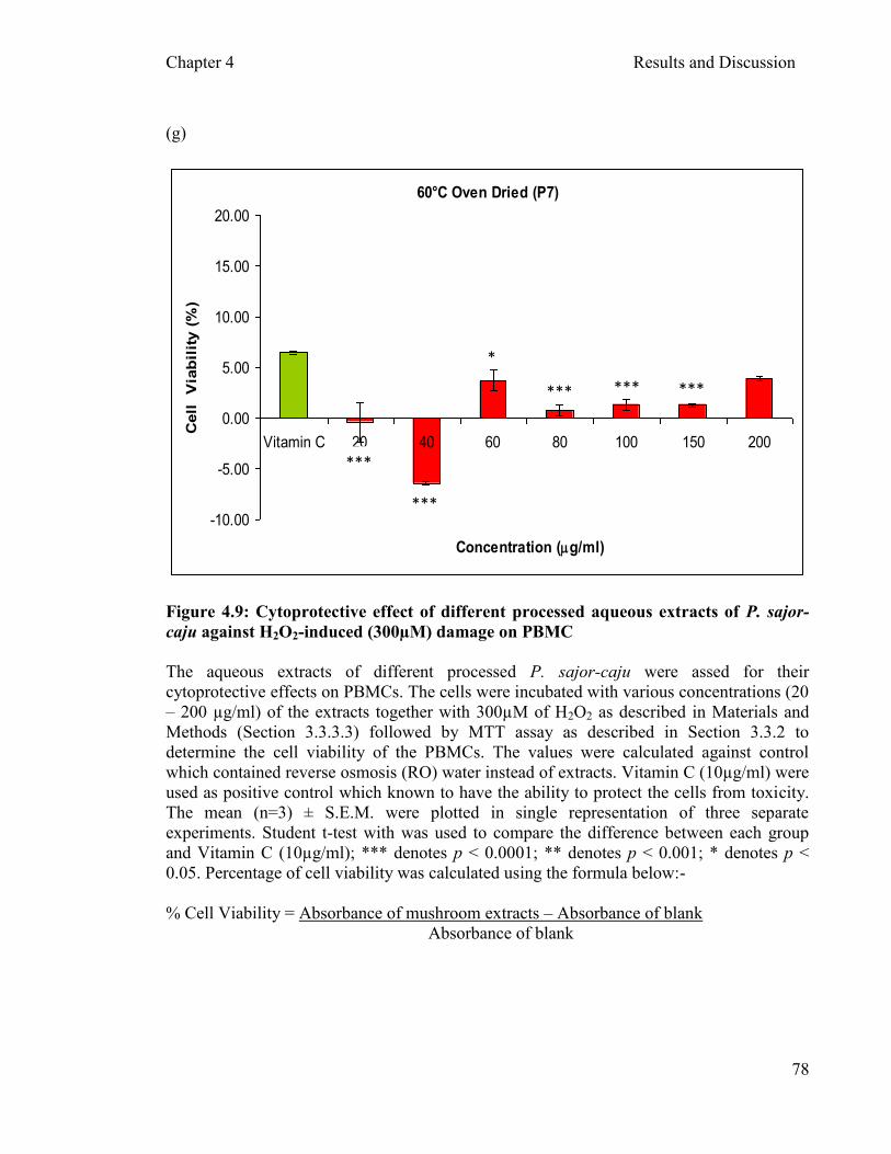
Chapter 4 Results and Discussion
78
(g)
60°C Oven Dried (P7)
-10.00
-5.00
0.00
5.00
10.00
15.00
20.00
Vitamin C 20 40 60 80 100 150 200
Concentration (mg/ml)
Cell
V
iab
ilit
y (
%)
Figure 4.9: Cytoprotective effect of different processed aqueous extracts of P. sajor-
caju against H2O2-induced (300µM) damage on PBMC
The aqueous extracts of different processed P. sajor-caju were assed for their
cytoprotective effects on PBMCs. The cells were incubated with various concentrations (20
– 200 µg/ml) of the extracts together with 300µM of H2O2 as described in Materials and
Methods (Section 3.3.3.3) followed by MTT assay as described in Section 3.3.2 to
determine the cell viability of the PBMCs. The values were calculated against control
which contained reverse osmosis (RO) water instead of extracts. Vitamin C (10µg/ml) were
used as positive control which known to have the ability to protect the cells from toxicity.
The mean (n=3) ± S.E.M. were plotted in single representation of three separate
experiments. Student t-test with was used to compare the difference between each group
and Vitamin C (10µg/ml); *** denotes p < 0.0001; ** denotes p < 0.001; * denotes p <
0.05. Percentage of cell viability was calculated using the formula below:-
% Cell Viability = Absorbance of mushroom extracts – Absorbance of blank
Absorbance of blank
***
***
*
*** *** ***

Chapter 4 Results and Discussion
79
In terms of processing methods, the sun dried (P2) mushroom demonstrated the
highest level of protection (Figure 4.9b) (9% higher at 100µg/ml compared to vitamin C).
This could be possibly caused by the elevation of vitamin D or the transformation of
ergosterol peroxidase into vitamin D (Imtiaj and Rahman, 2008). They reported that
exposure of shiitake to direct sunlight for 3 hour/ day increases the vitamin D content up to
5 times. The peroxide of the ergosterol, 5α, 8α-epidioxy-22E-ergosta-6, 22-dien-3β-ol
(ergosterol peroxide) is common in mushrooms such as Hypsizigus marmoreus, Pleurotus
eryngii, and Ganoderma lipsiense (Bok et al., 1999 and Takei et al., 2005). However, the
real mechanism/s involving ergosterol or vitamin D with the protective activity is still not
clear and further investigation should be carried out in future.
Biologically active compounds found in mushroom have been known for their
medicinal and nutritional benefits to human health. Many studies have reported that the
various types of polysaccharides isolated from mushrooms possess bioactive properties
such as anti-tumor, immunological, anti-inflammatory, anti-coagulant, and hypoglycemic
activities (Kim et al., 2007). Mushroom polysaccharide extracts were also reported to have
scavenging effects on superoxides and hydroxyl radicals produced by reactive oxygen
species (Liu et al., 1997). Mushroom polysaccharides are known to stimulate natural killer
cells, T-cells, B-cells and macrophage-dependent immune system responses (Wasser,
2002). Various effects of precursor T cells and macrophages on the response to cytokines
were produced by lymphocytes after specific recognition of tumour cells (Hamuro and
Chihara, 1985).

Chapter 4 Results and Discussion
80
As mushroom could be useful candidates as effective, non-toxic substances with
free radical scavenging activities as mentioned in section 4.2, the presence of antioxidant
and polyphenol activity in processed mushroom extracts shows that the processing method
does not affect the mushroom properties. In addition, the non-toxic properties of this
mushroom extracts enabled them to become a good candidate for protective activity on
human cells. Although, several biological activities of the mushroom polysaccharides
extracts have been reported, studies on their cytoprotective effects against H2O2 induced
damage on human mononuclear cells is limited. Therefore this study shed some light on the
protective effects of P. sajor-caju extracts which are grown extensively in Malaysia. In
addition, a time course study (24 hours or 48 hours) on the effect of H2O2 induced damage
on PBMC and other cell lines with processed P. sajor-caju extracts should be carried out in
future.
The experiments were continued to study the effects of H2O2 induced damage at the
DNA level of PBMCs. Extracts P1, P2, P3 and P4 which were processed at different
temperatures were further tested for the genotoxicity and genoprotective activity. From the
oven dried group of sample, 45°C oven dried (P4) were chosen for the genotoxicity and
genoprotective assays. In the food processing industry, a variety of oven drying
temperature ranging from 40 - 80°C was applied to achieve better products depending on
the type of food or fruits or mushrooms. All the oven dried extracts showed approximately
similar antioxidant activity as mentioned in Section 4.2 and 4.2.3. However, when the
cytoprotective activity was taken into consideration the extract P4 (Figure 4.9d) showed a
mild protection which is comparably better than the other oven dried extracts.
Chapter 4 Results and Discussion
81
4.4 The effect P. sajor-caju aqueous extracts on DNA damage of PBMCs
4.4.1 DNA staining and visualization (Comet assay)
Scoring of DNA damage was performed using a fluorescent microscope and
automated software scoring system with an image capture device attached to the
microscope. A grade was given to each comet image according to the intensity and length
of the comet tail visualized. Figure 4.12 illustrates the different grades of DNA damages.
Generally two or three gels per treatment were scored, and the mean and SEM of the gels
are presented as graph in Sections 4.4.2, 4.4.3 and 4.4.4.
The DNA-specific dye and the magnification used for comet visualization depend
largely on investigator-specific needs (Tice et al., 2000). There are many types of
florescent dyes that can be used for visualizing the DNA such as ethidium bromide,
propidium iodide, 4,6- diamidino-2-phenylindole (DAPI), SYBR Green and YOYO-1
(benzoxazolium- 4-quinolinum oxazole yellow homodimer) (Tice et al., 2000). The most
common florescent dye employed in DNA visualizing is the ethidium bromide. Kazilian et
al (1999) reported the use of non fluorescent technique for visualizing comets which are
based on staining with silver nitrate that is less harmful than the ethidium bromide
(carcinogenic). Comet image magnification can vary between 160X to 600X. However, the
most common magnification used varies between 200X to 400X (Tice et al., 2000).
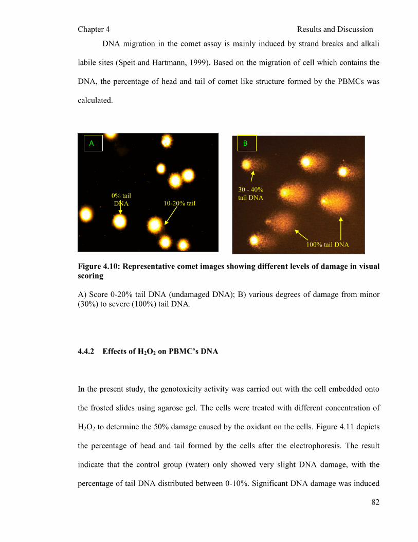
Chapter 4 Results and Discussion
82
DNA migration in the comet assay is mainly induced by strand breaks and alkali
labile sites (Speit and Hartmann, 1999). Based on the migration of cell which contains the
DNA, the percentage of head and tail of comet like structure formed by the PBMCs was
calculated.
Figure 4.10: Representative comet images showing different levels of damage in visual
scoring
A) Score 0-20% tail DNA (undamaged DNA); B) various degrees of damage from minor
(30%) to severe (100%) tail DNA.
4.4.2 Effects of H2O2 on PBMC’s DNA
In the present study, the genotoxicity activity was carried out with the cell embedded onto
the frosted slides using agarose gel. The cells were treated with different concentration of
H2O2 to determine the 50% damage caused by the oxidant on the cells. Figure 4.11 depicts
the percentage of head and tail formed by the cells after the electrophoresis. The result
indicate that the control group (water) only showed very slight DNA damage, with the
percentage of tail DNA distributed between 0-10%. Significant DNA damage was induced
100% tail DNA
30 - 40%
tail DNA
A B
0% tail
DNA 10-20% tail
DNA

Chapter 4 Results and Discussion
83
by H2O2 concentrations greater than 200µM. At 300 µM H2O2, most of the cells exhibited
tailed DNA, indicating extensive damage (Figure 4.11). The higher head DNA represents
minimum DNA strand breaks. The toxicity of oxidant i.e. H2O2 was considered significant
when the tail DNA is more than 30% and the head DNA is less than 70%. The tail DNA
exceeded 30% in PBMC incubated with H2O2 concentrations higher than 0.15mM whereas
50% tail DNA (IC50) was achieved at 0.20mM. Thus, this concentration was selected for
further investigation on the protective effect of mushroom extracts on H2O2 induced
genotoxicity.
The realization that the DNA damage and mutation arise from endogenous products
of cellular metabolism has been a major concern for the past two decades (Ames, 1989)
The generation of damaged bases and strand breaks are the consequence of oxygen radicals
that are produced during the reduction of O2 which attack DNA bases or deoxyribose
residues (Ward, 1987). Replication of this damaged strands leads to mutation or apoptosis
(Johnson, 1996). Ames et al. (1993) reported that low levels of reactive oxygen species
(ROS) have useful roles in signal transduction and in the modulation of gene expression but
are extremely harmful to DNA and other biological macromolecules at higher
concentrations. Injury to DNA is caused by the accumulation of ROS resulting from
overproduction and/or inefficient intracellular antioxidant defense systems (Halliwell,
1999). The ROS are important as direct and indirect initiators as well as promoters of
mutagenesis and carcinogenesis. They also increase the lipid peroxidation, which in turn
alter the integrity of membrane bound enzymes (Halliwell and Gutteridge, 2003). The free
radical scavenging efficiency of the extract thus might play an important role in the
antimutagenic activity.

Chapter 4 Results and Discussion
84
Figure 4.11: DNA damage in PBMC exposed to H2O2
Peripheral blood mononuclear cells (PBMC) (1,000,000 cells/ml) were incubated with
various concentrations of H2O2 (0.05mM – 0.30mM) for 30 minutes at 37ºC and 5% of
CO2. The treated PBMCs were electrophoresed using single cell gel electrophoresis
(SCGE) technique and stained with EtBr (Section 3.3.4.3 and 3.3.5). The normal control (0
mM) comprised cells with RO water in media. The mean (n=150) ± S.E.M. were plotted in
single representation of three separate experiments.
0.00
10.00
20.00
30.00
40.00
50.00
60.00
70.00
80.00
90.00
100.00
0/H2 0 0.05 0.10 0.15 0.20 0.25 0.30
Concentrations (mM)
Perc
en
tag
e o
f D
NA
(%
)
(%)
Tail
Extent of DNA damage on PBMC induced with various concentration of H2O2
Head
Chapter 4 Results and Discussion
85
The damage caused by these free radicals can be detected using comet assay.
Theoretically, the comet assay can be applied to any eukaryotic cell; however, the human
white cells mainly lymphocyte cells are most frequently employed (Vijayalaxmi et al.,
1993). White cells are easily harvested from venous or capillary blood, whereas it is
generally difficult to obtain other types of nucleated cells from human subjects. Besides
human lymphocytes, other cell types such as spermatocytes, fibroblasts, epithelial cells
from the buccal (oral) cavity, stomach, tear duct, nose and bladder have been used,
although not always successfully because of their resistance to lysis buffer, high level of
background and DNA damage (Wong et al., 2005). Various white cell lines also can be
used, such as RAJI and TK6 from B-cell lines and HUT-78 from T-cell lines (Wong et al.,
2005).
The advantages of the comet assay are its speed, simplicity, low cost, the small
number of cells required (<10,000 cells), its sensitivity, and its widespread applicability
with eukaryotic cells, whether proliferating or non-proliferating (Tice et al., 2000).
Furthermore, effects on different cell types can be explored. This is an advantage because
genotoxic and genoprotective effects can be tissue or cell type specific. Tice et al. (2000)
also reported that another advantages of the comet assay is its flexibility; different
combinations of unwinding and electrophoresis conditions and lesion-specific enzymes can
be used to detect different types and levels of DNA damage (Tice et al., 2000).

Chapter 4 Results and Discussion
86
4.4.3 Effects of P. sajor-caju on PBMC’s DNA
In the present study, the genotoxic activity of the crude water extracts of P. sajor-
caju was established using the single cell gel electrophoresis (Comet assay). The
genotoxicity assessment of the processed P. sajor-caju extracts were carried out using the
methods described Materials and Methods (Section 3.3.4.5). Figures 4.12a-d show the
result obtained from the comet assay of different processed P. sajor-caju. None of the
processed P. sajor-caju extracts were genotoxic. As the result shown in Figure 4.12a-d, the
processed P. sajor-caju extracts caused a slight DNA damage at very low and very high
concentrations. The tail DNA was distributed between 0-20% at concentration of 50 µg/ml
to 200 µg/ml, this was significantly (p < 0.05) less that the damage induced by H2O2
(0.2mM).
Efforts to develop dietary supplements and preventive treatments for offsetting the
detrimental effects of ROS have focused on identifying antioxidants from a variety of
natural products (Shi et al., 2002b). More recently, mushrooms have been identified as a
potential source of natural antioxidants. This special group of fungi has long been
acknowledged in Eastern cultures as possessing a wide range of medicinal properties
(Chang and Buswell, 1996), and modern techniques have identified numerous bioactive
mushroom components which have been reported to exhibit anti-cancer, anti-tumour, anti-
viral, immunomodulatory, hypocholesterolaemic and hepatoprotective activities.

Chapter 4 Results and Discussion
87
Commonly, it was possible to see a relationship between cytotoxicity and
genotoxicity determined in the comet assay (Machado et al., 2005). The cytoprotective and
genoprotective effects of A. bisporus extracts showed a positive dose-dependent
correlation. However, other non cytotoxic concentration showed genotoxicity. It is
important to perform comet assay at non cytotoxic concentrations to better evaluate the
DNA damage (Hartmann and Speit, 1997). Therefore, it is evident that a cytotoxicity test
should be performed concomitantly with genotoxicity tests (Machado et al., 2005).
Nevertheless, the results obtained for the genotoxic activity of P. sajor-caju as shown in
Figure 4.12a-d correspond/ correlate to the pattern of cytotoxic activity in section 4.3.2.
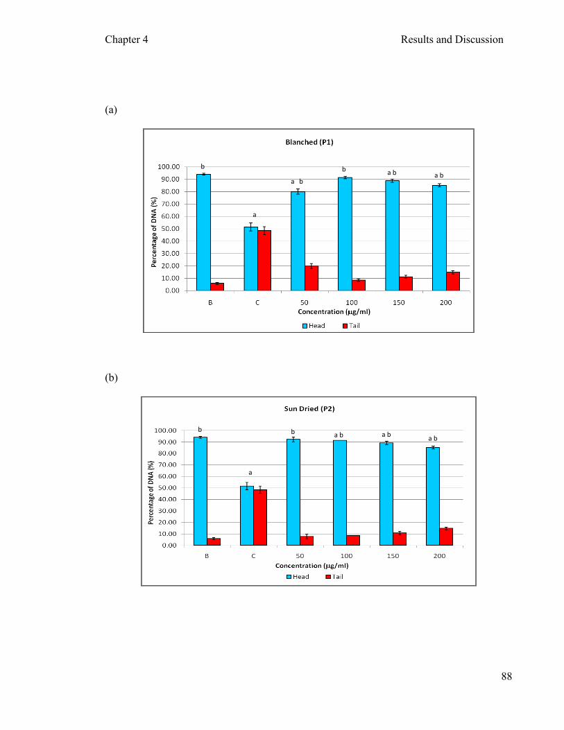
Chapter 4 Results and Discussion
88
(a)
(b)
a
b b
b a b a b
a
a b
a
a b b b a b

Chapter 4 Results and Discussion
89
(c)
(d)
Figure 4.12: Genotoxicity effects of aqueous extracts of different processed P. sajor-
caju on DNA damage in PBMCs
The aqueous extracts of different processed P. sajor-caju were assessed for their genotoxic
effects on PBMCs. The cells were incubated with various concentrations of extracts
(50µg/ml - 200µg/ml) as described in Material and Methods (Section 3.3.4.5) followed
with comet assay as described in Section 3.3.4.2, 3.3.4.3 and 3.3.5 to determine the level of
DNA damage in the cells. B: The normal control comprised cells with RO water in media,
and C: Cells treated with 0.2mM H2O2. The mean (n=150) ± S.E.M. were plotted in single
representation of three separate experiments. Student t-test with was used to compare the
difference between groups; a: denotes p < 0.0001 compared with B; b: denotes p < 0.0001
compared with C.
a
b b a b a b a b
a
b b b
a b a b
Chapter 4 Results and Discussion
90
In addition, the mild damage in DNA could be caused by the natural antioxidant
such as vitamin C that is present in the mushroom extracts. Anderson et al. (1994) reported
that ascorbic acid can induce DNA damage in human lymphocytes under certain
concentrations in SCGE assay. It had an inhibitory effect on DNA damage of human
lymphocytes inducted by H2O2 under incubation with lymphocytes and H2O2 at the same
time. In addition, Duthie et al. (1997) also reported that some flavonoids induced
genotoxicity and cytotoxicity in cells under higher concentrations. Therefore, in the present
study, processed P. sajor-caju may have caused mild DNA damage (not significant) at
concentrations as mentioned above because it contained significant amounts of phenolic
compounds (Figure 4.1) and ascorbic acid (as mentioned in section 2.5.1).
Pleurotus species a well known basidiomycetes species is consumed as food
especially in oriental region for centuries. Thus, it can be concluded here that the extracts
obtained via different processing method (temperatures) of P. sajor-caju did not exert any
significant genotoxicity to normal human cell.
4.4.4 Protective effects of mushroom extracts on PBMC’s DNA damage by H2O2
The genoprotective activity of P. sajor-caju was carried out only for extract P1, P2,
P3 and P4. Figure 4.13a-d depicts the genoprotective effect of extracts P1, P2, P3 and P4
respectively. The cells were incubated with extracts and were compared with cells
incubated with water. As the concentration of extract increases the protection level of the
mushroom against H2O2 induced genotoxicity also increased with highest protection level
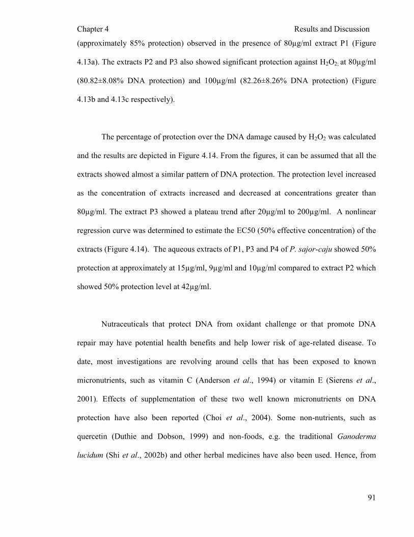
Chapter 4 Results and Discussion
91
(approximately 85% protection) observed in the presence of 80µg/ml extract P1 (Figure
4.13a). The extracts P2 and P3 also showed significant protection against H2O2; at 80µg/ml
(80.82±8.08% DNA protection) and 100µg/ml (82.26±8.26% DNA protection) (Figure
4.13b and 4.13c respectively).
The percentage of protection over the DNA damage caused by H2O2 was calculated
and the results are depicted in Figure 4.14. From the figures, it can be assumed that all the
extracts showed almost a similar pattern of DNA protection. The protection level increased
as the concentration of extracts increased and decreased at concentrations greater than
80µg/ml. The extract P3 showed a plateau trend after 20µg/ml to 200µg/ml. A nonlinear
regression curve was determined to estimate the EC50 (50% effective concentration) of the
extracts (Figure 4.14). The aqueous extracts of P1, P3 and P4 of P. sajor-caju showed 50%
protection at approximately at 15µg/ml, 9µg/ml and 10µg/ml compared to extract P2 which
showed 50% protection level at 42µg/ml.
Nutraceuticals that protect DNA from oxidant challenge or that promote DNA
repair may have potential health benefits and help lower risk of age-related disease. To
date, most investigations are revolving around cells that has been exposed to known
micronutrients, such as vitamin C (Anderson et al., 1994) or vitamin E (Sierens et al.,
2001). Effects of supplementation of these two well known micronutrients on DNA
protection have also been reported (Choi et al., 2004). Some non-nutrients, such as
quercetin (Duthie and Dobson, 1999) and non-foods, e.g. the traditional Ganoderma
lucidum (Shi et al., 2002b) and other herbal medicines have also been used. Hence, from

Chapter 4 Results and Discussion
92
this study we could see the potential effects of P. sajor-caju as a good genoprotective agent
that are available cheaply compared to other micronutrients.
The potential genoprotective or genotoxic effect of whole foods, simple mixtures
and purified components in in vitro, animal and human supplementation trials can be
investigated quickly with simple assays. The comet assay provides this simple and
relatively quick way of investigation (Wachtel-Galor et al., 2004). It is well known that
reactive free radicals attack cellular DNA and nucleotide pools to form various types of
oxidized bases; e.g., 8-OHdG (Kasai et al., 1986), 5-hydroxydeoxycytidine (Feig et al.,
1994) and 2 hydroxydeoxyadenosine (Kamiya and Kasai, 1995). Among these oxidized
bases, 8-OHdG has especially received much attention because of the accumulated data
indicating that it may play crucial roles in a chain of events from reactive oxygen and/or
nitrogen oxide species–induced stress (RONOSS) to carcinogenesis (Kasai, 1997). 8-
OHdG pairs with adenine as well as cytosine, subsequently yielding GC-to-TA
transversions upon replication by DNA polymerases (Cheng et al., 1992; Shibutani et al.,
1991). This mutation is considered to contribute to the activation of oncogenes and/ or the
inactivation of tumor suppressor genes, leading to carcinogenesis (LePage et al., 1995).
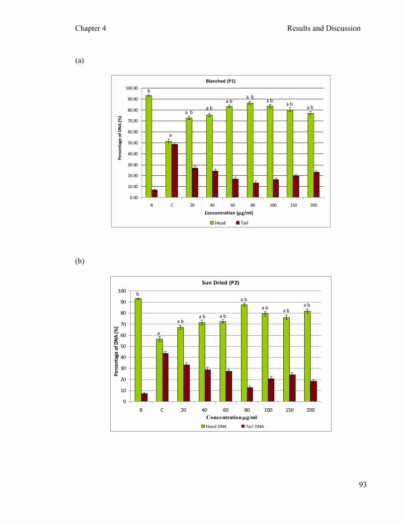
Chapter 4 Results and Discussion
93
(a)
Blanched (P1)
0.00
10.00
20.00
30.00
40.00
50.00
60.00
70.00
80.00
90.00
100.00
B C 20 40 60 80 100 150 200
Concentration (mg/ml)
Pe
rce
nta
ge o
f D
NA
(%
)
Head Tail
(b)
Sun Dried (P2)
0
10
20
30
40
50
60
70
80
90
100
B C 20 40 60 80 100 150 200
Concentration mg/ml
Perc
enta
ge o
f DN
A (%
)
Head DNA Tail DNA
a
a b a b
a b a b
b a b
a b a b
a b a b
a b
a b a b
a b
a b
a
b

Chapter 4 Results and Discussion
94
(c)
Freeze Dried (P3)
0
10
20
30
40
50
60
70
80
90
100
B C 20 40 60 80 100 150 200Concentration mg/ml
Perc
enta
ge o
f DN
A (%
)
Head DNA Tail DNA
(d)
45°C Oven Dried (P4)
0.00
10.00
20.00
30.00
40.00
50.00
60.00
70.00
80.00
90.00
100.00
B C 20 40 60 80 100 150 200
Concentration (mg/ml)
Perc
enta
ge o
f DN
A (%
)
Head DNA Tai l DNA
Figure 4.13: Genoprotective effects of aqueous extracts of P. sajor-caju on PBMC
against H2O2-induced DNA damage
The aqueous extracts of different processed P. sajor-caju were assessed for their
genoprotective effects against H2O2-induced DNA damage (0.2mM) on PBMCs. The cells
were incubated with various concentrations of extracts (20µg/ml - 200µg/ml) as described
in Material and Methods (Section 3.3.4.6) followed with comet assay as described in
Section 3.3.4.2, 3.3.4.3 and 3.3.5 to determine the level of DNA damage in the cells. B:
The normal control comprised cells with RO water in media, and C: Cells treated with
0.2mM H2O2. The mean (n=150) ± S.E.M. were plotted in single representation of three
separate experiments. Student t-test with was used to compare the difference between
groups; a: denotes p < 0.0001 compared with B; b: denotes p < 0.0001 compared with C.
a
b
a b a b
a b a b a b a b a b
b
a
a b
a b a b
a b a b a b a b
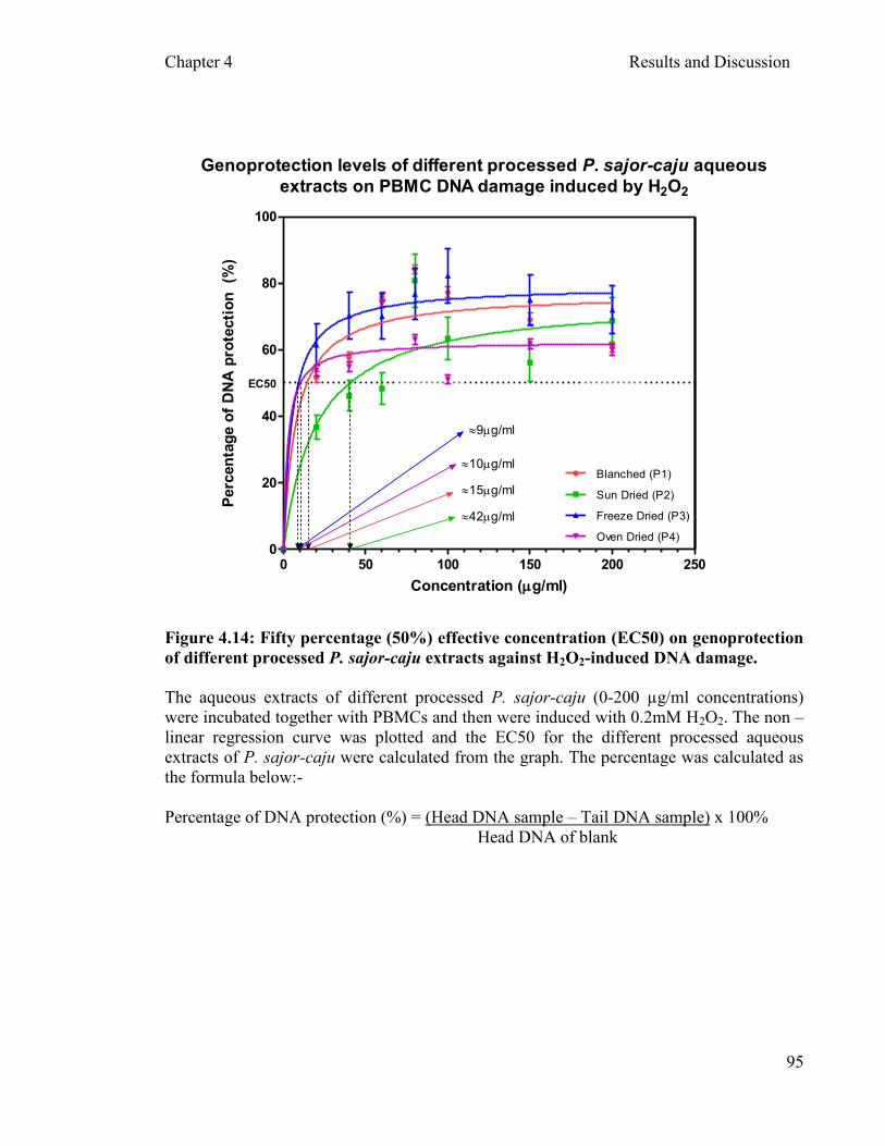
Chapter 4 Results and Discussion
95
Genoprotection levels of different processed P. sajor-caju aqueous
extracts on PBMC DNA damage induced by H2O2
0 50 100 150 200 2500
20
40
60
80
100
Blanched (P1)
Sun Dried (P2)
Freeze Dried (P3)
Oven Dried (P4)
EC50
42mg/ml
9mg/ml
10mg/ml
15mg/ml
Concentration (mg/ml)
Perc
en
tag
e o
f D
NA
pro
tecti
on
(%
)
Figure 4.14: Fifty percentage (50%) effective concentration (EC50) on genoprotection
of different processed P. sajor-caju extracts against H2O2-induced DNA damage.
The aqueous extracts of different processed P. sajor-caju (0-200 µg/ml concentrations)
were incubated together with PBMCs and then were induced with 0.2mM H2O2. The non –
linear regression curve was plotted and the EC50 for the different processed aqueous
extracts of P. sajor-caju were calculated from the graph. The percentage was calculated as
the formula below:-
Percentage of DNA protection (%) = (Head DNA sample – Tail DNA sample) x 100%
Head DNA of blank
Chapter 4 Results and Discussion
96
Shi et al. (2002a) has reported that tyrosinase plays an important role in
genoprotection activity/effect of button mushroom. The nature of the genoprotective
activity of tyrosinase is dependent upon the two associated catalytic activities of the
enzyme, namely hydroxylation of tyrosine to L-DOPA and the subsequent oxidation of L-
DOPA to dopaquinone (Shi et al., 2002a). The basis of the observed genoprotective effects
of tyrosinase-generated L-DOPA oxidation products is not known (Shi et al., 2002a).
However, in addition to its pro-oxidant properties, L-DOPA also stimulates cellular
antioxidant defense mechanisms under ambiguous conditions. Although tyrosinase activity
in Pleurotus sp. (Ratcliffe et al., 1994) is lower than in Agaricus sp., it is possible to
speculate that tyrosinase-generated L-DOPA oxidation products could contribute to the
genoprotective effects observed in this study. However, further investigation to elucidate
the underlying mechanisms would have to be carried out.
The phenolic compounds in P. sajor-caju might play an important role in
modulating H2O2-mediated DNA damage in PBMCs. However, the absorption and
bioavailability of P. sajor-caju need further evaluation and investigation. As result shown
in Figure 4.13 and Figure 4.15, P. sajor-caju had a protective effect against H2O2 induced
damage in PBMCs. Pleurotus sajor-caju had antioxidant activity, scavenging activity on
free radicals or ROS (Figure 4.1 - 4.3). Thus, the antioxidant and scavenging activity of P.
sajor-caju on free radicals induced by H2O2 might be mainly related to its protective effect
on DNA damage on PBMCs.
The potential inhibition of P. sajor-caju on H2O2 induced oxidative DNA damage,
suggests that P. sajor-caju has prospective role in the chemopreventive function.
Chapter 4 Results and Discussion
97
4.5 Future Investigation
The following studies are needed to validate the findings of this study:
I. A time dependent study had to be carried out to determine the appropriate time of
incubation for the extracts to show a percentage of viability of cell more than
30%.
II. The polysaccharides, specifically beta glucans are well known for their ability to
protect the cells. Hence, in future study, if the polysaccharides from this
mushroom could be extracted and the antioxidant properties, cytoprotective and
genoprotective ability studied. It could give an idea of the actual process
involved in the protective effect of the P. sajor-caju extracts.
III. In comet assay, it was shown that P. sajor-caju extracts had genoprotective activity.
The potential of P. sajor-caju extracts to repair the DNA, if any, need further
investigation.
IV. Fractionation of the processed mushroom extract using column chromatography or
HPLC/ anion exchange chromatography could be carried out and the active
compound/s that give protective effect against H2O2 induced toxicity need to
identified.

Chapter 4 Results and Discussion
98
V. The laboratory rats that were treated with oxidant or tumor induced can be given
processed P. sajor-caju in their diet. The lymphocytes can then be isolated and
Comet assay and MTT assay can be carried out to study if there are in vivo
protective effects of P. sajor-caju extracts.

CHAPTER 5
CONCLUSION
Chapter 5 Conclusion
99
CHAPTER 5
CONCLUSION
5.1 Conclusion
In this study, antioxidant, cytotoxicity, cytoprotective, genotoxicity and
genoprotective effect of different processed P. sajor-caju were analyzed. Studies of
antioxidant activity with P. sajor-caju extracts of blanched (P1), sun dried (P2), freeze
dried (P3), 45°C oven dried (P4), 50°C oven dried (P5), 55°C oven dried (P6) and 60°C
oven dried (P7), showed that processing methods did not affect the content of antioxidant
in the fruiting bodies of P. sajor-caju. The low percentage of DPPH scavenging activity
showed by the processed extracts in this study compared to other reports which depicted a
higher percentage of DPPH scavenging activity could be caused by the limiting factor such
as solubility of the extracts and also the method of extraction employed in this study. The
concentrations of the extracts above 5mg/ml were not soluble. This problem could be
overcome in future by diluting the samples with different solvents or by using different
type of extraction methods i.e. solvent extractions. The correlation between FRAP, DPPH
scavenging abilities and total phenols were low to high respectively. The highest
correlation was shown by DPPH scavenging abilities of mushrooms extracts and total
phenol content. Thus, it can be concluded that the higher content of total phenols in seven
different fruit bodies of P. sajor-caju extracts were responsible for their better antioxidant
level.
Chapter 5 Conclusion
100
Aqueous extracts of P. sajor-caju did not show toxicity when it was incubated
together with PBMCs. These effects were compared to PBMCs treated with H2O2 (known
oxidant) that could cause damage to the DNA of the cells. Cytotoxicity is one of the
chemotherapeutic targets of antitumor activity on tumor cells but in some cases these
agents turn out to be toxic to normal adjacent cells. The in vitro study on the non toxic
effect of P. sajor-caju extracts despite of their processing methods shed some light on the
ability of them to be implicated in chemopreventive treatment without harming normal
cells. In addition, all the processed extracts showed mild protection against H2O2 induced
toxicity. In this brief study on the protective effect of processed fruiting bodies of P. sajor-
caju, the extracts actually maintained the viability of PBMCs from further destruction by
the H2O2
(300μM) which showed percentage of cell inhibition approximately 25%. The
protective effect could be caused by the presence of significant antioxidant level in the
processed mushroom.
In the in vitro comet assay for genotoxicity in PBMCs, control (water) and
processed P. sajor-caju were comparable. Obvious DNA damage in PBMCs after exposure
to H2O2 (200μM) were observed. The DNA damage cause by this concentration of H2O2
was approximately 50%. This concentration was used to study the genoprotective activity
of processed P. sajor-caju extracts. The extract P1 and P2 showed a protection of
approximately 80% at 80μg/ml, where as extract P3 showed about the same level of
protection at 100μg/ml. However, the freeze dried P. sajor-caju extract (P3) showed 50%
effective concentration (EC50) at approximately 9μg/ml, in contrast to P1 and P2 which
illustrated EC50 at 10μg/ml and 42μg/ml respectively. Hence, the processed mushroom
showed different protective level on DNA damage induced with H2O2.
Chapter 5 Conclusion
101
Total phenols are reported to be the major antioxidant component found in
mushrooms. According to the results of this study, it is clearly indicated that the aqueous
extracts of processed P. sajor-caju has significant antioxidant activity against various in
vitro antioxidant system. Furthermore, the mushroom species can be used as an easily
accessible source of natural antioxidant and to add on, they have become more popular in
oriental and western cuisine. Processing or cooking the mushroom at different temperature
showed that the antioxidant activities of P. sajor-caju were refrained although they showed
a slight different in their properties. The preserved antioxidants were able to protect not
only the integrity of plasma membrane of the normal cells but also the DNA of the cells.
The results underline the interest of further investigations of P. sajor-caju into
possible chemopreventive activities and properties such as the isolation of possible
polysaccharides or proteins with medicinal properties. The intake of more local edible
mushroom especially oyster mushrooms in our daily diet could be an effective or one of the
early strategies to reduce the risk of cancer. Pleurotus sajor-caju has a high potential to be
produced economically on large scale in Malaysia. The nutritive and nutraceutical value or
fact of this fungus should be emphasized to increase the mushroom intake in daily diet of
Malaysian (2kg/year). By doing so, the popularity of P. sajor-caju can wide spread to
standards of other known medicinal or edible mushrooms such as L. edodes (shitake), H.
erinaceus (lion’s mane), G. lucidum (ling-zhi), Tuber sp. (truffles) and C. sinensis
(cordyseps).

REFERENCES

References
102
REFERENCES
Abdellah, M. M. F., Emara, M. F. Z. and Mohammady, T. F. (2000). Open field
interplanting of oyster mushroom with cabbage and its effect on the subsequent eggplant crop. Annual Horticultural Science (Cairo), 45:281-293.
Ahmad, N. and Mukhtar, H. (1999). Green tea polyphenols and cancer: biologic
mechanisms and practical implications. Nutrition Review, 57:78–83. Ajith, T.A. and Janardhanan, K.K. (2003). Cytototoxic and antitumor activities of a
polypore macrofungus, Phellinus rimosus (Berk) Pilat. Journal of Ethnopharmacology, 84:157-162.
Ajith, T.A., Jose, N. and Janardhanan, K.K. (2002). Amelioration of cisplatin induced
nephrotoxicity in mice by ethyl acetate extract of a pore fungus Phellinus rimosus. Journal of Experimental Clinical Cancer Research, 21:487-491.
Amarowicz, R., Pegg, R.B., Moghaddam, P.R., Barl, B. and Weil, J.A. (2004). Free-radical
scavenging capacity and antioxidant activity of selected plant species from the Canadian Prairies. Food Chemistry, 84: 551-562.
Ames, B.N. (1989). Endogenous DNA damage as related to cancer and aging.Mutation
Research, 214:41-46. Ames, B.N., Shigenaga, M.K. and Hagen, T.M. (1993). Oxidants, antioxidants, and the
degenerative diseases of aging. Proceedings of the National Academy of Sciences, USA, 90:7915–22.
Anderson, D., Yu, T.-W., Phillips, B. J. and Schmezer, P. (1994). The effect of various
antioxidants and other modifying agents on oxygen-radical-generated DNA damage in human lymphocytes in the comet assay. Mutation Research. 307:261-271.
Angelis, K. J., Dusinska, M. and Collins, A. R. (1999). Single cell gel electrophoresis:
detection of DNA damage at different levels of sensitivity. Electrophoresis, 20(10): 2133-2138.
Axtell, C., Johnston, C. G. and Bumpus, J. A. (2000). Bioremediation of soil contaminated
with explosives at the Naval Weapons Station Yorktown. Soil and Sediment Contamination, 9:537-548.
Baars, J. J. P., Sonnenberg, A. S. M., Mikosch, T. S. P. and Griensven, L. J. L. D. V.
(
2000). Development of a sporeless strain of oyster mushroom Pleurotus ostreatus. In L. J. L. D. van Griensven (ed.), Science and Cultivation of Edible Fungi, Vol. 1. A. A. Balkema, Rotterdam, The Netherlands. Pp: 317- 323.
Ballero, M., Mascia, E., Rescigno, A. and di Teulada, E. S. (1990). Use of Pleurotus for transformation of polyphenols in waste waters from olive presses into proteins. Micol. Italiana, 19:39-41.
References
103
Balunas, M.J. and Kinghorn, A.D. (2005). Drug discovery from medicinal plants. Life
Science; 78:431-441. Bano, Z. and Rajarathnam, S. (1988). Pleurotus mushrooms, Part II. Chemical
composition, nutritional value, post-harvest physiology, preservation and role as human food. Critical Reviews in Food Science and Nutrition, 27(2):87-158.
Bhattaram, V.A., Graefe, U., Kohlert, C., Veit, M. and Derendorf, H. (2002).
Pharmacokinetics and bioavailability of herbal medicinal products. Phytomedicine, 9:1-33.
Beckman, J.S., Beckman, T.W., Chen, J., Marshall, P.A. and Freeman, B.E. (1990).
Apparent hydroxyl radical production by peroxynitrite: implications for endothelial injury from nitric oxide and superoxide. The Proceedings of the National Academy of Sciences. USA, 87:1620–1624.
Benzie, I. F. and Strain, J. J. (1996). The ferric reducing ability of plasma (FRAP) as a
measure of "antioxidant power": the FRAP assay. Analytical Biochemistry, 239(1): 70-76.
Bok, J.W., Lermer, L., Chilton, J., Klingeman, H.G. and Towers, G.H.N. (1999). Antitumor
sterols from the mycelia of Cordyceps sinensis. Phytochemistry, 51: 891–8. Boyum, A. (1968). Isolation of mononuclear cells and granulocytes from human blood.
Isolation of monuclear cells by one centrifugation, and of granulocytes by combining centrifugation and sedimentation at 1 g. Scandinavian Journal of Clinical and Laboratory Investigation Supplement, 97: 77-89.
Brand-Williams, W., Cuvelier, M.E. and Berset, C. (1995). Use of a free radical method to
evaluate antioxidant activity. Food Science and Technology, 28:25–30. Breene, W.B. (1990). Nutritional and medicinal value of specialty mushrooms. Journal of
Food Protection, 53(10): 883-894. Brekhman, I.I. (1980). Man and Biologically Active Substances. Pergamon Press, New
York. Pp: 1-89. Brochers, A.T., Keen, C.L., and Gershwin, M.E. (1999). Mushrooms, tumors and
immunity. Proceedings of the Society
for Experimental Biology and Medicine, 221:281-93.
Burney, S., Niles, J.C., Dedon, P.C. and Tannenbaum, S.R. (1999). DNA damage in deoxynucleosides and oligonucleotides treated with peroxynitrite. Chemical Research. Toxicology, 12:513–520.
Cadet, J., Delatour, T., Douki, T., Gasparutto, D., Pouget, J.-P., Ravanat, J.-L. and
Sauvaigo, S. (1999). Hydroxyl radicals and DNA base damage. Mutation Research: Fundamental of Molecular Mechanism Mutagen, 424:9–21.

References
104
Caglarirmak, N. (2007). The nutrientsof exotic mushrooms (Lentinula edodes and Pleurotus species) and an estimated approach to the volatile compound. Food chemistry, 105: 1188-1194.
Chang, R. (1996). Functional properties of edible mushrooms. Nutritional Review, 54(2):
591-593. Chang, S.T. (1999). Global impact of edible and medicinal mushrooms on human welfare
in the 21st century: non green revolution. International Journal of Medicinal Mushrooms, 1:1–8.
Chang, S.T. and Buswell, J.A. (1996). Mushroom nutriceuticals. World Journal of
Microbiology Biotechnology, 12:473-476. Chang, S.T. and Mshigeni, K.E. (2001). Mushroom and Human Health: Their growing
significance as potent dietary supplements. The University of Namibia, Windhoek. Pp:1-79.
Chen, C., Pearson, A.M. and Gray, J.I. (1992). Effects of synthetic antioxidants (BHA,
BHT and PG) on the mutagenicity of IQ-like compounds. Food Chemistry, 43:177-183.
Cheng, K.C., Cahill, D.S. and Kasai, H. (1992). 8-Hydroxyguanine, an abundant form of
oxidative DNA damage, causes G-T and A-C substitutions. Journal of Biological Chemistry, 267:166-172.
Cheung, L.M., Cheung, P.C.K. Ooi, V.E.C. (2003). Antioxidant activity and total phenolics
of edible mushroom extracts. Food Chemistry, 81: 249-255. Choi, S.W., Benzie, I.F.F., Collins, A.R., Hannigan, B.M. and Strain, J.J. (2004).Vitamins
C and E: acute interactive effects on biomarkers of antioxidant defense and oxidative stress. Mutation Research, 551:109-117.
Choi, Y., Lee, S.M., Chun, J., Lee, H.B. and Lee, J. (2005). Influence of heat treatment on
the antioxidant activities and polyphenolic compounds of Shiitake (Lentinus edodes) mushroom, Food Chemistry. 99:381–387.
Cimerman, N.G. (1999). Medicinal value of genus Pleurotus (Fr.) P.Karst. (Agaricales s.i.,
Basidiomycetes). International Journal of Medicinal Mushroom, 1: 69-80. Clingen, P. H., Lowe, J.E. and Green, M.H.L. (2000). Measurementof DNA damage and
repair capacity as a function of age using the comet assay. In Barnett, Y. A. and Barnett, C.R (Ed.), Aging methods and protocols (pp. 143-157). Totowa: Humana Press.
Cochran, K.W. (1978). Medicinal Effects. In: Chang, S.T. and Hayes, W.A. The Biology
and Cultivation of Edible Mushrooms. Academic Press Inc. New York. Pp: 169-167.
References
105
Cook, P. R., Brazell, I. A. and Jost, E. (1976). Characterization of nuclear structures containing superhelical DNA. Journal of Cell Science, 22(2): 303-324.
Cun, Z., Mizuno, T., Ito, H., Shimura, K., Sumiya, T. and Kawade, M. (1994). Antitumor
activity and immunological property of polysaccharides from the mycelium of liquid-cultured Grifola frondosa. Journal of Japanese Society of Food Science and Technology, 41:724–732.
Croteau, R, Kutchan, T. M. and Lewis, N.G. (2000). Natural Products (Secondary
Metabolites). In: B. Buchanan, W. Gruissem, R. Jones, Eds. Biochemistry & Molecular Biology of Plants, American Society of Plant Physiologists. Pp 1250-1318.
Decker, E. A. (1997). Phenolics: prooxidants or antioxidants? Nutrition Reviews, 55:396–
407. Denizot, F. and Lang, R. (1986). Rapid colorimetric assay for cell growth and survival.
Modifications to the tetrazolium dye procedure giving improved sensitivity and reliability. Journal of Immunological Methods, 89(2): 271-277.
Deshpande, S.S. and Irani, K. (2002). Oxidant signaling in carcinogenesis: a commentary.
Human Expression Toxicology, 21:63-4. Dizdaroglu, M. and Gajewski, E. (1990). Selected-ion mass spectrometry: assays of
oxidative DNA damage. Methods Enzymology, 186:530–544. Dong, M.K., Chui, W.P., Han, G.K. and Won, M.P. (2000). Isolation of antimicrobial
substance from Hericium erinaceum. Mycobiology, 28: 33-38. Duthie, S.J., Collins, A.R., Duthie, G.G. and Dobson, V.L. (1997). Quercetin and myricetin
protect against hydrogen peroxide-induced DNA damage (strand breaks and oxidized pyrimidines) in human lymphocytes. Mutation Research, 393:223-231.
Duthie, S.J. and Dobson, V.L. (1999). Dietary flavonoids protect human colonocyte DNA
from oxidative attack in vitro. European Journal of Nutrition, 38:28-34. Feig, D.I., Sowers, L.C. and Loeb, L.A. (1994). Reverse chemical mutagenesis:
identification of the mutagenic lesions resulting from reactive oxygen species-mediated damage to DNA. Proceeding of National Academy of Sciences USA, 91: 6609-13.
Fotakis, G. and Timbrell, J.A. (2006). In vitro cytotoxicity assays: Comparison of LDH,
neutral red, MTT and protein assay in hepatoma cell lines following exposure to cadmium chloride. Toxicology Letters, 160 (2):171-177.
Gerhauser, C., Klimo, K., Heiss, E., Neumann, I., Gamal-Eldeen, A., Knauft, J., Liu, G. Y.,
Sitthimonchai, S. and Frank, N. (2003). Mechanism-based in vitro screening of potential cancer chemopreventive agents. Mutation Research, 523-524: 163-172.

References
106
Greenwald, P. (2002). Cancer chemopreventation. British Medical Journal, 324(7339): 714-718.
Grisham, M.B. and McCord, J.M. (1986). Chemistry and cytotoxicity of reactive oxygen
metabolites. In Taylor,A.E., Matalan,S. and Ward,P.A. (eds) Physiology of Oxygen Radicals. American Physiological Society, Bethesda, MD. Pp:1–18.
Guzman, G. (2000). Genus Pleurotus (Jacq.:Fr.) P.Kumm. (Agaricomydetideae): diversity,
taxonomic problems and cultural and traditional medicinal uses. International Journal of Medicinal Mushrooms, 2:95-123.
Halliwell, B. (1999). Antioxidant defense mechanisms: from the beginning to the end (of
the beginning). Free Radical Research, 31:261–72. Halliwell, B. (2003). Oxidative stress in cell culture: an under-appreciated problem? FEBS
Letters, 540:3-6. Halliwell, B., Clement, M.V. and Lee, H. L. (2000). Hydrogen peroxide in the human body.
FEBS Letters, 486:10-13. Halliwell, B. and Gutteridge, J.M.C. (1990). Role of free radicals and catalytic metal ions
in human disease: an overview. Methods Enzymology, 186:1-85. Halliwell, B. and Gutteridge, J.M.C. (2003) Free Radicals in Biology and Medicine, 3rd
edn., Clarendon Press, Oxford. Pp:617-783. Halliwell, B., and Gutteridge, J.M.C. (1985). The chemistry of oxygen radicals and other
oxygen-derived species. In: Free Radicals in Biology and Medicine (Eds). New York: Oxford University Press. Pp: 20-64.
Hamuro, J. and Chihara, G. (1985) Lentinan, a T-cell oriented immunopotentiator: its
experimental and clinical applications and possible mechanism of immune modulation. In: Fenichel RL, Chirigos MA (eds) Immunomodulation agents and their mechanisms. Dekker, New York, Pp: 409–436.
Hartmann A. and Speit G. (1997). The contribution of cytotoxicity to DNA – effects in the
single cell gel test(comet assay). Toxicology Letters, 90: 181-188. Harvey, A.L. (1999). Medicines from nature: are natural products still relevant to drug
discovery? Trends in Pharmaceutical Science, 20(5): 196-198. Hasler, C.M. (1998). A new look at an ancient concept. Chemical Industry, 2: 84-89. Hathcock, J.N. (2004). Vitamin C and Mineral Safety. 2nd
Edition. Council of Responsible Nutrition, 66: 427-437.
Henle, E.S. and Linn, S. (1997). Formation, prevention and repair of DNA damage by iron/hydrogen peroxide. Journal of Biological Chemistry. 272:19095–19098.

References
107
Holt, G. A. and Chandra, A. (2002). Herbs in the modern healthcare environment – An overview of uses, legalities and the role of the healthcare professional. Clinical Research and Regulatory Affairs, 19(1): 83 – 107.
Ikekawa, T. (2001). Beneficial effects of edible and medicinal mushrooms in health care. International Journal of Medicinal Mushrooms. 3: 291–298.
Imtiaj, A. and Rahman, S.A. (2008). Economic viability of mushrooms cultivation to
poverty reduction in Bangladesh. Tropical and subtropical agroecosystems, 8:93-99.
Itoh, H., Ito, H., Amano, H. and Noda, H. (1994). Inhibitory action of a (1 → 6)-_-d-
glucan–protein complex (FIII-2b) isolated from A. blazei Murrill (Himematsutake) on Meth A fibrosarcoma bearing mice and its antitumor mechanism, Japanese Journal of Pharmacology, 66: 265–271.
Jayakumar, T., Thomas, P.A. and Geraldine P. (2007). Protective effect of an extract of the
oyster mushroom, Pleurotus ostreatus, on antioxidants of major organs of aged rats. Experimental Gerontology, 42:183–191.
Jeng, L.M., Hsiu, C.L. and Si, F.S. (2002). Antioxidant properties of several specialty
mushrooms. Food Research International, 35: 519-526. Johnson, T. M., Yu, Z. X., Ferrans, V. J., Lowenstein, R. A. and Finkel, T. (1996). Reactive
oxygen species are downstream mediators of p53-dependent apoptosis. The Proceedings of the National Academy of Sciences. USA, 93: 11848–11852.
Jones, W.P., Chin, Y.W. and Kinghorn, A.D. (2006). The role of pharmacognosy in
modern medicine and pharmacy. Current Drug Targets, 7: 247-264. Jong, S.C. and Donovick, R. (1989). Antitumor and antiviral substances from fungi. Adv in
Applied Microbiology. 34: 183-261. Jose, N., Ajith, T.A. and Janardhanan, K.K. (2002). Antioxidant, anti-inflammatory and
antitumor activities of culinary-medicinal mushroom Pleurotus pulmonarius (Fr.) Quel. (Agaricomycetideae). International Journal of Medicinal Mushrooms, 4: 329-335.
Jose, N. and Janardhanan, K.K. (2000). Antioxidant and antitumor activities of Pleurotus
florida. Current Science, 79(7): 941-943. Kamiya, K. and Kasai, H. (1995). Formation of 2- hydroxydeoxyadenosine triphosphate, an
oxidatively damaged nucleotide, and its incorporation by DNA polymerases: Steadystate kinetics of the incorporation. Journal of Biological Chemistry, 270: 19446-50.
Kasai, H. (1997). Analysis of a form of oxidative DNA damage, 8-hydroxy-2’-
deoxyguanosine, as a marker of cellular oxidative stress during carcinogenesis. Mutation Research, 387:147-63.

References
108
Kasai, H., Crain, P.F. and Kuchino, Y. (1986). Formation of 8-hydroxyguanine moiety in cellular DNA by agents producing oxygen radicals and evidence for its repair. Carcinogenesis, 7:1849-51.
Kaul, T.N. (2002). Biology and conservation of mushrooms. Science Publishers, Inc UK. Kazilian, N., Wilkins, R.C., Reinhardt, P., Ferrarotto, C., McLean, J.R.N. and Mc-Namee,
J.P. (1999). Silver-stained comet assay for detection of apoptosis. Biotechniques, 27: 926 –930.
Khanna, P.K., Bhandari, G.L., Soni, G.L., and Garcha, H.S. (1992). Evaluation of
Pleurotus spp for growth, nutritive value and antifungal activity. Indian Journal of Microbiology, 32:197-200.
Kho, Y.S., Vikineswary, S., Noorlidah Abdullah, Kuppusamy, U.R. and Oh, H.I. (2009).
Antioxidant capacity of fresh and processed fruit bodies and mycelium of Auricularia auricula-judae (Fr.) Quél. Journal of Medicinal Food, 12(1): 167-174.
Kim, J.S., Chung, H.Y. and Na, K. (2007) Cytoprotective effect of polysaccharide isolated
from different mushrooms against 7-ketocholesterol induced damage in mouse liver cell line (BNL CL. 2). Nutrition Research and Practical, 1(3): 180-183.
Kim, Y.S., Park, K.S., Park, H.K. and Kim, S.W. (1994). Compositional sugar analysis of
polysaccharides by high perfomance liquid chromatography and gas chromatography. Archive of Pharmacal Research (Seoul), 17: 337–342.
Kimura, X., Taniguchi, M. and Baba, K. (2002). Antitumor and antimetastatic effects on
liver of triterpenoid fractions of Ganoderma lucidum: mechanism of action and isolation of an active substance. Anticancer Research, 22: 3309–18.
Klivenyi, P., Andreassen, O.A., Aerrante, R.J., Dedeoglu, A., Mueller, G., Lancelot, E.,
Bogdanov, M., Andersen, J.K., Jiang, D. and Beal, M.F. (2000). Mice deficient in cellular glutathione peroxidase show increased vulnerability to malonate, 3-nitroproprionicacid, and 1-methyl-4- phenyl-1,2,5,6-etrahydropyridine. Journal of Neuroscience, 20:1–7.
Knaapen, M., Neijl, G.P.R., Roel, P., Schins, F., Paul, J., Borm, A. and Schooten F.J.V.
(2006). Neutrophils and respiratory tract DNA damage and mutagenesis: a review. Mutagenesis, 21(4): 225-236.
Koehn, F.E. and Carter, G.T. (2005). The evolving role of natural products in drug
discovery. Nature Review of Drug Discovery, 4: 206-220. Konecny M., Cejpek K., Cechovsk a, L. and Velisek J. (2009). Transformation pathways of
reductones in the advanced Maillard reaction. Czech Journal of Food Science, 27: 149-152.
References
109
Koppenol, W.H., Moreno, J.J., Pryor, W.A., Ischiropoulos, H. and Beckman, J.S. (1992) Peroxynitrite: a cloaked oxidant from superoxide and nitric oxide. Chemical in Research Toxicology, 5: 834–842.
Korzetz, A., Chagnac, A., Weinstein, T., Ori, Y., Malachi, T. and Gafter, U. (1999). H2O2
induces DNA repair in mononuclear cells: Evidence for association with cytosolic Ca2+ fluxes. Journal of Laboratory Clinical Medicine, 133:362-369.
Kosema, N., Hanb, Y.H. and Moongkarndia, P. (2007). Antioxidant and cytoprotective activities of methanolic extract from Garcinia mangostana hulls. ScienceAsia, 33: 283-292.
Kuppusamy, U.R., Chong, Y.L., Mahmood, A.A., Indran, M., Noorlidah Abdullah and
Vikineswary, A. (2009). Lentinula edodes (Shiitake) mushroom extract protects against hydrogen peroxide induced cytotoxicity in peripheral blood mononuclear cells. Indian Journal of Biochemistry and Biophysics, 46:161-165.
Lee, B.C., Bae, J.T., Pyo, H.B., Choe, T.B., Kim, S.W., Hwang, H.J. and Yun, J.W. (2003).
Biological activities of the polysaccharides produced from submerged culture of the edible Basidiomycete Grifola frondosa. Enzyme and Microbial Technology, 32: 574–581.
Lee, Y.L., Huang, G.W., Liang, Z.C. and Mau, J.L. (2007). Antioxidant properties of three
extracts from Pleurotus citrinopileatus. Food Science and Technology, 40: 823-833. Lee, Y.L., Jian, S.Y., Lian, P.Y., and Mau, J.L. (2008). Antioxidant properties of extracts
from a white mutant of the mushroom Hypsizigus marmoreus. Journal of Food Composition and Analysis, 21: 116–124.
LePage, F., Margot, A. and Grollman, A.P. (1995). Mutagenicity of aunique 8-oxoguanine
in a human Ha-ras sequence inmammalian cells. Carcinogenesis, 16: 2779-84. Liu, F., Ooi, V.E.C. and Chang, S.T. (1997). Free radical scavenging activities of
mushroom polysacharide extracts. Life Science, 64: 1005–1011. Lindequist, U., Teuscher, E. and Narbe, G. (2005). Neue Wirkstoffe aus Basidiomyceten. Z
Phytotherapy Research, 11:139–49. Lindsay, D.G. and Astley, S.B. (2002). European research on the functional effects of
dietary antioxidant: EUROFEDA. Molecular Aspects in Medicine, 23:1–38. Machado, M.P., Filho., E.R., Terezan., A.P., Riberio., L.R. and Mantovani., M. S. (2005).
Cytotoxicity, genotoxicity and mutagenecity of hexane extracts of Agaricus blazei determined in vitro by the comet assay and CHO/HGPRT gene mutation assay. Toxicology in Vitro, 19:533-539.

References
110
Madhavi, D.L., Singhal, R.S., Kulkarni, P.R., 1996. Technological aspects of food antioxidants. In: Madhavi, D.L., Deshpande, S.S., Salunkhe, D.K. (Eds.), Food Antioxidants: Technological, Toxicological, and Health Perspectives. Marcel Dekker, New York, USA, pp. 159–265.
Manzi, P., Aguzzi, A., Vivanti, V., Paci, M., Pizzoferrato, L. (1999). Mushrooms as a
source of functional ingredients. European Food Chem X European Conference on Functional Foods. A New Challenge for the Food Chemist, vol. 1, 22–24 September 1999, Budapest, Hungary, Pp: 86–93.
Manzi, P., Aguzzi, A. and Pizzoferrato, L. (2001). Nutritional value of mushrooms widely
consumed in Italy. Food Chemistry, 73(3): 321–325 Manzi, P. and Pizzoferrato, L. (2000). Beta glucans in edible mushrooms. Food Chemistry,
68(3): 315 – 318. Marcy, J.E., Hansen, A.P., and Graumilch, T.R. (1989). Effect of storage temperature on
the stability of aseptically packaged concentrated orange juice and concentrated orange drink. Journal of Food Science, 54: 227-230.
Marnett, L.J. (2000). Oxyradicals and DNA damage. Carcinogenesis, 21:316–370. Mascarenhas, M. (1994). Structure-activity characterization, a quick method to screen
mushrooms for the presence of antitumor glucans. Mushroom Research, 3: 77-80. Mattila, P., Salo-Va a’na’ nen, P., Ko’ nko’ , K., Aro, H. and Jalava, T. (2002). Basic
composition and amino acid contents of mushrooms cultivated in Finland. Journal of Agricultural and Food Chemistry, 50: 6419–6422.
Matilla, P., Sounpaa, K. and Piironen, V. (2000). Functional properties of edible
mushrooms. Nutrition, 16: 694-696. Mau, J.L., Chen, P.R. and Yang, J.H. (1998). Ultraviolet irradiation increased vitamin D2
content in edible mushrooms. Journal of Agricultural and Food Chemistry, 46: 5269–5272.
Mau, J.L., Chao, G.R. and Wu, K.T. (2001). Antioxidant properties of methanolic extract
from several ear mushrooms. Journal Agricultural of Food Chemistry, 49: 5461–5467.
Mau, J.L., Lin, H.C., and Song, S.F. (2002a). Antioxidant properties of several specialty
mushroom. Food Research International, 35: 519–526. Mau, J.L., Lin, H.C. and Chen, C.C. (2002b). Antioxidant properties of several medicinal
mushrooms. Journal of Agricultural and Food Chemistry, 50: 6072-6077. Mayell, M. (2001). Maitake extracts and their therapeutics potential - A review. Alternative
Medicine Review, 6: 48-60.

References
111
Maynard, C. (2001). Antioxidants: Food for your body’s defense. Current Health 2, 27:27. Mizuno, T. (1999). The extraction and development of antitumor-active polysaccharides
from medicinal mushrooms in Japan. International Journal of Medicinal Mushrooms, 1: 9–30.
Mizuno, T., Yeohlui, P., Kinoshita, T., Zhuang, C., Ito, H., and Mayuzumi, Y. (1996). Antitumor activity and chemical modification of polysaccharides from Niohshimeji mushroom, Tricholoma giganteum. Bioscience, Biotechnology and Biochemistry, 60: 30–33.
Mizuno, T. and Zhuang, C. (1995). Maitake, Grifola frondosa: Pharmacological effects.
Food Review International, 11:135–149. Mizuno, T., Zhuang, C., Abe, K., Okamoto, H., Kiho, T., Ukai, S., Leclerc, S. and Meijer,
L. (1999). Antitumor and hypoglycemic activities of polysaccharides from the sclerotia and mycelia of Inonotus obliquus (Pers.: Fr.) Pil. (Aphyllophoromycetideae). International Journal of Medicinal Mushrooms, 1: 301–316.
Mosmann, T. (1983). Rapid colorimetric assay for cellular growth and survival: application
to proliferation and cytotoxicity assays. Journal of Immunological Methods, 65(1-2): 55-63.
Murcia, A.M., Martinez-Tome, M., Jimenez, A.M., Vera, A.M., Honrubia, M. and Parras,
P. (2002). Antioxidant activity of edible fungi (truffles and mushrooms): losses during industrial processing. Journal of Food Protection. 65(10): 1614–1622.
O’Hare, S. and Atterwill, C.K. (1995). In vitro toxicity testing protocols, methods in
molecular biology,. Humana Press, Totowa, NJ, 43. Oki, T., Masuda, M., Furuta, S., Nishiba, Y., Terahara, N. and Suda, I. (2002). Involvement
of anthocyanins and other phenolic compounds in radical-scavenging activity of purple-fleshed sweet potato cultivars, Journal of Food Science, 67
:1752–1756.
Olive, P. L. (2002). The comet assay. An overview of techniques. Methods in Molecular Biology, 203: 179-194.
Ono, T., Arimoto, K. and Kano, K. (1974). Vitamin D2
formation in Lentinus edodes (Shitake) by irradiation with a flourescent sunlamp. Mushroom Science, 9: 435.
Ostling, O. and Johanson, K. J. (1984). Microelectrophoretic study of radiation-induced DNA damages in individual mammalian cells. Biochemical and Biophysical Research Communications, 123(1): 291-298.
Osaki, Y., Kato, T., Yamamoto, K., Okubo, J. and Miyazaki, T. (1994). Antimutagenic and
bactericidal substances in the fruit body of a Basidiomycete Agaricus blazei, Yakugaku-Zasshi, 114: 342–350.

References
112
Outila, T., Matilla, P., Piironen, V. and Lamberg-Allardt, C. (1999). Bioavailability of vitamin D from edible mushrooms (Chanterellus tubaeformis) as measured with human bioassay. American Journal of Clinical Nutrition, 69: 95.
Panayiotidis, M. and Collins, A. R. (1997). Ex vivo assessment of lymphocyte antioxidant
status using the comet assay. Free Radical Research, 27(5): 533-537. Puniya, A. K., Shah, K. G., Hire, S. A., Ahire, R. N., Rathod, M. P. and Mali, R. S. (1996).
Bioreactor for solid-state fermentation of agro-industrial wastes. Indian Journal of Microbiology, 36:177-178.
Phillipson, J.D. (2001). Phytochemistry and medicinal plants. Phytochemistry, 56: 237-
243. Purkayastha, R.P. and Chandra , A. (1985). Manual of Indian edible mushrooms. Today
and Tomorrow's Printers and Publishers, New Delhi. Pp: 300. Quimio, T.H. (2001). Edible mushroom: Potential and low cost technologies for tropical
cultivation in trichomycetes and other fungal groups. Science Publishers Inc. Radi, R. (1998). Peroxynitrite reactions and diffusion in biology. Chemical Research in
Toxicology, 11: 720–721. Rangaswami, G., Kandaswamy, T.K. and Ramaswamy, K. (1975). Pleurotus sajor-caju
Singer. a protein rich nitrogen fixing mushroom fungus. Current Science, 44: 403-403.
Ratcliffe, B., Flurkey, W. H., Kiglin, J. and Dawley, R. (1994). Tyrosinase, laccase, and
peroxidase in mushrooms (Agaricus, crimini, oyster, and shiitake). Journal of Food Science, 59: 824-827.
Rates, S.M.K. (2001). Plants as source of drugs. Toxicon, 39(5): 6003-613. Reddy, .M.V., Gupta, R.C., Randerath, E. and Randerath K. (1984). 32P-postlabeling test
for covalent DNA binding of chemicals in vivo: application to a variety of aromatic carcinogens and methylating agents. Carcinogenesis. 5
(2):231–243.
Reddy, L., Odhav, B. and Bhoola, K.D. (2003). Natural products for cancer prevention: A global perspective. Pharmacology and Therapeutics, 99 (1): 1-13.
Renzo, L.D., Yefenof, E. and Klein, E. (1991). The function of human NK cells is
enhanced by beta-glucan, a ligand of CR3 (CD11b/CD18). European Journal of Immunology,
21:1755 – 1758.
Rocha, A.B., Lopes, R.M. and Schwartsmann, G. (2001). Natural products in anticancer therapy. Current Opinion in Pharmacology, 1: 364–369.
References
113
Rodriguez-Amaya, D.B. (1997). Carotenoids and food preparation: The retention of provitamin A carotenoids in prepared, processed and stored foods. Office of Health and Nutrition, Bureau for Global Programs, Field Support and Research, U.S. Pp 39-77.
Rojas, E., Lopez, M.C. and Valverde, M (1999). Single cell gel electrophoresis assay:
methodology and applications. Journal of Chromatography, 722: 225–254. Royse, D. J.
(1996). Speciality mushrooms. In J. Janick (ed.), Progress in new crops. ASHS Press, Arlington, Va. Pp:464-475
Ruiz-Herrera, J. (1956). Fungal cell wall: Structure, synthesis, and assembly. Boca Raton, Ann Arbor, London: CRC press.
Ryley, J. and Kajda, P. (1994). Vitamins in thermal processing. Food Chemistry, 49:119-
129. Sakanaka, S., Tachibana, Y., Ishihara, N. and Juneja, L.R. (2004). Antioxidant properties
activity of egg-yolk protein hydrolysates in a linoleic acid oxidation system. Food Chemistry, 86: 99-103.
Sasaki, M.S., Ejima, Y., Tachibana, A., Yamada, T., Ishizaki, K., Shimizu, T.and Nomura,
T. (2002). DNA damage response pathway in radio adaptive response. Mutation Research, 504: 101–118.
Schieven, G.I., Kirihara, S.M., Myers, D.E., Ledbetter, J.A. and Uckun, F.M. (1993).
Reactive oxygen intermediates activate NFB in a tyrosine kinase -dependant mechanism and in combination with vanadate activate the p56lck and p59fyn
tyrosine kinases in human lymphocytes. Blood, 82:1212-1220.
Sherwin, E.R., Branen, A.L., Davidson, P.M. and Salminen, S. (1990). Food additives. Marvel Dekker Inc., New York, Pp. 139–193.
Shi, Y.L., Benzie, I.F.F. and Buswell, J.A. (2002a). Role of tyrosinase in the
genoprotective effect of the edible mushroom, Agaricus bisporus. Life Sciences, 70(14): 1595-1608.
Shi, Y.L., James, A.E., Benzie, I.F.F. and Buswell, J.A. (2002b). Mushroom-derived
preparations in the prevention of H2O2- induced oxidative damage to cellular DNA. Teratogenesis, Carcinogenesis, and Mutagenesis, 22: 103–11.
Shibutani, S., Takeshita, M. and Grollman, A.P. (1991). Insertion of specificbases during
DNA synthesis past the oxidation-damaged base 8-oxodG. Nature, 349: 431-4. Shimada, K., Fujikawa, K., Yahara, K., and Nakamura, T. (1992). Antioxidative properties
of xanthan on the autoxidation of soybean oil in cyclodextrin emulsion. Journal of Agricultural and Food Chemistry, 40: 945–948.

References
114
Shimura, K., Ito, H. and Hibasami H. (1983). Screening of host-mediated antitumor polysaccharides by crossed immunoelectrophoresis using fresh human serum, Japanese Journal of Pharmacology, 33: 403–408.
Sierens, J., Hartley, J.A., Campbell, M.J., Leathem, A.J.C. and Woodside, J.V. (2001).
Effect of phytoestrogen and antioxidant supplementation on oxidative DNA damage assessed using the comet assay. Mutation Research, 485:169-176.
Sieuwerts, A. M., Klijn, J. G. M., Peters, H. A. and Foekens. J. A. (1995). The MTT
tetrazolium salt assay scrutinized: how to use this assay reliably to measure metabolic activity of cell cultures in vitro for the assessment of growth characteristics, IC50-values and cell survival. European Journal of Clinical Chemistry and Clinical Biochemistry, 33: 813-823.
Simic, M.G. (1988). Mechanisms of inhibition of free radical processed in mutagenesis and
carcinogenesis. Mutation Research, 202: 337-386. Singh, N. P. (1996). Microgel electrophoresis of DNA from individual cells: principles and
methodology. In: Pfeifer GP (Ed.), Technologies for detection of DNA damage and mutations (pp. 3–24). New York: Plenum Press.
Singh, N. P., McCoy, M.T., Tice, R.R. and Schneider, E.L. (1988). A simple technique for
quantitation of low levels of DNA damage in individual cells. Experimental Cell Research, 175: 184-191.
Singleton, V. L. and Rossi, J. A. jr. (1965). Colorimetry of total phenolics with
phosphomolybdic-phosphotungstic acid reagents. American Journal of Enology and Viticulture, 16: 144–158.
Speit, G. and Hartmann, A.A. (1999) Sensitive genotoxicity test for detection of DNA
damage and repair. Methods of Molecular Biology, 113: 203–212. Spencer, J.P.E., Jenner, A., Aruoma, O.I., Cross, C.E., Wu, R. and Halliwell, B. (1996).
Oxidative DNA damage in human respiratory tract epithelial cells. Time course in relation to DNA strand breakage. Biochemical and Biophysical Research Communication, 224:17-22.
Stamets, P. (1993). Growing gourmet and medicinal mushrooms. Ten Speed Press:
California, USA. Pp: 574. Suffness, M. and Pezzuto, J.M. (1991). Methods in plant biochemistry, vol. V1. Academic
Press, New York. Pp: 71. Sujatha, R. (2005). Cytotoxic and antioxidant activities of selected Pleurotus spp. grown in
Malaysia. (Thesis).University of Malaya.

References
115
Suzuki, H., Okubo, A., Yamazaki, S., Suzuki, K., Mitsuya, H. and Toda, S. (1989) Inhibition of the infectivity and cytopathic effect of human immunodeficiency virus by water-soluble lignin in an extract of the culture medium of Lentinus edodes mycelia (LEM). Biochemical and Biophysics Research in Community, 160: 367–73.
Takamura, K., Hoshino, H., Sugahara, T. and Amano, H. (1991). Determination of vitamin
D in shiitake mushroom (Lentinula edodes) by high-performance liquid chromatography. Journal of Chromatography, 545: 201.
Takei, T., Yoshida, M., Ohnishi-Kameyama, M. and Kobori, M. (2005). Ergosterol
peroxide, an apoptosis-inducing component isolated from Sarcodon aspratus (Berk.) S. Ito. Bioscience, Biotechnology, and Biochemistry, 69: 212–215.
Tan Y.H. and Wahab M.N. (1997). Extracellular enzyme production during anamorphic growth in the edible mushroom, Pleurotus sajor-caju. World Journal of Microbiology and Biotechnology, 13 (6): 613-617.
Tanaka, M., Kuie, C.W., Nagashima, Y. and Taguchi, T. (1988). Application of antioxidative Maillard reaction products from histidine and glucose to sadine products. Nippon Suisan Gakkaishi, 54:409–1414.
Teissedre, P. L., Frankel, E. N., Waterhouse, A. L., Peleg, H. and German, J. B. (1996).
Inhibition of in vitro human LDL oxidation by phenolic antioxidants from grapes and wines. Journal of the Science of Food and Agriculture, 70: 55–61.
Teissedre, P.L. and Landrault, N. (2000). Wine phenolics: contribution to dietary intake
and bioavailability. Food Research International, 33: 461-467. Tice, R. R., Agurell, E., Anderson, D., Burlinson, B., Hartmann, A., Kobayashi, H.,
Miyamae, Y., Rojas, E., Ryu, J. C. and Sasaki, Y. F. (2000). Single cell gel/comet assay: guidelines for in vitro and in vivo genetic toxicology testing. Environmental and Molecular Mutagenesis, 35(3): 206-221.
Tochikura, T.S., Nakashima, H., Ohashi, Y. and Yamamoto, N. (1988). Inhibition (in vitro)
of replication and of the cytopathic effect of human immunodeficiency virus by an extract of the culture medium of Lentinus edodes mycelia. Medical Microbiology and Immunology, 177: 235–44.
Tsai, S.Y., Huang, S.J. and Mau, J.L. (2006). Antioxidant properties of hot water extracts
from Agrocybe cylindracea. Food Chemistry, 98: 868–875. Tsai, S.Y., Tsai, H.L. and Mau, J.L. (2007). Antioxidant properties of Agaricus blazei,
Agrocybe cylindracea, and Boletus edulis. Swiss Society of Food Science and Technology, 40:1392-1402.
Ueda, J., Saito, N., Shimazu, Y. and Ozawa, T. (1996) Arch. Biochem. Biophysics, 333:
377-384.
References
116
Velioglu, Y. S., Mazza, G., Gao, L. and Oomah, B. D. (1998). Antioxidant activity and total phenolics in selected fruits, vegetables, and grain products. Journal of Agricultural and Food Chemistry, 46: 4113–4117.
Vijayalaxmi, Strauss, G.H.S. and Tice, R.R. (1993). An analysis of gamma-ray-induced
DNA damage in human blood leukocytes, lymphocytes and granulocytes. Mutation Research, 292:123-128.
Vikineswary, S., Wong, K.H., Kho, Y.S., Noorlidah Abdullah, Murali, N. and Kuppusamy,
U.R. (2008). Mushrooms in healthy diet: Fresh or processed. In: Current topics on bioprocesses in food industries. Chapter 32. (Eds Ashok Pandey et al.). Asiatech Publ. India. Pp: 494-501.
Wachtel-Galor, S., Tomlinson, B. and Benzie, I.F.F. (2004). Ganoderma lucidum
(‘Lingzhi’), a Chinese medicinal mushroom: biomarker response in a controlled human supplementation study. British Journal of Nutrition. 91: 263-269.
Wang, H.X. and Ng, B.(2004). Eryngin, a novel antifungal peptide from fruiting bodies of
the edible mushroom Pleurotus eryngii. Peptides, 25:1–5. Wasser, S.P. (2002). Medicinal mushrooms as a source of antitumor and
immunomodulating polysaccharides. Applied Microbiology and Biotechnology, 60: 258–274.
Wasser, S.P. and Weis, A.L. (1999). Medicinal properties of substances occurring in higher
basidiomycete mushrooms: current perspectives (review). International Journal of Medicinal Mushrooms, 1: 31-62.
Ward, J.F. (1988) DNA damage produced by ionizing radiation in mammalian cells:
identities, mechanisms of formation and reparability. Progress in Nucleic Acid Research and Molecular Biology
, 35: 95–125.
Ward, J.F., Limoli, C.L., Calabro-Jones, P. and Evans, J.W. (1987) Radiation versus chemical damage to DNA. In Cerutti, P.A., Nygaard, O.F. and Simic, M.G. (eds) Anticarcinogenesis and Radiation Protection. Plenum, New York. Pp: 321–327.
Weng, W.J. and Chen.J.S. (1996). The eastern perspective on functional foods based on
traditional Chinese medicine. Nutraceutical Reviews, 54(110): 511-516. Weyermann, J., Lochmann, D. and Zimmer, A. (2005).
A practical note on the use of cytotoxicity assays. International Journal of Pharmaceutics, 288(2): 369-376
Wichi, H.P. (1988). Enhanced tumor development by butylated hydroxyanisole (BHA) from the prospective of effect on forestomach and oesophageal squamous epithelium. Food Chemistry and Toxicology, 26: 717– 723.
References
117
Wiesenberg, I., Missbach, M., Kahlen, J.P., Schrader, M. and Carlberg, C. (1995). Transcriptional activation of the nuclear receptor RZR alpha by the pineal gland hormone melatonin and identification of CGP 52608 as a synthetic ligand. Nucleic Acids Research, 23: 327–333.
Weisberger, J.H. and Williams, G.M. (2000). The distinction between genotoxic and
epigenetic carcinogens and implications for cancer risk. Toxicology Science, 57: 4–5.
Williams, J.A., Stone, E.M., Millar, B.C., Gusterson, B.A., Grover, P.L. and Phillips, D.H.
(1998). Determination of the enzymes responsible for activation of the heterocyclic amine 2-amino-3-methylimidazo[4,5-f]quinolinein the human breast. Pharmacogenetics, 8: 519–528.
Wilson, A.P. (1986). Cytotoxicity and viability assays in animal cell culture – a practical
approach. Edited by Freshny, R.I. IRL Press Limited, New York. Wong, K.H., Sabaratnam, V., Abdullah, N., Kuppusamy, U.R., and Naidu, M. (2009).
Effects of cultivation techniques and processing on antimicrobial and antioxidant activities of Hericium erinaceus (Bull.:Fr.) Pers. extracts. Food Technology and Biotechnology, 47(1): 47–55.
Wong, V.W.C., Szeto, Y.T., Collins, A.R. and Benzie, I.F.F. (2005). The Comet assay: a
biomonitoring tool for nutraceutical research. Current Topics in Nutritional Research, 3 (1):1-14.
Yang, J.H., Lin, H.C. and Mau, J.L. (2002). Antioxidant properties of several commercial mushrooms. Food Chemistry, 77(2): 229-235.
Yang, J.H., Lin, H.C. and Mau, J.L. (2001). Non-volatile taste components of several commercial mushrooms. Food Chemistry, 72: 465-471.
Yang, Q.Y., Jong, S.C., Li, X.Y., Zhou, J.X., Chen, R.T. and Xu, L.Z (1992). Antitumour
and immunomudulating activities of the polysaccharide –peptide (PSP) of Coriolus versicolor. Journal of Immunology and Immunopharmacology, 12: 29-34.
Yasukawa, K., Aoki, T., Takido, M., Ikekawa, T., Saito, H. and Matsuzawa, T. (1994).
Inhibitory effects of ergosterol isolated from the edible mushroom Hypsizigus marmoreus on TPA-induced inflammatory ear oedema and tumour promotion in mice. Phytotherapy Research, 8:10–13.
Yuan, H., Song, J., Zhang, W., Li., X., Li., N. and Gao., X. (2006). Antioxidant activity and
cytoprotective effect of κ-carrageenam oligosaccharides and their different derivatives. Bioorganic and Medicinal Chemistry Letters, 16:1329-1334.
Yuan, X., Wang, J. and Yao, H. (2005). Antioxidant activity of feruloylated
oligosaccharides from wheat bran. Food Chemistry, 90: 759-764.

References
118
Zadril, F. (1978). Cultivation of Pleurottus. In the biology and the Cultivation of Edible Mushrooms, ed. By Chang, St.t. and Hayes, W.A., Academics Press, New York. Pp:445-449.
Zhang, M., Cheung, P. C. K. and Zangh, L. (2001). Evaluation of mushroom dietary fibre
(nonstarch polysaccharides) from sclerotia of Pleurotus tuber-regium (Fries) Singer as a potental antitumor agent. Journal Agricultural of Food Chemistry, 49: 5059–5062.
Zhang, M., Cui, S.W., Cheung, P.C.K and Wang, Q. (2007). Antitumor polysaccharides
from mushrooms: a review on their isolation process, structural characteristics and antitumor activity. Food Science and Technology, 18: 4-19.
Zhuang, C., Mizuno, T., Shimada, A., Ito, H., Suzuki, C., Mayuzumi, Y., Okamoto, H., Ma,
Y. and Li, J. (1993). Antitumor protein-containing polysaccharides from a Chinese mushroom Fengweigu or Houbitake, Pleurotus sajor-caju (Fr.) Sing. Bioscience, Biotechnology and Biochemistry, 57: 901–906.
APPENDICES
Appendices
119
Appendix A: Preparation of standard reagents and solutions
1. 0.00197 g of powder DPPH (Sigma) was dissolved in 50 ml ethanol and was used for the antioxidant test. It should be prepared freshly prior to experiments.
DPPH Solution
2.
0.0155 g of powder sodium acetate (Merck) was dissolved in 40 ml of distilled water and 800 µl of acetic acid. The mixture was topped up to 50 ml. Then, 0.0156 g of TPTZ powder (Sigma) was dissolved in 0.2 ml of hydrochloric acid (1M) and topped up with distilled water to 5 ml. Both prepared solution were added together with 5 ml of 20mM of FeCl
FRAP Reagent
3.6H2
O (Merck). It should be prepared freshly prior to experiments.
3. One mililitre ml of Folin Ciocaltue (Sigma) was topped up with 9 ml of distilled water. It should be prepared fresh prior to polyphenol test.
10% Folin Ciocaltue
4.
Three gram of powder sodium carbonate (Sigma) was dissolved in 300 ml of distilled water. It should be prepared fresh prior to polyphenol test.
10% Sodium carbonate
5.
90 ml of basic medium RPMI 1640 (Sigma), 10 ml of FBS, 1ml of Penicillin/ Streptomycin (P/S) and L-Glutathione were added in a flask. The medium was kept at 4ºC prior to use.
RPMI (10%)
6.
One tablet PBS (ICN) was dissolved in 100ml of distilled water. The solution was sterilized at 121ºC for 20 minutes if it is used for cell culture purposes.
Phosphate Buffered Saline (PBS)
7.
50 mg of powder MTT (Sigma) was dissolved in 10 ml of PBS solution. The prepared solution was filter sterilized and kept in aliquots of 1 ml Eppendorf tube in -20ºC. The solution was used for MTT assay.
MTT solution
8.
A stock solution containing 146.4 g of sodium chloride (Sigma), 37.2 g of sodium EDTA (Merck), 1.2 g tris base (Sigma) and 8 g of sodium hydroxide (Merck) were prepared by dissolving to 890 ml distilled water. The pH should be adjusted to 10 with either HCl or NAOH. The stock solution was kept at room temperature for further use. The working solution of lysing solution was prepared by adding 10ml of triton-X 100 (Merck) and 10 g of SDS powder (Merck) into the stock and stir. The working solution should be kept at 4°C, 30 minutes before use.
Lysing solution
9.
One gram of agarose powder (Sigma) was dissolved in 100 ml of PBS solution. Heat required for the agarose dissolving properly in the buffer.
1% agarose

Appendices
120
10.
Stock solution of 10N of NAOH (Merck) and 200mM of EDTA (Sigma) were prepared. The working solution was prepared by adding 30 ml of NAOH and 5 ml of EDTA together and were topped up to 1 litre with distilled water. . The working solution should be kept at 4°C, 30 minutes before use.
Electrophoresis buffer
11.
48.5 g of tris base powder (Sigma) was dissolved in with 700 ml of distilled water. The pH was adjusted to 7.5 with HCL. Later, the solution was topped up to 1 litre.
Neutralization buffer
12.
4.85 g of tris powder (Sigma) was added together with 0.37g of sodium EDTA (Merck) into 700 ml of distilled water. The pH was adjusted to 8. Later, the solution was topped up to 1 litre.
TAE buffer
13.
Two microlitre of ethidium bromide (10mg/ml) (Sigma) was diluted in 998 µl of distilled water. The Effendorf tube was covered with aluminium foil.
Ethidium bromide solution
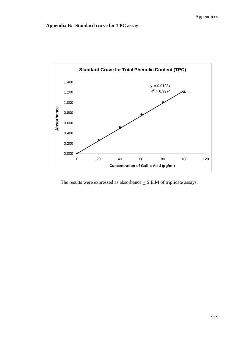
Appendices
121
Appendix B: Standard curve for TPC assay
Standard Cruve for Total Phenolic Content (TPC)
y = 0.0123xR2 = 0.9974
0.000
0.200
0.400
0.600
0.800
1.000
1.200
1.400
0 20 40 60 80 100 120
Concentration of Gallic Acid (µg/ml)
Abs
orba
nce
The results were expressed as absorbance + S.E.M of triplicate assays.
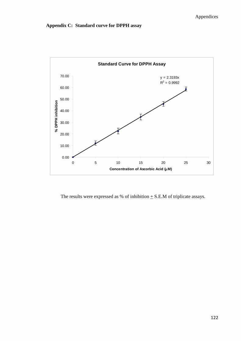
Appendices
122
Appendix C: Standard curve for DPPH assay
Standard Curve for DPPH Assay
y = 2.3193xR2 = 0.9992
0.00
10.00
20.00
30.00
40.00
50.00
60.00
70.00
0 5 10 15 20 25 30
Concentration of Ascorbic Acid (µM)
% D
PPH
inhi
bitio
n
The results were expressed as % of inhibition + S.E.M of triplicate assays.

Appendices
123
Appendix D: Standard curve for FRAP assay
Standard Curve for FRAP Assay
y = 0.0007xR2 = 0.9998
-0.1
0
0.1
0.2
0.3
0.4
0.5
0.6
0.7
0.8
0 200 400 600 800 1000 1200
Concentration of FeSO4 (µΜ )
Abso
rban
ce
The results were expressed as absorbance + S.E.M of triplicate assays.
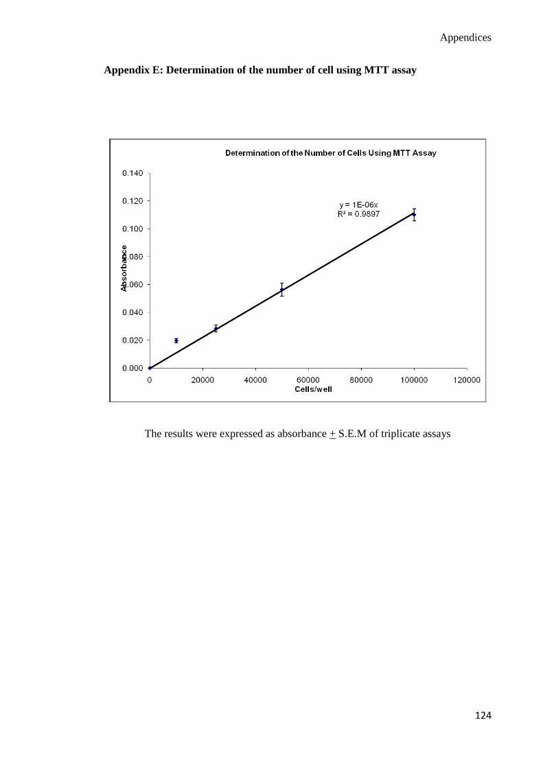
Appendices
124
Appendix E: Determination of the number of cell using MTT assay
The results were expressed as absorbance + S.E.M of triplicate assays